1. Introduction
One of main problems in cancer diseases is preventing connections between programmed cell death protein (PD-1) on a T-cell, and the programmed death-ligand 1 (PD-L1) on the tumor cells. Interactions between these cell surface proteins are involved in the suppression of the immune system and limit the killing of tumor cells by T-cells [
1]. Thus, use of an inhibitor that blocks the interaction between PD-L1 and the PD-1 receptor can prevent the cancer from evading the immune system [
2]. To date, many PD-1/PD-L1 inhibitors have been approved, which could be used for the treatment of different forms of cancers, such as melanoma, non-small cell lung cancer, renal cell carcinoma, bladder cancer and Hodgkin lymphoma [
3,
4]. For example, some authors have summarized and discussed the current understanding of mechanisms promoting resistance to anti-PD1/PDL1 therapies, and how combination strategies, which target these pathways, might yield better outcomes for patients [
5]. Another investigation has shown that the approved checkpoint inhibitors (PD1 and PDL1 inhibitors) have similar efficacy and safety profiles for some cancers, such as bladder cancer, but they vary in dose, frequency and cost burden. However, only pembrolizumab has shown superiority over standard chemotherapy in a randomized Phase III setting [
6]. Other authors have disclosed that most of the patients cannot benefit from anti-PD1/PDL1 therapy. Furthermore, a large group of responders would develop acquired resistance after initial responses. To break these resistances and improve anti-PD1/PDL1 methods, they have considered the strategies that may relieve the anti-PD1/PDL1 resistance [
7]. Another study has shown that the expression of PD-L1 is highly prevalent in some cancers, such as prostate cancer. PD-L1 is closely related to the Gleason score and may be a co-factor associated with the progression of prostate cancer [
8]. Another research has suggested that PD1/PDL1 expression and immune response varied at different microsatellite statuses in some cancers, such as gastric cancer. PD1/PDL1 expression was correlated with CD8+ T cell/CD68+ M densities in this cancer at different microsatellite statuses, especially at the invasion front [
9]. Another article has indicated that combined treatment of expanded natural killer (NK) cells and PD1-blockade resulted in robust synergistic tumor destruction of initially non-responding patient tumors. Thus, expanded NK cells may overcome the critical roadblocks to extending the prodigious benefits of PD1-blockade therapy to more patients with lung cancer and other tumor types [
10].
Most of these considerations show that some of present drugs and treatments for PD-1/PD-L1 connections are expensive and may be non-helpful or effective. Now, the question arises whether there is any probability that electromagnetic waves act as effective PD-1/PD-L1 inhibitors. Recently, some scientists are using electromagnetic waves to cure cancers. For example, current experimental evidence suggests that tumor-specific modulation frequencies regulate the expression of genes involved in migration and invasion and disrupt the mitotic spindle. This novel targeted treatment approach is emerging as an appealing therapeutic option for patients with advanced cancer given its excellent tolerability [
11]. Some authors have reviewed the experimental and clinical evidence of pulsed electromagnetic field (PEMF) therapy, discussing future perspectives in its use in oncology [
12]. Other authors have argued that specific ranges of extremely low frequency electromagnetic waves inhibit cancer cell proliferation. Their thermodynamic model selects the frequency of ELF-EMF specific for each cancer [
13]. Some investigators have shown that extremely low frequency variable electromagnetic fields affect cancer and noncancerous cells in vitro differently [
14]. Another paper has discussed that cancerous tissues have natural frequencies different from the natural frequencies of normal tissues, and thus, one can hypothesize to irradiate cancerous tissues using EMFs at natural frequencies of cancer cells, causing resonant interaction with cellular membrane channels, inducing increasing of ions’ flux across cellular channels and damaging the cellular functions of cancer cells [
15]. Another work has shown that extremely low-frequency electromagnetic field altered PPARγ and CCL2 levels and suppressed CD44
+/CD24
− breast cancer cell characteristics [
16]. This research and many other investigations show that electromagnetic waves could act as a strong tool in curing cancers.
The question may be raised as to why electromagnetic waves have these effects on cells. To answer, it should be remembered that the heart as the origin of life emits both types of electrical and magnetic waves. As of now, many models have been proposed to consider the origin of heart waves and most of them have focused on the action potentials on the cell membranes or between cells [
17,
18]. However, it seems that there is a direct relation between these potentials and DNA waves [
19,
20]. In this paper, we will propose a theoretical model for controlling information transmission between cells and reducing PD-1/PD-L1 connections by using spinor waves. We will show that DNAs, RNAs and other biological matter within biological cells are built from spinors like electrons, and for this reason, one of the waves for exchanging information is using spinor waves. We will formulate the model and propose some mathematical relations, which describe the dependency of correlations between spinors within biological systems in terms of energies and temperatures. Any change in one spin within an entangled or correlated system of spinors may cause a change in other spins. For example, when a cell changes, and a tumor is formed, its spinor waves change, and this change could be understood by spinors within the heart or brain and other controller cells. These cells may send some spinor waves towards the T-cells and encourage them to attack tumor cells. However, these waves are taken by tumor cells and they become ready to respond. We will show that by adding new spinors, the correlations between the initial spinors decrease mathematically. This means that by adding extra spinors around tumor cells, real information may be lost, and these cells cannot understand the closing of T-cells. This gives the opportunity for T-cells to kill tumor cells.
The outline of paper is as follows. In
Section 2, we will describe the model theoretically and consider the role of hemoglobin and biological spinors in exchanging information between the heart and other controller cells and biological cells. In
Section 3, we will formulate the model and obtain the correlation functions for biological spinors within the cells mathematically. In
Section 4, we will review some observations, which may confirm the model.
2. The Model: The Role of Hemoglobin in Exchanging Information between Heart and Cells and Reducing PD-1/PD-L1 Connections
To date, it has been known that hemoglobin plays the main role in exchanging oxygen with cells. They also have some physical and electromagnetic properties [
21,
22]. However, these molecules include iron atoms which could attract magnetic waves and induce some quantum waves into multi-gonal molecules. Thus, we could suppose that these molecules could store some waves in some places and then give them to other molecules in other places. The question then arises as to how these molecules store information.
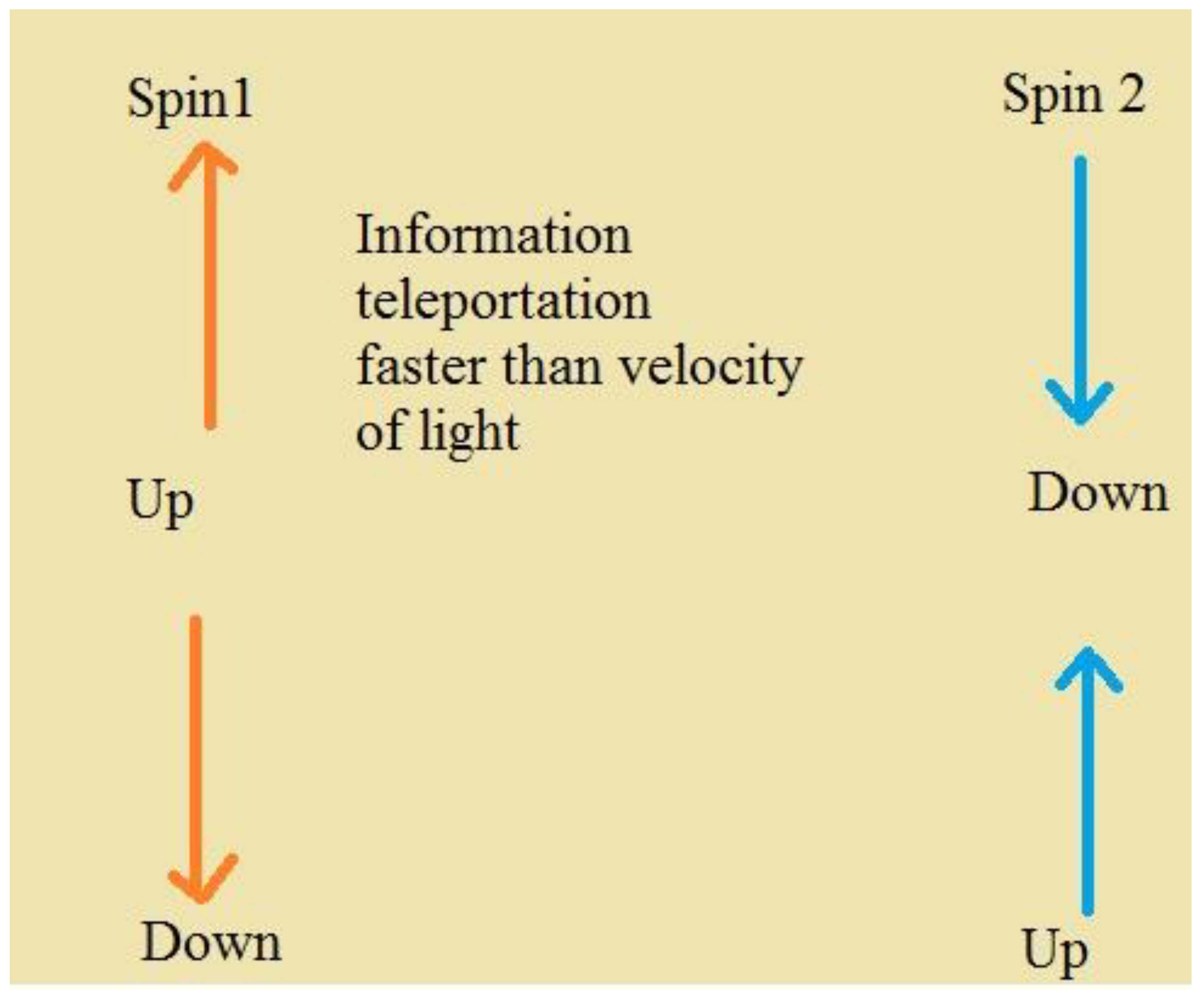
The best quantum method for storing information is using spinor waves. Spinors are particles like electrons with spins = 1/2, 3/2, … These particles obey Pauli’s repulsive principle. This means that two parallel spins repel each other, and two anti-parallel spins attract each other. The velocity of exchanging information between two spins is more than velocity of light (See
Figure 1). For example, in a pair of two spins, when the arrow of one spin changes, the arrow of the other spin changes very fast. This entanglement between two spinors could be affected by adding some extra spinors. For example, an extra spinor produces new entanglement with one of two entangled spinors and decreases previous entanglement (See
Figure 2). Now, the question becomes how these concepts could help us to consider the methods for storing information in hemoglobin.
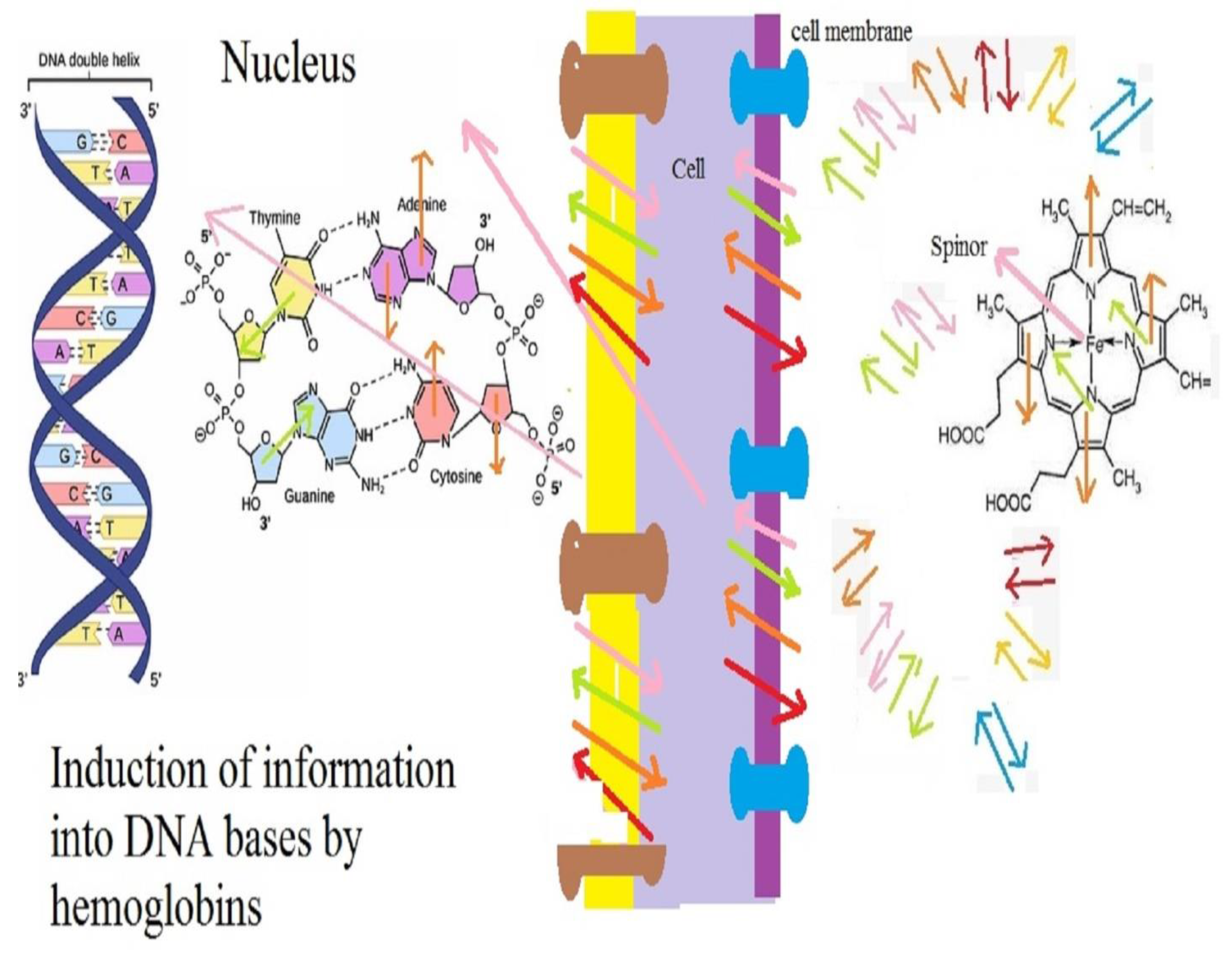
To answer this question, first, we return to DNAs within cells, especially heart cells, and more especially, those cells which produce electromagnetic waves. For the most part, scientists assume that the origin of these waves are some action potentials. However, the question arises as to what the origin of these potentials is. We could go a step more and deeper into heart cells. Suppose that a DNA is built from charged particles and by its motion, some waves are generated. In fact, we have two types of cellular waves. Some of them are produced by electrical currents within and around cells. These currents are produced by the motion of charges of DNAs, cellular membranes and ions around and within cells. These currents emit some waves for which their collections could be seen as heart waves. These waves may be detected by some detectors, especially those which use superconducting materials. The magnitude of these waves may be very less than 10−4 and around 10−10 Tesla. In addition to these waves, the spins of electrons and other spinors produce some magnetic waves which only biological spinors could detect. These waves are produced by vibrations of spinors, such as electrons within cells, and could be absorbed by spinors outside cells. The consideration of changes in these two types of waves may help us to image events within the human body. For example, if a cell changes and a tumor emerges, then its spinor and electromagnetic waves change, and these changes are transmitted along biological cells and reach to the heart and brain and other controller cells. Thus, by considering the evolutions of waves, we can diagnose some diseases, such as cancers.
In
Figure 3, we consider the cellular waves in more details:
DNAs, proteins, cellular membranes and other biological material are built from charged and spinning particles, such as electrons.
Charges emit electrical fields:
- 3.
By motions of charges within and around cells, some currents are generated. These currents emit some magnetic fields. We have:
- 4.
Each electron within the structure of DNAs, proteins, cellular membranes and other biological materials has a spin which may be entangled with the spins of other electrons. Any change in one spin causes many changes in the spins of other spinors within an entangled system. Thus, spinor waves could help the biological system to transmit information. For example, a cell could change spinors around it and consequently, all spinors change and a message could be transmitted.
- 5.
Each spin has a relation with quantum magnetic fields:
- 6.
Quantum magnetic fields, which are produced by spinors within the biological cells of the heart, could be taken by spinors and also iron atoms within hemoglobin and spinors on other biological molecules. These molecules move along blood vessels and could take these waves to other cells.
The heart thus could carry out two processes:
In
Figure 4, we show that spinors within and around heart cells and hemoglobin could be entangled. A hemoglobin is built from iron atoms, nitrogen, carbon and related electrons and spinors. These spinors could be correlated with spinors within heart cells. Any change in one spin could cause changes of other spins. Thus, heart cells could store information within hemoglobin through changing spins.
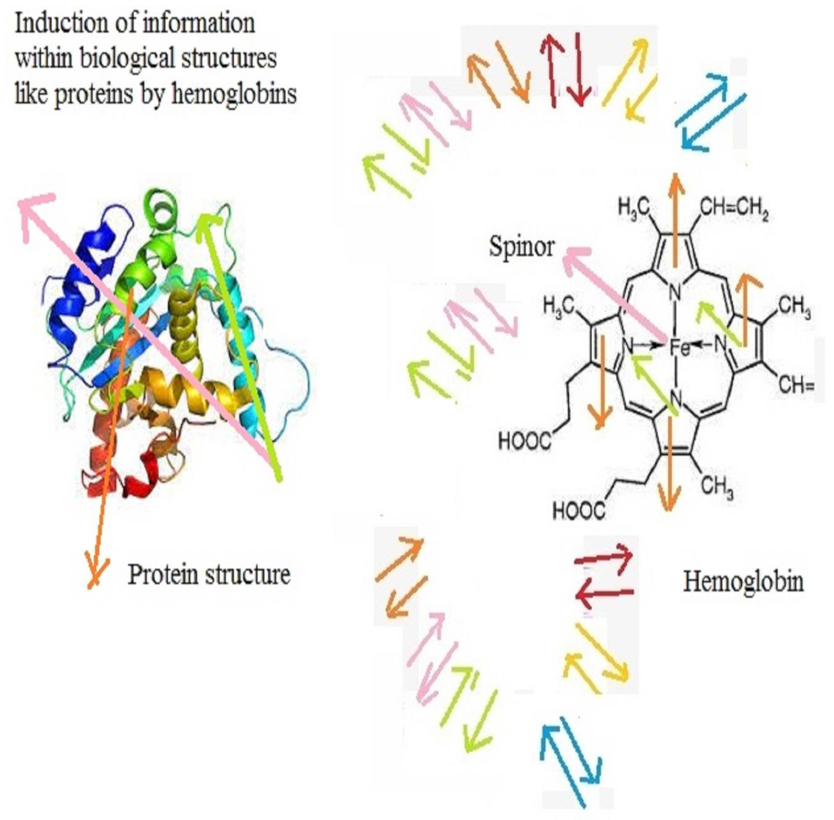
Hemoglobin could be located within blood cells, move along vessels and reach special cells. Then, hemoglobin could induce its information as spinor waves into hexagonal/pentagonal molecules of a DNA (See
Figure 5) or within the coil-like structures of some proteins (See
Figure 6).
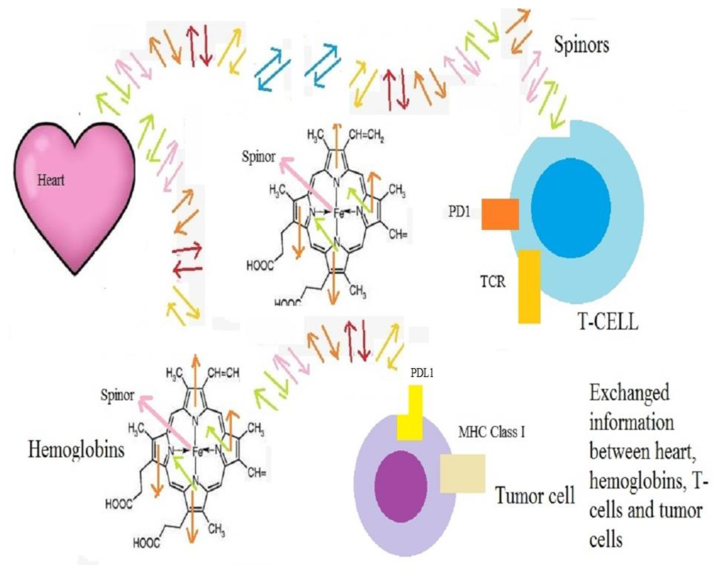
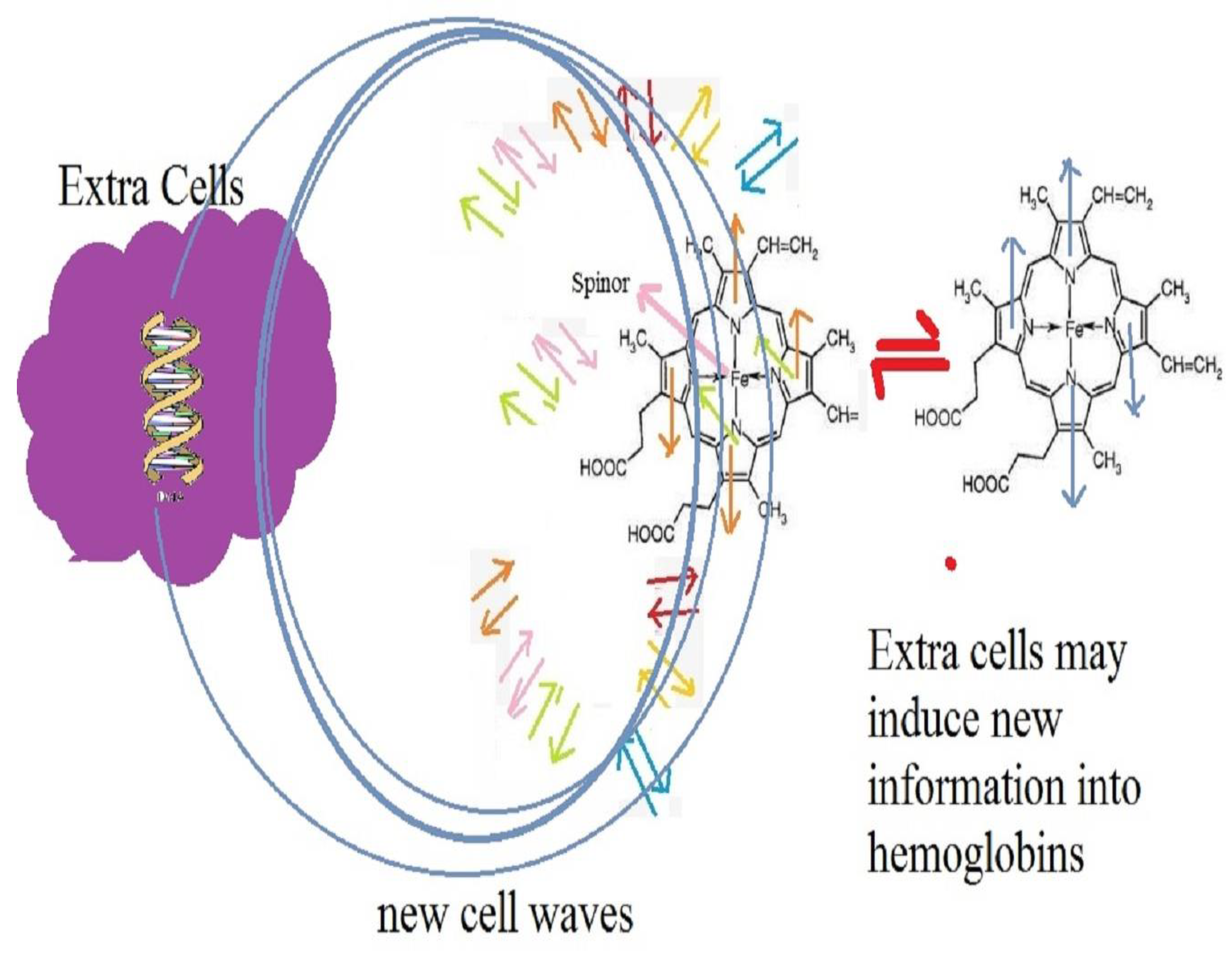
The question might aris as to how this mechanism could be used in curing cancers. In response, we should remember that blood includes many ions and spinors. Thus, the heart could induce any new information into all spinors within blood vessels and change information which is stored in a hemoglobin. For example, when a tumor emerges, its changes have a direct effect on spinor waves and these waves and information are received by heart cells immediately. Then, the heart cells send new information to all cells and special T-cells for creating the best conditions in curing and attacking the tumors. Unfortunately, this information is taken up by tumor cells, and they become ready to respond to any attack. For example, they produce some PD-1/PD-L1 connections and kill T-cells (See
Figure 7).
Now, one could ask how we could prevent the tumor cells from receiving this information. To answer this question, we suggest several ways:
We could use some extra harmless cells, which interact with the hemoglobin around tumor cells and produce some noise in the information received by the tumor (See
Figure 8).
We can use some extra hemoglobin, which interacts with the hemoglobin around tumor cells and produces noise in the main waves (See
Figure 9).
In the next section, we carry out some calculations, which show that by increasing numbers of molecules or cells including spinors, we can break the entanglement between the heart spinors and hemoglobin around tumor cells. Consequently, the tumor cells do not notice the new messages exchanged between other parts of the body and do not wait for any attack of T-cells, and they can kill the cancer cells. In fact, the mathematical result shows the needed spinors which produce some noise around tumor cells and reduce the probability for producing PD1/PD-L1 connections.
3. Mathematical Results
In the previous section, we described the model qualitatively, and this section, we will formulate it quantitatively. DNAs, proteins, cellular membranes and other biological matters within or around cells and hemoglobin are formed from charges, spins and spinors. All of the spinors have spins 1/2 and obey the Pauli exclusion principle. This means that they are maximally entangled and by changing the arrow of one of the spinors, the arrow of the other spinor will change. On the other hand, spins 1,2, … obey a different mechanism and entanglement. For a system of two spinors with spins 1/2, we have:
The above equation shows that by changing the direction of the arrow of one spinor, the direction of the arrow of the other spinor changes. For example, suppose that there are two electrons in two sides of the world. When the spin of an electron changes, the spin of another electron could change immediately. However, for spins 1, 2, …, we have many free degrees and could write:
where 1 denotes the spin 1 and up or down denotes the direction of spin. The above equations have been written for a non-thermal system. Previously, the correlation functions for spinors and spins in a thermal system have been proposed. We can follow those considerations and write a function, which includes both types of spinors and spins. For a system which includes both types of spinors and spins, we can write [
23,
24]:
where
i labels the spinor
i and
j labels spin
j. In the above equation, (
s) are the quantum states which show the quantum entanglement between biological spinors (see Equations (4) and (5)) and (
) is the cross product between states. Also, F is a functions of quantum states related to spinors and spins. Equations (4) and (5) are related to quantum states in a non-thermal system. However, previous considerations have shown that the temperature has a direct effect on the correlation, entanglement and change states [
23,
24]. For example, for two quantum spinors in a thermal system, we write [
23,
24]:
where
is the related spinor energy in the system and
is temperature. We also could write the equation below for two spins states in a thermal system:
All quantum states should be maximally entangled, and their functions should be unified.
which gives causes to the result below:
where
and
Maybe, a quantum state emerges between three spinors. In that case, we could write:
or for three spins:
Maybe, N spinors join one another and form N-quantum spinors. In that case, we will have:
Additionally, n quantum spins may join to each other and form a unitary space:
Moreover, a system may include N spinors and spins:
Thus, we should rewrite the total function of quantum states as shown below:
where
The above equation shows that by adding new particles and increasing the number of spinors and spins, the correlation function decreases. This is because those extra spinors and spins interact with initial spinors and spins, and decrease their maximal connections and entanglement.
The above equations help us to formulate the model. Each biological object, such as a DNA or protein, is built from spinors, such as electrons. These spinors could be entangled and correlated. We have obtained the correlation function in terms of energy and temperature. Any change in one spin may change all other spins. Thus, cells could use this technique in exchanging information. For example, when a tumor is formed, spins around cells change and these changes cause immediate changes of spins around the heart or brain and other controller cells. Then, these cells send spinor waves towards T-cells and encourage them to move towards the tumor cells. Tumor cells also take this message and become ready to respond. We have shown that by adding new spinors to a system of correlated spinors, the initial correlations decrease mathematically. Thus, one can add new spinors around tumor cells, decrease the initial correlations between spinors and make a noise in real information.
4. A Review on Some Observations
The question may arise around the reason for proposing this theoretical model. To date, it has been observed that by the emergence of some types of cancers, some significant changes in blood cells occur. For example, anemia is a common condition of cancer patients. However, most scientists believe that cancers cause inflammation, which decreases red blood cell production. In addition, many chemotherapies are myelosuppressive, meaning they slow down the production of new blood cells by the bone marrow [
25]. There may also be another reason. It means that, for example, controller cells within the heart send some messages to hematopoietic cells to decrease the amount of red blood cells and prevent the exchanging of information between normal and cancer cells. In addition to anemia, there are other changes in the blood cells, which may occur by tumors and cancers [
26]. The reason for these changes may be the response of the human body by producing noise in exchanged information between cancerous and normal cells.
We can have a review on the relation between the amount of hemoglobin, T-cells, heart diseases and cancer. To date, many investigations have shown that cancers could use different factors and change the amount of hemoglobin. For example, some authors have shown that increased levels of serum glycosylated hemoglobin are associated with depressive symptoms in a population with cancer [
27]. Another interesting paper argued that the ratio of the hemoglobin to red cell distribution width combined with the ratio of platelets to lymphocytes can predict the survival of patients with gastric cancer liver metastasis [
28]. Other authors have considered the nitration, chlorination, and oxidation in the hemoglobin of breast cancer patients by using nanoflow liquid chromatography tandem mass spectrometry [
29]. Another article has discussed about prognostic significance of hemoglobin-to-red cell distribution width the ratio in patients with metastatic renal cancer [
30]. Some other authors have predicted the value of the hemoglobin, albumin, lymphocyte, and platelet (HALP) score and lymphocyte-to-monocyte ratio (LMR) in patients with non-small cell lung cancer after radical lung cancer surgery [
31]. There are many other similar research papers, which show the exact relation between the quantity of hemoglobin and different types of cancers. On the other hand, some researchers focus on the interaction between T-cells and hemoglobin. For example, some authors have found that CD8
+ T-cell responses against hemoglobin-β prevents solid tumor growth [
32]. Or some authors have argued that T cells specific for alpha-beta interface regions of hemoglobin recognize the isolated subunit but not the tetramer and indicate presentation without processing [
33]. Moreover, some authors have reviewed the roles of cell-free hemoglobin in the innate immune system, focusing on the plausible interactions among hemoglobin, pathogens, host cell components and innate immune cells [
34]. Besides these studies, many investigations have been carried out on the interactions between hemoglobin and heart cells. For example, one study has shown that anemia is frequently present in patients with chronic heart failure. Lower hemoglobin is associated with greater disease severity, a greater left ventricular mass index, and higher hospitalization and mortality rates [
35]. Another study has reviewed the main pathophysiological links between red blood cell disorders and cardiovascular diseases and considered the implications for clinical practice and therapy. This review indicated that anemia is associated with a special risk in proatherosclerotic conditions and heart disease and became a new therapeutic target [
36].
The above results show that there is a direct relation between hemoglobin, heart activities and cells, T-cells and cancer cells. Thus, we can speculate that these elements of a body can communicate with each other. All of these biological objects have the ability to emit electromagnetic waves. Also, they are formed from electrons and atoms. Some of these charges have spin, and thus exchanging information between spinors may be very natural in the human body. In fact, if biological systems want to send some information with a velocity higher than the velocity of light, it is better to make use of spin properties. For this reason, we cannot ignore this quantum property, which may help us in curing hard diseases, such as cancers.
5. Discussion
To cure some diseases, such as cancer, we need to know the mechanism of transformation of information between biological cells. To date, many chemical and electromagnetic mechanisms, which may be used by cells in transmitting information have been introduced. However, the exchanged spinor waves between spins on biological matters have been ignored. All elements of biological cells, such as cellular membranes, DNAs, RNAs, proteins and other material are built from electrons, which have spin ½ and could be regarded as spinors. Consequently, it is natural that spinor methods be one of the ways for exchanging information between cells. To describe this method, we should review some concepts of quantum mechanics. According to quantum sciences, two entangled spinors could exchange information with a velocity more than that of light. For example, when, the spin of one spinor changes, the other spin changes promptly. In the above calculations, we showed that the quantum entanglement between these spinors could depend on their energies and the temperature of the system. If instead of two spinors, we have N entangled spinors, it is needed to introduce a more complicated function, which depends on the energies of all spinors. However, these spinors could act like a string and by any change in one spinor, all spinors change. In the previous section, we formulate this method and introduce a function for biological spinors. This mechanism could be applied in biological systems. For example, within the human body, spinors within biological cells and hemoglobin could be entangled. Thus, any change in a normal cell could be understood by other cells. When a normal cell changes and a tumor cell is formed, its radiated spinors and charges change. This change in spinors around tumor cells causes changes in the spinors within the biological string and could be diagnosed by the heart or brain or other controller cells. These cells use some new quantum spinor waves and send some messages to T-cells. These T-cells move towards the tumor cells. However, tumor cells take these spinor waves and become ready to produce some harmful connections and kill T-cells. To prevent these connections, we can produce some noise around tumor cells. In the previous section, we have shown that by adding new spinors to a system of entangled spinors, the correlations between the initial spinors decrease. This is because the new spinors build new correlations with initial spinors and reduce their initial entanglement. The same method could be used for producing noise in biological systems. We can add some extra hemoglobin or biological cells and material, which includes spinors around tumor cells and reduces the quantum entanglements between spinors on tumor cells and initial hemoglobin. Consequently, some noises emerge and information is lost.
6. Conclusions
Recent considerations have shown that cancerous cells could notice the approach of T-cells and become ready to response by producing PD-1/PD-L1 connections and other connections with them. It seems that these cells obtain some information of the motion and closing T-cells. Now, the question arises as to how tumor cells could understand the presence of T-cells. In response, we should consider different mechanisms, which cells may use for transferring information. By knowing these mechanisms, maybe we could propose some medical methods for reducing PD-1/PD-L1 connections and curing tumors. In this research, we suggest a new theoretical method for exchanging information between hemoglobin, T-cells, tumors and other biological cells by using spinor waves. Spinors could be entangled and exchange information much faster than any other way. For example, by a change or reversing the spin of one spinor within an entangled system, the spins of other spinors change immediately. If human cells have some spinors which are entangled with each other, the velocity of the exchanged information between them could be more than the velocity of light. This theory may be true. DNAs, RNAs, proteins and other biological material within biological cells of the heart or brain and other controllers are constructed from some spinors, such as electrons, and by their motion, some spinor waves are emitted. Spinors within hemoglobin and other molecules around these cells could absorb these spinor waves, move along the blood vessels and give their related information to other biological cells and matters. All of the spinors within the blood vessels and biological cells could be entangled, and by any change in one spin, other spins change immediately. Consequently, any change in one cell could be understood by other cells. For example, when a normal cell converts to a tumor cell, its spinor waves are changed, and these evolutions are transmitted to the heart cells, brain cells and other controller cells immediately. These cells receive these spinor waves, and by using the quantum entanglements between spinors within the biological structure of the human body, send some new spinor waves to T-cells. In addition to T-cells, tumor cells also take these spinor waves and become aware of the closing T-cells. Consequently, they become ready to kill T-cells through the production of PD-1/PD-L1 and other connections. To reduce these connections, we should prevent the reception of spinor waves by tumor cells. To this aim, we can produce some extra spinor waves, which act as noise and real information is lost. These noises could be produced by extra hemoglobin cells, and other biological material, which includes spinors around tumor cells. To formulate the model, we first consider the quantum entanglement between two spinors. These spinors may be located on two hemoglobin molecules, or one on a hemoglobin molecule and another within a heart cell. This entanglement depends on the energy of the spinors and the temperature of the system. Any change in one spin could cause a change in the other spin. Then, we generalize the equations by regarding the quantum entanglements between all spinors which are located on human cells and hemoglobin. By changing a spin, all spins could change. This helps us to transmit information immediately. Next, we add some extra spinors to the system and show that quantum correlations between spinors decrease. This is because extra spinors form new entanglements with initial spinors and reduce previous entanglements. Thus, we can cause the extra spinors to decrease the quantum entanglements between spinors around tumor cells and prevent the reception of real information by them. Finally, to propose some observational reasons for the correctness the model, a review on previous investigations on the connections between hemoglobin, T-cells, heart and cancer has been carried out.


