Article

Thermodynamic Interpretation of a Machine-Learning-Based Response Surface Model and Its Application to Pharmacodynamic Synergy between Propofol and Opioids

1
Department of Anesthesiology, Taipei Veterans General Hospital and National Yang Ming Chiao Tung University, Taipei 11217, Taiwan
2
Department of Biomedical Sciences and Engineering, National Central University, Taoyuan City 32001, Taiwan
3
Department of Biomedical Engineering, National Yang Ming Chiao Tung University, Taipei 11217, Taiwan
*
Author to whom correspondence should be addressed.
Mathematics 2022, 10(10), 1651; https://doi.org/10.3390/math10101651
Submission received: 31 March 2022 / Revised: 22 April 2022 / Accepted: 10 May 2022 / Published: 12 May 2022
(This article belongs to the Special Issue Theory of Algorithms and Recursion Theory)

Abstract

:
Propofol and fentanyl are commonly used agents for the induction of anesthesia, and are often associated with hemodynamic disturbances. Understanding pharmacodynamic impacts is vital for parasympathetic and sympathetic tones during the anesthesia induction period. Inspired by the thermodynamic interaction between drug concentrations and effects, we established a machine-learning-based response surface model (MLRSM) to address this predicament. Then, we investigated and modeled the biomedical phenomena in the autonomic nervous system. Our study prospectively enrolled 60 patients, and the participants were assigned to two groups randomly and equally. Group 1 received propofol first, followed by fentanyl, and the drug sequence followed an inverse procedure in Group 2. Then, we extracted and analyzed the spectrograms of electrocardiography (ECG) and pulse photoplethysmography (PPG) signals after induction of propofol and fentanyl. Eventually, we utilized the proposed MLRSM to evaluate the relationship between anesthetics and the integrity/balance of sympathetic and parasympathetic activity by employing the power of high-frequency (HF) and low-frequency (LF) bands and PPG amplitude (PPGA). It is worth emphasizing that the proposed MLRSM exhibits a similar mathematical form to the conventional Greco model, but with better computational performance. Furthermore, the MLRSM has a theoretical foundation and flexibility for arbitrary numbers of drug combinations. The modeling results are consistent with the previous literature. We employed the bootstrap algorithm to inspect the results’ consistency and measure the various statistical fluctuations. Then, the comparison between the modeling and the bootstrapping results was used to validate the statistical stability and the feasibility of the proposed MLRSM.

1. Introduction

The induction of anesthesia—the transition process from an awake to an anesthetized state—gradually increases the effects of hypnotics and opioids [1,2], which may cause complex dynamic changes in the activity of the autonomic nervous system (ANS). The ANS plays a vital role in the maintenance of physiological homeostasis. Some clinical trials have evaluated different intravenous anesthetic effects on ANS tone [3,4,5,6]. However, these trials were limited to individual anesthetic techniques, and analyzed heart rate variability (HRV) through classical spectral analysis, which assumes data stationarity, and is thus unsuitable for the short induction course of anesthesia. Spectral analysis of heartbeat-to-heartbeat intervals is a widely used non-invasive technique to assess the autonomic integrity of—and the balance between—the sympathetic and parasympathetic tones [7]. From the perspective of wavelet-based spectral analysis, signal fitting with multiple wavelet filters would offer a higher time resolution [8]. This technique improves the understanding of how co-administered propofol and fentanyl modify the activity of the ANS. However, there is no available method to deal with the instantaneous modulation of the ANS during the induction of anesthesia with propofol and fentanyl.
The response surface model (RSM) is a mathematical representation of the concentration–effect relationships between combinations of multiple drugs over a continuous surface space [1,2,9,10,11,12,13,14]. It illustrates the type and degree of drug interactions [15,16]. The RSM method can elucidate the interactions between two drugs—such as propofol and fentanyl in our study. It also clarifies the drugs’ effects, including loss of response to verbal or tactile stimuli, or as surrogates for surgical pain [1,10]. However, methods to measure the immediate sympathetic and parasympathetic effects during the induction of anesthesia are still lacking [2,3,9,14]. To solve this problem, we extended the framework of the thermodynamic mechanism to accommodate the conventional RSMs. We established a machine-learning-based response surface model (MLRSM) to explain the autonomic effects of drug stimulation. This data-driven MLRSM, based on thermodynamic mechanisms and statistical learning methods, can reflect the human physiological responses affected by anesthetic drugs. Our purpose was to seek the least amount of local anesthetics and, thus, shorten the recovery time after surgery. The conventional RSMs rely on the initial guesses in the maximum likelihood estimates, and would offer ill-suited results when using singular matrices [9,10,11,12,13,14]. Compared to conventional RSMs, the proposed MLRSM could significantly reduce computational time consumption and maintain an acceptable level of accuracy. Eventually, we hope that the proposed MLRSM can offer an optimal depth of anesthesia for autonomic modulation in the perioperative period.
In a nutshell, the proposed MLRSM connects the physical essence of thermodynamics and the statistical properties of experimental datasets, and then extracts the corresponding indispensable parameters by learning from the clinical data. We hypothesized that this proposed propofol–fentanyl MLRSM, combined with the wavelet transformation of HRV, could provide precise information on the optimal anesthesia status for transient autonomic modulation, even in the short timeframes of general anesthesia. This may lay the foundation for improving the reliability of anesthetic depth monitoring and the development of new clinical applications. The goal of this study was to provide a data-driven response surface model based on sophisticated machine learning methods. The MLRSM can offer a high-efficiency model construction with extremely low computational complexity. It can also derive analytical formulae for an arbitrary number of drug combinations. This achievement would reinforce the relevant development of clinical practices.

2. Materials and Methods

2.1. Data Collection and Preprocessing

2.1.1. Clinical Trials

A prospective observational study was conducted on 60 patients with the American Society of Anesthesiologists (ASA) physical status I–II who underwent elective surgery with the induction of propofol and fentanyl. All subjects gave their informed consent for inclusion before they participated in the study. The study was conducted in accordance with the Declaration of Helsinki, and the protocol was approved by the Ethics Committee of the Institutional Review Boards (IRB) of Taipei Veterans General Hospital (No. 2017-07-009CC) and Landseed Hospital (No. 16-033-B1), and registered at ClinicalTrials.gov (NCT03613961 and NCT03072069). We excluded cases of emergency surgery. This study adheres to the applicable STROBE guidelines. Patients were studied while supine, and standard monitoring was employed and recorded before and throughout the induction of anesthesia with propofol and fentanyl. The ECG (electrocardiogram) waveforms were continuously recorded by a multichannel polygraphic system (Embla N7000, Natus, Pleasanton, CA, USA), and data were saved directly to a memory card within the device at a rate of 1024 Hz for offline analysis of HRV and pulse photoplethysmography (PPG). Patients were divided into two groups for characterizing the individual ANS modulation effects of propofol and fentanyl, and for exploring the interactions in these two distinct induction groups. Patients in Group 1 received propofol first, followed by fentanyl, while the drug sequence followed an inverse procedure in Group 2. A brief period of individual drug effects was observable before adding the second induction drug. This period was pooled and modeled separately to investigate the single-drug ANS effects. The MLRSM then performed the interaction estimation when the two drugs existed simultaneously. Each patient received propofol (Propofol-Lipuro 1%, 20 mL, B BRAUN) and fentanyl (fentanyl “PPCD”, 0.1 mg/2 mL) via a 20-gauge catheter. When each surgery was completed uneventfully, so were the data collection and the general anesthesia.
We found that after both drugs were administered, both groups seemed identical. However, pharmacology is not a simple linear combination. If one drug is administered after another drug, we could not determine the individual effects of the latter drug, and vice versa. The delay of drug onset would result in very different paired peak and trough concentrations [17,18]. Thus, for a two-drug model, as in our study, we needed to identify the effects of propofol alone, of fentanyl alone, and then of both drugs simultaneously. The stepwise dosing technique is used frequently in pharmacodynamic studies [19,20,21,22,23]. Furthermore, it would be unethical and excessive to recall the patients for a second dosing trial.

2.1.2. Data Preprocessing and Pharmacokinetic Simulation

Our previous methods provided a preprocessing technique for ECG data, R–R wavelet intervals, and photoplethysmography beat-to-beat pulse amplitudes (PPGA) [24]. This technique offered estimations of the power over the high-frequency (HF) and low-frequency (LF) bands (HF, 0.15–0.4 Hz; LF, 0.04–0.15 Hz) in every second via a continuous wavelet function, representing the instantaneous function of the ANS. The HF of the series of heartbeats represents a marker of vagal modulation [25], and the ratio of the LF to the HF (LHR) becomes an indicator of the balance between sympathetic and vagal modulation [7,26]. The PPG signals reflect the peripheral sympathetic responses [27,28]. Since the normalization of the data would eliminate the inter-subject differences between baseline autonomic functions, we utilized Equation (1) to transform the signals individually into the region of [0, 1] through their maximum and minimum values [24]:
X X X m i n X m a x X m i n × 100 ,
where X is the original value in the series, and X m i n and X m a x are the original minimum and maximum values, respectively, while X is the transformed value. Thus, the transformed variables had the same normalized range, and the distribution of each patient response had the same normalized unit.
The pharmacokinetic profiles of plasma (Cp) and effect-site concentrations (Ce) were estimated using the Tivatrainer simulation program (version 8, EURO Siva). Then, we employed Shafer’s pharmacokinetic model [29] to estimate the fentanyl effect-site concentrations and Schnider’s pharmacokinetic model [30] to calculate propofol effect-site concentrations. Finally, we estimated the mean HRV using the technique of continuous wavelet transform [24], and graphically extracted the values of drug concentrations at the same time points using plots.

2.2. Model Derivation

2.2.1. Thermodynamic Interpretation

From the perspective of thermodynamics, we can estimate the macroscopic properties of a system of interest through the integrative manifestations of a large number of microscopic particles inside the system. Then, we can clarify the mapping transform between this macro–micro relationship by utilizing the number of possible microstates Ω . For a system that contains N particles, if we naturally classify these particles into several subgroups ( n 1 ,   n 2 ,   , n m ) according to their intrinsic microscopic properties, we can directly define the number of possible microstates as Ω = N ! / ( n 1 ! n 2 ! n m ! ) , where n 1 + n 2 + + n m = N . Each subgroup n i here represents a specific microstate. By introducing this relationship to the mechanism of drug stimulation, we may elaborate on the human physiological responses after integrative manifestations of a set of drug stimuli. Here, we assumed that the human body is an isolated and isothermal system, and that the drug stimulation could immediately affect this system. In other words, the human physiological responses under anesthetic stimuli would instantly reflect the integrative manifestations of the drugs’ effects. This means that we can describe the average consequences of human responses after anesthetic stimuli by mapping the performance of drug effects to the microstates in a physical space. This concept connects the number of possible microstates to the drug effects and the corresponding drug concentrations.
Specific anesthetic drug effects directly lead to relevant human physiological responses in clinical practice. Thus, we can safely assume that the microstates and the performance of the drugs’ effects are isomorphic. To be specific, the number of possible microstates can have a simple expression according to the mapping relationship between the microstates and the specific effects of the individual anesthetic drugs, and this expression has the simple form Ω = E m a x ! / [ E ! ( E m a x E ) ! ] . The parameter E represents the subgroup of successful drug stimuli, while E m a x E represents the subgroup with insignificant or unsuccessful drug effects. In other words, these two terms in the denominator of Ω indicate that two possible subgroups were adopted to model the two different states of the drug effects. From the perspective of thermodynamics, the parameters E and E m a x are also the state number of successful drug stimulation and the total state number of drug stimuli, respectively, throughout the duration of drug stimulation.
Because of the response differences in the drug concentrations of each subject, we first normalized the drug concentration as C / C 50 for the model’s construction. The parameter C is the drug concentration given by injection, while C 50 is the drug concentration at which 50% of subjects experienced the maximum clinical effect. Under this scheme, the upper bound of the total drug effects statistically becomes H = i = 1 E C / C 50 = ( C / C 50 ) E after E -time successful and independent drug stimulation. Since each of the state numbers of these two subgroups is enormous, we can find the maximum likelihood of Ω subject to the constraint condition by applying Stirling’s approximation to the natural logarithm of Ω and the constraint condition. Thus, we obtained [31]:
ln Ω + γ [ ln ( C C 50 ) E ln H ] E m a x ln E m a x E ln E ( E m a x E ) ln ( E m a x E ) + γ [ ln ( C C 50 ) E ln H ] .
The parameter γ is a Lagrangian multiplier. By taking the calculus of variations to Equation (2) with the value of successful drug stimulation E , we obtained δ [ ln Ω + γ ln ( C / C 50 ) E ] [ ln E + ln ( E m a x E ) + γ ln ( C / C 50 ) ] δ E = 0 . Therefore, we obtained an analytical model that connected the drugs’ effects with the drug concentrations in a set of individual anesthetic drug stimuli:
E = E m a x × ( C C 50 ) γ ( C C 50 ) γ + 1 = E m a x 1 + ( C C 50 ) γ .
It should be noted that the proposed analytical model is the well-known Greco model with individual drug effects used to resolve the RSM-related problems [15,16]. The total state number E m a x of Equation (3) is a measured maximum of human physiological responses with drug stimulation in the Greco model [9,10,11,12,13,14]. It should also be emphasized that we obtained the RSM-type model by adopting the concept of the macro–micro relationship. The Lagrangian multiplier γ becomes a measure of the steepness of the drug concentration–effect configuration. Moreover, instead of the iteration processes utilized in parameter extraction by conventional RSMs [1,2,9,10,11,12,13,14,15,16], Equation (3) offers an avenue of data-driven processing for the RSM-type model. It connects the statistical properties of experimental data and the corresponding indispensable parameters by learning from the clinical data.

2.2.2. Machine-Learning-Based Response Surface Model

Conventional iteration procedures help to numerically estimate parameters in resolving the RSM problems [9,10,11,12,13,14], but they also cause undesired issues, such as high computational complexity, lower accuracy, high-cost hardware dependency, and irregular data structure [31,32]. We thus proposed an alternative solution by endowing probabilistic meanings to the data-driven model. From a statistical perspective, we can reconstruct Equation (3) as a conditional probability density function (PDF):
f ( C | γ , C 50 ) E E m a x = 1 1 + ( C C 50 ) γ .
Thus, the PDF f ( C | γ , C 50 ) is conditional on the parameters γ and C 50 , and behaves similarly to a sigmoid function. Since we converted the conventional RSM-type model to a PDF by squeezing the data into a specific domain, the PDF curves collected from every subject should pass through the data centroid according to the similarity of the physiological responses. To technically measure the data centroid ( C 0 ,   f 0 ) , we utilized a machine learning method—Gaussian mixture models [31]—to search for the most probable positions of the centroid from the experimental data, and to estimate the corresponding statistical distributions of the data.
To resolve the steepness γ , we took a partial derivative of Equation (4) and obtained f / C = f ( 1 f ) ( γ / C ) . Then, we redefined the centroid as a coordinate origin to inspect the curve behavior near the data centroid. The trends of the curve’s steepness would then conform to the statistical meanings of the data covariance matrix. By introducing the concept of principal component analysis [33], we can determine the direction and the extent of curve declination utilizing the covariance matrix. First, the declination direction of the curve corresponds to the sign of off-diagonal elements C o v ( f , C ) . Since the value of C o v ( f , C ) would also affect the extent of the curve’s declination, we employed its unit vector form to measure the declination direction, i.e., C o v ( f , C ) / C o v ( f , C ) . Secondly, the ratio of eigenvalues λ f / λ C of the covariance matrix can directly reflect the extent of curve declination. From the following secular equation, we can obtain the corresponding eigenvalues of the covariance matrix [33]:
λ f , C = 1 2 [ C o v ( f , f ) + C o v ( C , C ) ] ± 1 2 [ C o v ( f , f ) C o v ( C , C ) ] 1 ( 2 C o v ( f , C ) C o v ( f , f ) C o v ( C , C ) ) 2 ,
Once the statistical condition | C o v ( f , f ) C o v ( C , C ) | 2 C o v ( f , C ) is satisfied, the eigenvalues λ f and λ C approach the diagonal elements of the data covariance matrices C o v ( f , f ) and C o v ( C , C ) , respectively. Since the domain of the PDF f is in [ 0 ,   1 ] , the statistical condition is always satisfied in clinical practice. Then, we can estimate the extent of the curve’s declination using the square root ratio of λ f and λ C , i.e., C o v ( f , f ) / C o v ( C , C ) . The value of the curve’s steepness at the centroid point ( C 0 ,   f 0 ) can be expressed as follows:
f ( 1 f ) γ C | ( C 0 ,   f 0 ) C o v ( f , C ) C o v ( f , C ) C o v ( f , f ) C o v ( C , C ) = r r · S f S C .
The factor r / r is a normalized correlation coefficient of the clinical data, and it also represents the declination direction of the curve’s steepness. The statistical measures S f and S C are the standard deviations of f and C , respectively.
Eventually, by substituting Equation (4) into Equation (6), the analytical form of the steepness γ with the data-driven term ( C 0 ,   f 0 ) can be expressed as follows:
γ = C 0   f 0 ( 1 f 0 ) r | r | · S f S C .
In the representation of the Greco model [15,16], we have
γ = C 0 E 0 ( 1 E 0 / E m a x ) r | r | · S f S C .
where E 0 is the average drug effect. Then, we can obtain the normalization factor C 50 by substituting γ and ( C 0 ,   f 0 ) into Equation (4):
C 50 = C 0 ( 1 f 0 f 0 ) 1 / γ .
In the representation of the Greco model, similarly, we have
C 50 = C 0 ( E m a x E 0 E 0 ) 1 / γ .
Therefore, we proposed a machine-learning-based RSM associated with the analytic forms of data-driven γ and C 50 . Under the framework of the proposed MLRSM, the curve steepness γ is dimensionless as its statistical property. Since the statistical operations of the MLRSM rely on machine learning methods and whole clinical data, the MLRSM has a high tolerance for data centroid mismatching, and can offer flexible model formation.

2.2.3. Multi-Drug MLRSM

Under the circumstance of a multi-drug stimulation, the total drug effects can be expressed linearly as i = 1 m C i / C 50 , i = M by considering m kinds of independent drug stimulation. However, the interaction between drugs would also participate in the effects on human physiological responses. We assumed the drug interaction as an additional effect on the linear drug combination M ( 1 + α C r e / C 50 , r e ) , where the parameter α is a ratio to modulate the drug interaction and the linear drug combination. Then, we utilized the reduced concentration C r e / C 50 , r e to describe the drug interaction:
C r e C 50 , r e = [ i = 1 m ( C i C 50 , i ) 1 ] 1 R .
Therefore, the upper bound of total drug effects after E -time successful and multi-drug stimulation statistically becomes:
i = 1 E M ( 1 + α R ) = [ ( i = 1 m C i C 50 , i ) ( 1 + α C r e C 50 , r e ) ] E .
Under this new constraint condition, for instance, the proposed MLRSM with two different drug stimuli becomes:
E E m a x = f ( C 1 ,   C 2 | α ,   γ i n t , C 50 , 1 , C 50 , 2 ) = 1 1 + ( C 1 C 50 , 1 + C 2 C 50 , 2 ) γ i n t ( 1 + α C r e C 50 , r e ) γ i n t .
Thus, we obtained an analytical model under a set of two anesthetic drug stimuli. It is obvious that once we modify the term ( C 1 C 50 , 1 + C 2 C 50 , 2 ) γ i n t ( 1 + α C r e C 50 , r e ) γ i n t to be [ C 1 C 50 , 1 + C 2 C 50 , 2 + α ( C 1 C 50 , 1 C 2 C 50 , 2 ) ] γ i n t , Equation (13) is indeed the general form of the Greco model [9,10,11,12,13,14,15,16]. It is worth emphasizing that according to the new constraint condition in Equation (12), the proposed MLRSM has high feasibility and flexibility for the cases of arbitrary usage numbers of anesthetic drugs in clinical practice.
To determine the interaction steepness γ i n t , we can repeat the steps from Equations (5)–(8). However, it is hard to find eigenvalues of a high-order covariance by resolving the corresponding secular equation. Furthermore, the definition of Equation (6) might not be suitable for high-dimensional datasets. At the moment, the concept of Bliss independence [34] provides an excellent foundation for establishing the proposed multi-drug MLRSM. It elaborates on the cooperation of drugs that behave independently of one another. Thus, according to the concept of Bliss independence, we safely assumed that the solution of γ i n t can have a first-order approximation by directly multiplying the corresponding PDFs:
1 1 + ( C 1 C 50 , 1 + C 2 C 50 , 2 ) γ i n t ( 1 + α C r e C 50 , r e ) γ i n t 1 1 + ( C 1 C 50 , 1 ) γ 1 1 1 + ( C 2 C 50 , 2 ) γ 2 .
Under this scenario, the PDFs are the one-sided limits of the two-drug MLRSM. By comparing the two-drug MLRSM to its one-sided limits, we obtained the corresponding equations:
{ M γ i n t = i = 1 2 ( C i C 50 , i ) γ i                                                                                         ( 1 + α R ) γ i n t = 1 + i = 1 2 ( C i C 50 , i ) γ i / i = 1 2 ( C i C 50 , i ) γ i ,
where C i , c 50 , i , and γ i are the drug concentration, that of 50% clinical effects, and the steepness of the i th drug at its one-sided limit, respectively. By redefining i = 1 2 ( C i / C 50 , i ) γ i S and i = 1 2 ( C i / C 50 , i ) γ i T , we obtained the compact expressions and corresponding values of the interaction steepness and the interaction ratio:
{ γ i n t = sgn ( i = 1 2 γ i ) ln S ln M | ( C 1 ,   C 2 ,   f ) = ( C 0 , 1 ,   C 0 , 2 ,   f 0 )   α = 1 R [ ( 1 + T S ) 1 / γ i n t 1 ] ( C 1 ,   C 2 ,   f ) = ( C 0 , 1 ,   C 0 , 2 ,   f 0 ) ,
where ( C 0 , 1 ,   C 0 , 2 ,   f 0 ) is the corresponding centroid point. It should be noted that the sign function used in Equation (16) represents the direction of the interaction steepness of the two-drug system. By following Equation (15), the derivations of the interaction steepness and the interaction ratio should be γ i n t = ln S / ln M and α = [ ( 1 + T / S ) 1 / γ i n t 1 ] / R , respectively. In practice, however, the formula of α would become divergent on some specific coordinates due to the term 1 / γ i n t . This predicament might arise from the utilization of the first-order approximation. Since the declination direction of the response surface depends on the signs of γ 1 and γ 2 , we utilized the sign function to determine the corresponding declination direction. Only when γ 1 and γ 2 have different signs would there be a twist of the response surface. Thus the sign of 1 / γ i n t also relies on the results of the sign function. These operations led to the outcomes of Equation (16).
To avoid data bias between subjects, we also transformed the values of drug concentrations C using the min–max normalization [24]:
C C C m i n   C M a x C m i n ,
where C m i n and C M a x are the minimum and the maximum values of the drug concentrations in each subject, respectively. Thus, the parameter C used in the proposed MLRSM becomes dimensionless. Additionally, we employed the bootstrap algorithm [35] in both groups for the statistical analysis to ensure consistency of the results and measure the various statistical fluctuations. The number of iterations of each model estimate was 500.

3. Results

The included patients were scheduled for orthopedic, breast, gynecological, and urological surgery, under general anesthesia. General anesthesia was induced with propofol (1.5–2.0 mg/kg) and fentanyl (3–5 μg/kg). Patients were divided into two groups to characterize the individual ANS modulation effects of propofol and fentanyl, and to explore the interaction between these two distinct induction groups. The 30 patients in Group 1 received propofol first, followed by fentanyl, while the drug sequence followed an inverse procedure in the 30 patients in Group 2. We collected Group 2’s patients to clarify both the single-sided ANS effects and their synergy. In Group 1, the average age was 44.8 ± 15.74 years, and 56% of the patients were women. Their mean height and weight were 160.50 ± 7.56 cm and 62.28 ± 12.31 kg, respectively, and their mean body mass index was 24.10 ± 3.94 kg/m2. In Group 2, the average patient age was 48.79 ± 12.63 years, and 51% of the patients were women. Their mean height and weight were 163.73 ± 9.43 cm and 65.68 ± 12.59 kg, respectively, and their mean body mass index was 24.5 ± 4.45 kg/m2.

3.1. Validation and Visualization of the Single-Drug MLRSM

To validate the proposed MLRSM, we collected clinical data from the two distinguished groups. First, we inspected the performance of the single-drug MLRSM by utilizing single-sided propofol and fentanyl. The averaging processing times of the effects of pure propofol (in Group 1) and pure fentanyl (in Group 2) were 178 and 107 s, respectively. The MLRSM extracted the data of normalized LF (LFn) and HF (HFn), LHR, and PPGA from the start of each administration of propofol or fentanyl. Figure 1 depicts the raw data distributions and the MLRSM predictions of the LFn, HFn, LHR, and PPGA signals versus the effect-site concentrations of pure propofol (Cep) and fentanyl (Cef). Table 1 lists their corresponding modeling and sampling steepness γ and normalization factor C 50 . The comparison between the sampling and modeling results exhibits the results’ consistency and the statistical stability of the single-drug MLRSM. Each plot in Figure 1 demonstrates the Cep, the Cef, and their corresponding effects on the ANS during the initial brief period of single-drug induction.

3.2. Two-Drug MLRSM vs. Conventional RSMs

We also demonstrated the three-dimensional morphologies of the propofol–fentanyl interaction MLRSM, as shown in Figure 2. The contour levels of 5%, 50%, and 95% delineated on each plot indicate the performance of the drug interactions. Table 2 exhibits the corresponding modeling and sampling parameters, and the comparison between parameters also verifies the results’ consistency and the statistical stability of the multi-drug MLRSM. The values of steepness γ 1 and γ 2 of PPGA listed in the Cp column of Table 2 achieve a negative interaction steepness γ i n t ; thus, the three-dimensional morphology of PPGA is reasonable, as shown in Figure 2a. It should be noted that since the morphologies of Groups 1 and 2 and their mixture are similar, we only exhibit that of the mixture data in Figure 2. Furthermore, the steepnesses and normalization factors listed in Table 2 are different from those in Table 1. This is because these two groups have different initial and boundary conditions.
We then demonstrated the global morphologies of MLRSM by extending the centroids in Equation (16) to the whole surface space. The morphologies of Figure 3 have significant differences compared to the outcomes shown in Figure 2. The red arrows in Figure 3 describe the paths of drugs with negative steepness. For instance, the Cep and Cef of PPGA in the term Cp, as shown in Figure 3a, all have negative steepnesses. Thus, the morphology exhibits the corresponding consequences on its two boundaries. On the other hand, the red paths on LHR and PPGA in the term Ce in Figure 3b show the performance of the drugs with negative steepness. In this case, the performance would have some interaction with the drugs with positive steepness. Thus, the surface becomes more flattened on the junction, as shown in Figure 3b. In other words, the global MLRSM seems to have a better capability for drug behavior predictions than conventional RSMs. However, the global MLRSM still requires more clinical evidence to verify its feasibility. Moreover, the values of the interaction steepness and ratio listed in Equation (16) still play a vital role in clinical research and applications. The validation of the global MLRSM is beyond the scope of this article.

4. Discussion

According to the single-drug MLRSM estimations, the depressions of LFn and HFn occur while increasing the Cep and Cef in the two groups. This study is the first to use a single-drug MLRSM to evaluate the autonomic effects of propofol and fentanyl. From a clinical perspective, low HFn indicates high parasympathetic suppression, whereas the increasing LHR represents the relative temporal elevation of cardiac sympathovagal balance. Thus, the phenomenon of high parasympathetic suppression occurs in these two groups. The sympathetic activity illustrated by PPGA was markedly suppressed after the administration of propofol in Group 1. However, no changes were seen in LHR and PPGA after the administration of fentanyl in Group 2. These findings indicate that there was an overall reduction in the autonomic response after the administration of pure propofol or fentanyl, as shown by the reduced HFn and LFn values (see Figure 1a,b), and a continuous increase in the normalized PPGA (see Figure 1a). Propofol and fentanyl are widely used induction agents due to the rapid onset of anesthesia after their administration. We believe that ANS disturbances occur primarily at this stage, and we need more efficient models to identify drugs’ effects on ANS activity. Wang et al. [24] state that the induction of propofol results in a significant and immediate rise in cardiac sympathovagal balance and reduced sympathetic activity. The same individual effects of propofol on the Cep term were also observed based on the single-drug MLRSM. Fentanyl (an opioid) is an agent used to suppress hemodynamic alterations by increasing analgesia and abolishing sympathetic discharge during surgery [36]. Previous studies have reported that fentanyl reduces the power of LF and LHR [37]. The single-drug MLRSM also reflects the same outcomes in Figure 1b. Vettorello et al. [38] found that low-dose fentanyl did not have a direct sympatholytic effect. Our findings based on the single-drug MLRSM of fentanyl’s effects in reducing sympathetic activity are consistent with previously published literature.
To fairly compare the conventional RSMs [9,10,11,12,13,14] to the two-drug MLRSM, we also demonstrated the three-dimensional morphologies of activities measured by plasma concentrations (Cp). Thus the Ce and Cp MLRSMs are graphed separately and shown in Figure 2. The modeling response surfaces of LFn, HFn, and LHR, shown in Figure 2a, appeared to share similar morphologies to that established using the conventional RSM methods [1,2,9,10,11,12,13,14,15,16]. In other words, the proposed two-drug MLRSM seems to resemble the traditional RSM methods in this regard. However, previous research has not reported the morphology of the PPGA response surface. It should be noted that the steepnesses of propofol ( γ 1 ) and fentanyl ( γ 2 ) in terms of LHR and PPGA listed in the Ce column of Table 2 have different signs. Their negative interaction steepnesses cause the corresponding morphologies, with decreasing trends. Then, this result inhibits the effects of positive steepnesses. We also noted that the sampling γ 2 s of LHR in both the Cp and Ce terms has significant standard deviations. This may be the root cause of this predicament. The single-sided steepness γ describes the boundary curvature of a response surface. According to Equations (9) and (16), it also affects its corresponding normalization factor C 50 , as well as the interaction steepness.
On the other hand, the bootstrapping results of γ imply that there might exist data inconsistency in the LHR term. Fortunately, the response surface constructed from the MLRSM could prevent these problems. The theoretical formulae directly provide the analytical solutions. We also found similar situations with regard to the γ i n t of LFn in the Cp term and the α of HFn in Group 1, but their modeling and sampling values were comparable. Furthermore, the abnormal interaction ratios that occurred in the LFn of Group 1 (67.5643) and the LHR of Group 2 (508.7802) did not show any influence on the results of the mixture part of the groups. Their corresponding sampling values were both infinite. These consequences become hard to explain, since the performance of each group should affect the outcomes. The outcomes seemed to smooth the performance of the abnormal interaction ratios. Only defining the local interaction steepness and ratio might not be a good strategy for the response surface modeling. Thus, we demonstrated the global morphologies of activities by extending the centroids in Equation (16) to the whole surface space, as shown in Figure 3.
We also discussed the limitations of the proposed MLRSM from the perspective of clinical practice. The duration of the individual anesthetic drugs obtained in this period varies according to clinical dosing preferences. The single-drug MLRSM only evaluated the brief period before adding the second induction drug in this observational study. The peak drug concentration of fentanyl (Cef) occurred at the 183rd second, after the time of the second drug’s administration (the 107th second). Our single-drug model may not capture the peak effect of the Cef on the ANS. We could only observe the trend of decreasing HFn and LFn, but were uncertain about the LHR balance and PPGA under the influence of Cef. On the other hand, the peak drug concentration of propofol (Cep) occurred at the 97th second, but the administration time of the second drug was at the 177th second. This may be the reason that we obtained the undesirable LHR and PPGA in Figure 1, and we may not have obtained a complete and true representation of the peak drug effects on ANS response. The time-dependent properties of RSM should be discussed in the future. On the other hand, fair comparisons in multi-drug scenarios between our MLRSM and other works are not easy, because we might have used different experimental conditions, protocols, and strategies for drug use. We only can compare the morphologies of the response surfaces and the corresponding boundaries under the effect of drugs with similar attributes. As mentioned above, the results from the traditional RSM methods [1,2,9,10,11,12,13,14,15,16] validate the performance of the two-drug MLRSM.

5. Conclusions

Based on the proposed MLRSM, the participants could study and model the performance of pure drugs or their combination. The MLRSM also allows an arbitrary number of combinations of drugs, since we derived the equations of the drug interaction. The vital clinical parameters estimated from the MLRSM would be of benefit to clinical research and applications. The proposed MLRSM offers a high-efficiency model construction with extremely low computational complexity, and it also provides vital clinical information. Furthermore, we found that the local MLRSM could not reflect the differences between the steepnesses with opposite signs. A global MLRSM could resolve this predicament, but we do not have enough clinical evidence at the moment. The parameters extracted from the local MLRSM still play a vital role in clinical practices. Thus, a well-developed global MLRSM is our next task.

Author Contributions

Conceptualization, H.-Y.W., J.-Y.L. and C.-C.C.; methodology, H.-Y.W., J.-Y.L. and C.-C.C.; software, J.-Y.L., C.L. and C.-C.C.; validation, H.-Y.W., J.-Y.L., C.L., C.-K.T., W.-K.C., M.-T.L. and C.-C.C.; formal analysis, H.-Y.W., J.-Y.L., C.L. and C.-C.C.; investigation, H.-Y.W., J.-Y.L., C.L., C.-K.T., W.-K.C., M.-T.L. and C.-C.C.; experimental curation, H.-Y.W., J.-Y.L., C.-K.T. and W.-K.C.; writing—original draft preparation, H.-Y.W., J.-Y.L. and C.-C.C.; writing—review and editing, H.-Y.W., J.-Y.L. and C.-C.C.; project administration, C.-K.T., W.-K.C., M.-T.L. and C.-C.C.; funding acquisition, C.-C.C. All authors have read and agreed to the published version of the manuscript.

Funding

This research was funded by the Ministry of Science and Technology, Taiwan, grant numbers MOST 108-2221-E-008-081-MY3 and 110-2622-E-008-012.

Institutional Review Board Statement

The study was conducted in accordance with the Declaration of Helsinki, and approved by the Institutional Review Board of Taipei Veterans General Hospital (No. 2017-07-009CC, 1 August 2017) and Landseed Hospital (No. 16-033-B1), and registered at ClinicalTrials.gov (NCT03613961 and NCT03072069).

Informed Consent Statement

Informed consent was obtained from all subjects involved in the study.

Data Availability Statement

Not applicable.

Acknowledgments

The authors thank the support from the Higher Education Sprout Project of National Central University, and the College Student Research Scholarship funded by the Ministry of Science and Technology, Taiwan, grant number MOST 108-2813-C-008-003-E.

Conflicts of Interest

The authors declare no conflict of interest.

References

  1. LaPierre, C.D.; Johnson, K.B.; Randall, B.R.; White, J.L.; Egan, T.D. An exploration of remifentanil-propofol combinations that lead to a loss of response to esophageal instrumentation, a loss of responsiveness, and/or onset of intolerable ventilatory depression. Anesth. Analg. 2011, 113, 490–499. [Google Scholar] [CrossRef] [PubMed]
  2. Hendrickx, J.F.A.; Eger, E.; Sonner, J.M.; Shafer, S.L. Is synergy the rule? A review of anesthetic interactions producing hypnosis and immobility. Anesth. Analg. 2008, 107, 494–506. [Google Scholar] [CrossRef] [PubMed]
  3. Estafanous, F.G.; Brum, J.M.; Ribeiro, M.P.; Estafanous, M.; Starr, N.; Ferrario, C. Analysis of heart rate variability to assess hemodynamic alterations following induction of anesthesia. J. Cardiothorac. Vasc. Anesth. 1992, 6, 651–657. [Google Scholar] [CrossRef]
  4. Galletly, D.C.; Corfiatis, T.; Westenberg, A.M.; Robinson, B.J. Heart rate periodicities during induction of propofol-nitrous oxide-isoflurane anaesthesia. Br. J. Anaesth. 1992, 68, 360–364. [Google Scholar] [CrossRef] [Green Version]
  5. Win, N.N.; Fukayama, H.; Kohase, H.; Umino, M. The different effects of intravenous propofol and midazolam sedation on hemodynamic and heart rate variability. Anesth. Analg. 2005, 101, 97–102. [Google Scholar] [CrossRef] [PubMed]
  6. Zickmann, B.; Hofmann, H.C.; Pottkämper, C.; Knothe, C.; Boldt, J.; Hempelmann, G. Changes in heart rate variability during induction of anesthesia with fentanyl and midazolam. J. Cardiothorac. Vasc. Anesth. 1996, 10, 609–613. [Google Scholar] [CrossRef]
  7. Malliani, A.; Pagani, M.; Lombardi, F.; Cerutti, S. Cardiovascular neural regulation explored in the frequency domain. Circulation 1991, 84, 482–492. [Google Scholar] [CrossRef] [Green Version]
  8. Gans, F.; Schumann, A.Y.; Kantelhardt, J.W.; Penzel, T.; Fietze, I. Cross-modulated amplitudes and frequencies characterize interacting components in complex systems. Phys. Rev. Lett. 2009, 102, 098701. [Google Scholar] [CrossRef]
  9. Diz, J.; Río, R.D.; Lamas, A.; Mendoza, M.; Durán, M.; Ferreira, L. Analysis of pharmacodynamic interaction of sevoflurane and propofol on Bispectral Index during general anaesthesia using a response surface model. Br. J. Anaesth. 2010, 104, 733–739. [Google Scholar] [CrossRef] [Green Version]
  10. Johnson, K.B.; Syroid, N.D.; Gupta, D.K.; Manyam, S.C.; Egan, T.D.; Huntington, J.; White, J.L.; Tyler, D.; Westenskow, D.R. An evaluation of remifentanil propofol response surfaces for loss of responsiveness, loss of response to surrogates of painful stimuli and laryngoscopy in patients undergoing elective surgery. Anesth. Analg. 2008, 106, 471–479. [Google Scholar] [CrossRef] [Green Version]
  11. Kim, W.H.; Ahn, H.J.; Kim, J.A. Interactions of propofol and remifentanil on bispectral index under 66% N(2)O: Analysis by dose-effect curve, isobologram, and combination index. Korean J. Anesthesiol. 2010, 59, 371–376. [Google Scholar] [CrossRef] [PubMed] [Green Version]
  12. Kern, S.E.; Xie, G.; White, J.L.; Egan, T.D. A Response Surface Analysis of Propofol–Remifentanil Pharmacodynamic Interaction in Volunteers. Anesthesiology 2004, 100, 1373–1381. [Google Scholar] [CrossRef] [PubMed]
  13. Mertens, M.J.; Olofsen, E.; Engbers, F.H.M.; Burm, A.G.L.; Bovill, J.G.; Vuyk, J. Propofol reduces perioperative remifentanil requirements in a synergistic manner response surface modeling of perioperative remifentanil-propofol interactions. Anesthesiology 2003, 99, 347–359. [Google Scholar] [CrossRef] [PubMed]
  14. Heyse, B.; Proost, J.H.; Hannivoort, L.N.; Eleveld, D.J.; Luginbühl, M.; Struys, M.M.R.F.; Vereecke, H.E.M. A response surface model approach for continuous measures of hypnotic and analgesic effect during sevoflurane-remifentanil interaction: Quantifying the pharmacodynamic shift evoked by stimulation. Anesthesiology 2014, 120, 1390–1399. [Google Scholar] [CrossRef] [PubMed] [Green Version]
  15. Greco, W.R.; Bravo, G.; Parsons, J.C. The search for synergy: A critical review from a response surface perspective. Pharmacol. Rev. 1995, 47, 331–385. [Google Scholar]
  16. Bol, C.J.; Vogelaar, J.P.; Tang, J.P.; Mandema, J.W. Quantification of pharmacodynamic interactions between dexmedetomidine and midazolam in the rat. J. Pharmacol. Exp. Ther. 2000, 294, 347–355. [Google Scholar]
  17. Miller, R.D.; Eriksson, L.I.; Fleisher, L.A.; Wiener-Kronish, J.P.; Cohen, N.H.; Young, W.L. Miller’s Anesthesia E-Book; Elsevier Health Sciences: Amsterdam, The Netherlands, 2014. [Google Scholar]
  18. Schnider, T.W.; Minto, C.F.; Shafer, S.L.; Gambus, P.L.; Andresen, C.; Goodale, D.B.; Youngs, E.J. The influence of age on propofol pharmacodynamics. Anesthesiology 1999, 90, 1502–1516. [Google Scholar] [CrossRef]
  19. Vuyk, J.; Lim, T.; Engbers, F.H.; Burm, A.G.; Vletter, A.A.; Bovill, J.G. The pharmacodynamic interaction of propofol and alfentanil during lower abdominal surgery in women. Anesthesiology 1995, 83, 8–22. [Google Scholar] [CrossRef]
  20. Bouillon, T.; Bruhn, J.; Radu-Radulescu, L.; Bertaccini, E.; Park, S.; Shafer, S. Non-steady state analysis of the pharmacokinetic interaction between propofol and remifentanil. Anesthesiology 2002, 97, 1350–1362. [Google Scholar] [CrossRef]
  21. Koitabashi, T.; Johansen, J.W.; Sebel, P.S. Remifentanil dose/electroencephalogram bispectral response during combined propofol/regional anesthesia. Anesth. Analg. 2002, 94, 1530–1533. [Google Scholar] [CrossRef]
  22. Vinik, H.R.; Bradley, E.L.; Kissin, I., Jr. Isobolographic analysis of propofol-thiopental hypnotic interaction in surgical patients. Anesth. Analg. 1999, 88, 667–670. [Google Scholar] [PubMed]
  23. Nieuwenhuijs, D.J.; Olofsen, E.; Romberg, R.R.; Sarton, E.; Ward, D.; Engbers, F.; Vuyk, J.; Mooren, R.; Teppema, L.J.; Dahan, A. Response surface modeling of remifentanil-propofol interaction on cardiorespiratory control and bispectral index. Anesthesiology 2003, 98, 312–322. [Google Scholar] [CrossRef] [PubMed] [Green Version]
  24. Wang, H.-Y.; Lo, M.-T.; Chen, K.-H.; Mandell, S.; Chang, W.-K.; Lin, C.; Ting, C.-K. Strong Early Phase Parasympathetic Inhibition Followed by Sympathetic Withdrawal During Propofol Induction: Temporal Response Assessed by Wavelet-Based Spectral Analysis and Photoplethysmography. Front. Physiol. 2021, 12, 705153. [Google Scholar] [CrossRef] [PubMed]
  25. Pomeranz, B.; Macaulay, R.J.; Caudill, M.A.; Kutz, I.; Adam, D.; Gordon, D.A.V.I.D.; Kilborn, K.M.; Barger, A.C.; Shannon, D.C.; Cohen, R.J. Assessment of autonomic function in humans by heart rate spectral analysis. Am. J. Physiol. 1985, 248, H151–H153. [Google Scholar] [CrossRef]
  26. Montano, N.; Ruscone, T.G.; Porta, A.; Lombardi, F.; Pagani, M.; Malliani, A. Power spectrum analysis of heart rate variability to assess the changes in sympathovagal balance during graded orthostatic tilt. Circulation 1994, 90, 1826–1831. [Google Scholar] [CrossRef] [Green Version]
  27. Colombo, R.; Marchi, A.; Borghi, B.; Fossali, T.; Rech, R.; Castelli, A.; Corona, A.; Guzzetti, S.; Raimondi, F. Pulse Photoplethysmographic Analysis Estimates the Sympathetic Activity Directed to Heart and Vessels. Anesthesiology 2015, 123, 336–345. [Google Scholar] [CrossRef] [Green Version]
  28. Babchenko, A.; Davidson, E.; Ginosar, Y.; Kurz, V.; Faib, I.; Adler, D.; Nitzan, M. Photoplethysmographic measurement of changes in total and pulsatile tissue blood volume, following sympathetic blockade. Physiol. Meas. 2001, 22, 389–396. [Google Scholar] [CrossRef]
  29. Shafer, S.L.; Varvel, J.R.; Aziz, N.; Scott, J.C. Pharmacokinetics of fentanyl administered by computer-controlled infusion pump. Anesthesiology 1990, 73, 1091–1102. [Google Scholar] [CrossRef]
  30. Schnider, T.W.; Minto, C.F.; Gambus, P.L.; Andresen, C.; Goodale, D.B.; Shafer, S.L.; Youngs, E.J. The influence of method of administration and covariates on the pharmacokinetics of propofol in adult volunteers. Anesthesiology 1998, 88, 1170–1182. [Google Scholar] [CrossRef]
  31. Chen, C.-C.; Juan, H.-H.; Tsai, M.-Y.; Lu, H.H.-S. Unsupervised Learning and Pattern Recognition of Biological Data Structures with Density Functional Theory and Machine Learning. Sci. Rep. 2018, 8, 557. [Google Scholar] [CrossRef] [Green Version]
  32. Tai, Y.-L.; Huang, S.-J.; Chen, C.-C.; Lu, H.H.-S. Computational Complexity Reduction of Neural Networks of Brain Tumor Image Segmentation by Introducing Fermi–Dirac Correction Functions. Entropy 2021, 23, 223. [Google Scholar] [CrossRef] [PubMed]
  33. Abdi, H.; Williams, L.J. Principal component analysis. Wiley Interdiscip. Rev. Comput. Stat. 2010, 2, 433–459. [Google Scholar] [CrossRef]
  34. Lee, S.-I. Drug interaction: Focusing on response surface models. Korean J. Anesthesiol. 2010, 58, 421–434. [Google Scholar] [CrossRef] [PubMed]
  35. Martínez-Camblor, P.; Corral, N. A general bootstrap algorithm for hypothesis testing. J. Stat. Plan. Inference 2012, 142, 589–600. [Google Scholar] [CrossRef]
  36. Watso, J.C.; Huang, M.; Belval, L.N.; Cimino, F.A.; Jarrard, C.P.; Hendrix, J.M.; Hinojosa-Laborde, C.; Crandall, C.G. Low-dose fentanyl reduces pain perception, muscle sympathetic nerve activity responses, and blood pressure responses during the cold pressor test. Am. J. Physiol. Regul. Integr. Comp. Physiol. 2022, 322, R64–R76. [Google Scholar] [CrossRef]
  37. Hasegawa, G.; Hirata, N.; Yoshikawa, Y.; Yamakage, M. Differential effects of remimazolam and propofol on heart rate variability during anesthesia induction. J. Anesth. 2022, 36, 239–245. [Google Scholar] [CrossRef]
  38. Vettorello, M.; Colombo, R.; Grandis, C.E.D.; Costantini, E.; Raimondi, F. Effect of fentanyl on heart rate variability during spontaneous and paced breathing in healthy volunteers. Acta. Anaesthesiol. Scand. 2008, 52, 1064–1070. [Google Scholar] [CrossRef]
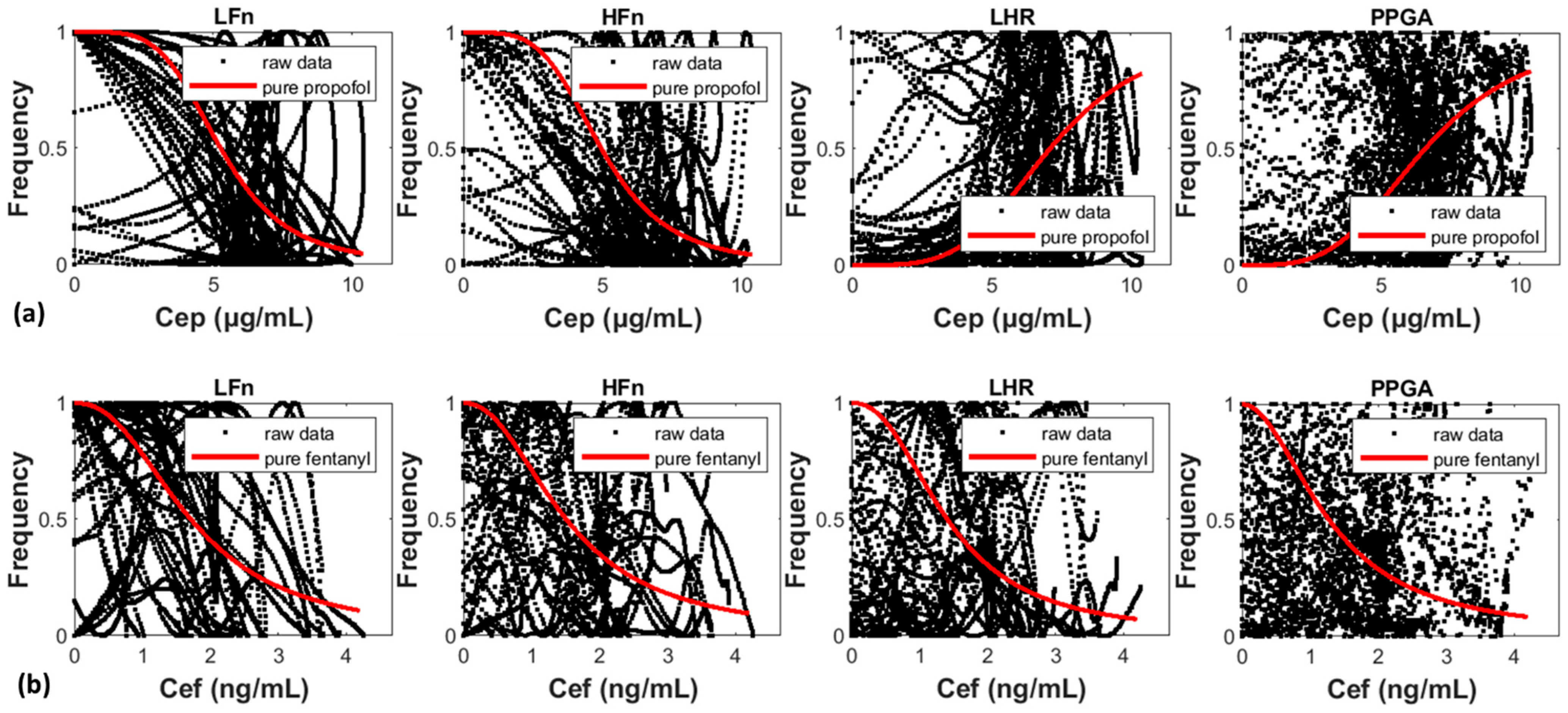
Figure 1. (a,b) The corresponding effects on the ANS caused by the effect-site concentrations of pure propofol (Cep) and fentanyl (Cef) in Groups 1 and 2, respectively. The black dots and red lines represent the raw data and the MLRSM predictions, respectively.
Mathematics 10 01651 g001
Figure 2. (a,b) The three-dimensional morphologies of activities on the ANS in terms of plasma concentrations (Cp) and effect-site concentrations (Ce), respectively. The level contours exhibit the corresponding performance of the drug interaction. Panel (b) exhibits only the mixed morphologies of Groups 1 and 2.
Mathematics 10 01651 g002
Figure 3. (a,b) The global three-dimensional morphologies of activities on the ANS in terms of plasma concentrations (Cp) and effect-site concentrations (Ce), respectively. This figure exhibits only the mixture of Groups 1 and 2. The red arrows represent the changing path of the drug with negative steepness.
Mathematics 10 01651 g003
Table 1. The modeling and sampling parameters of pure propofol and fentanyl MLRSM.
ActivityParametersPropofolFentanyl
ModelingSamplingModelingSampling
LFnSteepness γ −4.4084−4.4105 (0.0570) *−2.3600−2.3624 (0.0275)
Normalization factor C 50 5.26115.2613 (0.0307)1.71591.7161 (0.0184)
HFnSteepness γ −4.1503−4.1510 (0.0592)−2.2044−2.2052 (0.0281)
Normalization factor C 50 4.97244.9740 (0.0322)1.51341.5129 (0.0184)
LHRSteepness γ 4.02424.0220 (0.0521)−2.3588−2.3409 (0.0285)
Normalization factor C 50 7.08967.0899 (0.0379)1.40911.4075 (0.0199)
PPGASteepness γ 3.54423.5432 (0.0429)−2.0138−2.0371 (0.0291)
Normalization factor C 50 6.57996.5787 (0.0360)1.28361.2795 (0.0186)
*: Standard deviation.
Table 2. The modeling and sampling parameters of the propofol–fentanyl interaction MLRSM.
ActivityPara-
Meters
CpCe (Mixture)Ce (Group 1)Ce (Group 2)
ModelingSamplingModelingSamplingModelingSamplingModelingSampling
LFn γ 1
γ 2
1.2797
1.7051
1.28 (0.01)
1.70 (0.01)
−3.2024
−2.8392
−3.20 (0.03)
−2.84 (0.02)
-- *
--
−3.20 (0.03)
−2.84 (0.02)
--
--
−3.20 (0.03)
−2.84 (0.02)
C 50 , 1
C 50 , 2
13.1206
10.3580
13.11 (0.15)
10.36 (0.09)
3.9268
2.0578
3.93 (0.02)
2.06 (0.01)
--
--
3.93 (0.02)
2.06 (0.01)
--
--
3.93 (0.02)
2.06 (0.01)
C 0 , 1
C 0 , 2
5.3293
6.4379
5.33 (0.06)
6.44 (0.06)
4.2351
2.3693
4.23 (0.02)
2.37 (0.01)
4.4430
2.8881
4.44 (0.03)
2.89 (0.01)
4.0676
1.9515
4.07 (0.03)
1.95 (0.01)
γ i n t 10.029012.19 (9.95)−0.4678−0.47 (0.02)−0.0579−0.06 (0.02)−1.0517−1.05 (0.04)
α 0.06930.07 (0.03)1.67781.68 (0.07)67.5643Inf0.95980.96 (0.02)
HFn γ 1
γ 2
1.2892
1.5988
1.29 (0.01)
1.60 (0.01)
−3.2263
−2.6622
−3.23 (0.03)
−2.66 (0.03)
--
--
−3.23 (0.03)
−2.66 (0.03)
--
--
−3.23 (0.03)
−2.66 (0.02)
C 50 , 1
C 50 , 2
15.0960
11.8426
15.10 (0.19)
11.85 (0.11)
3.7128
1.8988
3.71 (0.02)
1.90 (0.01)
--
--
3.71 (0.02)
1.90 (0.01)
--
--
3.71 (0.02)
1.90 (0.01)
C 0 , 1
C 0 , 2
5.3293
6.4379
5.42 (0.06)
6.44 (0.06)
4.2351
2.3693
4.22 (0.02)
2.36 (0.01)
4.4430
2.8881
4.44 (0.03)
2.89 (0.01)
4.0676
1.9515
4.07 (0.03)
1.95 (0.01)
γ i n t 4.11074.13 (0.26)−0.2176−0.22 (0.02)−0.1191−0.12 (0.02)−0.6847−0.68 (0.03)
α 0.16600.17 (0.01)3.92634.02 (0.60)5.73356.42 (3.38)1.24051.24 (0.04)
LHR γ 1
γ 2
1.2451
1.5897
1.25 (0.01)
1.47 (0.59)
3.1158
−2.6172
3.12 (0.03)
−2.03 (1.65)
--
--
3.12 (0.03)
−2.02 (1.67)
--
--
3.12 (0.03)
−2.06 (1.61)
C 50 , 1
C 50 , 2
14.8138
12.4434
14.80 (0.20)
12.11 (1.48)
7.1478
1.8258
7.14 (0.04)
1.95 (0.34)
--
--
7.15 (0.04)
1.91 (0.30)
--
--
7.15 (0.04)
1.96 (0.35)
C 0 , 1
C 0 , 2
5.4163
6.4236
5.42 (0.06)
6.42 (0.06)
4.2173
2.3599
4.22 (0.02)
2.36 (0.01)
4.3982
2.8534
4.40 (0.03)
2.85 (0.01)
4.0676
1.9515
4.07 (0.03)
1.95 (0.01)
γ i n t 3.60843.02 (2.47)−0.5546−0.50 (0.71)−0.8130−0.53 (0.73)−0.0255−0.45 (1.61)
α 0.19290.22 (0.15)0.65890.59 (0.15)0.36420.42 (0.23)508.7802Inf
PPGA γ 1
γ 2
−1.0789
−1.4841
−1.08 (0.01)
−1.49 (0.01)
−2.7005
2.3635
−2.70 (0.02)
2.36 (0.02)
--
--
−2.70 (0.02)
2.36 (0.02)
--
--
−2.70 (0.02)
2.36 (0.02)
C 50 , 1
C 50 , 2
4.9809
6.5615
4.98 (0.07)
6.56 (0.07)
4.6255
2.5710
4.62 (0.03)
2.57 (0.02)
--
--
4.63 (0.03)
2.57 (0.02)
--
--
4.63 (0.03)
2.57 (0.02)
C 0 , 1
C 0 , 2
5.2263
6.5400
5.22 (0.06)
6.54 (0.06)
4.1871
2.2863
4.19 (0.02)
2.29 (0.01)
4.3317
2.7539
4.33 (0.03)
2.75 (0.01)
4.0815
1.9444
4.08 (0.03)
1.94 (0.01)
γ i n t −0.9360−0.94 (0.03)−1.2411−1.24 (0.03)−1.2383−1.24 (0.03)−1.3195−1.32 (0.05)
α 1.03531.04 (0.02)0.82780.83 (0.02)0.91290.91 (0.01)0.67470.68 (0.02)
* --: The same with the mixture data.
Publisher’s Note: MDPI stays neutral with regard to jurisdictional claims in published maps and institutional affiliations.