Abstract
This work aims to evaluate students’ opinions on the materials normally used in anatomy practical classes (fixed and plastinated) compared to 3D anatomical prints. For this purpose, students of anatomy enrolled in the Degree in Veterinary Medicine from the University of Murcia filled out a satisfaction survey about both kinds of material. The students rated the fixed material with a satisfaction percentage close to 100% and the plastinated material with a percentage higher than 75%. Regarding the 3D prints, the percentage obtained was consistently higher than 50% except for two issues: the identification of the vascular structures of the dolphin’s head and the usefulness for surgery of the viscera and vascular structures of the cat, both of which scored less than 50%. This could be related to the lesser knowledge of dolphins of the veterinary students as well as the complexity of these structures. However, the other questions, such as usefulness for learning and exam preparation, the quality of the anatomical piece, the identification of the feline spleen and kidneys, etc. obtained a satisfaction percentage between 58 and 90.40%. This reflects the good acceptance by students of the 3D prints and may allow for a reduction in the number of cadavers used.
1. Introduction
In anatomy education, in addition to the quality of the theoretical courses in veterinary faculties, practical classes are highly necessary and important for students to acquire knowledge about the real dimensions of the organs, topographical relationships, etc. Conventional cadaver model anatomy training presents several difficulties including the cost of the cadaver, ethical issues, and the application of formalin preservatives (). There are also factors intrinsic to the body donated to universities that can interfere with the quality and preparation of the pieces, such as rapid deterioration due to poor preservation of the cadavers, the presence of any pathology or poor body condition due to the cause of death (road traffic accident, etc.), obesity, age, and other factors (; ; ). Other anatomical models, such as plastinated specimens, are currently used as an alternative and/or complement to cadavers in human and veterinary medicine, with good results in learning and acceptance by students and professionals (). However, these specimens are also obtained from cadavers and through different procedures, which can also involve some toxic products (; ). The introduction of new technologies such as mobiles and tablets is leading to an evolution in the style of the teaching and learning of anatomy in faculties of health sciences (). Thus, software containing 3D visuals, virtual reality, and augmented reality is widely utilized (; ; ). Three-dimensional models provide advantages in being readily available and robust, thus presenting less deterioration and more anatomical details, whereas preserved bones allow students to have direct contact with the real tissue (). Thus, 3D-printed anatomical models can replace cadavers or human anatomical structures as teaching tools for medical students, as real cadavers have many deficiencies in practical medical teaching. For example, to prevent decomposition, formalin immersion is required, which is often too harsh for allergic students. There are also limits to the supply of bodies available ().
In recent years, 3D printing has been presented as an innovative educational methodology that can both complement the conventional material used in practical classes on human and veterinary anatomy and reduce the need for cadavers. Three-dimensional printing is a process in which a 3D computer model is transformed into a physical object (; ). Through computer control, the “printed materials” are stacked layer by layer until the physical object matches the blueprint on the computer. Commonly used materials for 3D printing include durable nylon, gypsum, aluminium, textile materials, and polylactic acid (; ; ). Laser-induced graphene and resin are also used (; ; ).
() carried out an experiment to study the external cardiac anatomy through different teaching methods. These authors divided first-year medical school students into three groups: studies on three-dimensional models; cadavers; and both combined. The group of students who used only 3D models had a statistically significant superior performance in the post-test than the group who used only cadavers and the group that used the combined materials. () presented previously created 3D models of the skull and jaw to students of anatomy in the Faculties of Health Sciences of the University of Salamanca, Spain, including the odontology, medicine, occupational therapy nursing, health sciences, and physiotherapy faculties. A survey was conducted among 280 students to assess the usefulness of these 3D models in the practical study of anatomy. The analysis of the results presented a positive evaluation of the use of 3D models by the students, showing a greater preference for the applicability of 3D models compared to real bones. () produced four biomodels based on cadaver-derived bones. These biomodels were presented to veterinary students for review along with cadaver bones. A survey was applied to a total of 298 participants. In this survey, the participants’ opinions about practical anatomy class, the class materials used, and use of biomodels were gathered. According to the results, only 21.2% of the participants found the practical anatomy classes adequate, and 49.7% of the participants stated that they are disturbed by the smell when they use bones in practical classes. The percentage of participants who found that biomodels were like real bones appeared to be 75.5%. In addition, the percentage of participants who found the use of biomodels beneficial in learning the skeletal system was 64.8%. Other studies in veterinary medicine have demonstrated the potential of 3D printing technology to be effectively applied to spinal stabilization surgeries for small breeds of dogs, allowing for precise screw placement and minimizing peri- and postoperative complications (; ). () carried out an integrative review on the use of 3D printing in the teaching of human anatomy. These authors found that in some studies, most students preferred the use of 3D printing to traditional methods (; ; ; ; ; ). Other studies have shown the importance of the use of 3D printing as a complementary tool to traditional methods of teaching anatomy (; ; ; ; ; ). () concluded that among the benefits of using 3D parts are accuracy, durability, ease of production, good cost–benefit ratio, and reductions in security risks linked to the fixation of cadaver and plastinated specimens. Added to this, the results found comparative studies which suggest other benefits of using 3D printing, such as efficiency in the reproducibility of models; fidelity to original anatomical structures; the possibility of representing common pathologies; ease of deployment; and reduction in ethical–legal issues (; ; ; ; ).
The aim of the present work is to produce an evaluation of different 3D anatomical impressions by veterinary students in practical anatomy classes as well as their comparison with the anatomical specimens usually used in practical rooms, such as fixed cadavers and plastinated material. Currently, it is difficult to obtain cadavers due to the Zero-Sacrifice Law, which was included in the animal welfare law (Law 6/2017, of November 8, on the protection and defence of companion animals in the Region of Murcia, Spain). This law prohibits the sacrifice of animals, except to prevent their suffering or for animal health, public health, safety, or environmental reasons. This law arose due to the growing awareness of the population regarding animal welfare, which has prompted a search for alternatives that reduce or eliminate the need to obtain cadavers. In addition, conventional methods of fixing cadavers, such as the use of formaldehyde, are harmful to health, which makes it necessary to search for other types of anatomical materials, such as plastinated material and 3D anatomical prints, that are non-toxic and perennial materials.
The processes of elaboration and interpretation of the 3D pieces included in this work were published in previous works carried out by our research team (, ; , ; ). In these works, the 3D pieces were analyzed from the point of view of the technical process necessary for their production, as well as from the point of view of the anatomical details that these pieces were capable of reproducing. The present work is focused on understanding the application of these 3D prints as educational models in practical sessions of veterinary anatomy, compared to the models normally used in our practical sessions (fixed and plastinated pieces).
2. Materials and Methods
This work was carried out with the students at the Veterinary Faculty of the University of Murcia. All students were studying the Anatomy II subject of the Degree in Veterinary Medicine. The study was carried out over four academic years (2020–21; 2021–22; 2022–23; and 2023–24) in which 84, 91, 86, and 87 students, respectively, participated, making up a total of 348 students. This subject is taught in the second semester of the first year of the Degree in Veterinary Medicine, within the Veterinary Anatomy Area (Department of Comparative Anatomy and Pathological Anatomy of the University of Murcia). This subject has six ECTS (European Credit Transfer System) credits assigned to it (50% practical credits and 50% theoretical credits). The practical credits are taught in the Dissection Room and the Veterinary Anatomical Museum of the Faculty of Veterinary Medicine of the University of Murcia. The materials used in our practical classes of veterinary anatomy include the following: skeletons and bones, joints, viscera, animal cadavers, and animal prosections. Anatomical specimens can be used fresh, but most of them are preserved in conventional fixation liquids. We specifically use a conventional fixation method, modified by us to reduce the percentage of formaldehyde. This liquid contains a mixture of 8.3% formaldehyde, 69.4% methyl alcohol, 5.5% phenol and 16.6% glycerine. This mixture produces a good colour and generates few odours, which lead to general acceptance by the students. Other teaching materials that we also use in our practical sessions are anatomical moulds or models, which are pieces made of a resistant synthetic material that show highly detailed anatomical structures. In addition, we have a plastination laboratory where we obtain plastinated material that is regularly used by anatomy students in our practical classes, as detailed by (). In addition to all the teaching material mentioned, our department has numerous interactive materials that students can consult, as well as bibliographic material (atlas, texts, etc.). This material is open access and freely available through the following website of our department: https://www.um.es/web/anatvet/docencia/recursos-docentes; https://www.um.es/web/anatvet/docencia/libros-materiales; https://www.um.es/web/plastinacion/), etc. URL (accessed on 30 October 2003).
In the present work, a general survey (Table 1 and Table 2) was developed with the aim of knowing the opinions of the students regarding the fixed material that is usually used in practical anatomy classes versus plastinated material, which students also use as complementary material. This survey also included questions aimed at checking the level of knowledge of the students regarding the origin and preparation process of cadavers to contextualize their ability to evaluate all the teaching material. In addition, this survey also collected students’ opinions regarding 3D-printed anatomical pieces. The surveys consisted of eleven questions (1–11). The response options for these eleven questions were two: Yes (100% agree) or No (100% disagree).
In recent years, the teaching staff of our department have produced 3D prints of anatomical pieces, to incorporate them as teaching materials in practical classes. These impressions have been made at the University of Murcia, according to the procedure detailed in previous works published by our research team (, ; , ; ). In the cited studies, the pieces were described from the technical and anatomical points of view. The present work aimed to assess their usefulness in the learning of veterinary anatomy. To this end, six 3D-printed anatomical pieces were included in the present work: piece 1 (3D-printed viscera and vascular structures of a cat); piece 2 (3D printing of vascular structures of a dolphin’s head), piece 3 (3D-printed leopard, cheetah, and cat skulls); piece 4 (3D printing of the skeleton of the gilthead sea bream, Sparus aurata L.); piece 5 (3D printing of the arteries, veins and bile ducts of a cat’s liver); piece 6 (3D printing of the visceral branches of a cat’s abdominal Aorta). Images of these 3D impressions are shown in Figure 1, Figure 2, Figure 3, Figure 4, Figure 5 and Figure 6. However, the anatomical details of each piece as well as the origin of the animals and the technical details necessary for obtaining them have been included in this work as a Supplementary Material (Figures S1–S6).
These six-3D printed anatomical pieces were evaluated by all students through different questions (Table 3, Table 4, Table 5, Table 6, Table 7 and Table 8; Figure 7, Figure 8, Figure 9, Figure 10, Figure 11, Figure 12, Figure 13, Figure 14, Figure 15, Figure 16, Figure 17 and Figure 18). The student rating scale that was used to assess student satisfaction with respect to 3D-printed pieces was divided into the following ratings: not at all, somewhat, quite a bit, a lot. and very much. These criteria were then converted to the following percentage values: 0, 25, 50, 75, and 100%, respectively. Similarly, () also used quality criteria that were then converted to percentages. To evaluate the 3D prints, these were displayed in the practice room, together with the other pieces that are usually used in the practical sessions of veterinary anatomy (fixed and plastinated pieces). At the end of each practical session, all the students evaluated the 3D pieces individually and filled out all the surveys that were included in the present work.
All surveys were conducted at the end of each academic year. The first academic year included in this work was 2020–21, which coincided with the first in-person course after the coronavirus (COVID-19) pandemic. Therefore, this course and the following ones represented a significant change for the students. For this reason, in order to determine the influence of the different circumstances that may have influenced each academic year, the results were analyzed independently for each course. However, the results were also analyzed jointly, in order to observe the general trend throughout the four academic years studied.

Table 1.
Questions posed to students regarding material for the study of veterinary anatomy (conventional versus new teaching materials).
Table 1.
Questions posed to students regarding material for the study of veterinary anatomy (conventional versus new teaching materials).
| Question Number | Questions |
|---|---|
| 1 | Do you regularly attend practical classes? |
| 2 | Do you consider that the material preserved by fixation methods, which is commonly used in anatomy practice rooms, is suitable for the development of good anatomy practices in the dissection room? |
| 3 | Did you know that the Zero-Slaughter Law 1 produces a shortage of cadavers, which generates a lack of material for practices? |
| 4 | Do you think that the Zero-Slaughter Law will mean a return to veterinary studies to medieval times? |
| 5 | Do you think that the Zero-Slaughter Law should have contemplated the possibility of obtaining 5% of animals for training purposes in Veterinary Medicine (Anatomy, Pathological Anatomy, Toxicology, Parasitology, Surgery, Medical Pathology, etc.) |
| 6 | Did you know that most of the teaching material for practices with dogs and cats came from the Zoonosis Centres of the City Councils? |
| 7 | Did you know that the few cadavers that are donated by veterinary clinics are frozen and are not heparinized? Since heparinization is necessary to avoid clots, this fact prevents proper preservation of the cadavers. |
| 8 | Would you be willing to dissect cadavers of porcine or ovine species? |
| 9 | Did you know that the possibility of buying cadavers to make up for their lack multiplies by four the budget available for anatomy practices? |
| 10 | Do you think that new methods of preserving cadavers, such as plastination, which is commonly used like a complement to the fixed anatomical pieces in practical veterinary anatomy classes, can partially alleviate the lack of cadavers? |
| 11 | Do you think that plastination and 3D impressions represent the future of veterinary anatomy? |
1 The Zero-Sacrifice Law refers to the protection of animals. It is included in the animal welfare law (Law 6/2017, of November 8, on the protection and defence of companion animals in the Region of Murcia, Spain), which prohibits the sacrifice of animals, except to prevent their suffering or for animal health, public health, safety or environmental reasons.
2.1. Piece 1: 3D-Printed Viscera and Vascular Structures of a Cat
The evaluation of this piece (Figure 1) by the students was carried out in the four academic years mentioned, through several questions related to the degree of satisfaction of this piece (Figure 7 and Figure 8; Table 3).
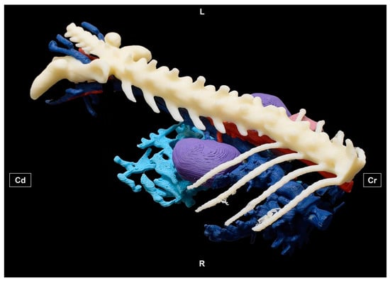
Figure 1.
Three-dimensionally printed viscera and vascular structures of a cat. Dorsolateral view. L = Left side. R = Right side. Cr = Cranial. Cd = Caudal.
2.2. Piece 2: 3D Printing of Vascular Structures of a Dolphin’s Head
This piece (Figure 2) was evaluated in the same way and in the same academic years as described for piece 1 (Figure 9 and Figure 10; Table 4).
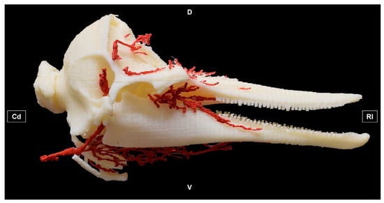
Figure 2.
Three-dimensional printing of the vascular structures of a dolphin’s head. Right lateral view. D = Dorsal. V = ventral. Rl = Rostral. Cd = Caudal.
2.3. Piece 3: 3D Printings of Lion, Leopard, Cat and Cheetah Skulls
This piece (Figure 3) was evaluated in the same way and in the same academic years as described for pieces 1 and 2 (Figure 11 and Figure 12; Table 5).
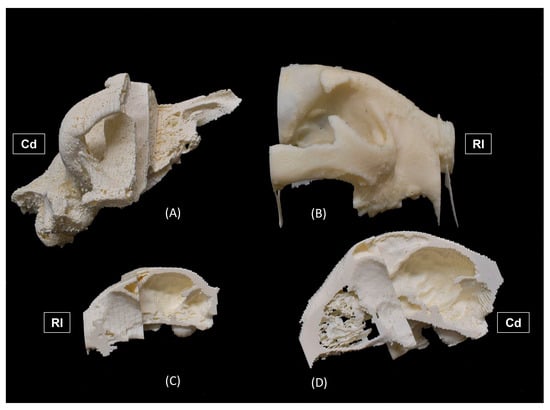
Figure 3.
Three-dimensional printing of (A) lion, (B) leopard, (C) cat, and (D) cheetah skulls. (A,B) Right lateral views. (C,D) Left lateral views; Rl = Rostral. Cd = Caudal.
2.4. Piece 4: 3D Printing of the Skeleton of the Gilthead Sea Bream Sparus aurata L.
This piece (Figure 4) was evaluated in the same way and in the same academic years as described for pieces 1–3 (Figure 13 and Figure 14; Table 6).
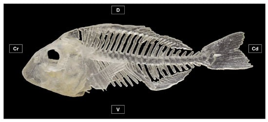
Figure 4.
Three-dimensional printing of the skeleton of the gilthead sea bream Sparus aurata L. Left lateral view. Cr = Cranial. Cd = Caudal. D = Dorsal. V = Ventral.
2.5. Piece 5: 3D Printing of Arteries, Veins and Bile Ducts of a Cat’s Liver
This piece (Figure 5) was evaluated in the same way as described in pieces 1–4, but it was only evaluated in the 2021–22, 2022–23, and 2023–24 academic years (Figure 15 and Figure 16; Table 7).
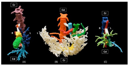
Figure 5.
Three-dimensional printing of (A) bile ducts, (B) veins, and (C) arteries. Ventral views. R = Right. L = Left. Cr = Cranial. Cd = Caudal.
2.6. Piece 6: 3D Printing of the Visceral Branches of a Cat’s Abdominal Aorta
This piece (Figure 6) was evaluated in the same way as described in pieces 1–5, but it was only evaluated in the 2022–23 and 2023–24 academic years (Figure 17 and Figure 18; Table 8).
Figure 6.
Three-dimensional printing of the visceral branches of a female cat’s abdominal aorta and caudal vena cava. Ventral view. R = Right. L = Left. Cr = Cranial. Cd = Caudal.
2.7. Statistical Analysis
Statistical differences among the academic years were obtained for both the general eleven questions and the specific questions for each 3D anatomical print. Also, the statistical differences among the general and specific questions were obtained from the data of all the academic years surveyed. Data were expressed in percentages. The statistical package SPSS 28.0.1.1. (IBM, New York, NY, USA) was used for the statistical analysis. The data distribution and the homogeneity of variances were analyzed by the Shapiro–Wilk and the Levene test, respectively, for p < 0.05. For most of the questions, both tests showed values of p > 0.05. Hence, the analysis of variance (ANOVA) and a post-hoc Tukey test were used, for p < 0.05. In the cases with values of p < 0.05 in the Shapiro–Wilk and the Levene tests, nonparametric tests (U of Mann–Whitney and Z of Kolmogorov–Smirnov tests) were used.
3. Results
3.1. General Questions Related to the Usual Practical Classes in Veterinary Anatomy Practice Rooms
Table 2 shows the results obtained from the eleven general questions that were asked to the students from each academic year. These questions are shown in Table 1.
Table 2.
The first four rows of the table indicate the percentage of students from each academic year who answered affirmatively to the general eleven questions. Different superscripts between these rows indicate significant differences (p < 0.05) between academic years for each question. In the last row of the table, the average value ± SEM obtained from all the academic years is also represented. In this last row, different superscripts between columns indicate significant differences between issues. Questions 1–11 are shown in Table 1.
| Academic Years | Questions | ||||||||||
|---|---|---|---|---|---|---|---|---|---|---|---|
| 1 | 2 | 3 | 4 | 5 | 6 | 7 | 8 | 9 | 10 | 11 | |
| 2020–21 | 100 a | 98.8 a | 54.8 a | 42.2 a | 57.1 a,b,c | 35.7 a | 3.6 a | 97.6 a,b | 8.3 a | 80.5 a | 73.5 a |
| 2021–22 | 98.9 a | 100 a | 54.9 a | 56.04 a,b | 76.5 a | 25.3 a | 30.8 b | 100 a | 10.99 a | 76.1 a | 84.4 a |
| 2022–23 | 95.3 a | 97.7 a | 40.7 a | 74.4 b | 65.8 a,b | 34.9 a | 19.8 b | 94.2 b | 9.3 a | 75.9 a | 78.6 a |
| 2023–24 | 98.8 a | 100 a | 41.4 a | 60.7 a,b | 44.3 c | 19.5 a | 17.2 a,b | 98.8 a,b | 10.3 a | 75.56 a | 88.4 a |
| 2020–2024 | 98.2 a ± 1.02 | 99.2 a ± 0.55 | 47.95 b ± 3.99 | 58.33 b,c ± 6.64 | 60.92 b,c ± 6.81 | 28.85 d ± 3.91 | 17.85 d ± 5.59 | 97.65 a ± 1.25 | 9.72 d ± 0.59 | 77.01 c ± 1.17 | 81.22 a ± 3.27 |
According to the first question, almost 100% of the students attended practical classes in all the academic years studied (p > 0.05).
In relation to the to the second question, most students considered that the fixed material that is used by them in anatomy practices is well prepared and is suitable for practical classes in the dissection room (p > 0.05).
According to the third question, approximately half of the students are unaware that the Law of Zero Sacrifice of Animals has caused a lack of cadavers, which has decreased the possibility of dissecting and obtaining anatomical pieces (p > 0.05).
Regarding the fourth, approximately half of the students of the first two academic years considered that the Zero-Slaughter Law may produce a setback in veterinary studies because it drastically reduces access to cadavers, which in turn prevents dissection and obtaining the anatomical specimens necessary for veterinary studies. This opinion increased significantly in the students of the following two academic years (p < 0.05).
In relation to the fifth question, the percentage of students who considered that the Zero-Slaughter Law should have established that at least 5% of the animals were used for veterinary training purposes varied between academic years, being higher in the 2021–22 academic year and lower in the 2023–24 academic year (p < 0.05). However, the mean value for this question was 60.92%.
Regarding the sixth question, most students do not know the origin of most of the dogs and cats that are commonly used in veterinary anatomy practices (p > 0.05).
In relation to the seventh question, most students are unaware of the state of conservation of the cadavers that are donated by veterinary clinics, as well as the consequences that derive from this fact. This lack of knowledge was especially significant among students in the 2020–21 academic year (p < 0.05).
Regarding the eighth question, most students would be willing to dissect swine or ovine cadavers. This fact indicates the students’ interest in dissecting, even if it is on livestock species.
In relation to the ninth question, most students (p > 0.05) are unaware of the economic cost of purchasing cadavers.
Regarding the tenth question, more than 75% of the students (p > 0.05) consider that the new systems for preserving cadavers, such as the plastinated pieces that we usually use in practical classes as a complement to the fixed anatomical material, could partially alleviate the lack of cadavers, which shows the receptivity of students to new conservation methods that compensate for the lack of cadavers. Since plastinated anatomical pieces are commonly used in our practical veterinary anatomy classes, students can evaluate them from their experience. However, when we compare the percentage of satisfaction of the plastinated pieces versus the fixed pieces (question 10 versus question 2), we can observe that the students preferred the fixed pieces (99% satisfaction percentage) to the plastinated pieces (77% satisfaction percentage), this result being significant (p < 0.05).
Finally, in relation the eleventh question, 81.22% of students (the average value obtained from all academic years) considered that new teaching materials such as 3D-printed and plastinated pieces, represent the future of anatomy veterinary science, which shows the students’ appreciation of these pieces and their ability to adapt to new teaching methodologies.
3.2. Assessment of 3D Prints of Anatomical Pieces
3.2.1. Three-Dimensional Print of the Viscera and Vascular Structures of a Cat (Piece 1)
Figure 7 shows the results obtained by academic year for each question for this 3D printing anatomical model, according to a rating scale, ranging from not at all to somewhat, quite a bit, a lot, and very much, which were equivalent to a satisfaction percentage of 0, 25, 50, 75, and 100%, respectively. Figure 8 shows the average percentages of satisfaction (+SEM) based on the data obtained from all students in each academic year. Table 3 shows the average values obtained from all the academic years surveyed. According to the data that are represented in Figure 7 and Figure 8 and Table 3, the results obtained for this 3D-printed anatomical model were the following.
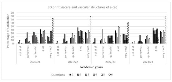
Figure 7.
Percentage of satisfaction that was obtained for 3D-printed viscera and vascular structures of a cat, in relation to six questions. 1: Usefulness for learning; 2: useful for exam preparation 3: usefulness for future preparation in surgery; 4: quality of the anatomical piece; 5: whether the vascular structures are well identified. 6: whether the spleen and kidneys are well identified. Student rating scale: not at all, somewhat, quite a bit, a lot, and very much (0, 25, 50, 75, and 100% satisfaction, respectively).
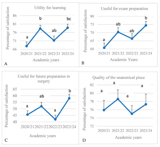
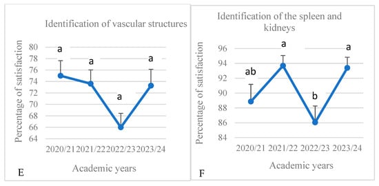
Figure 8.
Average percentage of satisfaction (+SEM) that was obtained for 3D-printed viscera and vascular structures of a cat, in relation to questions 1–6 (A–F), from each academic year. Different letters on each value indicate significant differences between academic years.
Table 3.
Average percentage of satisfaction ± SEM obtained from the data of all academic years studied for the 3D-printed of viscera and vascular structures of a cat. Different superscripts between rows indicate significant differences between questions (p < 0.05).
Table 3.
Average percentage of satisfaction ± SEM obtained from the data of all academic years studied for the 3D-printed of viscera and vascular structures of a cat. Different superscripts between rows indicate significant differences between questions (p < 0.05).
| 3D-Printed Model | Question | Average Percentage of Satisfaction |
|---|---|---|
| 3D-printed viscera and vascular structures of a cat. | 1. Usefulness for learning | 74.04 a ± 2.69 |
| 2. Useful for exam preparation | 67.94 a ± 2.93 | |
| 3. Usefulness for future preparation in surgery | 49.34 c ± 3.57 | |
| 4. Quality of the anatomical piece | 76.69 a,b ± 0.78 | |
| 5. Identification of vascular structures | 72.0 a ± 2.04 | |
| 6. Identification of the spleen and kidneys | 90.49 b ± 1.85 |
3.2.2. Three-Dimensional Print of Vascular Structures of a Dolphin’s Head (Piece 2)
The results for this 3D-printed model are shown in Figure 9 and Figure 10, Table 4 and are described below.
- -
- -
- -
- -
- -
- Question 5: The students showed difficulty in identifying the vascular structures, with satisfaction percentages below 45% in all academic years (Figure 9 and Figure 10E). The lowest satisfaction percentages were observed in the 2022–23 academic year. Table 4 shows that the percentage of satisfaction for this question was significantly lower than for the other questions (p < 0.05).
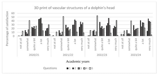
Figure 9.
Percentage of satisfaction obtained for the 3D print of the vascular structures of a dolphin’s head, in relation to five questions. 1: Usefulness for learning; 2: usefulness for understanding the vascularization of the dolphin head; 3: quality of the anatomical piece; 4: usefulness for the training of future veterinarians specializing in cetaceans; 5: whether the vascular structures are well identified. Student rating scale: not at all, somewhat, quite a bit, a lot, and very much (0, 25, 50, 75, and 100 % satisfaction, respectively).
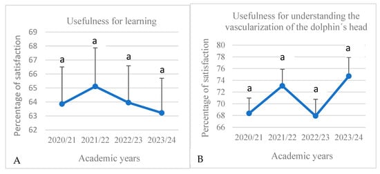
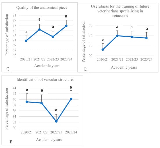
Figure 10.
Average percentage of satisfaction (+SEM) that was obtained for a 3D print of the vascular structures of a dolphin’s head, in relation to five questions (A–E). Different letters on each value indicate significant differences between academic years.
Table 4.
Average percentage of satisfaction (± SEM) obtained from the data of all academic years studied for the 3D print of the vascular structures of a dolphin’s head. Different superscripts between rows indicate significant differences between questions (p < 0.05).
Table 4.
Average percentage of satisfaction (± SEM) obtained from the data of all academic years studied for the 3D print of the vascular structures of a dolphin’s head. Different superscripts between rows indicate significant differences between questions (p < 0.05).
| 3D-Printed Model | Question | Average Percentage of Satisfaction |
|---|---|---|
| 3D print of the vascular structures of a dolphin’s head. | 1. Usefulness for learning | 64.03 a ± 0.39 |
| 2. Usefulness for understanding the vascularization of the dolphin head | 71.02 a ± 1.69 | |
| 3. Quality of the anatomical piece | 73.77 a ± 1.36 | |
| 4. Usefulness for the training of future veterinarians specializing in cetaceans | 72.54 a ± 1.6 | |
| 5. Identification of vascular structures | 37.6 b ± 1.8 |
3.2.3. Three-Dimensional Print of Lion, Leopard, Cat and Cheetah Skulls (Piece 3)
- Question 4: the values were higher than 56%, except in the 2022–23 academic year, in which the satisfaction percentage was less than 50% (p < 0.05) (Figure 12D).
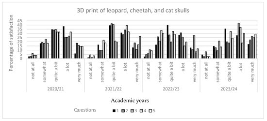
Figure 11.
Percentage of satisfaction that was obtained for 3D prints of lion, leopard, cat, and cheetah skulls, in relation to five questions. 1: Usefulness for learning; 2: usefulness for understanding the ethmoidal labyrinth and paranasal sinuses of feline species; 3: usefulness for the training of future veterinarians specializing in feline species; 4: whether any paranasal sinuses were identified; 5: whether the ethmoidal labyrinth was located. Student rating scale: not at all, somewhat, quite a bit, a lot, and very much (0, 25, 50, 75, and 100% satisfaction, respectively).
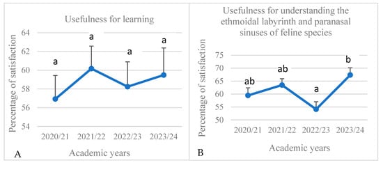
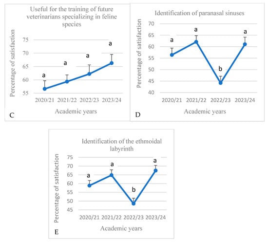
Figure 12.
Average percentage of satisfaction (+SEM) that was obtained for 3D prints of lion, leopard, cat, and cheetah skulls, in relation to five questions (A–E). Different letters on each value indicate significant differences between academic years (p < 0.05).
Table 5.
Average percentage of satisfaction ± SEM obtained from the data of all academic years studied for the 3D prints of lion, leopard, cat, and cheetah skulls. Different superscripts between rows indicate significant differences between questions (p < 0.05).
Table 5.
Average percentage of satisfaction ± SEM obtained from the data of all academic years studied for the 3D prints of lion, leopard, cat, and cheetah skulls. Different superscripts between rows indicate significant differences between questions (p < 0.05).
| 3D-Printed Model | Question | Average Percentage of Satisfaction |
|---|---|---|
| 3D prints of lion, leopard, cat, and cheetah skulls. | 1.Usefulness for learning | 58.70 a ± 0.71 |
| 2. Usefulness for understanding the ethmoidal labyrinth and paranasal sinuses of feline species | 61.09 a ± 2.83 | |
| 3. Useful for the training of future veterinarians specializing in feline species | 61.11 a ± 2.06 | |
| 4. Identification of the paranasal sinuses | 55.93 a ± 4.1 | |
| 5. Identification of the ethmoidal labyrinth | 59.92 a ± 4.2 |
3.2.4. Three-Dimensional Print of the Skeleton of the Gilthead Sea Bream, Sparus aurata L. (Piece 4)
- Question 3: The students of the 2021–22 and 2023–24 academic years showed a degree of satisfaction above 50% in this proposal, especially the students of the 2021–22 and 2023–24 academic years (Figure 14C). However, the percentage of satisfaction for this question was lower than for the question 1 (p < 0.05) (Table 6).
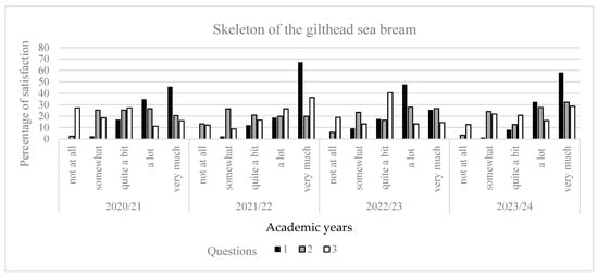
Figure 13.
Percentage of satisfaction that was obtained for 3D print of the skeleton of the gilthead sea bream, in relation to three questions. 1: Assessment of the quality of the anatomical piece; 2: positive evaluation of the incorporation of viscera into the anatomical piece; 3: considering this type of 3D printing useful in veterinary studies. Student rating scale: not at all, somewhat, quite a bit, a lot, and very much (0, 25, 50, 75, and 100% satisfaction, respectively).
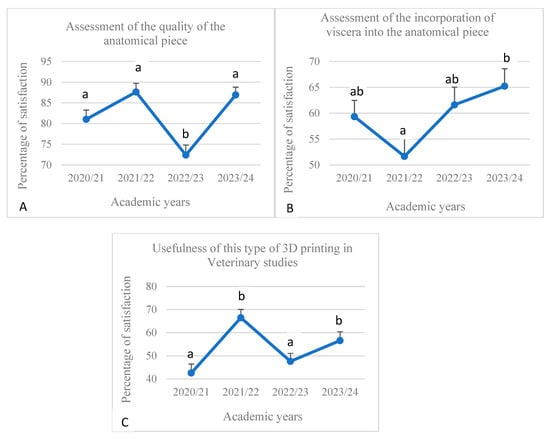
Figure 14.
Average percentage of satisfaction (+ SEM) that was obtained for 3D print of the skeleton of the gilthead sea bream, in relation to three questions (A–C). Different letters on each value indicate significant differences between academic years (p < 0.05).
Table 6.
Average percentage of satisfaction ± SEM obtained from the data of all academic years studied for the 3D print of the skeleton of the gilthead sea bream, Sparus aurata L. Different superscripts between rows indicate significant differences between questions (p < 0.05).
Table 6.
Average percentage of satisfaction ± SEM obtained from the data of all academic years studied for the 3D print of the skeleton of the gilthead sea bream, Sparus aurata L. Different superscripts between rows indicate significant differences between questions (p < 0.05).
| 3D-Printed Model | Question | Average Percentage of Satisfaction |
|---|---|---|
| 3D print of the skeleton of the gilthead sea bream, Sparus aurata L. | 1.Assessment of the quality of the anatomical piece | 81.99 a ± 3.53 |
| 2. Assessment of whether the incorporation of viscera would increase the usefulness of the 3D anatomical piece. | 59.46 b ± 2.87 | |
| 3. Assessment of the use of 3D anatomical prints in veterinary studies | 53.32 b ± 5.26 |
3.2.5. Three-Dimensional Print of Arteries, Veins and Bile Ducts of a Cat’s Liver (Piece 5)
- Question 2: the percentage of satisfaction was higher than 62% in all academic years (p > 0.05), with the lowest values in the 2022–23 academic year (Figure 16B).
- Question 3: the percentage of satisfaction was higher than 59% in all academic course (p > 0.05), with the lowest values in the 2022–23 academic year (Figure 16C).
- Question 4: The satisfaction percentage was higher than 50% in all academic years, except in the 2022–23 academic year, in which it was 45.29% (Figure 16D). When comparing with the other questions, the average percentage of satisfaction was significantly lower than for the questions 1 and 5 (p < 0.05) (Table 7).
- Question 6: the satisfaction percentage was higher than 50% in all academic years (p > 0.05), with the lowest values in the 2022–23 academic year (Figure 16F).
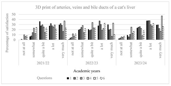
Figure 15.
Percentage of satisfaction that was obtained for a 3D print of the arteries, veins and bile ducts of a cat’s liver, in relation to six questions. 1: Utility for learning; 2: usefulness for knowledge of arterial and venous irrigation; 3: usefulness for knowledge of the biliary system; 4: identification of vascular structures; 5: identification of the gallbladder; 6: identification of the portal vein and hepatic veins. Student rating scale: not at all, somewhat, quite a bit, a lot, and very much (0, 25, 50, 75, and 100% satisfaction, respectively).
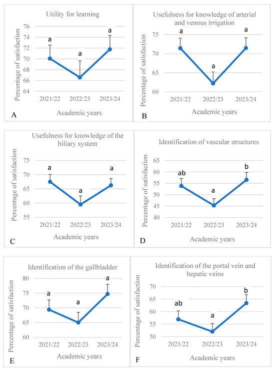
Figure 16.
Average percentage of satisfaction (+SEM) that was obtained for a 3D print of the arteries, veins, and bile ducts of a cat’s liver in relation to six questions (A–F). Different letters on each value indicate significant differences between academic years (p < 0.05).
Table 7.
Average percentage of satisfaction ± SEM obtained from the data of all academic years studied for the 3D print of the arteries, veins, and bile ducts of a cat’s liver. Different superscripts between rows indicate significant differences between questions (p < 0.05).
Table 7.
Average percentage of satisfaction ± SEM obtained from the data of all academic years studied for the 3D print of the arteries, veins, and bile ducts of a cat’s liver. Different superscripts between rows indicate significant differences between questions (p < 0.05).
| 3D-Printed Model | Question | Average Percentage of Satisfaction |
|---|---|---|
| 3D print of the arteries, veins and bile ducts of a cat’s liver | 1. Utility for learning | 69.46 a ± 1.53 |
| 2. Usefulness for knowledge of arterial and venous irrigation | 68.37 a,b ± 3.08 | |
| 3. Usefulness for knowledge of the biliary system | 64.42 a,b ± 2.48 | |
| 4. Identification of vascular structures | 51.9 b ± 3.39 | |
| 5. Identification of the gallbladder | 69.7 a ± 2.8 | |
| 6. Identification of the portal vein and hepatic veins | 57.45 a,b ± 3.29 |
3.2.6. Three-Dimensional Print of the Visceral Branches of a Cat’s Abdominal Aorta (Piece 6)
- Question 1: the percentage of satisfaction was higher than 70% in the two academic years that were studied (2022–23 and 2023–24), with the 2022–23 academic year showing the lowest values (p > 0.05) (Figure 18A).
- Question 2: the percentage of satisfaction was higher than 65% in the two academic years studied, with the 2022–23 academic year being the one that showed the lowest values (p > 0.05) (Figure 18B).
- Question 3: The satisfaction percentage was higher than 50% in the 2023–24 academic year, while in the 2022–23 academic year, this was 47.65% (p < 0.05) (Figure 18C). When comparing with the other questions, the values of satisfaction were lower than those obtained for questions 1 and 5 (p < 0.05) (Table 8).
- Question 4: the quality of the piece was rated better in the 2023–24 academic year than in the 2022–23 academic year (78.74 versus 66.28%, respectively) (p < 0.05) (Figure 18D).
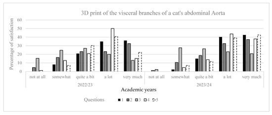
Figure 17.
Percentage of satisfaction that was obtained for a 3D print of the visceral branches of a cat’s abdominal aorta, in relation to five questions. 1: Utility for learning; 2: usefulness for exam preparation; 3: utility for future preparation in surgery: 4: quality of the anatomical piece; 5: identification of vascular structures. Student rating scale: not at all, somewhat, quite a bit, a lot, and very much (0, 25, 50, 75, and 100% satisfaction, respectively).
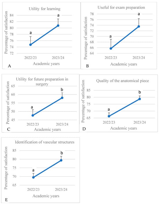
Figure 18.
Average percentage of satisfaction (+ SEM) that was obtained for a 3D print of the visceral branches of a cat’s abdominal aorta, in relation to five questions (A–E). Different letters on each value indicate significant differences between academic years.
Table 8.
Average percentage of satisfaction ± SEM obtained from the data of all academic years studied for the 3D print of the visceral branches of a cat’s abdominal aorta. Different superscripts between rows indicate significant differences between questions (p < 0.05).
Table 8.
Average percentage of satisfaction ± SEM obtained from the data of all academic years studied for the 3D print of the visceral branches of a cat’s abdominal aorta. Different superscripts between rows indicate significant differences between questions (p < 0.05).
| 3D-Printed Model | Question | Average Percentage of Satisfaction |
|---|---|---|
| 3D print of the visceral branches of a cat’s abdominal aorta | 1.Utility for learning | 77.73 a ± 3.02 |
| 2. Useful for exam preparation | 69.62 a,b ± 3.92 | |
| 3. Utility for future preparation in surgery | 52.85 b ± 5.2 | |
| 4. Quality of the anatomical piece | 72.51 a,b ± 6.23 | |
| 5. Identification of vascular structures | 74.39 a ± 4.91 |
4. Discussion
Different studies have shown the current interest in the use of 3D printing for learning and studying human and veterinary anatomy (; , ; ; ; ; ). According to these studies, the implementation of 3D-printed models in the teaching of anatomy improves the effectiveness of the learning of morphological structures and avoids the inconveniences generated by using cadavers and other anatomical pieces derived from said cadavers, such as the use of toxic products for their obtainment and/or preservation, the legal and ethical aspects, the cost and time of obtaining, etc. (; ; ).
In the present work, a survey was conducted among veterinary anatomy students in order to obtain their assessment of the veterinary anatomy material that they usually use in practical classes, as well as of different 3D anatomical impressions obtained from different animal species and different anatomical areas. The results found in this work are discussed below.
4.1. General Issues Related to Veterinary Anatomical Material That Is Commonly Used by the Students in Practice Rooms
Most students of the present work regularly attended practical classes and considered that the anatomical pieces that are usually used in practical veterinary anatomy classes are well prepared. In this sense, we must specify that the fixation system that we usually use in our practice rooms consists of a mixture of diluted formaldehyde, methyl alcohol, phenol and glycerine, as explained in material and methods section of the present work. This system allows good fixation, without excess formaldehyde, achieving anatomical pieces with good texture and that do not emit excess vapours or unpleasant odours, which justifies the good acceptance of these fixed pieces by the students. This contrasts with the results found in other published articles, in which students showed a rejection of fixed anatomical pieces due to the odours released, inappropriate texture, etc. (; ; ).
On the other hand, our students showed a lack of knowledge regarding the Law of Zero Sacrifice, as well as its consequences for teaching. This law prohibits the sacrifice of animals—except to prevent their suffering or for animal health, public health, safety, or environmental reasons—as explained in the Material and Methods section. In this regard, they are critical of the lack of cadavers for dissection. Likewise, most students are unaware of the animal cadaver donation system and the state of conservation of the donated cadavers, as well as the consequences of this for obtaining anatomical pieces. Similarly, other publications have documented different drawbacks in the donation system: poor conservation of cadavers, rapid deterioration, age, and other factors (; ; ).
Moreover, our students also use plastinated material from our plastination laboratory. Thus, they are accustomed to using it and consider it very useful for practical veterinary anatomy classes as a complement to the material fixed by conventional methods. These results coincide with those found by (). These authors assessed the efficacy of plastinated organs as teaching resources. For this purpose, they were studying three different subject areas of the University of Murcia: veterinary anatomy, human anatomy, and veterinary surgery. They studied two types of groups in each subject area: experimental groups that only used plastinated organs and control groups that used wet organs and anatomy sections with classical fixative solutions. The authors used a pre-test and a post-test in both control and experimental groups to evaluate the quantity and quality of the knowledge and skills students had acquired because of the use of plastinated material. Students’ results confirmed the efficacy of the use of plastinated specimens as teaching resources. Hence, the authors asserted that the use of plastinated material as a teaching resource does improve the quality of teaching and learning in anatomy. In the present work, when the students were asked about the usefulness of new teaching materials, such as 3D-printed and plastinated material, they considered that these are a good alternative to traditional conservation methods, being receptive to their use in veterinary education as a complement to traditional methods.
This is consistent with all the works reviewed, in which students showed good acceptance of the new methodologies due mainly to the ethical issues involved in obtaining cadavers as well as the health issues involved in using conservation methods, since the traditional systems use toxic products such as formaldehyde, phenol, etc. (; , ; ; ; ; ).
4.2. Specific Issues Related to 3D Anatomical Printing
After analyzing student satisfaction with 3D-printed models, we first observed that the results varied in relation to the academic year studied. Thus, in general, students from the 2020–21 and 2022–2023 academic years were those who showed the lowest satisfaction values. The lower values of the 2020–21 academic year may be related to the fact that it was the first in-person academic year after the coronavirus pandemic (COVID-19) which took place in 2019–2020. During the pandemic, teaching was online; therefore, it is likely that the students who began face-to-face classes in the 2020–21 academic year had greater difficulty in restarting the face-to-face teaching methodology, and this also caused them greater difficulty in assessing and analyzing anatomical pieces in 3D. In fact, the academic results of these students regarding the Anatomy II subject were worse than the results obtained in the other academic years studied in the present work. However, the satisfaction data for the 2022–23 academic year cannot be attributed to the circumstances mentioned for the 2020–21 academic year and can therefore only be attributed to a greater critical feeling among students in this academic year. For their part, students of the 2021–22 and 2023–24 academic years showed increased appreciation for the new 3D prints, probably indicating greater ease in their analysis and interpretation, which also coincides with better academic results than those obtained by the students of the academic year 2020–21.
On the other hand, overall, the average percentage of satisfaction of our students with respect to the 3D-printed anatomical models was acceptable, usually being higher than 60%. However, these values were lower than those found for the materials commonly used in our practice rooms, since the fixed and plastinated material is considered by students to be suitable and very useful for practical classes, as we have explained previously (Section 4.1). However, the average satisfaction of our students regarding the 3D-printed models was acceptable, which indicates good receptivity to new teaching methodologies, since these new anatomical materials do not require traditional conservation methods and reduce the need for cadavers for practical classes. () found similar results. In the cited study, 298 veterinary students were asked to evaluate four different bone biomodels that were 3D-modeled and -printed with reference to cadaver-derived bones. According to the survey, 75.5% of the students stated that their biomodel resembled the reference bones. In addition, 64.8% of these students stated that the use of biomodels can be efficient in learning the skeletal system. In this study, biomodels were assessed as the second most useful tool after bone. On the other hand, considering that 49.7% of the participants were disturbed by the smell while using bones in practical classes, the advantage of utilizing biomodel stands out even further. () reported that the students were particularly hesitant in touching pig, cat, and dog cadavers. The fact that the biomodels are like the real ones and not taken from live animals increases the motivation of students to learn by touching.
() presented 3D-printed models of skulls and jaws to the students of Faculties of Health Sciences of the University of Salamanca (Spain). A survey was carried out to assess the usefulness of these 3D models in the practical study of anatomy. The total number of students included in the survey was 280. The analysis of the responses showed that the participants gave more preference to the applicability of 3D models in comparison to the real bones. According to these authors, the combination of 3D models with the original bone pieces can be very advantageous.
Conversely, in our study, the percentage of satisfaction depended on the anatomical model evaluated and the question asked regarding each of the models. Thus, when we analyzed the difficulty level in relation to each of the 3D prints, we observed that the highest difficulty was found in identifying vascular structures in the 3D print of the dolphin’s head. We think that this was due to the fact that the dolphin is not a domestic species that students typically study in veterinary anatomy, and this may have created difficulty for students in identifying vascular structures. Furthermore, the cephalic territory is a complex anatomical area, according to our teaching experience with our veterinary anatomy students. Thus, the 3D-printed model of lion, leopard, cat, and cheetah skulls also created difficulty in the questions related to the identification of the paranasal sinuses and the ethmoidal labyrinth. Furthermore, the identification of vascular structures also often causes difficulty among anatomy students. Thus, the students in the present work showed high levels of difficulty in identifying vascular structures in the 3D-printed model of the arteries, veins, and bile ducts of a cat’s liver. In the case of the skeleton of the gilthead sea bream, the satisfaction percentage was higher than 50% in all cases, except regarding its usefulness in veterinary studies. We think this is due to the fact that this piece showed less anatomical detail than the other pieces; furthermore, it is a fish species that is not usually studied among the species included in veterinary anatomy studies. Similarly, () conducted a review regarding the role of 3D printing in teaching human anatomy and found that the results varied depending on the anatomical model chosen. For example, five studies compared 3D prints of the heart with conventional models of the heart (; ; ; ; ). The opinion of the participants (100 in the group evaluating the 3D print and 102 in the group evaluating the conventional model) showed no significant differences. However, other studies compared 3D models of the anatomy of the abdomen with conventional models (; ), and the results were more satisfactory for the 3D group than for the conventional group. Other studies have compared 3D models with cadavers (; ; ; ) and found that the 3D group showed more satisfactory results than the group using cadavers.
According to the studies reviewed, we found that students’ reasons for preferring 3D models over conventional ones are generally ethical and health reasons, as they reduce the use of cadavers and toxic products (formaldehyde, phenol, etc.) for their acquisition and/or maintenance. Our students also valued these aspects positively but considered that these 3D models cannot completely replace the material normally used. This opinion is shared by other authors, such as () and (), who believe that these biomodels cannot completely substitute the traditional models.
On the other hand, the percentage of satisfaction of our students also varied depending on the question posed for each 3D model. For example, in the 3D printing of the viscera and vascular structures of the cat, we found that the utility for surgery was not very satisfactory for the students (49.34%), while the identification of the spleen and kidneys in this 3D print reached an average of 90.49% satisfaction among all participants. Similarly, the utility for surgery was the lowest-rated issue for the 3D-printed model of the visceral branches of a cat’s abdominal aorta. Thus, these prints can be very useful for some aspects but not for others, such as, for example, the limiting factor of the actual size of the anatomical piece, which is usually “to scale”, since it cannot always be reproduced at actual size. This disadvantage has also been pointed out by other authors (; ).
Similarly, the studies reviewed also show that there are aspects related to 3D-printed models that are better valued by participants than others. Thus, () performed a review of the application of 3D printing in the teaching of human anatomy. These studies showed advantages in some aspects, such as effectiveness for teaching, reproducibility, customizability and manipulability, time savings, integration of functional anatomy, and better mental rotation ability, among other factors. The main disadvantages were related to the design’s consistency, lack of detail or transparency, overly bright colors, long printing time, and high cost. Hence, the accuracy of 3D-printed anatomical models still needs to be perfected, as other authors have indicated (; ). According to some authors, the costs associated with various materials and equipment are also a problem (). Furthermore, ethical issues related to 3D-printed models should not be ignored in aspects such as donor provenance and consent, etc. ().
However, our results show that although these pieces are useful, they cannot completely replace cadaver dissection. This opinion is shared by other authors, such as () and (), who believe that the experience gained from dissection practice and necropsy training cannot be substituted adequately by these biomodels (). However, the development of biomodels of the various species in veterinary medicine still requires a long time. These models still need characteristics such as size, flexibility, color, etc., to be perfected. Studies on the 3D modelling and printing of these structures are ongoing (), and it is expected that the development of these biomodels will go hand in hand with the development of image-processing technologies ().
5. Conclusions
Veterinary students showed a high percentage of satisfaction in relation to the materials commonly used in our practice rooms (fixed and plastinated anatomical materials). However, 3D-printed animal models were well accepted by our students as a complement to the methods usually used in practice rooms.
Students’ satisfaction with the 3D-printed models varied depending on the model evaluated. The greatest difficulty was found for the 3D print of the vascular structures of the dolphin’s head, as it is a species that has been little studied by the participants and because the cephalic territory and vascular structures usually generate difficulties among students. In fact, the cephalic and vascular structures of the other 3D-printed models included in this work also generated difficulties for the students in relation to their identification. In addition, the degree of satisfaction varied depending on the question asked for each model, with the utility for surgery being generally the least valued issue.
In light to the above, it is necessary to improve 3D-printed models in terms of their color, size, texture, flexibility, etc. in order to improve the reproduction of anatomical structures and their application in surgery.
Supplementary Materials
The following supporting information can be downloaded at: https://www.mdpi.com/article/10.3390/educsci15030355/s1, Figure S1. 3D printing viscera and vascular structures of a cat. Dorsolateral view. L = Left side. R = Right side. Cr = Cranial. Cd = Caudal. 1. Thoracic vertebrae, 2. Ribs, 3. Lumbar vertebrae; 4. Sacral vertebrae (os sacrum); 5. Caudal vertebrae; 6. Pelvic bones; 7. Thoracic aorta: descending aorta; 8. Abdominal aorta; 9. Caudal vena cava; 10. Cranial mesenteric veins; 11. Hepatic veins; 12. Spleen; 13. Left kidney; 14. Right kidney; 15. Iliac arteries and veins. The identification of these structures was not available for the students that participated in the surveys to discern whether students were able to distinguish the anatomical structures in these prints. Figure S2. 3D printing of vascular structures of a dolphin’s head. Right lateral view. D = Dorsal. V = ventral. Rl = Rostral. Cd = Caudal. 1. Maxilla; 2. Incisive bone; 3. Frontal bone; 4. Lacrimocigomatic bone; 5. Interparietal bone; 6. Parietal bone; 7. Occipital bone: squamous part; 8. Temporal bone; 9. Occipital bone: lateral part; 10. Atlas and axis bones; 11. Right mandible; 12. Hyoid apparatus; 13. Common carotid artery; 14. External carotid artery; 15. Ophthalmic artery: rami; 16. Supraorbitary arteries; 17. Maxillary artery: rami. The identification of these structures was not available for the students that participated in the surveys to discern whether students were able to distinguish the anatomical structures in these prints. Figure S3. 3D printing of (A) lion, (B) leopard, (C) cat and (D) cheetah skulls. (A,B) Right lateral views. (C,D) Left lateral views; Rl = Rostral. Cd = Caudal. 1. Nasal bone; 2. Frontal bone; 3. Zygomatic bone: temporal process; 4. Temporal bone: zygomatic process; 5. Temporal bone: squama; 6. Orbital fossa; 7. Ethmoid labyrinth; 8. Cranial fossa. The identification of these structures was not available for the students that participated in the surveys in order to discern whether students were able to distinguish the anatomical structures in these prints. Figure S4. 3D printing of the skeleton of the gilthead sea bream Sparus aurata L. Left lateral view. Cr = Cranial. Cd = Caudal. D = Dorsal. V = Ventral. 1. Head bones; 2. Mandibles; 3. Vertebrae: body; 4. Vertebrae: spinous process; 5. Vertebrae: costal process; 6. Dorsal fin; 7. Pectoral fin; 8. Anal fin; 9. Caudal fin. The identification of these structures was not available for the students that participated in the surveys to discern whether students were able to distinguish the anatomical structures in these prints. Figure S5. 3D printing of (A) bile ducts, (B) veins and (C) arteries. Ventral views. R = Right. L = Left. Cr = Cranial. Cd = Caudal. 1. Thoracic vertebrae; 2. Lumbar vertebrae: body; 3. Right hepatic ducts; 4. Left hepatic ducts; 5. Gallbladder; 6. Duodenum; 7. Portal vein; 8. Caudal vena cava; 9. Thoracic aorta; 10. Hepatic arteries. The identification of these structures was not available for the students that participated in the surveys to discern whether students were able to distinguish the anatomical structures in these prints. Figure S6. 3D printing of the visceral branches of a female cat’s abdominal aorta and caudal vena cava. Ventral view. R = Right. L = Left. Cr = Cranial. Cd = Caudal. 1. Thoracic vertebrae, 2. Ribs, 3. Lumbar vertebrae; 4. Sacral vertebrae (os sacrum); 5. Caudal vertebrae; 6. Pelvic bones; 7. Femur; 8. Thoracic aorta: descending aorta; 9. Abdominal aorta; 10. Caudal vena cava; 11. Kidneys; 12. Ovaries. The identification of these structures was not available for the students that participated in the surveys to discern whether students were able to distinguish the anatomical structures in these prints.
Author Contributions
Conceptualization, G.J.R.Z., M.D.A.F. and E.D.M., methodology, E.D.M., G.J.R.Z., M.S.L., M.D.A.F. and D.R.R.; software, G.J.R.Z., M.I.G.G., A.O.L., M.L.S. and D.R.R.; validation, A.A.E., D.K., F.G.C., E.D.M. and M.S.L.; formal analysis, M.D.A.F., D.C.-F., A.O.L., M.L.S. and D.R.R.; investigation, M.S.L., E.D.M. and M.D.A.F.; resources, M.I.G.G., F.M.G., C.S.C. and D.R.R.; data curation, M.D.A.F., A.A.E., M.S.L. E.D.M. and G.J.R.Z.; writing—original draft preparation, M.D.A.F., E.D.M., G.J.R.Z., M.S.L. and A.A.E.; writing—review and editing, M.D.A.F., E.D.M., M.S.L., G.J.R.Z., A.A.E. and D.K.; visualization, F.M.G., C.S.C., D.R.R. and F.G.C.; supervision, M.D.A.F., A.A.E., M.S.L., D.K. and G.J.R.Z.; project administration, G.J.R.Z., A.A.E., M.S.L. and M.D.A.F. All authors have read and agreed to the published version of the manuscript.
Funding
This research received no external funding.
Institutional Review Board Statement
The animal care and ethics committee of the University of Las Palmas de Gran Canaria, Spain, approved and supervised the research project for the piece 1. The research protocols of the pieces 2–6 were authorized by the Animal Ethical Committee of Veterinary Medicine of the University of Murcia, Spain.
Informed Consent Statement
Written informed consents have been obtained from the owners of the animals involved in this study.
Data Availability Statement
The information can be requested from elena.diaz@mivet.com and ricardo.n.l.@um.es.
Conflicts of Interest
The authors declare no conflicts of interest.
References
- Backhouse, S., Taylor, D., & Armitage, J. A. (2019). Is this mine to keep? Three-dimensional printing enables active, personalized learning in anatomy. Anatomical Sciences Education, 12(5), 518–528. [Google Scholar] [CrossRef] [PubMed]
- Bangeas, P., Drevelegas, K., Agorastou, C., Tzounis, L., Chorti, A., Paramythiotis, D., Michalopoulos, A., Tsoulfas, G., Papadopoulos, V. N., Exadaktylos, A., & Suri, J. S. (2019). Three-dimensional printing as an educational tool in colorectal surgery. Frontiers in bioscience (Elite Edition), 11(1), 29–37. [Google Scholar]
- Barreto, J. E. F., Kubrusly, B. S., Lemos Filho, C. N. R., Silva, R. S., Façanha, S. de O., Santos, J. C. C., & Cerqueira, G. S. (2022). 3D printing as a tool in anatomy teaching: An integrative review. International Journal for Innovation Education and Research, 10(6), 58–71. [Google Scholar] [CrossRef]
- Brumpt, E., Bertin, E., Tatu, L., & Louvrier, A. (2023). 3D printing as a pedagogical tool for teaching normal human anatomy: A systematic review. BMC Medical Education, 23(1), 783. [Google Scholar] [CrossRef]
- Ceballos-Francisco, D., Carrillo, N. G., Pardo-Fernández, F. J., Cuesta, A., & Esteban, M. Á. (2020a). Radiological characterization of gilthead seabream (Sparus aurata) by X-ray computed tomography. Journal of Fish Biology, 97(5), 1440–1447. [Google Scholar]
- Ceballos-Francisco, D., García-Carrillo, N., Cuesta, A., & Esteban, M. Á. (2020b). Radiological characterization of gilthead seabream (Sparus aurata) fat by X-ray micro-computed tomography. Scientific Reports, 10(1), 10527. [Google Scholar] [CrossRef]
- Chen, S., Pan, Z., Wu, Y., Gu, Z., Li, M., Liang, Z., Zhu, H., Yao, Y., Shui, W., Shen, Z., Zhao, J., & Pan, H. (2017). The role of three-dimensional printed models of skull in anatomy education: A randomized controlled trail. Scientific Reports, 7(1), 575. [Google Scholar] [CrossRef]
- Chen, Y., Qian, C., Shen, R., Wu, D., Bian, L., Qu, H., Fan, X., Liu, Z., Li, Y., & Xia, J. (2020). 3D printing technology improves medical interns’ understanding of anatomy of gastrocolic trunk. Journal of Surgical Education, 77(5), 1279–1284. [Google Scholar] [CrossRef]
- Clifton, W., Damon, A., Soares, C., Nottmeier, E., & Pichelmann, M. (2021). Investigation of a three-dimensional printed dynamic cervical spine model for anatomy and physiology education. Clinical Anatomy, 34(1), 30–39. [Google Scholar] [CrossRef]
- Crafts, T. D., Ellsperman, S. E., Wannemuehler, T. J., Bellicchi, T. D., Shipchandler, T. Z., & Mantravadi, A. V. (2017). Three-dimensional printing and its applications in otorhinolaryngology-head and neck surgery. Otolaryngology—Head and Neck Surgery: Official Journal of American Academy of Otolaryngology-Head and Neck Surgery, 156(6), 999–1010. [Google Scholar] [CrossRef]
- Díaz Martínez, E., Arencibia Espinosa, A., Soler Laguía, M., Ayala Florenciano, M. D., Kilroy, D., García García, M. I., Martínez Gomariz, F., Sánchez Collado, C., Gil Cano, F., Jaber, J. R., & Ramírez Zarzosa, G. (2024). The bony nasal cavity and paranasal sinuses of big felids and domestic cat: A study using anatomical techniques, computed tomographic images reconstructed in maximum-intensity projection, volume rendering and 3D printing models. Animals, 14(17), 2609. [Google Scholar] [CrossRef]
- Erolin, C. (2019). Interactive 3D digital models for anatomy and medical education. Advances in Experimental Medicine and Biology, 1138, 1–16. [Google Scholar] [PubMed]
- Fafenrot, S., Korger, M., & Ehrmann, A. (2018). Mechanical properties of composites from textiles and three-dimensional printed materials. In M. Jawaid, M. Thariq, & N. Saba (Eds.), Mechanical and physical testing of biocomposites, fibre-reinforced composites and hybrid composites (1st ed., pp. 409–425). Woodhead Publishing. [Google Scholar]
- Fleming, C., Sadaghiani, M. S., Stellon, M. A., & Javan, R. (2020). Effectiveness of three-dimensionally printed models in anatomy education for medical students and resident physicians: Systematic review and meta-analysis. Journal of the American College of Radiology, 17(9), 1220–1229. [Google Scholar] [CrossRef] [PubMed]
- Garas, M., Vaccarezza, M., Newland, G., McVay-Doornbusch, K., & Hasani, J. (2018). 3D-printed specimens as a valuable tool in anatomy education: A pilot study. Annals of Anatomy 219, 57–64. [Google Scholar] [CrossRef] [PubMed]
- Granger, N. A. (2004). Dissection laboratory is vital to medical gross anatomy education. Anatomical Record B: New Anatomist, 281(1), 6–8. [Google Scholar] [CrossRef]
- Hart, L. A., Wood, M. W., & Weng, H. Y. (2005). Mainstreaming alternatives in veterinary medical education: Resource development and curricular reform. Journal of Veterinary Medical Education, 32(4), 473–480. [Google Scholar] [CrossRef]
- Huang, Z., Song, W., Zhang, Y., Zhang, Q., Zhou, D., Zhou, X., & He, Y. (2018). Three-dimensional printing model improves morphological understanding in acetabular fracture learning: A multicenter, randomized, controlled study. PLoS ONE, 13(3), e0191328. [Google Scholar] [CrossRef]
- Jones, T. W., & Seckeler, M. D. (2017). Use of 3D models of vascular rings and slings to improve resident education. Congenital Heart Disease, 12(5), 578–582. [Google Scholar] [CrossRef]
- Kamishina, H., Sugawara, T., Nakata, K., Nishida, H., Yada, N., Fujioka, T., Nagata, Y., Doi, A., Konno, N., Uchida, F., & Maeda, S. (2019). Clinical application of 3D printing technology to the surgical treatment of atlantoaxial subluxation in small breed dogs. PLoS ONE, 14(2), e0216445. [Google Scholar] [CrossRef]
- Kocyigit, A., Ari, H., & Uslu, B. (2023). An insight into veterinary students’ perceptions on the use of 3D-printed bone biomodels in anatomy learning. Slovak Veterinary Research, 60(2), 155–160. [Google Scholar]
- Kong, X., Nie, L., Zhang, H., Wang, Z., Ye, Q., Tang, L., Huang, W., & Li, J. (2016). Do 3D printing models improve anatomical teaching about hepatic segments to medical students? A randomized controlled study. World Journal of Surgery, 40(9), 1969–1976. [Google Scholar] [CrossRef]
- Küçükaslan, Ö., Erdoğan, S., & Bulut, İ. (2019). Turkish undergraduate veterinary students’ attitudes to use of animals and other teaching alternatives for learning anatomy. Journal of Veterinary Medical Education, 46(1), 116–127. [Google Scholar] [CrossRef] [PubMed]
- Latorre, R. M., García-Sanz, M. P., Moreno, M., Hernández, F., Gil, F., López, O., Ayala, M. D., Ramírez, G., Vázquez, J. M., Arencibia, A., & Henry, R. W. (2007). How useful is plastination in learning anatomy? Journal of Veterinary Medical Education, 34(2), 172–176. [Google Scholar] [CrossRef] [PubMed]
- Lim, K. H., Loo, Z. Y., Goldie, S. J., Adams, J. W., & McMenamin, P. G. (2016). Use of 3D printed models in medical education: A randomized control trial comparing 3D prints versus cadaveric materials for learning external cardiac anatomy. Anatomical Sciences Education, 9(3), 213–221. [Google Scholar] [CrossRef]
- Loke, Y. H., Harahsheh, A. S., Krieger, A., & Olivieri, L. J. (2017). Usage of 3D models of tetralogy of Fallot for medical education: Impact on learning congenital heart disease. BMC Medical Education, 17, 54. [Google Scholar] [CrossRef]
- Luong, D. X., Subramanian, A. K., Silva, G. A. L., Yoon, J., Cofer, S., Yang, K., Owuor, P. S., Wang, T., Wang, Z., Lou, J., Ajayan, P. M., & Tour, J. M. (2018). Laminated object manufacturing of 3D-printed laser-induced graphene foams. Advanced Materials, 30(6), e1707416. [Google Scholar] [CrossRef]
- McMenamin, P. G., Quayle, M. R., McHenry, C. R., & Adams, J. W. (2014). The production of anatomical teaching resources using three-dimensional (3D) printing technology. Anatomical Sciences Education, 7(6), 479–486. [Google Scholar] [CrossRef]
- Mennecart, B., & Costeur, L. (2016). A Dorcatherium (Mammalia, Ruminantia, Middle Miocene) petrosal bone and the tragulid ear region. Journal of Vertebrate Paleontology, 36(3), e1211665. [Google Scholar] [CrossRef]
- Pereira, K. F., Santos, F. F., dos Minuzzo, L. G., & Salles, V. E. R. (2023). Dissecação anatômica como estratégia para estudo do sistema neural em fetos humanos. Arquivos de Ciências da Saúde da UNIPAR, 27, 291–312. [Google Scholar] [CrossRef]
- Rojo Ríos, D., Ramírez Zarzosa, G., Soler Laguía, M., Kilroy, D., Martínez Gomariz, F., Sánchez Collado, C., Gil Cano, F., García García, M. I., Ayala Florenciano, M. D., & Arencibia Espinosa, A. (2023a). Anatomical and three-dimensional study of the female feline abdominal and pelvic vascular system using dissections, computed tomography angiography, and magnetic resonance angiography. Veterinary Sciences, 10, 704. [Google Scholar] [CrossRef]
- Rojo Ríos, D., Ramírez Zarzosa, G., Soler Laguía, M., Kilroy, D., Martínez Gomariz, F., Sánchez Collado, C., Gil Cano, F., García García, M. I., Jáber, J. R., & Arencibia Espinosa, A. (2023b). Creation of three-dimensional anatomical vascular and biliary models for the study of the feline liver (Felis silvestris catus L.): A comparative CT, volume rendering (VR), cast, and 3D printing study. Animals, 13, 1573. [Google Scholar] [CrossRef]
- Sharma, S., & Goel, S. (2019). 3D printing and its future in the medical world. Journal of Medical Research and Innovation, 3, e000141. [Google Scholar] [CrossRef]
- Silver, A. (2019). Five innovative ways to use 3D printing in the laboratory. Nature, 565, 123–124. [Google Scholar] [CrossRef] [PubMed]
- Skrzat, J., Zdilla, M. J., Brzegowy, P., & Hołda, M. (2019). 3D printed replica of the human temporal bone intended for teaching gross anatomy. Folia Medica Cracoviensia, 59, 23–30. [Google Scholar] [PubMed]
- Smith, C. F., Tollemache, N., Covill, D., & Johnston, M. (2018). Take away body parts! An investigation into the use of 3D-printed anatomical models in undergraduate anatomy education. Anatomical Sciences Education, 11, 44–53. [Google Scholar] [CrossRef]
- Su, W., Xiao, Y., He, S., Huang, P., & Deng, X. (2018). Three-dimensional printing models in congenital heart disease education for medical students: A controlled comparative study. BMC Medical Education, 18, 178. [Google Scholar] [CrossRef]
- Sutton, J. T., Rajan, K., Harper, D. P., & Chmely, S. C. (2018). Lignin-containing photoactive resins for 3D printing by stereolithography. ACS Applied Materials & Interfaces, 10, 36456–36463. [Google Scholar]
- Tam, C. H. A., Chan, Y. C., Law, Y., & Cheng, S. W. K. (2018). The role of three-dimensional printing in contemporary vascular and endovascular surgery: A systematic review. Annals of Vascular Surgery, 53, 243–254. [Google Scholar] [CrossRef]
- Tanner, J. A., Jethwa, B., Jackson, J., Bartanuszova, M., King, T. S., Bhattacharya, A., & Sharma, R. (2020). A three-dimensional print model of the pterygopalatine fossa significantly enhances the learning experience. Anatomical Sciences Education, 13, 568–580. [Google Scholar] [CrossRef]
- Teixeira, C., Silva, R., Souza, V., Souza, N., Fazan, V., & Cerqueira, G. (2023). The role of 3D printing and image exams in human anatomy teaching. Journal of Morphological Sciences, 40, 363–369. [Google Scholar]
- Ugidos Lozano, M. T., Haro, F. B., Ruggiero, A., Manzoor, S., & Juanes Méndez, J. A. (2019). Evaluation of the applicability of 3D models as perceived by the students of health sciences. Journal of Medical Systems, 43, 108. [Google Scholar] [CrossRef]
- Vaccarezza, M., & Papa, V. (2015). 3D printing: A valuable resource in human anatomy education. Anatomy Science International, 90, 64–65. [Google Scholar] [CrossRef] [PubMed]
- Wang, Z., Liu, Y., Luo, H., Gao, C., Zhang, J., & Dai, Y. (2017). Is a three-dimensional printing model better than a traditional cardiac model for medical education? A pilot randomized controlled study. Acta Cardiologica Sinica, 33, 664–669. [Google Scholar] [PubMed]
- Wilk, R., Likus, W., Hudecki, A., Syguła, M., Różycka-Nechoritis, A., & Nechoritis, K. (2020). What would you like to print? Students’ opinions on the use of 3D printing technology in medicine. PLoS ONE, 15, e0230851. [Google Scholar] [CrossRef] [PubMed]
- Wu, A. M., Wang, K., Wang, J. S., Chen, C. H., Yang, X. D., Ni, W. F., & Hu, Y. Z. (2018). The addition of 3D printed models to enhance the teaching and learning of bone spatial anatomy and fractures for undergraduate students: A randomized controlled study. Annals of Translational Medicine, 6, 403. [Google Scholar] [CrossRef]
- Ye, Z., Dun, A., Jiang, H., Nie, C., Zhao, S., Wang, T., & Zhai, J. (2020). The role of 3D printed models in the teaching of human anatomy: A systematic review and meta-analysis. BMC Medical Education, 20, 335. [Google Scholar] [CrossRef]
- Ye, Z., Jiang, H., Bai, S., Wang, T., Yang, D., Hou, H., Zhang, Y., & Yi, S. (2023). Meta-analyzing the efficacy of 3D printed models in anatomy education. Frontiers in Bioengineering and Biotechnology, 11, 1117555. [Google Scholar] [CrossRef]
Disclaimer/Publisher’s Note: The statements, opinions and data contained in all publications are solely those of the individual author(s) and contributor(s) and not of MDPI and/or the editor(s). MDPI and/or the editor(s) disclaim responsibility for any injury to people or property resulting from any ideas, methods, instructions or products referred to in the content. |
© 2025 by the authors. Licensee MDPI, Basel, Switzerland. This article is an open access article distributed under the terms and conditions of the Creative Commons Attribution (CC BY) license (https://creativecommons.org/licenses/by/4.0/).