Review

From Cadavers to Neural Networks: A Narrative Review on Artificial Intelligence Tools in Anatomy Teaching

by
Srinivasa Rao Sirasanagandla
1,
Sharmila Saran Rajendran
2,
Sreenivasulu Reddy Mogali
3,
Yassine Bouchareb
4,
Noushath Shaffi
5 and
Adham Al-Rahbi
6,*
1
Department of Human & Clinical Anatomy, College of Medicine and Health Sciences, Sultan Qaboos University, Muscat 123, Oman
2
Department of Physiology, Anatomy and Genetics, University of Oxford, Oxford OX1 3PT, UK
3
Lee Kong Chian School of Medicine, Nanyang Technological University Singapore, Singapore 308232, Singapore
4
Department of Radiology & Molecular Imaging, College of Medicine and Health Sciences, Sultan Qaboos University, Muscat 123, Oman
5
Department of Computer Science, College of Science, Sultan Qaboos University, Muscat 123, Oman
6
College of Medicine and Health Sciences, Sultan Qaboos University, Muscat 123, Oman
*
Author to whom correspondence should be addressed.
Educ. Sci. 2025, 15(3), 283; https://doi.org/10.3390/educsci15030283
Submission received: 20 October 2024 / Revised: 2 February 2025 / Accepted: 8 February 2025 / Published: 24 February 2025

Abstract

:
The application of artificial intelligence (AI) in anatomy teaching is gaining attention due to its potential to support personalized learning and its ability to provide customized, real-time feedback. While the potential impact of complete AI integration in medical education remains unclear, there is a suspicion that it could revolutionize pedagogical and assessment practices. Traditional anatomy teaching strategies that use donated human resources hinder continuous learning due to accessibility and ethical challenges. Existing resources, such as anatomy atlases, may not provide knowledge of spatial relationships. AI-powered applications enable students to access more flexible and accessible learning material beyond physical classrooms. This review critically evaluates current advances and the possible impacts of AI in learning anatomy based on the reported empirical original studies. Additionally, it recognizes the challenges and provides possible solutions for them. Most of the initiatives to integrate AI in anatomy teaching are directed towards the development of customized anatomy chatbots and their integration with virtual reality (VR). Although the crucial role of medical imaging in the anatomy curriculum is recognized, currently, no AI application has been developed to target this field. This review discusses the currently available AI tools for anatomy teaching. Additionally, the knowledge gaps and future directions of AI in medical education, especially anatomy education, are also discussed. With the present advances in AI technologies, their application in anatomical education is still deficient. This review paper provides an overview of recent tools used in anatomy teaching and learning.

1. Introduction

1.1. Artificial Intelligence (AI)

Artificial intelligence (AI) is an umbrella term encompassing several sub-fields of computer sciences such as machine learning, deep learning, neural networks, natural language processing, and others (Patra et al., 2022). In the 1940s, the development of programmable digital computers was credited with the emergence of the modern version of AI, which was formalized at the 1956 Dartmouth Conference (Najjar, 2023). The abilities of AI were further broadened in 1986 by the introduction of decision trees and neural networks. This was followed by the creation of support vector machines in 1995. According to UNISCO, there is no single definition for AI; however, AI can be referred to as the replication of human intelligence by machines in problem-solving, cognition, and the processing of complex tasks that require higher mental abilities (Sabzalieva et al., 2023). Machine learning (ML) is a subcategory of AI. ML models are comprised of multiple algorithms that enable computers to learn from repeated experience and observed data. Using those algorithms, ML models can identify patterns and make decisions (Najjar, 2023). Additionally, there is a subset of ML called deep learning (DL). DL modes analyze data and makes decisions using multi-layered artificial neural networks. DL is more potent in image or speech recognition as it requires feature extraction functionality (Xu et al., 2022). These attributes hold immense potential in the development of AI-powered tools and their integration into existing teaching and learning practices, which would thereby improve the learning outcomes and experiences of students.

1.2. Traditional Anatomy Teaching and AI Potential

In the past, anatomy education relied exclusively on cadavers, which are limited due to the ethical, religious, financial, and time constraints of dissection-based learning. These barriers restrict learners from having a comprehensive understanding of internal anatomy. As the prosections of different body regions improved, and with the aid of artists, anatomical illustrations started to be included in textbooks and atlases. These visual resources greatly enhanced anatomical knowledge (Kurt et al., 2013; Dissabandara et al., 2015). In the late 1800s, new standards were brought into anatomy education and it became more formalized, with standardized curricula and textbooks (Al-Gailani, 2016). The introduction of X-rays, computed tomography (CT) scans, and magnetic resonance imaging (MRI) provided methods for the non-invasive visualization of internal structures, further complementing anatomical education, and these methods have evolved significantly since their inception. This brought about the use of categories of individual study such as gross anatomy, microanatomy, living anatomy, neuroanatomy, and embryology (Abdellatif et al., 2022). Recently, the post-pandemic era advent of virtual learning tools has further boosted and complemented anatomy education (Asad et al., 2023). Textbook images hinder the comprehensive understanding of spatial relationships by anatomy learners. The two-dimensional (2D) image visualization in textbooks does not translate into a valuable three-dimensional (3D) understanding of spatial relationships within the body (Usmani et al., 2022). The limited accessibility of cadavers and models beyond the classroom often restricts learning to the physical classroom, limiting accessibility for students with busy schedules or geographical limitations (Khasawneh, 2021). The integration of AI into the medical field is evolving, and it is now incorporated into diagnosis, disease grading, prognosis prediction, treatment planning, and both patient and doctor education (Abdellatif et al., 2022; Al-Rahbi et al., 2024). AI, with its powerful abilities, encourages self-directed learning and provides a tailored learning experience (Patra et al., 2022). The use of AI chatbots allows an interactive learning experience (Arun et al., 2024). It engages the learners in conversations with reliable resources. AI could be applied in education in various ways, for example it could be used in curriculum development, prediction of learner knowledge level, curriculum analysis, assessment methods generation, and the provision of feedback to improve student performance. (Patra et al., 2022; Sousa et al., 2021; Wood et al., 2021). The growing acceptance of AI applications needs to be acknowledged, and these methods need to be used in combination with other methods to complement each other. AI could completely transform education. Thus, exploring current advancements in AI incorporation in anatomy education is crucial for a better understanding of its benefits, challenges, and barriers to implementation. It can improve educators’ ability to prepare high-quality learning materials and provide comprehensive assessment ideas that target certain objectives, for example creating a summary of musculoskeletal anatomy followed by questions. AI integration in education forces educational guidelines and policies to be updated to accommodate the new changes (Al-Rahbi et al., 2024). Figure 1 represents the different areas of AI applications in anatomy education (Figure 1).
Across many parts of the world at various timelines, numerous scholars from Egypt, Roman, Greece, India, Arabic, and China have worked to explore the human anatomy, whether for religious practices, to remove organs, or to prepare bodies for systemic human cadaveric dissection. Later, their recognized work was transferred into the form of art as illustrations and documented the intricate details of human anatomy, and the scholastic work by Hippocrates, Herophilus, Erasistratus of Ceos, Galen, Cornelius Celsus, Abu ibn Sina, and Andreus Versalis was also greatly appreciated and recognized (Moxham & Plaisant, 2014; Habbal, 2017; Papa et al., 2019). Nevertheless, the quest for anatomical knowledge continued.
Eventually, the anatomy schools flourished, and the demand for cadavers rose, but their scarcity became an issue. To bridge this gap, educators embraced innovative approaches such as using anatomical wax models, 2D illustrated textbooks, natural bones, wax musculature models, embryology wax models, painted plaster models, Tuson’s human muscle myology atlas, The Edinburgh Stereoscopic Atlas of Anatomy, and plastinated specimens by Gunther von Hagens, which were becoming popular due to being reusable. The use of these various solutions was sustained for an extended period and perfectly filled the space that arose due to the lack of cadaver availability (Ayache, 2021; Papa et al., 2019). In the 21st century, anatomy learning has advanced with the advent of versatile imaging modalities and digital tools. Projects like the Visible Human Project, Korean Visible Human Project, and Chinese Visible Human Project are very popular even today for their cryosection of cross-section images. Advances in microscopy and staining techniques have also enhanced microscopic anatomy. Today, anatomy education integrates traditional approaches with modern approaches, utilizing digital tools such as anatomy software like 3D complete Anatomy, 3D Primal anatomy, virtual reality with 3D organon and Medical Holodeck, 3D printing, 3D model reconstruction, and social media (Papa et al., 2019; Ayache, 2021; Iwanaga et al., 2020).
These technologies address current global challenges like cadaver scarcity, high facility maintenance costs, inadequate ventilation systems, and the hazards of chemical fixatives like formaldehyde. Additionally, anatomy curricula vary widely among universities, with less time being dedicated to anatomy due to various factors. To overcome the challenges of cadaver scarcity, awareness of body donation, funding for infrastructure to have proper ventilation tables, and alternative formalin-free fixatives would be ideal (D. Chen et al., 2018; Kramer, 2023; Jacques, 2024; Fan et al., 2022). In addition, integrating evolving technological advancements would help to update knowledge on utilization and complement the limited practical lab hours or self-learning space in anatomy education. In this way, anatomy teaching continues to flourish and evolve, guiding surgeons and improving patient diagnosis.
The COVID-19 pandemic has taught us valuable lessons about sustaining education through digital platforms. Despite initial challenges in the navigation of digital platforms, they have played a crucial role in ensuring the continuity of teaching and learning. Integrating artificial intelligence (AI) into anatomy education holds immense potential to transform teaching methods. AI-powered tools can provide personalized learning experiences, allowing students to study independently. This can be particularly beneficial for students who hesitate to ask questions in traditional classroom settings. By leveraging AI, educators can create interactive, adaptive learning environments that cater to individual needs, enhance understanding, and foster active engagement in the study of anatomy. This technological advancement paves the way for innovative teaching and learning approaches, ensuring that education remains accessible, inclusive, and effective. Although the good side of AI exists, certain ‘uncertainty’ factors exist regarding the limitations, inaccuracies, hallucination of facts, and risks associated with the use of AI anatomy education (Lazarus et al., 2022). Therefore, exploring the potential of AI, evaluating and balancing the pros and cons, ascertaining its reliability and accuracy in clinical diagnosis, establishing AI ethics and policies, and implementing proper AI training would potentially set effective boundaries and enable its use in anatomy education.
Recent advancements include the use of AI assistance in endoscopic pituitary gland surgery, where the participants have benefited from improved safety procedures (Khan et al., 2024). Deep learning algorithms have proved to have high accuracy for detecting aortic stenosis in ECG (Kwon et al., 2020) although, on the other hand, experienced radiologists have outperformed AI neural network analysis in screening mammograms. However, with trained deep learning, an artificial neural network could detect breast cancer with high accuracy (Becker et al., 2017). Proper AI training will benefit medical image analysis, surgical simulation, anatomy, and surgical education.

2. AI Tools in Anatomy Teaching

The integration of AI in anatomy teaching is diverting the teaching process into more interactive and adaptive methodologies. It enhances the learning experience by providing learners with advanced tools that include the subfields of AI, such as ML, DL, and natural language processing (NLP). AI algorithms are integrated into anatomy teaching in an innovative way to visualize and understand the complexities of human anatomy. There are many algorithms and tools used by researchers around the world to incorporate AI into anatomy teaching, some of which are shown in Table 1.
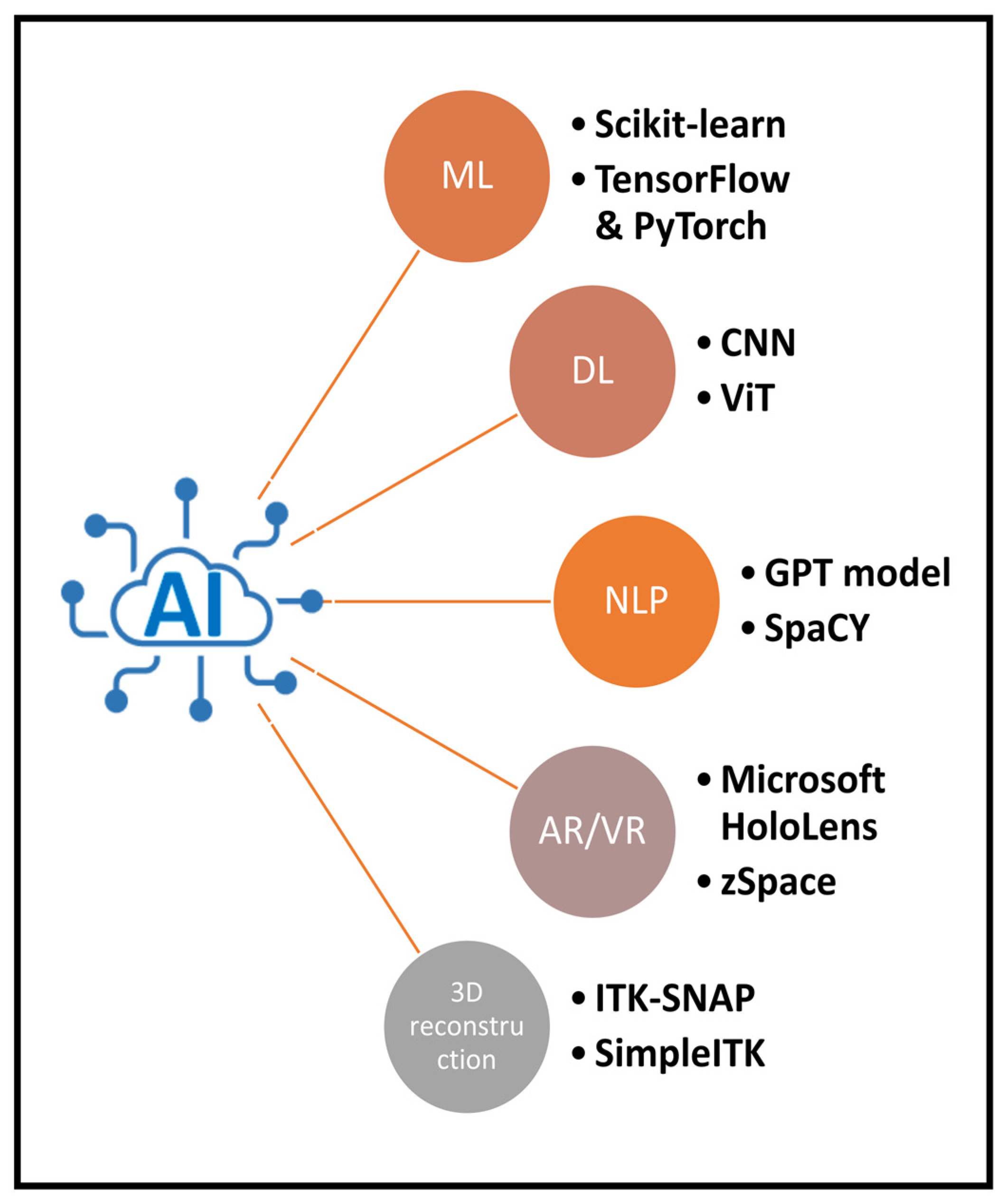
The possibilities for using AI in anatomy teaching are constantly evolving. With the implementation of AI applications in education and clinical practices, it is important to equip learners and teachers with the fundamentals of AI skills and their applications (Abdellatif et al., 2022). In the existing literature, there is a knowledge gap that highlights the current advances of AI models that have been developed for use in anatomy education. Hence, this review aims to investigate the current state of available novel AI models and their impact on anatomical education. Figure 2 demonstrates various AI tools that can be used in anatomy teaching (Figure 2).

3. Methods

The aim of the present narrative review was to summarize the current knowledge of AI integration in anatomy teaching and its subfields, which include gross anatomy, histology, embryology, and radiological anatomy. In addition, this review also highlighted the practical tools to acknowledge the current potential, challenges, and future directions. To achieve this aim, a comprehensive search was conducted in multiple electronic databases, including Scopus, Google Scholar, and PubMed. All materials published before 30 September 2024 were included in this review. The keywords used to retrieve the relevant published data include “anatomy”, “education”, “learning”, “teaching”, “radiological anatomy”, “histology”, “artificial intelligence”, “machine learning”, “neural networks”, and “deep learning”. The inclusion criteria included all full-text primary articles which are written in English and cover AI technology application in anatomy teaching for health science undergraduate students. The exclusion criteria include review articles, meta-analyses, letters to the editor, abstract-only articles, commentary, non-human author, and non-English articles.

4. Results and Discussion

Searching various databases revealed that the use of AI in anatomy education is an evolving topic with a promising future. The results are divided into the use of AI in gross anatomy, in histology, and in anatomical medical imaging, with the challenges and practical tools discussed for each. Many technologies are utilized to teach anatomy to provide the best achievable understanding. In an attempt to merge the capabilities of AI with 3D technologies, Chheang et al. (2024) integrated an AI assistant chatbot with a 3D platform to provide an interactive anatomy learning experience. It has been observed that most AI technology developed for anatomy teaching is in the form of a customized virtual assistant. For example, Arun et al. (2024) built a customized chatbot called Anatbuddy and evaluated its performance, which was found to exceed that of open AI chatbots (ChatGPT3.5) in accuracy because it is tailored to and trained in the specific area of anatomy education. In another study, Li et al. (2021) used an open-source machine learning architecture. Next, they fine-tuned it to create an AI chatbot for anatomy teaching. Collins et al. (2024) used GPT-builder to formalize an AI-powered anatomy tutor that was found to be more specific than ChatGPT as it was customized to anatomy topics. Castellano et al. (2023) combined gamified learning techniques with an AI-powered virtual assistant for learning anatomy in a single mobile application. They recommended integrating this method into a wide range of anatomy teaching such as that for other health-related educational programs, as it supports adaptive learning and personalized learning scenarios (Castellano et al., 2023). Ilgaz and Çelik (2023) examined effectiveness of large language models such as ChatGPT and Google Bard in education. Despite the ability to formulate correct questions for self-testing, these models require more improvement in essay writing.
The second most studied area in the teaching of anatomy using AI is image creation. Noel (2023) used the Microsoft Bing Image Creator powered by DALL-E, Stable Diffusion, and Craiyon V3 to create images from text, which can be used by anatomy course students. While Buzzaccarini et al. (2024) tested the use of Midjourney to create realistic and accurate anatomical images, they unfortunately found that it still requires more improvement and more collaboration between anatomists and AI engineers (Buzzaccarini et al., 2024). In radiological anatomy teaching, we did not find any AI applications specifically targeting radiological anatomy teaching. Figure 3 represents the currently available AI tools that could be used in anatomy teaching.

4.1. AI in Anatomy Teaching

Anatomy teaching is integral to medical education and optimizing it will positively impact future doctors’ performance. Therefore, anatomy educators and medical schools should adapt and embrace digital upgrades and apply new methods in addition to traditional ones (Khasawneh, 2021). Although the exact start of AI integration in medical education is not clear, we can use some clues to trace it. AI appeared in medical field research some 30 years ago (Lillehaug & Lajoie, 1998). Two decades ago, AI in medical education emerged as a topic in academic research (Lillehaug & Lajoie, 1998). According to Zarei et al. (2024), AI integration into medical education did not become widely spread until the 1980s. There has been an increase in the number of articles published about AI use in medical education in the last two decades, which reflects the expansion of its application in the real world (Zarei et al., 2024). COVID-19 acted as a catalyst for the rapid adoption and implementation of AI in medical curricula, especially in anatomy education, because clustering around cadavers in anatomy museums is no longer possible. The studies that utilized AI tools in anatomy teaching are summarized in Table 2. The initial use of computer-based anatomy teaching in anatomy education was performed via 3D interactive anatomy platforms (Abdellatif et al., 2022). Arantes et al. (2018) studied the tools used in neuroanatomy teaching and found that 3D tools are more effective in teaching anatomy. Furthermore, the use of assistant chatbots is already implemented in some medical schools as part of their educational projects (Sugand et al., 2010).
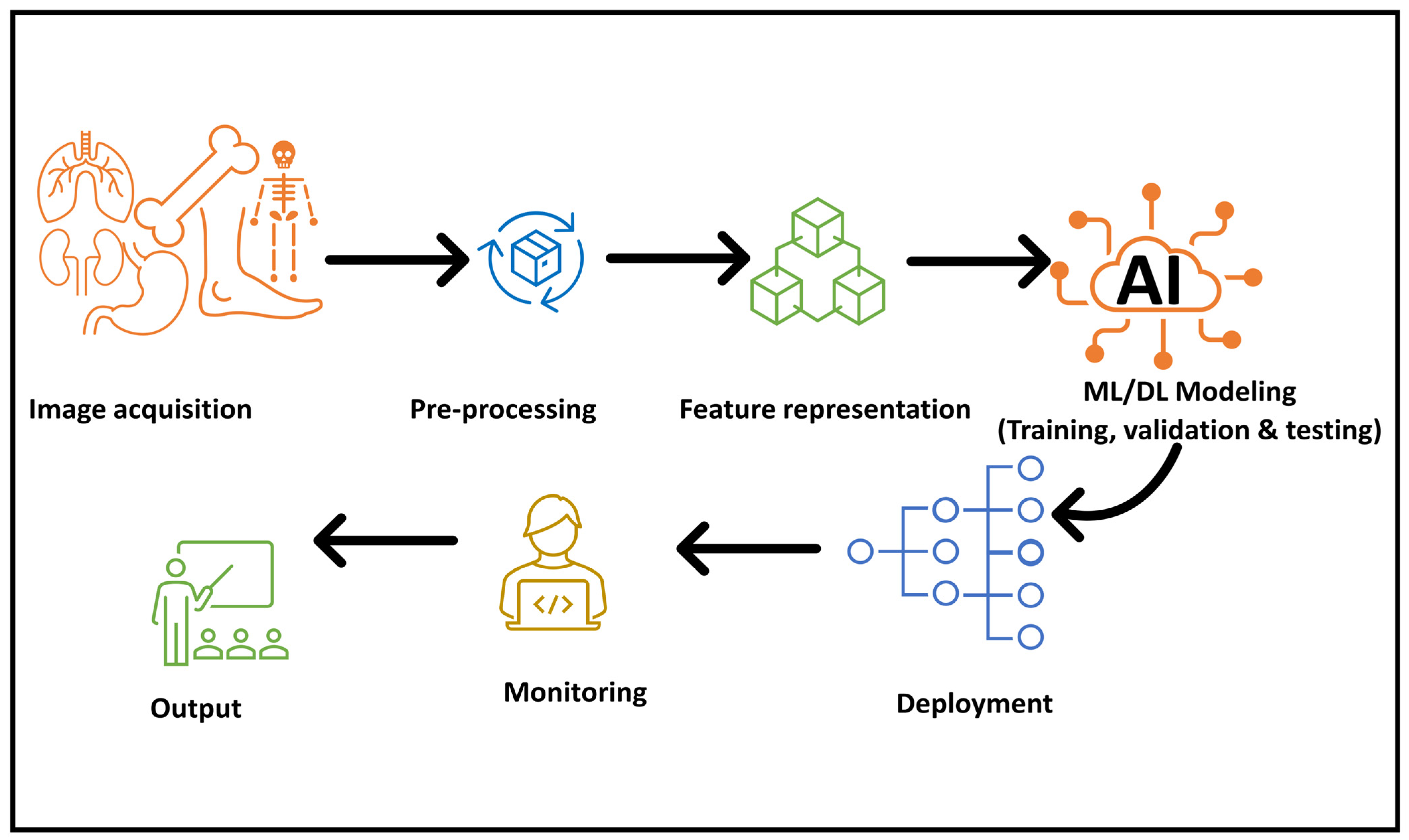
NLP is the most commonly used field of AI to create applications that analyze big data. A schematic representation of major steps in anatomical data preparation, modelling, deployment, and education regarding the effective use of AI in anatomy education is shown in Figure 4. The growing acceptance of AI applications needs to be acknowledged, and these applications need to be used in combination with other methods. The exploration of current advancements in the use of AI applications in anatomy education and the evaluation of its challenges is the main focus of this paper.

4.1.1. AI-Powered Interactive 3D Anatomical Models

With the provision of interactive 3D anatomical models, teaching becomes more effective (Moro et al., 2017). This way of teaching makes the learning process more interactive and accessible due to overcoming the limited access to cadaveric bodies (Usmani et al., 2022). The exploration and manipulation of bodies in a virtual environment give students a better understanding of spatial relationships that are deficient in 2D models. Chheang et al. (2024) created a model integrating AI assistants with 3D platforms to provide an interactive anatomy learning experience. Additionally, Castellano et al. (2023) combined a mobile gamified technological tool with a virtual assistant for use in learning anatomy and recommended the integration of this method in a wide range of anatomy teaching for other health-related educational programs, as it supports adaptive learning and personalized learning scenarios. In a systematic review for the evaluation of current advances and the efficacy of immersive virtual reality in teaching students and residents, they found it more effective when used with surgical checklists that require good anatomical understanding (Mao et al., 2021). The 3D visualization of anatomical organs can be physical, using AI-powered personalized 3D printing, or can be virtual, via AI-powered virtual reality (VR) dissection.

4.1.2. AI-Powered Virtual Reality (VR) Dissection

Another creative idea is AI-powered virtual reality (VR) dissection, which serves as a virtual immersive laboratory where users can dissect pre-programmed 3D models of anatomical structures. Chheang et al. (2024) merged a VR environment with a generative AI-powered assistant chatbot. They found that such a combination is a powerful and effective way of providing an interactive learning environment. This application comes with multiple levels of complexity in dissection and the user can choose from them, then the AI assistant highlights the key structures and provides information about their functions (Castellano et al., 2023). AI powered augmented reality (AR) and VR tools include but are not exclusive to Microsoft HoloLens and zSpace. Microsoft HoloLens creates 3D anatomy models and uses AI in a sensor-packed holographic computing headset to create mixed reality for a better visualization experience (Microsoft, n.d.; Roach, 2023). zSpace provides a virtual human anatomy atlas with a detailed visualization experience (zSpace, Inc., 2024). This virtual anatomy model is integrated with AI-curated content and an AI-powered assistant called career coach AI (DENVER & zSpace, Inc., 2024). Learning anatomy in such a way simulates real-world dissection without the ethical challenges related to cadaver accessibility (Usmani et al., 2022). It provides an active learning experience rather than a passive one. This method of learning anatomy is not only for medical students in their initial steps of learning anatomy but also for those who specialize in forensic medicine. Alharbi et al. (2020) used 3D virtual reality in their experiment regarding anatomy teaching. It was found to be an effective learning tool to improve students’ knowledge, retention, engagement, and real-life simulation. Furthermore, the authors still encourage the complementary use of traditional methods alongside the newly suggested methods (Alharbi et al., 2020; Kurul et al., 2020).

4.1.3. AI-Powered Personalized 3D Printing

AI-powered 3D printing is another way to allow a personalized experience through printing models of specific anatomical structures or organs based on student interest. This system allows the learner to customize the model’s size, level of detail, and labeling options. The 3D printed model facilitates in-depth understanding of spatial relationships and anatomical features (Ma et al., 2023). AI in 3D printing is used in multiple phases, in pre-printing image processing and analysis, in the printing phase, and in the post-printing phase. In pre-printing, it predicts the print suitability of materials to reduce some trial and error. Additionally, it works in image segmentation as the traditional methods require manual segmentation, which is error-prone and wastes time, money, and effort. AI also helps to reach the best parameters to match the ink properties (Murphy et al., 2012). In the printing phase, in 3D printing, closed-loop AI is used. It is a method to identify the printing errors in model printing and automatically correct them, achieving higher precision with less waste. In the post-printing phase, it is rarely used because the product is already made; however, it is mainly applied in quality checks and providing possible ways to avoid errors (Ma et al., 2023). Not only can anatomy learners benefit from this method, but educators can also use it to print specific models for objective structured practical exams, providing an unlimited number of new anatomical models to be tested on. Additionally, they can use environmentally friendly reusable printing material, so they can re-print new models without destroying the environment. Ma et al. (2023) describe the use of AI applications in 3D organ printing in depth.

4.1.4. AI-Powered Multilingual Anatomy Tutor

AI-powered multilingual anatomy tutor is an idea to develop a chatbot specialized in the anatomy field. This chatbot should be multilingual to provide information without restriction to certain languages. Students with different linguistic abilities can interact with the chatbot, asking questions about anatomical concepts, requesting clarifications, and receiving explanations in their native tongue (Ilgaz & Çelik, 2023). Natural language processing (NLP) capabilities can be used by this chatbot to understand student queries and provide accurate and culturally sensitive responses, promoting inclusivity and accessibility in anatomy education. This chatbot can act as a learning companion for medical students and even for educators, helping to prepare exam questions and suggesting new ways of teaching certain anatomy chapters (Y. Chen et al., 2022; Morton & Colbert-Getz, 2016).
A customized chatbot like Anatbuddy surpasses open AI chatbots (ChatGPT3.5) in accuracy because Anatbuddy is tailored and trained in a specific area, anatomy education (Arun et al., 2024). It is worth mentioning that both models score similarly in the completeness, relevance, coherence, and fluency of information generated. Anatbuddy is able to answer questions that require analysis, for example it compares the right and left lung, and the 1st and 12th ribs; however, ChatGPT3.5 finds it difficult to answer such questions correctly (Arun et al., 2024).
AI tools have their inherent weaknesses, and therefore will never replace human educators, but can instead support them. These are a small portion of the present solutions provided by AI, and the applications of AI in anatomy teaching are constantly evolving.

4.1.5. AI-Based Teaching Platforms with Immediate Feedback

Specific students’ needs are not considered by the traditional teaching methods. The current approach is one size made to fit all types of individual learning styles, which obviously leads to gaps in understanding. Subsequently, the new methods relying on AI start to address this issue by providing immediate and detailed feedback. AI helps to identify areas of improvement from quizzes and exercises that have been given. As a result, the learner can further study and refine his knowledge. Nevertheless, AI-personalized learning continues to generate quizzes and case studies depending on the cumulative knowledge about the weakness of certain learners. This strategy allows the learning process to be based on the learner’s needs and abilities rather than traditional passive methods. (Vella, n.d.) AI-based learning provides adaptive learning, which refers to the process of continuous system customization based on individual performance, fostering personalized learning (Usmani et al., 2022).
An example of similar platforms are the Smart Sparrow and Coursera AI-powered courses. Smart Sparrow provides an adaptive anatomy lesson that is customized to specific learners’ needs (Lukan, 2024; Frsa, 2024). Furthermore, Coursera provides anatomy courses, uses AI in its platform to provide immediate and informative feedback for learners, and uses AI to translate its courses for various languages to increase the accessibility of knowledge worldwide (Kayser et al., 2010). Generally, traditional learning methods are inadequate alone, and AI-powered anatomy learning provides a personalized interactive learning experience for better comprehensive understanding.

4.2. AI in Histology Teaching

AI is revolutionizing histology teaching by enhancing undergraduate medical students’ training and education through advanced technologies like whole-slide imaging. The anatomy and pathology fields have already implemented virtual slides in their continuous education (I. Kim et al., 2022). These AI applications aim to extract image patches for training and create morphometric analysis methods for quantitative histomorphometry. AI integration in histology teaching contributes to standardized learning because it ensures consistency and quality in the learning experience across all students, regardless of individual instructors (C. P. Chen et al., 2019). The integration of AI into medical education holds significant promise, particularly in areas like histology and the microscopic study of tissues. While there are not currently any widely adopted AI platforms specifically designed for histology teaching in undergraduate programs, several projects and initiatives are actively exploring the potential of AI in this field and can be used for histology teaching. Below are a few examples.

4.2.1. PathXL

PathXL is a platform licensed initially by the department of pathology in the University of Pittsburgh School of Medicine (UPSOM) Division of Informatics (Select Science, n.d.; Par Equity, n.d.; D. Kim et al., 2020). PathXL was created in 2005 and was initially called i-Path. A multidisciplinary team of experts in tissue-based research and practice collaborated to create this innovative platform. Using their knowledge and experience, they make groundbreaking solutions that change the field of pathology and tissue research (Select Science, n.d.). PathXL received a valuable investment from Par Equity in 2012. Later, in 2016, this project was acquired by Philips Healthcare, followed by an acquisition from Cirdan in 2020 (D. Kim et al., 2020).
Philips Healthcare improved the diagnostic capabilities for PathXL via its integration into their broad healthcare technology system, while Cirdan tends to have a more educational focus and enhance its training features. (Philips, 2016) PathXL is an innovative virtual learning environment designed for pathology and histology education. It provides the interactive discussion of cases with the ability to navigate through tissue samples. Additionally, it contains clinical history and examination findings to allow for comprehensive understanding in order to reach correct diagnoses. It adapts to the performance of specific learners, providing a personalized level of complexity.

4.2.2. Virtual Slide Platforms

Virtual slide platforms are developed by various companies to host and view digital histology slides, for example Aperio ImageScope and Philips PathologyViewer (Philips, 2016). Many institutions around the world have started to adopt such technology to present a better experience. It is noteworthy to mention that the Philips pathology viewer is an image viewer that is used as a management system within Philips’ IntelliSite Pathology Solution, while PathXL provides workflow software in addition to image analysis (Betmouni, 2021).
These AI-powered platforms are not strictly used for education but are considered valuable tools for histology education. They can be used for learning and material creation using digitalized histology slides. Additionally, they encourage self-directed learning by allowing remote access to histology slides. They can also be used to support traditional classrooms, facilitating discussion and further understanding of histology.

4.2.3. Whole-Slide Imaging Also Called Virtual Microscopy

Whole-slide imaging is also called virtual microscopy, but there is a slight difference between the two terms. Whole-slide imaging (WSI) involves scanning the whole histologic slide, facilitating remote collaboration and teaching. The application of AI algorithms to slides is called virtual microscopy/virtual slides. These tools enhance the learning experience and allow interactive navigation through histology slides. Such platforms consist of virtual slides that support continuous learning by providing access to high-resolution slides and exploring histology in depth for better understanding (Abdellatif et al., 2022). WSI techniques provide a huge dataset that can be used to train AI models for teaching and diagnosis. Generative AI models, after being trained on such cases, are able to generate new cases. These cases can be tailored to specific learning objectives and enhance understanding of complex medical concepts. Additionally, they enhance learning, as they can improve student learning outcomes, promoting a better understanding of complex concepts and fostering deeper engagement with the subject matter (C. P. Chen et al., 2019). Certain AI models increase education efficiency due to their capability to automate training, slide patching, and feedback provision, freeing up educators’ time for more interactive and personalized instruction (Philips, 2016).

4.2.4. AI-Powered 3D Reconstruction

Various research groups have tried to explore the use of AI abilities in reconstructing 3D models of tissues from digital slides (Zhang et al., 2024; Chan-Zuckerberg Initiative, n.d.). 3D image reconstruction from 2D images that display anatomical images or histologic slides is a promising field. It helps with in-depth understanding and the detailed visualization of internal structures. Tools that can assist in the construction of 3D images include but are not exclusive to ITK-SNAP and SimpleITK (Yushkevich et al., 2016; United States National Library of Medicine (NLM) et al., n.d.; Sieben et al., 2017). ITK-SNAP is used to segment MRI and CT images prior to their reconstruction into 3D images, while SimpleITK is a simplified version of ITK for the purpose of 3D image reconstruction (Sieben et al., 2017). Despite being an active research field, there is still no platform empowered to carry out 3D reconstruction from digital slides. One potential application of this technology in histology teaching could be via the provision of the interactive immersive visualization of spatial relationships and structures within tissue. This technology is promising in encouraging a better understanding of tissue architecture and pathology (Lobachev et al., 2021; Ossa et al., 2022). The incorporation of such 3D reconstruction of histology slides into an interactive learning platform enabling virtual slide exploration and the identification of structures with real-time feedback would provide a dynamic and personalized learning environment, complementing traditional teaching methods (Rakha et al., 2020).

4.2.5. Chatbots and Interactive Platforms

AI chatbots can be used in histology teaching. They are used to engage histology learners in conversations with reliable resources. Chatbots provide valuable interactive learning experiences and real-time feedback. Additionally, they can generate quizzes, answer learner questions, and offer personalized study recommendations (Ilgaz & Çelik, 2023; Y. Chen et al., 2022). AI chatbots can personalize learning for each student by recognizing individual strengths and weaknesses and adapting learning materials accordingly (Ganske et al., 2006). This helps students learn more effectively and efficiently. The use of AI-generated, interactive cases aids in case-based learning skills, as it allows students to practice their diagnostic skills in simulated clinical settings (Rakha et al., 2020).

4.3. AI in Anatomical Medical Imaging

Radiological anatomy teaching in medical education is advancing globally. Learning radiological anatomy is important in the early years of medical education (Long et al., 2022). Currently, radiological anatomy is taught through identifying structures in radiographs in an anatomy museum with the help of anatomists (Long et al., 2022). Many researchers have tried to integrate modern technologies into radiological anatomy education. Long et al. (2022) used augmented reality in radiological anatomy education, while Bork et al. (2019) evaluated two VR models, namely a magnetic mirror and an anatomage virtual dissection table, measuring their advantages over traditional radiological techniques. In another study, Tam (2010) provided a comprehensive guide for building VR models using radiological images, facilitating the pathway for future researchers. To blend the gross anatomy and radiographs, Rabbo et al. (2015) used two cadavers at the same time, one for dissection and the other to acknowledge the ultrasound view of the knee anatomy, while Ramos-Bossini et al. (2024) used radiologic images to construct 3D models which are used for anatomy teaching.
In the field of AI integration in radiology, Almansi et al. (2024) developed an AI-powered model to detect abnormalities in inner-ear radiographs. Additionally, Vats et al. (2021) used an AI model called iDoc-X to interpret chest X-rays of tuberculosis. Barger et al. (2023) found that online radiological anatomy courses are effective in improving the performance of anatomy learners and educators; they also address the need for more courses in this field.
The need for the incorporation of more radiology images into anatomy courses is increasing globally. The number of medical schools integrating radiology into anatomy has increased; however, the proportion of radiologists who become instructors has decreased dramatically (Phillips et al., 2013). Anatomy educators ask for more radiology instructors to be recruited; however, the main challenge to this is the financial and promotional incentives. Thus, to overcome these challenge, there is a need to develop AI-based models that integrate AI power to facilitate interactive, reliable learning experiences that involve radiological images as part of anatomy courses. Additionally, future research needs to focus on developing gamification applications that involve analyzing virtual images, for example X-rays, CT scans, and MRI images, and answering the questions provided by the game regarding possible etiologies. As the world is moving toward providing personalized learning experiences, self-instructed learning, and making knowledge accessible to everyone everywhere, AI is a promising tool in consideration of all of these aspects, and does not have the high cost of the recruitment of human radiologists as instructors. To the best of our knowledge, there is a scarcity of AI-powered applications targeting radiological anatomy education.

5. Challenges and Considerations of AI Applications

AI is promising in many fields, especially medical education, However, it is crucial to acknowledge the challenges and possible limitations associated with AI integration in education. The cost is a challenge and can be divided into two areas: the cost of buying or developing AI tools itself, and the cost of its maintenance by AI professionals, adding to the cost of the prior teaching for tutors and students. These cost-related challenges are restricting developing countries from integrating web-based AI tools. This is one of the limitations that restricts the wide adoption of AI in anatomy teaching. The limited access to such new technologies in low-income countries is still a major challenge in democratizing AI, and the development of low-cost or free AI tools will help to overcome it. Many anatomists are not well-equipped with enough knowledge about possible uses of AI in education. There is a need for faculty development programs to train educators in possible applications of AI in curriculum formulation, knowledge provision, and assessments (Lazarus et al., 2022). AI is a rapidly evolving technology, i.e., what was known last month seems to be outdated now. Therefore, keeping up with the technology is the biggest challenge. Data privacy and security are a major concern regarding student information used by AI systems. There is a need to ensure responsible use of these data to improve AI systems themselves (Chan & Zary, 2019; Lo, 2023). There is a possibility of over-reliance on technology and it is required to strike a balance between critical thinking and human interaction. Additionally, it is important to preserve a healthy balance between AI-powered learning and the advancement of critical thinking and interpersonal communication abilities. The training of AI models could contribute to existing biases. AI capabilities depend on multiple factors and one of them is the data that the AI is trained on: an AI model that is trained on cadavers of a certain ethnic group might not have a high accuracy when exposed to other human variations. Additionally, Lazarus et al. (2022) noted that AI could possibly exacerbate the biases in content delivery methods and the content itself. It is noteworthy to mention the need to overcome the challenge of developing unbiased AI models that represent the diversity of human anatomy and actively mitigate any potential biases.

6. Future Directions

In anatomy education, there is a need for interactive mobile applications that leverage AI in anatomy education. This will improve the ease of learning anywhere and anytime because these applications are accessible from phones and laptops. These applications provide more flexibility and accessibility beyond physical classrooms and anatomy museums (Khasawneh, 2021). An anatomy teaching application could be designed as a detective game powered by AI. There are creative ideas for AI models and applications in anatomy education. AI-powered anatomy detective games provide a gamified learning experience. They use virtual reality to show students virtual patients, allowing them to use anatomical knowledge to analyze real patients’ anatomy. Additionally, students can analyze virtual images and answer the questions provided by the games regarding possible etiologies. Furthermore, this type of game will provide real-time feedback, guiding students towards the correct answers and potential knowledge gaps. This type of game uses AI analysis algorithms and can provide a prediction of the user knowledge level and knowledge gaps to provide a better user experience and incorporate difficulty levels to enhance engagement and competition. This gaming experience can help anatomy students while preparing them for their exams and can be used in public education about human anatomy and its complexity in exhibitions and conferences.
In the current era, there is a need for an AI-powered collaborative platform, which is an idea that will leverage AI capabilities to facilitate collaborative learning experiences among anatomy students. This platform would allow students from around the world to work together on discussing and interpreting virtual anatomy cases with supervision and assistance from AI assistant tools. This platform could provide AI-powered feedback mechanisms to enhance critical thinking skills and encourage collaboration among learners despite their different ethnic and cultural backgrounds. This platform will overcome the assumption that the adoption of the new technologies contributes to self-isolation and loneliness among the new generations. These are just a few creative ideas, and the possibilities for using AI in anatomy teaching are constantly evolving. It is important to consider the specific needs and learning styles of students when developing AI models, ensuring they are both effective and engaging for future generations of medical professionals.
The present review has the following limitations. This study provides a comprehensive summary of AI tools developed for use in anatomy teaching; however, it still has its limitations. Types of articles other than peer-reviewed original articles may also be reviewed, for example conference papers and commentary, which may be used by professionals to share state-of-art ideas with the scientific community. There is a scarcity of systematic reviews with meta-analyses in this field, so it is required to provide in-depth insight into advances in the use of AI tools in anatomy teaching. Furthermore, an attempt towards AI in anatomy curriculum development has not been performed. It is worth mentioning that there is a need for further trials to understand the effectiveness of AI-based tools in anatomy teaching, as it has not been fully elucidated.

7. Conclusions

In education, AI is integrated in multiple fields, and medicine is not an exception. In this regard, researchers have developed customized AI chatbots that are trained specifically on anatomy subjects. Others have focused on AI image generation and its use in anatomy teaching. AI-generated gamification and other interactive tools were also attempted. Researchers posit that the thoughtful integration of AI across various aspects of anatomy and histology education can create a dynamic and engaging learning environment. The benefits of AI use in anatomy teaching may boost students’ objective outcomes and learning experiences. Unfortunately, there is a noticeable deficiency of AI models that have been developed specifically for teaching radiological anatomy. Additionally, the effectiveness of AI applications in anatomy education is still undiscovered. Future research needs to focus on developing AI tools that are customized and specifically designed for anatomy teaching. The future attempts to develop AI tools for anatomy teaching need to incorporate clinical reasoning with basic anatomy for the better utilization of such promising technology. As AI is growing drastically, one should be ready to embrace it, face the challenges associated with it, and provide customized and ethical solutions for its optimal use and effectiveness in learning.

Author Contributions

Conceptualization, A.A.-R. and S.R.S.; methodology, A.A.-R., S.S.R. and S.R.S.; software, A.A.-R., S.R.M. and N.S.; validation, A.A.-R., S.R.S. and S.R.M.; formal analysis, A.A.-R. and Y.B.; resources, A.A.-R., N.S. and Y.B.; data curation, A.A.-R., S.R.S., S.S.R. and N.S.; writing—original draft preparation, A.A.-R., S.R.S., S.S.R., Y.B. and N.S.; writing—review and editing, A.A.-R., S.R.M. and S.R.S.; supervision, A.A.-R. and S.R.S.; All authors have read and agreed to the published version of the manuscript.

Funding

This research received no external funding.

Conflicts of Interest

The authors declare no conflict of interest.

Abbreviations

AIArtificial intelligence
MLMachine learning
DLDeep learning
UPSOMThe University of Pittsburgh School of Medicine
NLPNatural language processing
VRVirtual reality
WSIWhole-slide imaging
CTComputed tomography
MRIMagnetic resonance imaging
USUltrasonography
TBTuberculosis
LLMLarge language model

References

  1. Abadi, M., Barham, P., Chen, J., Chen, Z., Davis, A., Dean, J., Devin, M., Ghemawat, S., Irving, G., Isard, M., Kudlur, M., Levenberg, J., Monga, R., Moore, S., Murray, D. G., Steiner, B., Tucker, P., Vasudevan, V., Warden, P., . . . Zheng, X. (2016). TensorFlow: A system for large-scale machine learning. arXiv, arXiv:1605.08695. [Google Scholar]
  2. Abdellatif, H., Mushaiqri, M. A., Albalushi, H., Al-Zaabi, A. A., Roychoudhury, S., & Das, S. (2022). Teaching, learning and assessing anatomy with artificial intelligence: The road to a better future. International Journal of Environmental Research and Public Health, 19(21), 14209. [Google Scholar] [CrossRef]
  3. Al-Gailani, S. (2016). The “Ice Age” of anatomy and obstetrics: Hand and eye in the promotion of frozen sections around 1900. Bulletin of the History of Medicine, 90(4), 611–642. [Google Scholar] [CrossRef]
  4. Alharbi, Y., Al-Mansour, M., Al-Saffar, R., Garman, A., & Al-Radadi, A. (2020). Three-dimensional Virtual reality as an innovative teaching and learning tool for human anatomy courses in Medical Education: A Mixed Methods study. Cureus, 12(2), e7085. [Google Scholar] [CrossRef] [PubMed]
  5. Almansi, A. A., Sugarova, S., Alsanosi, A., Almuhawas, F., Hofmeyr, L., Wagner, F., Kedves, E., Sriperumbudur, K., Dhanasingh, A., & Kedves, A. (2024). A novel radiological software prototype for automatically detecting the inner ear and classifying normal from malformed anatomy. Computers in Biology and Medicine, 171, 108168. [Google Scholar] [CrossRef]
  6. Al-Rahbi, A., Al-Mahrouqi, O., & Al-Saadi, T. (2024). Uses of artificial intelligence in glioma: A systematic review. Medicine International, 4(4), 40. [Google Scholar] [CrossRef] [PubMed]
  7. Arantes, M., Arantes, J., & Ferreira, M. A. (2018). Tools and resources for neuroanatomy education: A systematic review. BMC Medical Education, 18(1), 94. [Google Scholar] [CrossRef] [PubMed]
  8. Arun, G., Perumal, V., Urias, F. P. J. B., Ler, Y. E., Tan, B. W. T., Vallabhajosyula, R., Tan, E., Ng, O., Ng, K. B., & Mogali, S. R. (2024). ChatGPT versus a customized AI chatbot (Anatbuddy) for anatomy education: A comparative pilot study. Anatomical Sciences Education, 17(7), 1396–1405. [Google Scholar] [CrossRef] [PubMed]
  9. Asad, M. R., Mutairi, A. A., AlZahrani, R. E., Ahmed, M. M., Nazeer, M., & Taha, M. (2023). Role of living anatomy in medical education: A narrative review. Journal of Pharmacy and Bioallied Sciences, 15(Suppl. S2), S843–S845. [Google Scholar] [CrossRef]
  10. Ayache, N. (2021). Digital anatomy. In Human-computer interaction series. Springer. [Google Scholar] [CrossRef]
  11. Barger, J. B., Resuehr, D., & Edwards, D. N. (2023). Radiology for anatomy educators: Success of an online, 2-day course for radiology training. Anatomical Sciences Education, 16(5), 958–968. [Google Scholar] [CrossRef]
  12. Becker, A. S., Marcon, M., Ghafoor, S., Wurnig, M. C., Frauenfelder, T., & Boss, A. (2017). Deep learning in mammography. Investigative Radiology, 52(7), 434–440. [Google Scholar] [CrossRef]
  13. Betmouni, S. (2021). Diagnostic digital pathology implementation: Learning from the digital health experience. Digital Health, 7, 20552076211020240. [Google Scholar] [CrossRef] [PubMed]
  14. Bork, F., Stratmann, L., Enssle, S., Eck, U., Navab, N., Waschke, J., & Kugelmann, D. (2019). The benefits of an augmented Reality Magic Mirror system for integrated radiology teaching in gross anatomy. Anatomical Sciences Education, 12(6), 585–598. [Google Scholar] [CrossRef]
  15. Buzzaccarini, G., Degliuomini, R. S., Borin, M., Fidanza, A., Salmeri, N., Schiraldi, L., Di Summa, P. G., Vercesi, F., Vanni, V. S., Candiani, M., & Pagliardini, L. (2024). The promise and pitfalls of AI-Generated anatomical images: Evaluating midjourney for aesthetic surgery applications. Aesthetic Plastic Surgery, 48(9), 1874–1883. [Google Scholar] [CrossRef]
  16. Castellano, M. S., Contreras-McKay, I., Neyem, A., Farfán, E., Inzunza, O., Ottone, N. E., Del Sol, M., Alario-Hoyos, C., Alvarado, M. S., & Tubbs, R. S. (2023). Empowering human anatomy education through gamification and artificial intelligence: An innovative approach to knowledge appropriation. Clinical Anatomy, 37(1), 12–24. [Google Scholar] [CrossRef] [PubMed]
  17. Chan, K. S., & Zary, N. (2019). Applications and challenges of implementing artificial intelligence in medical education: Integrative review. JMIR Medical Education, 5(1), e13930. [Google Scholar] [CrossRef]
  18. Chan-Zuckerberg Initiative. (n.d.). ITK-SNAP home. ITK-SNAP. Available online: http://www.itksnap.org/pmwiki/pmwiki.php (accessed on 11 October 2024).
  19. Chen, C. P., Clifford, B. M., O’Leary, M. J., Hartman, D. J., & Picarsic, J. L. (2019). Improving medical students’ understanding of pediatric diseases through an innovative and tailored web-based digital pathology program with philips Pathology tutor (Formerly PathXL). Journal of Pathology Informatics, 10(1), 18. [Google Scholar] [CrossRef]
  20. Chen, D., Zhang, Q., Deng, J., Cai, Y., Huang, J., Li, F., & Xiong, K. (2018). A shortage of cadavers: The predicament of regional anatomy education in mainland China. Anatomical Sciences Education, 11(4), 397–402. [Google Scholar] [CrossRef]
  21. Chen, Y., Jensen, S., Albert, L. J., Gupta, S., & Lee, T. (2022). Artificial intelligence (AI) student assistants in the classroom: Designing chatbots to support student success. Information Systems Frontiers, 25(1), 161–182. [Google Scholar] [CrossRef]
  22. Chheang, V., Sharmin, S., Márquez-Hernández, R., Patel, M., Rajasekaran, D., Caulfield, G., Kiafar, B., Li, J., Kullu, P., & Barmaki, R. L. (2024, January 17–19). Towards anatomy education with generative AI-based virtual assistants in immersive virtual reality environments. 2024 IEEE International Conference on Artificial Intelligence and eXtended and Virtual Reality (AIxVR) (pp. 21–30), Los Angeles, CA, USA. [Google Scholar] [CrossRef]
  23. Collins, B. R., Black, E. W., & Rarey, K. E. (2024). Introducing AnatomyGPT: A customized artificial intelligence application for anatomical sciences education. Clinical Anatomy, 37(6), 661–669. [Google Scholar] [CrossRef]
  24. DENVER & zSpace, Inc. 2024 June 24. zSpace introduces zSpace AI: Enhancing education with artificial intelligence. zSpace. Available online: https://zspace.com/newsroom/zspace-introduces-zspace-ai-enhancing-education-with-artificial-intelligence (accessed on 14 October 2024).
  25. Dissabandara, L. O., Nirthanan, S. N., Khoo, T. K., & Tedman, R. (2015). Role of cadaveric dissections in modern medical curricula: A study on student perceptions. Anatomy & Cell Biology, 48(3), 205. [Google Scholar] [CrossRef]
  26. Dosovitskiy, A., Beyer, L., Kolesnikov, A., Weissenborn, D., Zhai, X., Unterthiner, T., Dehghani, M., Minderer, M., Heigold, G., Gelly, S., Uszkoreit, J., & Houlsby, N. (2020). An image is worth 16 × 16 words: Transformers for image recognition at scale. arXiv, arXiv:2010.11929. [Google Scholar]
  27. Explosion. (n.d.). spaCy industrial-strength natural language processing in Python. SpaCY. Available online: https://spacy.io/ (accessed on 14 October 2024).
  28. Fan, X., Li, M., Rolker, H., Li, Y., Du, J., Wang, D., & Li, E. (2022). Knowledge, attitudes and willingness to organ donation among the general public: A cross-sectional survey in China. BMC Public Health, 22(1), 918. [Google Scholar] [CrossRef] [PubMed]
  29. Frsa, C. D. 2024 January 30. Growth of coursera through AI. LinkedIn. Available online: https://www.linkedin.com/pulse/growth-coursera-through-ai-carl-dawson-frsa-yygme#:~:text=Coursera%20has%20used%20AI%20for,personalized%20learning%20companion%20named%20Coach (accessed on 14 October 2024).
  30. Gallifant, J., Fiske, A., Strekalova, Y. A. L., Osorio-Valencia, J. S., Parke, R., Mwavu, R., Martinez, N., Gichoya, J. W., Ghassemi, M., Demner-Fushman, D., McCoy, L. G., Celi, L. A., & Pierce, R. (2024). Peer review of GPT-4 technical report and systems card. PLoS Digital Health, 3(1), e0000417. [Google Scholar] [CrossRef]
  31. Ganske, I., Su, T., Loukas, M., & Shaffer, K. (2006). Teaching methods in anatomy courses in North American medical schools. Academic Radiology, 13(8), 1038–1046. [Google Scholar] [CrossRef]
  32. Habbal, O. (2017). The science of anatomy: A historical timeline. Sultan Qaboos University Medical Journal, 17(1), e18–e22. [Google Scholar] [CrossRef]
  33. Ilgaz, H. B., & Çelik, Z. (2023). The significance of artificial intelligence platforms in anatomy education: An experience with ChatGPT and Google Bard. Cureus, 15, e45301. [Google Scholar] [CrossRef]
  34. Iwanaga, J., Loukas, M., Dumont, A. S., & Tubbs, R. S. (2020). A review of anatomy education during and after the COVID-19 pandemic: Revisiting traditional and modern methods to achieve future innovation. Clinical Anatomy, 34(1), 108–114. [Google Scholar] [CrossRef] [PubMed]
  35. Jacques, S. (2024). Body donation for medical education and training: The importance of cadavers in teaching anatomy. Bulletin of the Royal College of Surgeons of England, 106(5), 286–289. [Google Scholar] [CrossRef]
  36. Jin, C., Udupa, J. K., Zhao, L., Tong, Y., Odhner, D., Pednekar, G., Nag, S., Lewis, S., Poole, N., Mannikeri, S., Govindasamy, S., Singh, A., Camaratta, J., Owens, S., & Torigian, D. A. (2022). Object recognition in medical images via anatomy-guided deep learning. Medical Image Analysis, 81, 102527. [Google Scholar] [CrossRef]
  37. Kayser, K., GĂśrtler, J., Bogovac, M., Bogovac, A., Goldmann, T., Vollmer, E., & Kayser, G. (2010). AI (artificial intelligence) in histopathology—From image analysis to automated diagnosis. Folia Histochemica Et Cytobiologica, 47(3), 355–361. [Google Scholar] [CrossRef] [PubMed]
  38. Khan, D. Z., Valetopoulou, A., Das, A., Hanrahan, J. G., Williams, S. C., Bano, S., Borg, A., Dorward, N. L., Barbarisi, S., Culshaw, L., Kerr, K., Luengo, I., Stoyanov, D., & Marcus, H. J. (2024). Artificial intelligence assisted operative anatomy recognition in endoscopic pituitary surgery. NPJ Digital Medicine, 7(1), 314. [Google Scholar] [CrossRef]
  39. Khasawneh, R. R. (2021). Anatomy education of medical students during the COVID 19 pandemic. International Journal of Morphology, 39(5), 1264–1269. [Google Scholar] [CrossRef]
  40. Kim, D., Pantanowitz, L., Schüttler, P., Yarlagadda, D. V. K., Ardon, O., Reuter, V. E., Hameed, M., Klimstra, D. S., & Hanna, M. G. (2020). (Re) defining the High-Power Field for digital pathology. Journal of Pathology Informatics, 11(1), 33. [Google Scholar] [CrossRef] [PubMed]
  41. Kim, I., Kang, K., Song, Y., & Kim, T. (2022). Application of artificial intelligence in Pathology: Trends and challenges. Diagnostics, 12(11), 2794. [Google Scholar] [CrossRef] [PubMed]
  42. Kramer, B. (2023). Challenges to sourcing human bodies for teaching and research in Africa: Are the challenges insurmountable? Annals of Anatomy—Anatomischer Anzeiger, 252, 152196. [Google Scholar] [CrossRef]
  43. Kurt, E., Yurdakul, S., & Ataç, A. (2013). An overview of the technologies used for anatomy education in terms of medical history. Procedia—Social and Behavioral Sciences, 103, 109–115. [Google Scholar] [CrossRef]
  44. Kurul, R., Ögün, M. N., Narin, A. N., Avci, Ş., & Yazgan, B. (2020). An alternative method for anatomy training: Immersive virtual reality. Anatomical Sciences Education, 13(5), 648–656. [Google Scholar] [CrossRef]
  45. Kwon, J., Lee, S. Y., Jeon, K., Lee, Y., Kim, K., Park, J., Oh, B., & Lee, M. (2020). Deep learning–Based algorithm for detecting aortic stenosis using electrocardiography. Journal of the American Heart Association, 9(7), e014717. [Google Scholar] [CrossRef]
  46. Lazarus, M. D., Truong, M., Douglas, P., & Selwyn, N. (2022). Artificial intelligence and clinical anatomical education: Promises and perils. Anatomical Sciences Education, 17(2), 249–262. [Google Scholar] [CrossRef] [PubMed]
  47. Li, Y. S., Lam, C. S. N., & See, C. (2021). Using a machine learning architecture to create an AI-Powered chatbot for anatomy education. Medical Science Educator, 31(6), 1729–1730. [Google Scholar] [CrossRef]
  48. Lillehaug, S., & Lajoie, S. P. (1998). AI in medical education—Another grand challenge for medical informatics. Artificial Intelligence in Medicine, 12(3), 197–225. [Google Scholar] [CrossRef] [PubMed]
  49. Lo, C. K. (2023). What is the impact of CHATGPT on education? A rapid review of the literature. Education Sciences, 13(4), 410. [Google Scholar] [CrossRef]
  50. Lobachev, O., Berthold, M., Pfeffer, H., Guthe, M., & Steiniger, B. S. (2021). Inspection of histological 3D reconstructions in virtual reality. Frontiers in Virtual Reality, 2, 628449. [Google Scholar] [CrossRef]
  51. Long, M., Dhanaliwala, A., Morley, C., Choudhry, O., Nadolski, G., Hunt, S., & Gade, T. (2022). Abstract No. 530 Focused anatomy lessons using augmented reality for teaching interventional radiology procedures. Journal of Vascular and Interventional Radiology, 33(6), S196. [Google Scholar] [CrossRef]
  52. Lukan, E. (2024, September 16). 8 uses of AI in education. Synthesia. Available online: https://www.synthesia.io/learn/ai-applications/education#:~:text=AI%20adaptive%20learning%20platforms%20like,interactive%2C%20they%20make%20it%20stick (accessed on 13 October 2024).
  53. Ma, L., Yu, S., Xu, X., Amadi, S. M., Zhang, J., & Wang, Z. (2023). Application of artificial intelligence in 3D printing physical organ models. Materials Today Bio, 23, 100792. [Google Scholar] [CrossRef] [PubMed]
  54. Mao, R. Q., Lan, L., Kay, J., Lohre, R., Ayeni, O. R., Goel, D. P., & De Sa, D. (2021). Immersive virtual reality for surgical training: A systematic review. Journal of Surgical Research, 268, 40–58. [Google Scholar] [CrossRef] [PubMed]
  55. Microsoft. (n.d.). Microsoft hololens. Microsoft learn. Available online: https://learn.microsoft.com/en-us/hololens/ (accessed on 14 October 2024).
  56. Moro, C., Štromberga, Z., Raikos, A., & Stirling, A. (2017). The effectiveness of virtual and augmented reality in health sciences and medical anatomy. Anatomical Sciences Education, 10(6), 549–559. [Google Scholar] [CrossRef]
  57. Morton, D. A., & Colbert-Getz, J. M. (2016). Measuring the impact of the flipped anatomy classroom: The importance of categorizing an assessment by Bloom’s taxonomy. Anatomical Sciences Education, 10(2), 170–175. [Google Scholar] [CrossRef] [PubMed]
  58. Moxham, B. J., & Plaisant, O. (2014). The history of the teaching of gross anatomy—How we got to where we are! European Journal of Anatomy, 18(3), 219–244. Available online: https://eurjanat.com/v1/journal/paper.php?id=140164bm (accessed on 28 January 2025).
  59. Murphy, S. V., Skardal, A., & Atala, A. (2012). Evaluation of hydrogels for bio-printing applications. Journal of Biomedical Materials Research Part A, 101A(1), 272–284. [Google Scholar] [CrossRef] [PubMed]
  60. Najjar, R. (2023). Redefining Radiology: A review of Artificial intelligence integration in medical imaging. Diagnostics, 13(17), 2760. [Google Scholar] [CrossRef] [PubMed]
  61. Noel, G. P. J. C. (2023). Evaluating AI-powered text-to-image generators for anatomical illustration: A comparative study. Anatomical Sciences Education, 17(5), 979–983. [Google Scholar] [CrossRef] [PubMed]
  62. Ossa, L. A., Rost, M., Lorenzini, G., Shaw, D. M., & Elger, B. S. (2022). A smarter perspective: Learning with and from AI-cases. Artificial Intelligence in Medicine, 135, 102458. [Google Scholar] [CrossRef] [PubMed]
  63. Papa, V., Varotto, E., Vaccarezza, M., Ballestriero, R., Tafuri, D., & Galassi, F. M. (2019). The teaching of anatomy throughout the centuries: From herophilus to plastination and beyond. Medicina Historica, 3(2), 69–77. [Google Scholar]
  64. Par Equity. (n.d.). PathXL|Par equity. Available online: https://www.parequity.com/portfolio/pathxl (accessed on 25 June 2024).
  65. Paszke, A., Gross, S., Massa, F., Lerer, A., Bradbury, J., Chanan, G., Killeen, T., Lin, Z., Gimelshein, N., Antiga, L., Desmaison, A., Köpf, A., Yang, E., DeVito, Z., Raison, M., Tejani, A., Chilamkurthy, S., Steiner, B., Fang, L., . . . Chintala, S. (2019). PyTorch: An imperative style, high-performance deep learning library. arXiv, arXiv:1912.01703. [Google Scholar]
  66. Patra, A., Asghar, A., Chaudhary, P., & Ravi, K. S. (2022). Integration of innovative educational technologies in anatomy teaching: New normal in anatomy education. Surgical and Radiologic Anatomy, 44(1), 25–32. [Google Scholar] [CrossRef] [PubMed]
  67. Pedregosa, F., Varoquaux, G., Gramfort, A., Michel, V., Thirion, B., Grisel, O., Blondel, M., Prettenhofer, P., Weiss, R., Dubourg, V., Vanderplas, J., Passos, A., Cournapeau, D., Brucher, M., Perrot, M., & Duchesnay, E. (2012). SciKit-Learn: Machine learning in Python. arXiv, arXiv:1201.0490. [Google Scholar]
  68. Philips. (2016, June 21). Philips expands digital pathology solutions. Available online: https://www.philips.co.uk/healthcare/sites/pathology/release-press/20160621-philips-expands-its-digital-pathology-solutions-portfolio-with-the-acquisition-of-pathxl (accessed on 18 August 2024).
  69. Phillips, A. W., Smith, S. G., & Straus, C. M. (2013). The role of radiology in preclinical anatomy. Academic Radiology, 20(3), 297–304.e1. [Google Scholar] [CrossRef] [PubMed]
  70. Rabbo, F. A., Garrigues, F., Lefèvre, C., & Seizeur, R. (2015). Interactive anatomical teaching: Integrating radiological anatomy within topographic anatomy. Morphologie, 100(328), 17–23. [Google Scholar] [CrossRef]
  71. Rakha, E. A., Toss, M., Shiino, S., Gamble, P., Jaroensri, R., Mermel, C. H., & Chen, P. C. (2020). Current and future applications of artificial intelligence in pathology: A clinical perspective. Journal of Clinical Pathology, 74(7), 409–414. [Google Scholar] [CrossRef] [PubMed]
  72. Ramos-Bossini, A. J. L., Cornejo, D. L., Guerrero, P. R., & Santiago, F. R. (2024). The educational impact of radiology in anatomy teaching: A field study using cross-sectional imaging and 3D printing for the study of the spine. Academic Radiology, 31(1), 329–337. [Google Scholar] [CrossRef] [PubMed]
  73. Roach, J. R. 2023 July 24. The making of the HoloLens 2: How advanced AI built Microsoft’s vision for ubiquitous computing—Source. Microsoft. Available online: https://news.microsoft.com/source/features/innovation/hololens-2-shipping-to-customers/#:~:text=The%20sensor%2Dpacked%20holographic%20computing,basic%20human%20impulses%3A%20exchanging%20knowledge (accessed on 14 October 2024).
  74. Sabzalieva, E., Valentini, A., & UNESCO IESALC. (2023). The new regional convention for the recognition of studies, degrees and diplomas in latin america and the caribbean (2019) [Report]. In UNESCO international institute for higher education in Latin America and the Caribbean (IESALC) (p. 7). United Nations Educational, Scientific and Cultural Organization. Available online: https://www.unesco.org/en/legal-affairs/regional-convention-recognition-studies-diplomas-and-degrees-higher-education-latin-america-and-0 (accessed on 16 August 2024).
  75. Select Science. (n.d.). Selectscience. Available online: https://www.selectscience.net/company/pathxl#company_description (accessed on 16 August 2024).
  76. Sieben, A., Oparka, R., & Erolin, C. (2017). Histology in 3D: Development of an online interactive student resource on epithelium. Journal of Visual Communication in Medicine, 40(2), 58–65. [Google Scholar] [CrossRef] [PubMed]
  77. Sousa, M. J., Mas, F. D., Pesqueira, A., Lemos, C., Verde, J. M., & Cobianchi, L. (2021). The potential of AI in health higher education to increase the students’ learning outcomes. TEM Journal, 10, 488–497. [Google Scholar] [CrossRef]
  78. Sugand, K., Abrahams, P., & Khurana, A. (2010). The anatomy of anatomy: A review for its modernization. Anatomical Sciences Education, 3(2), 83–93. [Google Scholar] [CrossRef]
  79. Tam, M. D. (2010). Building virtual models by postprocessing radiology images: A guide for anatomy faculty. Anatomical Sciences Education, 3(5), 261–266. [Google Scholar] [CrossRef]
  80. United States National Library of Medicine (NLM), Mayo Clinic, Kitware Inc., The University of Iowa & NLM’s Intramural Research Program. (n.d.). SimpleITK—Home. SimpleITK. Available online: https://simpleitk.org/ (accessed on 12 October 2024).
  81. Usmani, A., Imran, M., & Javaid, Q. (2022). Usage of artificial intelligence and virtual reality in medical studies. Pakistan Journal of Medical Sciences, 38(4), 777. [Google Scholar] [CrossRef]
  82. Vats, S., Singh, S., Kala, G., Tarar, R., & Dhawan, S. (2021). iDoc-X: An artificial intelligence model for tuberculosis diagnosis and localization. Journal of Discrete Mathematical Sciences and Cryptography, 24(5), 1257–1272. [Google Scholar] [CrossRef]
  83. Vella, D. (n.d.). Cellular transport. Available online: https://www.smartsparrow.com/demos/v-lab-cellular-transport/ (accessed on 14 October 2024).
  84. Wood, E. A., Ange, B. L., & Miller, D. D. (2021). Are we ready to integrate artificial intelligence literacy into medical school curriculum: Students and faculty survey. Journal of Medical Education and Curricular Development, 8, 23821205211024078. [Google Scholar] [CrossRef] [PubMed]
  85. Xu, J., Meng, Y., Qiu, K., Topatana, W., Li, S., Wei, C., Chen, T., Chen, M., Ding, Z., & Niu, G. (2022). Applications of artificial intelligence based on medical imaging in Glioma: Current state and future challenges. Frontiers in Oncology, 12, 892056. [Google Scholar] [CrossRef] [PubMed]
  86. Yu, D., Yao, K., & Zhang, Y. (2015). The computational network toolkit [Best of the Web]. IEEE Signal Processing Magazine, 32(6), 123–126. [Google Scholar] [CrossRef]
  87. Yushkevich, P. A., Gao, Y., & Gerig, G. (2016, August 16–20). ITK-SNAP: An interactive tool for semi-automatic segmentation of multi-modality biomedical images. 2016 38th Annual International Conference of the IEEE Engineering in Medicine and Biology Society (EMBC) (pp. 3342–3345), Orlando, FL, USA. [Google Scholar] [CrossRef]
  88. Zarei, M., Mamaghani, H. E., Abbasi, A., & Hosseini, M. (2024). Application of artificial intelligence in medical education: A review of benefits, challenges, and solutions. Medicina Clínica Práctica, 7(2), 100422. [Google Scholar] [CrossRef]
  89. Zhang, Y., Feng, H., Zhao, Y., & Zhang, S. (2024). Exploring the application of the artificial-intelligence-integrated platform 3D Slicer in medical imaging education. Diagnostics, 14(2), 146. [Google Scholar] [CrossRef]
  90. zSpace, Inc. (2024). Human anatomy atlas by visible body. zSpace. Available online: https://zspace.com/edu/info/human-anatomy-atlas-for-zspace (accessed on 13 October 2024).
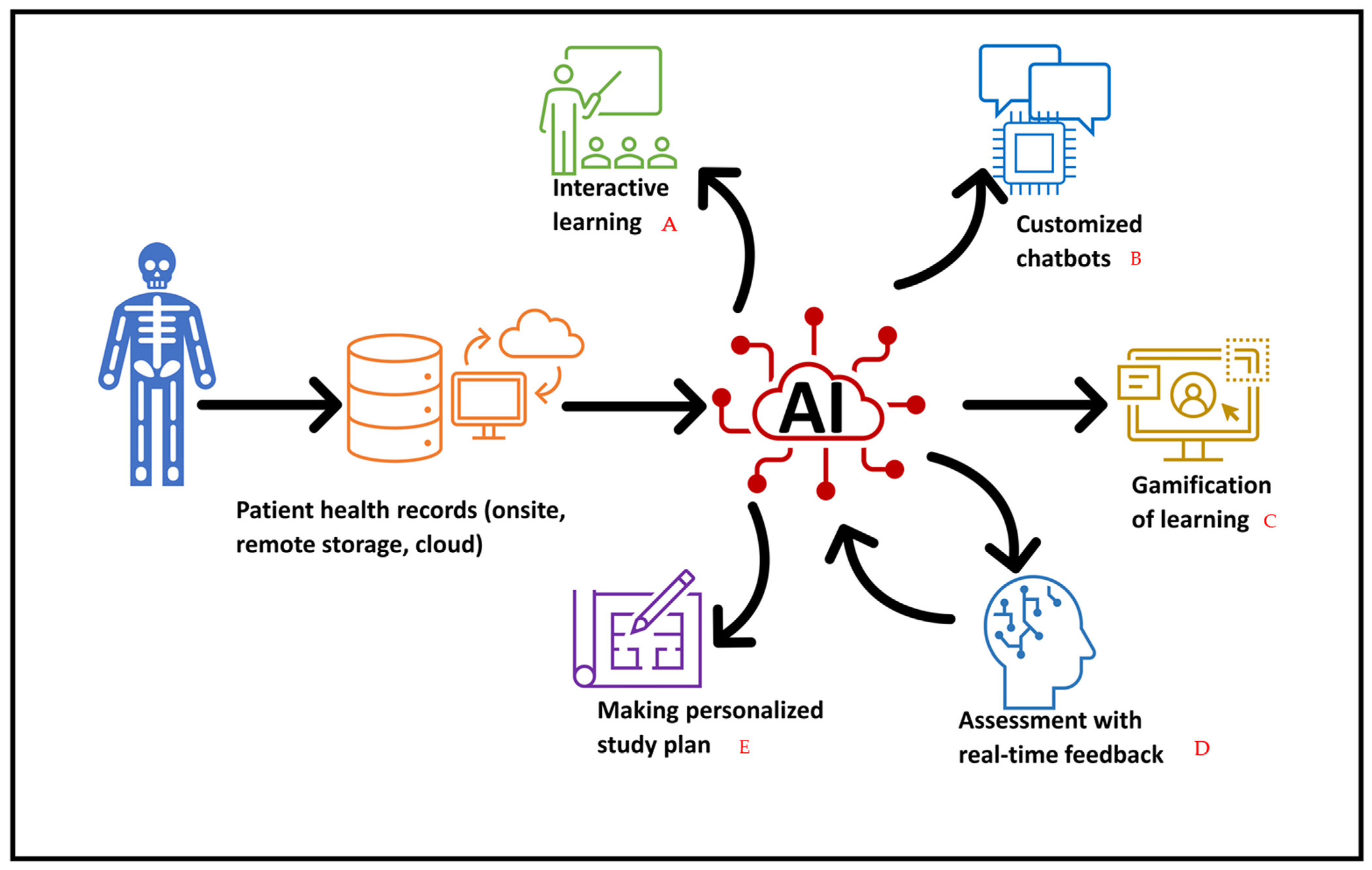
Figure 1. Various AI applications in anatomy teaching. A—AI makes the learning interactive by providing virtual simulations of anatomy and the AI assistant answers questions while exploring the anatomical organ to improve critical thinking. B—Customized chatbots provide more reliable answers for anatomy teaching as they are trained on specific databases. C—Gamification of the learning makes the learning process more favorable and interesting. It allows students to spend more time in application as they are learning with fun, similar to what has been done in the study by Castellano et al. (2023). D—Real-time feedback to the AI model of the learner’s performance on already-answered questions will provide the learners with feedback about the performance and allow the model to give them questions according to their level of knowledge. E—AI assistant, with its generative abilities, creates a study plan for each element or chapter of anatomy when provided with a time frame and specific objectives.
Education 15 00283 g001
Figure 2. Various AI tools used in anatomy teaching. Machine learning (ML), deep learning (DL), natural language processing (NLP), virtual reality (VR), convolutional neural networks (CNNs).
Education 15 00283 g002
Figure 3. AI models used for anatomy teaching and learning.
Education 15 00283 g003
Figure 4. Major steps in anatomical data preparation, modeling, deployment, and education for effective use of AI in anatomy education.
Education 15 00283 g004
Table 1. Algorithms and tools used in anatomy teaching.
ML algorithms
ML divided into supervised, semi-supervised, and non-supervised models. Supervised learning techniques are used to analyze medical images and predict outcomes. These techniques can be integrated into anatomy teaching to identify anatomical organs and detect abnormalities. The most used ML tools are:Scikit-learn 1.6 which is a Python 3.8 library for data analysis and mining. It can be used to build models to identify organs and structures from given images and illustrations (Pedregosa et al., 2012).
TensorFlow 2.16.1 and PyTorch 2.6 both can be used to develop DL models that perform complex tasks like image segmentation and classification (Abadi et al., 2016; Paszke et al., 2019).
DL algorithms
Convolutional neural networks (CNNs) and vision transformers (ViTs) are used in recognizing certain organ features from radiographs, CTs, and MRIs (Yu et al., 2015d; Dosovitskiy et al., 2020). From an anatomy teaching perspective, they can be used in teaching students to understand radiological anatomy. Some examples of DL algorithms include:CNNs including ResNet and U-net are useful for image segmentation and detection of organs of interest.
Recurrent neural networks (RNNs) are used to translate certain anatomy courses to different languages (Yu et al., 2015).
ViT is integrated into complex datasets like histological slides and CT to identify structures of interest (Dosovitskiy et al., 2020).
CycleGAN is a powerful tool for image generation that can be used to create state-of-the-art illustrations for anatomy teaching (Dosovitskiy et al., 2020).
NLP algorithms
NLP algorithms are used to generate content automatically after searching the available databases. Additionally, they are commonly used to create customized chatbots able to answer anatomy-related questions with a high degree of accuracy. Examples of NLPs are as follows:GPT models, with chatGPT being the most popular model, and which was developed by openAI (Gallifant et al., 2024). It can assist in answering questions, creating plans, and creating educational content.
SpaCY is an open Python library used to process medical texts. It helps to conduct NLP on large texts with high speed. It can be used to facilitate students understanding of anatomy concepts and terminologies (Explosion, n.d.).
Table 2. Summary of various studies exploring the use of AI tools in anatomy teaching.
NArticle CitationAimStudy Method/TypeSoftware Used (AI Model Used, ML or DL)Parameters
Studied
Recommendation Future Work Limitations
1Noel (2023)Explored the capabilities of various AI-powered text-to-image generators in generating detailed and accurate anatomical illustrations of the human skull, human heart, and human brainExperimental researchMicrosoft Bing Image Creator Powered by DALL-E 3, Stable Diffusion, and Craiyon V3Degree of details and accuracy.
compare different AI-powered tools to determine their effectiveness and reliability

examining the output of these generators and comparing them to established standards of accuracy
To improve the training databases for AI-powered text-to-image generators by incorporating a larger collection of anatomically correct images. Including a diverse range of accurate anatomical references.Not Available (NA)Compared only three software’s.
2Ilgaz and Çelik (2023)Evaluated how various features of open AI platforms such as ChatGPT and Google Bard in their current form can contribute to anatomy education with the perspective of questioning, answering, and writing articles, and aimed to respond to some controversial topics.Experimental researchChatGPT 3.5 and Google BardThe correctness in answering the questions of the Medical Specialty Exam (MSE) last five years a total of 131 questions in anatomy.
-The validity of the anatomical information’s and degree of details in question generation
And article writing
The need for continuous improvement and validation of LLMs for reliable healthcare practices.

Both models were able to generate multiple-choice questions with a high degree of accuracy. However, the performance of the models in article writing was not yet at a sufficient level. The study also found that the use of LLMs in medical education requires caution.
Further research is needed to increase the accuracy of the models and to better understand how they can be used effectively in educational settings.

There is a need for future studies on the application of 2D and 3D figures in anatomy education with other AI applications.
Limitations in dealing with potential inaccuracies.
3Arun et al. (2024)Evaluated AI’s ability to provide clear and accurate anatomy information and generated a custom interactive and intelligent chatbot (Anatbuddy)Experimental researchOpen AI Application Programming Interface (API) to build interactive chat potFactual accuracy, relevance, completeness, coherence, and fluencyAnatomy profession should develop a custom AI chatbot for anatomy education utilizing a
carefully curated knowledge base to ensure accuracy.
-Anatbuddy was trained only on thoracic anatomy content from open-source anatomy materials.
-Inter-rater agreement could not be computed due to the study’s design.
-Improve the capabilities of customizable chatbots to improve students’ learning experience.
-More research is needed to expand its capabilities across anatomical regions and incorporate image-based information.
-Further research is needed to ascertain the acceptance of the technology by educators and students.
4Chheang et al. (2024)Introduced a VR environment with a generative AI-embodied virtual assistant to help in responding to anatomy questions varying in cognitive complexity with the ability to communicate verbally.Pilot experimental research3D by Unity game engine (version2019.4.34f1)
-ChatGPT 3.5, and AI-based library (Avatar SDK), and Microsof Azure Speech service to enable natural interactions
Scores obtained from knowledge- and analysis-based questionsUtilization of AI models in many fields of anatomy as the combination of both virtual assistant configurations has the potential to offer a comprehensive solution for assisting and enhancing the learning experience.-Should investigate the conversations’ transcripts and the responses’ accuracy.
-Levels of experience should be considered as a covariate, or a balance of the groups based on their level of prior VR/virtual assistant.
-It is a pilot study was conducted with 16 participants.
-The way a participant phrased a question could impact the response they received.
-The lack of transparency and unclear information on the data source used, and other related consequences.
5Li et al. (2021)Utilized an open-source machine learning architecture and fine-tuned it with a customized database to train an AI dialogue system to teach medical students’ anatomy.Experimental researchML
Bidirectional Encoder Representations from Transformers’ BERT
self-reported confidenceAI chatbots provide high level of student engagement.
Recommend building AI systems based on open-source resources for medical education.
NANA
6Jin et al. (2022)Integrated neural intelligence with artificial intelligence to obtain objects from medical images by formalizing an anatomy-guided deep learning object recognition approach named AAR-DL.Experimental research4 modules: AA-R module: Fuzzy anatomy module, DL-R module, refinement, final detection moduleN/MHigh-level anatomy guidance improves recognition performance of DL methods.
Anatomy guidance brings stability and robustness to DL approaches for object localization and reduces training time.
Develop the model by including the formulation of anatomy-focused loss functions, injecting anatomic knowledge at deeper layers of the networksRelated to modelability of the objects and the appropriate incorporation of the models into AAR-DL.
7Buzzaccarini et al. (2024)Aimed to understand whether Midjourney could create images that were not only realistic, but also correct in terms of anatomy.Experimental researchMidjourney 5.2Accuracy, anatomical correctness, and visual impact.Collaboration between AI developers and medical experts might pave the way for more accurate and clinically relevant images.-Test other tools and compare them.
-Collaboration to develop better versions.
Used and tested only one single AI tool, Midjourney. Other tools are available worldwide and may be more accurate and correct than Midjourney
8Collins et al. (2024)-Developed AI-powered anatomy tutor.
-Assess the efficacy and performance of anatomyGPT
Experimental researchGPT-builderPerformance, answers accuracy,Instructors and students could create their own custom GPTs for teaching and learning anatomy.
Research is needed to develop the potential of GPTs for anatomy education.
Compare GPT-4 with other large language models on achievement tests and tutoring tasks.
Design, deploy, and study GPTs for other specific theoretical perspective such as the ACT-R theory of cognition.
First, the size of the question sets to evaluate the performance of the GPTs was relatively small
The challenge of acquiring open-source anatomy resources to incorporate into GPTs.
9Castellano et al. (2023)Investigated effectiveness of the mobile gamified technological tool with AI virtual assistant in learning Anatomy.Experimental research (empirical study and quasi-experiment statistical technique)AIEd gamification technique and virtual assistant chatbot based on NLPAcademic performance pre-test and post-test.Gamified components support students in learning anatomy.
In addition, the virtual assistant recommendations enabled the students to improve with feedback.
Search the area of reinforcement learning, and to study the influence of the gamified technological tool in other health-related programs.It does not allow the strategies delivered by the recommender system to be redirected directly to a gamified component, issues in sampling error, and didn’t record their score in final exam, just in pre and posttest.
Disclaimer/Publisher’s Note: The statements, opinions and data contained in all publications are solely those of the individual author(s) and contributor(s) and not of MDPI and/or the editor(s). MDPI and/or the editor(s) disclaim responsibility for any injury to people or property resulting from any ideas, methods, instructions or products referred to in the content.