Article

An Open-Source Neonatal Phototherapy Device

1
Department of Electrical & Computer Engineering, Thompson Engineering Building, Western University, 1151 Richmond St., London, ON N6A 3K7, Canada
2
Department of Electrical & Electronic Engineering, Kenyatta University, Nairobi 00100, Kenya
3
Ivey School of Business, Western University, London, ON N6A 3K7, Canada
*
Author to whom correspondence should be addressed.
Technologies 2025, 13(11), 499; https://doi.org/10.3390/technologies13110499 (registering DOI)
Submission received: 12 September 2025 / Revised: 25 October 2025 / Accepted: 28 October 2025 / Published: 31 October 2025
(This article belongs to the Special Issue Breakthroughs in Bioinformatics and Biomedical Engineering)

Abstract

Severe neonatal hyperbilirubinemia (SNH) (jaundice) is responsible for over 114,000 preventable neonatal deaths annually, as the technology that can treat the condition is cost-prohibitive for low- and middle-income countries. In this study an open-source neonatal phototherapy device (NPTD) to treat SNH was designed, built, and validated against the phototherapy technical specifications set by the American Academy of Pediatrics and UNICEF. The open-source device can be built for a tenth of the cost of the least expensive proprietary one on the market, with treatment metrics equivalent to or exceeding commercial devices available in developed nations. This device, whose material costs are USD 93.00, was shown to deliver an irradiance up to 80 µW/cm2/nm, within the acceptable wavelength range of 420–500 nm. It was further demonstrated that the unit could deliver a uniform distribution of irradiance (34.5 ± 4.3 µW/cm2/nm) over a surface area exceeding 3200 cm2. These findings show that the open-source NPTD is capable of delivering accurate, consistent, and reliable irradiances for the management of SNH. By releasing full documentation in an open-source manner, the device may be broadly used to ensure affordable and consistent low-cost means of improving the quality of care for SNH.

1. Introduction

Severe neonatal hyperbilirubinemia (SNH), or jaundice, is responsible for an estimated 114,100 neonatal deaths annually [1]. Another 75,000 newborns survive, yet they may develop kernicterus spectrum disorder (KSD) for the remainder of their lives [2]—a chronic condition characterized by motor dysfunction and oculomotor and auditory impairments [3]. The majority of these cases occur in low- and middle-income countries (LMICs), particularly in Sub-Saharan Africa, South Asia, and the Eastern Mediterranean [4]. A systematic review and meta–analysis by Slusher et al. [5] concluded that high-income countries have an incidence rate of SNH of 3.7 per 10,000, while LMICs have an incidence rate of 244.1 per 10,000. This disparity represents the highly treatable nature of SNH [6] if access to technology, resources, and training is provided [7]. Current devices used for treating SNH (Table 1) are manufactured in high-income countries and intended to serve a high-income consumer base, where sales margins are highest.
Thus the devices are not only cost-prohibitive in LMICs, but repairing these devices is challenging [13] and may lead to ‘medical equipment graveyards’ [14].
One way to overcome this challenge is the design and digital-distributed manufacturing of open-source medical devices [15,16], which was somewhat normalized in response to the COVID-19 pandemic [17]. The best practice for open hardware [18,19] involves designing hardware that can be easily replicated in a wide range of low-cost digital manufacturing tools like open-source RepRap-class 3-D printers [20,21], open-source autoinjectors [22], open-source cell culture incubators [23], and open-source microscopes [24]. This has been shown to produce high-quality medical and scientific hardware for a small fraction of the cost of proprietary products [25,26].
Treatment for SNH is most commonly administered through phototherapy [27,28] by exposing an infant to a light source of sufficient intensity (irradiance ≥ 30 µW/cm2/nm) [29] and appropriate wavelength (420–500 nm) [30] to photoconvert excess bilirubin into excretable photoproducts [31]. The use of exchange transfusion carries additional risks such as infection, blood clots, and mortality and produces less effective patient outcomes [32]. Intravenous immunoglobulin administration poses several concerns given its low efficacy, higher cost, and donor requirements [33]. Phototherapy is the preferred method of treatment [34]. This paper uses the open-hardware model to develop a low-cost NPTD capable of being manufactured in LMICs. In conjunction with an open-source validated irradiance meter [35] which, when paired with the phototherapy device, creates a system to effectively treat SNH at minimal cost.
This study validated the performance of the device by measuring its output wavelength, intensity, and coverage, and compared those values with the standard set by the American Academy of Pediatrics (AAP) and UNICEF phototherapy technical specifications [36]. A further cost and performance analysis was also made against modern commercial units sold in North America [37]. The results from deployment and testing at Kenyetta University, Kenya, are presented and discussed in the context of providing low-cost phototherapy everywhere in the world.

2. Materials and Methods

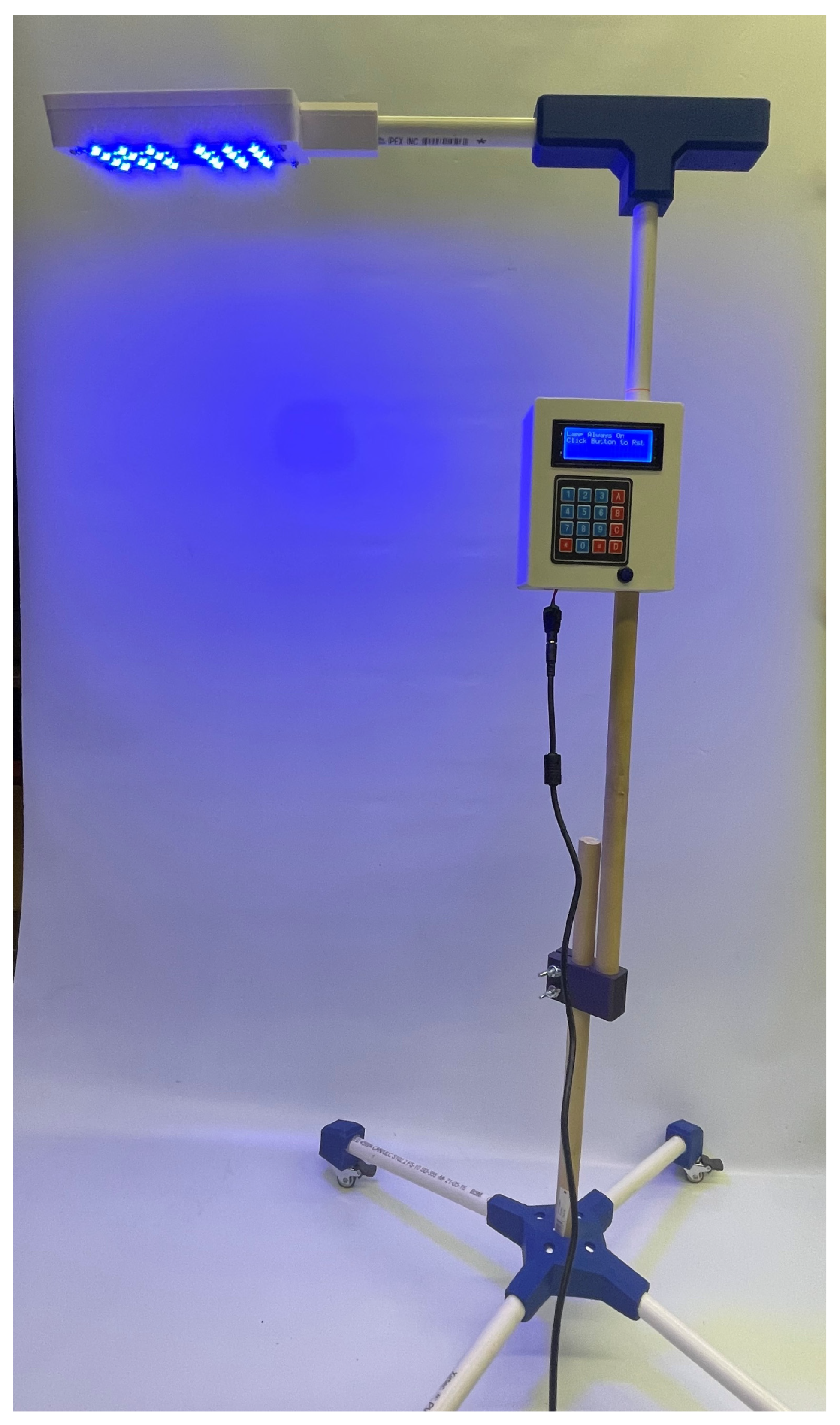
A fully assembled open-source NPTD is shown in Figure 1 and was constructed in three sub-assemblies:
(a)
The structural body including the 3D-printed electronic housings, PVC pipes, and joints connecting the pipes.
(b)
The microprocessor, power delivery, and interface electronics.
(c)
The light source and heat dissipation assembly.
The detailed assembly of the open-source NPTD is presented in Appendix B. A calibrated Ocean Insight UV-VIS FLAME spectrometer (Ocean Insight, Orlando, FL, USA) [38] was used to measure both the intensity and peak wavelength over the range of 200–800 nm. Multiple readings over a 2 h time span were taken to verify consistent spectral output. A mapping of the illuminated surface was created by holding the source at distances of 33 and 45 cm and measuring irradiance at equally spaced intervals of 10 cm.
The intensity, wavelength, and irradiated surface area were then compared with the standard set by the AAP for effective phototherapy treatment for SNH [27,28] and commercial products currently in use. The UNICEF phototherapy technical specifications [36] were used to determine if the device offered all the required functionalities for hospital operation. Finally, a bill of materials (BOM) was developed (Appendix A) to determine the cost compared with commercial units available in North America.

3. Results

3.1. Spectral Analyses

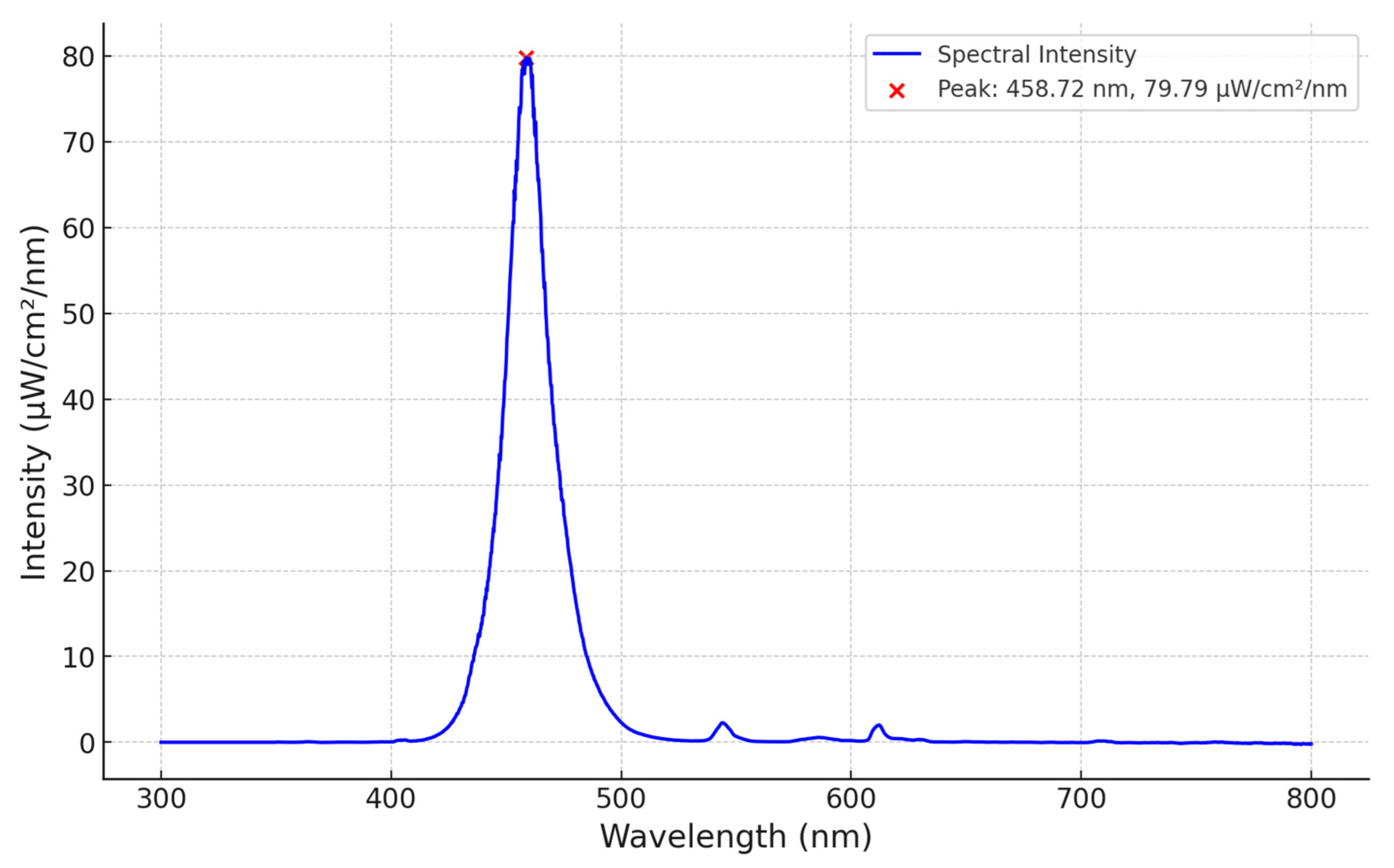
A calibrated Ocean Insight spectrometer was placed 33 cm below the neonatal lamp. A distinct peak of 80 µW/cm2/nm at 459 nm is shown in Figure 2. The total bandwidth is 420–500 nm, with a half-power bandwidth of 22 nm. The performance of the open-source NPTD is comparable to that of the commercial devices (Table 2). No variation in irradiance was observed over the 2 h period in which the test was conducted. A 2 h test period was determined to be sufficient given the stability of LEDs over long periods of time [39].

3.2. Irradiance Footprint

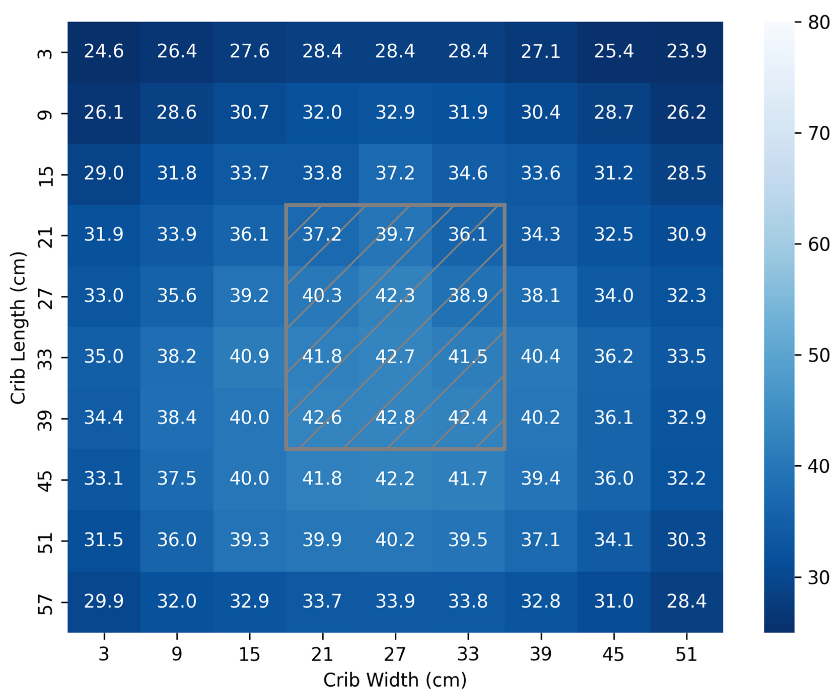
Figure 3 illustrates the light distribution when the source is placed 45 cm above the bed. The location of the lamp head is represented by the hatched gray box. It is observed that the distribution of the light was relatively uniform and that 81% of the surface is exposed to the minimum recommended dose for SNH of 30 µW/cm2/nm [29,30,31,40,41,42]. The total surface area mapped was 3240 cm2. The mean distribution of irradiance was 34.5 ± 4.3 µW/cm2/nm, as shown in Figure 3.
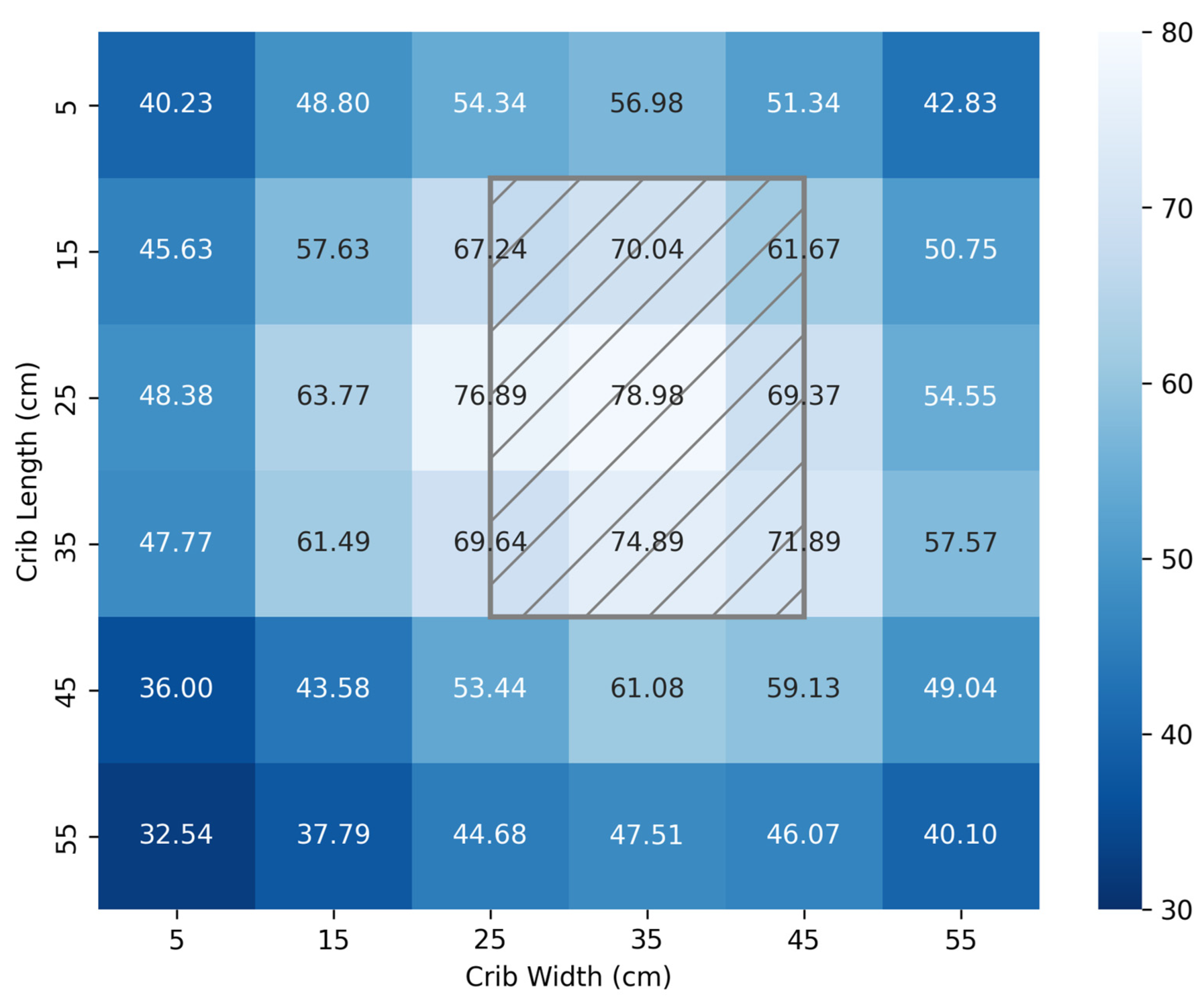
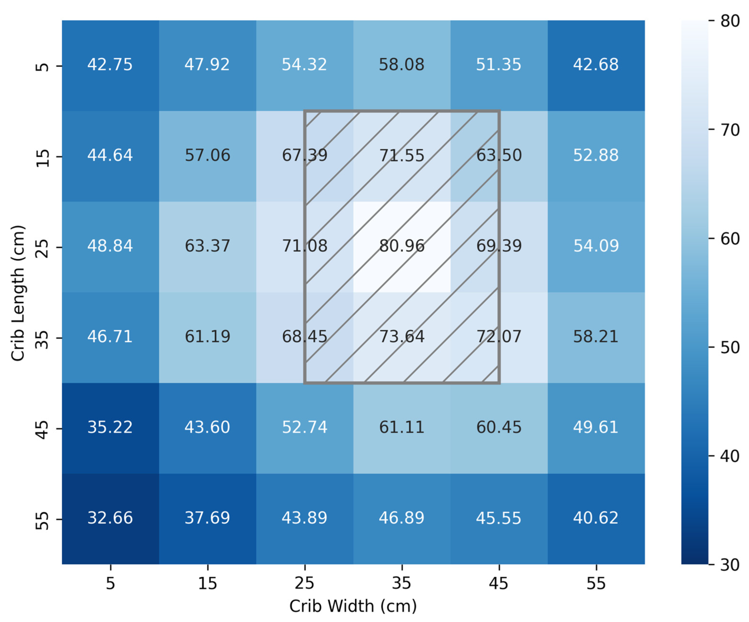
Figure 4 and Figure 5 illustrate mappings using the calibrated Ocean Insight spectrometer and the open-source FAST irradiance meter, respectively. The hatched gray rectangle represents the location of the light source, and the right-hand gradient indicates the intensity of the light in µW/cm2/nm. Measurements were taken at 10 cm intervals for a crib area of 3600 cm2 and a distance from the source of 33 cm.
The irradiance mapping using the Ocean Insight spectrometer (Figure 4) showed a mean of 54.8 ± 12.0 µW/cm2/nm, while measurements taken using the FAST irradiance meter (Figure 5) showed a mean of 55.1 ± 11.8 µW/cm2/nm. The absolute mean difference in measured values between the two devices was 0.88 µW/cm2/nm.
Table 2 summarizes parameters of commercial therapy units originally listed in the AAP technical report on phototherapy to prevent severe neonatal hyperbilirubinemia [27,28]. The parameters of the NPTD are also included for comparison.
Table 1 shows the cost of typical commercial devices in use in North America. The products selected for market analysis were chosen based on similarity to the NPTD; specifically, they are all overhead light emitting diode (LED) devices. As shown in Appendix A, the cost of building the developed NPTD in Canada is CAD 130.03, or USD 92.93. The cost of this NPTD is one tenth the cost of the least expensive proprietary commercial option on the market.
Irradiance measurements were taken from the NPTD by the two light meters, as shown in Table 3. It should be pointed out that there was minimal variance recorded between each mapping, suggesting sufficient accuracy and the reliability of the locally assembled devices (both the open-source NPTD and the locally fabricated 3D-printed open-source FAST light meter) with reference to the established reference devices.

4. Discussion

Phototherapy is the primary treatment for SNH [42], utilizing light with an irradiance of at least 30 µW/cm2/nm and a wavelength range of 420–500 nm [40] to facilitate the breakdown of excess bilirubin in the blood [41].
High-income countries experience far lower rates of SNH compared with LMICs, where there is limited access to necessary equipment [5]. As a result LMICs are disproportionately affected by kernicterus spectrum disorder (KSD) [43], a preventable chronic disease characterized by abnormal motor function, oculomotor impairment, and auditory complications [44]. KSD is caused by the buildup of bilirubin in the neonate’s blood and requires immediate phototherapy to lower the level of bilirubin [45]. KSD severely limits the abilities and opportunities of those afflicted for the remainder of their lives [43]. This paper has shown that construction of appropriate and cost-effective phototherapy equipment can be implemented through open-source hardware to effectively treat SNH in LMICs.
Spectroscopic analysis showed that the NPTD emitted a light at a wavelength of 460 nm with half-power bandwidth of 22 nm. This falls within the spectrum shown to effectively photodegrade bilirubin as cited by Porter [46] and Maisels [47]. At a distance of 45 cm from the NPTD, the mean irradiance was 34.5 ± 4.3 µW/cm2/nm, which compares well with the commercial products presented in Table 1 [27].
The total illuminated surface area available for therapy is 3600 cm2, which exceeds UNICEF’s required technical specifications [36] of a minimum effective surface area ≥ 1220 cm2. It also exceeds the area of commercial devices referred to in Table 2 [27]. Several infants are sometimes placed together to receive phototherapy in LMICs, which may lead to overcrowded cribs [48]. The larger effective area provided by the open-source NPTD may reduce the risk of overcrowding cribs.
The total material cost to construct these developed NPTDs is USD 93.00 (CAD 130.00), which represents a 92.3% to 98.7% cost reduction compared with commercial devices. In LMICs, access to effective treatment of SNH has not only been hindered by high costs, but also by technologies that are ill-suited to these resource-limited settings [49]. This NPTD significantly lowers this financial barrier.
Another limitation to effective treatment is the inability to consistently monitor the irradiance of delivered phototherapy [6]. It has also been demonstrated that the open-source FAST irradiance meter [35] is equally effective at measuring irradiance as a calibrated spectrometer, with an absolute mean difference of less than 1 µW/cm2/nm. The NPTD in conjunction with the FAST irradiance meter establishes an open-source system to treat SNH at a price point significantly lower than that a set of commercial equipment.
Further device refinements should be explored, such as incorporation of reflective material to increase irradiance homogeneity [50], evaluation of long-term operation of the device, increasing power delivery, and installation of multiple LED lamps per unit. Clinical validation will be required before the device can be approved for clinical use. In addition to regulatory requirements, testing within a clinical context will also inform further developments to improve device durability and maintenance feasibility and provide feedback for changes to the user interface. No tests involving humans or animals have been conducted. The NPTD meets most of the UNICEF-required technical specifications [36], with the exception of the following: battery operation for power outages less than 10 min and the incorporation of white LED lamps for examination. These are considered lower-priority features and will add cost and not improve patient outcomes.

5. Conclusions

One of the leading causes of severe neonatal hyperbilirubinemia in LMICs is the lack of cost-effective and appropriate technology to prevent the condition. It has been demonstrated that an open-source NPTD to effectively treat SNH can be built at a significantly lower cost while producing treatment metrics equivalent to or exceeding commercial devices available in developed nations. The developed device, whose material costs are USD 93.00 (CAD 130.00), was shown to produce an irradiance of 80 µW/cm2/nm within the acceptable range of 420–500 nm. It was further demonstrated that the unit could produce a uniform distribution of (34.5 ± 4.3) µW/cm2/nm over a surface area exceeding 3200 cm2. The device has been shown to meet or exceed commercial device parameters and has been tested in a LMIC context at the University of Kenyetta, Kenya; however, it requires clinical certification and regulatory testing before clinical deployment. By releasing documentation, building instructions, and design in an open-source manner, the device may be broadly distributed. In conjunction with the open-source irradiance meter, the pair is technically validated, pending clinical validation, to treat SNH in resource-limited communities.

Author Contributions

Conceptualization, J.M.P.; methodology, J.G., A.W., J.M. and J.M.P.; validation, J.G., A.W., J.N., G.N. and G.M.; formal analysis, J.G., A.W., J.N., G.N., G.M., J.M. and J.M.P.; investigation, J.G., A.W., J.N., G.N. and G.M.; resources, J.M. and J.M.P.; data curation, J.G. and A.W.; writing—original draft preparation, J.G. and J.M.P.; writing—review and editing, J.G., A.W., J.N., G.N., G.M., J.M. and J.M.P.; visualization, J.G.; supervision, J.M. and J.M.P.; project administration, J.M. and J.M.P.; funding acquisition, J.M.P. All authors have read and agreed to the published version of the manuscript.

Funding

This research was funded by the Frugal Biomed Grant and the Thompson Endowment.

Institutional Review Board Statement

Not applicable.

Informed Consent Statement

Not applicable.

Data Availability Statement

All data and design files are available on the OSF: https://osf.io/93gnj/ (accessed on 27 October 2025).

Conflicts of Interest

The authors declare no conflicts of interest. The funders had no role in the design of the study; in the collection, analyses, or interpretation of data; in the writing of the manuscript; or in the decision to publish the results.

Appendix A. Neonatal Phototherapy Device Bill of Materials

ComponentDescriptionQuantityCost (CAD)
LED246.00
2″ Caster Wheels43.08
1/2″ PVC Pipe8 ft14.37
Arduino Nano11.89
LCD11.09
Buck Converter10.37
12 V 24 W Wall Adapter12.74
PCB224.20
Button11.41
SUB TOTAL65.15
Shipping, Misc. Standard Elec-tronics *100% of Total65.15
TOTAL130.30

Appendix B. Build Documentation

Appendix B.1. Structural Sub-Assembly

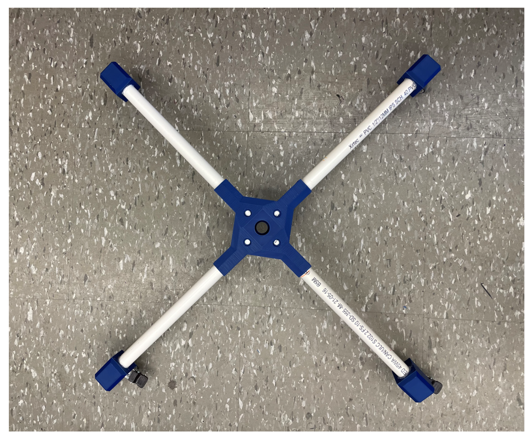
Figure A1 illustrates the Structural Sub-Assembly, consisting of six 12 mm (1/2″) polyvinyl-chloride (PVC) pipes with an outer diameter of 21.4 mm (0.84″). The approximate lengths of each are shown in Table A1. It was found that the PVC pipe used for pipe C can be extended to 80 cm before bending becomes a significant factor. It is recommended that for a unit with a desired height exceeding 130 cm, pipe C should be replaced by a wooden dowel of equivalent diameter.
Table A1. Pipe fittings.
IDDescriptionLength (cm)
AT-Connector to Light Source Housing~45
BElectronics Housing to T-Connector~45
CBase to Electronics Housing~70–130
D–GFour Legs~45
All remaining components are 3D-printed parts. They include the light source housing, electronics housing, T-connector, base connector, and caster wheel adapters. The model files can be found at https://osf.io/93gnj/ (accessed on 27 October 2025).
Once the relevant components are printed, each of the caster wheel adapters are secured to the pipes D–G with a nut and bolt (or optionally ABS cement) Figure A2.
Figure A1. Structural sub-assembly.
Technologies 13 00499 g0a1
Figure A2. Caster wheels fitted to feet.
Technologies 13 00499 g0a2
Each foot can then be connected through the base connector, as shown in Figure A3. If only two of your casters have brakes, it is recommended that they are positioned adjacent to one another to maximize braking ability. A nut and bolt securing the base to each pipe is recommended. Secure pipe C to the center of the base connector with ABS cement.
Figure A3. Pipes D-G secured to base connector.
Technologies 13 00499 g0a3
A height-adjustment mechanism is implemented, as shown in Figure A4, so that irradiance dosing is more easily varied.
Figure A4. Height-adjustment mechanism.
Technologies 13 00499 g0a4

Appendix B.2. Electronics Sub-Assembly
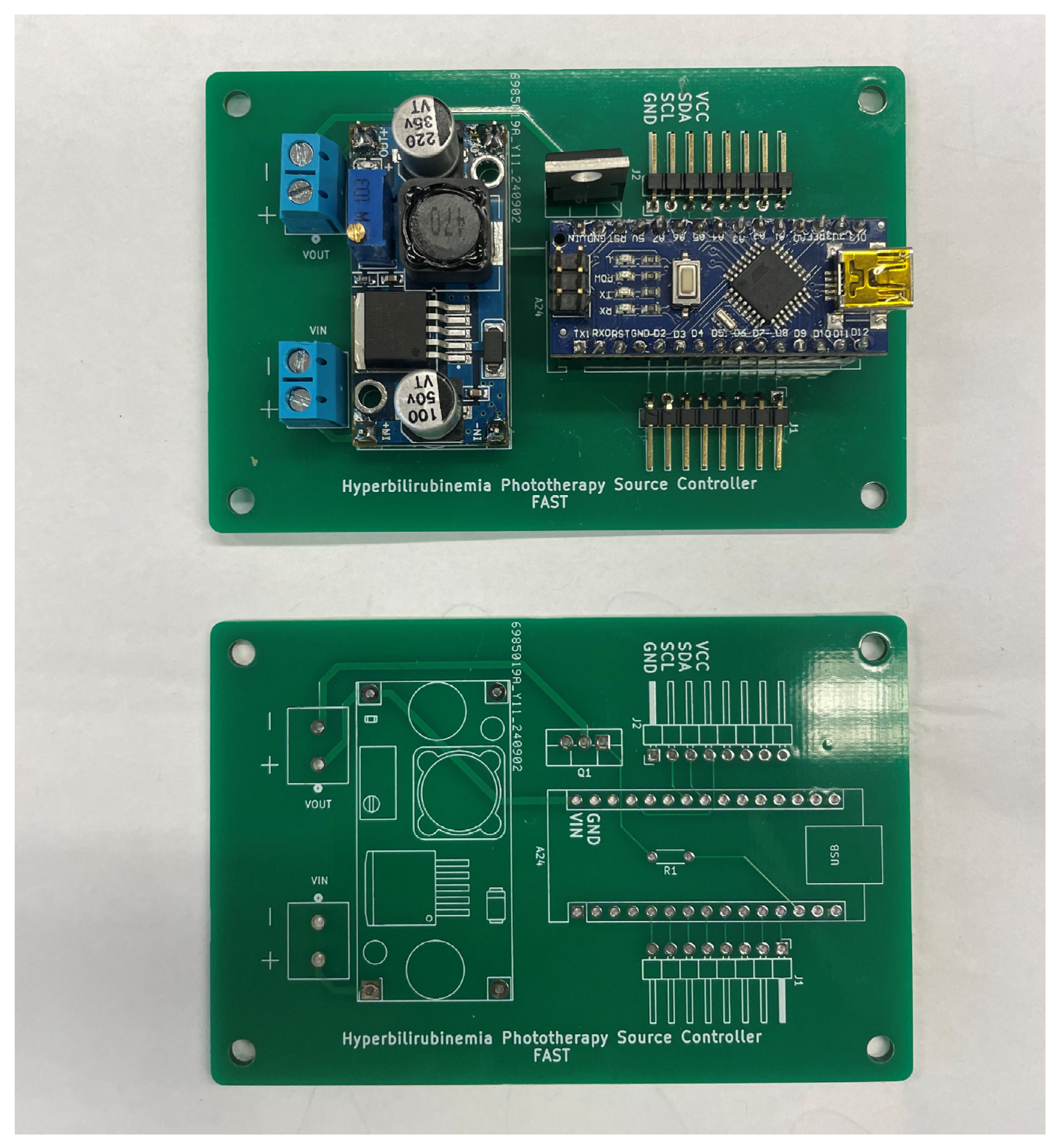
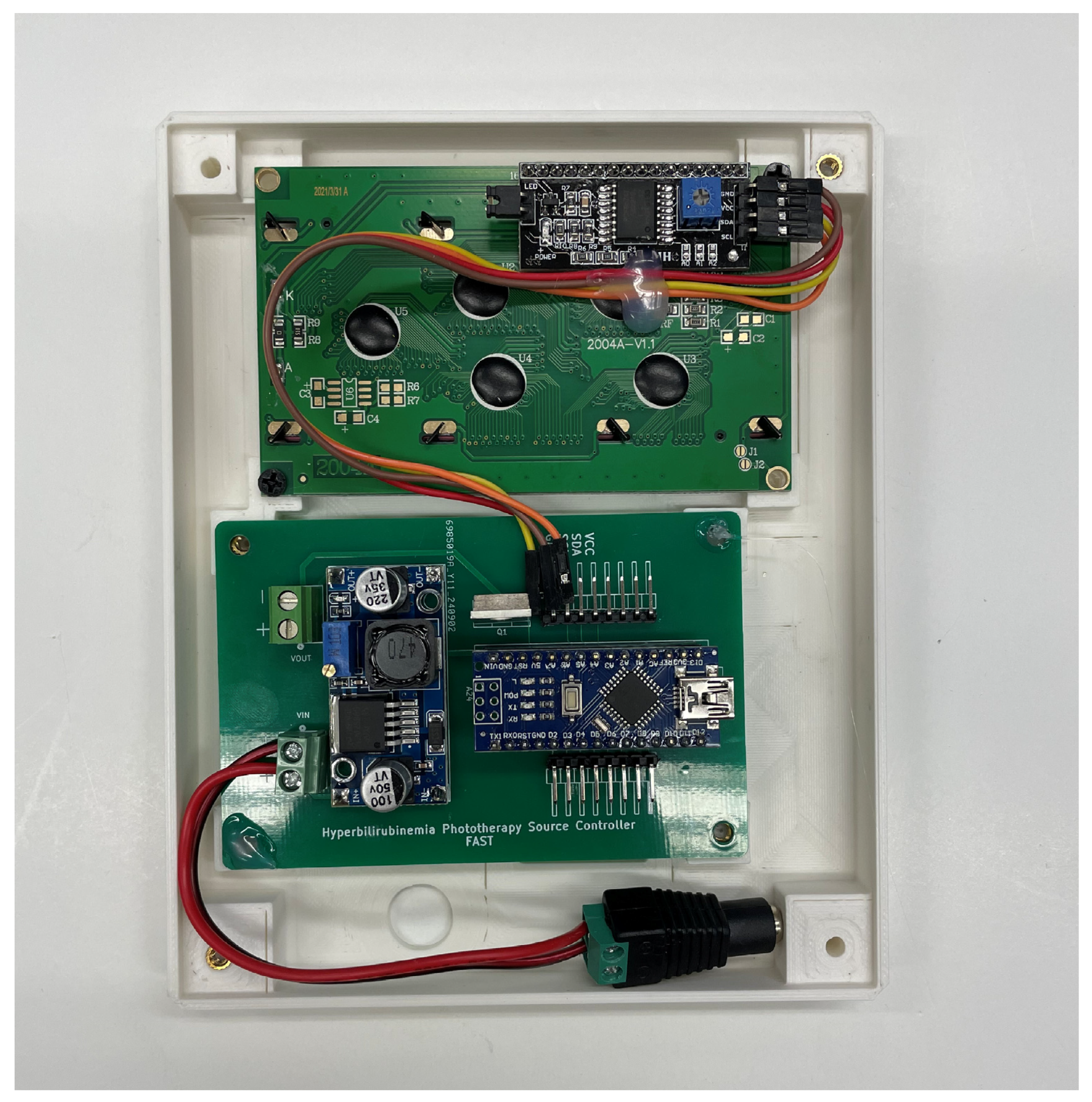
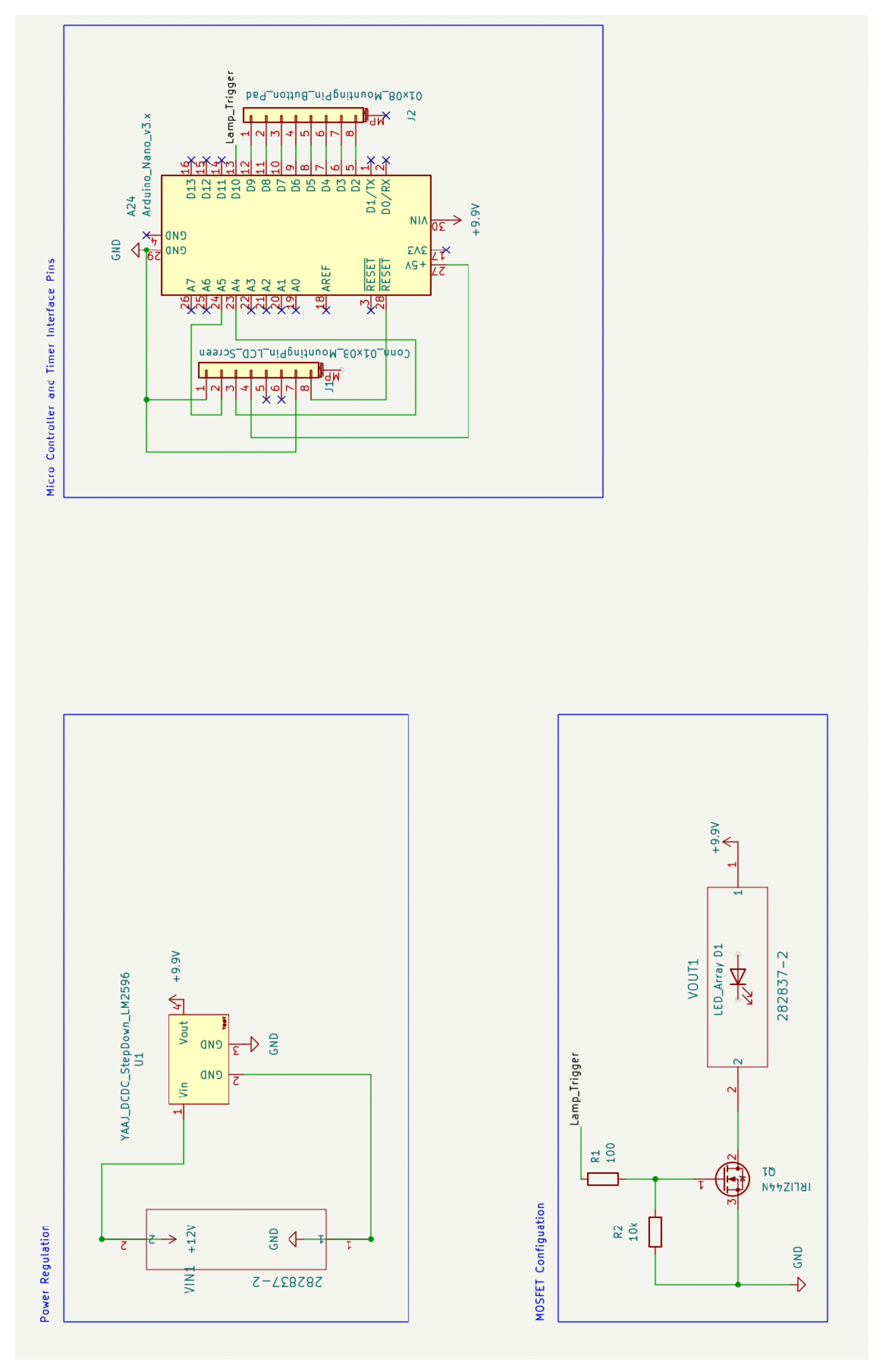
This sub-assembly consists of power conversion and regulation circuitry, user interface hardware, and the system controller. Power is converted from 110 V 60 Hz AC input from a wall outlet to 12 V DC with a maximum current output of 10 A using a standard AC power supply. The 12 V DC line is stepped down through a voltage regulator to a stable 9.9 V using the LM-2596 DC-DC converter. The output is then routed to provide continuous power to the Arduino Nano microcontroller. The output is also connected to the drain side of an N-Channel MOSFET whose gate input is triggered by a pin from the Arduino Nano. When the gate is pulled high, current is allowed to flow through the MOSFET and provide power to the lamp source, which is further described in the Lamp Source Sub-Assembly. See Figure A4 for the PCB assembly of the power converter, microcontroller, and MOSFET.
Figure A5. PCB pre- and post-assembly.
Technologies 13 00499 g0a5
The Arduino is also responsible for monitoring the duration for which the lamp is on. This is accomplished using a timer interface. A number pad is presented to the administrator, who can then select the hours and minutes for the lamp to be on. An LCD screen is included to assist with this process and display the remaining time before shutting off. Figure A5 shows the LCD screen connected to the controller PCB. A detailed description of how to use the interface is provided in Appendix C.
Figure A6. Screen and controller PCB installed in face of electronics box.
Technologies 13 00499 g0a6
The schematic diagram and a printed circuit board drawing are presented in Figure A6 and Figure A7, respectively. The files required to order these boards can be found at https://osf.io/93gnj/ (accessed on 27 October 2025).
Figure A7. Schematic diagram.
Technologies 13 00499 g0a7
Figure A8. PCB layout.
Technologies 13 00499 g0a8

Appendix B.3. Lamp Source Sub-Assembly
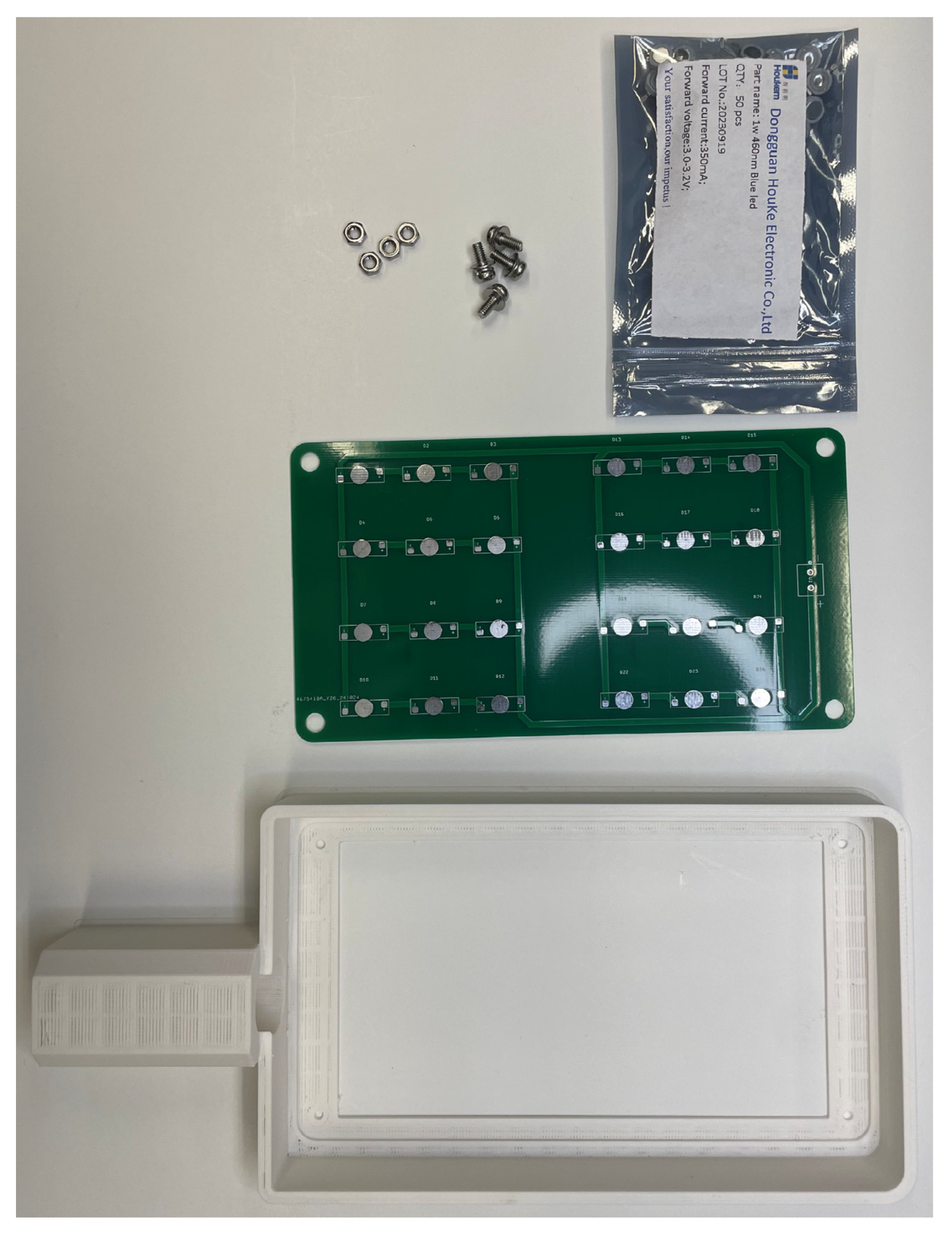
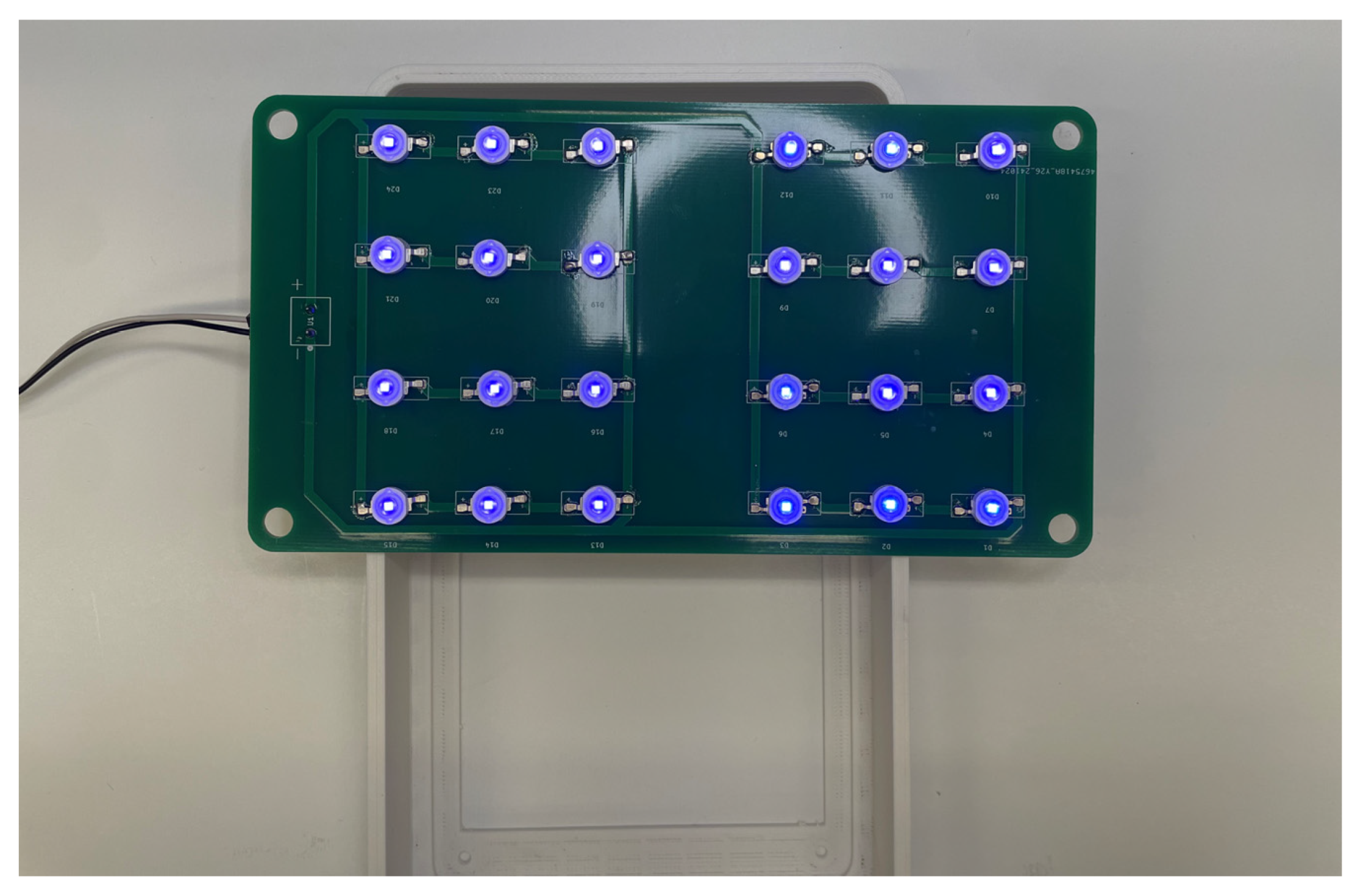
This sub-assembly consists of the light source providing the treatment to the infant and the wires providing power. The source consists of an array of 24 LEDs, each operating at 3 V and a maximum constant current of 350 mA. The source can draw a total of 12.6 watts and requires a heat sink to prevent overheating during long periods of operation.
The lamp source before and after assembly can be seen in Figure A9 and Figure A10, respectively. The PCB drawing of the LED array circuit is shown in Figure A11. Before assembly, a small amount of thermal paste is applied to each round pad, while solder paste is applied to each adjacent electrical pad. Each LED is then placed on the PCB, oriented by the correct polarity, before a hot-air solder station applies heat to secure the LEDs to the PCB.
The files required to order these boards can be found here: https://osf.io/93gnj/(accessed on 27 October 2025).
Figure A9. Lamp source pre-assembly.
Technologies 13 00499 g0a9
Figure A10. Source post-assembly with minimal test voltage applied.
Technologies 13 00499 g0a10
Figure A11. LED array PCB.
Technologies 13 00499 g0a11

Appendix B.4. Final Assembly

The electronics sub-assembly should be secured to the top of pipe C. Connect a pair of 18-gauge wires to the ‘Vout’ pin header and route the wires up through the top of the electronics sub-assembly box and into pipe B. Pipe B is then inserted into the top of the box. Route the wires through the T-connector and into pipe A. Finally, secure the ends of each wire to their respective terminal in the lamp source assembly. Secure the lamp source sub-assembly to pipe A with ABS cement.
Put the lamp in “Always On” mode using the steps outlined in Appendix C. Measure the voltage across the LED assembly and adjust the voltage output on the DC-DC converter until you read 9.1–9.8 volts across the light source. Verify that the system operates as expected before closing the front of the electronic sub-assembly housing with M3 × 20 mm screws. The final assembly is shown in Figure A12.
Figure A12. Device operation after final assembly.
Technologies 13 00499 g0a12

Appendix C. User Interface Guide

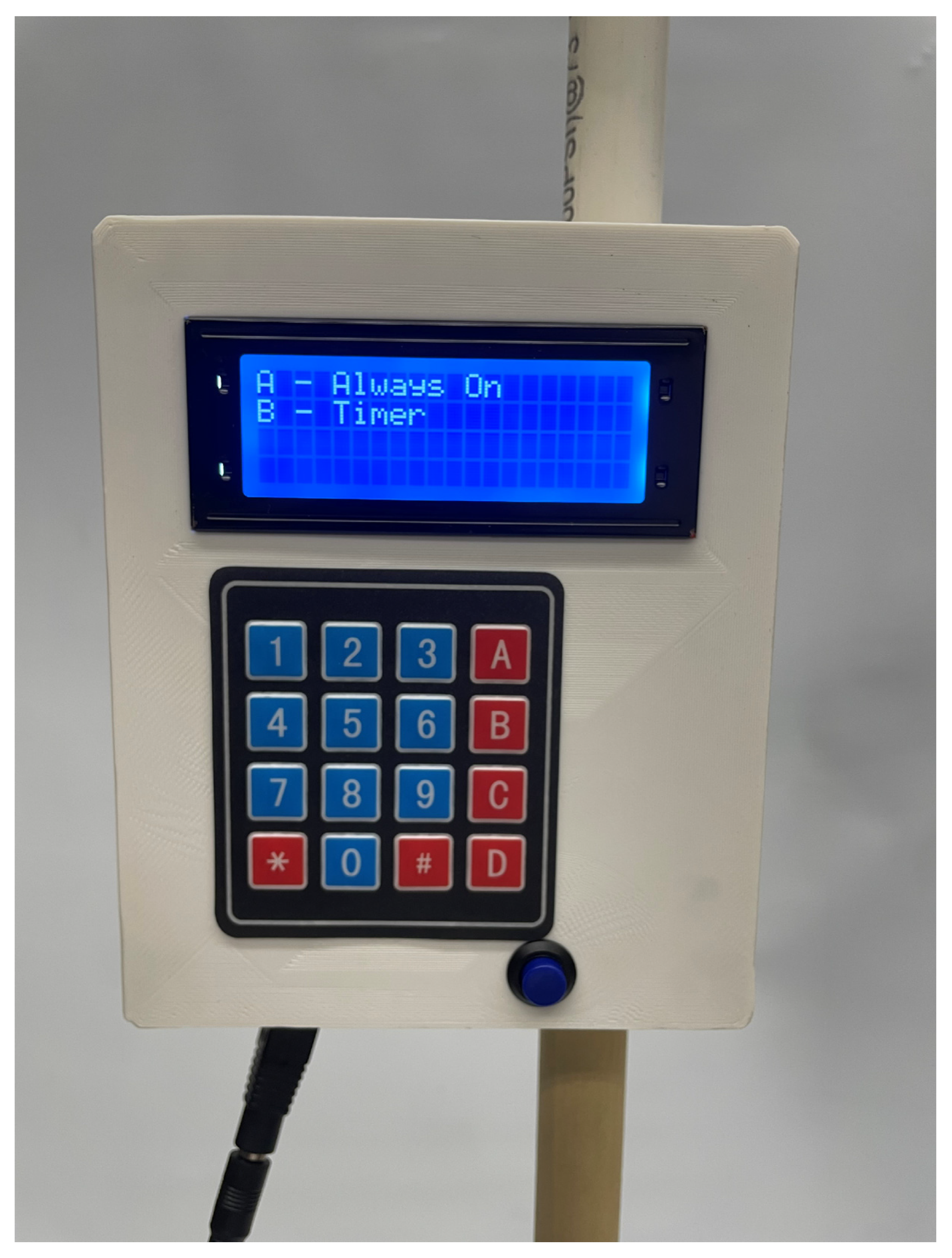
When power is supplied, the screen in Figure A11 will appear. Selecting A will immediately toggle the lamp on, and it will remain on until the blue reset button is triggered.
Figure A13. Mode selection screen.
Technologies 13 00499 g0a13
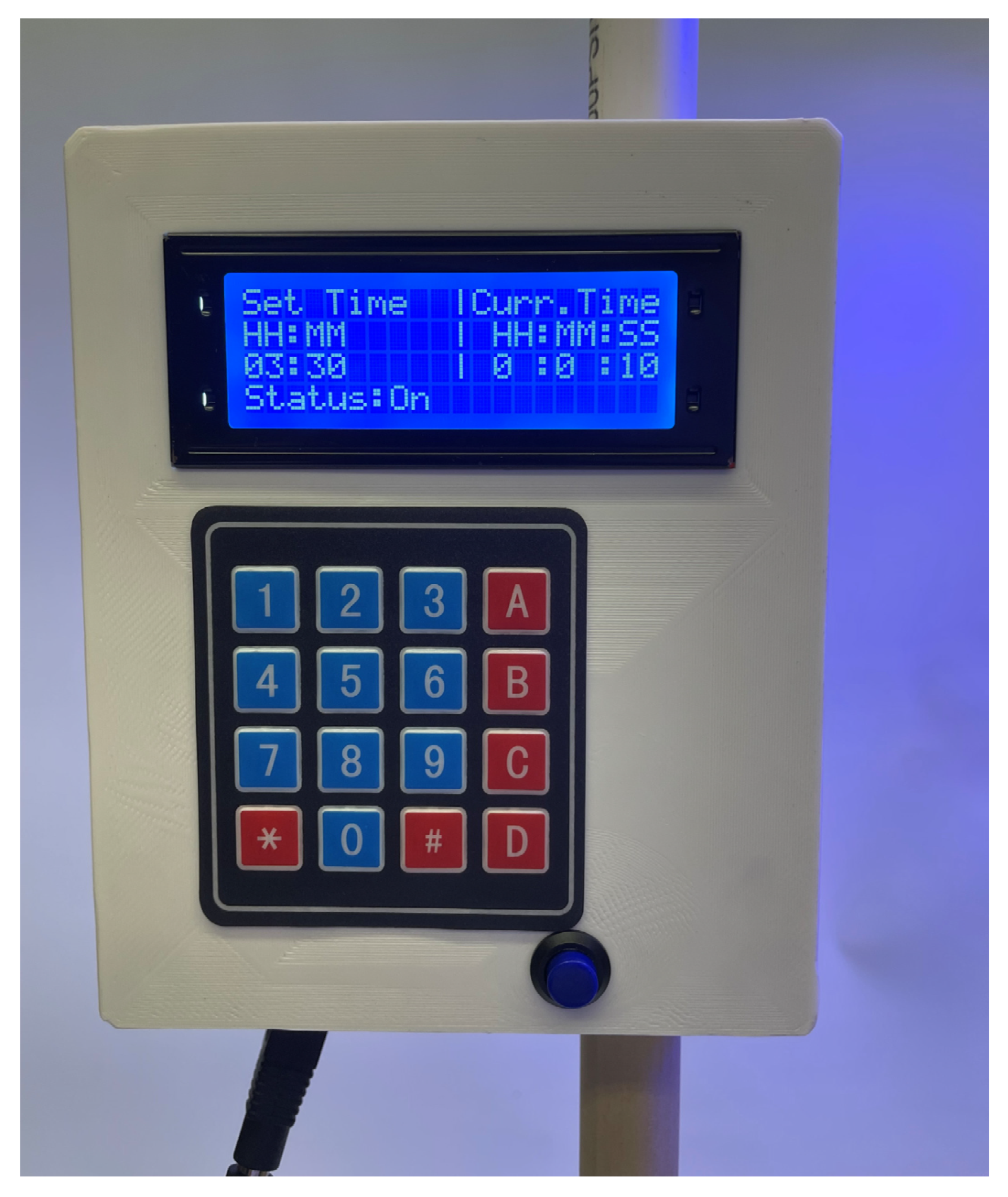
If B is selected, the unit will be placed into timer mode, as shown in Figure A12. The unit will wait until four digits are pressed, representing the hours and minutes it will be set to. The two hour digits are entered first, followed by the minute digits. The lamp will immediately turn on once the fourth digit is entered, and the elapsed time will update every second under the ‘Curr. Time’ column, as shown in Figure A13. Once the current time is equal to the set time, the unit will turn off. The blue reset button must be pressed to return to the start screen.
Figure A14. Timer interface awaiting input.
Technologies 13 00499 g0a14
Figure A15. Timer showing elapsed time.
Technologies 13 00499 g0a15

References

  1. Bhutani, V.K.; Zipursky, A.; Blencowe, H.; Khanna, R.; Sgro, M.; Ebbesen, F.; Bell, J.; Mori, R.; Slusher, T.M.; Fahmy, N.; et al. Neonatal Hyperbilirubinemia and Rhesus Disease of the Newborn: Incidence and Impairment Estimates for 2010 at Regional and Global Levels. Pediatr. Res. 2013, 74, 86–100. [Google Scholar] [CrossRef]
  2. Diala, U.M.; Usman, F.; Appiah, D.; Hassan, L.; Ogundele, T.; Abdullahi, F.; Satrom, K.M.; Bakker, C.J.; Lee, B.W.; Slusher, T.M. Global Prevalence of Severe Neonatal Jaundice among Hospital Admissions: A Systematic Review and Meta-Analysis. J. Clin. Med. 2023, 12, 3738. [Google Scholar] [CrossRef]
  3. Kasirer, Y.; Kaplan, M.; Hammerman, C. Kernicterus on the Spectrum. NeoReviews 2023, 24, e329–e342. [Google Scholar] [CrossRef] [PubMed]
  4. Olusanya, B.O.; Kaplan, M.; Hansen, T.W.R. Neonatal Hyperbilirubinaemia: A Global Perspective. Lancet Child Adolesc. Health 2018, 2, 610–620. [Google Scholar] [CrossRef] [PubMed]
  5. Slusher, T.M.; Zamora, T.G.; Appiah, D.; Stanke, J.U.; Strand, M.A.; Lee, B.W.; Richardson, S.B.; Keating, E.M.; Siddappa, A.M.; Olusanya, B.O. Burden of Severe Neonatal Jaundice: A Systematic Review and Meta-Analysis. BMJ Paediatr. Open 2017, 1, e000105. [Google Scholar] [CrossRef] [PubMed]
  6. Satrom, K.M.; Farouk, Z.L.; Slusher, T.M. Management Challenges in the Treatment of Severe Hyperbilirubinemia in Low- and Middle-Income Countries: Encouraging Advancements, Remaining Gaps, and Future Opportunities. Front. Pediatr. 2023, 11, 1001141. [Google Scholar] [CrossRef]
  7. Olusanya, B.O.; Ogunlesi, T.A.; Slusher, T.M. Why Is Kernicterus Still a Major Cause of Death and Disability in Low-Income and Middle-Income Countries? Arch. Dis. Child. 2014, 99, 1117–1121. [Google Scholar] [CrossRef]
  8. neoBLUE® LED Phototherapy System. Available online: https://natus.com/sensory/neoblue-led-phototherapy-system/ (accessed on 24 November 2024).
  9. neoBLUE® Compact LED Phototherapy System for Newborn Jaundice. Available online: https://natus.com/sensory/neoblue-compact/ (accessed on 24 November 2024).
  10. Pocket Nurse® Infant Phototherapy Unit for Simulation. Available online: https://emrn.ca/pocket-nurse-infant-phototherapy-unit-for-simulation/ (accessed on 24 November 2024).
  11. Bistos BT-400 Neonatal Blue LED Phototherapy Equipment. Available online: https://medlabamerica.com/products/bistos-bt-400-neonatal-blue-led-phototherapy-equipment (accessed on 12 September 2025).
  12. Bistos BT-550 Infant Warmer w/LCD Display. Available online: https://medlabamerica.com/products/bistos-bt-550-infant-warmer (accessed on 12 September 2025).
  13. Coker, A.; Achi, C.; Idowu, O.; Olayinka, O.; Oturu, K. Remanufacture of Medical Equipment: A Viable Sustainable Strategy for Medical Waste Reduction. In Proceedings of the International Conference on Remanufacturing, Brighton, UK, 24–25 May 2021; Available online: https://www.researchgate.net/publication/361969995_Remanufacture_of_Medical_Equipment_A_viable_sustainable_strategy_for_medical_waste_reduction (accessed on 12 September 2025).
  14. Ademe, B.W.; Tebeje, B.; Molla, A. Availability and Utilization of Medical Devices in Jimma Zone Hospitals, Southwest Ethiopia: A Case Study. BMC Health Serv. Res. 2016, 16, 287. [Google Scholar] [CrossRef]
  15. De Maria, C.; Di Pietro, L.; Ravizza, A.; Lantada, A.D.; Ahluwalia, A.D. Chapter 2—Open-Source Medical Devices: Healthcare Solutions for Low-, Middle-, and High-Resource Settings. In Clinical Engineering Handbook, 2nd ed.; Iadanza, E., Ed.; Academic Press: Cambridge, MA, USA, 2020; pp. 7–14. ISBN 978-0-12-813467-2. [Google Scholar]
  16. Otero, J.; Pearce, J.M.; Gozal, D.; Farré, R. Open-Source Design of Medical Devices. Nat. Rev. Bioeng. 2024, 2, 280–281. [Google Scholar] [CrossRef]
  17. Pearce, J.M. Distributed Manufacturing of Open Source Medical Hardware for Pandemics. J. Manuf. Mater. Process. 2020, 4, 49. [Google Scholar] [CrossRef]
  18. Gibb, A. Building Open Source Hardware: DIY Manufacturing for Hackers and Makers; Addison-Wesley Professional: Boston, MA, USA, 2014; ISBN 978-0-13-337390-5. [Google Scholar]
  19. Oberloier, S.; Pearce, J.M. General Design Procedure for Free and Open-Source Hardware for Scientific Equipment. Designs 2018, 2, 2. [Google Scholar] [CrossRef]
  20. Jones, R.; Haufe, P.; Sells, E.; Iravani, P.; Olliver, V.; Palmer, C.; Bowyer, A. RepRap—The Replicating Rapid Prototyper. Robotica 2011, 29, 177–191. [Google Scholar] [CrossRef]
  21. Sells, E.; Bailard, S.; Smith, Z.; Bowyer, A.; Olliver, V. RepRap: The Replicating Rapid Prototyper: Maximizing Customizability by Breeding the Means of Production. In Handbook of Research in Mass Customization and Personalization; World Scientific Publishing Company: Singapore, 2009; pp. 568–580. ISBN 978-981-4280-25-9. [Google Scholar]
  22. Selvaraj, A.; Kulkarni, A.; Pearce, J.M. Open-Source 3-D Printable Autoinjector: Design, Testing, and Regulatory Limitations. PLoS ONE 2023, 18, e0288696. [Google Scholar] [CrossRef]
  23. Newharvest-Incubator-Perfusion/README.Md at Main IRNAS/Newharvest-Incubator-Perfusion. Available online: https://github.com/IRNAS/newharvest-incubator-perfusion/blob/main/README.md (accessed on 1 December 2024).
  24. Collins, J.T.; Knapper, J.; Stirling, J.; Mduda, J.; Mkindi, C.; Mayagaya, V.; Mwakajinga, G.A.; Nyakyi, P.T.; Sanga, V.L.; Carbery, D.; et al. Robotic Microscopy for Everyone: The OpenFlexure Microscope. Biomed Opt Express 2020, 11, 2447–2460. [Google Scholar] [CrossRef] [PubMed]
  25. Pearce, J.M. Cut Costs with Open-Source Hardware. Nature 2014, 505, 618. [Google Scholar] [CrossRef] [PubMed]
  26. Pearce, J.M. Economic Savings for Scientific Free and Open Source Technology: A Review. HardwareX 2020, 8, e00139. [Google Scholar] [CrossRef] [PubMed]
  27. Bhutani, V.K.; the Committee on Fetus and Newborn. Phototherapy to Prevent Severe Neonatal Hyperbilirubinemia in the Newborn Infant 35 or More Weeks of Gestation. Pediatrics 2011, 128, e1046–e1052. [Google Scholar] [CrossRef]
  28. Bhutani, V.K.; Wong, R.J.; Turkewitz, D.; Rauch, D.A.; Mowitz, M.E.; Barfield, W.D.; Eichenwald, E.; Ambalavanan, N.; Guillory, C.; Committee on Fetus & Newborn. Phototherapy to Prevent Severe Neonatal Hyperbilirubinemia in the Newborn Infant 35 or More Weeks of Gestation: Technical Report. Pediatrics 2024, 154, e2024068026. [Google Scholar] [CrossRef]
  29. Itoh, S.; Okada, H.; Kuboi, T.; Kusaka, T. Phototherapy for Neonatal Hyperbilirubinemia. Pediatr. Int. 2017, 59, 959–966. [Google Scholar] [CrossRef]
  30. Ansong-Assoku, B.; Shah, S.D.; Adnan, M.; Ankola, P.A. Neonatal Jaundice. In StatPearls; StatPearls Publishing: Treasure Island, FL, USA, 2023. [Google Scholar]
  31. Kumar, P.; Chawla, D.; Deorari, A. Light-Emitting Diode Phototherapy for Unconjugated Hyperbilirubinaemia in Neonates. Cochrane Database Syst. Rev. 2011, 2011, CD007969. [Google Scholar] [CrossRef]
  32. Abe, S.; Fujioka, K. Can Exchange Transfusion Be Replaced by Double-LED Phototherapy? Open Med. 2021, 16, 992–996. [Google Scholar] [CrossRef] [PubMed]
  33. Mohan, D.R.; Lu, H.; McClary, J.; Marasch, J.; Nock, M.L.; Ryan, R.M. Evaluation of Intravenous Immunoglobulin Administration for Hyperbilirubinemia in Newborn Infants with Hemolytic Disease. Children 2023, 10, 496. [Google Scholar] [CrossRef]
  34. Tan, K.L. Comparison of the Effectiveness of Phototherapy and Exchange Transfusion in the Management of Nonhemolytic Neonatal Hyperbilirubinemia. J. Pediatr. 1975, 87, 609–612. [Google Scholar] [CrossRef] [PubMed]
  35. Givans, J.T.M.; Waswa, A.; Madete, J.; Pearce, J.M. Open-Source Light Calibration System for Hyperbilirubinemia Phototherapy Treatments 2025. Available online: https://doi.org/10.1101/2025.08.01.25332669 (accessed on 24 October 2025).
  36. Photo Therapy Unit,w/Access. Available online: https://supply.unicef.org/s0002032.html (accessed on 16 November 2024).
  37. Wentworth, S.D. Neonatal Phototherapy–Today’s Lights, Lamps and Devices. Infant 2005, 1, 14–19. [Google Scholar]
  38. SR2. Available online: https://www.oceanoptics.com/spectrometer/sr2/ (accessed on 12 November 2024).
  39. Trevisanello, L.-R.; Meneghini, M.; Mura, G.; Sanna, C.; Buso, S.; Spiazzi, G.; Vanzi, M.; Meneghesso, G.; Zanoni, E. Thermal Stability Analysis of High Brightness LED during High Temperature and Electrical Aging. Proc. SPIE Int. Soc. Opt. Eng. 2007, 6669, 666913. [Google Scholar]
  40. Wang, J.; Guo, G.; Li, A.; Cai, W.-Q.; Wang, X. Challenges of Phototherapy for Neonatal Hyperbilirubinemia (Review). Exp. Ther. Med. 2021, 21, 231. [Google Scholar] [CrossRef] [PubMed]
  41. Sisson, T.R.C. Molecular Basis of Hyperbilirubinemia and Phototherapy. J. Investig. Dermatol. 1981, 77, 158–161. [Google Scholar] [CrossRef]
  42. Sisson, T.R.C. Visible Light Therapy of Neonatal Hyperbilirubinemia. In Photochemical and Photobiological Reviews: Volume 1; Smith, K.C., Ed.; Springer US: Boston, MA, USA, 1976; pp. 241–268. ISBN 978-1-4684-2574-1. [Google Scholar]
  43. Hansen, T.W.R. Kernicterus: An International Perspective. Semin. Neonatol. 2002, 7, 103–109. [Google Scholar] [CrossRef]
  44. Das, S.; van Landeghem, F.K.H. Clinicopathological Spectrum of Bilirubin Encephalopathy/Kernicterus. Diagnostics 2019, 9, 24. [Google Scholar] [CrossRef]
  45. Muchowski, K.E. Evaluation and Treatment of Neonatal Hyperbilirubinemia. Am. Fam. Physician 2014, 89, 873–878. [Google Scholar]
  46. Porter, M.L.; Dennis, B.L. Hyperbilirubinemia in the Term Newborn. Am. Fam. Physician 2002, 65, 599–607. [Google Scholar]
  47. Maisels, M.J.; McDonagh, A.F. Phototherapy for Neonatal Jaundice. N. Engl. J. Med. 2008, 358, 920–928. [Google Scholar] [CrossRef] [PubMed]
  48. Adam, C.G.; Ethan, M.T.; Hendrik, J.V.; Tina, M.S. Neonatal Hyperbilirubinemia in Low-Income African Countries. Int. J. Pediatr. Res. 2021, 7, 073. [Google Scholar] [CrossRef]
  49. Slusher, T.M.; Zipursky, A.; Bhutani, V.K. A Global Need for Affordable Neonatal Jaundice Technologies. Semin. Perinatol. 2011, 35, 185–191. [Google Scholar] [CrossRef] [PubMed]
  50. Cline, B.K.; Vreman, H.J.; Faber, K.; Lou, H.; Donaldson, K.M.; Amuabunosi, E.; Ofovwe, G.; Bhutani, V.K.; Olusanya, B.O.; Slusher, T.M. Phototherapy Device Effectiveness in Nigeria: Irradiance Assessment and Potential for Improvement. J. Trop. Pediatr. 2013, 59, 321–325. [Google Scholar] [CrossRef]
Figure 1. The open-source neonatal phototherapy device.
Technologies 13 00499 g001
Figure 2. Spectral analysis of neonatal lamp.
Technologies 13 00499 g002
Figure 3. Irradiance mapping at 45 cm from the open-source NPTD (lamp head in the grey box).
Technologies 13 00499 g003
Figure 4. Ocean Insight spectrometer irradiance mapping at 33 cm from the open-source NPTD.
Technologies 13 00499 g004
Figure 5. FAST irradiance meter irradiance mapping at 33 cm from the open-source NPTD.
Technologies 13 00499 g005
Table 1. Market analysis of common neonatal photo therapy devices (NPTDs) used in North America.
DeviceCost (CAD) Ref.
NEOBLUE OVERHEAD W/ROLLSTAND11,685.00[8]
NEOBLUE COMPACT LED PHOTOTHERAPY SYSTEM4315.00[9]
Pocket Nurse Infant Phototherapy Unit2046.02[10]
Bistos BT-400 Neonatal Blue LED Phototherapy Equipment1265.39[11]
Bistos BT-550 Infant Warmer w/LCD Display6481.13[12]
Table 2. Common commercial NPTDs, adapted from V. Bhutani et al. [25], compared with FAST NLTD.
DeviceDistance to Patient (cm)Footprint Area (Length × Width, cm2)Spectrum, Total (nm)Bandwidth (nm)Peak (nm)Footprint Irradiance (μW/cm2/nm) Max
MinMaxMean ± SD
LEDneoBLUE301152 (48 × 24)420–54020462123730 ± 7
PortaBed≥51740 (30 × 58)425–54027463147067 ± 8
FAST NPTD30–453600 (60 × 54)420–5002245923.94335 ± 4
FluorescentBiliLITE CW/BB452928 (48 × 61)380–7206857861010 ± 1
BiliLITE BB452928 (48 × 61)400–55055437111717 ± 2
BiliLITE TL52452928 (48 × 61)400–5503543781313 ± 2
BiliBed0693 (21 × 33)400–58080450488 ± 2
HalogenMiniBiliite45490 (25 diam)350–800190500<133 ± 1
Phototherapy Lite45490 (25 diam)370–850200590<1177 ± 5
Table 3. Light meter comparison.
Distance from Light (cm)3D-Printed Light Meter (µW/cm2/nm)MTTS Light Meter (µW/cm2/nm)
1034.2835
1517.6421
2013.1114
2510.4010
309.607
404.684
503.263
602.652
Disclaimer/Publisher’s Note: The statements, opinions and data contained in all publications are solely those of the individual author(s) and contributor(s) and not of MDPI and/or the editor(s). MDPI and/or the editor(s) disclaim responsibility for any injury to people or property resulting from any ideas, methods, instructions or products referred to in the content.