Article

Non-Invasive Multimodal and Multiscale Bioelectrical Sensor System for Proactive Holistic Plant Assessment

1
Department of Electronics and Computer Engineering, De La Salle University, Manila 1004, Philippines
2
Department of Manufacturing Engineering and Management, De La Salle University, Manila 1004, Philippines
3
Department of Mechanical Engineering, De La Salle University, Manila 1004, Philippines
4
School of Mathematics, Computer Science and Engineering, Liverpool Hope University, Liverpool L16 9JD, UK
*
Author to whom correspondence should be addressed.
Technologies 2025, 13(11), 496; https://doi.org/10.3390/technologies13110496
Submission received: 15 August 2025 / Revised: 16 September 2025 / Accepted: 26 September 2025 / Published: 30 October 2025
(This article belongs to the Special Issue New Technologies for Sensors)

Abstract

Global crop losses of 20–40% continue because traditional plant assessment methods are either invasive, damaging plant tissues, or reactive, detecting stress only after visible symptoms. Recent developments have remained fragmented, focusing on single modalities, individual organs, or limited frequency ranges. This study developed a unified bioelectrical sensor system capable of non-invasive, multimodal, multiscale, and integrative assessment by integrating capabilities that existing methods address only separately. The system combines spectroscopy and tomography within a single platform, enabling simultaneous evaluation of multiple organs. Unlike approaches confined to narrow frequencies, it captures complete physiological responses across scales. Validation on strawberry (Fragaria × ananassaSweet Charlie’) demonstrated comprehensive multi-organ assessment: 98.3% accuracy for fruit categorization, 95.8% for leaf water status, and 88.2% for stem productivity. Tomographic performance reached 2.6–2.8 mm resolution for 3D root mapping and 2.8–3.0 mm for 2D postharvest fruit sorting. Correlations with reference metrics were used exclusively for validation, confirming that the extracted features reflect genuine physiological variations. Importantly, the system detects stress before visible symptoms, enabling intervention within the reversible window. By unifying spectroscopy and tomography with complete frequency coverage and multi-organ capability, this platform overcomes existing fragmentation and establishes a foundation for proactive, comprehensive plant monitoring essential for sustainable agriculture.

Graphical Abstract

1. Introduction

Global agriculture loses 20–40% of crops each year because traditional plant assessment methods rely on invasive techniques that damage tissues and reactive management that detects stress only after symptoms appear [1,2,3]. Studies show that stress indicators emerge before visible symptoms, meaning early intervention windows are often missed [4,5,6,7]. Once irreversible plant damage occurs, recovery is no longer possible [8,9]. This underscores the need for proactive rather than reactive strategies, with non-invasive methods becoming increasingly important as agriculture faces climate change and resource depletion [10,11,12].
Invasive techniques, such as tissue sampling and molecular extraction, remain limited because they require destructive sampling, laboratory facilities, and extended processing times. They are also hampered by low bacterial sensitivity, contamination risks, and extraction inhibitors, making them unsuitable for field use [13,14]. Non-invasive alternatives preserve tissue integrity but still depend on indirect measurements and limited data, delaying detection and contributing to yield losses [15,16,17]. Moreover, most of these approaches remain reactive, constrained by accuracy challenges and difficulties in interpreting field data [18,19,20].
These limitations explain why existing methods fail to provide a comprehensive assessment. They either damage tissues, focus only on surface properties, or remain limited to a single modality and a narrow physiological scale. Effective assessment requires preserving tissue integrity while capturing both features and spatial patterns across multiple scales [21,22,23]. Current technologies achieve only fragments of this integration. For example, hyperspectral imaging (350–2500 nm) and multispectral imaging (4–10 bands) capture surface reflectance but miss internal physiological changes [24,25,26]. Vis-NIR (400–1000 nm) can assess chlorophyll and water indices but provides no information on membrane integrity or ion transport [27]. Thermal imaging detects temperature variations with 73–89% accuracy but cannot identify ion deficiencies or membrane changes [28,29,30]. Chlorophyll fluorescence measures photosynthetic efficiency but not water status or structural integrity [31,32]. Electrochemical sensors are limited to specific analytes [33,34], while molecular methods still suffer from inhibitors and low bacterial sensitivity [35,36].
Attempts to integrate methods partially address these constraints. Wearable sensors enable multiple measurements at discrete points [37,38,39], and IoT systems link sensors for broader coverage but often miss physiological transitions between intervals [40,41]. Machine learning can enhance detection but remains limited by the quality of the sensor inputs [41,42,43]. Advanced methods such as X-ray micro-CT, Raman spectroscopy, terahertz spectroscopy, and magnetic resonance can measure multiple parameters, but they bring challenges, including radiation exposure, optical interference, limited penetration, high costs, and infrastructure requirements [44,45,46,47,48,49,50,51].
Bioelectrical measurements overcome many of these issues by directly probing cellular electrical properties through spectroscopy [52,53,54,55]. Unlike surface-limited methods, bioelectrical signals penetrate tissues, capturing internal physiological changes through frequency-dependent responses [56,57,58,59,60,61]. Specifically, α-dispersion (<10 kHz) reveals ion transport dynamics, while β-dispersion (100 kHz–1 MHz) reflects membrane integrity [62,63,64]. Recent studies have demonstrated strong correlations between bioelectrical properties and water potential [65,66,67,68], nutrient status [69,70,71,72,73], membrane integrity [74,75,76], and stress indicators [77,78,79,80,81]. Despite this promise, most bioelectrical studies remain fragmented: they employ either spectroscopy or tomography in isolation, often restricted to narrow frequency ranges, and they typically rely on raw bioelectrical data rather than systematic feature extraction with advanced machine learning [81,82,83].
Strawberry (Fragaria × ananassaSweet Charlie’) was chosen as the model crop to validate this integrated system for four reasons: (1) its rapid stress progression requires early intervention [84,85,86]; (2) complete organ accessibility allows for simultaneous multi-organ validation [85,87,88]; (3) its high economic value makes it particularly vulnerable to losses from reactive management [89,90,91,92]; and (4) its bioelectrical response patterns across physiological transitions are well documented [85,93]
This system targets critical gaps in strawberry production: reactive drought detection through proactive leaf water status classification; subjective fruit categorization through physiological maturity assessment; inefficient allocation of resources to non-productive stems through productivity classification; hidden root stress through 3D conductivity mapping; and postharvest sorting limitations through 2D tomographic defect sorting
This study addresses the fragmentation in existing bioelectrical approaches through the following objectives:
  • Unified platform development: Integrate multiple electrode configurations and operational modes, enabling both spectroscopy and tomography within a single deployable system.
  • Organ-specific optimization: Establish a systematic methodology for identifying optimal frequency ranges and extracting physiologically meaningful features for each organ.
  • Physiological validation framework: Correlate bioelectrical features with established reference metrics across all organs.
  • Tomographic capability: Develop both 2D and 3D conductivity imaging for spatial physiological assessment.
  • Proof-of-concept demonstration: Validate the integrated system on strawberry, establishing a transferable methodology for broader agricultural applications.
Together, these objectives culminate in a non-invasive, multimodal, and multiscale bioelectrical sensor system that transitions plant assessment from reactive detection to proactive intervention, providing the comprehensive assessment capability required for sustainable intensification under climate change.

2. Materials and Methods

The study comprises three phases, as shown in Figure 1: (1) system development and experimental preparation, (2) data acquisition and feature processing, and (3) dual-pathway spectroscopic and tomographic analysis. The analysis employs spectroscopic classification (solid arrows) for leaves, stems, and fruits with physiological correlation; tomographic reconstruction analysis (dotted arrows) for 3D root zone conductivity mapping; and 2D conductivity localization for postharvest fruit sorting. Both pathways converge in a unified performance evaluation.
The methodology and framework in this study have been validated through rigorous experimental trials, statistical evaluations, and comparison with established reference measurements done by existing studies. All design choices and claims, specifically in strawberry model crops, are supported by evidence.

2.1. Modified Analog Signal-Processing Module for Multi-Configuration Integration

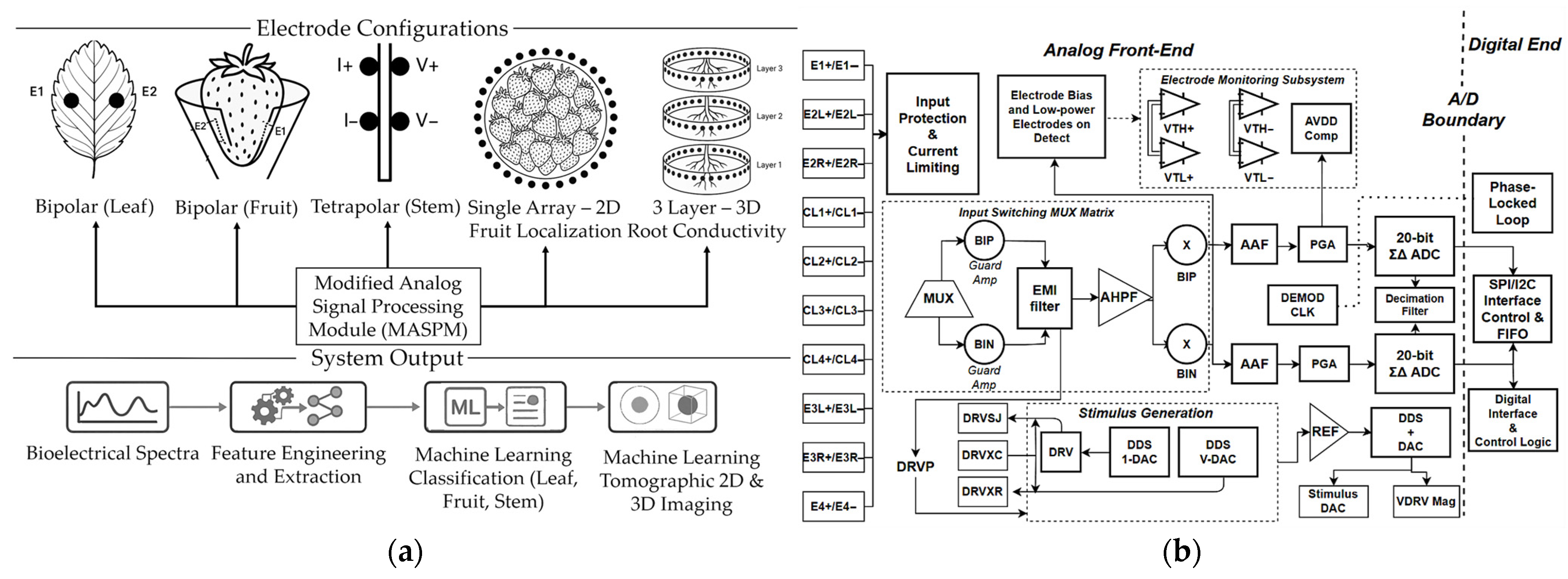
Figure 2 presents the measurement system with the modified analog signal-processing module (MASPM) architecture that provides multi-configuration switching and signal processing for all measurement modes.
Figure 2a illustrates the electrode configurations. Bipolar electrodes were used for leaves and fruits, tetrapolar for stems. A single 32-electrode array for 2D fruit tomography, and triple-layer arrays for 3D root tomography. Together, these setups allow the system to convert bioelectrical signals into both physiological state classifications and spatial conductivity maps. Figure 2b presents the internal architecture of the MASPM. Differential electrode pairs are connected through input protection and current-limiting circuits. A switching matrix with multiplexers and guard amplifiers provides noise reduction and EMI filtering. The electrode-monitoring unit ensures reliable connections by detecting voltage thresholds with supply compensation. Signal processing is carried out through dual channels, each employing anti-aliasing filters and programmable gain amplifiers feeding ADCs. Bioelectrical signals are simultaneously measured using cross-multiplication with phase-locked demodulation. The stimulus generation block uses dual DDS modules to deliver current and voltage outputs with programmable reference control, maintaining stable excitation across different plant tissues. Finally, digital control coordinates timing, data filtering, and communication protocols.

2.2. Multimodal Hardware Integration for Spectroscopic and Tomographic Operation

The bioelectrical measurement system employs configurations sharing core circuitry. Bipolar configuration (Supplementary Figure S1) implements two-electrode measurements for homogeneous tissues. Its H-bridge circuit establishes reference voltages using 4.7 kΩ resistors (0.1% tolerance), while OPA333 amplifiers (Texas Instruments, Dallas, TX, USA) minimize tissue loading. Protection combines SMAJ5.0A TVS devices (Littelfuse, Chicago, IL, USA) (±5 V clamp) with EMI filtering. INA333 instrumentation amplifiers (Texas Instruments, Dallas, TX, USA) (CMRR >100 dB) with sense resistors enable overcurrent detection. The circuit interfaces with MASPM through DRVP/DRVN and BIP/BIN signal paths, controlled via SPI/I2C protocols. Signal conditioning employs three-stage RC networks (HPF, BPF, LPF) with corner frequencies and −60 dB/decade roll-off (detailed in Supplementary Figure S4). The tetrapolar configuration (Supplementary Figure S2) separates current injection from voltage sensing to eliminate polarization artifacts in heterogeneous stem tissues. A Howland current source utilizing OPA2376 amplifiers (Texas Instruments, Dallas, TX, USA), controlled by an AD5171 potentiometer (Analog Devices, Wilmington, MA, USA) and an AD5663 DAC (Analog Devices, Wilmington, MA, USA), delivers programmable currents (1 µA–10 mA) with ±12 V compliance. Voltage paths use OPA333 buffers (Texas Instruments, Dallas, TX, USA) with guard drive techniques, reducing stray capacitance. Current monitoring combines INA333 (Texas Instruments, Dallas, TX, USA) with a 24-bit ADS1256 ADC (Texas Instruments, Dallas, TX, USA) for resolving differential voltages across channels. ADG1404 multiplexers (Analog Devices, Wilmington, MA, USA) manage electrode routing and source switching. Both spectroscopic configurations utilize ADG714 multiplexers (Analog Devices, Wilmington, MA, USA) for calibration resistor switching, AD9833 DDS (Analog Devices, Wilmington, MA, USA) with AD835 multipliers (Analog Devices, Wilmington, MA, USA) for frequency generation, and an STM32H755 dual-core processor (STMicroelectronics, Geneva, Switzerland). The Cortex-M7 core handles current source control, while the Cortex-M4 manages voltage acquisition and data processing, enabling parallel operation. Signal conditioning includes OPA4188 (Texas Instruments, Dallas, TX, USA) and REF5025 (Texas Instruments, Dallas, TX, USA) for reference stability. The system operates from linear ±12 V supplies (LM7812/7912, STMicroelectronics, Geneva, Switzerland) with a <10 mVpp ripple.
For tomographic configuration, it employs 32-electrode arrays where four ADG1408 multiplexers cascade through dual ADG1409 multiplexers (Supplementary Figure S3), with an XC7A35RT controller managing switching, phase-locked sampling, and I/Q demodulation for reconstruction. The selection of 32 electrodes and phantom dimensions was not arbitrary but rather determined through optimization of measurement sensitivity and spatial resolution. This penetration depth is governed by the following equation:
δ p e n e t r a t i o n = 1 π f μ σ ,
where f is the excitation frequency, μ is the magnetic permeability, and σ is the nominal plant tissue conductivity. Reported tissue conductivity values range from 0.05–0.2 S/m based on physiological changes [94,95]. The 70 mm radius ensures current distribution based on the two-fold penetration depth criterion [96], while the 60 mm height provides 924 cm3 volume. Within these constraints, 32 electrodes were calculated via sensitivity optimization analysis, maximizing volume [97,98]. The angular electrode spacing is computed as follows:
θres = 360°/Nelectrodes,
where θres represents the angular resolution between adjacent electrodes, and Nelectrodes denotes the total electrode count. This arrangement aims to balance circumferential coverage with the system. The parallelotope volume for measurement sensitivity, calculated as follows:
V p a r a l l e l o t o p e =   ( d e t ( J ( M ) J ( M ) ) ,
where J(M) represents the Jacobian matrix sensitivity matrix of selected measurements M, with each row containing partial derivatives ∂Ui/∂σj, relating voltage measurements Ui to conductivity changes ∂σj. The superscript “*” represents a conjugate transpose, and det indicates the determinant operation. This correlates voltage measurements to conductivity changes. Theoretical spatial resolution is estimated as follows:
Δsspatial = rphantom × sin(θres/2)
where Δsspatial is the minimum resolvable feature size, rphantom is the phantom radius (70 mm), and θres/2 accounts for the half-angle between adjacent electrodes. This equation derives from geometric considerations where the spatial resolution depends on both the angular separation and radial distance from the array center. Current density distribution was modeled to evaluate penetration characteristics:
J ( r , θ ) = ( I inj / 2 π r )   ×   Σ n = 1 N / 2 cos ( n ( θ     θ inj ) )   ×   e ( n r / R ) ,
where J(r,θ) is the current density at coordinates (r,θ), Iinj is the injection current, r is the radial distance from the center, θ is the angular position, θinj is the injection electrode angular position, n is the harmonic number, N is the total electrode count, and R is the phantom radius. This ensures current reaches the phantom center (r = 0) with <5% sensitivity loss compared to the boundary, consistent with prior simulations [96,97,98]. These computations generate conductivity slices and volumetric reconstructions (Figure 3).

2.3. Development of Calibration Network for Multi-Configuration and Multimodal Operation

The challenge in plant bioelectrical measurements is that different organs have vastly different electrical properties. The adaptive calibration network solves this problem by enabling one system to accurately measure all plant organs. The network centers on a precision resistor matrix that automatically adjusts to match each organ’s measurement range (Supplementary Figure S4). Four programmable control signals manage this adaptation. PLANT_CAL_EN selects the organ type, while GSR_LOAD_EN handles conductivity variations. PLANT_GSR_RSEL sets the appropriate measurement range. Together, these signals allow the system to reconfigure itself for accurate measurements. To maintain non-invasive measurements, the network carefully manages electrode polarization. DC blocking uses 100 nF capacitors paired with 100 kΩ bias resistors to prevent polarization buildup. For frequency-specific responses, the system employs 10 nF and 10 kΩ networks to optimize α-dispersion measurements in the cellular response range, while 100 nH inductors with 4.7 kΩ resistors extend performance into higher frequencies of β-dispersion. All these parameters have been validated against traceable standards, including ENOB, noise floor, and THD specifications, ensuring consistent results across different organs and measurement sessions.

2.4. Electrode Design, Selection, and Performance Evaluation

The electrodes used to plant tissue connection influence the measurement quality. This requires the selection of optimal electrode types and configurations for both tomographic and spectroscopic evaluation. For tomographic measurements, electrode materials were selected based on established agricultural applications. Gold-plated electrodes [99,100,101,102,103] were evaluated as the reference, given their proven conductivity and stability. Ag/AgCl electrodes [104,105,106,107,108] were included based on widespread adoption and cost-effectiveness. Performance was compared using the spatial resolution through point spread function (PSF) analysis, and current uniformity via FEM. For spectroscopic measurements, electrode selection followed established plant bioelectrical studies. For leaf measurements, hydrogel surface [109,110,111] and Ag/AgCl [112,113,114,115] electrodes were employed. For fruit measurements, adhesive patches [116,117,118,119] and screen-printed electrodes [120,121,122,123] for non-invasive surface contact were employed. For stem measurements, Ag/AgCl clip-type [124,125,126,127] and clamp-type [128,129,130,131] configurations were employed. Each electrode type was evaluated in both bipolar and tetrapolar configurations to identify optimal organ-specific pairings. Performance metrics included the signal-to-noise ratio (SNR), contact resistance, measurement repeatability, and contact area.

2.5. Sample Collection and Experimental Design

Having established the complete measurement platform, validation experiments were conducted using strawberries (Fragaria × ananassaSweet Charlie’) to demonstrate sensor system performance. Experiments were conducted at an existing controlled farm in Bukal, Majayjay, Laguna, the Philippines (14.1447° N, 121.4723° E, 750 m elevation). Environmental conditions were carefully controlled throughout the experiments. The measurement protocol drew from established bioelectrical studies and incorporated advice from local farmers familiar with the site’s microclimate. Three Three HOBO U23-001 Pro v2 data loggers (Onset Computer Corporation, Bourne, MA, USA; ±0.21 °C, ±2.5% RH) monitored conditions continuously, recording every 5 min. Measurements were conducted between 9:00 and 11:00 a.m. for several reasons. This window minimizes circadian-related measurement drift [132]. Local farmers confirmed temperature stability during these hours, which preliminary tests validated. Ten test runs showed that the temperature varied less than 2 °C during morning hours, compared to over 5 °C variations in the afternoon. The protocol successfully maintained conditions within the target range of 24–27 °C and 75–85% relative humidity. These ranges match those used in previous strawberry bioelectrical studies [85,86,120]. Measurement stability was verified through repeated testing. Twenty reference measurements yielded a coefficient of variation below 2.5% across the environmental range, consistent with the expected 2–3%/°C temperature coefficient for plant tissues. The stable electrode–tissue contact parameters shown in the results Section 3.1 further confirmed the protocol’s effectiveness.
For leaf water status classification, 400 leaf samples were collected from plants across eight beds. Water treatments utilized controlled irrigation: “well-watered” plants (n = 200) received 250–300 mL daily, maintaining more than 75% field capacity; “water-stressed” plants (n = 200) received 100–150 mL every third day, maintaining less than 40% field capacity. For validation, the soil moisture was monitored using capacitance probes at a 10 cm depth.
For market-based fruit categorization, two types were identified: (1) physiologically ripe (PR) fruits, with advanced internal changes that enhance flavor but shorten shelf life, and (2) commercially mature (CM) fruits, retaining firmness [133] and transport and storage potential [92]. These differences are natural from the farm’s environmental conditions: (1) Sunlight exposure accelerated internal physiological maturation, with bed-edge plants yielding more PR fruits and shaded inner plants producing CM fruits. (2) Primary crown fruits changed earlier than those from lateral branches. (3) Plants near irrigation emitters produced more PR fruits than those at line ends. (4) Topography is also a factor, with elevated and well-drained areas favoring PR fruits, while lower, wetter areas favored CM fruits. A total of 300 fruits were collected with the help of experienced farmers in identifying the categories using a visual and firmness approach. PR fruits showed red color (including the tip), strong aroma, and softness. CM fruits displayed 85–90% red color (excluding the tip) and remained firm. For further validation, established postharvest assessment methods were employed. PR samples (n =150) averaged 22–24 °Brix with a firmness of 0.8–1.2 N, while CM samples (n =150) averaged 18–20 °Brix with a firmness of 2.0–2.5 N.
For stem productivity assessment, 200 samples were collected from plants that underwent a seven-week-long controlled-management period to create productivity categories (high vs. low productive). Productivity was generated using combined treatments. High-productive stems received daily watering (250–300 mL), weekly fertilization (20 g), and normal flowering, while low-productive stems received reduced watering (100–150 mL every third day), no fertilizer, and weekly flower removal. These treatments created clear phenotypic contrasts for system validation. Single-factor experiments were not conducted, as the focus of this study was classification of distinct productivity groups rather than isolating individual variable effects. For root zone conductivity mapping, four plants were utilized for tomographic mapping capabilities through contrasting root conditions. This validation employed a 2 × 2 factorial design (two irrigation levels × two replicates) monitored over seven weeks to allow for complete root system development. Plants were grown in the system’s phantom with the same organic medium used in farm beds, ensuring relevance to field conditions. Since the roots were grown in the system’s phantom, both water and fertilizer inputs were computationally reduced to prevent baseline conductivity shifts and ensure measurements reflected genuine physiological changes. Two plants received normal care, with 100 mL water daily with diluted 5 g organic fertilizer. Then, the other two plants underwent controlled stress, with only 50 mL daily, with no fertilizer. These treatment levels were calibrated to create detectable conductivity contrasts between healthy and stressed root systems while preventing plant death.
For conductivity localization for postharvest fruit sorting, tomographic validation focused on detecting fruits with internal defects, specifically hollow centers that appear normal externally but contain voids detectable through conductivity differences. Also, natural factors on farm conditions produced these defects: (1) Sun-exposed fruits create hollow centers despite normal appearance. (2) Irrigation inconsistencies at 15–20% water variation along drip lines caused cycles of dehydration and rehydration that collapsed internal tissues while external surfaces recovered. Uneven fertilizer distribution created nutrient-poor zones where fruits developed internal cavities due to insufficient ion availability. A total of 100 fruits were strategically harvested with guidance from an experienced farmer. Following 48 h of controlled storage at 25 °C and 35% RH to accentuate quality differences, bioelectrical prescreening via conductivity measurement was conducted. Fruits with high conductivity (≥0.5 S/m) were classified as good quality, indicating intact cellular structure, while those with low conductivity (≤0.3 S/m) were classified as poor quality, characteristic of internal voids. This correlated with visual inspection, confirming external appearance often masked internal defects detectable only through bioelectrical assessment. The prescreened fruits were allocated into three primary experimental configurations using 70 fruits, while the remaining 30 fruits were used for preliminary algorithm optimization and repeatability testing. Configuration 1 used 22 uniform good-quality fruits as the baseline validation; Configuration 2 assessed boundary detection with 10 poor-quality fruits centered among 12 good-quality fruits; and Configuration 3 tested resolution limits with 26 smaller poor-quality fruits arranged at a higher density. This distribution provided sufficient replication across scenarios to detect medium effect sizes (Cohen’s d ≈ 0.5) with ≥80% statistical power, ensuring robust comparative analysis.

2.6. Bioelectrical Measurements Organ-Specific Protocols

The 1000 samples (400 leaves, 300 fruits, 200 stems, 100 postharvest fruits) from Section 2.5 were measured by the bioelectrical system using electrode configurations determined in Section 2.4. The methodologies are adapted from validated bioelectrical measurement approaches and tailored to strawberry-specific applications.
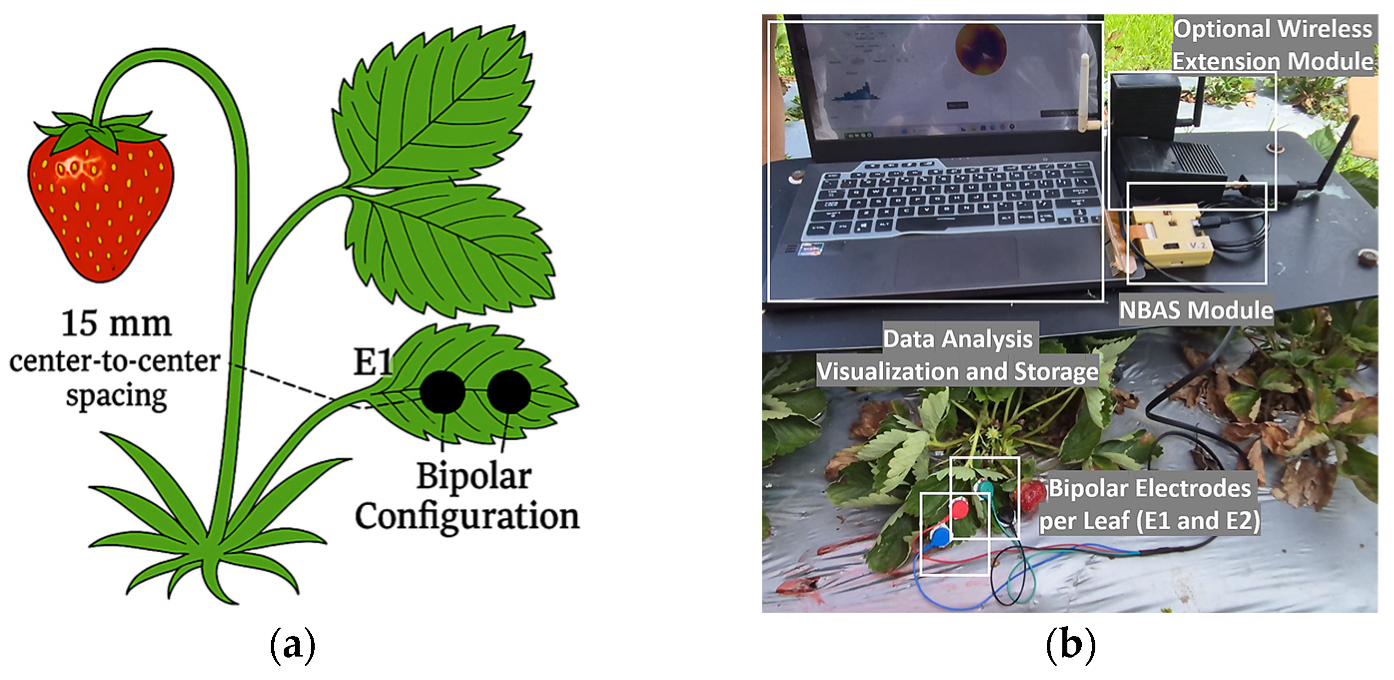
For leaf measurements, the hydrogel electrodes selected in Section 2.4 were specifically H-series models with integrated adhesive, comparable to Covidien and Medtronic types that represent the standard in plant bioelectrical studies. These particular specifications were chosen because the electrodes adhere through hydrogen bonding with leaf moisture, eliminating additional adhesives that could interfere with measurements. The measurement protocol involved surface preparation with deionized water cleaning and gentle drying, followed by application of Spectra-electrode gel to ensure consistent contact on irregular leaf surfaces. Unlike recent sensors designed for continuous growth monitoring [112,115], this study focused on measurement stability, physiological correlations, and absence of bioelectrical drift, rather than direct growth impacts. Figure 4 illustrates the electrode arrangement (Figure 4a) and the actual field implementation (Figure 4b). Bipolar electrodes were positioned on abaxial leaf surfaces with 15 mm center-to-center spacing.
Fruit measurements employed screen-printed electrodes embedded in conical substrates with a 60° apex angle, using bipolar configuration. This conical design was specifically chosen to conform to the fruit’s curved surface, distributing pressure evenly to prevent damage while maintaining consistent electrode spacing. The electrodes were positioned at the fruit’s equatorial plane. Each electrode’s contact area of approximately 3 mm2 was small enough to accommodate surface irregularities yet large enough to ensure proper current distribution across the tissue. The conical substrate design proved particularly effective for measuring attached fruits without causing tissue damage, an important consideration for non-invasive assessment. Figure 5 shows both the electrode arrangement schematic (a) and the actual field implementation (b).
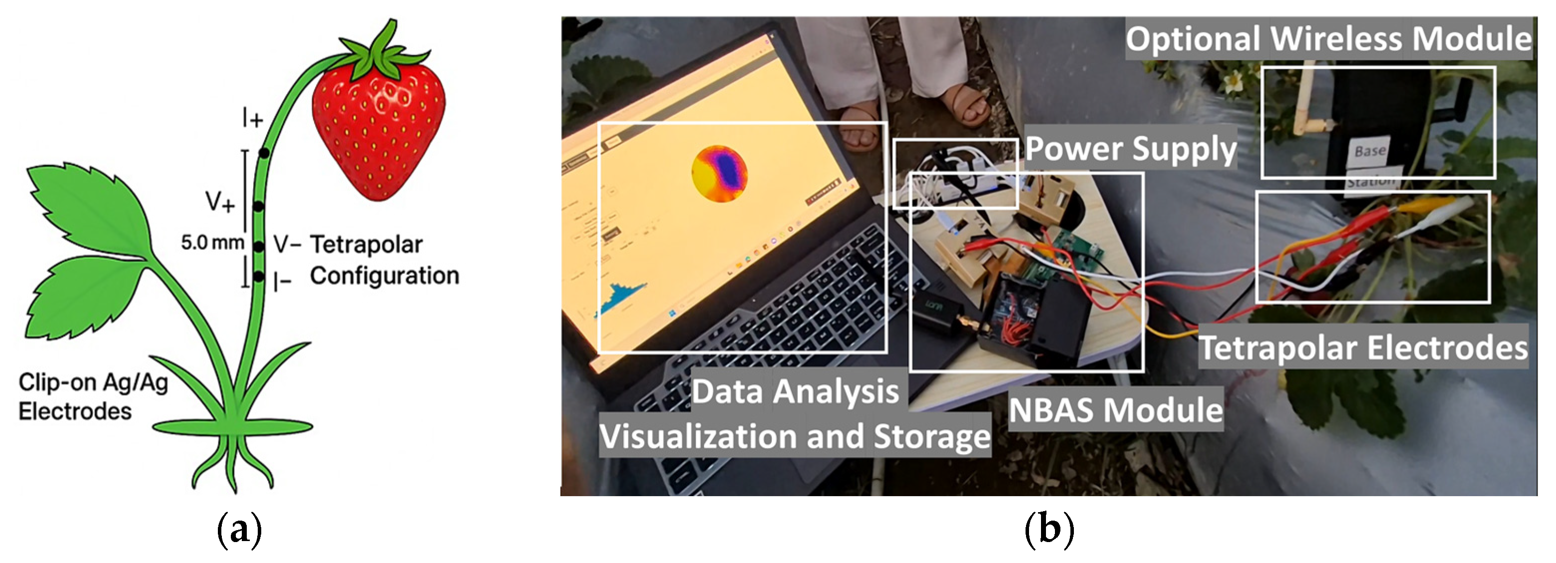
For stem measurement, clip-on Ag/AgCl electrodes in tetrapolar configuration were selected based on their superior performance detailed in Section 3.2. This configuration excelled specifically because the stem’s complex structure, with conductive vascular bundles requires separation of current injection from voltage sensing to minimize interface effects. The tetrapolar arrangement achieves this separation. Figure 6 illustrates the electrode arrangement in stem (Figure 6a) and the actual field implementation of it (Figure 6b). The electrode was placed linearly with 5 mm electrode spacing suitable for 5–8 mm diameter strawberry stem. This enabled complete current penetration while maintaining injection measurement separation, capturing the dual dispersion characteristics of stem tissues. While the clip-on mechanism of the electrodes provided consistent pressure accommodating diameter variations.
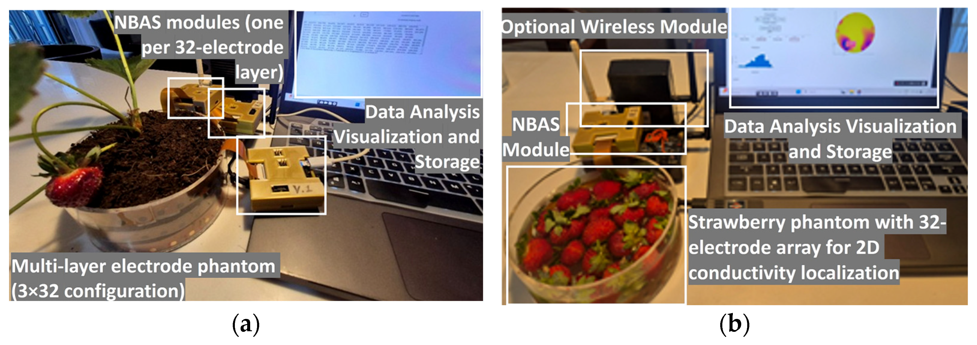
Tomographic measurements utilized the computed 32-electrode array configuration, with gold-plated electrodes selected through comprehensive evaluation, including current uniformity via FEM to validate conductivity field distribution and spatial resolution through point spread function (PSF) analysis to verify localization accuracy and temporal stability. These measurements per frequency through adjacent-pair current injection and phase-locked FPGA acquisition, enabled both temporal monitoring and spatial reconstruction. Figure 7 illustrates the experimental setups for both 3D root conductivity mapping (Figure 7a) and 2D postharvest fruit quality assessment (Figure 7b).

2.7. Data Preprocessing Filtering and Normalization

Raw bioelectrical data requires preprocessing to remove artifacts. Noise suppression employed Savitzky–Golay filtering through localized polynomial fitting:
y j s m o o t h = 1 N i = m 1 2 m 1 2 c i y j + i ,
where y j s m o o t h represents the filtered value at frequency point j, calculated as the weighted average of N neighboring points using polynomial coefficients c i . The summation extends from i = −(m − 1)/2 to (m − 1)/2, where m defines the window size. The selection of m balanced noise reduction and feature preservation based on each organ: 5-point windows for leaves were selected to preserve sharp frequency transitions in narrow spectral peaks, 7-point for fruits broader spectral patterns, and 9-point for stems capturing dual-dispersion transitions. For anomaly detection, a modified z-score was utilized to identify electrode contact losses and transient interference:
M i = 0.67 x i x ~ M A D ,
This metric compares each measurement x i against the local median x ~ , normalized by the median absolute deviation ( M A D ) , with 0.67 ensuring equivalence to standard deviation units under normal distribution assumptions. Tissue-specific thresholds were established based on plant organ structural complexity: 3.0 for uniform leaf tissue, 3.5 for moderately variable fruits, and 4.0 for complex stem vascular tissue. Standardization through z-score normalization addressed electrode contact variations:
P f n o r m a l i z e d = P f μ P σ P ,
where bioelectrical parameters P(f) at each frequency are centered by their dataset mean μ P and scaled by standard deviation σ P , producing zero mean unit variance distributions, enabling direct comparison across plant organ samples regardless of measurement scales.

2.8. Key Frequency Selection and Statistical Validation of Organ-Specific Ranges

After ensuring data quality through preprocessing, multi-frequency bioelectrical measurements were reduced to identify the most physiologically informative regions for each organ type. The selection process began with statistical evaluation of parameter differences at each frequency point using two-sample Welch’s t-tests:
t = ( μ 1 μ 2 ) / ( s 1 2 / n 1 + s 2 2 / n 2 ) ,
where μ1 and μ2 represent mean parameters for contrasting physiological states, s12 and s22 are sample variances, and n1 and n2 are group sizes. A Benjamini–Hochberg false discovery rate correction-controlled Type I error inflation from multiple comparisons was adopted, retaining frequencies with adjusted p-values less than 0.05. Significance was evaluated through Cohen’s d effect size to distinguish substantial from negligible differences, expressed as follows:
d   = μ 1   μ 2 s p o o l e d ,
This metric expresses group mean differences in pooled standard deviation units (μ1 and μ2), providing scale-independent effect magnitude. And the pooled standard deviation accounts for unequal variances through weighted averaging:
s p o o l e d =   n 1 1 s 1 2 +   n 2 1 s 2 2 \ o v e r   n 1 + n 2 2 ,
where s12 and s22 represent group variances weighted by their respective degrees of freedom (n1 − 1 and n2 − 1). This provides an unbiased estimate of population standard deviation when group sizes differ. Only frequencies with d ≥ 1.0 were retained, indicating separation greater than one standard deviation between states, ensuring detection beyond measurement variability. The final selection criterion employed mutual information ( M I ) analysis. Unlike correlation coefficients with limited linear relationships, M I captures both linear and nonlinear dependencies. M I is expressed as follows:
M I X ; Y =   x , y p x , y log p x , y p x p y ,
This measures uncertainty reduction about physiological state Y when the bioelectrical parameter X is known at each frequency. Joint probability p(x,y) captures parameter-state co-occurrence, while the product p(x)p(y) represents the independence expectation. Larger logarithmic ratios indicate stronger associations. Frequencies meeting all three criteria were selected for feature generation: statistical significance after FDR correction, effect size ≥ 1.0, and normalized mutual information ≥ 0.7. This integrated approach was designed to balance statistical rigor with physiological relevance.

2.9. Bioelectrical Feature Extraction for Physiological Interpretation

With optimal frequencies identified, the bioelectrical measurements underwent systematic feature extraction. This process transformed measurements into physiologically interpretable parameters, avoiding direct reliance on raw data. Central to this transformation is the Cole–Cole model, which describes the frequency-dependent bioelectrical response of biological tissues through physically meaningful parameters:
Z ω =   R + R 0   R 1   +   j ω τ α
where Z ω is the complex impedance at angular frequency ω, R0 represents resistance at low frequencies reflecting extracellular pathways, R∞ represents resistance at high frequencies representing total tissue resistance, τ represents the relaxation time that indicates how quickly cells respond to electrical changes, and α is the dispersion parameter ranging from 0 to 1, which indicates tissue homogeneity. Derived parameters include membrane capacitance ( C m ), indicating structural integrity and expressed as follows:
C m = τ R 0     R
reflecting the charge storage capability, with higher values suggesting intact membranes. Characteristic frequency ( f c ) identifies optimal measurement points, expressed as follows:
f c = 1 2 π τ
representing the resistive to capacitive transition where cellular effects dominate. Spectral energy distribution (P(f)) quantifies how magnitude varies across the frequency spectrum
P (f) = |Z(f)|2/Σ|Z(f)|2,
where |Z(f)| is magnitude at frequency f, normalized by total spectral energy. This reveals which frequencies contain maximum physiological information. While spectral centroid ( f c ) locates the frequency response center:
f c = f   × P S D f P S D f
Lower values indicate slow cellular processes, and higher values suggest faster responses. The spectral bandwidth (BW) measures the spread around this center:
B W   =   f     f c 2 × P S D f P S D f
Narrow bandwidths indicate uniform tissue; wide bandwidths suggest heterogeneous structures. Dynamic analysis examines rapid bioelectrical changes with frequency, revealing tissue complexity:
d Z d f = Z f   +   Δ f   Z f     Δ f 2 Δ f
Steep derivatives indicate tissue boundaries, while gradual slopes suggest uniform regions. Large derivatives occur at critical frequencies where conduction transitions from ionic to electronic pathways. Morphological analysis extracts patterns from reactance vs. resistance trends. Extreme phase angles indicate capacitive and resistive dominance at physiological transitions.

2.10. Optimal Parameter Identification and Feature Selection

Following feature extraction in Section 2.9, this section will discuss the selection of the most discriminative features from the expanded feature pool for organ-specific classification. Recursive Feature Elimination with Cross-Validation (RFECV) was implemented to remove features with minimal classification contribution. This was evaluated through fivefold cross validation. Feature retention followed these criteria: (1) feature had to yield at least 1.0% improvement in accuracy, (2) the training to validation performance gap was kept below 3.0% to avoid overfitting, and (3) individual feature importance exceeded 5.0% of the model contribution, as determined by permutation testing. Redundancy was addressed via hierarchical clustering of correlation, with features showing correlation coefficients above 0.85 considered redundant. Among these, only the representative feature with the highest M I was retained, computed as follows:
M I F ; Y =   p f , y l o g p f , y p f p y
This metric captures the nonlinear dependencies between features F and target labels Y that linear correlation overlook.
This selection process identified organ-specific optimal features reflecting physiological traits: fruits 12 features (sample-to-feature ratio 25:1), leaves 10 features (ratio 40:1), and stems 8 features (ratio 25:1). This feature subsets enable also balance classification performance with efficiency.

2.11. Multi-Frequency Tomographic Data Acquisition Method

While spectroscopic measurements utilized selected frequencies for feature extraction, tomographic imaging required comprehensive frequency sweeps to enable spatial conductivity reconstruction. Frequency sweeps were designed based on penetration depth calculations for the standard phantoms detailed in Section 2.4, and computed as follows:
δ f = 1 π f × 4 π × 10 7 × σ 0 1 + j ω τ α 1 ,
where δ f represents the penetration depth (m), f is the excitation frequency (Hz), σ 0 is conductivity (S/m), ω = 2πf represents the angular frequency, τ is the Cole–Cole relaxation time (s), and α represents the dispersion parameter. Logarithmically spaced frequency points were implemented to optimize sampling, expressed as follows:
f k = f m i n × 10 k · log 10 f m a x f m i n N 1    
This spacing ensured adequate coverage while maintaining acquisition times less than 100 ms per frequency. Spatial data organization employed adjacent electrode current injections. For the 32-electrode array, systematic rotation yields the following matrix:
Z s p a t i a l i , j = V m e a s u r e d i , j I i n j e c t e d i , j × exp j ϕ i , j ,
where V m e a s u r e d i , j represents differential voltage between electrode pair i , j , I i n j e c t e d i , j is constant current, and ϕ i , j denotes the measured phase angle. The frame synchronization maintains coherence across the multi-electrode array through the equation below:
t s y n c k =   k   ·   T f r a m e +   ϕ o f f s e t ,
where T f r a m e represents the frame acquisition period (s), and ϕ o f f s e t compensates for system propagation delays, achieving a synchronization accuracy of ± 10 μs, which is essential for demodulation across all channels. The reconstruction domain employed cylindrical coordinates with homogeneous Neumann boundary conditions (∂ϕ/∂n=0), at phantom walls and mixed conditions at electrode interfaces. Multi-frequency data integration was implemented using weighted averaging, calculated as follows:
σ r e c o n s t r u c t e d x , y , z = k = 1 50 w k ·   σ k x , y , z k = 1 50 w k ,
where frequency-specific weights ( w k ) combine SNR with penetration depth characteristics. The weighting scheme assigns higher importance to low frequencies for central region reconstruction and emphasizes higher frequencies for boundary definition, represented as follows:
w k = S N R f k × δ f k β ,
with β = 0.5 for balanced depth and resolution. This multi-frequency approach provides the spatial richness necessary for conductivity differentiation.
To validate these penetration depth calculations and frequency-dependent weightings, a comprehensive evaluation was performed through established methods of assessment: (1) penetration depth via progressive electrode measurements, (2) spatial resolution through point spread function analysis (PSF), (3) sensitivity via conductivity perturbation tests, and (4) calculation of a performance index

2.12. Algorithm Selection and Implementation

Following feature extraction and protocol establishment, algorithms were selected to match each organ’s data characteristics and reconstruction requirements. For classification tasks, four machine learning approaches were evaluated. TabPFN was chosen as the primary classifier due to its effectiveness with the extracted features from raw spectral data. Three gradient-boosting methods provided comparative baselines: CatBoost for its robust handling of mixed features and overfitting resistance, LightGBM for computational efficiency with feature sets, and XGBoost as the established benchmark with strong regularization capabilities. Tomographic reconstruction required different approaches for 2D and 3D applications. The 3D root conductivity mapping employed a U-Net architecture, whose skip connections excel at reconstructing detailed conductivity patterns from sparse electrode measurements. Filtered Back Projection (FBP) served as the analytical standard for comparison. For 2D fruit quality assessment, SIRT was selected for its stability with circular electrode arrays and effective noise suppression. Additionally, a Modified Back-Projection (MBP) algorithm was developed specifically for detecting defect boundaries:
σ * = a r g m i n σ J σ Δ V 22 + λ σ 1
where σ * is the conductivity distribution, J is the Jacobian sensitivity matrix, Δ V is the measured voltage differences, λ controls the regularization strength, and σ 1 is the L1 norm of the conductivity gradient. The σ preserves edges while suppressing noise, essential for localizing defect boundaries.

2.13. System Validation and Physiological Correlation

Following standard bioelectrical protocols, we employ (1) classification accuracy (%) as the primary performance metric and (2) correlation (r) to validate physiological relevance only. Leaf validation employed computation of the relative water content (RWC) through gravimetric analysis. The direct relationship between RWC and bioelectrical properties is well-established through ionic conductivity changes, evidence correlations across multiple crop species [134,135]. It is computed as:
  R W C   % = W f   W d W t   W d × 100 ,
where W f , represents fresh weight, W d , represents dry and W t represents turgid weights. W f was recorded immediately after harvest. W t , was recorded after leaves were immersed in distilled water for 4 h at 20 °C. After hydration, surface water was blotted by filter papers before weighing. W d , followed 12 h desiccation at 70 °C. Constant mass was confirmed every hour checking for less than 0.005 g threshold change. Values outside 50–100% indicated measurement error. Replicate variation remained below 5%. Key requirements included turgid weight exceeding fresh weight, complete surface water removal, and gentle sample handling. Second validation is chlorophyll content. This was measured via SPAD-502Plus that affect cellular capacitance and ion transport. SPAD validations demonstrate high correlations with water stress indicators [135]. The temporal precedence of electrical changes over chlorophyll decline provides additional validation support. Leaf thickness demonstrates correlations with bioelectrical parameters through effects on current distribution pathways.
For fruits, features were correlated with reference metrics using established strawberry assessment protocols, as consistently applied in previous studies. Sugar content (°Brix) was measured at the equatorial plane, 90° from the ventral suture, validated for representative sugar content in strawberry [136]. Measurements were made using an Atago PAL-3 refractometer. Firmness (N) was measured using a TA.XT2 texture analyzer with a 5 mm probe applied at two opposite cheeks on the equatorial plane [133].
Stem validation employed correlation with its diameter and attached fruit quality (using fruit validation metrics). Productive stems yielded normal-sized fruits with standard quality metrics, and non-productive stems produced smaller, poorly developed fruits. A correlation analysis of all plant organ’s optimal features and established reference measurements employed Pearson coefficients, calculated as follows:
r x y = i = 1 n x i   x - y i   ȳ i = 1 n x i   x - 2 i = 1 n y i   ȳ 2 ,
where selected bioelectrical features ( x i ) (12 features in fruits, 10 features in leaves, 8 features in stems) correlated with validation metrics y i . Multiple comparison correction employed Bonferroni adjustment with α = 0.05/m for m parameter-metric pairs; 72 for fruits (12 features × 6 quality metrics), 30 for leaves (10 features × 3 quality metrics), and 56 for stems (8 features × 7 quality metrics). Reference measurements followed standardized protocols with uncertainties documented in Table A1.
For tomographic localization quality, relative residual error was calculated as the normalized difference between measured and reconstructed voltage patterns following established tomography protocols.

3. Results

This section presents the experimental results for the validation of the sensor system, which was comprehensively evaluated and calibrated using model crops, strawberries (Fragaria × ananassa ‘Sweet Charlie’).

3.1. Comparative Performance of Electrode Types and Configurations for Organ-Specific Measurements

Electrode performance varied significantly across plant organs, reflecting their distinct properties (Table 1). Values represent mean ± standard deviation from measurements per configuration. While comprehensive visualization of electrode performance evaluation across all configurations is provided in Supplementary Figure S5.
These stable parameters confirm the non-invasive nature of the contact-based measurements. Tissue damage would normally produce measurement drift, degrade SNR, or increase measurement variation, but none of these effects were observed across all samples. Established electrodes and configurations from previous studies was also employed and validated for non-invasiveness. Hydrogel patches providing minimize stress on leaves, screen-printed electrodes shown to be safe for repeated fruit assessments, and Ag/AgCl electrodes validated for continuous stem monitoring without vascular damage. Combined with stable repeatability (CV < 2.5%), these results verify that the tissue integrity was preserved.
The tomographic reconstruction required different electrode configurations to ensure uniform current distribution across 32-electrode arrays. Both gold-plated and Ag/AgCl electrodes were evaluated in 2D single-layer and 3D three-layer configurations. Gold-plated electrodes outperformed Ag/AgCl across all metrics: spatial resolution reached 5.5 ± 0.4 mm versus 6.3 ± 0.5 mm in 2D configuration and 5.9 ± 0.4 mm versus 6.8 ± 0.5 mm in 3D configuration, while current injection uniformity achieved 89.2 ± 1.8% versus 83.7 ± 2.4%, as validated through PSF analysis and FEM modeling in EIDORS. This baseline performances improved further with frequency optimization discussed in the next section.

3.2. Optimal Multi-Scale Frequency Across Plant Organs

The frequency selection protocol in Section 2.8 identified which frequencies produced the strongest bioelectrical signatures for each organ. Statistical analysis revealed distinct dispersion patterns that proved useful for both spectroscopic and tomographic measurements. These optimal frequency ranges matched the dispersion characteristics previously documented in plant studies [62,63,64]. Table 2 lists the specific frequencies selected for spectroscopic measurements, while Supplementary Figure S6 shows statistical validation and selection process.
All selected frequencies show p < 0.001 (statistical significance), Cohen’s d > 1.0 (effect size), and mutual information MI > 0.7. This validation confirms the representation of changes, not random noise or equipment errors.
While in the tomographic frequency selection, detailed in Section 2.11, different frequency ranges were evaluated based on penetration depth, spatial resolution, sensitivity, and overall performance. Lower frequencies penetrated deeper but gave coarser resolution, while higher frequencies provided finer resolution with shallower penetration, as shown in Supplementary Figure S7. Table 3 presents the quantitative results from this evaluation.
Table 3 reveals that successful conductivity reconstruction requires balancing multiple parameters rather than optimizing any single metric. The performance index (PI) captures this balance and identifies optimal frequency ranges. For 3D root zone mapping, three distinct patterns emerged. The lowest frequencies (0.01–0.1 kHz) penetrated deepest at 70 mm with 0.85 sensitivity, but their poor 6.2 mm resolution merged adjacent zones, making it impossible to distinguish localized water and nutrient uptake regions (PI = 0.68). Mid-range frequencies (0.1–10 kHz) offered the best compromise: 48 mm penetration reached active root zones, 3.8 mm resolution separated individual conductivity gradients, and 0.71 sensitivity detected meaningful changes, yielding the highest PI of 0.92. Higher frequencies (10–100 kHz) achieved 2.7 mm resolution but penetrated only 22 mm, missing deeper variations entirely, with sensitivity dropping to 0.34 (PI = 0.42). Similar trade-offs appeared in 2D fruit localization. Low frequencies (0.1–1 kHz) detected conductivity differences with 0.78 sensitivity but blurred fruit boundaries at 5.1 mm resolution (PI = 0.72). The middle range (1–50 kHz) again proved optimal, balancing 28 mm penetration for complete fruit coverage, 3.1 mm resolution to separate quality regions, and 0.62 sensitivity above discrimination threshold (PI = 0.94). High frequencies (50–500 kHz) provided 2.1 mm resolution but penetrated only 12 mm with an inadequate 0.35 sensitivity (PI = 0.35). These results establish 0.1–10 kHz as optimal for root zone monitoring and 1–50 kHz for fruit quality assessment.

3.3. Optimal Features for Classification

Feature extraction and selection (Section 2.9 and Section 2.10) identified organ-specific parameter subsets achieving optimal classification, all with statistical significance (p < 0.001). Table A2 lists these parameters and weights. Supplementary Figure S8 shows the RFECV results for optimal feature selection. Each organ has different bioelectrical features matching its physiology. Fruits have 12 parameters dominated by high-frequency measurements (50–100 kHz) that detect ripening-related membrane changes and sugar accumulation. Leaves have 10 features primarily at low frequencies (0.5–5 kHz), where water stress directly affects ionic conductivity. Stems have 8 parameters, including two separate Cole–Cole relaxation times, revealing distinct pathways for water versus nutrient transport. Despite organ differences, a clear hierarchy emerged. Basic electrical properties (phase angles, resistances) always ranked highest, followed by Cole–Cole relaxation parameters capturing tissue dynamics, then derived metrics providing Supplementary Information. This consistent pattern across organs aligns with bioelectrical theory and demonstrates why effective plant monitoring requires organ-specific feature selection rather than one-size-fits-all approaches.

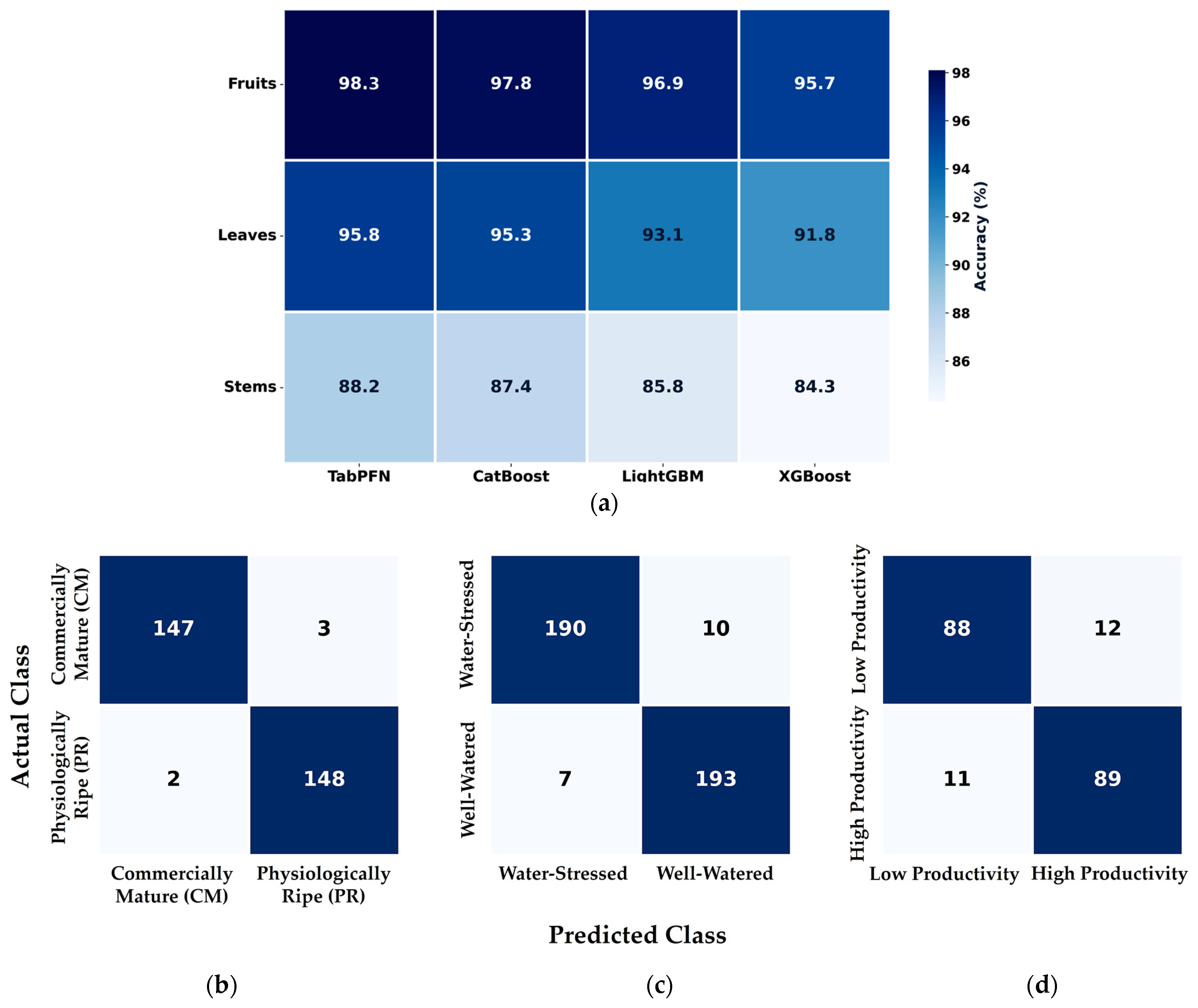
3.4. Performance Matrix and Confusion Matrices for Classification

Comparative evaluation of machine learning algorithms revealed TabPFN outperformed CatBoost, LightGBM, and XGBoost across all organs, achieving 98.3% accuracy for fruits, 95.8% for leaves, and 88.2% for stems (Figure 8). These accuracy levels reflect the directness of electrical–biological coupling in each organ. Fruit classification succeeded best because ripening creates clear conductivity shifts through sugar accumulation and cell wall softening. Leaf assessment performed well since the water content directly determines ionic conductivity. Stem productivity assessment measured current vascular transport properties to indicate production potential. Misclassification patterns provided additional insights. Errors clustered at physiological transition points rather than occurring randomly. Fruits misclassified were typically transitioning between commercial maturity and physiological ripeness, where electrical properties naturally overlap. Similarly, leaves at intermediate water stress levels and stems transitioning between high- and low-productivity states showed higher error rates, as their electrical signatures occupied the boundary zones between distinct categories.

3.5. Correlation Results

The correlation analysis served solely to validate that bioelectrical measurements reflect real physiological changes. Full correlation details appear in Table A4, Table A5 and Table A6 and Figure S9. A clear pattern emerged across organs that mirrors measurement directness. Fruits showed the strongest correlations because ripening directly alters electrical properties through sugar and ion changes. Leaves performed well since water loss immediately affects conductivity. Stems had the weakest correlations, which makes sense given that current vascular activity must predict productivity. Interestingly, chemical parameters consistently outperformed physical ones. In fruits, sugar content and pH correlated better than weight or size. This suggests bioelectrical measurements excel at detecting internal chemical changes rather than external physical or morphological traits. The measurement frequencies also matched organ physiology. Fruits responded best at higher frequencies where internal changes dominate, while leaves preferred lower frequencies where water-related ion transport occurs. This validation approach follows standard practice in bioelectrical studies, where classification provides practical performance while correlations confirm physiological relevance.

3.6. Tomography Results

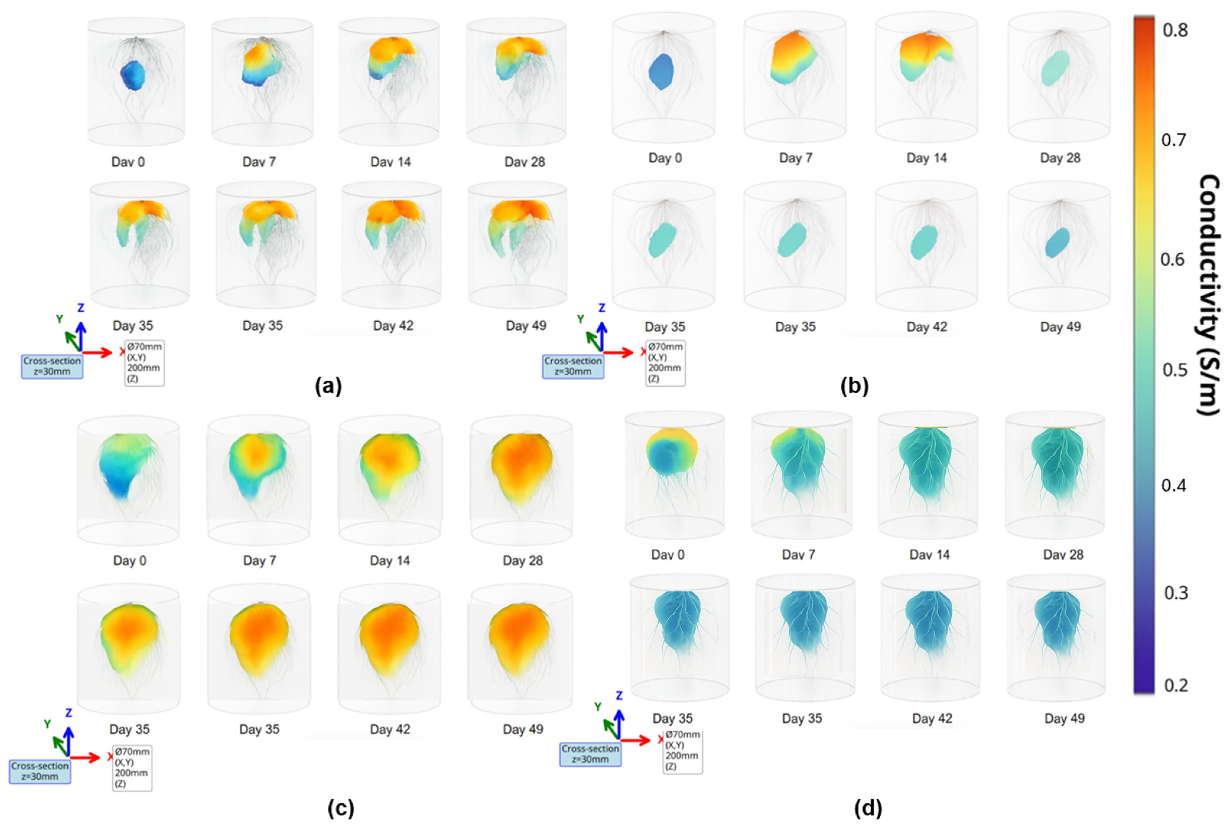
Temporal 3D root zone conductivity mapping over 49 days revealed conductivity patterns between stressed and control plants (Figure 9). Both groups initially showed blue regions (0.4–0.5 S/m) during the first week, indicating adaptation to the environment following transplantation.
Control plants subsequently developed healthy root systems visible as high-conductivity zones (orange to red, 0.7–0.8 S/m) concentrated in the upper root zone where primary water uptake occurs. These values remained stable throughout monitoring. In contrast, stressed plants under water and nutrient reduction showed progressive decline from an initial 0.6 S/m to below 0.4 S/m by day 49, appearing as expanding cyan blue regions. This 33% reduction suggests reduced ionic transport due to water and nutrient deprivation affecting the metabolic activity. The color progression from warm to cool tones reveals three distinct stress phases: initial resilience (days 0–14) with minimal change, rapid deterioration (days 14–28) as blue zones expanded inward from the root periphery, and stabilization at degraded levels after day 28 with persistent blue coloration. These measurements represent electrical conductivity patterns. While in algorithm evaluation, U-Net achieved a better resolution (2.6 ± 0.3 mm) than FBP (2.8 ± 0.3 mm), though FBP processed faster (8.7 ± 1.4 vs. 24.3 ± 3.2 s per timepoint).
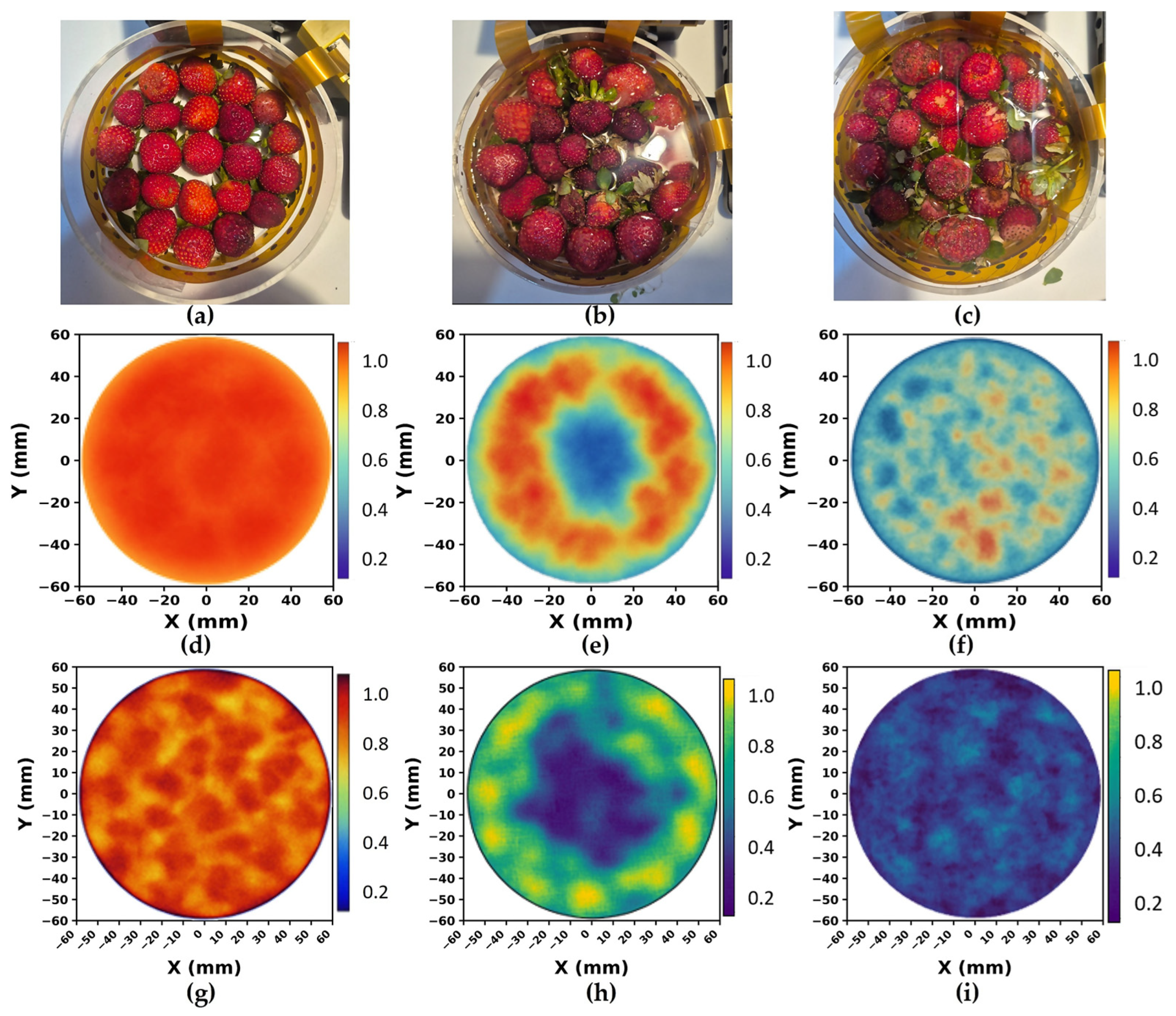
Figure 10 illustrates the postharvest strawberry quality assessment through conductivity localization starting from setup configurations (a–c), followed by SIRT (d–f) and Modified Back-Projection (g–i) reconstructions. Figure 10a–c shows the 2D tomographic reconstruction for postharvest fruit quality sorting using the three test configurations: uniform good-quality fruits, mixed quality with central defects, and uniform poor-quality fruits. Good-quality fruits showed high conductivity (≥0.5 S/m), while poor-quality fruits exhibited low conductivity (≤0.3 S/m), enabling clear differentiation.
Conductivity values were normalized (0–1 scale) to enable direct comparison between reconstruction algorithms using a unified color bar. Both SIRT and Modified Back-Projection successfully differentiated fruit quality across all three test configurations. SIRT reconstructions (Figure 10d–f) clearly identified uniform good quality, central poor-quality clusters, and distributed defects through distinct conductivity patterns. Modified Back-Projection (Figure 10g–i) produced the same quality differentiation but with notably sharper boundaries between good and poor fruit regions, particularly visible at quality transition zones. This boundary sharpness advantage aligns with MBP’s superior resolution metrics reported above. Modified Back-Projection achieved better resolution (2.8 ± 0.3 mm) and lower error (8.1 ± 0.9%) while processing faster at 13.7 ± 2.1 s per frame. SIRT required 19.2 ± 3.1 s with slightly coarser resolution (3.0 ± 0.3 mm) and higher error (10.3 ± 1.2%).
These tomographic capabilities were validated through focused sample sizes designed for system demonstration. Root measurements were performed on four plants, generating 41,664 individual observations (7 time points × 1488 measurements per scan), while fruit assessment involved 100 specimens, producing 34,720 measurements. This measurement density approach is consistent with established tomography validation protocols [60,94,106], where spatial and temporal multiplicity provide statistical power for technology validation despite limited biological replication. Similar studies have successfully demonstrated system capabilities with limited samples [44,45,51,94], as the emphasis is on validation rather than biological variation [96,97,98]. While these sample sizes were sufficient to demonstrate system performance, future agricultural deployment will require expanded trials across cultivars and seasons to establish population-level validity.

4. Discussion

This study presents a non-invasive, multimodal, and multiscale bioelectrical sensor system achieved through unified hardware integration and systematic methodology for organ-specific feature extraction. These features were correlated with physiological reference metrics for validation. The system further incorporates multimodality through 2D and 3D tomographic imaging. A proof-of-concept demonstration across multiple organs of Fragaria × ananassa (cv. ‘Sweet Charlie’) confirmed the system’s capability for holistic plant assessment and established a transferable foundation for broader applications across cultivars and crops.
This study addresses the fragmentation that limits existing bioelectrical approaches. Spectroscopy and tomography, previously deployed separately, here function as complementary outputs of a single system. Spectroscopy provides organ-specific indicators in leaves, fruits, and stems, while tomography enables spatial conductivity imaging in roots and postharvest fruits, offering converging physiological views across all plant organs. Direct cross-validation between modalities was not performed as biological metrics remain organ-specific, but the integrated framework (Table A3) enabled the following validated results.
Fruit categorization achieved 98.3% accuracy, comparable to standard and advanced optical methods. Leaf water status reached 95.8%, matching hyperspectral and developed approaches [137]. Stem productivity attained 88.2%, similar to existing electrochemical methods (detailed comparisons in Table A4). While some methods report higher accuracy, they detect only surface changes after symptom onset. This system identifies internal physiological changes within the reversible stress window, enabling proactive intervention.
These classification accuracies reflect the integrated assessment approach employed. The stem productivity assessment applied integrated stress variables, recognizing that plant responses arise from multiple interacting factors as documented in stress assessment literature [5,10,23]. This approach is consistent with methodologies reported in previous studies [53,68,73]. Similarly, fruit and leaf assessments were based on established studies, incorporating multiple parameters linked to standard quality traits [138,139,140,141] and to progressive water stress effects [142,143,144], respectively.
The correlation analysis further validates these results by confirming that the extracted features capture genuine physiological processes (Appendix A, Table A5, Table A6 and Table A7), with the correlation map presented in Supplementary Figure S9. Specifically, each organ exhibits distinct mechanisms that simultaneously affect both bioelectrical properties [145,146] and reference parameters.
In leaves, water stress drives parallel changes in RWC and bioelectrical conductivity through altered ionic regulation and conductivity [68,85]. SPAD correlations reflect stress pathways affecting both photosynthesis and conductivity, with electrical changes preceding chlorophyll decline [135,147,148]. Leaf thickness further influences current distribution pathways [149,150]. In fruits, ripening simultaneously triggers pectinase activity, ATPase modulation, and vacuolar changes that affect factors like sugar accumulation (Brix), conductivity, and structural changes [95,132,136], explaining strong correlations with quality metrics [70,89,95,120]. In stems, vascular transport processes, like xylem flow, phloem loading, and cambial activity, influence productivity metrics and electrical conductivity [125,127,131]. These parallel processes generate strong correlations without implying causation, following established bioelectrical validation approaches where classification accuracy provides primary performance metrics while correlations confirm physiological relevance [53,68,73,95,125]. This validates that bioelectrical measurements capture genuine physiological processes rather than artifacts.
Beyond these spectroscopic assessments, the system’s tomographic capabilities extend measurements to spatial imaging. Root system 3D mapping achieved 2.6–2.8 mm resolution, with conductivity patterns (0.8 to 0.4 S/m decline) matching documented stress responses [60,94,106,147,148], though direct physiological validation remains future work. Postharvest 2D fruit tomography achieved 2.8–3.0 mm resolution. These capabilities provide localized physiological information impossible with spectroscopic point measurements alone, demonstrating multimodal integration.
The system’s technical innovations succeeded through organ-specific electrode matching. Leaves and fruits have homogeneous tissues that maintain stable conductivity with bipolar measurements [95,109,111,120]. Stems, however, contain conductive vascular bundles within resistive tissue, creating heterogeneity that causes errors in bipolar mode [61,93,125,127,131]. Tetrapolar configuration corrects this by separating current injection from voltage sensing [61,125,131], consistent with bioelectrical theory [54,63]. This tissue-electrode matching explains why homogeneous organs achieve high SNR with bipolar configurations while heterogeneous stems require tetrapolar arrangements.
To address how the system achieves multimodal, multiscale, and holistic assessment, Table A3 summarizes the validation framework. Detailed technical specifications are provided in Table A8.
These technical capabilities involve practical trade-offs. Unlike rapid advanced optical methods, the system detects cellular changes before visible symptoms, enabling intervention to prevent the 20–40% losses typical of reactive management. As of now, the system’s architecture incorporates parallel multiplexing capability, though deployment awaits field validation for future scalability. This positions the system as a complementary tool for high-value crops or research requiring early stress detection or detailed physiological data.
While the current validation focused on strawberry (Fragaria × ananassa ‘Sweet Charlie’), the system’s capabilities demonstrated here apply across crops. Each crop will require threshold calibration, which is standard practice for any assessment method, with this study providing the methodological framework. Future work involves multi-cultivar validation, automated calibration algorithms, and integration with farm management systems.

5. Conclusions and Recommendations

This study developed and validated a unified bioelectrical sensor system for non-invasive, multimodal, and multiscale plant assessment. As a proof of concept using strawberry (Fragaria × ananassa ‘Sweet Charlie’), the system achieved high accuracy across multiple organs: 98.3% for fruit categorization, 95.8% for leaf water status, and 88.2% for stem productivity. Additionally, tomographic imaging reached 2.6–2.8 mm resolution for 3D root mapping and 2.8–3.0 mm for 2D fruit defect sorting. The platform integrates spectroscopy, which captures multiscale responses from cellular to tissue levels, with tomography, which provides spatial visualization. Strong physiological correlations confirmed that the features detected represent genuine physiological variations.
While validation was limited to a single cultivar and stem assessment remained indirect, these represent development opportunities rather than fundamental limitations. Future work should focus on enabling parallel measurements, expanding to other crops, and building automated calibration algorithms. Integration with farm management systems and standardized deployment protocols will allow for field-scale adoption. In the longer term, IoT implementation, controlled single-factor experiments, and crop-specific bioelectrical libraries will establish comprehensive assessment networks.
By detecting stress before irreversible damage, this proactive approach helps prevent the 20–40% yield losses typical of reactive management. It also supports the 40% production increase required by 2050, contributing to sustainable intensification in the face of climate change.

Supplementary Materials

The following supporting information can be downloaded at: https://www.mdpi.com/article/10.3390/technologies13110496/s1, Figure S1. Bipolar (2-electrode) configuration circuit implementation for bioelectrical spectroscopy. Figure S2. Tetrapolar (4-electrode) configuration circuit implementation for bioelectrical spectroscopy. Figure S3. 32-electrode array configuration with multi-stage multiplexing architecture for tomographic applications. Figure S4. Precision programmable resistor load network for bioelectrical sensor system calibration. Figure S5. Electrode performance evaluation for bioelectrical spectroscopy (a–c) and tomography (d–f) measurements across strawberry plant organs. Figure S6. Statistical validation and multi-criteria frequency selection showing (a–c) −log10(p-values) from t-tests and (d–f) normalized scores integrating statistical significance, effect size, and mutual information for leaves, fruits, and stems respectively. Figure S7. Frequency-dependent sensitivity and penetration analysis for tomographic applications showing (a) radial sensitivity profiles, (b) frequency response optimization, (c) 3-layer array root conductivity reconstruction, and (d) 2D fruit conductivity reconstruction. Figure S8. Recursive feature elimination with cross-validation (RFECV) results for optimal feature selection across (a) fruit maturity, (b) leaf water status, and (c) stem productivity classification tasks. Figure S9. Bioelectrical feature-validation metric correlation matrices for (a) fruits showing 12 features vs. 6 established validation metrics, (b) leaves showing 10 features vs. 3 metrics, and (c) stems showing 8 features vs. 7 metric.

Author Contributions

Conceptualization, J.A. and R.C.II; methodology, J.A., R.C.II and E.D.; software, J.A.; validation, R.C.II, R.N., E.D., A.B., R.R.V. and E.S.; formal analysis, J.A., R.C.II, A.B., R.R.V. and E.S.; investigation, J.A.; resources, E.D., A.B., R.R.V., E.S. and L.G.L.; data curation, J.A. and R.N.; writing—original draft preparation, J.A.; writing—review and editing, R.C.II; visualization, J.A., R.C.II and E.D.; supervision, E.D., A.B., R.R.V., E.S. and L.G.L.; project administration, E.D., A.B., R.R.V., E.S. and L.G.L.; funding acquisition, E.D. and E.S. All authors have read and agreed to the published version of the manuscript.

Funding

The APC was funded by the De La Salle University Science Foundation Inc.

Data Availability Statement

The data supporting the findings of this study are not publicly available as they are reserved for ongoing and future research and system development, but may be provided by the authors upon reasonable request.

Acknowledgments

This study is supported by the Engineering Research and Development for Technology of the Department of Science and Technology of the Philippines and the Office of the Vice President for Research and Innovation of De La Salle University.

Conflicts of Interest

The authors declare no conflict of interest.

Appendix A

Table A1. Reference methods and uncertainties for validating bioelectrical correlations with physiological parameters.
OrganParameterInstrument/MethodMeasurement UncertaintyRangeReferences
FruitSugar content (°Brix)Atago PAL-3 refractometer (Atago Co., Ltd., Tokyo, Japan)±0.2 °Brix6–14 °Brix[95,132,136]
Firmness (N)TA.XT2 texture analyzer (Stable Micro Systems, Godalming, UK)±0.15 N1.5–4.5 N[133,136]
Weight (g)Mettler Toledo MA204 balance (Mettler Toledo, Columbus, OH, USA)±0.003 g8–25 g[86,95,136]
Diameter (mm)Mitutoyo 500-196-30 caliper (Mitutoyo Corporation, Kawasaki, Japan)±0.03 mm20–40 mm[90,91,95]
pHMettler Toledo FP20 pH meter (Mettler Toledo, Columbus, OH, USA)±0.02 pH3.2–4.0[132,136]
EC (mS/cm)HI 9033 conductivity meter (Hanna Instruments, Woonsocket, RI, USA)±0.05 mS/cm2.0–5.0 mS/cm[95,120,132]
LeafRWC (%)Gravimetric method±3%65–95%[68,85,134]
Chlorophyll (SPAD)SPAD-502Plus chlorophyll meter (Konica Minolta, Tokyo, Japan)±1.5 units25–45 SPAD[85,88,135]
Thickness (mm)Mitutoyo 500-196-30 caliper (Mitutoyo Corporation, Kawasaki, Japan)±0.03 mm0.25–0.45 mm[68,85,86]
StemDiameter (mm)Mitutoyo CD-6AX caliper (Mitutoyo Corporation, Kawasaki, Japan)±0.03 mm4–12 mm[93,125]
Sugar content (°Brix) *Atago PAL-3 refractometer (Atago Co., Ltd., Tokyo, Japan)±0.2 °Brix6–14 °Brix[95,132,136]
Firmness (N) *TA.XT2 texture analyzer (Stable Micro Systems, Godalming, UK)±0.15 N1.5–4.5 N[133,136]
Weight (g) *Mettler Toledo MA204 balance (Mettler Toledo, Columbus, OH, USA)±0.003 g8–25 g[86,95,136]
Diameter (mm) *Mitutoyo 500-196-30 caliper (Mitutoyo Corporation, Kawasaki, Japan)±0.03 mm20–40 mm[90,91,95]
pH *Mettler Toledo FP20 pH meter (Mettler Toledo, Columbus, OH, USA)±0.02 pH3.2–4.0[132,136]
EC (mS/cm) *HI 9033 conductivity meter (Hanna Instruments, Woonsocket, RI, USA)±0.05 mS/cm2.0–5.0 mS/cm[95,120,132]
* Quality metrics measured from fruits attached to the assessed stems.
Table A2. Optimal bioelectrical features for plant organ classification.
Organ Type (Sample Size, Accuracy)RankFeature NameType/FrequencyRelative Weight
Fruits (n =300, 98.3% ± 1.0%)1θ (fruit50kHz)Phase angle at 50 kHz0.187
2τ (fruit50kHz)Cole-Cole parameter0.172
3L (fruit100kHz)Inductance at 100 kHz0.158
4Q (fruit50kHz)Derived at 50 kHz0.144
5α (fruitβ-dispersion)Dispersion parameter0.131
6 C m (fruit50kHz)Capacitance at 50 kHz0.118
7R0 (fruit1kHz)Resistance at 1 kHz0.105
8R∞ (fruit200kHz)Resistance at 200 kHz0.092
9Z (fruit10k/100k)Impedance ratio0.087
10Morphological (asymmetry)Derived geometric0.081
11Spectral centroid (fruitβ-dispersion)Statistical0.074
12Peak power (fruit50kHz)Derived0.068
Leaves (n =400, 95.8% ± 1.6%)1θ (leaf5kHz)Phase angle at 5 kHz0.156
2R0 (leaf0.5kHz)Resistance at 0.5 kHz0.142
3C (leaf5kHz)Capacitation at 5 kHz0.134
4τ (leaf5kHz)Cole-Cole at 5 kHz0.128
5R∞ (leaf50kHz)Resistance at 50 kHz0.121
6Z (leaf1k/25k)Impedance ratio0.115
7α (leafα-dispersion)Dispersion parameter0.108
8Z (ratio leaf5k/50k)Impedance ratio0.102
9Spectral centroid (leafα-dispersion)Statistical0.096
10Derivative magnitude (leafα-average)Derived0.089
Stems (n =200, 88.2% ± 1.9%)1θ (stem100kHz)Phase angle at 100 kHz0.198
2τ1 (stem100kHz)Cole-Cole parameter0.176
3τ2 (stem500kHz)Cole-Cole parameter0.149
4Z (ratio stem1k/250k)Impedance ratio0.132
5α (stemα-dispersion)Dispersion parameter0.118
6 C m (stem100kHz)Capacitance at 100 kHz0.097
7R0 (stem25kHz)Resistance at 25 kHz0.084
8Coupling factor (stem)Derived0.073
Table A3. Framework for achieving multimodal, multiscale, and holistic plant assessment.
AssessmentImplementationOrgan CoverageValidation Outcome
MultimodalSpectroscopy
(bipolar/tetrapolar)
Leaves, Fruits, StemsAccurate classification (95.8%, 98.3%, 88.2%)
Tomography
(2D & 3D arrays)
Fruits, RootsSpatial resolution 2.6–3.0 mm; conductivity-based root zone mapping and fruit defect sorting
Multiscaleα-dispersion LeavesCaptures water status dynamics
β-dispersion FruitsMarket-based fruit quality sorting
Dual dispersion StemsReflects vascular transport and stem productivity
HolisticMulti-organ protocol
on single platform
All four organsUnified hardware
with physiologically validated features
Table A4. Comparison of the proposed bioelectrical sensor system with standard and advanced methods reported in the literature.
MethodPerformance
Type and Metrics
Detection
Type
Key
Limitations
References
Fruit Quality Assessment
Hyperspectral imaging + AIClassification: 90–99%ReactiveEquipment cost,
surface only,
[24,26,137,140]
VIS-NIR spectroscopyRegression (R2): 0.73–0.91ReactiveCost, Species-specific, Lighting dependent[27,139]
Portable VIS-NIR Regression (R2): 0.82–0.89ReactiveSingle species, Lighting dependent, Inconclusive[27,65,141]
Advanced optical spectralClassification: 85–98%ReactiveLimited to optical parameters, affected by light[87,88,138]
Thermal imagingClassification: 73–89%ReactiveEnvironment dependent, surface temp only[28,29,30,83]
Chlorophyll fluorescenceDetection:
82–87%
Semi-proactiveLimited to
single parameter
[28,29,30,31,32]
Electrochemical spectroscopyClassification: 82–95% Semi-proactiveLimited features, Limited penetration,[52,53,64,95]
MolecularDetection: 70–85%ReactiveDestructive sampling[33,34,35,36]
This StudyClassification: 98.3%ProactiveContact required, Low-throughput Present work
Leaf Relative Water Content
Spectral indicesRegression (R2): 0.65–0.78Semi-proactiveSurface only, Inconclusive in field calibration[27,88,142]
Hyperspectral reflectanceRegression (R2): 0.71–0.83Semi-proactiveEquipment cost, Lab conditions only[24,88,143]
Hyperspectral stressClassification: 83–89%Semi-proactiveComplex, Equipment cost, Lab conditions only[24,25,26]
Plant-based sensorsCorrelation (r): 0.75–0.85MonitoringFixed installation, Environment dependent[40,77,144]
Thermal imagingDetection: 73–89%Semi-proactiveEnvironment dependent, surface temp only[28,29,30,31,32]
Chlorophyll fluorescenceClassification: 80–85%Semi-proactiveDark adaptation, affected by light and radiation[31,32,147,148,149]
Terahertz spectroscopyDetection: 76–82%Semi-proactiveLimited features, Limited penetration[48,49,50]
Electrochemical
spectroscopy
Classification: 87–90%Semi-proactiveNo feature extraction, Limited features, Limited penetration[53,67,68,85]
Wearable sensors, IoT networksSystem dependentMonitoringPower/durability, Integration complexity[37,38,39,40,41]
This StudyClassification: 95.8% ± 1.6%ProactiveContact required, Low-throughput compared to advanced imagingPresent work
Stem Assessment
Visual inspectionAccuracy: 70–75%ReactiveSubjective[70,92,93]
Electrochemical spectroscopyClassification: 87%ProactiveSpecific to peduncle only[124,125,126,127,128,129,130,131]
Pedicel AssessmentCorrelation (r): 0.65–0.72ReactiveSubjective Manual measurement, Operator-dependent[92,93]
Sap flow sensorsCorrelation (r): 0.70–0.80ReactiveLimited point measurement[77,127,144]
This StudyClassification: 88.2%ProactiveIndirect validationPresent work
Root Mapping
Electrochemical TomographyResolution: 5–10 mmSequentialLow resolution[56,57,94]
X-ray micro-CTResolution: 0.5–2 mmStaticRadiation exposure[43,44,45]
MRI Resolution: 1–3 mmTime-seriesExpensive equipment[50,51]
Ground penetrating radarDepth: 50–200 mmSingle sweepSoil dependent[57,60,94]
Conductivity root mappingResolution: 8–12 mmContinuousLimited penetration[56,59,60]
This Study Resolution: 2.6–2.8 mmReal-timePhantom-grown onlyPresent work
Fruit Defect Detection
X-ray imagingDetection: 85–92%Static Radiation required[44,45]
NIR transmittanceClassification: 78–86%Single scanTranslucent only[27,65,139]
Acoustic analysisClassification: 75–83%Pulse responseNoise sensitive[89,90,91]
Previous 2D EITResolution: 4–6 mmSequential Poor boundaries[56,61,95]
MRI internal imagingResolution: 1–2 mmTime-seriesVery expensive[49,50,51]
This Study Resolution: 2.8–3.0 mmNon-invasiveBinary defect detection onlyPresent work
Table A5. Correlation coefficients between optimal bioelectrical features and key quality attributes across all fruit samples (n =300), spanning both physiologically ripe and commercially mature categories.
Bioelectrical ParameterBrix Content (°Bx)Firmness (N)Weight (g)Diameter (mm)Juice pHJuice EC (mS/cm)
θ (fruit50kHz)0.9230.9120.6870.6540.8430.896
τ (fruit50kHz)0.9080.8940.6720.6410.8290.881
L (fruit100kHz)0.8960.8870.6980.6650.8560.874
Q (fruit50kHz)0.8870.8780.6810.6480.8340.869
α (fruitβ-dispersion)0.8740.8650.6630.6310.8210.856
C m (fruit50kHz)0.8690.8580.6560.6240.8120.847
R0 (fruit1kHz)0.8560.8470.6490.6170.7980.834
R∞ (fruit200kHz)0.8630.8520.6710.6390.8050.841
Z (fruit10k/100k)0.8470.8360.6920.6590.7890.823
Morphological (asymmetry)0.8340.8230.6780.6450.7760.812
Spectral centroid (fruitβ-dispersion)0.8210.8120.6650.6320.7630.798
Peak power (fruit50kHz)0.8120.8010.6580.6250.7540.789
Table A6. Correlation coefficients between optimal bioelectrical features and key quality attributes across all leaf samples (n =400), spanning both well-watered and water-stressed categories.
Bioelectrical ParameterWater Content (%)Chlorophyll (SPAD)Thickness (mm)
θ (leaf5kHz)0.8340.8210.808
R0 (leaf0.5kHz)0.8230.8120.798
C (leaf5kHz)0.8120.8010.789
τ (leaf5kHz)0.8010.7890.776
R∞ (leaf50kHz)0.7890.7780.765
Z (leaf1k/25k)0.7780.7670.754
α (leafα-dispersion)0.7670.7560.743
Z (ratio leaf5k/50k)0.7560.7450.732
Spectral centroid (leafα-dispersion)0.7450.7340.721
Derivative magnitude (leafα-average)0.7340.7230.712
Table A7. Correlation coefficients between optimal bioelectrical features and key quality attributes across all stem samples (n =200), spanning both productive and non-productive categories.
ParameterStem Diameter (mm)Fruit Brix (°Bx) *Fruit Firmness (N) *Fruit Weight (g) *Fruit Thickness (mm) *Fruit pH *Fruit EC (mS/cm) *
θ (stem100kHz)0.7120.6980.5840.5210.4870.6340.671
τ1 (stem100kHz)0.6980.6850.5710.5080.4740.6210.658
τ2 (stem500kHz)0.6850.6720.5580.4950.4610.6080.645
Z (ratio stem1k/250k)0.6720.6590.5450.4820.4480.5950.632
α (stemα-dispersion)0.6590.6460.5320.4690.4350.5820.619
C m (stem100kHz)0.6460.6330.5190.4560.4220.5690.606
R0 (stem25kHz)0.6330.6200.5060.4430.4090.5560.593
Coupling factor (stem)0.6200.6070.4930.4300.3960.5430.580
* All fruit metrics were taken from fruits attached to the stem.
Table A8. Hardware implementation and core processes in the bioelectrical measurement system.
ProcessHardware Implementation
Signal GenerationAD9833 DDS + AD835 multiplier
Electrode RoutingADG714, ADG1404, ADG1408/1409
Input ProtectionSMAJ5.0A TVS + EMI filtering
Signal Conditioning3-stage RC networks: HPF + BPF + LPF
Current SourceHowland: OPA2376/AD5171/AD5663
Voltage BufferingOPA333 with guard drive
InstrumentationINA333 + sense resistors
Reference Elements4.7 kΩ (H-bridge), calibration resistors
Data AcquisitionADS1256 (24-bit) + OPA4188 + REF5025
System ControlSTM32H755 + XC7A35RT FPGA
Power SystemLinear ±12 V (LM7812/7912)

References

  1. Food and Agriculture Organization. The State of Food Security and Nutrition in the World 2023; FAO: Rome, Italy, 2023; pp. 1–250. [Google Scholar]
  2. Taha, M.F.; Ahmed, S.M.; Hassan, R.K.; Al-Rashid, M.A.; Ibrahim, N.H.; Mahmoud, A.S. Emerging Technologies for Precision Crop Management Towards Agriculture 5.0: A Comprehensive Overview. Agriculture 2025, 15, 582. [Google Scholar] [CrossRef]
  3. Rahman, M.A.; Singh, P.; Kumar, V.; Patel, S.; Sharma, A.; Gupta, R. Ensuring food security through rapid and in-field detection of diseases in food crops using real time and portable sensors. Anal. Biochem. 2025, 705, 115925. [Google Scholar] [CrossRef] [PubMed]
  4. Alhabsi, A.; Ling, Y.; Crespi, M.; Reddy, A.S.N.; Mahfouz, M. Alternative Splicing Dynamics in Plant Adaptive Responses to Stress. Annu. Rev. Plant Biol. 2025, 76, 687–717. [Google Scholar] [CrossRef]
  5. Kumar, M.; Saifi, Z.; Krishnananda, S.D. Decoding the physiological response of plants to stress using deep learning for forecasting crop loss due to abiotic, biotic, and climatic variables. Sci. Rep. 2023, 13, 8598. [Google Scholar] [CrossRef]
  6. Moustaka, J.; Moustakas, M. Early-Stage Detection of Biotic and Abiotic Stress on Plants by Chlorophyll Fluorescence Imaging Analysis. Biosensors 2023, 13, 796. [Google Scholar] [CrossRef]
  7. Aina, O.; Bakare, O.O.; Fadaka, A.O.; Keyster, M.; Klein, A. Plant biomarkers as early detection tools in stress management in food crops: A review. Planta 2024, 259, 60. [Google Scholar] [CrossRef]
  8. Trifilò, P.; Abate, E.; Petruzzellis, F.; Azzarà, M.; Nardini, A. Critical water contents at leaf, stem and root level leading to irreversible drought-induced damage in two woody and one herbaceous species. Plant Cell Environ. 2022, 46, 119–132. [Google Scholar] [CrossRef]
  9. Liu, Z.; Li, Z.; Zhang, X.; Li, Y.; Wang, Y. Stress resilience in plants: The complex interplay between heat stress memory and resetting. New Phytol. 2025, 203, 203–217. [Google Scholar]
  10. IPCC. Climate Change 2023: Synthesis Report; Contribution of Working Groups I, II and III to the Sixth Assessment Report of the Intergovernmental Panel on Climate Change; Core Writing Team; Lee, H., Romero, J., Eds.; IPCC: Geneva, Switzerland, 2023. [Google Scholar]
  11. Ali, Q.; Rahman, S.; Ahmed, M.; Hassan, A.; Khan, R.; Malik, S. Research advances and applications of biosensing technology for the diagnosis of pathogens in sustainable agriculture. Environ. Sci. Pollut. Res. 2021, 28, 9002–9019. [Google Scholar] [CrossRef] [PubMed]
  12. Krishnamoorthi, S.; Patel, N.; Singh, K.; Kumar, A.; Sharma, R.; Gupta, M. Advancements in Plant Diagnostic and Sensing Technologies. Adv. Sens. Res. 2025, 4, 2500045. [Google Scholar] [CrossRef]
  13. Khakimov, A.; Salakhutdinov, I.; Omolikov, A.; Utaganov, S. Traditional and current-prospective methods of agricultural plant diseases detection: A review. IOP Conf. Ser. Earth Environ. Sci. 2022, 951, 012002. [Google Scholar] [CrossRef]
  14. Attaluri, S.; Dharavath, R. Novel plant disease detection techniques-a brief review. Mol. Biol. Rep. 2023, 50, 9677–9690. [Google Scholar] [CrossRef]
  15. Ang, M.C.-Y.; Lew, T.T.S. Non-destructive Technologies for Plant Health Diagnosis. Front. Plant Sci. 2022, 13, 884454. [Google Scholar] [CrossRef] [PubMed]
  16. Okere, E.E.; Arendse, E.; Nieuwoudt, H.; Fawole, O.A.; Perold, W.J.; Opara, U.L. Non-Invasive Methods for Predicting the Quality of Processed Horticultural Food Products, with Emphasis on Dried Powders, Juices and Oils: A Review. Foods 2021, 10, 3061. [Google Scholar] [CrossRef] [PubMed]
  17. Roper, J.M.; Garcia, J.F.; Tsutsui, H. Emerging Technologies for Monitoring Plant Health In Vivo. ACS Omega 2021, 6, 5101–5107. [Google Scholar] [CrossRef] [PubMed]
  18. Burynin, D.A. Screening in Plant Factories: A Review of Non-Invasive Plant Monitoring Techniques for Closed Regulated Agroecosystems. Agroengineering 2022, 6, 70–75. [Google Scholar] [CrossRef]
  19. Galieni, A.; D’Ascenzo, N.; Stagnari, F.; Pagnani, G.; Xie, Q.; Pisante, M. Past and Future of Plant Stress Detection: An Overview From Remote Sensing to Positron Emission Tomography. Front. Plant Sci. 2021, 11, 609155. [Google Scholar] [CrossRef]
  20. Yan, B.; Zhang, F.; Wang, M.; Zhang, Y.; Fu, S. Flexible wearable sensors for crop monitoring: A review. Front. Plant Sci. 2024, 15, 1406074. [Google Scholar] [CrossRef]
  21. Alejandrino, J.D.; Concepcion, R.; Culaba, A.; Dadios, E. Root Phenotyping Revolution: Science-based Policy Recommendations Amidst Green Revolution 2.0’s Transition from Invasive to Non-Invasive Tomography. In Proceedings of the 2021 IEEE 13th International Conference on Humanoid, Nanotechnology, Information Technology, Communication and Control, Environment, and Management (HNICEM), Manila, Philippines, 28–30 November 2021; pp. 1–6. [Google Scholar]
  22. Coatsworth, P.; Gonzalez-Macia, L.; Collins, A.S.P.; Bozkurt, T.; Güder, F. Continuous monitoring of chemical signals in plants under stress. Nat. Rev. Chem. 2023, 7, 7–25. [Google Scholar] [CrossRef]
  23. Savary, S. A Global Assessment of the State of Plant Health. Plant Dis. 2023, 107, 3649–3665. [Google Scholar] [CrossRef]
  24. Wan, G.; Liu, Y.; Zhang, H.; Wang, X.; Chen, M.; Li, S. Hyperspectral imaging technology for nondestructive identification of quality deterioration in fruits and vegetables: A review. Crit. Rev. Food Sci. Nutr. 2025, 65, 1–30. [Google Scholar] [CrossRef]
  25. Dabek, A.; Mantovani, L.; Mirabella, S.; Vignati, M.; Cinquemani, S. Advancements in Non-Destructive Detection of Biochemical Traits in Plants Through Spectral Imaging-Based Algorithms: A Systematic Review. Algorithms 2025, 18, 255. [Google Scholar] [CrossRef]
  26. Xu, J.; Wang, L.; Chen, Y.; Liu, H.; Zhang, K.; Zhao, M. Trend analysis of the application of multispectral technology in plant yield prediction: A bibliometric visualization analysis (2003–2024). Front. Sustain. Food Syst. 2025, 9, 1513690. [Google Scholar] [CrossRef]
  27. Zahir, S.A.D.M.; Omar, A.F.; Jamlos, M.F.; Azmi, M.A.M.; Muncan, J. A review of visible and near-infrared (Vis-NIR) spectroscopy application in plant stress detection. Sens. Actuators A Phys. 2022, 338, 113468. [Google Scholar] [CrossRef]
  28. Sharma, N.; Tarade, P.; Rathi, R.; Biradar, A.; Jahirabadkar, S. Stress Analysis of ‘Ashwagandha, a Medicinal Plant’ Using Thermal Imaging. In Proceedings of the International Conference on Advances in Electrical Technology (ICAET), Mumbai, India, 10–11 January 2025; pp. 1–8. [Google Scholar]
  29. Smigaj, M.; Gaulton, R.; Suárez, J.C.; Barr, S.L. Thermal Infrared Remote Sensing of Stress Responses in Forest Environments: A Review of Developments, Challenges, and Opportunities. Curr. For. Rep. 2023, 9, 424–443. [Google Scholar] [CrossRef]
  30. Omia, E.; Bae, H.; Park, E.; Kim, M.S.; Baek, I.; Kabenge, I.; Cho, B.-K. Remote Sensing in Field Crop Monitoring: A Comprehensive Review of Sensor Systems, Data Analyses and Recent Advances. Remote Sens. 2023, 15, 354. [Google Scholar] [CrossRef]
  31. Grishina, A.; Sherstneva, O.; Mysyagin, S.; Brilkina, A.; Vodeneev, V. Detecting Plant Infections: Prospects for Chlorophyll Fluorescence Imaging. Agronomy 2024, 14, 2600. [Google Scholar] [CrossRef]
  32. Li, Y.; Luo, J.; Ndikuryayo, F.; Chen, Y.; Liu, G.; Yang, W.-C. Advances in Fluorescence-based Probes for Abiotic Stress Detection in Plants. ACS Sens. 2025, 10, 2474–2486. [Google Scholar] [CrossRef]
  33. Shukla, A.; Yadav, D.; Mishra, U.K.; Dipake, S.; Mishra, R. Revolutionizing Plant Virus Detection: A Review on Molecular Breakthroughs and their Implications. Int. J. Plant Soil Sci. 2023, 35, 339–350. [Google Scholar] [CrossRef]
  34. Alsharksi, A.N.; Sirekbasan, S.; Gürkök-Tan, T.; Mustapha, A. From Tradition to Innovation: Diverse Molecular Techniques in the Fight Against Infectious Diseases. Diagnostics 2024, 14, 2876. [Google Scholar] [CrossRef]
  35. Trippa, D.; Scalenghe, R.; Basso, M.F.; Panno, S.; Davino, S.; Molinatto, G.; Brugaletta, V.; Mauceri, A.; Mercati, F.; Martinelli, F. Next-generation methods for early disease detection in crops. Pest Manag. Sci. 2024, 80, 245–261. [Google Scholar] [CrossRef] [PubMed]
  36. Ali, Q.; Ahmar, S.; Sohail, M.A.; Kamran, M.; Ali, M.; Saleem, M.H.; Rizwan, M.; Ahmed, A.M.; Mora-Poblete, F.; Júnior, A.T.A.; et al. Advances, limitations, and prospects of biosensing technology for detecting phytopathogenic bacteria. Chemosphere 2022, 296, 133773. [Google Scholar] [CrossRef] [PubMed]
  37. Yan, X.; Wang, Z.; Chen, Y.; Wu, Y.; Li, X.; Wang, S.; Liu, Z.; Sun, W.; Hu, Z. Wearable Sensors for Plants: Status and Prospects. Biosensors 2025, 15, 53. [Google Scholar] [CrossRef]
  38. Zhang, M.; Li, J.; Wang, X.; Chen, L.; Liu, Y.; Zhou, H.; Yang, S.; Zhao, Q. Exploration of the Plant World: Application and Innovation of Plant-Wearable Sensors for Real-Time Detection. Crit. Rev. Anal. Chem. 2025, 55, 1–17. [Google Scholar] [CrossRef]
  39. Li, X.; Li, M.; Li, J.; Gao, Y.; Liu, C.; Hao, G. Wearable sensor supports in-situ and continuous monitoring of plant health in precision agriculture era. Plant Biotechnol. J. 2024, 22, 14283. [Google Scholar] [CrossRef]
  40. Prithviraj, S.S.; Raksha, G.; Sahana, N.J.; Sayuj, P.N.; Faisal, P. Smart Plant Monitoring System: A Review of Approaches for Monitoring Condition of Plants. Int. J. Adv. Res. Sci. Commun. Technol. 2024, 13, 685–692. [Google Scholar] [CrossRef]
  41. Sharma, R.; Vaidya, P.; Sharma, B. A Review on Plant Growth Monitoring using Artificial Intelligence and the Internet of Things. In Proceedings of the 2024 11th International Conference on Computing for Sustainable Global Development (INDIACom), New Delhi, India, 28 February–1 March 2024. [Google Scholar]
  42. Gou, C.; Zhu, P.; Meng, Y.; Yang, F.; Xu, Y.; Xia, P.; Chen, J.; Li, J. Machine and Deep Learning: Artificial Intelligence Application in Biotic and Abiotic Stress Management in Plants. Front. Biosci. 2024, 29, 20. [Google Scholar] [CrossRef]
  43. Jackulin, C.; Murugavalli, S. A comprehensive review on detection of plant disease using machine learning and deep learning approaches. Meas. Sens. 2022, 24, 100441. [Google Scholar] [CrossRef]
  44. Karahara, I.; Yamauchi, D.; Uesugi, K.; Mineyuki, Y. Three-dimensional visualization of plant tissues and organs by X-ray micro-computed tomography. Microscopy 2023, 72, 310–325. [Google Scholar] [CrossRef]
  45. Zhang, H.; Qi, R.; Mine, Y.; Zhang, X.; Song, Z.; Meng, W.; Zhou, H. Applications of Computed Tomography (CT) in environmental soil and plant sciences. Soil Tillage Res. 2023, 226, 105574. [Google Scholar] [CrossRef]
  46. Saletnik, A.; Saletnik, B.; Zaguła, G.; Puchalski, C. Raman Spectroscopy for Plant Disease Detection in Next-Generation Agriculture. Sustainability 2024, 16, 5474. [Google Scholar] [CrossRef]
  47. Zavafer, A.; Ball, M.C. Good vibrations: Raman spectroscopy enables insights into plant biochemical composition. Funct. Plant Biol. 2023, 50, 1–16. [Google Scholar] [CrossRef] [PubMed]
  48. Peretti, R. Terahertz technology for biophotonics: A general overview and recent advances in THz spectroscopy toward biology. In Proceedings of the SPIE Optical Metrology, Munich, Germany, 26–29 June 2023. [Google Scholar]
  49. Greco, M.; Cinquemani, S.; Malavasi, M.; Martellosio, A.; Resta, V.; Alghisi, D.; Beccalli, R.; Chiesa, M.; Crespi, E.; D’Adda, F.; et al. Studies of leaf water content in smart agriculture using THz technologies: A review. J. Smart Environ. Green Comput. 2022, 2, 46–57. [Google Scholar] [CrossRef]
  50. Anand, A.; Sharma, A.; Kaur, H.; Khandelwal, M.; Singh, G.; Barnwal, S.; Rath, A.; Mohan, S.; Shah, R. Profiling of Plant Derived Natural Constituents by Using Magnetic Resonance Techniques. Concepts Magn. Reson. Part A 2022, 2022, 5705637. [Google Scholar] [CrossRef]
  51. Blystone, S.; Nuixe, M.; Traoré, A.S.; Cochard, H.; Picon-Cochard, C.; Pagès, G. Towards portable MRI in the plant sciences. Plant Methods 2024, 20, 31. [Google Scholar] [CrossRef]
  52. Alejandrino, J.; Concepcion, R.; Dadios, E. Ripening Degree Assessment of Organic Strawberry Based on Electrical Impedance Spectroscopy with Machine Learning. In Proceedings of the 2024 6th International Conference on Applied Electromagnetics, Signal Processing & Communication (AGRETA), Bhubaneswar, India, 26–28 September 2024; pp. 61–66. [Google Scholar]
  53. Hamed, S.; Altana, A.; Lugli, P.; Petti, L.; Ibba, P. Supervised classification and circuit parameter analysis of electrical bioimpedance spectroscopy data of water stress in tomato plants. Comput. Electron. Agric. 2024, 226, 109347. [Google Scholar] [CrossRef]
  54. Van Haeverbeke, M.; De Baets, B.; Stock, M. Plant impedance spectroscopy: A review of modeling approaches and applications. Front. Plant Sci. 2023, 14, 1187573. [Google Scholar] [CrossRef]
  55. La, Z.; Wang, Y.; Chen, X.; Li, J.; Zhang, M.; Liu, H.; Zhou, S.; Yang, L. Electrical Signal Characterization of Aloe vera Var. Chinensis Using Non-Parametric and Parametric Signal Analysis. Appl. Sci. 2025, 15, 1708. [Google Scholar] [CrossRef]
  56. Custodio, J.M.; Concepcion, R.; Alejandrino, J.; Relano, R.-J.; Bandala, A.; Mirjalili, S. Patterns and Positions of Concentric Ring Electrodes for Electrical Impedance Tomography. In Proceedings of the 2023 IEEE 15th International Conference on Humanoid, Nanotechnology, Information Technology, Communication and Control, Environment, and Management (HNICEM), Manila, Philippines, 28–30 November 2023. [Google Scholar]
  57. Basak, R.; Wahid, K.A. A Rapid, Low-Cost, and High-Precision Multifrequency Electrical Impedance Tomography Data Acquisition System for Plant Phenotyping. Remote Sens. 2022, 14, 3214. [Google Scholar] [CrossRef]
  58. Alejandrino, J.D.; Concepcion, R.S., II; Bandala, A.A.; Sybingco, E.; Vicerra, R.R.P.; Dadios, E.P. Non-Invasive Plant Root Tomography Through Optimized Sonar Array Transducer Antenna Design Using Genetic Swarm Metaheuristic. J. Adv. Comput. Intell. Intell. Inform. 2024, 28, 59–66. [Google Scholar] [CrossRef]
  59. Zhu, Y.; Wang, Z.; Li, X.; Chen, J.; Liu, Y.; Zhang, H. A New Portable Electrical Impedance Tomography System for Measuring Two-Dimensional Stem Water Content Distribution. Agric. For. Meteorol. 2022, 325, 109161. [Google Scholar] [CrossRef]
  60. Mary, B.; Vanella, D.; Consoli, S.; Cassiani, G. Imaging of the electrical activity in the root zone under limited-water-availability stress: A laboratory study for Vitis vinifera. Biogeosciences 2023, 20, 4625–4650. [Google Scholar] [CrossRef]
  61. Calvo, S.; Barezzi, M.; Demarchi, D.; Garlando, U. In-vivo proximal monitoring system for plant water stress and biological activity based on stem electrical impedance. In Proceedings of the 2023 9th International Workshop on Advances in Sensors and Interfaces (IWASI), Monopoli, Italy, 8–9 June 2023; pp. 80–85. [Google Scholar]
  62. Mohsen, M.; Said, L.A.; Madian, A.H.; Radwan, A.G.; Elwakil, A.S. Fractional-Order Bio-Impedance Modeling for Interdisciplinary Applications: A Review. IEEE Access 2021, 9, 33158–33168. [Google Scholar] [CrossRef]
  63. Hussain, M.I.; El-Keblawy, A.; Akhtar, N.; Elwakil, A.S. Electrical Impedance Spectroscopy in Plant Biology. In Sustainable Agriculture Reviews 52; Springer: Cham, Switzerland, 2021; pp. 395–416. [Google Scholar]
  64. Alejnikov, A.F.; Cheshkova, A.F.; Mineev, V.V. Choice of impedance parameter of strawberry tissue for detection of fungal diseases. IOP Conf. Ser. Earth Environ. Sci. 2020, 548, 032005. [Google Scholar] [CrossRef]
  65. Serrano-Finetti, E.; Castillo, E.; Alejos, S.; León, M. Toward Noninvasive Monitoring of Plant Leaf Water Content by Electrical Impedance Spectroscopy. Comput. Electron. Agric. 2023, 209, 107907. [Google Scholar] [CrossRef]
  66. Alejandrino, J.; Concepcion, R., II; Dadios, E. Impedance-Based Assessment of Ipomoea Batatas Leaf Water Status Using Hybrid Mutual Dependence and Variance Inflation Factor Regression Analysis. In Intelligent Computing and Optimization, Proceedings of the 7th International Conference on Intelligent Computing and Optimization 2023 (ICO2023), Phnom Penh, Cambodia, 26–27 October 2023; Lecture Notes in Networks and Systems; Springer: Singapore, 2024; pp. 139–150. [Google Scholar]
  67. Kang, J.; Park, Y.J.; Lee, J.; Wang, S.H.; Park, D.S. Flexible PI-Based Plant Drought Stress Sensor for Real-Time Monitoring System in Smart Farm. Electronics 2018, 7, 114. [Google Scholar]
  68. Basak, R.; Wahid, K.A.; Dinh, A.; Soolanayakanahally, R.; Fotouhi, R.; Mehr, A.S. Rapid and Efficient Determination of Relative Water Contents of Crop Leaves Using Electrical Impedance Spectroscopy in Vegetative Growth Stage. Remote Sens. 2020, 12, 1753. [Google Scholar] [CrossRef]
  69. Hamed, S.; Ibba, P.; Altana, A.; Lugli, P.; Petti, L. Towards Tomato Plant Iron Stress Monitoring Through Bioimpedance and Circuit Analysis. In Proceedings of the 2023 IEEE Conference on AgriFood Electronics (CAFE), Torino, Italy, 25–27 September 2023; pp. 20–24. [Google Scholar]
  70. Alejandrino, J.; Concepcion, R.; Bandala, A.; Sybingco, E.; Dadios, E.; Vicerra, R.R. Non-Invasive Bioelectrical Characterization of Strawberry Peduncles for Post-Harvest Physiological Maturity Classification. AgriEngineering 2025, 7, 223. [Google Scholar] [CrossRef]
  71. Altana, A.; Hamed, S.; Lugli, P.; Petti, L.; Ibba, P. Monitoring Iron Stress in Tomato Plants Through Bioimpedance and Machine-Learning-Enhanced Classification Based on Circuit Component Analysis. IEEE Trans. AgriFood Electron. 2024, 2, 190–197. [Google Scholar] [CrossRef]
  72. Muñoz-Huerta, R.; Ortiz-Melendez, A.; Guevara-Gonzalez, R.; Torres-Pacheco, I.; Herrera-Ruiz, G.; Contreras-Medina, L.; Prado-Olivarez, J.; Ocampo-Velazquez, R. An Analysis of Electrical Impedance Measurements Applied for Plant N Status Estimation in Lettuce (Lactuca sativa). Sensors 2014, 14, 11492–11503. [Google Scholar] [CrossRef]
  73. Tran, D.; Dutoit, F.; Najdenovska, E.; Wallbridge, N.; Plummer, C.; Mazza, M.; Raileanu, L.E.; Camps, C. Electrophysiological assessment of plant status outside a Faraday cage using supervised machine learning. Sci. Rep. 2019, 9, 17073. [Google Scholar] [CrossRef]
  74. Alejandrino, J.D.; Concepcion, R.; Bandala, A.; Dadios, E. Bioimpedance Spectroscopy Tool with Embedded Hybrid PCA-SVM Model for Post-Harvest Carrot Decay Detection. In Proceedings of the 2023 IEEE 15th International Conference on Humanoid, Nanotechnology, Information Technology, Communication and Control, Environment, and Management (HNICEM), Manila, Philippines, 28–30 November 2023; pp. 1–6. [Google Scholar]
  75. Cooper, R.L.; Thomas, M.A.; McLetchie, D.N. Impedance Measures for Detecting Electrical Responses during Acute Injury and Exposure of Compounds to Roots of Plants. Methods Protoc. 2022, 5, 56. [Google Scholar] [CrossRef]
  76. Kim, J.; Park, S.; Lee, Y.; Choi, H. Early detection of plant stress using the internal electrical conductivity of Capsicum annuum in response to temperature and salinity stress. Plant Growth Regul. 2021, 95, 359–371. [Google Scholar] [CrossRef]
  77. Laarabi, S. Characterization of Short-Term Stress Applied to the Root System by Electrical Impedance Measurement in the First Leaf of Corn (Zea mays L.) and Pumpkin (Cucurbita maxima L.). Am. J. Plant Sci. 2014, 5, 1285–1295. [Google Scholar] [CrossRef]
  78. Sneha, M.; Afijth Ravindranath, N.; Jayaraman, V. Electrochemical Investigations of Plant Response to Salt Stress: Insights Through Impedance Studies of OECT-Based Biosensor. ChemistrySelect 2024, 9, e202302928. [Google Scholar]
  79. Jócsák, I.; Droppa, M.; Horváth, G.; Bóka, K.; Vozáry, E. Electrical impedance measurement on plants: A review with some insights to other fields. Theor. Exp. Plant Physiol. 2019, 31, 359–375. [Google Scholar] [CrossRef]
  80. Alejandrino, J.D.; Concepcion, R.; Bandala, A.; Dadios, E. Detection of Chilling Injury in Cucumis Sativus Using Bioimpedance Spectroscopy and Machine Learning Algorithms. In Proceedings of the 2023 IEEE 15th International Conference on Humanoid, Nanotechnology, Information Technology, Communication and Control, Environment, and Management (HNICEM), Manila, Philippines, 28–30 November 2023; pp. 1–6. [Google Scholar]
  81. Reynolds, J.; Taggart, M.; Lobaton, E.; Daniele, M.; Rufty, T.; Bozkurt, A. An Environmental Station with Bioimpedance Capabilities for Agricultural Deployment. In Proceedings of the 2020 IEEE SENSORS, Rotterdam, The Netherlands, 25–28 October 2020. [Google Scholar]
  82. Zheng, C.; Abd-Elrahman, A.; Whitaker, V. Remote Sensing and Machine Learning in Crop Phenotyping and Management, with an Emphasis on Applications in Strawberry Farming. Remote Sens. 2021, 13, 531. [Google Scholar] [CrossRef]
  83. Wu, D. Detecting Plant Stress and Phenotyping in Live California Crops Using Thermal Imaging Technique. In Proceedings of the 2024 5th International Conference on Smart Sensors and Application (ICSSA), Kuala Lumpur, Malaysia, 24–25 September 2024; pp. 1–6. [Google Scholar]
  84. Arief, M.A.A.; Kim, H.; Kurniawan, H.; Nugroho, A.P.; Kim, T.; Cho, B.-K. Chlorophyll Fluorescence Imaging for Early Detection of Drought and Heat Stress in Strawberry Plants. Plants 2023, 12, 1387. [Google Scholar] [CrossRef]
  85. Zhou, J.; Chen, H.; Zhou, J.; Fu, X.; Ye, H.; Nguyen, H.T. Relationship among Electrical Signals, Chlorophyll Fluorescence, and Root Vitality of Strawberry Seedlings under Drought Stress. Agronomy 2022, 12, 1428. [Google Scholar] [CrossRef]
  86. Ullah, I.; Dawar, K.; Tariq, M.; Sharif, M.; Fahad, S.; Asghar, M.A.; Ahmad, M.; Abbas, A.; Refat, M.E. High-temperature stress in strawberry: Understanding physiological, biochemical and molecular responses. Planta 2024, 260, 118. [Google Scholar] [CrossRef]
  87. Raj, R.; Cosgun, A.; Kulić, D. Strawberry Water Content Estimation and Ripeness Classification Using Hyperspectral Sensing. Agronomy 2022, 12, 425. [Google Scholar] [CrossRef]
  88. Poobalasubramanian, M.; Park, E.-S.; Faqeerzada, M.A.; Kim, T.; Kim, M.S.; Baek, I.; Cho, B.-K. Identification of Early Heat and Water Stress in Strawberry Plants Using Chlorophyll-Fluorescence Indices Extracted via Hyperspectral Images. Sensors 2022, 22, 8706. [Google Scholar] [CrossRef] [PubMed]
  89. Corvino, A.; Brillante, L.; Benelli, A.; Bini, F.; Serni, E.; Nati, C.; Caruso, G.; Papini, R.; Gucci, R. Image analysis to predict the maturity index of strawberries. Adv. Hortic. Sci. 2023, 37, 83–87. [Google Scholar] [CrossRef]
  90. Wang, C.; Wang, H.; Han, Q.; Zhang, Z.; Kong, D.; Zou, X. Strawberry Detection and Ripeness Classification Using YOLOv8+ Model and Image Processing Method. Agriculture 2024, 14, 751. [Google Scholar] [CrossRef]
  91. Wang, D.; Wang, X.; Chen, Y.; Wu, Y.; Zhang, X. Strawberry ripeness classification method in facility environment based on red color ratio of fruit rind. Comput. Electron. Agric. 2023, 214, 108313. [Google Scholar] [CrossRef]
  92. Çezik, F.; Saraçoğlu, O. The Effect of Pedicel Length and Post-Harvest Calcium Chloride Application on the Storage Life of Strawberry Fruit. Appl. Fruit Sci. 2024, 66, 843–853. [Google Scholar] [CrossRef]
  93. Barezzi, M.; Cum, F.; Garlando, U.; Martina, M.; Demarchi, D. On the impact of the stem electrical impedance in neural network algorithms for plant monitoring applications. In Proceedings of the 2022 IEEE International Workshop on Metrology for Agriculture and Forestry (MetroAgriFor), Perugia, Italy, 3–5 November 2022; pp. 80–85. [Google Scholar]
  94. Corona-Lopez, D.D.J.; Sommer, S.; Rolfe, S.A.; Podd, F.; Grieve, B.D. Electrical impedance tomography as a tool for phenotyping plant roots. Plant Methods 2019, 15, 49. [Google Scholar] [CrossRef]
  95. Ibba, P.; Falco, A.; Abera, B.D.; Cantarella, G.; Petti, L.; Lugli, P. Bio-impedance and circuit parameters: An analysis for tracking fruit ripening. Postharvest Biol. Technol. 2020, 159, 110978. [Google Scholar] [CrossRef]
  96. Wu, Y.; Liu, J.; Wang, H.; Zhang, X.; Chen, L.; Li, M.; Zhou, S. Shape Reconstruction with Multiphase Conductivity for Electrical Impedance Tomography Using Improved Convolutional Neural Network Method. IEEE Sens. J. 2021, 21, 9277–9287. [Google Scholar] [CrossRef]
  97. Deng, J.; Wang, Y.; Zhang, L.; Liu, H.; Chen, X.; Li, Z.; Zhao, M. Imaging Algorithm and Optimal Measurement Protocol for 3D Impedance Tomography on 2D High Density Micro Electrode Array. IEEE Sens. J. 2024, 24, 24452–24465. [Google Scholar] [CrossRef]
  98. Dimas, C.; Alimisis, V.; Uzunoglu, N.; Sotiriadis, P. Advances in Electrical Impedance Tomography Inverse Problem Solution Methods: From Traditional Regularization to Deep Learning. IEEE Access 2024, 12, 47976–48015. [Google Scholar] [CrossRef]
  99. Bera, T.K.; Nagaraju, J. Gold electrode sensors for electrical impedance tomography (EIT) studies. In Proceedings of the 2011 IEEE Sensors Applications Symposium, San Antonio, TX, USA, 22–24 February 2011. [Google Scholar]
  100. Khan, Y.; Ostfeld, A.E.; Lochner, C.M.; Pierre, A.; Arias, A.C. Inkjet-Printed Flexible Gold Electrode Arrays for Bioelectronic Interfaces. Adv. Funct. Mater. 2016, 26, 1004–1013. [Google Scholar] [CrossRef]
  101. Lo Presti, D.; Romano, A.; Massaroni, C.; D’Abbraccio, J.; Massari, L.; Caponero, M.A.; Oddo, G.; Formica, D.; Schena, E. Current understanding, challenges and perspective on portable systems applied to plant monitoring and precision agriculture. Biosens. Bioelectron. 2023, 222, 115005. [Google Scholar] [CrossRef]
  102. Sun, J.-Q.; Zhao, X.-Z.; Liang, C.-Y.; Yang, Z.-X.; Liu, Y.; Qi, D.-P. The monitoring of plant physiology and ecology: From materials to flexible devices. Chin. J. Anal. Chem. 2023, 51, 100211. [Google Scholar] [CrossRef]
  103. Bera, T.K.; Nagaraju, J. A Gold Sensors Array for Imaging The Real Tissue Phantom in Electrical Impedance Tomography. IOP Conf. Ser. Mater. Sci. Eng. 2015, 73, 012083. [Google Scholar] [CrossRef]
  104. Zhang, X.; Zhong, Y. A silver/silver chloride woven electrode with convex based on electrical impedance tomography. J. Text. Inst. 2020, 112, 1067–1079. [Google Scholar] [CrossRef]
  105. Sasaki, S.; Yokota, K.; Hanagata, N. Study of salt permeation process into Vigna angularis using Ag/AgCl electrodes. Bioelectrochemistry 2004, 64, 29–31. [Google Scholar] [CrossRef]
  106. Ehosioke, S.; Garré, S.; Huisman, J.A.; Kemna, A.; Nguyen, F. Sensing the electrical properties of roots: A review. Vadose Zone J. 2020, 19, e20082. [Google Scholar] [CrossRef]
  107. Li, L.; Zhang, Q.; Huang, D. A Review of Imaging Techniques for Plant Phenotyping. Sensors 2014, 14, 20078–20111. [Google Scholar] [CrossRef] [PubMed]
  108. Kessouri, P.; Furman, A.; Huisman, J.A.; Martin, T.; Mellage, A.; Ntarlagiannis, D.; Bücker, M.; Ehosioke, S.; Fernandez, P.; Flores-Orozco, A.; et al. Induced polarization applied to biogeophysics: Recent advances and future prospects. Near Surf. Geophys. 2019, 17, 595–621. [Google Scholar] [CrossRef]
  109. Meder, F.; Saar, S.; Taccola, S.; Filippeschi, C.; Mattoli, V.; Mazzolai, B. Ultraconformable, Self-Adhering Surface Electrodes for Measuring Electrical Signals in Plants. Adv. Mater. Technol. 2021, 6, 2001182. [Google Scholar] [CrossRef]
  110. Son, D.; Park, J.; Lee, S.; Kim, J.J.; Chung, S. Integrating non-invasive VIS-NIR and bioimpedance spectroscopies for stress classification of sweet basil (Ocimum basilicum L.) with machine learning. Biosens. Bioelectron. 2024, 263, 116579. [Google Scholar] [CrossRef]
  111. Barbosa, J.A.; Freitas, V.M.S.; Vidotto, L.H.B.; Schleder, G.R.; de Oliveira, R.A.G.; Rocha, J.F.C.; Kubota, L.T.; Vieira, L.C.S.; Tolentino, H.C.N.; Neckel, I.T.; et al. Biocompatible Wearable Electrodes on Leaves toward the On-Site Monitoring of Water Loss from Plants. ACS Appl. Mater. Interfaces 2022, 14, 22989–23001. [Google Scholar] [CrossRef]
  112. Xu, H.; Zhang, Y.; Wang, L.; Chen, M.; Li, S.; Liu, J. A Highly Sensitive, Low Creep Hydrogel Sensor for Plant Growth Monitoring. Sensors 2024, 24, 6197. [Google Scholar] [CrossRef] [PubMed]
  113. Volkov, A.G.; Ranatunga, D.R.A.; Shtessel, Y.B.; Volkova, M.I.; Markin, V.S.; Jana, S. Electrotonic potentials in Aloe vera L.: Effects of intercellular and external electrodes arrangement. Bioelectrochemistry 2017, 113, 60–68. [Google Scholar] [CrossRef] [PubMed]
  114. Gurovich, L.A. Real-time Plant Water Potential Assessment Based on Electrical Signalling in Higher Plants. In Proceedings of the 2009 ASABE Annual International Meeting, Reno, NV, USA, 21–24 June 2009. [Google Scholar]
  115. Xu, H.; Chen, L.; Zhang, Y.; Wang, M.; Li, J.; Zhou, S. A plant-friendly wearable sensor for reducing interfacial abiotic stress effects and growth monitoring. J. Mater. Chem. A 2024, 12, 30012–30021. [Google Scholar] [CrossRef]
  116. Wang, L.; Zhang, H.; Liu, M.; Chen, X.; Li, Y. Mandarin orange ripening assessment using bioimpedance spectroscopy with commercial ECG electrodes. J. Food Eng. 2024, 358, 111654. [Google Scholar]
  117. Kluza, M.; Karpiel, I.; Duch, K.; Komorowski, D.; Siecinski, S. An Assessment of the Freshness of Fruits and Vegetables Through the Utilization of Bioimpedance Spectroscopy (BIS)—A Preliminary Study. Foods 2025, 14, 947. [Google Scholar] [CrossRef] [PubMed]
  118. Xiao, Y.; Wang, Z.; Li, M.; Chen, Y.; Zhang, X.; Liu, H. High-Adhesive Flexible Electrodes and Their Manufacture: A Review. Micromachines 2021, 12, 1505. [Google Scholar] [CrossRef]
  119. Zhang, Y.; Chen, Y.; Chang, Z.; Zhao, J.; Wang, X.; Xian, J. Detection of Localized Damage in Tomato Based on Bioelectrical Impedance Spectroscopy. Agronomy 2024, 14, 1822. [Google Scholar] [CrossRef]
  120. Ibba, P.; Falco, A.; Abdelhalim, A.; Cantarella, G.; Petti, L.; Lugli, P. Bio-impedance spectroscopy for the identification of strawberry ripeness. Sci. Rep. 2021, 11, 11409. [Google Scholar]
  121. Chen, W.; Liu, X.; Zhang, Y.; Wang, H.; Li, Z. Flexible screen-printed electrodes for real-time fruit quality monitoring during storage. Food Control 2024, 145, 109456. [Google Scholar]
  122. Patel, R.; Kumar, S.; Singh, A.; Sharma, M.; Gupta, N. Applications of chemically modified screen-printed electrodes in food analysis and quality monitoring: A review. RSC Adv. 2024, 14, 27957–27976. [Google Scholar] [CrossRef]
  123. Singh, S.; Wang, J.; Cinti, S. Review—An Overview on Recent Progress in Screen-Printed Electroanalytical (Bio)Sensors. ECS Sens. Plus 2022, 1, 023401. [Google Scholar] [CrossRef]
  124. Wang, X.; Liu, Y.; Zhang, H.; Chen, L.; Li, M. Supervised classification and circuit parameter analysis of electrical bioimpedance spectroscopy for tomato water stress detection. Comput. Electron. Agric. 2024, 223, 109087. [Google Scholar]
  125. Garlando, U.; Bar-On, L.; Ros, P.M.; Sanginario, A.; Calvo, S.; Martina, M.; Avni, A.; Shacham-Diamand, Y.; Demarchi, D. Analysis of in Vivo Plant Stem Impedance Variations in Relation with External Conditions Daily Cycle. In Proceedings of the 2021 IEEE International Symposium on Circuits and Systems (ISCAS), Daegu, Republic of Korea, 22–28 May 2021; pp. 1–5. [Google Scholar]
  126. Dotan, T.; Jog, A.; Kadan-Jamal, K.; Avni, A.; Shacham-Diamand, Y. In Vivo Plant Bio-Electrochemical Sensor Using Redox Cycling. Biosensors 2023, 13, 219. [Google Scholar] [CrossRef]
  127. Gurovich, L.; Hermosilla, P. Electric signalling in fruit trees in response to water applications and light–darkness conditions. J. Plant Physiol. 2009, 166, 290–300. [Google Scholar] [CrossRef]
  128. Kozlova, E.; Yudina, L.; Sukhova, E.; Sukhov, V. Analysis of Electrome as a Tool for Plant Monitoring: Progress and Perspectives. Plants 2025, 14, 1500. [Google Scholar] [CrossRef]
  129. Volkov, A. Plant Electrophysiology; Springer: Berlin/Heidelberg, Germany, 2012. [Google Scholar]
  130. Rajkai, K.; Végh, K.R.; Nacsa, T. Electrical capacitance of roots in relation to plant electrodes, measuring frequency and root media. Acta Agron. Hung. 2005, 53, 197–210. [Google Scholar] [CrossRef]
  131. Ríos-Rojas, L.; Tapia, F.; Gurovich, L.A. Electrophysiological assessment of water stress in fruit-bearing woody plants. J. Plant Physiol. 2014, 171, 799–806. [Google Scholar] [CrossRef]
  132. Hernanz, D.; Recamales, Á.F.; Meléndez-Martínez, A.J.; González-Miret, M.L.; Heredia, F.J. Assessment of the differences in the phenolic composition of five strawberry cultivars grown in two different soilless systems. J. Agric. Food Chem. 2007, 55, 1846–1852. [Google Scholar] [CrossRef]
  133. Døving, A.; Måge, F. Methods of testing strawberry fruit firmness. Acta Agric. Scand. B Soil Plant Sci. 2002, 52, 43–51. [Google Scholar] [CrossRef]
  134. Martínez-Vilalta, J.; Garcia-Forner, N. Water potential regulation, stomatal behaviour and hydraulic transport under drought: Deconstructing the iso/anisohydric concept. Plant Cell Environ. 2017, 40, 962–976. [Google Scholar] [CrossRef]
  135. Uddling, J.; Gelang-Alfredsson, J.; Piikki, K.; Pleijel, H. Evaluating the relationship between leaf chlorophyll concentration and SPAD-502 chlorophyll meter readings. Photosynth. Res. 2007, 91, 37–46. [Google Scholar] [CrossRef]
  136. Hanhineva, K.; Beleggia, R.; Dai, N.; Rogachev, I.; Nikiforova, V.J.; Fernie, A.R.; Aharoni, A. Reconfiguration of the achene and receptacle metabolic networks during strawberry fruit development. Plant Physiol. 2018, 148, 730–750. [Google Scholar]
  137. Huang, Y.; Ren, Z.; Li, D.; Liu, X. Phenotypic techniques and applications in fruit trees: A review. Plant Methods 2020, 16, 107. [Google Scholar] [CrossRef]
  138. Aline, U.; Bhattacharya, T.; Faqeerzada, M.A.; Kim, M.S.; Baek, I.; Cho, B.-K. Advancement of non-destructive spectral measurements for the quality of major tropical fruits and vegetables: A review. Front. Plant Sci. 2023, 14, 1240361. [Google Scholar] [CrossRef] [PubMed]
  139. Walsh, K.B.; Blasco, J.; Zude-Sasse, M.; Sun, X. Visible-NIR ‘point’ spectroscopy in postharvest fruit and vegetable assessment: The science behind three decades of commercial use. Postharvest Biol. Technol. 2020, 168, 111246. [Google Scholar] [CrossRef]
  140. Wieme, J.; Mollazade, K.; Malounas, I.; Zude-Sasse, M.; Zhao, M.; Gowen, A.; Argyropoulos, D.; Fountas, S.; Van Beek, J. Application of hyperspectral imaging systems and artificial intelligence for quality assessment of fruit, vegetables and mushrooms: A review. Biosyst. Eng. 2022, 222, 156–176. [Google Scholar] [CrossRef]
  141. Ecarnot, M.; Bączyk, P.; Tessarotto, L.; Chervin, C. Rapid phenotyping of the tomato fruit model, Micro-Tom, with a portable VIS–NIR spectrometer. Plant Physiol. Biochem. 2013, 70, 159–163. [Google Scholar] [CrossRef]
  142. Ballester, C.; Zarco-Tejada, P.J.; Nicolás, E.; Alarcón, J.J.; Fereres, E.; Intrigliolo, D.S.; Gonzalez-Dugo, V. Evaluating the performance of xanthophyll, chlorophyll and structure-sensitive spectral indices to detect water stress in five fruit tree species. Precis. Agric. 2017, 19, 178–193. [Google Scholar] [CrossRef]
  143. Zhou, J.-J.; Zhang, Y.-H.; Han, Z.-M.; Liu, X.-Y.; Jian, Y.-F.; Hu, C.-G.; Dian, Y.-Y. Evaluating the Performance of Hyperspectral Leaf Reflectance to Detect Water Stress and Estimation of Photosynthetic Capacities. Remote Sens. 2021, 13, 2160. [Google Scholar] [CrossRef]
  144. Scalisi, A.; Bresilla, K.; Grilo, F. Continuous determination of fruit tree water-status by plant-based sensors. Italus Hortus 2018, 25, 39–50. [Google Scholar] [CrossRef]
  145. Zhang, C.; Wu, Y.; Su, Y.; Li, H.; Fang, L.; Xing, D. Plant’s electrophysiological information manifests the composition and nutrient transport characteristics of membrane proteins. Plant Signal. Behav. 2021, 16, 1918867. [Google Scholar] [CrossRef]
  146. Okajima, M.; Nakagawa, H.; Sugiyama, M. Possibility of directly sensing plant stress under environment temperature changes using electrochemical impedance spectroscopy. Jpn. J. Appl. Phys. 2023, 62, 088002. [Google Scholar] [CrossRef]
  147. Shah, S.H.; Houborg, R.; McCabe, M. Response of chlorophyll, carotenoid and SPAD-502 measurement to salinity and nutrient stress in wheat. Agronomy 2017, 7, 61. [Google Scholar] [CrossRef]
  148. Cendrero-Mateo, M.; Carmo-Silva, A.; Porcar-Castell, A.; Hamerlynck, E.; Papuga, S.; Moran, M.S. Dynamic response of plant chlorophyll fluorescence to light, water and nutrient availability. Funct. Plant Biol. 2015, 42, 746–757. [Google Scholar] [CrossRef]
  149. Reynolds, J.; Taggart, M.; Lobaton, E.; Daniele, M.; Rufty, T.; Bozkurt, A. Rapid drought stress detection in plants using bioimpedance measurements and analysis. IEEE Trans. AgriFood Electron. 2023, 1, 135–144. [Google Scholar] [CrossRef]
  150. Afzal, A.; Duiker, S.; Watson, J. Leaf thickness and electrical capacitance as measures of plant water status. Trans. ASABE 2017, 60, 1063–1074. [Google Scholar] [CrossRef]
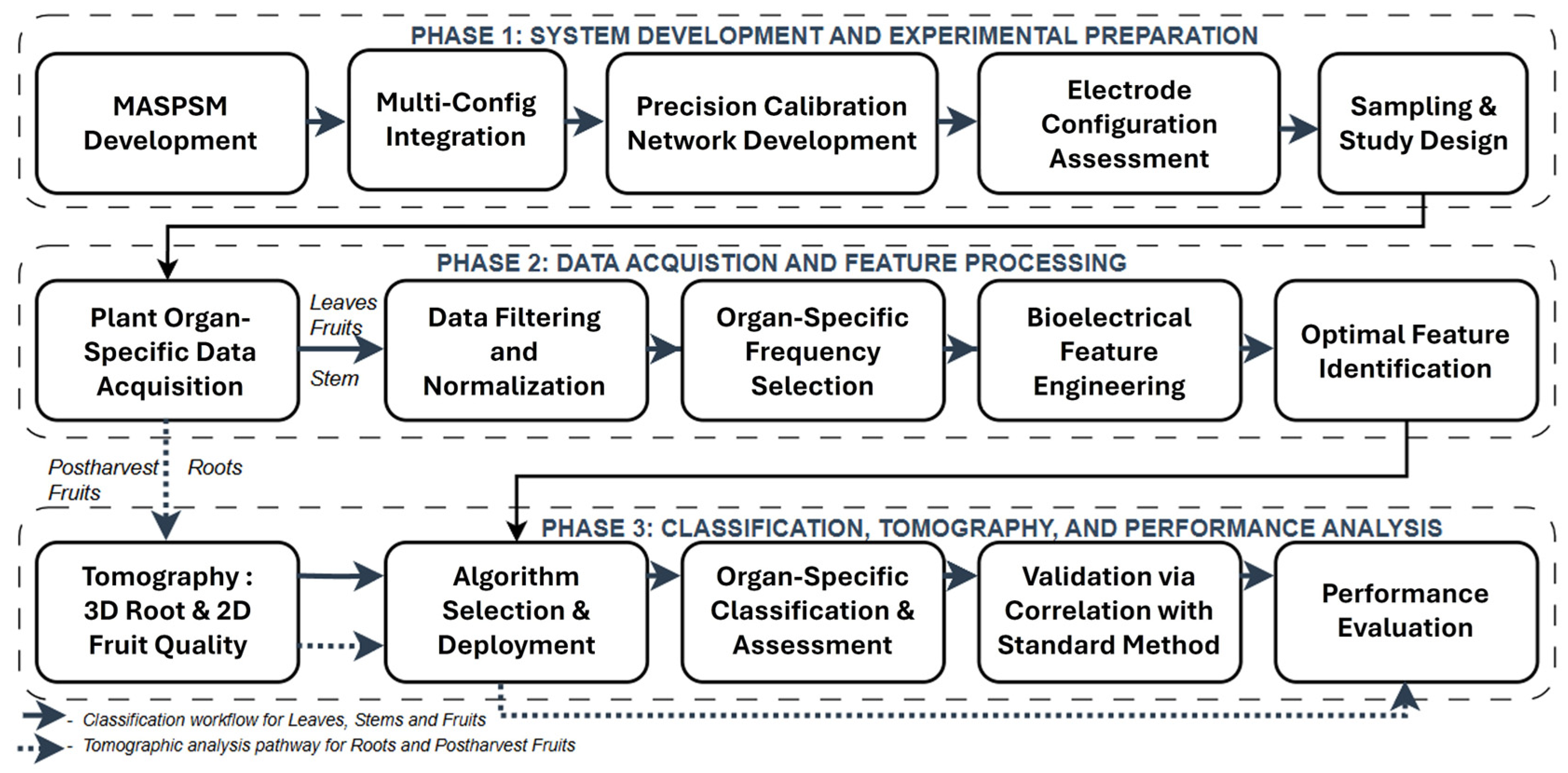
Figure 1. Comprehensive methodology workflow of the bioelectrical sensor system.
Technologies 13 00496 g001
Figure 2. Non-invasive, multimodal, and multi-configuration bioelectrical sensing system: (a) organ-specific electrode configurations and signal-processing architecture; (b) internal architecture of the modified analog signal-processing module (MASPM) enabling multi-configuration switching.
Technologies 13 00496 g002
Figure 3. Tomographic setup simulation: (a) 2D conductivity slices used to form volumetric reconstruction; (b) 3D conductivity volumetric reconstruction and conductivity distribution.
Technologies 13 00496 g003
Figure 4. Bipolar electrode configuration for leaf bioelectrical measurements: (a) illustration showing electrode placement on abaxial leaf surface; (b) field implementation showing the complete leaf bioelectrical measurement setup.
Technologies 13 00496 g004
Figure 5. Bipolar configuration for fruit bioelectrical measurements: (a) illustration showing screen-printed electrodes embedded in conical substrate; (b) field implementation showing the complete strawberry fruit bioelectrical measurement setup.
Technologies 13 00496 g005
Figure 6. Electrode configurations for stem bioelectrical measurements: (a) tetrapolar clip-on Ag/AgCl electrode configuration schematic; (b) field implementation of NBAS system measuring strawberry stem.
Technologies 13 00496 g006
Figure 7. Tomography measurement setups: (a) 3D roots zone conductivity mapping; (b) 2D conductivity localization for postharvest fruit sorting.
Technologies 13 00496 g007
Figure 8. Algorithm performance comparison and TabPFN classification results: (a) algorithm performance heatmap showing accuracy percentages across four machine learning algorithms (TabPFN, CatBoost, LightGBM, and XGBoost) for three organ types; (bd) TabPFN confusion matrices detailing classification performance for (b) fruit, (c) leaf water status, and (d) stem productivity.
Technologies 13 00496 g008
Figure 9. Temporal 3D root conductivity mapping showing (a) normal management with U-Net reconstruction, (b) stressed management with U-Net, (c) normal management with FBP, and (d) stressed management with FBP over 49 days.
Technologies 13 00496 g009
Figure 10. Postharvest strawberry quality assessment through conductivity localization showing (ac) original fruit arrangements with (a) uniform good quality, (b) mixed quality with central defects, (c) uniform poor quality, (df) SIRT reconstructions, and (gi) Modified Back-Projection reconstructions. Color bar indicates conductivity (S/m).
Technologies 13 00496 g010
Table 1. Comparative performance of electrode types and configurations for organ-specific spectroscopic measurements.
OrganElectrode TypeConfigurationSignal Quality (SNR, dB)Contact
Resistance(Ω)
Repeatability (CV, %)Contact Area (mm2)
Leaf Hydrogel Patch Bipolar 48.2 ± 1.8 950 ± 120 2.1 ± 0.4 3.14
Tetrapolar 46.1 ± 1.9 1050 ± 1402.6 ± 0.53.14
Clip Ag/AgCl Bipolar 42.8 ± 2.3 1280 ± 1703.4 ± 0.73.14
Tetrapolar 39.6 ± 2.6 1450 ± 1954.1 ± 0.92.83
Fruit Conical Screen Printed Bipolar 47.8 ± 1.6 890 ± 115 2.3 ± 0.4 7.07
Tetrapolar 45.2 ± 1.8 1020 ± 1352.7 ± 0.57.07
Adhesive Patch Bipolar 41.3 ± 2.2 1320 ± 1853.5 ± 0.82.83
Tetrapolar 38.9 ± 2.5 1480 ± 2104.2 ± 0.93.14
Stem Clip-on Ag/AgCl Bipolar 42.3 ± 2.1 1180 ± 1653.3 ± 0.77.07
Tetrapolar 46.8 ± 1.7 920 ± 125 2.4 ± 0.5 5.31
Clamp Ag/AgCl Bipolar 35.6 ± 2.8 1580 ± 2455.1 ± 1.24.91
Tetrapolar 40.1 ± 2.4 1350 ± 1903.8 ± 0.92.83
Table 2. Optimal multi-scale frequency with statistical validation across plant organs.
OrganFrequency (kHz)Cohen’s dMutual Information
Leaf0.51.340.847
1.01.280.823
5.01.670.912
10.01.450.876
25.01.230.798
50.01.180.765
Fruit1.01.120.734
10.01.410.856
50.01.780.934
75.01.650.898
100.01.520.867
200.01.340.812
Stem1.00.980.687
25.01.150.743
100.01.340.798
250.01.280.776
500.01.220.754
1000.01.080.712
Table 3. Evaluated frequency ranges for tomographic reconstruction.
ApplicationFrequency RangePenetration Depth (mm)Spatial Resolution (mm)SensitivityPerformance Index
3D Root
Conductivity
Mapping
0.01–0.1 kHz70 ± 86.2 ± 0.80.850.68
0.1–10 kHz48 ± 33.8 ± 0.30.710.92
10–100 kHz22 ± 32.7 ± 0.30.340.42
2D Fruit
Conductivity
Localization
0.1–1 kHz55 ± 65.1 ± 0.60.780.72
1–50 kHz28 ± 23.1 ± 0.20.620.94
50–500 kHz12 ± 22.1 ± 0.30.230.35
Disclaimer/Publisher’s Note: The statements, opinions and data contained in all publications are solely those of the individual author(s) and contributor(s) and not of MDPI and/or the editor(s). MDPI and/or the editor(s) disclaim responsibility for any injury to people or property resulting from any ideas, methods, instructions or products referred to in the content.