Article

AM-Induced Alteration in the Expression of Genes, Encoding Phosphorus Transporters and Enzymes of Carbohydrate Metabolism in Medicago lupulina

1
Laboratory of Ecology of Symbiotic and Associative Rhizobacteria/All-Russia Research Institute for Agricultural Microbiology, 196608 Saint Petersburg, Russia
2
Department of Geobotany and Plant Ecology/Saint Petersburg State University, 199034 Saint Petersburg, Russia
3
Laboratory of Molecular and Environmental Physiology/Komarov Botanical Institute of the Russian Academy of Sciences, 197376 Saint Petersburg, Russia
4
Laboratory of Biosystematics and Cytology/Komarov Botanical Institute of the Russian Academy of Sciences, 197376 Saint Petersburg, Russia
5
Laboratory of Genetics of Plant–Microbe Interactions/All-Russia Research Institute for Agricultural Microbiology, 196608 Saint Petersburg, Russia
6
Department of Plant Physiology and Biochemistry/Saint Petersburg State University, 199034 Saint Petersburg, Russia
*
Author to whom correspondence should be addressed.
Plants 2020, 9(4), 486; https://doi.org/10.3390/plants9040486
Submission received: 23 January 2020 / Revised: 5 April 2020 / Accepted: 8 April 2020 / Published: 10 April 2020
(This article belongs to the Special Issue Contribution of Mycorrhizal Symbiosis to Plant Growth)

Abstract

:
Plant–microbe interactions, including those of arbuscular mycorrhiza (AM), have been investigated for a wide spectrum of model plants. The present study focuses on an analysis of gene expression that encodes phosphate and sugar transporters and carbohydrate metabolic enzymes in a new model plant, the highly mycotrophic Medicago lupulina MLS-1 line under conditions of phosphorus deficiency and inoculation with Rhizophagus irregularis. Expression profiles were detected by RT-PCR at six plant stages of development (second leaf, third leaf, shooting, axillary shoot branching initiation, axillary shoot branching, flowering initiation). In comparison to control (without AM), the variant with AM inoculation exhibited a significant elevation of transcription levels of carbohydrate metabolic enzymes (MlSUS, MlHXK1) and sucrose transporters (MlSUC4) in M. lupulina leaves at the shooting stage. We suggest that this leads to a significant increase in the frequency of AM infection, an abundance of mycelium in roots and an increase in AM efficiency (which is calculated by the fresh weight of aerial parts and roots at the axillary shoot branching initiation stage). In roots, the specificity of MlPT4 and MlATP1 gene expressions were revealed for effective AM symbiosis. The level of MlPT4 transcripts in AM roots increased more than tenfold in comparison to that of non-specific MlPT1 and MlPT2. For the first time, MlPT1 expression was shown to increase sharply against MlPT2 in M. lupulina roots without AM at the shooting initiation stage. A significant increase in MlRUB expression was revealed at late stages in the host plant’s development, during axillary shoot branching and flowering initiation. The opposite changes characterized MlHXK1 expression. Alteration in MlHXK1 gene transcription was the same, but was more pronounced in roots. The obtained results indicate the importance of genes that encode phosphate transporters and the enzymes of carbohydrate metabolism for effective AM development at the shooting stage in the host plant.

1. Introduction

More than 80% of plant species can form arbuscular mycorrhiza (AM) with the Glomeromycotina subdivision of the Mucoromycota division in native ecosystems [1]. Nowadays, AM is being actively introduced in agricultural systems. Thus, the investigation of the mechanisms responsible for the symbiotic efficiency of plant interaction with AM fungi has both theoretical and practical importance. Molecular, physiological and biochemical approaches are widely employed with the aim of revealing these mechanisms.
In AM symbiosis, the host plant supplies AM fungus with photosynthates in the form of hexose [2] and, to a lesser extent, sucrose [2,3,4]. The plant, in turn, receives water and a number of macro- and microelements, especially phosphorus [5]. Thus, AM efficiency depends on the intensity of phosphate and carbohydrate transport processes between the partners. Alteration in the carbohydrate/mineral nutrient exchange might convert the plant–microbial interaction from a mutualistic to a parasitic one, which might affect the intensity of host plant development [5,6,7].
Plant growth depression was shown for spring wheat plants (Triticum aestivum) inoculated with Glomus intraradices (DAOM181602 strain, now classified as Rhizophagus irregularis) and Gigaspora margarita (WFVAM21 strain) in comparison to control plants without AM under conditions of different levels of phosphorus deficiency in the substrate. The elevation of AM efficiency (reduction in the growth depression) was detected with a rise in plant density per pot. It was supposed that such increasing AM efficiency was determined not only by less competition for soil phosphorus in AM plants (and greater competition in plants without AM), but also by reducing the cost of carbohydrate transfer to AM fungi interacting with a greater number of plants [7]. Thus, the intensity of supply in between the host plant and the microorganism contributes to the regulation of AM symbiotic efficiency. The importance of phosphate and carbohydrate transporters as well as enzymes of carbohydrate assimilation are of great interest for the investigation of efficient AM symbiosis formation and development. Regulation of the proteins’ activity appeared at different levels, including at the transcriptional level. The identification of genes encoding enzymes of carbohydrate metabolism, sugars and phosphate transporters, as well as the investigation of its expression, are major priorities for contemporary AM research.

1.1. Phosphate Transport in AM Plants

Soil phosphates are insoluble which makes them a limiting factor for plant growth. AM symbiosis is beneficial for plants because AM fungi absorb inorganic phosphorus from the soil, help transform it into polyphosphate, accumulate it, move it through hyphae, hydrolyze it, and then transport it to plant cells in the form of phosphate anions. Inside the root, AM fungi are represented as the mycelium, the vesicles, and the arbuscules. The latter is believed to play a key role in the mutual nutrition and signal exchange between partners [5]. It was shown that the formation of AM increases the intensity of phosphorus uptake by the roots of the host plant by two to four times, whereas 80% of phosphorus is absorbed by mycosymbionts [8]. AM fungi efficiently assimilate phosphates because of their high adsorption of mycelium and the fact that active excretion of organic acids and, in particular, phosphatases result in its ability to dissolve unavailable phosphorus [9]. The elevation of phosphorus uptake into AM plants is related to the activity of AM-specific phosphate transporters of the PHT1 family [5,10]. PHT1 transporters belong to the phosphate transporters of the H+-symporter family, PHS [11]. For example, MtPT4 is a key phosphate transporter from AM fungi to M. truncatula [12]. MtPT4 gene expression was discovered only in cells with arbuscules. The level of expression increased during AM development [13]. On the contrary, the expression of MtPT1 was suppressed in the presence of AM or at high phosphorous levels. The MtPT1 protein is localized in root epidermal cells. It is the main plant transporter of phosphorus uptake directly from the soil. A decrease in the amount of MtPT1 protein in the roots of M. truncatula with AM indicates that this transporter is not involved in symbiotic phosphate transport [14]. MtPT2 gene expression is also significantly suppressed with high levels of phosphorus in the substrate. The accumulation of MtPT3 gene transcripts decreases both in the presence of phosphorus and AM fungi. However, AM scarcely reduces the expression of the MtPT5 gene [12]. Despite the recent presentation of the data dynamics of MtPT6-MtPT9 [15,16,17] and MtPT10-MtPT13 [18], the nature of their expression is still puzzling.
A number of proteins were also shown to modulate the intensity of phosphate transport. The H+-ATPase (ATPase1, MtHA1) was found to be specific for AM and important due to the necessity to generate a proton gradient required for phosphate transport through the periarbuscular membrane [9,19,20]. PHR1 [21,22,23] and CBX1 [24] transcription factors regulate the activity of genes involved in the efficient transport of phosphate in AM structures. CBX1 presumably regulates the genes encoding AM-specific transporters of the PHT1 family (e.g., MtPT4) and H+-ATPase (e.g., MtHA1), as well as proteins involved in fatty acid biosynthesis [24].
Many of the mechanisms of phosphate transport in AM are still poorly understood. Data is still inadequate on the expression of the transporter genes of the PHT1 family and the mechanisms regulating their expression.

1.2. Carbohydrate Metabolism of AM Plants

Plant carbohydrate metabolism was shown to change significantly during AM formation. The host plant transfers up to 30% of assimilates towards the AM fungus [2,25]. Besides carbohydrates, it donates other metabolites, such as fatty acids, known to be used as a source of organic carbon directed in the synthesis of fungal lipids [26]. AM affects photosynthesis activity (increase in chlorophyll content, leaf surface area, and CO2 fixation rate) and enhances the photoassimilates’ flux towards host roots [5]. A similar complex effect is never obtained with the phosphorus fertilizer treatment. It is presumed that plants receive better benefit by effective symbiosis with AM fungi than by developing an equivalent level of root absorption.
The assumption is that AM affects the activity of symbiosis specific plant genes encoding metabolic enzymes and sugar transporters [27,28,29]. For example, in the case of symbiotic sucrose synthase MtSucS1 knockdown, the development of arbuscules in M. truncatula was inhibited [30]. However, little is known about the dynamics of the expression of genes encoding such important enzymes as ribulose-1,5-bisphosphate carboxylase/oxygenase (RUBISCO) and hexokinases in mycorrhizal plants. The hexokinase family of enzymes is involved in glucose phosphorylation; RUBISCO is responsible for carbon dioxide fixation in the Calvin cycle [31,32]. Such a lack of detailed information might be due to the complications of discovering specific pathways which integrate transport, deposition and catabolism during AM formation.
Plant sugar transporters are integrated in three key families: the sucrose/H+-symporter Sucrose Transporter (SUT), Monosaccharide Transporters (MST), including the Sugar Transport Protein subfamily (STP), etc., and Sugars Will Eventually be Exported Transporters (SWEETs). For some plant species, Sucrose Facilitator (SUF) and Chloroplast Maltose Exporter (MEX) transporters have additionally been identified [33]. These transporters have been shown to be involved in the transport of sugars in AM plants. An increase in Mtst1 (MtMST family) accumulation during mycorrhization has been previously shown [34]. Subsequent investigation has shed some light on the common mechanisms of sugar transport. These include: detection and analysis of sink cell hexose transporters MtMSTs in Medicago truncatula [27,35], the sucrose transporters SlSUTs in Solanum lycopersicum during mycorrhization (e.g., low expression of SlSUT2 leads to higher mycorrhization) [29,36], StSUT1 in S. tuberosum (overexpression of the gene leads to increased mycorrhization) [29,37], as well as SUT4 [29] and genes of the SWEET family, StSWEET2c, StSWEET7a, StSWEET12a in S. tuberosum (which have specific localization in arbuscular-containing root cells) [29,38] and LjSWEET3 in Lotus japonicus (increased expression in AM) [29,39]. Expression of the genes from the MtSTP family was revealed in arbuscule-containing cells of M. truncatula [29]. Thus, AM induces alterations in the representation of sugar transporters from different gene families; however, they are not well documented [40].
There are still gaps in the scheme of carbohydrate metabolism and phosphate/carbohydrate transport in plants with and without AM fungi [33]. New approaches such as transcriptomic, proteomic and metabolic analyses are used in recent studies. Thus, new data focusing on key enzymes and transporters in plants forming AM are anticipated.
The aim of this study is to evaluate the expression of genes encoding sugar and phosphate transporters, as well as important carbohydrate metabolism enzymes. The expression profile was detected in the plants of the Medicago lupulina MlS-1 line with and without Rhizophagus irregularis inoculation of AM fungi that were highly responsive to mycorrhization (highly mycotrophic). The comparison was carried outduring host plant development under conditions of low phosphorous content in the substrate.

2. Results

The analysis of mycorrhization parameters of the M. lupulina MLS-1 plant line with a highly intensive response to AM inoculation showed that the frequency of mycorrhizal infection (F) even at the second true leaf stage of plant development (2L) (for abbreviations for stages of development see Table 1), at 21 days after sowing (DAS), was already 39.6% and increased by 1.9 times up to FI (57 DAS) (Figure 1a). The abundance of mycelium without arbuscules is expressed by the “[100% − a]” value, another important parameter “[1 − a]/a” reflects the ratio of the mycelium abundance without arbuscules to the abundance of arbuscules in the root. The “[(1 − a)/a]” value (Figure 1b) rose substantially during development: slowly (1.8 times) up to 33 DAS (shooting (SH) stage of M. lupulina plants) and more sharply (3.5 times) up to 57 DAS (at FI). We observed an increase in the abundance of mycelium without vesicles in comparison with the abundance of vesicles. The parameter “[(1 − b)/b]” reflects the ratio of the mycelium abundance without vesicles to the abundance of fungal vesicles in the root. Its value rises by 2.6 times up to SH and is followed by a significant (p < 0.05) decrease from 33 DAS to 57 DAS at FI (Figure 1c). The latter stage of plant development is characterized by an elevation in the abundance of mycelium without vesicles which was five times higher than that with vesicles.
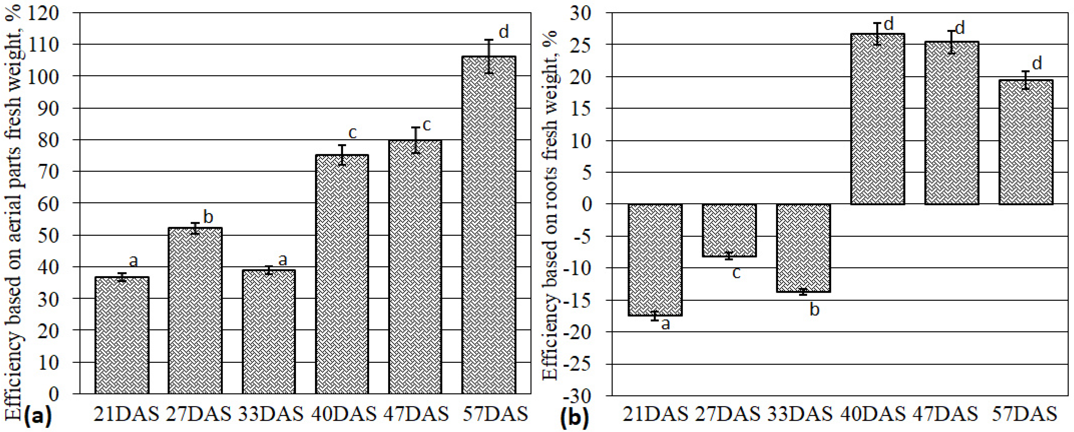
AM efficiency was calculated as a ratio of the difference in fresh weight between mycorrhized plants (+AM) and control plants (−AM) to that of the control variant. Significant increases in AM efficiency were observed (1.9 times from 33 DAS to 40 DAS; Figure 2a). Further on, an increase from 40 DAS to 57 DAS was determined (1.4 times; Figure 2a). At the same time, dramatic changes were detected in the AM efficiency calculated as function of the fresh root weight (Figure 2b). Control plants (−AM) had a more developed root system at earlier stages (including 33 DAS). Active root growth was observed in plants with AM starting from SH. Significant AM efficiency was shown at the next stage of M. lupulina development (axillary shoot branching initiation (SBI) at 40 DAS). AM efficiency for fresh root weight was maintained until the last stage of analysis, flowering initiation (FI).
The alteration in AM efficiency is linked to the intensity of phosphate and carbohydrate exchanges between the host plant and AM fungus. This investigation is focused on the expression of genes encoding representatives of the transporters involved in the exchange. The genes were selected and primers for these genes were designed (Table 2). Sequence data was obtained from the early performed transcriptome assembly of the analyzed highly responsive M. lupulina MLS-1 line within the frame of a separate investigation (SUBID: SUB6631257; BioProject: PRJNA592725; BioSample: SAMN13439142; Accession: GIDJ00000000; Organism: Medicago lupulina).
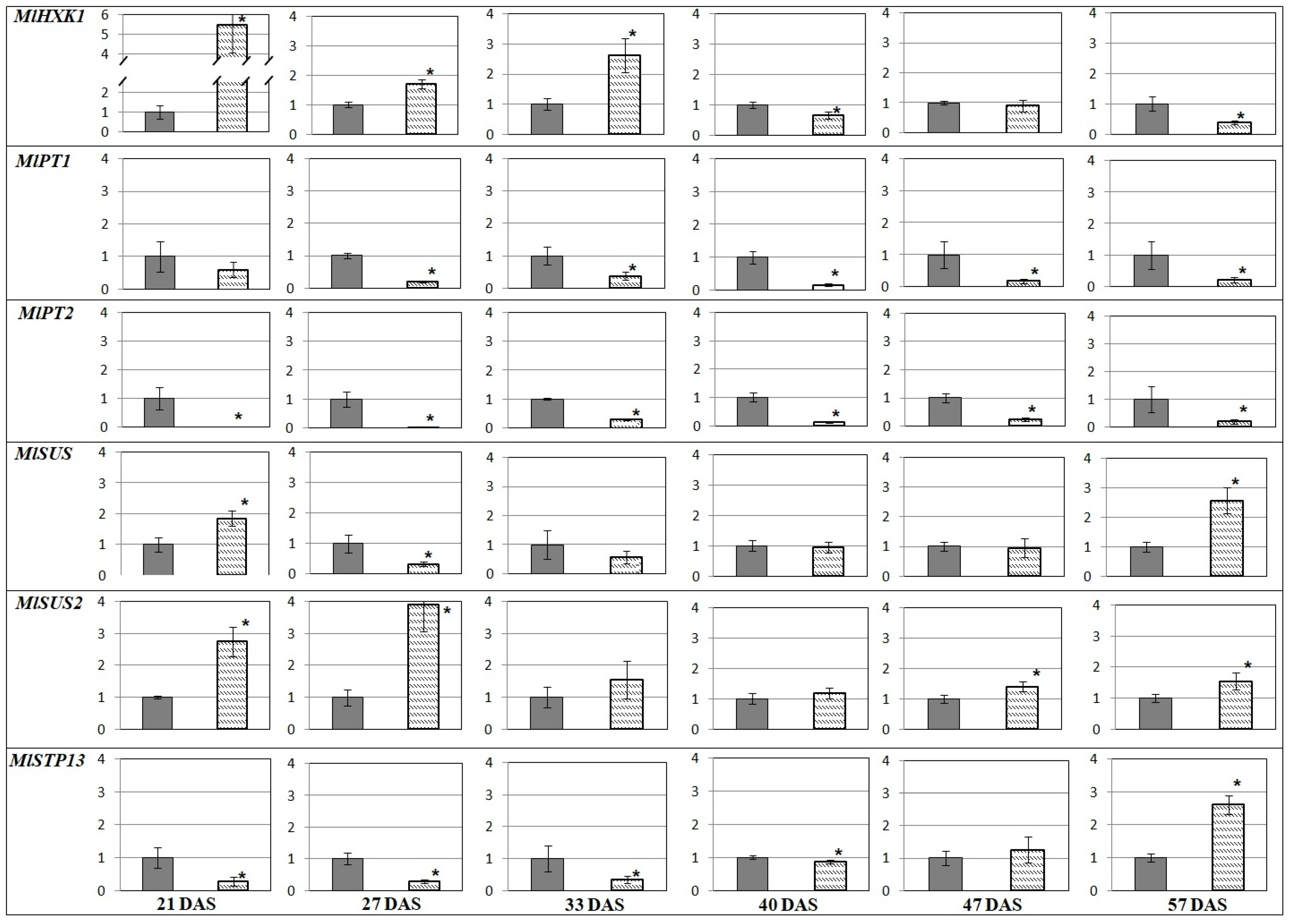
A comparison of the transcription profiles revealed that genes encoding the sugar transporter (MlSUC4) and the enzymes of carbohydrate metabolism (such as MlSUS and MlHXK1) had higher expression in AM plant leaves vs. the control (−AM) at SH (33 DAS; Figure 3). In roots, transcript levels were higher at the beginning (MlHXK1, MlSUS, MlSUS2) and the end of vegetation (MlSUS, MlSUS2, MlSTP13). MlPT1 and MlPT2 genes were continuously expressed at higher levels in plants without AM (Figure 4). The obtained results showed that the expression of genes encoding the RUBISCO small subunit (MlRUB) had complex dynamics (Figure 3). In our experiments, the accumulation of MlRUB transcripts was less intense in AM plant leaves at the initial stages of M. lupulina development (up to SH, 33 DAS). Then, up to SBI at 40 DAS, the MlRUB transcript level in AM plant leaves was aligned with the transcript level in the control plants, becoming higher at SB (47 DAS) and FI (57 DAS). In the latter stages of development, carbon assimilation apparently prevails over its degradation, because the decrease in MlHXK1 and increase in MlRUB transcript levels against control (−AM) in the latter stages of development (40–57 DAS; Figure 3).
The highest MlHXK1 transcript accumulation both in leaves (Figure 3) and in roots of AM plants (Figure 4) was estimated at 21 DAS and, at SH, 33 DAS.
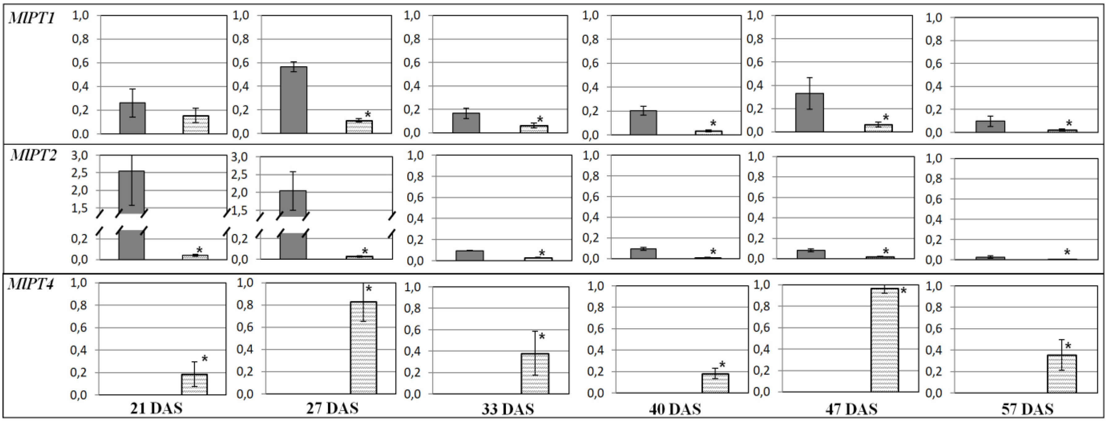
Expressions of genes encoding sucrose synthases, MlSUS and MlSUS2, in leaves had similar dynamics, except SH (33 DAS). The number of MlSUS transcripts was significantly larger than MlSUS2 transcripts. From SH, the expressions of both genes were significantly (p < 0.05) lower in the leaves of AM plants compared to the control plants (‒AM) (Figure 3). However, in roots, the expressions of these genes, on the contrary, were aligned at SBI and after. At FI, the sucrose synthase transcription levels in leaves of AM plants exceeded those in the control (Figure 4). The sucrose-H+ symporter gene MlSUC4 was less transcripted in leaves of AM plants throughout development, except SH at 33 DAS (Figure 3). Accumulation of MlSTP13 transcripts, the gene encoding the monosaccharide-H+ symporter, was also low and characterized by complicated dynamics in both the leaves and roots (Figure 3 and Figure 4). The first significant decrease in MlSTP13 expression in leaves of AM plants against control was observed at SH. In contrast, MlSTP13 expression levels in AM plant roots were less intensive than in the control at the initial stages of development (21–33 DAS), but expression levels then increased up to SBI (40 DAS). It was shown that the expression of MlPT1 and MlPT2 genes (Pi-transporters genes) was significantly weaker in the roots of AM plants at all stages of M. lupulina development (Figure 4). Presumably, Pi uptake in conditions of low Pi content in the substrate was mainly due to transporters specific of AM. MlATP1 (gene encoding H+-ATPase) and MlPT4 (Pi-transporter gene) are presumably involved in Pi transport across the periarbuscular membrane in cells of M. lupulina roots interacting with R. irregularis AM fungi. Since MlPT1, MlPT2 and MlPT4 belong to the same PHT1 gene family (like homologs in M. truncatula), we assumed that it is possible to compare the levels of their transcripts, normalized to the common reference gene (Figure 5). Such an approach of data comparison permitted highlighting the contribution of MlPT2 transcripts in M. lupulina roots without AM (between four and 10 times more than MlPT1 transcript levels at 2L and 3L stages). It changed to a higher contribution of MlPT1 transcripts (two to four times more than MlPT2 transcript levels) with a key shift at SH (33 DAS) and with a maximum at SBI. On the other hand, in inoculated plants at the 2L stage (21 DAS), the symbiotrophic Pi uptake was fulfilled by the MlPT4 transporter. Its transcription was probably at the level of the non-symbiotrophic Pi uptake with the participation of MlPT1 and MlPT2 transporters. However, the proportion of MlPT4 transcripts increased dramatically from 48% to 85% at the 3L stage, and grew until FI (57 DAS), when it reached 93% (almost 15 times more than total MlPT1 and MlPT2 transcript levels; see Figure 5). Thus, it can be supposed that the main Pi transport in M. lupulina roots occurred in a symbiotic way from R. irregularis AM fungi directly through the detected MlPT4 transporter.

3. Discussion

An analysis of the AM symbiotic efficiency in M. lupulina with R. irregularis showed significant (p < 0.05) decreases in the SH stage (Figure 2). Such a decrement might be caused by the necessity of energy and metabolites supporting developmental processes for the host plant. According to our earlier data, M. lupulina’s SH stage was characterized by a maximum increase in symbiotrophic Pi uptake, both in leaves and roots [41]. Further plant development from SH to SBI was accompanied by the sharp increase in symbiotic efficiency in the weight of both aerial parts and roots. Thus, M. lupulina plants achieved the most effective interaction with R. irregularis. The root weight efficiency reached the maximum (Figure 2b), while the weight of the aerial parts continued to grow (Figure 2a). Therefore, it can be expected that SH stage can be characterized by the key changes in the AM plants’ metabolism. Note that F did not change much before SH (33 DAS) (Figure 1a), but then elevated significantly. Zea mays was characterized by similar F alterations during stem development [42]. F is a complex parameter of AM development, which indicates that host plants from SH begin to accelerate development of AM fungi’s symbiotic structures, especially mycelium.
The abundance of intraradical mycelium “[1−a]” exceeds the abundance of arbuscules (a) in the latter stages of AM development after the SH stage (40–57; Figure 1b). Mycelium in roots can directly participate in Pi transfer to plants (the Pi level in intercellular hyphae was significantly higher than the Pi level in arbuscules) [43] and sucrose flux to AM fungi [44]. The possibility of active metabolic processes in symbiotic mycelium might be realized by diverse types of AM—for instance, the Paris type [45]. A significant role of mycelium in the effective AM symbiosis in the latter stages was shown for symbiosis of M. lupulina with R. irregularis [41]. The expression of sucrose fungal transporter gene MST2 (in M. truncatula + Glomus sp. symbiotic system) was determined not only in arbuscules but also in the intercellular hyphae, which indicated that both structures appear to be important for sugar unloading [44]. The mycelium of the AM fungi probably receives carbohydrates from the apoplast of the root cortex [5] as glucose [46], and less as sucrose [2]. The exact location and mechanism of carbon transfer from host plants to AM fungi is not yet fully discovered, but it is the focus of recent studies of plant–microbial symbiotic interactions [17,18,27,28,29,33].
Carbohydrate metabolism starts with sugar biosynthesis, so it is important to analyze the possible alteration in the expression of genes encoding RUBISCO subunits. This study focused on the expression of the MlRUB gene, encoding the small RUBISCO subunit (rbcS = RUB). rbcS plays a role in the regulation of the enzyme activity [47]. MlRUB was intensively expressed in both types of M. lupulina leaves (+/−AM). A slight decrease in its expression was detected during SH in AM plants vs. the control. At this stage (33 DAS), the control plants intensively developed (Figure 2) and, presumably, some energy resources were directed towards plant organ formation. This stage was also characterized by the intensive formation of effective AM. Later on (at SB and FI stages), the transcript level for MlRUB increased significantly (Figure 3), ensuring the maintenance of highly effective AM symbiosis. This might reflect the fact that the presence of AM intensified accumulation of chlorophyll and photosynthesis activity in M. lupulina [48].
HXK1 is a vital enzyme localized in cytosol on the outer membrane of the mitochondria in eudicots and in chloroplasts in source cells, as well as in cytosol and apoplasts in sink cells [49]. It should be noted that hexokinase, in addition to the usual function of redistribution of the glucose, also performs a signaling function, mediating the glucose-induced repression of genes associated with photosynthesis, such as rbcS [49,50,51]. The changes in MlHXK1 transcript accumulation are opposite to the dynamics of MlRUB transcript levels in leaves during host plant development (a distinct inverse relationship was revealed), a fact which is in agreement with some earlier results [51]. Unfortunately, there are no published data on the dynamics of rbcS and HXK1 expression levels in the other leguminous plants. However, it was shown that overexpression of the HXK1 gene in Arabidopsis, tomatoes and rice results in lower growth intensity, lower chlorophyll content, suppression of photosynthesis and negative impacts on rbcS expression [51].
A key result was that the MlHXK1 had similar alterations in the leaves and roots of AM plants (Figure 3 and Figure 4), which emphasizes the importance of the enzyme both for roots and aerial organs. At the SH stage, the number of MlHXK1 transcripts was significantly higher (almost 2.8 times) in AM plants compared to the control (Figure 3 and Figure 4). Higher intense sucrose transfer toward R. irregularis might be due to the active development of mycelium ([1−a]; Figure 1b) and other symbiotic structures at the SH stage (F; Figure 1a). The reduction in MlHXK1 transcript levels in M. lupulina AM roots vs. the control (−AM) (Figure 4) after the SH stage may be associated with a decrease in glucose unloading to amyloplasts. The amyloplasts change proliferation in the presence of arbuscules [52,53]; the number of amyloplasts in cells with arbuscules was either greater (Z. mays, Vitis vinifera [52,54,55]), or less (Lotus japonicus [56]) depending on the presence of the former. Thus, the number and distribution of amyloplasts in the root cortex cells with arbuscules are species-specific parameters, and it can be assumed that the content of sugars in the cytosol of root cortex cells depends intensively on their metabolism.
An essential enzyme for effective AM development is Sucrose Synthase (SUS), glycosyl transferase, which is believed to play an important role in the formation of uridine diphosphate glucose during cellulose synthesis. The MlSUS transcription level increased sharply during plants’ shooting stage with AM vs. the control (Figure 3). In our experiment, this period coincided with significant changes in AM development: (1) F significantly increased (Figure 1a); (2) the abundance of mycelium without arbuscules ([1−a]) aligned with the abundance of arbuscules (a) and continued to grow (Figure 1b). For MtSucS1 and MtSucS2, which are homologs of MlSUS and MlSUS2 in M. truncatula [30,57], gene expression was determined only at 50 DAS [30]. The authors suggested: (1) the important role of MtSucS1 in the maintenance of arbuscules must be taken into account; (2) the M. truncatula mutant lines with reduced expression of MtSucS1 showed abnormalities in their expression of symbiotic Pi-transporter MtPT4, and usually formed short-lived arbuscules; (3) the expression of MtSucS2 was two to three times higher than that of the wild-type line [30]. On the contrary, the highly mycotrophic MLS-1 line of M. lupulina in our experiment showed a high AM efficiency and a high level of MlSUS transcripts in leaves at SH. Synthesis of SUS is well known to be increased under stress conditions [58]. Therefore, its lower content in the latter stages of M. lupulina development might indicate that AM plants completely overcome the Pi deficiency. MlSUS2 transcript levels in leaves of AM plants are lower at all stages of development (Figure 3), so MlSUS2 is assumed to play a minor role in carbohydrate metabolism in the leaves of M. lupulina AM plants.
Nevertheless, MlSUS2 is expressed more actively than MlSUS in AM roots in comparison with the control (‒AM). This may highlight a significant redistribution of sucrose in sink cells and root cells with AM, because SUS catalyze a reversible reaction of synthesis/degradation of sucrose in sugar-consuming root cells [27,59]. According to the literature, genes of the family of sucrose synthases tend to be characterized by increased expression in roots with AM [30]. SUS1 seems to be involved in the induction of arbuscule formation and the maintenance of normal arbuscule maturation and activity [30].
Another method for sucrose reloading in mesophyll cells is possible extrusion from the plant vacuole into cytoplasm due to the sucrose symporter SUC4. The MlSUC4 showed a large transcript level only during the SH stage, when the intensive development of symbiotic structures was observed (Figure 3). Further higher expression of MlSUC4 in control plants was associated with phosphate starvation caused by low Pi level in the substrate, and therefore caused conditions where there was a constant lack of organic compounds for the normal development of the host plant. In M. truncatula, MtSUT4-1 (homolog to MlSUC4) is of greatest interest. It encodes the sucrose symporter, which is actively accumulated both in root cells with arbuscules and in the cells of leaves. MtSUT4-1 localizes in the tonoplast and is involved in sucrose transfer from vacuole to cytosol [60]. Thus, the sucrose in M. lupulina roots can be exported from the vacuole into the cytoplasm by MlSUC4. MlSUC4 in M. lupulina roots is very low. The mechanisms for sucrose unloading in roots (including SUC4 expression) have been studied very fragmentarily and partially based on indirect modeling [61].
A reverse influx of sugars from the cell wall to cytosol of mesophyll cells can also be provided by STP13. A clear trend in the decreasing transcript level of MlSTP13 (hexose transporter) was detected in the leaves of M. lupulina AM plants vs. the control (Figure 3). In contrast, the transcript level of MlSTP13 increased significantly in the roots of M. lupulina-mycorrhized plants in the latter stages (Figure 4). Such results obtained for M. lupulina may indicate an intensive flux of hexoses at the FI stage from the arbuscules of R. irregularis to the cytosol of plant cells, or hexose transport from the cell wall space to the arbuscule-containing cells [29].
Pi transfer associated with carbohydrate metabolism is localized mainly in peri-arbuscular space [62]. Specific Pi transporters were found in M. truncatula, Solanum tuberosum, Oryza sativa, and other plants [63,64,65]. This study showed, for the first time, that MlPT4 expression in M. lupulina roots was intensive and AM-specific. Transcript levels of AM-specific MlPT4 were higher than the levels of both MlPT1 and MlPT2 transcripts in M. lupulina roots at FI (Figure 5). Our conclusions are that: (1) a significant increase in AM efficiency in M. lupulina during SH, SBI and FI (Figure 2) is linked with high MlPT4 expression in the roots of AM plants. This may be associated with active development of intraradical mycelium (Figure 1b). (2) Mycelium can play an important role in the transport of phosphate and carbohydrates under conditions of low Pi levels in the substrate.
Symbiotic Pi uptake across the periarbuscular membrane requires the generation of a proton gradient by H+-ATPase [63,66], namely ATP1 (homolog to MtHA1; in M. truncatula, a single isoform, MtHA1, was specifically expressed in cortical cells containing arbuscules) [19,20,66]. MlATP1 expression in M. lupulina roots was AM-specific. Upregulation of H+-ATPase genes has been reported in the AM roots of several plant species: M. truncatula, Solanum lycopersicum and Nicotiana tabacum [19,67,68].
Nonspecific PT1 and PT2 transporters are not localized in the peri-arbuscular membrane [12,14,66,69]. The transcript levels of MlPT1 and MlPT2 in M. lupulina AM roots were lower (vs. control) at all stages of development (Figure 5). Starting from the SH stage, these low transcript levels of MlPT1 and MlPT2 in AM roots were compensated (in abundance) by the high expression of MlPT4. Equally important are the obtained results demonstrating an intensive decrease in the level of MlPT2 transcripts vs. MlPT1 in the roots of M. lupulina plants without AM from the 2L to 3L stages. The level of MlPT1 transcripts (vs. MlPT2) increased at this stage and continued rising until the SBI stage. The results are consistent with the literature data [12]. Thus, MtPT1 expression was higher in plants exposed to Pi starvation [14]. At the high levels of Pi in substrate, the expression of MtPT2 and MtPT1 genes was significantly inhibited [12,14]. The present study showed that M. lupulina plants without AM perhaps experienced an intensive lack of Pi, starting with the 3L stage, and the adaptation to low Pi levels did not occur. M. lupulina plants inoculated with R. irregularis did not experience a lack of Pi up to the FI stage, as confirmed by the low level of MlPT1 vs. MlPT4 transcripts (Figure 5).

4. Conclusions

In conclusion, this study was the first to analyze the expression of phosphate transport and carbohydrate transport, as well as carbohydrate metabolism in genes in M. lupulina AM plants that formed highly effective symbiosis with R. irregularis under conditions of Pi deficiency. Two genes specific for expression in AM, MlATP1 and MlPT4, were identified. The AM plants of M. lupulina did not experience Pi starvation, but M. lupulina plants without AM did not adapt to the low Pi level in the substrate. For the first time, it was shown that the proportion of MlPT1 vs. MlPT2 transcripts in plants without AM was lower at earlier stages of development, but from the stage of shooting initiation (when Pi deficiency begins to manifest itself), the proportion of MlPT1 vs. MlPT2 transcripts significantly increased. MlPT1 can play a key role in the transport of phosphate in M. lupulina without AM under conditions of Pi deficit in the substrate. Nevertheless, the transcript levels of genes encoding carbohydrate metabolic enzymes (MlSUS, MlHXK1) and sucrose transporters (MlSUC4) were higher in leaves of AM plants vs. the control plants at the shooting stage (33rd DAS). This was probably associated with a subsequent significant increase in AM efficiency, calculated by the fresh weight of M. lupulina plants at the axillary shoot branching initiation stage (40th DAS). Thus, the shooting stage was concluded to be the key point in triggering an efficient interaction between M. lupulina and R. irregularis.

5. Materials and Methods

5.1. Biomaterials

In this study, we used the black medic (Medicago lupulina L. subsp. vulgaris Koch; synonyms: black medick, black medic clover, hop clover, hop medic, nonesuch clover, yellow trefoil) MlS-1 line from VIK32 cultivar-population (cultivar-population selected by G.V. Stepanova from Federal Williams Research Center of Forage Production and Agroecology; MlS-1 line selected by A.P. Yurkov and L.M. Jacobi from ARRIAM) [70]. Black medic is a widespread species of Medicago genus, a self-pollinating diploid (2n = 16) with ~800 Mb genome size and high seed production up to 2500 seeds from one plant. MlS-1 plants have signs of dwarfism in the absence of AM fungus inoculation and low P (available for plants) levels in substrates. The highly effective Rhizophagus irregularis RCAM00320 strain (formerly known as CIAM8 Glomus intraradices Schenck & Smith, from the collection at ARRIAM (All-Russia Research Institute for Agricultural Microbiology) was used to inoculate M. lupulina plants. The authors recently carried out refined identification of this strain [71]. Since AM fungi are obligate symbionts, the strain as well as the entire collection of AM fungi were grown in an accumulative culture of Plectranthus australis R.Br. (synonyms: Swedish Ivy, P. verticillatus Druce, P. nummularius Briq.) under conditions of low Pi level in substrate at the Laboratory of Ecology of Symbiotic and Associative Rhizobacteria, ARRIAM [72].

5.2. Microvegetation Method

A.P. Yurkov et al. earlier described the plant cultivation protocol details using phytobox [70,73]. Plants were planted with two seedlings in one vessel filled with a soil–sand mixture weighing 210 g. Agrochemical soil characteristics: sod-podzolic loam-poor soil with a low P content; P2O5 and K2O contents (calculated using the Kirsanov reaction) were 23 mg and 78 mg per 1 kg of soil, respectively; organic matter content was 3.64%; pHKCl was 6.44 and pHH2O was 7.28. The first recording was performed on the 21st day after sowing during the development stage of the second true leaf. Subsequent recordings were carried out at the key stages of plant development. The final stage, flowering initiation, was estimated on the 57th day after sowing (Table 1).

5.3. Evaluation of Mycorrhization Parameters

During microscopic analysis of AM development, the authors used the method of macerating and staining the root samples, which was developed by J.M. Phillips and D.S. Hayman to evaluate AM infections in leguminous plant roots [74]. The method also included staining the roots with a trypan blue solution. For microscopic analysis of AM structures in crushed preparation, the authors used dried, macerated and cut (1 cm long) Medicago roots for analysis by light microscopy (Mikmed-2 var.2 microscope, LOMO, Russia; characteristics: eyepiece × 10, lens × 10, front lens in the flange of the binocular adjustment × 1.0). The estimated length of the microscope field of view was 1.69 mm. Subsequently, mycorrhization indices were calculated [75]: F (the frequency of mycorrhizal infection in the roots), a and b (the abundance of arbuscules and vesicles in the mycorrhized parts of roots, respectively). The minimum biological replication in each of studied variants was eight.

5.4. Evaluation of AM Symbiotic Efficiency

AM efficiency (mycorrhizal growth response) was calculated as the fresh weight of the aerial parts and roots of the plants, using Odum’s formula:
E = ([+АМ;] − [without AМ;]) × 100%/[without АМ;]
where E is the AM symbiotic efficiency; [+АМ] is the value of mycorrhized plant fresh weight; [without АМ] is the value of the fresh weight of plants without AM.

5.5. RNA Isolation and Expression Evaluation for Genes of Interest

Total RNA from plant material was isolated using the Trizol method [76]. PCR was performed with ubiquitin primers (Ubi-f: ATG CGA TYT TTG TGA AGA C and Ubi-r: ACC ACC ACG RAG ACG GAG) and isolated RNA, then PCR was performed after treatment with DNase, and negative results were obtained: DNA absent in RNA probes. The cDNA synthesis was performed using the Maxima First Strand cDNA Synthesis Kit with dsDNase according to the producer’s protocol (Thermo Scientific, Waltham, MA, USA). Approximately 1 μg of the total RNA was selected for synthesis. Genes of interest were selected based on an analysis of the literature [33,77]. Ten homologues of the phosphorus transport and carbohydrate metabolism genes in M. truncatula were selected. The “Ml” prefix was used in gene names to take M. lupulina species into consideration. Thus, MlPT1, MlPT2, MlPT4, MlATP1 phosphorus transport genes, and MlHXK1, MlSUS, MlSUS2, MlSUC4, MlRUB, and MlSTP13 carbohydrate metabolism genes were selected for analysis. Between two and three pairs of primers (synthesized in Evrogen, Moscow, Russian Federation) were chosen for all genes of interest. From these primers, the authors selected the effective pairs, which were tested in PCR. The effective pairs of selected primers for the genes of interest are presented in Table 2.
Changes in gene expression were studied by the qPCR-RT method with a BioRad CFX-96 thermal cycler (BioRad, Hercules, CA, USA) and using the reagent kit to carry out qPCR-RT in the presence of SYBR Green I. The amplification cycle parameters are: +95 °C, 10 min, 1 cycle; +95 °C, 15 s, +60 °C, 30 s, +72 °C, 30 s, 40 cycles. Amplification specificity was assessed by melting curve analysis. Changes in the expression level for the gene of interest in the experiment with AM were comparable to the expression level of this gene in the control without AM, the analysis for which was performed using the comparative 2−∆∆CT method (at the beginning, the normalization of the reference gene, beta actin, was carried out, followed by the normalization for the control sample without AM). Beta actin (β-actin), glyceraldehyde-3-phosphate dehydrogenase (GAPDH), translation elongation factor 1-alpha (EF1) are known as commonly used reference genes [38,78,79]. According to the results of a comparative analysis, β-actin is the most appropriate reference gene for expression level stability, depending on the concentration of cDNA. GAPDH and EF1 showed less stable expression (compared to β-actin) in roots and leaves when the cDNA concentration was changed. The PCR mixture (10 μL) contained 1 μL of 10× buffer B + SYBR Green, 1 μL of 2.5 mM dNTP, 1 μL of MgCl2 (25 mM), 0.3 μL of each pair of primers (10 mM per primer), 0.125 μL (0.625 U) of SynTaq DNA polymerase, 4.275 μL of ddH2O, and a 2 μL cDNA sample. The relative values of gene expression for each sample were evaluated. Biological replication was three, and technical replication was taken in three measurements (3 qPCR-RT reactions).

5.6. Statistical Analysis

One-way ANOVA and the Tukey’s HSD test (p < 0.05) as a post-hoc test were used to compare differences in AM efficiency and mycorrhization parameters at different stage of plant development. The Student’s t-test (p < 0.05 is the presence of significant differences) was used to estimate the significance of differences in the mean values of gene expression levels between the variants with AM and without AM (in control).

Author Contributions

Individual contribution in conceptualization: A.Y., A.K., M.S.; methodology: A.K., K.D., Y.M., A.S., A.A.; software: A.K., A.A.; validation: A.G., K.D.; formal analysis: A.Y., A.K.; investigation: A.Y., A.K., A.G.; resources: A.Y.; data curation: A.Y., A.K.; writing, draft preparation: A.Y.; review and editing: A.Y., A.K., A.G., K.D., A.S., Y.M., A.A., M.S.; visualization: A.Y., A.K., Y.M., K.D.; supervision: A.Y.; project administration: A.Y., M.S.; funding acquisition: A.Y. All authors have read and agreed to the published version of the manuscript.

Funding

This research was funded by the RFBR (grant number 18-016-00220; support was provided for analyzing expression of carbohydrate metabolism genes), RFBR (grant number 19-29-05275; support was provided for analyzing expression of phosphate transport genes and for analyzing symbiotic efficiency parameters) and RSF (grant number 16-16-00118; support was provided for M. lupulina transcriptome construction, for analyzing mycorrhization parameters, and for selection of effective primers for genes of interest).

Acknowledgments

The research was performed using equipment of the Core Center “Genomic Technologies, Proteomics and Cell Biology” in ARRIAM and the equipment of the Research Park “Center for Molecular and Cell Technologies” at St. Petersburg State University.

Conflicts of Interest

The authors declare no conflict of interest.

References

  1. Spatafora, J.W.; Chang, Y.; Benny, G.L.; Lazarus, K.; Smith, M.E.; Berbee, M.L.; Bonito, G.; Corradi, N.; Grigoriev, I.; Gryganskyi, A.; et al. A phylum-level phylogenetic classification of zygomycete fungi based on genome-scale data. Mycologia 2017, 108, 1028–1046. [Google Scholar] [CrossRef] [PubMed] [Green Version]
  2. Delano-Frier, J.P.; Tejeda-Sartorius, M. Unraveling the network: Novel developments in the understanding of signaling and nutrient exchange mechanisms in the arbuscular mycorrhizal symbiosis. Plant Signal. Behav. 2008, 3, 936–944. [Google Scholar] [CrossRef] [PubMed] [Green Version]
  3. Beilby, J.P.; Kidby, D.K. Biochemistry of ungerminated and germinated spores of the vesicular-arbuscular mycorrhizal fungus, Glomus caledonius: Changes in neutral and polar lipids. J. Lipid Res. 1980, 21, 739–750. [Google Scholar] [PubMed]
  4. Luginbuehl, L.H.; Oldroyd, G.E.D. Understanding the arbuscule at the heart of endomycorrhizal symbioses in plants. Curr. Biol. 2017, 27, R952–R963. [Google Scholar] [CrossRef] [PubMed]
  5. Smith, S.E.; Read, D.J. Mycorrhizal Symbiosis, 3rd ed.; Academic Press: London, UK, 2008; 787p. [Google Scholar]
  6. Fitter, A. Costs and benefits of mycorrhizas: Implications for functioning under natural conditions. Experientia 1991, 47, 350–355. [Google Scholar] [CrossRef]
  7. Li, H.; Smith, F.A.; Dickson, S.; Holloway, R.E.; Smith, S.E. Plant growth depressions in arbuscular mycorrhizal symbioses: Not just caused by carbon drain? New Phytol. 2008, 178, 852–862. [Google Scholar] [CrossRef]
  8. Schachtman, D.P.; Read, R.J.; Ayling, S.M. Phosphorus uptake by plants: From soil to cell. Plant Physiol. 1998, 116, 447–453. [Google Scholar] [CrossRef] [Green Version]
  9. Wang, X.X.; Hoffland, E.; Feng, G.; Kuyper, T.W. Phosphate uptake from phytate due to hyphae-mediated phytase activity by arbuscular mycorrhizal maize. Front. Plant Sci. 2017, 8, 684. [Google Scholar] [CrossRef] [Green Version]
  10. Xie, X.; Huang, W.; Liu, F.; Tang, N.; Liu, Y.; Lin, H.; Zhao, B. Functional analysis of the novel mycorrhiza-specific phosphate transporter AsPT1 and PHT1 family from Astragalus sinicus during the arbuscular mycorrhizal symbiosis. New Phytol. 2013, 198, 836–852. [Google Scholar] [CrossRef]
  11. Pao, S.S.; Paulsen, I.T.; Saier, M.H., Jr. Major facilitator superfamily. Microbiol. Mol. Biol. Rev. 1998, 62, 1–34. [Google Scholar] [CrossRef] [Green Version]
  12. Liu, J.; Versaw, W.K.; Pumplin, N.; Gomez, S.K.; Blaylock, L.A.; Harrison, M.J. Closely related members of the Medicago truncatula PHT1 phosphate transporter gene family encode phosphate transporters with distinct biochemical activities. J. Biol. Chem. 2008, 283, 24673–24681. [Google Scholar] [CrossRef] [PubMed] [Green Version]
  13. Voß, S.; Betz, R.; Heidt, S.; Corradi, N.; Requena, N. RiCRN1, a crinkler effector from the arbuscular mycorrhizal fungus Rhizophagus irregularis, functions in arbuscule development. Front. Microbiol. 2018, 9, 1–18. [Google Scholar] [CrossRef] [PubMed]
  14. Chiou, T.J.; Liu, H.; Harrison, M.J. The spatial expression patterns of a phosphate transporter (MtPT1) from Medicago truncatula indicate a role in phosphate transport at the root/soil interface. Plant J. 2001, 25, 281–293. [Google Scholar] [CrossRef] [PubMed]
  15. Grunwald, U.; Guo, W.; Fischer, K.; Isayenkov, S.; Ludwig-Müller, J.; Hause, B.; Yan, X.; Küster, H.; Franken, P. Overlapping expression patterns and differential transcript levels of phosphate transporter genes in arbuscular mycorrhizal, Pi-fertilised and phytohormone-treated Medicago truncatula roots. Planta 2009, 229, 1023–1034. [Google Scholar] [CrossRef] [Green Version]
  16. Pumplin, N.S. Cell Biology of the Arbuscular Mycorrhizal Symbiosis: Localization and Functional Studies of Medicago truncatula Phosphate Transporters and Vapyrin. Ph.D. Thesis, Cornell University, Ithaca, NY, USA, 20 October 2010. [Google Scholar]
  17. Floss, D.S.; Levy, J.G.; Lévesque-Tremblay, V.; Pumplin, N.; Harrison, M.J. DELLA proteins regulate arbuscule formation in arbuscular mycorrhizal symbiosis. Proc. Natl. Acad. Sci. USA 2013, 110, E5025–E5034. [Google Scholar] [CrossRef] [Green Version]
  18. Breuillin-Sessoms, F.; Floss, D.S.; Gomez, S.K.; Pumplin, N.; Ding, Y.; Levesque-Tremblay, V.; Noar, R.D.; Daniels, D.A.; Bravo, A.; Eaglesham, J.B.; et al. Suppression of Arbuscule Degeneration in Medicago truncatula phosphate transporter 4 Mutants is Dependent on the Ammonium Transporter 2 Family Protein AMT2;3. Plant Cell 2015, 27, 1352–1366. [Google Scholar] [CrossRef] [Green Version]
  19. Krajinski, F.; Hause, B.; Gianinazzi-Pearson, V.; Franken, P. Mtha1, a plasma membrane H+- ATPase gene from Medicago truncatula, shows arbusculespecific induced expression in mycorrhizal tissue. Plant Biol. 2002, 4, 754–761. [Google Scholar] [CrossRef]
  20. Krajinski, F.; Courty, P.E.; Sieh, D.; Franken, P.; Zhang, H.; Bucher, M.; Gerlach, N.; Kryvoruchko, I.; Zoeller, D.; Udvardi, M.; et al. The H+-ATPase HA1 of Medicago truncatula is essential for phosphate transport and plant growth during arbuscular mycorrhizal symbiosis. Plant Cell 2014, 26, 1808–1817. [Google Scholar] [CrossRef] [Green Version]
  21. Aung, K.; Lin, S.; Wu, C.; Huang, Y.; Su, C.; Chiou, T. pho2, a phosphate overaccumulator, is caused by a nonsense mutation in a microRNA399 target gene. Plant Physiol. 2006, 141, 1000–1011. [Google Scholar] [CrossRef] [Green Version]
  22. Bari, R.; Datt Pant, B.; Stitt, M.; Scheible, W.R. PHO2, microRNA399, and PHR1 define a phosphate-signaling pathway in plants. Plant Physiol. 2006, 141, 988–999. [Google Scholar] [CrossRef] [Green Version]
  23. Chiou, T.J.; Aung, K.; Lin, S.I.; Wu, C.C.; Chiang, S.F.; Su, C.L. Regulation of phosphate homeostasis by microRNA in Arabidopsis. Plant Cell 2006, 18, 412–421. [Google Scholar] [CrossRef] [PubMed] [Green Version]
  24. Xue, L.; Klinnawee, L.; Zhou, Y.; Saridis, G.; Vijayakumar, V.; Brands, M.; Dörmann, P.; Gigolashvili, T.; Turck, F.; Bucher, M. AP2 transcription factor CBX1 with a specific function in symbiotic exchange of nutrients in mycorrhizal Lotus japonicus. Proc. Natl. Acad. Sci. USA 2018, 115, E9239–E9246. [Google Scholar] [CrossRef] [PubMed] [Green Version]
  25. Bago, B.; Pfeffer, P.E.; Shachar-Hill, Y. Carbon metabolism and transport in arbuscular mycorrhizas. Plant Physiol. 2000, 124, 949–957. [Google Scholar] [CrossRef] [Green Version]
  26. Luginbuehl, L.H.; Menard, G.N.; Kurup, S.; van Erp, H.; Radhakrishnan, G.V.; Breakspear, A.; Oldroyd, G.E.D.; Eastmond, P.J. Fatty acids in arbuscular mycorrhizal fungi are synthesized by the host plant. Science 2017, 356, 1175–1178. [Google Scholar] [CrossRef] [PubMed] [Green Version]
  27. Doidy, J.; Grace, E.; Kühn, C.; Simon-Plas, F.; Casieri, L.; Wipf, D. Sugar transporters in plants and in their interactions with fungi. Trends Plant Sci. 2012, 17, 413–422. [Google Scholar] [CrossRef] [PubMed]
  28. Doidy, J.; van Tuinen, D.; Lamotte, O.; Corneillat, M.; Alcaraz, G.; Wipf, D. The Medicago truncatula sucrose transporter family: Characterization and implication of key members in carbon partitioning towards arbuscular mycorrhizal fungi. Mol. Plant 2012, 5, 1346–1358. [Google Scholar] [CrossRef] [PubMed] [Green Version]
  29. Hennion, N.; Durand, M.; Vriet, C.; Doidy, J.; Maurousset, L.; Lemoine, R.; Pourtau, N. Sugars en route to the roots. Transport, metabolism and storage within plant roots and towards microorganisms of the rhizosphere. Physiol. Plant 2019, 165, 44–57. [Google Scholar] [CrossRef] [Green Version]
  30. Baier, M.C.; Keck, M.; Godde, V.; Niehaus, K.; Kuster, H.; Hohnjec, N. Knockdown of the symbiotic sucrose synthase MtSucS1 affects arbuscule maturation and maintenance in mycorrhizal roots of Medicago truncatula. Plant Physiol. 2010, 152, 1000–1014. [Google Scholar] [CrossRef] [Green Version]
  31. Fliege, R.; Flugge, U.I.; Werdan, K.; Heldt, H.W. Specific transport of inorganic phosphate, 3-phosphoglycerate and triosephosphates across the inner membrane of the envelope in spinach chloroplasts. Biochim. Biophys. Acta 1978, 502, 232–247. [Google Scholar] [CrossRef]
  32. Flügge, U.I.; Fischer, K.; Gross, A.; Sebald, W.; Lottspeich, F.; Eckerskorn, C. The triose phosphate-3-phosphoglycerate-phosphate translocator from spinach chloroplasts: Nucleotide sequence of a full-length cDNA clone and import of the in vitro synthesized precursor protein into chloroplasts. EMBO J. 1989, 8, 39–46. [Google Scholar] [CrossRef]
  33. Yurkov, A.P.; Kryukov, A.A.; Gorbunova, A.O.; Afonin, A.M.; Kirpichnikova, A.A.; Dobryakova, K.S.; Machs, E.M.; Shishova, M.F. Molecular genetic mechanisms of sugar transport in plants in the absence and during arbuscular mycorrhyza development. Ecol. Genet. 2019, 17, 81–99. [Google Scholar] [CrossRef] [Green Version]
  34. Harrison, M.J. A sugar transporter from Medicago truncatula: Altered expression pattern in roots during vesicular-arbuscular (VA) mycorrhizal associations. Plant J. 1996, 9, 491–503. [Google Scholar] [CrossRef] [PubMed] [Green Version]
  35. Lefebvre, B.; Furt, F.; Hartmann, M.A.; Michaelson, L.V.; Carde, J.P.; Sargueil-Boiron, F.; Rossignol, M.; Napier, J.A.; Cullimore, J.; Bessoule, J.J.; et al. Characterization of lipid rafts from Medicago truncatula root plasma membranes: A proteomic study reveals the presence of a raft-associated redox system. Plant Physiol. 2007, 144, 402–418. [Google Scholar] [CrossRef] [PubMed] [Green Version]
  36. Bitterlich, M.; Krügel, U.; Boldt-Burisch, K.; Franken, P.; Kühn, C. The sucrose transporter SlSUT2 from tomato interacts with brassinosteroid functioning and affects arbuscular mycorrhiza formation. Plant J. 2014, 78, 877–889. [Google Scholar] [CrossRef] [PubMed]
  37. Gabriel-Neumann, E.; Neumann, G.; Leggewie, G.; George, E. Constitutive overexpression of the sucrose transporter SoSUT1 in potato plants increases arbuscular mycorrhiza fungal root colonization under high, but not under low, soil phosphorus availability. J. Plant Physiol. 2011, 168, 911–919. [Google Scholar] [CrossRef] [PubMed]
  38. Manck-Gotzenberger, J.; Requena, N. Arbuscular mycorrhiza symbiosis induces a major transcriptional reprogramming of the potato SWEET sugar transporter family. Front. Plant Sci. 2016, 7, 487. [Google Scholar] [CrossRef] [PubMed] [Green Version]
  39. Sugiyama, A.; Saida, Y.; Yoshimizu, M.; Takanashi, K.; Sosso, D.; Frommer, W.B.; Yazaki, K. Molecular characterization of LjSWEET3, a sugar transporter in nodules of Lotus japonicus. Plant Cell Physiol. 2016, 58, 298–306. [Google Scholar] [CrossRef]
  40. Duensing, N. Transport Processes in the Arbuscular Mycorrhizal Symbiosis. Ph.D. Thesis, University of Potsdam, Potsdam, Germany, 24 October 2013. [Google Scholar]
  41. Yurkov, A.P.; Veselova, S.V.; Jacobi, L.M.; Stepanova, G.V.; Yemelyanov, V.V.; Kudoyarova, G.R.; Shishova, M.F. The effect of inoculation with arbuscular mycorrhizal fungus Rhizophagus irregularis on cytokinin content in highly mycotrophic Medicago lupulina line under low phosphorus level in soil. Plant Soil Environ. 2017, 63, 519–524. [Google Scholar] [CrossRef] [Green Version]
  42. Janeeshma, E.; Mirshad, P.P.; Puthur, J.T. Dynamics of Arbuscular Mycorrhizal Development in Relation to Various Growth Stages of Zea mays L. 28th Kerala Science Congress (Jan 2016) Extended Abstracts; University of Calicut: Thenhipalam, Malappuram, 2016; pp. 1975–1979. [Google Scholar]
  43. Ryan, M.H.; McCully, M.E.; Huang, C.X. Location and quantification of phosphorus and other elements in fully hydrated, soil-grown arbuscular mycorrhizas: A cryo-analytical scanning electron microscopy study. New Phytol. 2003, 160, 429–441. [Google Scholar] [CrossRef]
  44. Helber, N.; Wippel, K.; Sauer, N.; Schaarschmidt, S.; Hause, B.; Requena, N. A versatile monosaccharide transporter that operates in the arbuscular mycorrhizal fungus Glomus sp is crucial for the symbiotic relationship with plants. Plant Cell 2011, 23, 3812–3823. [Google Scholar] [CrossRef] [Green Version]
  45. Dickson, S. The Arum-Paris continuum of mycorrhizal symbioses. New Phytol. 2004, 163, 187–200. [Google Scholar] [CrossRef]
  46. Solaiman, M.Z.; Saito, M. Use of sugars by intraradical hyphae of arbuscular mycorrhizal fungi revealed by radiorespirometry. New Phytol. 1997, 136, 533–538. [Google Scholar] [CrossRef]
  47. Suzuki, Y.; Makino, A. Availability of rubisco small subunit up-regulates the transcript levels of large subunit for stoichiometric assembly of its holoenzyme in rice. Plant Physiol. 2012, 160, 533–540. [Google Scholar] [CrossRef] [PubMed] [Green Version]
  48. Yurkov, A.P.; Kryukov, A.A.; Gorbunova, A.O.; Jacobi, L.M.; Vorobiev, N.I.; Shishova, M.F. The Effect of Arbuscular Mycorrhizal Fungus Rhizophagus irregularis on the Development of Black Medick under Phosphorus Deficiency conditions. In Proceedings of the Annual Meeting Society of Plant Physiologists of Russia, Irkutsk, Russia, 10–15 July 2018; I, pp. 840–843. [Google Scholar] [CrossRef]
  49. Granot, D.; David-Schwartz, R.; Kelly, G. Hexose kinases and their role in sugar-sensing and plant development. Front. Plant Sci. 2013, 4, 44. [Google Scholar] [CrossRef] [PubMed] [Green Version]
  50. Moore, B.; Zhou, L.; Rolland, F.; Hall, Q.; Cheng, W.H.; Liu, Y.X.; Hwang, I.; Jones, T.; Sheen, J. Role of the Arabidopsis glucose sensor HXK1 in nutrient, light, and hormonal signaling. Science 2003, 300, 332–336. [Google Scholar] [CrossRef] [PubMed] [Green Version]
  51. Van Dingenen, J.; Antoniou, C.; Filippou, P.; Pollier, J.; Gonzalez, N.; Dhondt, S.; Goossens, A.; Fotopoulos, V.; Inzé, D. Strobilurins as growth-promoting compounds: How stroby regulates Arabidopsis leaf growth. Plant Cell Environ. 2017, 40, 1748–1760. [Google Scholar] [CrossRef]
  52. Hans, J.; Hause, B.; Strack, D.; Walter, M.H. Cloning, characterization, and immunolocalization of a mycorrhizainducible 1-Deoxy-D-Xylulose 5-Phosphate reductoisomerase in arbuscule-containing cells of maize. Plant Physiol. 2004, 134, 614–624. [Google Scholar] [CrossRef] [Green Version]
  53. Genre, A.; Bonfante, P. Building a mycorrhizal cell: How to reach compatibility between plants and arbuscular mycorrhizal fungi. J. Plant Interact. 2005, 1, 3–13. [Google Scholar] [CrossRef]
  54. Bonfante-Fasolo, P. Some ultrastructural features of the vesicular-arbuscular mycorrhiza in the grapevine. Vitis 1978, 17, 386–395. [Google Scholar]
  55. Fester, T.; Strack, D.; Hause, B. Reorganization of tobacco root plastids during arbuscule development. Planta 2001, 213, 864–868. [Google Scholar] [CrossRef]
  56. Gutjahr, C.; Novero, M.; Guether, M.; Montanari, O.; Udvardi, M.; Bonfante, P. Presymbiotic factors released by the arbuscular mycorrhizal fungus Gigaspora margarita induce starch accumulation in Lotus japonicus roots. New Phytol. 2009, 183, 53–61. [Google Scholar] [CrossRef] [PubMed]
  57. Hohnjec, N.; Perlick, A.M.; Puhler, A.; Kuster, H. The Medicago truncatula sucrose synthase gene MtSucS1 is activated both in the infected region of root nodules and in the cortex of roots colonized by arbuscular mycorrhizal fungi. Mol. Plant-Microbe Interact. 2003, 16, 903–915. [Google Scholar] [CrossRef] [PubMed] [Green Version]
  58. Xiao, X.; Tang, C.; Fang, Y.; Yang, M.; Zhou, B.; Qi, J.; Zhang, Y. Structure and expression profile of the sucrose synthase gene family in the rubber tree: Indicative of roles in stress response and sucrose utilization in the laticifers. FEBS J. 2013, 281, 291–305. [Google Scholar] [CrossRef] [PubMed] [Green Version]
  59. Wang, W.; Shi, J.; Xie, Q.; Jiang, Y.; Yu, N.; Wang, E. Nutrient exchange and regulation in arbuscular mycorrhizal symbiosis. Mol. Plant 2017, 10, 1147–1158. [Google Scholar] [CrossRef] [Green Version]
  60. Reinders, A.; Sivitz, A.B.; Starker, C.G.; Gantt, J.S.; Ward, J.M. Functional analysis of LjSUT4, a vacuolar sucrose transporter from Lotus japonicas. Plant Mol. Biol. 2008, 68, 289–299. [Google Scholar] [CrossRef]
  61. Milne, R.J.; Grof, C.P.; Patrick, J.W. Mechanisms of phloem unloading: Shaped by cellular pathways, their conductances and sink function. Curr. Opin. Plant Biol. 2018, 43, 8–15. [Google Scholar] [CrossRef]
  62. Pumplin, N.; Harrison, M.J. Live-cell imaging reveals periarbuscular membrane domains and organelle location in Medicago truncatula roots during arbuscular mycorrhizal symbiosis. Plant Physiol. 2009, 151, 809–819. [Google Scholar] [CrossRef] [Green Version]
  63. Harrison, M.J.; Dewbre, G.R.; Liu, J. A phosphate transporter from Medicago truncatula involved in the acquisition of phosphate released by arbuscular mycorrhizal fungi. Plant Cell 2002, 14, 2413–2429. [Google Scholar] [CrossRef] [Green Version]
  64. Rausch, C.; Daram, P.; Brunner, S.; Jansa, J.; Laloi, M.; Leggewie, G.; Amrhein, N.; Bucher, M. A phosphate transporter expressed in arbuscule-containing cells in potato. Nature 2001, 414, 462–470. [Google Scholar] [CrossRef]
  65. Paszkowski, U.; Kroken, S.; Roux, C.; Briggs, S.P. Rice phosphate transporters include an evolutionarily divergent gene specifically activated in arbuscular mycorrhizal symbiosis. Proc. Natl. Acad. Sci. USA 2002, 99, 13324–13329. [Google Scholar] [CrossRef] [Green Version]
  66. Ferrol, N.; Azcón-Aguilar, C.; Pérez-Tienda, J. Review: Arbuscular mycorrhizas as key players in sustainable plant phosphorus acquisition: An overview on the mechanisms involved. Plant Sci. 2019, 280, 441–447. [Google Scholar] [CrossRef] [PubMed] [Green Version]
  67. Gianinazzi-Pearson, V.; Arnould, C.; Oufattole, M.; Arango, M.; Gianinazzi, S. Differential activation of H+-ATPase genes by an arbuscular mycorrhizal fungus in root cells of transgenic tobacco. Planta 2000, 211, 609–613. [Google Scholar] [CrossRef] [PubMed]
  68. Ferrol, N.; Pozo, M.J.; Antelo, M.; Azcón-Aguilar, C. Arbuscular mycorrhizal symbiosis regulates plasma membrane H+-ATPase gene expression in tomato plants. J. Exp. Bot. 2002, 53, 1683–1687. [Google Scholar] [CrossRef] [PubMed] [Green Version]
  69. Liu, H.; Trieu, A.T.; Blaylock, L.A.; Harrison, M.J. Cloning and characterization of two phosphate transporters from Medicago truncatula roots: Regulation in response to phosphate and to colonization by arbuscular mycorrhizal (AM) fungi. Mol. Plant-Microbe Interact. 1998, 11, 14–22. [Google Scholar] [CrossRef] [PubMed] [Green Version]
  70. Yurkov, A.P.; Jacobi, L.M.; Gapeeva, N.E.; Stepanova, G.V.; Shishova, M.F. Development of arbuscular mycorrhiza in highly responsive in the mycotrophic host plant—Black medick (Medicago lupulina L.). Russ. J. Dev. Biol. 2015, 46, 263–275. [Google Scholar] [CrossRef]
  71. Kryukov, A.A.; Yurkov, A.P. Optimization procedures for molecular-genetic identification of arbuscular mycorrhizal fungi in symbiotic phase on the example of two closely kindred strains. Mycol. Phytopathol. (Mikologiya i fitopatologiya) 2018, 52, 38–48. [Google Scholar]
  72. Yurkov, A.P.; Shishova, M.F.; Semenov, D.G. Features of Development of Black Medic with Endomycorrhizal Fungus; LAMBERT Acad. Publ.: Saarbrücken, Germany, 2010; pp. 54–56. [Google Scholar]
  73. Yurkov, A.P.; Veselova, S.V.; Jacobi, L.M.; Stepanova, G.V.; Kudoyarova, G.R.; Shishova, M.F. The effect of inoculation with arbuscular mycorrhizal fungus Rhizophagus irregularis on auxin content in highly mycotrophic black medick under low phosphorus level in soil. Sel’skokhozyaistvennaya Biologiya [Agric. Biol.] 2017, 52, 830–838. [Google Scholar] [CrossRef]
  74. Phillips, J.M.; Hayman, D.S. Improved procedures for clearing roots and staining parasitic and vesicular-arbuscular mycorrhizal fungi for rapid assessment of infection. Trans. Br. Mycol. Soc. 1970, 55, 158–161. [Google Scholar] [CrossRef]
  75. Trouvelot, A.; Kough, J.L.; Gianinazzi-Pearson, V. Mesure du Taux de Mycorhization VA d’un Système Radiculaire. Recherche de Méthodes Ayant une Signification Fonctionnelle. In Physiological and Genetical Aspects of Mycorrhizae; Gianinazzi-Pearson, V., Gianinazzi, S., Eds.; INRA-Press: Paris, France, 1986; pp. 217–221. [Google Scholar]
  76. MacRae, E. Extraction of plant RNA. Methods Mol. Biol. 2007, 353, 15–24. [Google Scholar] [CrossRef]
  77. Gaude, N.; Bortfeld, S.; Duensing, N.; Lohse, M.; Krajinski, F. Arbuscule-containing and non-colonized cortical cells of mycorrhizal roots undergo extensive and specific reprogramming during arbuscular mycorrhizal development. Plant J. 2012, 69, 510–528. [Google Scholar] [CrossRef]
  78. Wu, J.; Zhang, H.; Liu, L.; Li, W.; Wei, Y.; Shi, S. Validation of reference genes for RT-qPCR studies of gene expression in preharvest and postharvest longan fruits under different experimental conditions. Front. Plant Sci. 2016, 3, 780. [Google Scholar] [CrossRef] [PubMed] [Green Version]
  79. Zhang, M.F.; Liu, Q.; Jia, G.X. Reference gene selection for gene expression studies in lily using quantitative real-time PCR. Genet. Mol. Res. 2016, 15. [Google Scholar] [CrossRef] [PubMed]
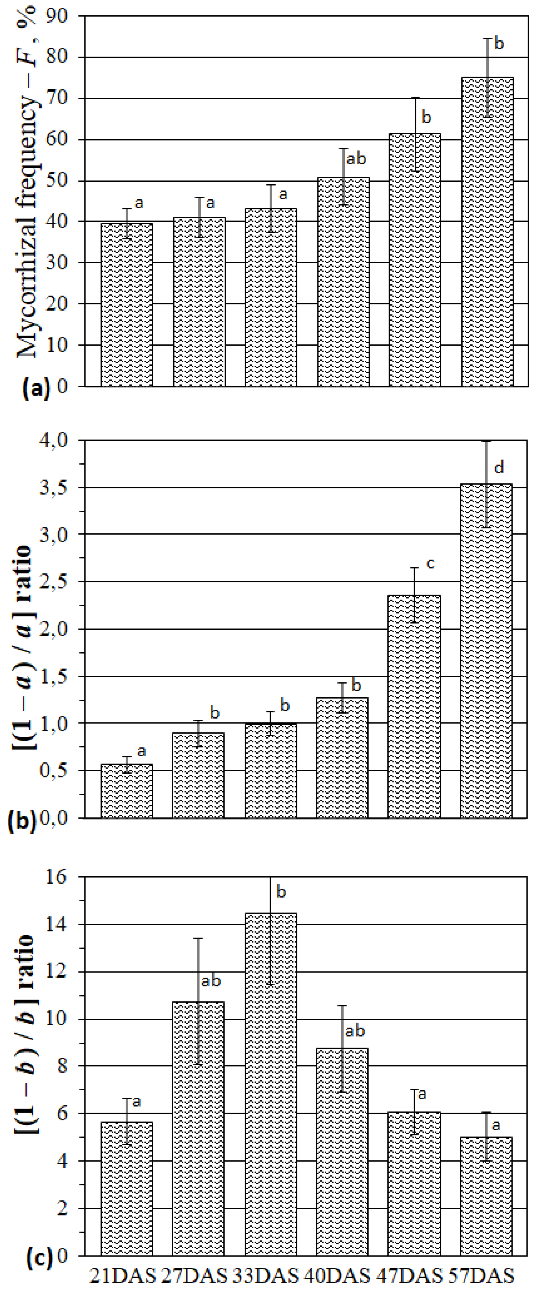
Figure 1. The mycorrhization parameters for M. lupulina roots: mycorrhizal frequency (a); ratio of mycelium abundance without arbuscules to arbuscule abundance, [(1 − a)/a] (b); ratio of mycelium abundance without vesicules to vesicule abundance, [(1 − b)/b] (c). Day after sowing (DAS). Different letters indicate significant differences within the same mycorrhizal parameter (ANOVA and Tukey’s test; p < 0.05). The average values (means of eight replicates) with standard errors are presented.
Plants 09 00486 g001
Figure 2. Arbuscular mycorrhiza (AM) symbiotic efficiency calculated by M. lupulina shoots (aerial parts) fresh weight (a) and roots fresh weight (b). Different letters indicate significant differences within the same efficiency parameter (ANOVA and Tukey’s test; p < 0.05). The average values (means of eight replicates) with standard errors are presented.
Plants 09 00486 g002
Figure 3. Relative transcript levels (2−ΔΔCt normalized values) in M. lupulina leaves. The average values (means of three replicates) with standard errors in the experiment with AM plants against control plants without AM (gray bar) are presented. * —significant (p < 0.05) differences using the t-student test. MlPT1, MlPT2, MlPT4 and MlATP1 genes not expressed.
Plants 09 00486 g003
Figure 4. Relative transcript levels (2−ΔΔCt normalized values) in M. lupulina roots. The average values (means of three replicates) with standard errors in the experiment with AM plants against control plants without AM (gray bar) are presented. * —significant (p < 0.05) differences using the t-student test. MlPT4 and MlATP1 genes were specifically expressed in roots with AM and therefore were not presented. MlSUC4 expression was very low in roots with/without AM and therefore was not analyzed.
Plants 09 00486 g004
Figure 5. Relative transcript levels (2−ΔCt normalized values) of genes from the same PHT1 gene family in M. lupulina roots. * —significant (p < 0.05) differences using the t-student test. MlPT4 was specifically expressed in plants with AM.
Plants 09 00486 g005
Table 1. The plant development stages in Medicago lupulina under conditions of low phosphorus level in substrate and with/without AM inoculation.
Analysis NoDay after Sowing and Inoculation (DAS)Host Plant Development Stage
without AM+AM
1212nd leaf initiation (2LI)2nd leaf development (2L)
2272nd leaf development (2L)shooting initiation, 3rd leaf (3L)
333shooting initiation, 3rd leaf (3L)4th leaf, shooting (SH)
4404th leaf, shooting (SH)5th leaf development, axillary shoot branching initiation (SBI)
5475th leaf development, axillary shoot branching initiation (SBI)6–7th leaf development, axillary shoot branching (SB)
6576–7th leaf development, axillary shoot branching, flowering initiation (FI)8th leaf, flowering initiation (FI)
Note: “without AM” is the variant without AM fungus inoculation, “+AM” is the variant inoculated with R. irregularis AM fungus.
Table 2. Primer sequences for Medicago lupulina genes of interest which are homologues of the phosphorus transport and carbohydrate metabolism genes in M. truncatula.
Gene NamesNames of the PrimersPrimer SequencesGene Bank Sequense Numbers
MlHXK1HXK1-1FCCCTGGAGAACAGATTTTTGAGAMN737455
HXK1-1RCCTCAATTTGCTTCCAACCTC
MlPT1PT1-2FTACTCACTCACATTCTTCTTTGCMN737457
PT1-2RCCAAGCATGATAAGAGAGTTCTTA
MlPT2PT2-2FCAGAACAAGGACAAGAGTAAAGC MN737458
PT2-2RCCGTTTTGTTATGAGAATGAGATTG
MlPT4PT4-1FGGGATTAGAAGTCCTTGAGGCMN737459
PT4-1RGGCCACACCGGTGACTAAG
MlSUSSUS-3FGGAGAGGCTTGATGAAACCTTMN756680
SUS-3RCCAAATGCACCATCAGTCAG
MlSUS2SUS2-2FGATACTCTTTCTGCTCACCGTAMN737460
SUS2-2RGACATTAACACGGACATATTCCC
MlSUC4SUC4-2FGGAGCTATTTGGGACATTCAGMN737461
SUC4-2RCTGAATTAAGCAATAAACCAAGTGC
MlRUBRUB-1FGGCTTCCTCTATGATCTCCTCMN737462
RUB-1RGCCAATAGGAGGCCACACC
MlATP1ATP1-2FCCTTTGTCATGGGTTATGGAAGMN737456
ATP1-2RCATTTTCCGTCGCGAAGTACC
MlSTP13STP13-2FGTTTGTGTCAACTTCCTCTTCACMN737463
STP13-2RCAATGTTGCTTCCACACTCTTTG
MlActinactin7-f2GGCAGATGCTGAGGATATTCAAMN756679
actin7-r2GTATGACGAGGTCGGCCAA
Note: by using the presented primers, the sequenced parts of genes of interest were deposited in GeneBank. Actin7 is beta actin. All transcript lengths for applied primers ranged from 127 bp to 254 bp.