Review

Shedding the Last Layer: Mechanisms of Root Cap Cell Release

by
Narender Kumar
and
Anjali S. Iyer-Pascuzzi
*
Department of Botany and Plant Pathology and Center for Plant Biology, Purdue University, West Lafayette, IN 47907, USA
*
Author to whom correspondence should be addressed.
Plants 2020, 9(3), 308; https://doi.org/10.3390/plants9030308
Submission received: 16 January 2020 / Revised: 21 February 2020 / Accepted: 24 February 2020 / Published: 1 March 2020

Abstract

:
The root cap, a small tissue at the tip of the root, protects the root from environmental stress and functions in gravity perception. To perform its functions, the position and size of the root cap remains stable throughout root growth. This occurs due to constant root cap cell turnover, in which the last layer of the root cap is released, and new root cap cells are produced. Cells in the last root cap layer are known as border cells or border-like cells, and have important functions in root protection against bacterial and fungal pathogens. Despite the importance of root cap cell release to root health and plant growth, the mechanisms regulating this phenomenon are not well understood. Recent work identified several factors including transcription factors, auxin, and small peptides with roles in the production and release of root cap cells. Here, we review the involvement of the known players in root cap cell release, compare the release of border-like cells and border cells, and discuss the importance of root cap cell release to root health and survival.

1. Introduction

Unlike animals, plant cells are fixed and attached together at the cell wall. However, there are several developmental processes in which cell separation is required and critical for plant survival. These include development of intercellular air spaces, leaf abscission, and seed dehiscence [1,2]. Another site of cell separation is the root cap, where the outer layer is sloughed off and released into the soil. The root cap is located at the tip of the root and protects the root apical meristem (RAM), a zone of dividing cells that produces all cells in the root (Figure 1). The root cap shields the RAM from abrasive damage and directs root growth by sensing environmental stimuli such as gravity, temperature gradients, water, touch, and chemicals [3,4,5,6].
Throughout root growth, the root cap maintains a constant size by coupling the production of new cells from root cap stem cells with the detachment of cells from the outermost layer of the root cap [6,7]. Root cap cell detachment results in a sleeve of loosely attached cells covering plant root tips. Because of their role at the root-soil interface, these cells are named “border cells (BCs)” [8,9,10]. In most plant species, BCs separate individually from the root cap and are viable after release from the root [10,11,12,13]. Studies in Arabidopsis, [14,15] and maize [11] have shown that BLCs and BCs can remain viable for days (Arabidopsis) to weeks (maize) after detachment. Unlike most other species, in Arabidopsis and other Brassicaceae members including Brassica napus (rapeseed), Brassica oleracea (Brussels sprout), and Sinapis alba (Mustard), BCs are released from the root tip as an organized layer of several cells that remain attached and are called “border-like cells (BLCs)” [10,14] (Figure 1). BLCs have also been reported in flax (Linum usitatissimum), a member of the Linaceae family [15]. BCs and BLCs protect the root from biotic and abiotic stress [8,12,15,16,17,18].
Root cap cell release is a developmentally programmed process that is linked with cell production and development. In this review, we will focus on molecular factors in the release of BLCs in Arabidopsis, comparisons of BLCs to BCs, and the contribution of root cap cell release to plant survival.

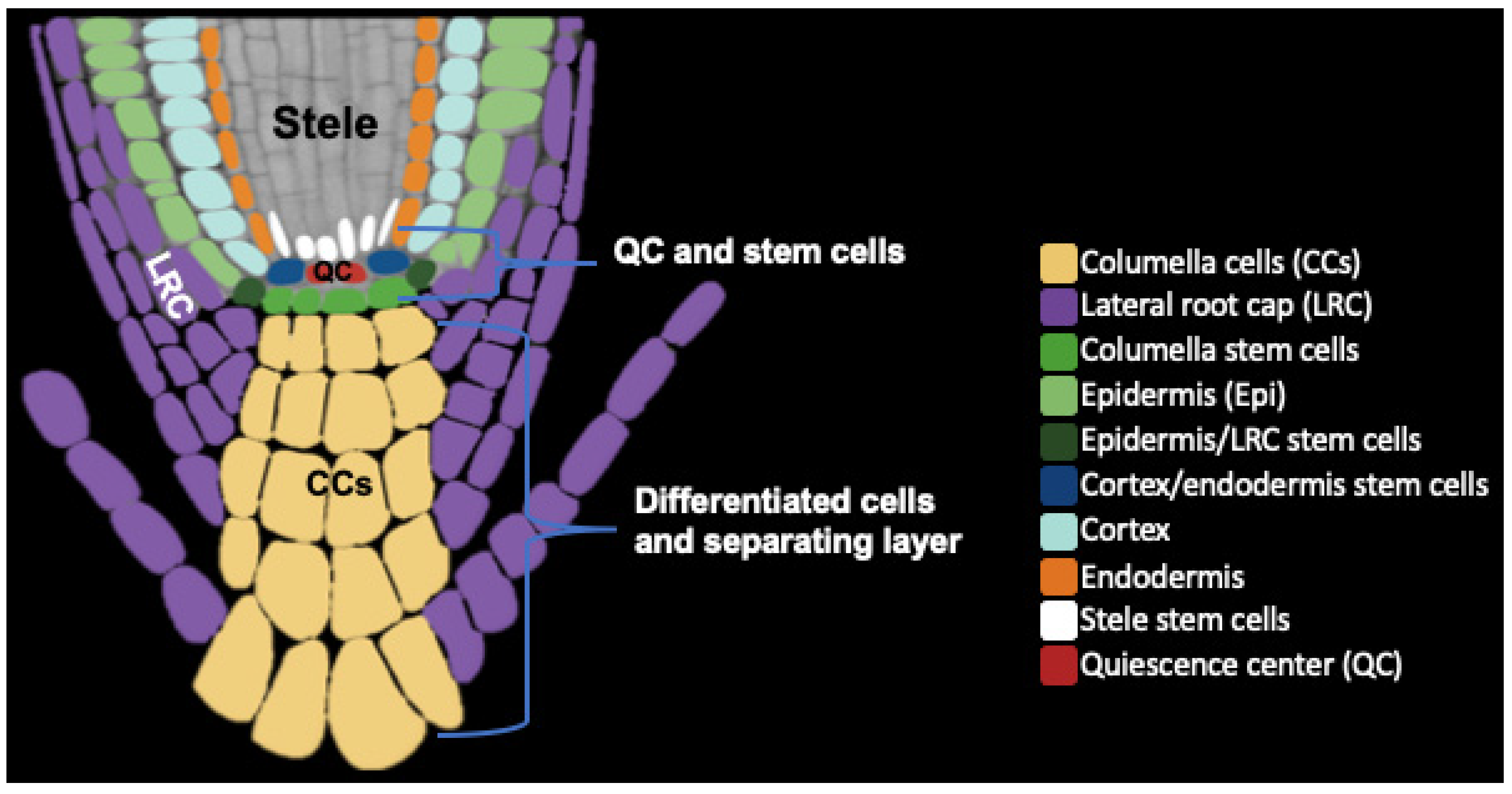
2. Root Cap Structure and BLC Release

The Arabidopsis root cap consists of several layers and is partitioned into two tissues: the columella (COL), at the tip of the root cap, and the lateral root cap (LRC) that surrounds the COL [19] (Figure 1). Both COL and LRC originate from distinct groups of stem cells located in the RAM. The RAM includes cortex/endodermis, stele, columella, and LRC/epidermis stem cells that surround a small group of infrequently dividing cells called the quiescent center (QC). COL stem cells divide asymmetrically to regenerate one stem cell and produce a daughter cell that undergoes differentiation and elongation without further division. The LRC originates from epidermis/LRC initials that are located laterally to the COL stem cells. LRC cells are located on the periphery of the COL and extend toward the shoot to the end of the root meristem. LRC cells undergo programmed cell death before their release [7]. Although some BCs and BLCs remain alive after separation [8,10,11,12,14,15], recent work suggests the presence of a programmed cell death mechanism active during COL cell separation [20]. The root cap secretes polysaccharide-containing mucilage that enhances the root’s ability to the move through the soil [21,22]. In young seedlings, a cuticle layer covers both the COL and LRC outer layers, but is not present at the root epidermis. This root cap cuticle acts as a protective barrier for the developing RAM and is observed primarily in one to three-day old seedlings and has mostly disappeared by 5 days, just before release of the first BLC layer [23]. The cuticle is present on the root caps of both primary and lateral roots, although the thickness and possibly molecular composition differ on each root cap [23].
In the first phase of root cap development, during the first four to five days after germination, the root cap grows through stem cell divisions without root cap cell release [24]. At the end of this first phase, the columella root cap has approximately five layers, including one stem cell tier, and four columella layers [24]. The first root cap layer is released once the Arabidopsis seedling is approximately five days old. At this point, coordination between stem cell division and separation of the outer root cap layer begins [24]. If columella stem cell activity is repressed, this will delay separation of the last layer [24]. Recent work used live imaging to reveal the temporal dynamics of root cap cell release [25]. Images were taken every 15 min for 64 h and 70 h, and revealed that root cap cell release is a multi-step process. In the first step, cell breaks occur in the LRC cell files just above the QC. Approximately 5 h later, another break occurs in the LRC around the youngest layer of COL cells. Finally, COL cells in the last root cap layer at the tip of the root separate from the root cap, and an intact layer of BLCs is released from the root [25]. The entire process takes an average of 18 h, and approximately 18 h passes prior to the initiation of the next release.
The release of BLC and BCs also depends on complex changes and rearrangements to cell wall polysaccharides, including the pectin homogalacturonan (HG) [26]. For example, in Arabidopsis, mutants defective in the synthesis of HG have altered BLC organization [27,28,29]. Additionally, in pea, the activity of pectin methylesterase (PME) is correlated with BC release [30,31], and inhibiting PME expression alters BC release [31].
To maintain a constant size and proper development, the root cap must manage regulation of cell division, cell differentiation, and the separation of the last layer. These processes are the outcome of coordination between transcriptional regulation, signal peptides, and hormone signaling. There has been progress in our understanding of the interplay between transcriptional factors and hormone signaling pathways during root cap development [32], and the relationship between root cap cell release and stem cell division is starting to emerge. In the following section, we will review small peptides and transcriptional regulation as well as hormone signaling in root cap development and BLC release.

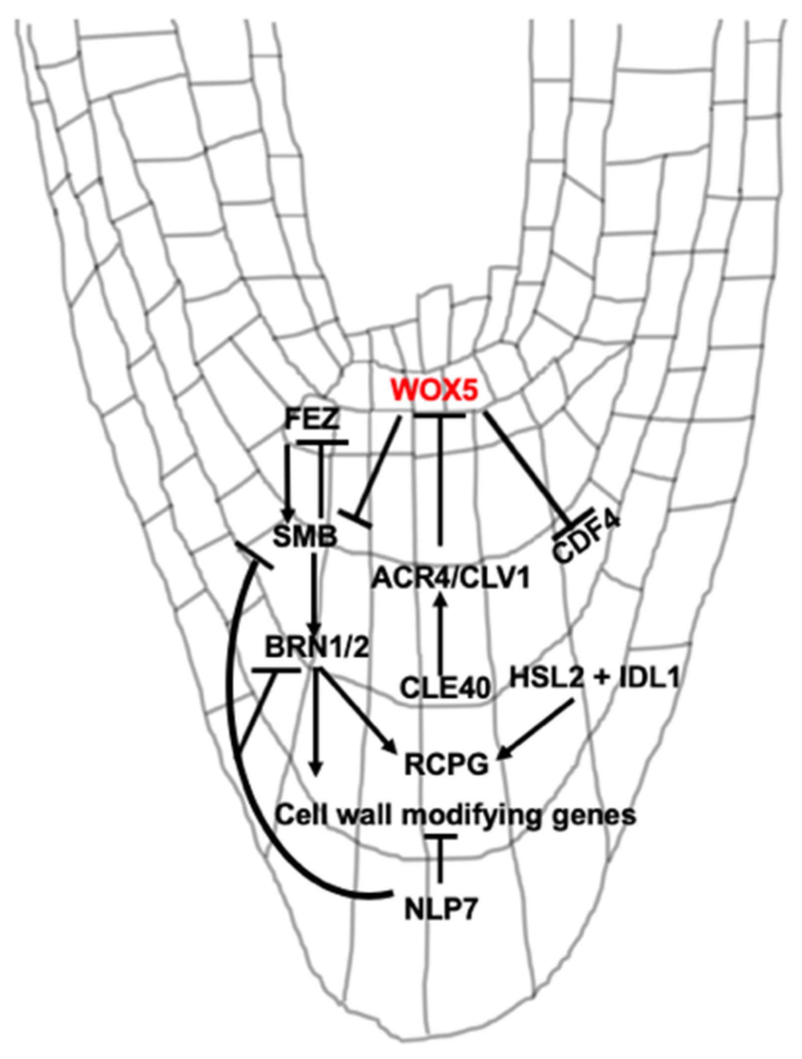
3. Signaling Peptide and Transcriptional Regulation of BLC Release

Transcription factors and signal peptides are major players in BLC release (Figure 2). Because of the importance of root cap cell production to root cap cell release, we briefly review the transcription factors and peptides involved in cell production. For more extensive review, the reader is directed to [32]. For recent reviews of root apical meristem development and the maintenance of the root stem cell niche, see [33,34,35].
New COL cells are produced by COL stem cells (CSCs), which form one or two layers distal to the QC (Figure 1). CSCs remain undifferentiated, while stem cell daughter cells differentiate into COL cells. Differentiation is observed by the presence of starch granules, which are used for gravity perception. The balance between CSCs and differentiated COL cells is accomplished by the antagonistic actions of pathways that either promote ‘stemness’ or promote differentiation. The homeodomain transcription factor WUSCHEL HOMEOBOX 5 (WOX5) is expressed in the QC where it suppresses QC cell division [36] and promotes the undifferentiated status of CSCs [37] (Figure 2). Mutations in WOX5 lead to differentiation of CSCs, while overexpression increases the number of CSC layers [37,38]. Differentiated COL cells produce the small signaling peptide CLE40 [39]. CLE40 is perceived by the receptor kinases ARABIDOPSIS CRINKLY4 (ACR4) and CLAVATA1 (CLV1) which promotes COL cell differentiation [40] and the position of the QC [41]. The CLE40 module may regulate the movement of factors from the QC that promote ‘stemness’ [40,42]. Mutations in CLE40, CLV1, and ACR4 increase CSC layers, while exogenous treatment of wild-type roots with CLE40 induces differentiation of CSCs [40]. In addition, another transcription factor, CYCLIN DOF FACTOR4 (CDF4) is expressed in COL cells and promotes their differentiation [43]. In CSCs, WOX5 represses expression of CDF4 via histone deacetylation [43], which again promotes ‘stemness’. Other factors acting independently of WOX5 also control the fate of CSCs [38,41,42].
Another signaling pair, the peptide IDA-LIKE1 (IDL1) and the receptor-like kinase HAESA-LIKE2 (HSL2), appears to coordinate the dynamics of root separation and production of new layers [25] (Figure 2). IDL1 is highly expressed in the COL, particularly in the last two layers. HSL2 expression is present in young LRC cells as well as upper tiers of the COL [25]. Overexpression of IDL1 in the root cap increased the frequency of BLC release, and this increased separation was compensated by the production of new COL cells [25]. How this signaling pair communicates to regulate the generation and separation of root cap layers is not fully clear.
Several NAC [NAC-domain stands for NAM (no apical meristem), ATAF, CUC (cup-shaped cotyledon)] transcription factors including FEZ, SOMBRERO (SMB), BEARSKIN1 (BRN1), BRN2, ANAC046, and ANAC087 have important roles in root cap development and cell release. FEZ is preferentially expressed in root cap stem cells and promotes periclinal divisions in these cells [44] (Figure 2). FEZ activates expression of SMB. SMB subsequently acts to repress FEZ in stem cell daughters, preventing additional stem cell-like divisions in these cells [44]. The fez mutant has reduced COL and LRC layers [44]. SMB is expressed in differentiated COL and LRC layers. SMB has been implicated in the differentiation, maturation, and cell death of LRC cells [7,44,45]. Mutations in SMB lead to additional cell divisions in the root cap [44] delayed differentiation [45] and LRC cell death [7] and defective root cap cell detachment [45]. In addition, to maintain CSCs, WOX5 appears to exclude SMB expression from the CSCs [38] (Figure 2).
SMB modifies the spatial expression of BRN1 and 2 [46], NAC-domain transcription factors that are important for root cap development. BRN1 and 2 function in cellular differentiation, maturation, and release of BLCs [38,45,46]. Expression of BRN1 and 2 overlaps and is found primarily in the outer layers of the root cap, suggesting a role in the later phase of differentiation [46]. BRN1 and 2 are also involved in mucilage accumulation [21]. BRN1 and 2, along with SMB, regulate the expression of genes associated with root cap differentiation and release of BLCs [45,46]. For example, the expression of cell wall degrading enzymes CELLULASE3 (CEL3) and CEL5 is reduced in smb brn1 brn2 triple mutants [45]. Further, BRN1 and 2 expression overlaps with ROOT CAP POLYGALACTURONASE (RCPG). RCPG is localized to the apoplast and is predicted to have a role in the degradation of cell wall pectins [46]. Overexpression of RCPG releases BLCs as individual cells, unlike a layer in wild-type Arabidopsis. RCPG is a direct target and activated by BRN1 (Figure 2) and RCPG expression is reduced in the brn1 brn2 double mutant. Together, these data suggest a role of BRN1, BRN2, and RCPG in the separation of BLCs [46].
In addition to NAC transcription factors, the transcription factor NIN-LIKE PROTEIN 7 (NLP7) functions in release of BLCs (Figure 2). The nlp7 mutant root releases a higher number of BLCs as single cells compared to wild-type plants [47]. Expression of SMB, BRN1 and 2, and cell wall loosening enzymes such as CEL5, a pectin lyase-like gene, and XYLOGLUCAN ENDOTRANSGLUCOSYLASE (XTH5) are upregulated in nlp7-1. NLP7 is expressed in multiple tissues in the root, including the root cap. In the root cap, NLP7 expression is highest in the last layer [47]. NLP7 is a major regulator of nitrate signaling in plants [48,49], but the functional connections between its roles in nitrate signaling and root cap development are not clear.
In the past few years, good progress has been made in understanding regulatory mechanisms of root cap development and release. It appears that an intricate transcriptional regulation maintains root cap stem cells, differentiation, and release of BLCs (Figure 2). However, several questions remain unanswered or require additional clarity, as discussed in future perspectives below.

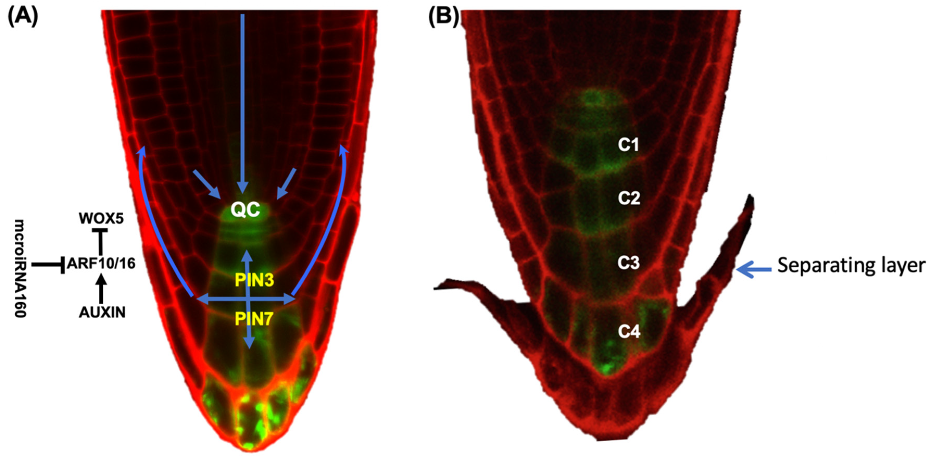
4. The Role of Hormones in BLC Release

The hormone auxin functions as an instructive signal to direct transcriptional events that lead to changes in cell division, differentiation, and morphogenesis. It is an essential hormone in establishing and maintaining the root meristem. A gradient of auxin in the COL root cap is critical to BLC release and proper root cap development [24]. Before root cap cell release initiates, auxin responses are higher in the outer root cap layer compared to inner layers. However, at root cap cell separation, auxin response levels in the outer root cap layer become very low, and a sharp decrease is found in the layer of newly separated BLCs [24] (Figure 3). Thus, root cap cell separation occurs at auxin response minima. Consistent with a requirement for auxin minima for root cap cell separation, expression of the bacterial auxin biosynthesis gene iaaM in the outermost root cap layer increased the number of attached columella layers [24]. These data suggest that auxin response levels act as a threshold below which BLC separation occurs. The number of COL layers, COL length and the duration of the CSC division-BLC release cycle are dependent on the concentration of auxin in the QC and last layer [24]. Consistent with a role for auxin in the regulation of BLC release, auxin biosynthesis mutants have a lower rate of division in the CSCs and separation of the last layer, and a reduced columella size.
The COL expressed auxin efflux transporters PIN-FORMED3 (PIN3), 4 and 7 are important for auxin homeostasis in the root cap [50,51]. PINs can actively transport auxin against a concentration gradient to generate auxin maxima. However, modeling suggested that the root cap auxin gradient is largely the result of auxin movement, decay and dilution from cell elongation and release rather than from PIN transporters actively concentrating auxin in specific cells [24]. PIN expression patterns are important, though, as PINs are repressed in the fourth COL layer, which isolates the fifth separating layer from auxin and likely reinforces the auxin minimum [24] (Figure 3).
Auxin also regulates root cap development through the auxin transcription factors Auxin Response Factor10 (ARF10) and ARF16. ARF10 and ARF16 repress WOX5 and limit it to the QC [52] (Figure 3). An increased number of cell divisions in the root cap stem cell area was observed in arf10/16 double mutants [53]. In addition, the arf10/16 smb triple mutant has an additive effect on the number of root cap cell divisions [38]. Together, these data suggest roles for ARFs 10/16 in root cap differentiation, although whether ARF10/16 function in the release of BLCs is not known. ARFs 10/16 are also regulated by microRNA160 (miR160), an auxin-independent negative regulator of auxin response factors [53,54] (Figure 3).

5. Variations in BC Release Among Species

Release of BCs as individual cells, groups of cells or as a layer appears to depend on the organization of the RAM. RAM organization is conserved within taxa and organized in dicots as open, intermediate-open, and closed patterns based on the tiers of stem initials [55]. For example, Arabidopsis thaliana has closed organization in which all stem cell initials are arranged in the clonally distinct tiers. Carrot (Daucus carota) has an intermediate-open organization where root cap, cortex and vascular tissue originate from the same stem cell. Comparatively, in pea (Pisum sativum), the open RAM organization lacks clonally distinct tiers of stem cells, and different cell types cannot be traced back to specific types of stem cells. Species with open RAM organization produce significantly more BCs compared to species with a closed RAM organization [55]. The number of BCs is conserved at a family level, which can be from a few hundred (Solanaceae) to thousands (Fabaceae, Poaceae, Cucurbitaceae) to nearly 10,000 or more (Pinaceae and cotton) [13,17,55].
The morphology of released BCs also varies from species to species. BLCs released from Arabidopsis are spherical and elongated, while those released from flax are spherical, filamentous, and elongated even though both have a similar type of BLC (i.e., a single layer) [15]. Pea (Pisum sativum) releases BCs with a curved shape. Most are moderately curved (up to 45°), but others are nearly circular [26]. The curvature may be due to the maturation and detachment process, in which BCs elongate and subsequently curve away from the root cap surface. This curvature process is the consequence of complex modifications in BC cell wall components, including changes in pectin and in the distribution of xyloglucans (XyG) and extensin epitopes [26]. In separating BCs, XyG and extensin epitopes were polarly distributed such that they were present on the cell wall facing away from the root [26]. XyG and extension may be important for cell wall rigidity, and the authors speculate that their polar distribution in separating pea BCs helps promotes the BC curvature process [26].
Brachypodium distachyon, a model for grasses, has homogeneous and less curved border cells compared to pea [56]. Recent work suggests that spatial modifications to the pectin homogalacturonan during BC release are similar in B. distachyon and pea [56].
Solanum tuberosum releases two types of BC populations, one near the root tip known as “small border cells” and “elongated border cells” near the elongation zone. These BCs are viable after their detachment from the root tip [57]. Whether differences in BC number or morphology are functionally important is not clear.

6. BC and BLC Protective Functions in Biotic and Abiotic Stresses

The root cap assists the root in making a path through compact soil by secreting mucilage and protecting the RAM, which is vulnerable to damage and infection as it moves through dense microbial communities in the soil. BC and BLC release may act as the first point of defense for plant roots (recently reviewed in [18]) BCs protect the continuously growing root tip by releasing compounds such as protein, polysaccharides, or phytoalexins [18,57,58,59,60] and extracellular DNA in the rhizosphere [16,61,62]. Moreover, BCs are critical to root growth and plant health because these cells contribute to the nutrient-rich exudates in the vicinity of a root and may influence the microbial population by altering the physical and chemical properties of soil [12,16,18,63,64].
BCs from different plant species appear to provide a barrier against pathogens. For example, bacterial [flagellin22 (flg22) and peptidoglycan] and fungal (chitin and fusaric acid) microbe-associated molecular patterns (MAMPs) activate defense responses by triggering the production of reactive oxygen species in BLCs of both Arabidopsis and flax [15]. After elicitation with MAMPs, callose, a well-known cellular marker of plant defense, was observed in BLCs of both species, suggesting active defense responses in these cells [15]. In response to flg22 and the soilborne pathogen Ralstonia solanacaerum, pea (Pisum sativum) border cells release extracellular DNA which entangles microbes [62]. Mutating the DNase-coding gene in R. solanacaerum and the fungal pathogen Cochliobolus heterostrophus results in reduced virulence of these microbes [62,65].
Metabolites secreted from BCs also may function in defense. For example, flavonoid 7,4′-dihydroxyflavone from Medicago truncatula BCs inhibits growth of the soilborne fungal pathogen Phymatotrichopsis omnivora, causal agent of cotton root rot [66]. Exudates from pea root caps inoculated with Nectria haematococca suppressed growth of the fungus [64].
Arabinogalactan proteins (AGPs) are secreted from the cell walls of BCs and BLCs of several species [14,26,67] and may be involved in early events of root infection. For example, zoospores from Aphanomyces euteiches, an oomycete pathogen of pea, were attracted to pea AGPs in a chemotaxis assay [67]. In addition, germination of A. euteiches zoospores appeared to be inhibited by pea AGPs [67]. Potato (Solanum tuberosum) root exudate, enriched in galactose-containing molecules including arabinogalactan proteins, impacts the growth of bacteria Pectobacterium atrosepticum that causes stem wilting of potatoes [57].
Besides protection from biotic stresses, BCs have been reported to protect plant roots from abiotic stress. For example, BCs of different crops immobilize aluminum in order to protect the growing root [22,68,69]. In soybean (Glycine max L.), exposure to Al for 48 h promoted mucilage production in BCs [22]. Compared to BCs from an Al-sensitive cultivar, 48 h after exposure to 400 µM Al, BCs from an Al-resistant cultivar had thicker mucilage. Mucilage from Al-resistant soybeans bound more Al compared to that of Al-susceptible [22]. At high concentrations of Al (400 µM), BCs from the Al-resistant cultivar had higher viability compared to those from the Al-sensitive cultivar [22]. In snapbean (Phaseolus vulgaris), the viability of Al-resistant border cells exposed to 200 µM Al in an agarose gel was higher than that of border cells from an Al-susceptible cultivar [70]. In pea, Al bound to the alkali-soluble pectin in cell walls of BCs, which may protect the root tip from Al stress [69]. High levels of Fe2+ toxicity result in decreased border cell viability in rice [71]. Compared to a more Fe2+ tolerant rice genotype, Fe2+-sensitive rice roots had fewer BCs after exposure to high levels of Fe2+ [71].

7. Conclusions and Future Perspectives

Root cap cell release is developmentally programmed and influenced by transcription factors, small peptides, and hormones. While we are beginning to understand the molecular players involved in this process, many questions remain, including:
  • How do external stresses impact the release of root cap cells and the molecular pathways that control release? How are such stresses communicated in the root cap such that root cap cell production and release remain linked during environmental stress?
  • Do differences in either the timing or type (BLC vs BC) of release impact whole root growth and root architecture? Do BLC and BC release impact root cap protection differently?
  • How are signals between the separating layer and stem cells communicated and what are additional signals?
  • What are the mechanisms used by the auxin pathway to regulate cell production from CSCs and the release of BLCs?
  • What other transcription factors are involved in BLC and BC release? Are there other hormones besides auxin involved in BLC release? Does WOX5 have an impact on BLC release?

Author Contributions

N.K. and A.S.I.-P. wrote the paper. All authors have read and agreed to the published version of the manuscript.

Funding

Root cap research in the AIP laboratory is supported by funding from the National Science Foundation (grant #1656392).

Acknowledgments

We thank members of the AIP laboratory for helpful discussions.

Conflicts of Interest

The authors declare no conflict of interest.

References

  1. Knox, J.P. Cell adhesion, cell separation and plant morphogenesis. Plant J. 1992, 2, 137–141. [Google Scholar] [CrossRef]
  2. Jarvis, M.C.; Briggs, S.P.H.; Knox, J.P. Intercellular adhesion and cell separation in plants. Plant Cell Environ. 2003, 26, 977–989. [Google Scholar] [CrossRef]
  3. Strohm, A.K.; Baldwin, K.L.; Masson, P.H. Molecular mechanisms of root gravity sensing and signal transduction. Wiley Interdiscip. Rev. Dev. Biol. 2012, 1, 276–285. [Google Scholar] [CrossRef] [PubMed]
  4. Tsugeki, R.; Fedoroff, N.V. Genetic ablation of root cap cells in Arabidopsis. Proc. Natl. Acad. Sci. USA 1999, 96, 12941–12946. [Google Scholar] [CrossRef] [Green Version]
  5. Massa, G.D.; Gilroy, S. Touch modulates gravity sensing to regulate the growth of primary roots of Arabidopsis thaliana. Plant J. 2003, 33, 435–445. [Google Scholar] [CrossRef] [Green Version]
  6. Barlow, P.W. The root cap: Cell dynamics, cell differentiation and cap function. J. Plant Growth Regul. 2002, 21, 261–286. [Google Scholar] [CrossRef]
  7. Fendrych, M.; Van Hautegem, T.; Van Durme, M.; Olvera-Carrillo, Y.; Huysmans, M.; Karimi, M.; Lippens, S.; Guérin, C.J.; Krebs, M.; Schumacher, K.; et al. Programmed cell death controlled by ANAC033/SOMBRERO determines root cap organ size in Arabidopsis. Curr. Biol. 2014, 24, 931–940. [Google Scholar] [CrossRef] [Green Version]
  8. Hawes, M.C.; Brigham, L.A.; Wen, F.; Woo, H.H.; Zhu, Y. FUNCTION OF ROOT BORDER CELLS IN PLANT HEALTH: Pioneers in the Rhizosphere. Annu. Rev. Phytopathol. 1998, 36, 311–327. [Google Scholar] [CrossRef]
  9. Hawes, M.C.; Lin, H.J. Correlation of pectolytic enzyme activity with the programmed release of cells from root caps of pea (Pisum sativum). Plant Physiol. 1990, 94, 1855–1859. [Google Scholar] [CrossRef] [Green Version]
  10. Driouich, A.; Durand, C.; Vicre-Gibouin, M. Formation and separation of root border cells. Trends Plant Sci. 2007, 12, 14–19. [Google Scholar] [CrossRef]
  11. Vermeer, J.; McCully, M.E. The rhizosphere in Zea: New insight into its structure and development. Planta 1982, 156, 45–61. [Google Scholar] [CrossRef] [PubMed]
  12. Hawes, M.C.; Gunawardena, U.; Miyasaka, S.; Zhao, X. The role of root border cells in plant defense. Trends Plant Sci. 2000, 5, 128–133. [Google Scholar] [CrossRef]
  13. Hawes, M.C.; Bengough, G.; Cassab, G.; Ponce, G. Root caps and rhizosphere. J. Plant Growth Regul. 2002, 21, 352–367. [Google Scholar] [CrossRef]
  14. Vicré, M.; Santaella, C.; Blanchet, S.; Gateau, A.; Driouich, A. Root border-like cells of Arabidopsis. Microscopical characterization and role in the interaction with rhizobacteria. Plant Physiol. 2005, 138, 998–1008. [Google Scholar] [CrossRef] [PubMed] [Green Version]
  15. Plancot, B.; Santaella, C.; Jaber, R.; Kiefer-Meyer, M.C.; Follet-Gueye, M.L.; Leprince, J.; Gattin, I.; Souc, C.; Driouich, A.; Vicré-Gibouin, M. Deciphering the responses of root border-like cells of Arabidopsis and flax to pathogen-derived elicitors. Plant Physiol. 2013, 163, 1584–1597. [Google Scholar] [CrossRef] [PubMed] [Green Version]
  16. Driouich, A.; Follett-Gueye, M.; Vicre-Gibouin, M.; Hawes, M. Root border cells and secretions are critical elements in plant host defense. Curr. Opin. Plant Biol. 2013, 16, 489–495. [Google Scholar] [CrossRef]
  17. Driouich, A.; Durand, C.; Cannesan, M.A.; Percoco, G.; Vicré-Gibouin, M. Border cells versus border-like cells: Are they alike? J. Exp. Bot. 2010, 61, 3827–3831. [Google Scholar] [CrossRef]
  18. Hawes, M.; Allen, C.; Turgeon, B.G.; Curlango-Rivera, G.; Tran, T.M.; Huskey, D.A.; Xiong, Z. Root Border Cells and Their Role in Plant Defense. Annu. Rev. Phytopathol. 2016, 54, 143–161. [Google Scholar] [CrossRef]
  19. Dolan, L.; Janmaat, K.; Willemsen, V.; Linstead, P.; Poethig, S.; Roberts, K.; Scheres, B. Cellular organisation of the Arabidopsis thaliana root. Development 1993, 119, 71–84. [Google Scholar]
  20. Huysmans, M.; Buono, R.A.; Skorzinski, N.; Radio, M.C.; De Winter, F.; Parizot, B.; Mertens, J.; Karimi, M.; Fendrych, M.; Nowack, M.K. NAC transcription factors ANAC087 and ANAC046 control distinct aspects of programmed cell death in the Arabidopsis columella and lateral root cap. Plant Cell 2018, 30, 2197–2213. [Google Scholar] [CrossRef] [Green Version]
  21. Maeda, K.; Kunieda, T.; Tamura, K.; Hatano, K.; Hara-Nishimura, I.; Shimada, T. Identification of periplasmic root-cap mucilage in developing columella cells of Arabidopsis thaliana. Plant Cell Physiol. 2019, 60, 1296–1303. [Google Scholar] [CrossRef]
  22. Cai, M.; Wang, N.; Xing, C.; Wang, F.; Wu, K.; Du, X. Immobilization of aluminum with mucilage secreted by root cap and root border cells is related to aluminum resistance in Glycine max L. Environ. Sci. Pollut. Res. 2013, 20, 8924–8933. [Google Scholar] [CrossRef] [PubMed]
  23. Berhin, A.; de Bellis, D.; Franke, R.B.; Buono, R.A.; Nowack, M.K.; Nawrath, C. The Root Cap Cuticle: A Cell Wall Structure for Seedling Establishment and Lateral Root Formation. Cell 2019, 176, 1367–1378. [Google Scholar] [CrossRef] [PubMed] [Green Version]
  24. Dubreuil, C.; Jin, X.; Grönlund, A.; Fischer, U. A Local Auxin Gradient Regulates Root Cap Self-Renewal and Size Homeostasis. Curr. Biol. 2018, 28, 2581–2587. [Google Scholar] [CrossRef] [Green Version]
  25. Shi, C.L.; von Wangenheim, D.; Herrmann, U.; Wildhagen, M.; Kulik, I.; Kopf, A.; Ishida, T.; Olsson, V.; Anker, M.K.; Albert, M.; et al. The dynamics of root cap sloughing in Arabidopsis is regulated by peptide signalling. Nat. Plants 2018, 4, 596–604. [Google Scholar] [CrossRef] [PubMed]
  26. Mravec, J.; Guo, X.; Hansen, A.R.; Schückel, J.; Kračun, S.K.; Mikkelsen, M.D.; Mouille, G.; Johansen, I.E.; Ulvskov, P.; Domozych, D.S.; et al. Pea border cell maturation and release involve complex cell wall structural dynamics. Plant Physiol. 2017, 174, 1051–1066. [Google Scholar] [CrossRef] [PubMed] [Green Version]
  27. Durand, C.; Vicré-Gibouin, M.; Follet-Gueye, M.L.; Duponchel, L.; Moreau, M.; Lerouge, P.; Driouich, A. The organization pattern of root border-like cells of Arabidopsis is dependent on cell wall homogalacturonan. Plant Physiol. 2009, 150, 1411–1421. [Google Scholar] [CrossRef] [Green Version]
  28. Bouton, S.; Leboeuf, E.; Mouille, G.; Leydecker, M.-T.; Talbotec, J.; Granier, F.; Lahaye, M.; Höfte, H.; Truong, H.-N. QUASIMODO1 Encodes a Putative Membrane-Bound Glycosyltransferase Required for Normal Pectin Synthesis and Cell Adhesion in Arabidopsis. Plant Cell 2002, 14, 2577–2590. [Google Scholar] [CrossRef] [Green Version]
  29. Mravec, J.; Kračun, S.K.; Rydahl, M.G.; Westereng, B.; Miart, F.; Clausen, M.H.; Fangel, J.U.; Daugaard, M.; Van Cutsem, P.; De Fine Licht, H.H.; et al. Tracking Developmentally Regulated Post-Synthetic Processing of Homogalacturonan and Chitin Using Reciprocal Oligosaccharide Probes. Development 2014, 141, 4841–4850. [Google Scholar] [CrossRef] [Green Version]
  30. Stephenson, M.B.; Hawes, M.C. Correlation of Pectin Methylesterase Activity in Root Caps of Pea with Root Border Cell Separation. Plant Physiol. 1994, 106, 739–745. [Google Scholar] [CrossRef] [Green Version]
  31. Wen, F.; Zhu, Y.; Hawes, M.C. Effect of Pectin Methylesterase Gene Expression on Pea Root Development. Plant Cell 1999, 11, 1129–1140. [Google Scholar] [CrossRef] [Green Version]
  32. Kumpf, R.P.; Nowack, M.K. The root cap: A short story of life and death. J. Exp. Bot. 2015, 66, 5651–5662. [Google Scholar] [CrossRef] [Green Version]
  33. Drisch, R.C.; Stahl, Y. Function and regulation of transcription factors involved in root apical meristem and stem cell maintenance. Front. Plant Sci. 2015, 6, 1–8. [Google Scholar] [CrossRef] [PubMed] [Green Version]
  34. Rahni, R.; Efroni, I.; Birnbaum, K.D. A Case for Distributed Control of Local Stem Cell Behavior in Plants. Dev. Cell 2016, 6, 635–642. [Google Scholar] [CrossRef] [PubMed] [Green Version]
  35. Augstein, F.; Carlsbecker, A. Getting to the roots: A developmental genetic view of root anatomy and function from Arabidopsis to lycophytes. Front. Plant Sci. 2018, 9, 1–15. [Google Scholar] [CrossRef] [PubMed] [Green Version]
  36. Forzani, C.; Aichinger, E.; Sornay, E.; Willemsen, V.; Laux, T.; Dewitte, W.; Murray, J.A.H. WOX5 suppresses CYCLIN D activity to establish quiescence at the center of the root stem cell niche. Curr. Biol. 2014, 24, 1939–1944. [Google Scholar] [CrossRef] [PubMed] [Green Version]
  37. Sarkar, A.K.; Luijten, M.; Miyashima, S.; Lenhard, M.; Hashimoto, T.; Nakajima, K.; Scheres, B.; Heidstra, R.; Laux, T. Conserved factors regulate signalling in Arabidopsis thaliana shoot and root stem cell organizers. Nature 2007, 446, 811–814. [Google Scholar] [CrossRef]
  38. Bennett, T.; van den Toorn, A.; Willemsen, V.; Scheres, B. Precise control of plant stem cell activity through parallel regulatory inputs. Development 2014, 141, 4055–4064. [Google Scholar] [CrossRef] [Green Version]
  39. Stahl, Y.; Wink, R.H.; Ingram, G.C.; Simon, R. A Signaling Module Controlling the Stem Cell Niche in Arabidopsis Root Meristems. Curr. Biol. 2009, 19, 909–914. [Google Scholar] [CrossRef] [Green Version]
  40. Stahl, Y.; Grabowski, S.; Bleckmann, A.; Kühnemuth, R.; Weidtkamp-Peters, S.; Pinto, K.G.; Kirschner, G.K.; Schmid, J.B.; Wink, R.H.; Hülsewede, A.; et al. Moderation of Arabidopsis root stemness by CLAVATA1 and ARABIDOPSIS CRINKLY4 receptor kinase complexes. Curr. Biol. 2013, 23, 362–371. [Google Scholar] [CrossRef] [Green Version]
  41. Berckmans, B.; Kirschner, G.; Gerlitz, N.; Stadler, R.; Simmon, R. CLE40 signalling regulates root stem cell fate. Plant Physiol. 2019. [Google Scholar] [CrossRef] [PubMed] [Green Version]
  42. Richards, S.; Wink, R.H.; Simon, R. Mathematical modelling of WOX5-and CLE40-mediated columella stem cell homeostasis in Arabidopsis. J. Exp. Bot. 2015, 66, 5375–5384. [Google Scholar] [CrossRef] [PubMed] [Green Version]
  43. Pi, L.; Aichinger, E.; van der Graaff, E.; Llavata-Peris, C.I.; Weijers, D.; Hennig, L.; Groot, E.; Laux, T. Organizer-Derived WOX5 Signal Maintains Root Columella Stem Cells through Chromatin-Mediated Repression of CDF4 Expression. Dev. Cell 2015, 33, 576–588. [Google Scholar] [CrossRef] [PubMed] [Green Version]
  44. Willemsen, V.; Bauch, M.; Bennett, T.; Campilho, A.; Wolkenfelt, H.; Xu, J.; Haseloff, J.; Scheres, B. The NAC Domain Transcription Factors FEZ and SOMBRERO Control the Orientation of Cell Division Plane in Arabidopsis Root Stem Cells. Dev. Cell 2008, 15, 913–922. [Google Scholar] [CrossRef] [PubMed] [Green Version]
  45. Bennett, T.; van den Toorn, A.; Sanchez-Perez, G.F.; Campilho, A.; Willemsen, V.; Snel, B.; Scheres, B. SOMBRERO, BEARSKIN1, and BEARSKIN2 regulate root cap maturation in Arabidopsis. Plant Cell 2010, 22, 640–654. [Google Scholar] [CrossRef] [Green Version]
  46. Kamiya, M.; Higashio, S.Y.; Isomoto, A.; Kim, J.M.; Seki, M.; Miyashima, S.; Nakajima, K. Control of root cap maturation and cell detachment by BEARSKIN transcription factors in Arabidopsis. Development 2016, 143, 4063–4072. [Google Scholar] [CrossRef] [Green Version]
  47. Karve, R.; Suárez-Román, F.; Iyer-Pascuzzi, A.S. The transcription factor NIN-LIKE PROTEIN7 controls border-like cell release. Plant Physiol. 2016, 171, 2101–2111. [Google Scholar] [CrossRef] [Green Version]
  48. Castaings, L.; Camargo, A.; Pocholle, D.; Gaudon, V.; Texier, Y.; Boutet-Mercey, S.; Taconnat, L.; Renou, J.P.; Daniel-Vedele, F.; Fernandez, E.; et al. The nodule inception-like protein 7 modulates nitrate sensing and metabolism in Arabidopsis. Plant J. 2009, 57, 426–435. [Google Scholar] [CrossRef]
  49. Marchive, C.; Roudier, F.; Castaings, L.; Bréhaut, V.; Blondet, E.; Colot, V.; Meyer, C.; Krapp, A. Nuclear retention of the transcription factor NLP7 orchestrates the early response to nitrate in plants. Nat. Commun. 2013, 4, 1–9. [Google Scholar] [CrossRef]
  50. Grieneisen, V.A.; Xu, J.; Marée, A.F.M.; Hogeweg, P.; Scheres, B. Auxin transport is sufficient to generate a maximum and gradient guiding root growth. Nature 2007, 449, 1008–1013. [Google Scholar] [CrossRef] [Green Version]
  51. Billou, I.; Xu, J.; Wildwater, M.; Willemsen, V.; Paponov, I.; Frimi, J.; Heldstra, R.; Aida, M.; Palme, K.; Scheres, B. The PIN auxin efflux facilitator network controls growth and patterning in Arabidopsis roots. Nature 2005, 433, 39–44. [Google Scholar] [CrossRef] [PubMed]
  52. Ding, Z.; Friml, J. Auxin regulates distal stem cell differentiation in Arabidopsis roots. Proc. Natl. Acad. Sci. USA 2010, 107, 12046–12051. [Google Scholar] [CrossRef] [PubMed] [Green Version]
  53. Wang, J.W.; Wang, L.J.; Mao, Y.B.; Cai, W.J.; Xue, H.W.; Chen, X.Y. Control of root cap formation by MicroRNA-targeted auxin response factors in Arabidopsis. Plant Cell 2005, 17, 2204–2216. [Google Scholar] [CrossRef] [PubMed] [Green Version]
  54. Liu, P.P.; Montgomery, T.A.; Fahlgren, N.; Kasschau, K.D.; Nonogaki, H.; Carrington, J.C. Repression of AUXIN RESPONSE FACTOR10 by microRNA160 is critical for seed germination and post-germination stages. Plant J. 2007, 52, 133–146. [Google Scholar] [CrossRef] [PubMed]
  55. Hamamoto, L.; Hawes, M.C.; Rost, T.L. The production and release of living root cap border cells is a function of root apical meristem type in dicotyledonous angiosperm plants. Ann. Bot. 2006, 97, 917–923. [Google Scholar] [CrossRef] [Green Version]
  56. Mravec, J. Border cell release: Cell separation without cell wall degradation? Plant Signal. Behav. 2017, 12, 1–3. [Google Scholar] [CrossRef] [PubMed] [Green Version]
  57. Koroney, A.S.; Plasson, C.; Pawlak, B.; Sidikou, R.; Driouich, A.; Menu-Bouaouiche, L.; Vicré-Gibouin, M. Root exudate of Solanum tuberosum is enriched in galactose-containing molecules and impacts the growth of Pectobacterium atrosepticum. Ann. Bot. 2016, 118, 797–808. [Google Scholar] [CrossRef] [Green Version]
  58. Brigham, L.A.; Woo, H.H.; Nicoll, S.M.; Hawes, M.C. Differential expression of proteins and mRNAs from border cells and root tips of pea. Plant Physiol. 1995, 109, 457–463. [Google Scholar] [CrossRef] [Green Version]
  59. Wen, F.; Vanetten, H.D.; Tsaprailis, G.; Hawes, M.C. Extracellular proteins in pea root tip and border cell exudates. Plant Physiol. 2007, 143, 773–783. [Google Scholar] [CrossRef] [Green Version]
  60. Cannesan, M.A.; Gangneux, C.; Lanoue, A.; Giron, D.; Laval, K.; Hawes, M.; Driouich, A.; Vicré-Gibouin, M. Association between border cell responses and localized root infection by pathogenic Aphanomyces euteiches. Ann. Bot. 2011, 108, 459–469. [Google Scholar] [CrossRef] [Green Version]
  61. Wen, F.; White, G.J.; Vanetten, H.D.; Xiong, Z.; Hawes, M.C. Extracellular DNA is required for root tip resistance to fungal infection. Plant Physiol. 2009, 151, 820–829. [Google Scholar] [CrossRef] [PubMed] [Green Version]
  62. Tran, T.M.; MacIntyre, A.; Hawes, M.; Allen, C. Escaping Underground Nets: Extracellular DNases Degrade Plant Extracellular Traps and Contribute to Virulence of the Plant Pathogenic Bacterium Ralstonia solanacearum. PLoS Pathog. 2016, 12, 1–26. [Google Scholar] [CrossRef] [PubMed]
  63. Gunawardena, U.; Hawes, M.C. Tissue specific localization of root infection by fungal pathogens: Role of root border cells. Mol. Plant-Microbe Interact. 2002, 15, 1128–1136. [Google Scholar] [CrossRef] [PubMed] [Green Version]
  64. Gunawardena, U.; Rodriguez, M.; Straney, D.; Romeo, J.T.; VanEtten, H.D.; Hawes, M.C. Tissue-specific localization of pea root infection by Nectria haematococca. Mechanisms and consequences. Plant Physiol. 2005, 137, 1363–1374. [Google Scholar] [CrossRef] [PubMed] [Green Version]
  65. Park, H.J.; Wang, W.; Curlango-Rivera, G.; Xiong, Z.; Lin, Z.; Huskey, D.A.; Hawes, M.C.; Vanetten, H.D.; Turgeon, B.G. A DNase from a fungal phytopathogen is a virulence factor likely deployed as counter defense against host-secreted extracellular dna. MBio 2019, 10, 1–13. [Google Scholar] [CrossRef] [PubMed] [Green Version]
  66. Watson, B.S.; Bedair, M.F.; Urbanczyk-Wochniak, E.; Huhman, D.V.; Yang, D.S.; Allen, S.N.; Li, W.; Tang, Y.; Sumner, L.W. Integrated metabolomics and transcriptomics reveal enhanced specialized metabolism in Medicago truncatula root border cells. Plant Physiol. 2015, 167, 1699–1716. [Google Scholar] [CrossRef] [Green Version]
  67. Cannesan, M.A.; Durand, C.; Burel, C.; Gangneux, C.; Lerouge, P.; Ishii, T.; Laval, K.; Follet-Gueye, M.L.; Driouich, A.; Vicré-Gibouin, M. Effect of arabinogalactan proteins from the root caps of pea and Brassica napus on Aphanomyces euteiches zoospore chemotaxis and germination. Plant Physiol. 2012, 159, 1658–1670. [Google Scholar] [CrossRef] [Green Version]
  68. Nagayama, T.; Nakamura, A.; Yamaji, N.; Satoh, S.; Furukawa, J.; Iwai, H. Changes in the Distribution of Pectin in Root Border Cells Under Aluminum Stress. Front. Plant Sci. 2019, 10, 1–8. [Google Scholar] [CrossRef]
  69. Yang, J.; Qu, M.; Fang, J.; Shen, R.F.; Feng, Y.M.; Liu, J.Y.; Bian, J.F.; Wu, L.S.; He, Y.M.; Yu, M. Alkali-soluble pectin is the primary target of aluminum immobilization in root border cells of pea (Pisum sativum). Front. Plant Sci. 2016, 7, 1–7. [Google Scholar] [CrossRef] [Green Version]
  70. Miyasaka, S.C.; Hawes, M.C. Possible Role of Root Border Cells in Detection and Avoidance of Aluminum Toxicity. Plant Physiol. 2001, 125, 1978–1987. [Google Scholar] [CrossRef] [Green Version]
  71. Zhang, Y.; Wu, Y.; Gendi, X.; Song, J.; Wu, T.; Mei, X.; Liu, P. Effects of Iron Toxicity on the Morphological and Biological Characteristics of Rice Root Border Cells. J. Plant Nutr. 2017, 40, 332–343. [Google Scholar] [CrossRef]
Figure 1. Schematic diagram showing different tissue organization of the root cap (CCs and LRC) and root apical meristem (QC and stem cells) regions. The last root cap layer is separating as a single intact layer known as BLCs (here colored purple and maize). Different cell types and tissues are colored differently.
Plants 09 00308 g001
Figure 2. Involvement of different transcription factors and signaling peptides in Arabidopsis root cap development and BLC release. Arrows and barred lines indicate positive and negative regulation, respectively. Locations of proteins and peptides shown in the root are approximate.
Plants 09 00308 g002
Figure 3. Auxin gradient in five-day old Arabidopsis root cap. (A) Columella localized PIN-FORMED (PIN) 3 and 7 and their flux direction. Auxin responsive factors (ARFs) 10/16 influence WOX5 to control columella cell differentiation and are regulated by auxin and miR160. Blue arrows indicate the direction of auxin flux. PIN3 and PIN7 are critical for auxin flux in the columella root cap, but additional PIN auxin transporters are required for flux in other cell types. Arrows and barred lines indicate positive and negative regulation, respectively. (B) The auxin response disappears from the separating layer. Auxin responsive synthetic promoter (pDR5:GFP) is used to show auxin response (green color). Columella (C) layers from C1 to C4. Arrows and barred lines indicate positive and negative regulations, respectively. Propidium iodide (red color) is used to visualize cells.
Plants 09 00308 g003

