Article

Genome-Wide Identification of the LdARF Gene Family in Lilium davidii var. unicolor and Transient Functional Analysis of LdARF17 in Bulblet Regeneration


1
Institute of Horticultural Biotechnology, Fujian Agriculture and Forestry University, Fuzhou 350002, China
2
Fujian Provincial Key Laboratory of Agricultural Genetic Engineering, Biotechnology Research Institute, Fujian Academy of Agricultural Science, Fuzhou 350003, China
3
Beijing Key Laboratory of Development and Quality Control of Ornamental Crops, Department of Ornamental Horticulture, China Agricultural University, Beijing 100193, China
*
Authors to whom correspondence should be addressed.
These authors contributed equally to this work.
Plants 2026, 15(17), 2581; https://doi.org/10.3390/plants15172581
Submission received: 14 July 2026 / Revised: 17 August 2026 / Accepted: 21 August 2026 / Published: 24 August 2026
(This article belongs to the Section Plant Genetics, Genomics and Biotechnology)

Abstract

Auxin response factors (ARFs) are key transcriptional regulators of the auxin signaling pathway and play important roles in plant organogenesis and regeneration. However, the functions of ARF family genes in lily scale-derived bulblet regeneration remain largely unclear. In this study, 24 LdARF genes were identified from the genome of Lilium davidii var. unicolor. Phylogenetic analysis revealed that LdARF proteins showed evolutionary conservation with ARF homologs from other monocot species. Genome-wide identification, phylogenetic analysis, and expression profiling revealed functional divergence among LdARF genes during scale-derived bulblet regeneration. Among them, LdARF17 exhibited a distinct regeneration-associated expression pattern, characterized by rapid induction after scale excision and sustained high expression during subsequent bulblet initiation and formation. Subcellular localization analysis demonstrated that LdARF17 is localized in the nucleus. Transient overexpression of LdARF17 significantly promoted bulblet regeneration and was associated with increased expression of auxin-responsive and regeneration-related genes, including IAA14, LBD16, and LBD29. These findings suggest that LdARF17 acts as a positive regulator of lily scale regeneration and may influence auxin-responsive transcriptional processes associated with early cell proliferation, providing new insights into the molecular mechanisms underlying vegetative regeneration in lilies.

1. Introduction

Plant regeneration is a highly coordinated developmental process controlled by complex hormonal and transcriptional regulatory networks. Among phytohormones, auxin plays a central role in regulating cell dedifferentiation, proliferation, and organ formation during regeneration [1,2,3]. During wound-induced regeneration, local auxin accumulation activates cell division, induces regeneration-related gene expression, and promotes the establishment of new meristematic tissues, ultimately leading to organogenesis [3,4,5].
Auxin response factors (ARFs) are key transcriptional regulators that mediate auxin signaling by controlling downstream gene expression. In Arabidopsis, ARF7 and ARF19 regulate lateral root formation and callus induction by activating LATERAL ORGAN BOUNDARIES DOMAIN (LBD) genes such as LBD16 and LBD29, functioning as central regulators of organ regeneration [6,7]. In addition, ARFs interact with WUSCHEL-related homeobox (WOX) transcription factors to regulate cell fate reprogramming and promote the formation of adventitious organs [8,9]. Together with PIN-mediated auxin transport, ARF-dependent transcriptional regulation contributes to the establishment of spatial auxin responses and tissue patterning during regeneration [10,11,12]. Similar ARF-mediated regulatory modules have been identified in rice, maize, and other plant species, indicating the conserved roles of ARFs in regeneration processes [13,14,15,16,17,18]. Moreover, comparative analyses of ARF gene families across horticultural plants have demonstrated that ARF proteins are evolutionarily conserved while exhibiting species-specific diversification patterns [19].
Lanzhou lily (Lilium davidii var. unicolor) is a perennial bulbous plant with both medicinal and edible value and is widely cultivated in China due to its large bulbs, high nutritional quality, and considerable economic importance [20,21,22]. As an important edible lily germplasm resource, it plays a key role in regional horticultural production and the bulb industry. However, large-scale propagation of Lanzhou lily relies predominantly on vegetative reproduction through bulb division and scale-derived bulblet regeneration [23]. In practice, this regeneration process is often inefficient and highly variable, which severely limits commercial production and breeding efficiency. This limitation has become a major bottleneck in the industrial utilization of Lanzhou lily. During scale propagation, regeneration efficiency is influenced by multiple factors, including the physiological status of donor scales, pathogen infection, and environmental conditions [24,25]. Notably, scale explants exhibit strong regenerative potential at their basal regions, where adventitious bulblets can be induced, making them an ideal experimental system for studying plant regeneration mechanisms [25,26,27].
Previous transcriptomic and physiological studies have demonstrated that hormone signaling pathways, especially auxin-related processes, are closely associated with lily bulblet initiation and development. For example, exogenous auxin treatment was shown to promote bulblet formation by modulating auxin-responsive genes and hormone-related pathways in lily [28]. In addition, transcriptome analyses and functional studies have identified several transcription factors, such as LOB domain-containing proteins, that participate in the regulation of lily bulblet formation [29]. Despite these advances, the upstream transcriptional regulators that coordinate auxin responses during lily regeneration remain largely unknown, and the specific roles of ARF family members in lily vegetative regeneration have not been systematically characterized. Furthermore, the large and complex genome of lily, together with extensive expansion of gene families, has hindered the functional characterization of key regulatory genes [30,31,32].
Therefore, identifying ARF members involved in lily regeneration and elucidating their potential regulatory roles will provide new insights into the molecular mechanisms underlying bulblet regeneration. In this study, we performed a genome-wide identification and systematic characterization of the ARF gene family in Lilium davidii var. unicolor. The expression profiles of LdARF genes were analyzed throughout different stages of scale-derived bulblet regeneration, and LdARF17 was identified as a regeneration-associated candidate gene. Subcellular localization and transient transformation assays were performed to investigate the potential role of LdARF17 in bulblet regeneration. Furthermore, the expression patterns of auxin-responsive and regeneration-related genes, including members of the IAA, LBD, WOX, and PIN gene families, were analyzed to explore the possible regulatory pathways associated with LdARF17 during lily bulblet regeneration.

2. Results

2.1. Identification, Chromosomal Distribution, and Physicochemical Properties of the LdARF Gene Family

A total of 24 LdARF genes were identified in the L. davidii var. unicolor genome (Table 1). These genes were unevenly distributed across 12 chromosomes, with the highest number observed on chromosome LG05 (five members). Based on chromosomal localization, the genes were designated as LdARF1–LdARF24 (Figure S1). The predicted protein lengths of LdARF members ranged from 370 to 1144 amino acids (aa). The predicted instability index values of LdARF proteins ranged from 44.80 to 73.40. The theoretical isoelectric points (pI) ranged from 5.16 to 9.81, with 18 acidic proteins (pI < 7) and six basic proteins (pI > 7), indicating that the majority of LdARF proteins exhibit acidic characteristics. The aliphatic index ranged from 65.77 to 87.17, and the GRAVY values were negative (−0.750 to −0.165), suggesting that LdARF proteins are hydrophilic. Subcellular localization prediction showed that 16 LdARF proteins were predicted to be localized in the nucleus (nucl), while the remaining members were predicted to be localized in the chloroplast (chlo), cytoplasm (cyto), endoplasmic reticulum (ER), peroxisome (pero), vacuole (vacu), and Golgi apparatus (golg). These results suggest potential differences in the subcellular localization of LdARF proteins, which may contribute to their functional diversification.

2.2. Conserved Motifs, Domain Architecture, and Gene Structure of LdARF Proteins

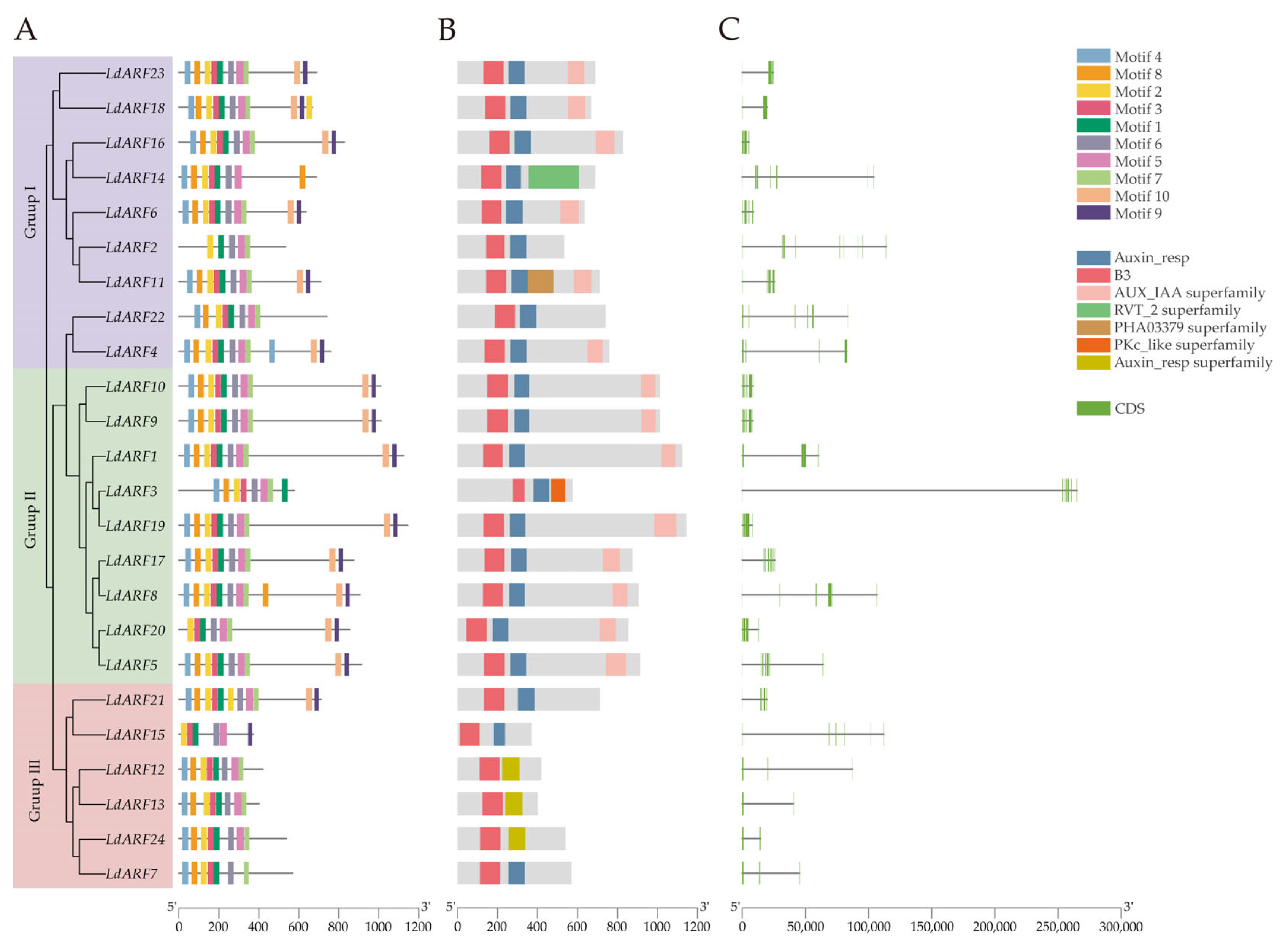
Phylogenetic relationships, conserved motifs, protein domains, and gene structures of LdARF proteins were analyzed (Figure 1). Motif analysis showed that Motif 1, Motif 2, and Motif 6 were present in all LdARF members. Members within the same phylogenetic clade exhibited similar motif composition and arrangement patterns, while differences were observed between clades. Some members, including LdARF2, LdARF3, and LdARF15, showed variation in motif composition, with some motifs being absent or additionally detected. Domain analysis indicated that all LdARF proteins contained a conserved B3 domain. Twenty-one LdARF proteins contained an Auxin_resp domain, whereas three members (LdARF12, LdARF13, and LdARF24) were annotated only with an Auxin_resp superfamily domain. The AUX/IAA domain was predominantly detected in LdARF proteins belonging to Group I and Group III. Furthermore, exon–intron structure analysis showed substantial differences in gene length and intron distribution among the 24 LdARF members. Members clustered within the same phylogenetic group displayed relatively conserved exon–intron organization, whereas genes from different groups showed distinct structural patterns. Among them, LdARF3 exhibited the largest gene length, mainly characterized by an extremely long intronic region, resulting in a distinct genomic structure compared with other LdARF genes.

2.3. Phylogenetic Analysis and Synteny of the LdARF Gene Family

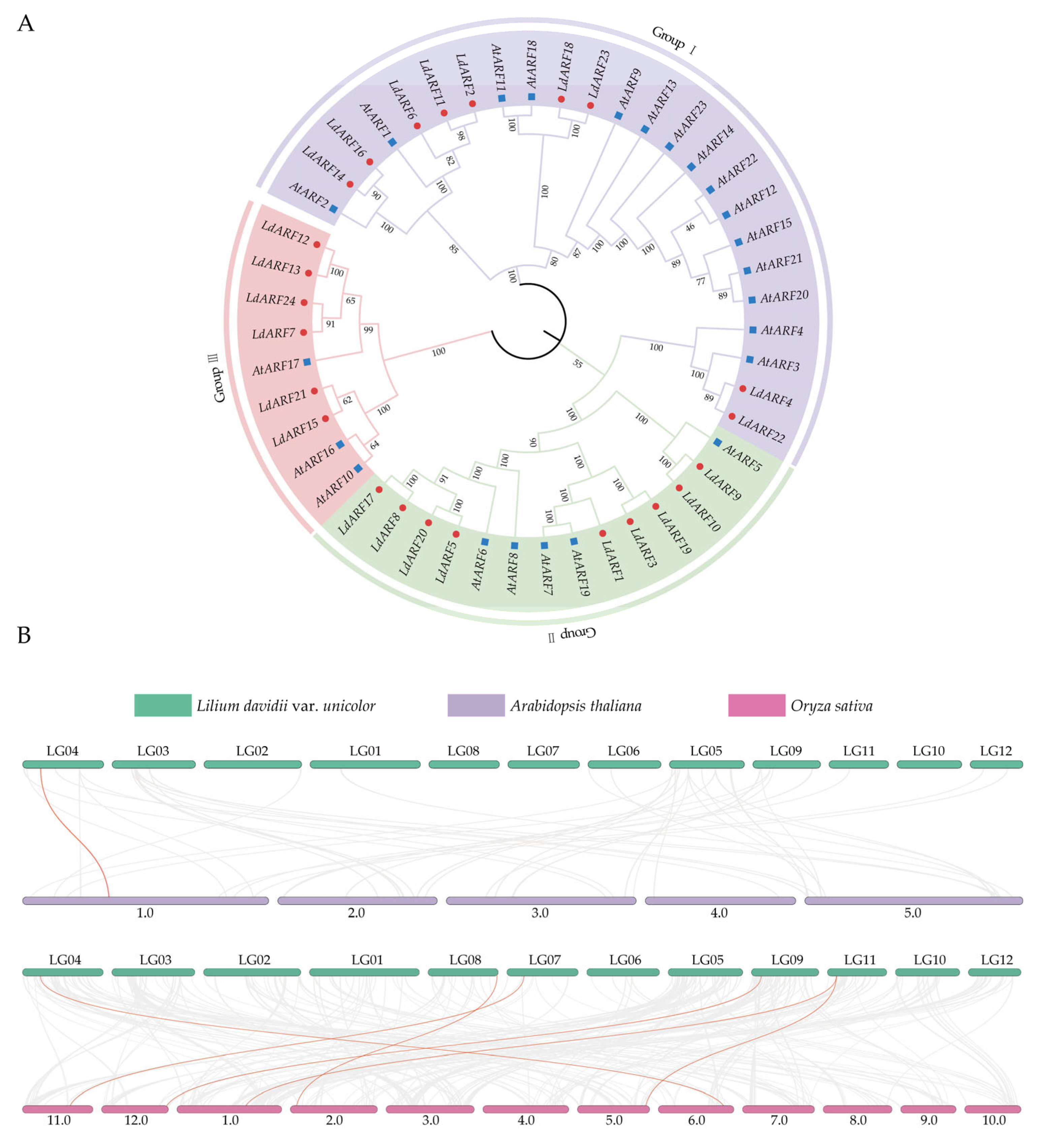
A phylogenetic tree was constructed using the neighbor-joining (NJ) method based on aligned ARF protein sequences from A. thaliana and L. davidii var. unicolor (Figure 2A). According to the classification of the model species A. thaliana, the ARF proteins were divided into three subfamilies, designated Group I, Group II, and Group III. The 24 LdARF proteins were unevenly distributed among these subfamilies, with Group I and Group II containing nine members each, and Group III containing six members.
To investigate the evolutionary relationships and potential expansion mechanisms of the LdARF gene family, synteny analysis was performed between Lilium davidii var. unicolor, Arabidopsis thaliana, and Oryza sativa (Figure 2B). Syntenic blocks were detected between lily and both reference species, indicating the presence of conserved syntenic relationships among ARF genes across species. In the L. davidii var. unicolor genome, LdARF genes were distributed across 12 linkage groups (LG01–LG12), showing a broad chromosomal distribution pattern. In addition, six orthologous gene pairs were identified between lily and rice, suggesting conserved evolutionary relationships between ARF genes in these two species.
Furthermore, intraspecific synteny analysis was performed to investigate duplication events within the L. davidii var. unicolor genome (Figure S2). One segmental duplication pair was identified among the LdARF genes, corresponding to LdARF7 and LdARF12, whereas no tandem duplication events were detected. To evaluate the evolutionary pressure acting on this duplicated gene pair, Ka/Ks analysis was conducted. The Ka/Ks ratio of the LdARF7LdARF12 duplicated gene pair was calculated as 0.276, which was lower than 1, indicating that this duplicated gene pair has evolved under purifying selection.

2.4. Cis-Acting Regulatory Elements in the Promoters of LdARF Genes

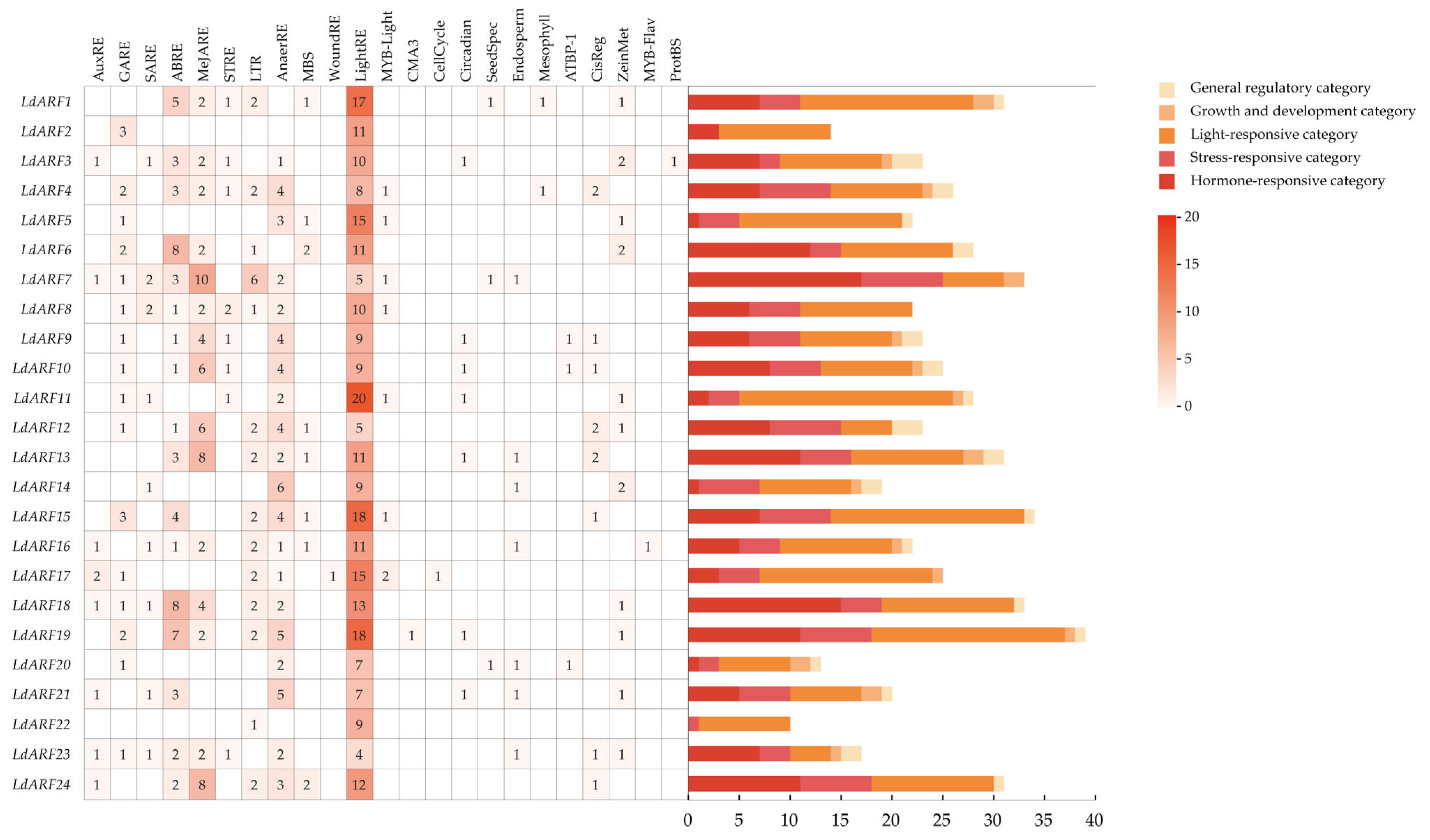
The 2000 bp upstream promoter regions of LdARF genes were extracted and analyzed for cis-acting regulatory elements (Figure S3). To visualize the types and distribution of predicted cis-elements, all identified elements were classified and quantitatively summarized (Figure 3). The results showed that multiple types of regulatory elements were present in the promoters of LdARF genes, which were categorized into five major groups, including hormone-responsive, light-responsive, stress-responsive, growth and development-related, and general regulatory elements. Light-responsive elements were highly abundant in the promoters of all LdARF genes. In addition, hormone-responsive elements such as AuxRE, GARE, and ABRE were widely detected in the promoter regions, indicating the presence of multiple hormone-associated regulatory components. Stress-related elements, including W-box, LTR, STRE, and MBS, were also identified in most LdARF promoters. Quantitative analysis based on stacked bar plots showed that the total number of cis-elements varied among LdARF genes, ranging from approximately 10 to over 30. Different LdARF members displayed distinct compositions and proportions of cis-element categories. Some genes exhibited dominance of a single category, whereas others contained multiple categories of regulatory elements. Overall, variation in the composition and abundance of cis-acting elements was observed among LdARF family members, indicating diversity in promoter architecture.

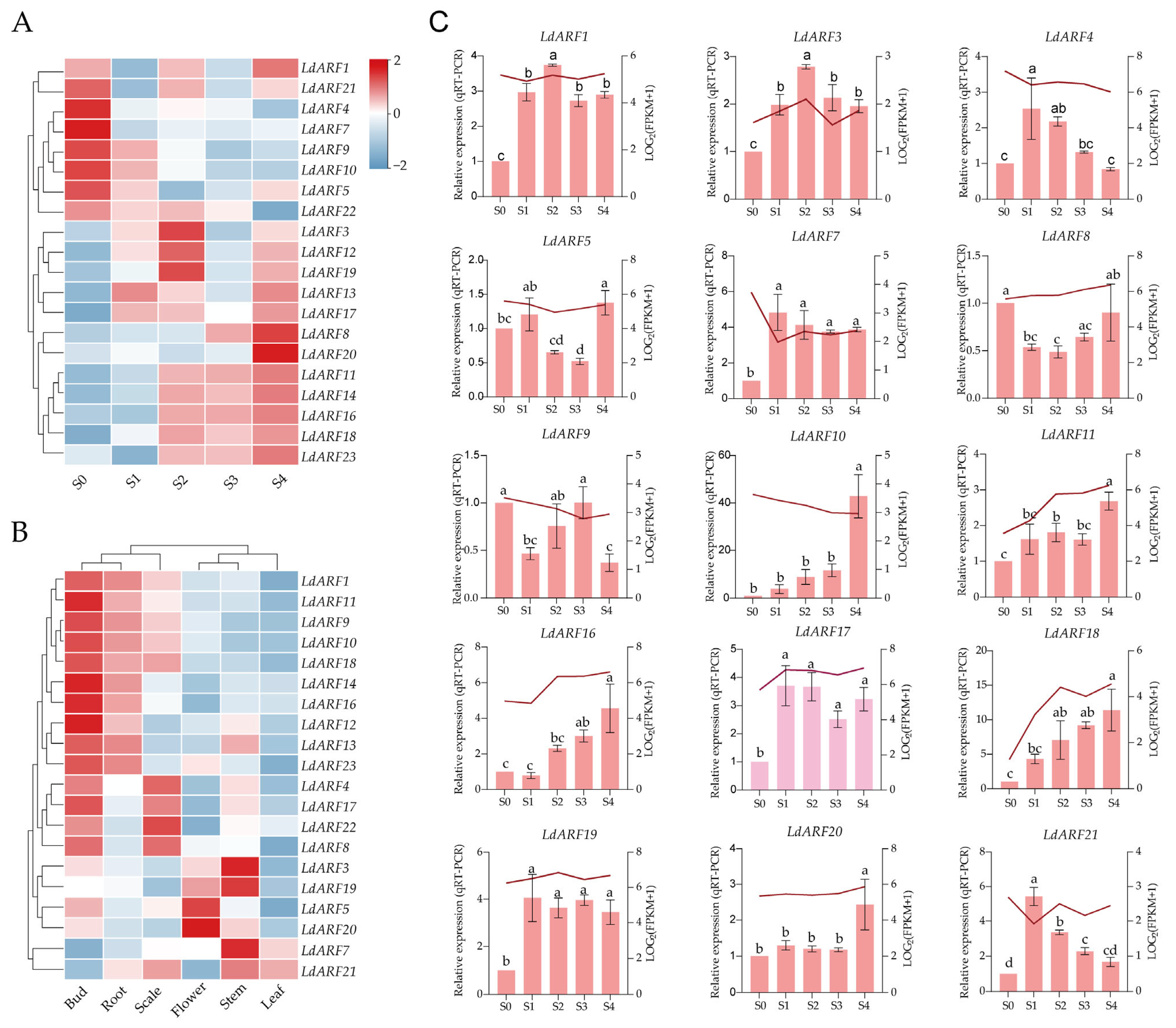
2.5. Expression Patterns of the LdARF Gene Family

Transcriptome data from five developmental stages (S0–S4) of scale-derived bulblet regeneration in the cultivar ‘Siberia’, together with multiple tissues (root, stem, leaf, flower, bud, and scale), were used to analyze ARF homolog expression profiles corresponding to the LdARF gene family. Because a reference genome of ‘Siberia’ is currently unavailable, these transcriptome datasets were annotated using the published L. davidii var. unicolor genome as the reference (Tables S2 and S3). Although derived from different lily genetic backgrounds, the ‘Siberia’ transcriptome dataset was used as a reference for identifying regeneration-associated ARF expression patterns, and candidate genes were further validated in L. davidii var. unicolor by qRT-PCR (Figure 4C and Figure S4, Table S4). Tissue-specific expression analysis revealed that ARF homologs of the LdARF genes were broadly expressed across different organs. Buds exhibited the highest number of highly expressed genes, whereas scales and stems showed moderate expression levels, and roots and leaves contained relatively fewer highly expressed members (Figure 4B). In scale tissues, homologs corresponding to LdARF4, LdARF8, LdARF17, and LdARF22 showed relatively higher expression levels. During scale-derived bulblet regeneration, ARF homologs displayed distinct stage-dependent expression patterns (Figure 4A,C). A subset of genes, including LdARF1, LdARF3, LdARF7, LdARF17, and LdARF19, showed increased expression during early stages (S1–S2). Among them, the LdARF17 homolog exhibited a marked increase in expression during early regeneration and reached its highest expression level at S2–S3 stages. By contrast, the expression levels of LdARF10, LdARF11, LdARF16, and LdARF18 exhibited a sustained increase throughout the process. Expression of LdARF4 and LdARF21 gradually decreased during S1–S4, while LdARF5, LdARF8, LdARF9, and LdARF20 showed fluctuating expression patterns.
Among the identified LdARF genes, LdARF17 displayed a distinct regeneration-associated expression pattern characterized by rapid induction after scale excision and sustained expression during early bulblet initiation and formation. This temporal expression pattern was observed in the ‘Siberia’ transcriptome dataset and was further supported by qRT-PCR analysis in L. davidii var. unicolor. Phylogenetic analysis revealed that LdARF17 clustered within the AtARF6/8 subgroup, which contains ARF members involved in auxin-mediated developmental regulation. In addition, promoter analysis revealed that the upstream regulatory region of LdARF17 contains multiple predicted auxin-responsive elements (AuxREs) and wound-responsive cis-elements, suggesting its potential involvement in responses to auxin and wound-related signaling during regeneration. Based on the combined evidence from its distinct regeneration-associated expression dynamics, evolutionary relationship, and predicted regulatory characteristics, LdARF17 was selected as a candidate gene for further functional characterization.

2.6. LdARF17 Silencing Negatively Affects Bulblet Regeneration in Lily Scales

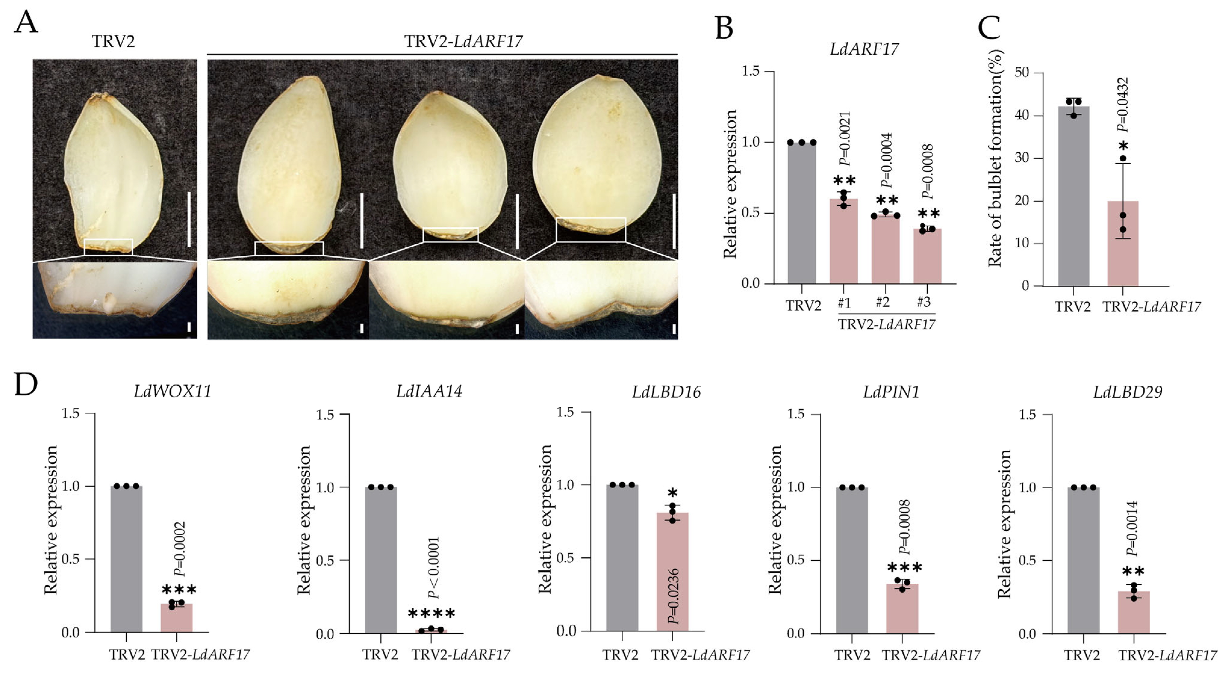
To further explore the potential role of LdARF17 in bulblet regeneration, a TRV2-LdARF17 construct was generated and introduced into scale explants via Agrobacterium-mediated infiltration. PCR detection of the CP (coat protein) gene produced a specific band of the expected size in treated samples, indicating that the viral vector had successfully entered the plant tissues (Figure S4). To evaluate potential off-target effects, the expression levels of three randomly selected ARF family members (LdARF3, LdARF9, and LdARF21) were examined by qRT-PCR. These genes did not show a consistent reduction pattern across independent TRV2-LdARF17 silencing lines (Figure S4). Compared with the control group (TRV2), LdARF17-silenced explants showed a reduction in bulblet formation at the basal region of scales, although the extent of the phenotype varied among independent biological replicates (Figure 5A–C). qRT-PCR analysis showed reduced expression levels of LdARF17 in silenced tissues and revealed altered expression patterns of several auxin- and regeneration-related genes (Figure 5D). Expression levels of IAA14, LBD16, LBD29, WOX11, and PIN1 were all reduced in the silencing group. Among these, IAA14 showed the strongest reduction, whereas LBD16 displayed a moderate decrease.

2.7. Transient Overexpression of LdARF17 Promotes Bulblet Regeneration in Lily Scales

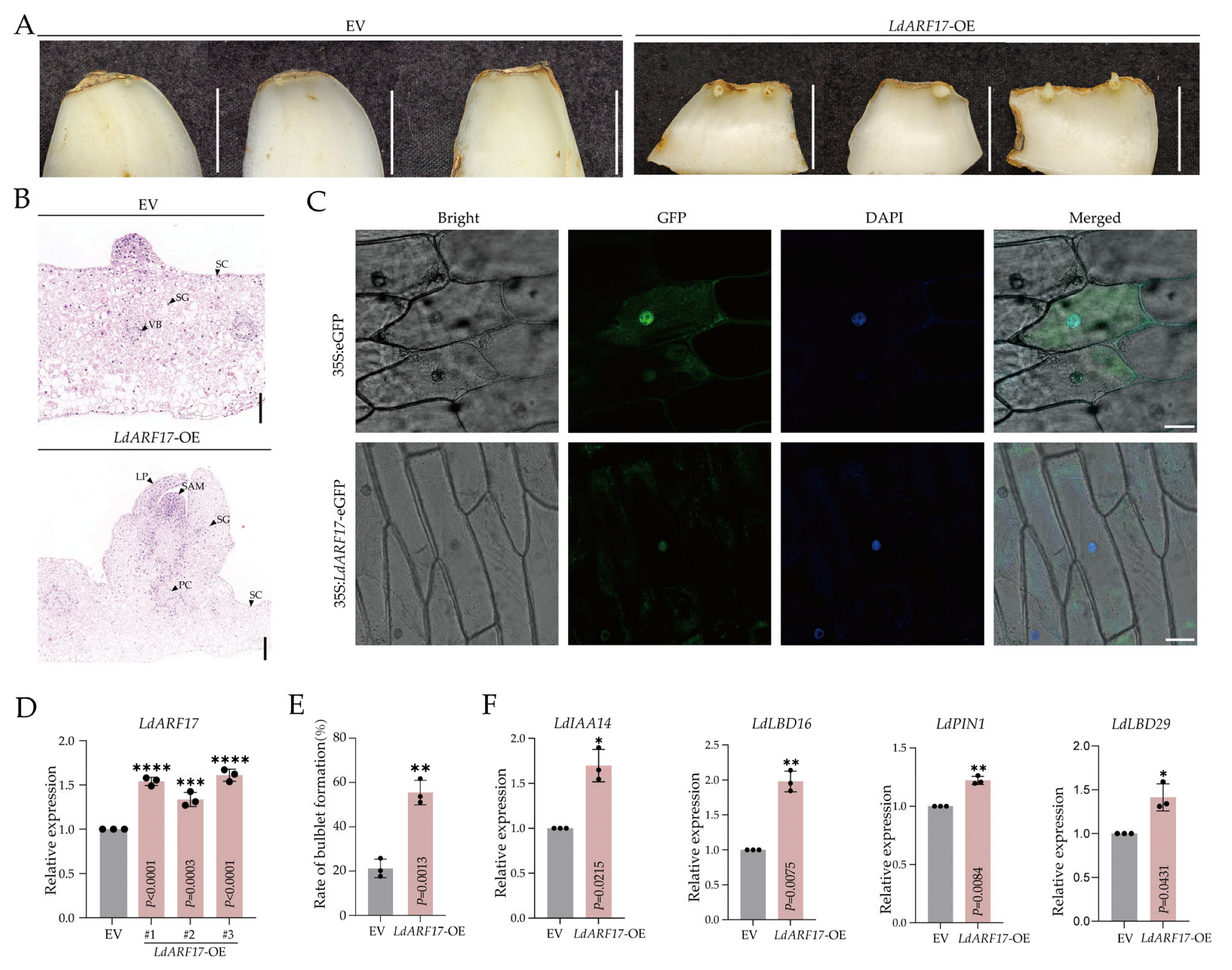
A p35S::LdARF17::GUS overexpression construct was generated and introduced into lily scale explants via vacuum infiltration, with empty vector (EV, p35S::GUS) serving as the control. Regeneration performance was evaluated 15 days after infiltration, and bulblet formation was statistically analyzed. PCR amplification confirmed the presence of GUS and Hyg fragments in transformed tissues, indicating successful introduction of the recombinant construct (Figure S6). qRT-PCR analysis further showed significantly elevated expression of LdARF17 in three independent OE lines compared with the EV control (Figure 6D). Phenotypic observation revealed that overexpression of LdARF17 promoted bulblet formation at the basal region of scale explants compared with the control (Figure 6A). Histological analysis revealed more pronounced bulblet primordia formation at the basal region of OE scales, accompanied by increased starch granule accumulation compared with the EV control (Figure 6B). Quantitative analysis further demonstrated a significant increase in bulblet formation frequency in the OE group (Figure 6E). Expression analysis of auxin- and regeneration-related genes showed upregulation of IAA14, LBD16, PIN1 and LBD29 in LdARF17-overexpressing tissues (Figure 6D,F).

2.8. Subcellular Localization Analysis of LdARF17

Subcellular localization analysis in onion epidermal cells showed that LdARF17 was predominantly localized in the nucleus (Figure 6C). In cells expressing the pCAMBIA2300-eGFP-LdARF17 fusion protein, green fluorescence signals were observed mainly in the nucleus and overlapped with DAPI-stained nuclear signals. In contrast, free eGFP signals in the control were distributed throughout both the cytoplasm and nucleus, showing no specific localization pattern.

3. Discussion

3.1. Evolutionary Conservation and Functional Divergence of the LdARF Gene Family in Lily

Auxin response factors (ARFs) are key transcriptional regulators that mediate auxin-responsive gene expression and participate in diverse aspects of plant growth and development. The typical ARF protein structure, consisting of an N-terminal B3 DNA-binding domain, a middle regulatory region, and a C-terminal Auxin_resp domain, provides the structural basis for auxin-dependent transcriptional regulation [2,33,34]. Although this conserved architecture is generally maintained among plant species, sequence divergence and structural variation among individual ARF members may contribute to functional specialization during plant evolution.
In this study, 24 LdARF genes were identified in Lilium davidii var. unicolor (Table 1), with a family size comparable to those reported for A. thaliana and O. sativa [35,36,37]. Phylogenetic analysis classified LdARF proteins into three major clades, consistent with the classical classification of ARF members in A. thaliana (Figure 2A). The clustering of lily ARF members with their corresponding Arabidopsis ARF homologs within the same phylogenetic branches supports the hypothesis that the major evolutionary lineages of ARFs were established early in plant evolution and have been conserved throughout the diversification of monocots and dicots.
Despite the conserved phylogenetic framework, structural diversification was observed among LdARF proteins. Several LdARFs, including LdARF12, LdARF13, and LdARF24, exhibited variations in their Auxin_resp domain annotations, suggesting potential differences in protein regulatory properties (Figure 1B). Similar atypical ARF members have been identified in A. thaliana and other plant species [38,39,40]. For example, Arabidopsis ARF3/ETTIN lacks the canonical C-terminal PB1 domain that mediates interaction with Aux/IAA proteins, but retains its function in regulating floral organ development, indicating that divergent domain architectures do not necessarily impair ARF activity and may contribute to alternative regulatory mechanisms [41]. Therefore, the atypical domain organizations observed in several LdARF proteins may reflect evolutionary diversification within the ARF family and contribute to functional diversity among ARF members. However, whether these divergent LdARF proteins retain canonical ARF activities or possess specialized regulatory functions remains to be experimentally determined.
Motif composition and gene structure analyses further supported functional diversification of LdARF genes (Figure 1A,C). Members within the same phylogenetic clade generally showed similar motif arrangements and exon–intron organization, whereas considerable structural variation was observed between different clades. Similar patterns have been reported in rice, maize, and other plant species, suggesting that gene duplication followed by sequence divergence has contributed to the functional differentiation of ARF family members [36,42,43]. Notably, LdARF3 contained an exceptionally long intron, which may be associated with the accumulation of repetitive elements and genome structural expansion characteristic of the giant lily genome [31,32]. In addition, the limited number of duplication events, conserved syntenic relationships, and low Ka/Ks ratio of the duplicated gene pair suggest that LdARF family expansion was not mainly driven by recent duplication events and that duplicated ARF genes have been maintained under strong evolutionary constraints.
Promoter analysis revealed that LdARF genes contain diverse cis-regulatory elements associated with hormone responses, light signaling, and stress responses, consistent with previous reports in soybean and other plant species (Figure 3) [44,45]. The presence of these regulatory elements suggests that LdARFs may integrate developmental signals and environmental responses. This feature may be particularly relevant in lily scale-derived regeneration, a process involving wound responses, cellular reprogramming, and organ initiation.
Collectively, the LdARF family exhibits both evolutionary conservation and structural diversification. The conserved ARF domain organization provides the structural basis for the fundamental roles of LdARF proteins in auxin-responsive regulation, whereas sequence and structural variations among family members may contribute to specialized developmental functions. As a bulbous monocot with strong regenerative capacity, lily provides a valuable system for exploring the functional diversification of ARF-mediated auxin regulation during plant regeneration.

3.2. Dynamic Expression Patterns of LdARF Genes Reveal Their Potential Roles in Lily Bulblet Regeneration

Although LdARF members share conserved evolutionary characteristics, their distinct expression profiles indicate functional divergence among family members (Figure 4B). Transcriptome analysis revealed that LdARF genes displayed tissue-preferential expression patterns across buds, roots, scales, flowers, stems, and leaves, suggesting that different ARF members may participate in specific developmental processes. Similar expression divergence has been observed in monocot crops, where OsARF and ZmARF genes exhibit distinct organ-specific expression patterns associated with diverse developmental functions [36,42].
In addition to regulating normal plant development, ARFs have been widely associated with regeneration-related processes, including wound responses, cellular dedifferentiation, and organ initiation. In A. thaliana, ARF6 and ARF8 participate in adventitious root formation, whereas ARF7 and ARF19 regulate auxin-mediated organogenesis through downstream developmental pathways [6,46,47]. In terms of underground storage organ formation, studies in potato, carrot, and sugar beet have demonstrated that auxin signaling networks are involved in tuber initiation and expansion processes, while ARFs, as core auxin-responsive transcription factors, may participate in this regulatory mechanism [48]. These findings highlight the conserved involvement of ARF-mediated transcriptional regulation in developmental transitions.
Lily bulblet regeneration is a complex developmental process involving sequential events, including wound responses, acquisition of regenerative competence, meristem initiation, primordium formation, and bulblet enlargement [26]. Integrated transcriptome analysis and qRT-PCR validation revealed distinct temporal expression patterns among LdARF genes during regeneration, suggesting that different ARFs may function at specific developmental stages (Figure 4A). Early-induced LdARFs may contribute to the activation of regeneration-related responses following scale excision, whereas members with sustained or late-stage expression may participate in subsequent organ formation and bulblet development. Other dynamically expressed members may contribute to maintaining auxin signaling balance during developmental transitions.
A limitation of this study is that the transcriptome dataset used for stage-specific expression analysis was generated from the cultivar ‘Siberia’, whereas genome-based gene identification and functional analyses were performed in Lilium davidii var. unicolor. Because a reference genome for ‘Siberia’ is currently unavailable, the transcriptome reads were aligned to the published L. davidii var. unicolor genome. Although this strategy enabled the identification of homologous genes and characterization of temporal expression patterns, cultivar-specific genetic variation may influence transcript abundance. For example, LdARF7 exhibited a significant negative correlation between the RNA-seq and qRT-PCR datasets (r = −0.96, p = 0.01), indicating an opposite expression trend between the two datasets (Table S4). Therefore, the transcriptome data were interpreted primarily as evidence of expression dynamics rather than direct quantitative comparisons between these two genetic backgrounds.

3.3. Transient Functional Analysis of LdARF17 Suggests Its Positive Role in Lily Bulblet Regeneration

Among the identified LdARF members, LdARF17 showed a characteristic regeneration-associated expression pattern and was selected for preliminary functional characterization. Although transient expression in onion epidermal cells confirmed the nuclear localization pattern of LdARF17, this heterologous system may not fully represent its localization behavior in lily cells. Future studies using a homologous lily transformation system or co-localization with nuclear markers will provide further confirmation of LdARF17 subcellular localization. Transient manipulation of LdARF17 expression demonstrated that silencing of LdARF17 reduced bulblet formation efficiency (Figure 5), whereas overexpression was associated with enhanced regeneration (Figure 6). Although transient overexpression of LdARF17 resulted in a significant increase in bulblet formation efficiency, the observed increase in LdARF17 transcript abundance was relatively moderate. This apparent discrepancy between transcriptional activation and phenotypic output may be attributed to the characteristics of the transient expression system and the temporal dynamics of transgene expression. Because samples for qRT-PCR analysis were collected 15 days after infiltration, when bulblet regeneration phenotypes had already been established, the transcript abundance detected at this stage may not fully represent the initial expression level or potential peak accumulation of LdARF17 during the early regeneration process [49]. In addition, although the CaMV 35S promoter has been widely used for constitutive expression in both dicotyledonous and monocotyledonous plants, its activity can vary depending on plant species, tissue type, and experimental conditions [50]. Therefore, the moderate increase in LdARF17 transcript abundance observed in lily tissues may partly reflect the expression characteristics of the CaMV 35S promoter in this specific transient transformation system. Nevertheless, the enhanced bulblet regeneration phenotype consistently observed in transient LdARF17-overexpression materials supports a positive regulatory role of LdARF17 in lily bulblet regeneration.
To further explore the potential role of LdARF17 in regeneration, several regeneration-associated genes were examined. In LdARF17-silenced and transient LdARF17-overexpressing materials, the expression patterns of PIN1, WOX11, LBD16, LBD29, and IAA14 were altered and were associated with changes in LdARF17 expression. WOX11 plays an important role in cellular fate transition and the establishment of regenerative competence by promoting the acquisition of meristematic characteristics [51,52]. LBD16 and LBD29 are auxin-responsive transcription factors involved in lateral organ development and auxin-regulated developmental processes [7,53,54]. PIN1-mediated polar auxin transport plays an essential role in establishing local auxin maxima required for organ initiation and meristem development [11,55,56,57]. Aux/IAA proteins are important regulators of auxin signaling, as they interact with ARF transcription factors and modulate their transcriptional activity [58,59,60,61].
The altered expression of these regeneration- and auxin-related genes suggests that LdARF17 may be associated with auxin-related regulatory processes during bulblet regeneration. However, whether these genes are directly regulated by LdARF17 or act as downstream components requires further experimental validation. Such coordinated expression changes may contribute to maintaining auxin signaling stability during the transition from cellular reprogramming to organ formation.
Overall, genome-wide identification and expression profiling revealed extensive conservation and diversification within the LdARF gene family. Among these members, LdARF17 displayed a regeneration-associated expression pattern and positively influenced bulblet formation, suggesting that specific ARF members may acquire specialized functions during lily regeneration. These findings expand the understanding of ARF family evolution and provide candidate genes for further investigating auxin-mediated regulatory networks underlying lily scale-derived regeneration.

4. Materials and Methods

4.1. Plant Materials and Regeneration System

Scale-derived bulblet regeneration was induced using disease-free L. davidii var. unicolor plants. The outer 2–3 scales were removed, and the middle scales were cultured in a substrate (peat:vermiculite = 3:1, v/v) in a growth chamber at 24 °C under dark conditions. The substrate was sterilized with carbendazim prior to use.
Regeneration stages were defined as follows: S0, scale excision; S1, 3 days after planting; S2, slight swelling at the basal region (9 days); S3, visible meristem-like structures; and S4, fully developed bulblets or adventitious roots, according to the classification criteria described by Kang et al. [26]. Approximately 0.5 cm of basal tissue from each stage was collected, immediately frozen in liquid nitrogen, and stored for RNA extraction and gene expression analysis.

4.2. Genome-Wide Identification and Characterization of the LdARF Gene Family

ARF genes were identified from the L. davidii var. unicolor genome using a combination of BLASTP and hidden Markov model (HMM)-based searches. The protein sequences of A. thaliana ARF family members were used as queries for BLASTP searches against the lily protein database using the BLASTP function implemented in TBtools v2.472, with an E-value cutoff of 1 × 10⁻⁵. In parallel, the HMM profile of the ARF domain (PF06507) was obtained from the Pfam database and used to search the lily protein database using the Simple HMM Search function implemented in TBtools v2.472 with default parameters. Candidate ARF proteins identified by both BLASTP and HMM-based searches were retained for further analysis. The conserved domains of candidate proteins were subsequently verified using the NCBI Conserved Domain Database (CDD) and InterPro database. Proteins lacking complete ARF domains or containing incomplete ARF domains were excluded. Finally, a total of 24 LdARF genes were identified and used for subsequent analyses. Physicochemical properties, including amino acid length, molecular weight, and isoelectric point, were predicted using TBtools v2.472. Chromosomal locations were determined based on GFF3 annotation files, and genes were renamed according to chromosomal order [62]. Subcellular localization was predicted using WoLF PSORT.

4.3. Motif, Gene Structure, and Promoter Analysis

Conserved motifs were identified using MEME Suite v5.5.9 with the Classic mode. The maximum number of motifs was set to 10, with motif widths ranging from 6 to 50 amino acids. The motif occurrence model was set to zero or one occurrence per sequence (ZOOPS), and the significance of identified motifs was evaluated based on the E-values calculated by MEME. Conserved domains were analyzed using the NCBI Batch CD-Search tool. Gene structures were visualized using TBtools v2.472 based on genome annotation files. Phylogenetic relationships and structural features (motifs, domains, and gene structures) were integrated using TBtools [62]. For promoter analysis, 2000 bp upstream sequences were extracted using TBtools. Cis-acting regulatory elements were predicted using PlantCARE. Element abundance was summarized in Microsoft Excel 2021, and heatmaps were generated using TBtools [62].

4.4. Phylogenetic and Synteny Analyses

A neighbor-joining (NJ) phylogenetic tree was constructed using MEGA 12.0 (Pennsylvania State University, State College, PA, USA) with 1000 bootstrap replicates. Genomic synteny analysis was conducted using the Oryza sativa genome as a reference. The one-step MCScanX pipeline in TBtools was employed to perform genome-wide collinearity analysis between Lilium davidii var. unicolor and the two reference species, Arabidopsis thaliana and Oryza sativa, and the resulting syntenic relationships were visualized [62].

4.5. Expression Profiling and qRT-PCR Validation

Based on sequence homology, transcriptome data from different stages (S0–S4) of scale-derived bulblet regeneration in the cultivar ‘Siberia’, previously generated by our research group, were analyzed to investigate the temporal expression patterns of ARF homologs corresponding to L. davidii var. unicolor ARF genes. Since a reference genome of ‘Siberia’ is currently unavailable, clean reads were aligned to the published L. davidii var. unicolor reference genome using HISAT2 for splice-aware alignment. Gene expression levels were quantified based on the annotated gene models of L. davidii var. unicolor. The mapping statistics are provided in Supplementary Tables S2 and S3. Heatmaps were generated using TBtools v2.472 [62]. A total of 15 LdARF genes were selected for qRT-PCR analysis. Total RNA was extracted from the basal tissues of scales at five regeneration stages using the HiPure Plant RNA Mini Kit (Magen, Shanghai, China; R4151-02). First-strand cDNA was synthesized using the HiScript III All-in-one RT SuperMix kit (Vazyme, Nanjing, China; R333-01) and used as the template for qRT-PCR. qRT-PCR was performed using Taq Pro Universal SYBR qPCR Master Mix (Vazyme, Nanjing, China; Q712-02) on a QuantStudio™ 1 Plus Real-Time PCR system (Thermo Fisher Scientific, Waltham, MA, USA). Each experiment included three biological replicates and three technical replicates. Primers for LdARF genes, regeneration-related genes (WOX11, LBD16, and LBD29), and auxin-related genes (IAA14 and PIN1) were designed using the GenScript online tool. LdACTIN was used as the internal reference gene [63]. Relative expression levels were calculated using the 2−ΔΔCt method.

4.6. Virus-Induced Gene Silencing of LdARF17

TRV vectors were provided by the Key Laboratory of Ornamental Plants, China Agricultural University. A specific non-conserved fragment of LdARF17 was cloned into TRV2 using BamHI and XhoI restriction sites. Recombinant constructs TRV1, TRV2, and TRV2-LdARF17 were transformed into Agrobacterium tumefaciens GV3101. Two groups were established: control (TRV2) and silencing (TRV2-LdARF17), each with three biological replicates (30 scales per replicate). Agrobacterium cultures were grown in LB medium with kanamycin and rifampicin. Cells were resuspended in infiltration buffer (10 mM MgCl2, 200 μM acetosyringone, 10 mM MES, pH 5.6) and adjusted to OD600 = 0.8. Equal volumes of TRV1 and TRV2 constructs were mixed. Vacuum infiltration was performed at 0.8 MPa for 15 min. After infiltration, scales were incubated on moist cotton pads. Bulblet regeneration was evaluated after 15 days, and infection efficiency was confirmed by RT-PCR using TRV coat protein primers.

4.7. Transient Overexpression of LdARF17

The full-length ORF of LdARF17 was cloned into the pCAMBIA1301-GUS vector under the CaMV 35S promoter (p35S::LdARF17::GUS). The construct was introduced into Agrobacterium tumefaciens GV3101. Agrobacterium infiltration was performed as described in Section 4.6. Two groups were established: empty vector control group (EV, p35S::GUS) and LdARF17 overexpression group (p35S::LdARF17::GUS), each with three biological replicates (30 scales per replicate). Bulblet regeneration was evaluated 15 days after infiltration, and samples for gene expression analysis were collected at the same time point. Transformation was confirmed by RT-PCR detection of GUS and Hyg genes.

4.8. Subcellular Localization of LdARF17

The coding sequence of LdARF17 was cloned into the pCAMBIA2300-eGFP vector using BamHI and SalI sites. The recombinant construct was introduced into Agrobacterium tumefaciens strain GV3101. Transient expression was performed in onion epidermal cells via Agrobacterium-mediated infiltration. Empty vector (eGFP) served as a control. Samples were incubated at 28 °C in darkness for 24 h, and fluorescence signals were observed 2–3 days post-infiltration using a confocal laser scanning microscope (Leica Microsystems, Wetzlar, Germany).

4.9. Statistical Analysis

Data were analyzed using one-way analysis of variance (ANOVA). For multi-group comparisons with a single control group, Dunnett’s multiple comparisons test was performed. For comparisons between two independent groups, two-tailed Student’s t-tests were used. All analyses were conducted using GraphPad Prism 10.5.0.

5. Conclusions

In this study, 24 LdARF gene family members were identified from the whole-genome data of Lilium davidii var. unicolor. Systematic analyses of chromosomal distribution, protein structure, phylogeny, cis-acting elements, and expression profiles revealed the structural diversity and potential functional differentiation of the LdARF family. LdARF genes exhibited stage-specific expression patterns during scale regeneration and were classified into four categories: early continuously upregulated, late continuously upregulated, late continuously downregulated, and fluctuating types. LdARF17 showed dynamic expression during key regeneration stages and LdARF17 protein was localized in the nucleus. Functional analyses demonstrated that silencing of LdARF17 inhibited scale regeneration and bulblet formation, whereas transient overexpression promoted these processes, accompanied by altered expression of regeneration-related genes. These results support the conclusion that LdARF17 acts as a positive regulator of lily scale regeneration, and that changes in LdARF17 expression are associated with altered expression patterns of regeneration-related genes, suggesting its potential role in promoting organ regeneration. The study refines the functional framework of the LdARF gene family in lily and provides new insights into the regulatory mechanisms underlying bulblet regeneration, offering candidate genetic resources for future functional studies and molecular breeding in lily.

Supplementary Materials

The following supporting information can be downloaded at https://www.mdpi.com/article/10.3390/plants15172581/s1, Figure S1: The distribution of LdARF genes on chromosomes; Figure S2: Chromosomal localization, gene density distribution, and synteny relationships of the LdARF gene family in Lilium davidii var. unicolor. Figure S3: Analysis of cis-acting regulatory elements in the promoter regions of LdARF genes in Lilium davidii var. unicolor; Figure S4: Expression patterns of LdARF17 during bulblet regeneration in Lilium davidii var. unicolor. Figure S5: Validation of TRV infection and relative expression of LdARF3, LdARF9, and LdARF21 in VIGS-treated Lilium davidii var. unicolor scales; Figure S6: Molecular confirmation of transient transformation in Lilium davidii var. unicolor scales; Figure S7: Melting curve analysis of qRT-PCR products for LdARF genes; Table S1: The primers used in this study; Table S2: Summary of ‘Siberia’ tissue transcriptome reads mapped to the Lilium davidii var. unicolor reference genome. Table S3: Summary of ‘Siberia’ scale-derived bulblet regeneration transcriptome reads mapped to the Lilium davidii var. unicolor reference genome. Table S4: Pearson correlation analysis of LdARF expression profiles between ‘Siberia’ lily RNA-seq data and Lilium davidii var. unicolor qRT-PCR data during bulblet regeneration. Table S5: The function of LdARF17 in the scale-induced bulblet regeneration verified by virus-mediated gene silencing technology; Table S6: Overexpression of LdARF17 promotes scale-induced bulblet regeneration; Table S7: The relative expression of LdARF family members during different bulblet development stages (S0, S1, S2, S3 and S4) derived from the scaling propagation method; Table S8: The relative expression of LdARF17, bulblet regeneration-related genes and auxin-related genes in TRV2 and TRV2-LdARF17 lines; Table S9: The relative expression of LdARF17, bulblet regeneration-related genes and auxin-related genes in EV and LdARF17-OE lines.

Author Contributions

Conceptualization, Y.T. and X.X.; methodology, Y.T. and X.X.; software, Y.T. and Y.W.; validation, Y.T. and S.G.; formal analysis, Y.T.; investigation, Y.T. and S.G.; resources, C.Y., X.X. and W.G.; data curation, Y.T., X.X., and C.Y.; writing—original draft preparation, Y.T.; writing—review and editing, Y.T., Y.W., X.X. and Z.L.; visualization, Y.T. and Y.W.; supervision, Z.L., C.Y. and X.X.; project administration, Z.L., S.Z. and C.Y.; funding acquisition, C.Y., X.X. and S.F. All authors have read and agreed to the published version of the manuscript.

Funding

This research was funded by the Open Fund Projects of the Fujian Provincial Key Laboratory of Agricultural Genetic Engineering, grant numbers KFJJ20250408 and KFJJ20260330; the National Natural Science Foundation of China (32302595); and the Extended Research Project of the National Natural Science Foundation of China of Fujian Academy of Agricultural Sciences (grant number GJYS202407).

Data Availability Statement

Data supporting the findings of this study are available within the article and its Supplementary Materials. All primers used in this study are provided in Supplementary Table S1. The RNA-seq datasets generated and analyzed in this study have been deposited in the National Genomics Data Center (NGDC) under accession numbers PRJCA060256 and PRJCA068453, corresponding to the scale-derived bulblet regeneration transcriptome dataset and the tissue transcriptome dataset from different organs of Lilium ‘Siberia’, respectively.

Acknowledgments

During the preparation of this manuscript, the authors used ChatGPT (OpenAI, GPT-5.5-mini) for language editing and improvement of manuscript readability. The authors have reviewed and edited the generated content and take full responsibility for the content of this publication.

Conflicts of Interest

The authors declare no conflicts of interest.

Abbreviations

The following abbreviations are used in this manuscript:
ARFAuxin Response Factor
Aux/IAAAuxin/Indole-3-Acetic Acid
CDSCoding Sequence
LTRLong Terminal Repeat
NJNeighbor-Joining
qRT-PCRQuantitative Real-Time Polymerase Chain Reaction
RNA-seqRNA Sequencing
RNAiRNA Interference
TFTranscription Factor
TRVTobacco Rattle Virus
VIGSVirus-Induced Gene Silencing
GUSβ-Glucuronidase
HygHygromycin resistance gene
HMMHidden Markov Model
BLASTPBasic Local Alignment Search Tool for Protein sequences
MEGAMolecular Evolutionary Genetics Analysis
CDDConserved Domain Database
MEMEMultiple Em for Motif Elicitation
Ka/KsNon-synonymous substitution rate/Synonymous substitution rate

References

  1. Hernandez-Garcia, J.; Weijers, D. The origin and evolution of auxin as a plant signaling molecule. Curr. Biol. 2026, 36, R545–R552. [Google Scholar] [CrossRef] [Scilit] [PubMed]
  2. Yu, Z.; Zhang, F.; Friml, J.; Ding, Z. Auxin signaling: Research advances over the past 30 years. J. Integr. Plant Biol. 2022, 64, 371–392. [Google Scholar] [CrossRef] [Scilit] [PubMed]
  3. Ikeuchi, M.; Ogawa, Y.; Iwase, A.; Sugimoto, K. Plant regeneration: Cellular origins and molecular mechanisms. Development 2016, 143, 1442–1451. [Google Scholar] [CrossRef] [Scilit] [PubMed]
  4. Sakamoto, Y.; Kawamura, A.; Suzuki, T.; Segami, S.; Maeshima, M.; Polyn, S.; De Veylder, L.; Sugimoto, K. Transcriptional activation of auxin biosynthesis drives developmental reprogramming of differentiated cells. Plant Cell 2022, 34, 4348–4365. [Google Scholar] [CrossRef] [Scilit] [PubMed]
  5. Matosevich, R.; Cohen, I.; Gil-Yarom, N.; Modrego, A.; Friedlander-Shani, L.; Verna, C.; Scarpella, E.; Efroni, I. Local auxin biosynthesis is required for root regeneration after wounding. Nat. Plants 2020, 6, 1020–1030. [Google Scholar] [CrossRef] [Scilit] [PubMed]
  6. Okushima, Y.; Fukaki, H.; Onoda, M.; Theologis, A.; Tasaka, M. ARF7 and ARF19 regulate lateral root formation via direct activation of LBD/ASL genes in Arabidopsis. Plant Cell 2007, 19, 118–130. [Google Scholar] [CrossRef] [Scilit] [PubMed]
  7. Fan, M.; Xu, C.; Xu, K.; Hu, Y. LATERAL ORGAN BOUNDARIES DOMAIN transcription factors direct callus formation in Arabidopsis regeneration. Cell Res. 2012, 22, 1169–1180. [Google Scholar] [CrossRef] [Scilit] [PubMed]
  8. Hu, X.; Xu, L. Transcription Factors WOX11/12 Directly Activate WOX5/7 to Promote Root Primordia Initiation and Organogenesis. Plant Physiol. 2016, 172, 2363–2373. [Google Scholar] [CrossRef] [Scilit] [PubMed]
  9. Liu, J.; Sheng, L.; Xu, Y.; Li, J.; Yang, Z.; Huang, H.; Xu, L. WOX11 and 12 are involved in the first-step cell fate transition during de novo root organogenesis in Arabidopsis. Plant Cell 2014, 26, 1081–1093. [Google Scholar] [CrossRef] [Scilit] [PubMed]
  10. Hajný, J.; Tan, S.; Friml, J. Auxin canalization: From speculative models toward molecular players. Curr. Opin. Plant Biol. 2022, 65, 102174. [Google Scholar] [CrossRef] [Scilit] [PubMed]
  11. Leyser, O. Auxin Distribution and Plant Pattern Formation: How Many Angels Can Dance on the Point of PIN? Cell 2005, 121, 819–822. [Google Scholar] [CrossRef] [Scilit] [PubMed]
  12. Moller, B.; Weijers, D. Auxin Control of Embryo Patterning. Cold Spring Harb. Perspect. Biol. 2009, 1, a001545. [Google Scholar] [CrossRef] [Scilit] [PubMed]
  13. Song, X.; Xiong, Y.; Kong, X.; Huang, G. Roles of auxin response factors in rice development and stress responses. Plant Cell Environ. 2023, 46, 1075–1086. [Google Scholar] [CrossRef] [Scilit] [PubMed]
  14. Hu, B.; Zhang, G.; Liu, W.; Shi, J.; Wang, H.; Qi, M.; Li, J.; Qin, P.; Ruan, Y.; Huang, H.; et al. Divergent regeneration-competent cells adopt a common mechanism for callus initiation in angiosperms. Regeneration 2017, 4, 132–139. [Google Scholar] [CrossRef] [Scilit] [PubMed]
  15. López-Ruiz, B.A.; Juárez-González, V.T.; Gómez-Felipe, A.; De Folter, S.; Dinkova, T.D. tasiR-ARFs Production and Target Regulation during In Vitro Maize Plant Regeneration. Plants 2020, 9, 849. [Google Scholar] [CrossRef] [Scilit] [PubMed]
  16. Bannoud, F.; Bellini, C. Adventitious Rooting in Populus Species: Update and Perspectives. Front. Plant Sci. 2021, 12, 668837. [Google Scholar] [CrossRef] [Scilit] [PubMed]
  17. Yang, C.; Xu, M.; Xuan, L.; Jiang, X.; Huang, M. Identification and expression analysis of twenty ARF genes in Populus. Gene 2014, 544, 134–144. [Google Scholar] [CrossRef] [Scilit] [PubMed]
  18. Li, S.-B.; OuYang, W.-Z.; Hou, X.-J.; Xie, L.-L.; Hu, C.-G.; Zhang, J.-Z. Genome-wide identification, isolation and expression analysis of auxin response factor (ARF) gene family in sweet orange (Citrus sinensis). Front. Plant Sci. 2015, 6, 119. [Google Scholar] [CrossRef] [Scilit] [PubMed]
  19. Feng, S.; Li, N.; Chen, H.; Liu, Z.; Li, C.; Zhou, R.; Zhang, Y.; Cao, R.; Ma, X.; Song, X. Large-scale analysis of the ARF and Aux/IAA gene families in 406 horticultural and other plants. Mol. Hortic. 2024, 4, 15. [Google Scholar] [CrossRef] [Scilit] [PubMed]
  20. Mi, L.; Wang, K.; Gan, Z.; Lin, Y.; Wang, X.; Yang, S.; Xu, Z. A comparative metabolomics study on two fresh edible lilies for vegetable: Lilium brownii var. viridulum and Lilium davidii var. unicolor. Food Biosci. 2024, 57, 103583. [Google Scholar] [CrossRef] [Scilit]
  21. Li, W.; Wang, Y.; Wei, H.; Zhang, Y.; Guo, Z.; Qiu, Y.; Wen, L.; Xie, Z. Structural characterization of Lanzhou lily (Lilium davidii var. unicolor) polysaccharides and determination of their associated antioxidant activity. J. Sci. Food Agric. 2020, 100, 5603–5616. [Google Scholar] [CrossRef] [Scilit] [PubMed]
  22. Li, Y.; Wang, H.; Zhang, W.; Wu, H.; Wang, Z. Evaluation of nutrition components in Lanzhou lily bulb by confocal Raman microscopy. Spectrochim. Acta Part A Mol. Biomol. Spectrosc. 2021, 244, 118837. [Google Scholar] [CrossRef] [Scilit] [PubMed]
  23. Wu, L.; Wan, L.; Cui, L.; Xiao, K.; Zhong, J.; Liu, Y.; Zeng, J.; Sun, Y.; Zhou, S. Analysis of the cross-compatibility of Lilium brownii var. viridulum and L. davidii var. unicolor. Sci. Hortic. 2021, 284, 110130. [Google Scholar] [CrossRef] [Scilit]
  24. Varshney, A.; Dhawan, V.; Srivastava, P.S. A protocol for in vitro mass propagation of asiatic hybrids of lily through liquid stationary culture. In Vitro Cell. Dev. Biol. Plant 2000, 36, 383–391. [Google Scholar] [CrossRef] [Scilit]
  25. Ren, Z.-M.; Cui, L.; Gao, C.; Wu, Y.; Cui, Q.; Li, D.-Z.; Zheng, L.-Q.; Xia, Y.-P. Effects of Morphological Characteristics, Nutritional Status and Light on the Scale Propagation of Lilium. Agriculture 2023, 13, 1341. [Google Scholar] [CrossRef] [Scilit]
  26. Kang, J.; Yang, Z.; Qin, M.; Ling, L.; Hao, Y.; Guo, Z.; Li, Z.; Xia, Y.; Wu, Y. Development of a simplified source-sink research model based on the aerial bulblet-induction system in lily. Sci. Hortic. 2026, 359, 114752. [Google Scholar] [CrossRef] [Scilit]
  27. Askari, N.; Visser, R.G.F. The role of scale explants in the growth of regenerating lily bulblets in vitro. Plant Cell Tissue Organ Cult. (PCTOC) 2022, 149, 589–598. [Google Scholar] [CrossRef] [Scilit]
  28. Gao, C.; Zhang, L.; Xu, Y.; Liu, Y.; Xiao, X.; Cui, L.; Xia, Y.; Wu, Y.; Ren, Z. Full-length transcriptome analysis revealed that 2,4-dichlorophenoxyacetic acid promoted in vitro bulblet initiation in lily by affecting carbohydrate metabolism and auxin signaling. Front. Plant Sci. 2023, 14, 1236315. [Google Scholar] [CrossRef] [Scilit] [PubMed]
  29. Fang, S.; Yang, C.; Ali, M.M.; Lin, M.; Tian, S.; Zhang, L.; Chen, F.; Lin, Z. Transcriptome Analysis Reveals the Molecular Regularity Mechanism Underlying Stem Bulblet Formation in Oriental Lily ‘Siberia’; Functional Characterization of the LoLOB18 Gene. Int. J. Mol. Sci. 2022, 23, 15246. [Google Scholar] [CrossRef] [Scilit] [PubMed]
  30. Liang, Y.; Gao, Q.; Li, F.; Du, Y.; Wu, J.; Pan, W.; Wang, S.; Zhang, X.; Zhang, M.; Song, X.; et al. The giant genome of lily provides insights into the hybridization of cultivated lilies. Nat. Commun. 2025, 16, 45. [Google Scholar] [CrossRef] [Scilit] [PubMed]
  31. Sun, J.; Wang, X.; Wang, K.; Meng, D.; Mu, Y.; Zhang, L.; Wang, J.; Yao, G.; Guo, L. Genomic and epigenomic insight into giga-chromosome architecture and adaptive evolution of royal lily (Lilium regale). Nat. Commun. 2025, 16, 5617. [Google Scholar] [CrossRef] [Scilit] [PubMed]
  32. Xu, S.; Chen, R.; Zhang, X.; Wu, Y.; Yang, L.; Sun, Z.; Zhu, Z.; Song, A.; Wu, Z.; Li, T.; et al. The evolutionary tale of lilies: Giant genomes derived from transposon insertions and polyploidization. Innovation 2024, 5, 100726. [Google Scholar] [CrossRef] [Scilit] [PubMed]
  33. Ulmasov, T.; Hagen, G.; Guilfoyle, T.J. Activation and repression of transcription by auxin-response factors. Proc. Natl. Acad. Sci. USA 1999, 96, 5844–5849. [Google Scholar] [CrossRef] [Scilit] [PubMed]
  34. Ulmasov, T.; Hagen, G.; Guilfoyle, T.J. Dimerization and DNA binding of auxin response factors. Plant J. 1999, 19, 309–319. [Google Scholar] [CrossRef] [Scilit] [PubMed]
  35. Guilfoyle, T.J.; Hagen, G. Auxin response factors. Curr. Opin. Plant Biol. 2007, 10, 453–460. [Google Scholar] [CrossRef] [Scilit] [PubMed]
  36. Wang, D.; Pei, K.; Fu, Y.; Sun, Z.; Li, S.; Liu, H.; Tang, K.; Han, B.; Tao, Y. Genome-wide analysis of the auxin response factors (ARF) gene family in rice (Oryza sativa). Gene 2007, 394, 13–24. [Google Scholar] [CrossRef] [Scilit] [PubMed]
  37. Remington, D.L.; Vision, T.J.; Guilfoyle, T.J.; Reed, J.W. Contrasting Modes of Diversification in the Aux/IAA and ARF Gene Families. Plant Physiol. 2004, 135, 1738–1752. [Google Scholar] [CrossRef] [Scilit] [PubMed]
  38. Luo, X.C.; Sun, M.H.; Xu, R.R.; Shu, H.R.; Wang, J.W.; Zhang, S.Z. Genomewide identification and expression analysis of the ARF gene family in apple. J. Genet. 2014, 93, 785–797. [Google Scholar] [CrossRef] [Scilit] [PubMed]
  39. Zouine, M.; Fu, Y.; Chateigner-Boutin, A.L.; Mila, I.; Frasse, P.; Wang, H.; Audran, C.; Roustan, J.P.; Bouzayen, M. Characterization of the tomato ARF gene family uncovers a multi-levels post-transcriptional regulation including alternative splicing. PLoS ONE 2014, 9, e84203. [Google Scholar] [CrossRef] [Scilit] [PubMed]
  40. Simonini, S.; Mas, P.J.; Mas, C.M.V.S.; Østergaard, L.; Hart, D.J. Auxin sensing is a property of an unstructured domain in the Auxin Response Factor ETTIN of Arabidopsis thaliana. Sci. Rep. 2018, 8, 13403. [Google Scholar] [CrossRef] [Scilit] [PubMed]
  41. Simonini, S.; Deb, J.; Moubayidin, L.; Stephenson, P.; Valluru, M.; Freire-Rios, A.; Sorefan, K.; Weijers, D.; Friml, J.; Østergaard, L. A noncanonical auxin-sensing mechanism is required for organ morphogenesis in Arabidopsis. Genes Dev. 2016, 30, 2286–2296. [Google Scholar] [CrossRef] [Scilit] [PubMed]
  42. Xing, H.; Pudake, R.N.; Guo, G.; Xing, G.; Hu, Z.; Zhang, Y.; Sun, Q.; Ni, Z. Genome-wide identification and expression profiling of auxin response factor (ARF) gene family in maize. BMC Genom. 2011, 12, 178. [Google Scholar] [CrossRef] [Scilit] [PubMed]
  43. Song, S.; Hao, L.; Zhao, P.; Xu, Y.; Zhong, N.; Zhang, H.; Liu, N. Genome-wide Identification, Expression Profiling and Evolutionary Analysis of Auxin Response Factor Gene Family in Potato (Solanum tuberosum Group Phureja). Sci. Rep. 2019, 9, 10266. [Google Scholar] [CrossRef] [Scilit] [PubMed]
  44. Ha, C.V.; Le, D.T.; Nishiyama, R.; Watanabe, Y.; Sulieman, S.; Tran, U.T.; Mochida, K.; Dong, N.V.; Yamaguchi-Shinozaki, K.; Shinozaki, K.; et al. The auxin response factor transcription factor family in soybean: Genome-wide identification and expression analyses during development and water stress. DNA Res. 2013, 20, 511–524. [Google Scholar] [CrossRef] [Scilit] [PubMed]
  45. Yan, M.; Yan, Y.; Wang, P.; Wang, Y.; Piao, X.; Di, P.; Yang, D.-C. Genome-Wide Identification and Expression Analysis of Auxin Response Factor (ARF) Gene Family in Panax ginseng Indicates Its Possible Roles in Root Development. Plants 2023, 12, 3943. [Google Scholar] [CrossRef] [Scilit] [PubMed]
  46. Gutierrez, L.; Bussell, J.D.; Pacurar, D.I.; Schwambach, J.; Pacurar, M.; Bellini, C. Phenotypic plasticity of adventitious rooting in Arabidopsis is controlled by complex regulation of AUXIN RESPONSE FACTOR transcripts and microRNA abundance. Plant Cell 2009, 21, 3119–3132. [Google Scholar] [CrossRef] [Scilit] [PubMed]
  47. Wójcikowska, B.; Gaj, M.D. Expression profiling of AUXIN RESPONSE FACTOR genes during somatic embryogenesis induction in Arabidopsis. Plant Cell Rep. 2017, 36, 843–858. [Google Scholar] [CrossRef] [Scilit] [PubMed]
  48. Kondhare, K.R.; Patil, A.B.; Giri, A.P. Auxin: An emerging regulator of tuber and storage root development. Plant Sci. 2021, 306, 110854. [Google Scholar] [CrossRef] [Scilit] [PubMed]
  49. Sainsbury, F.; Thuenemann, E.C.; Lomonossoff, G.P. pEAQ: Versatile expression vectors for easy and quick transient expression of heterologous proteins in plants. Plant Biotechnol. J. 2009, 7, 682–693. [Google Scholar] [CrossRef] [Scilit] [PubMed]
  50. Benfey, P.N.; Ren, L.; Chua, N.H. The CaMV 35S enhancer contains at least two domains which can confer different developmental and tissue-specific expression patterns. EMBO J. 1989, 8, 2195–2202. [Google Scholar] [CrossRef] [Scilit] [PubMed]
  51. Zhang, L.; Wang, F.; Luo, B.; Chen, N.; Wang, Y.; Zhang, X. Mechanism and Application of Developmental Factors in Plant Genetic Transformation. Int. J. Mol. Sci. 2025, 26, 10135. [Google Scholar] [CrossRef] [Scilit] [PubMed]
  52. Wan, Q.; Zhai, N.; Xie, D.; Liu, W.; Xu, L. WOX11: The founder of plant organ regeneration. Cell Regen. 2023, 12, 1. [Google Scholar] [CrossRef] [Scilit] [PubMed]
  53. Liu, L.; Yahaya, B.S.; Li, J.; Wu, F. Enigmatic role of auxin response factors in plant growth and stress tolerance. Front. Plant Sci. 2024, 15, 1398818. [Google Scholar] [CrossRef] [Scilit] [PubMed]
  54. Long, Y.; Yang, Y.; Pan, G.; Shen, Y. New Insights Into Tissue Culture Plant-Regeneration Mechanisms. Front. Plant Sci. 2022, 13, 926752. [Google Scholar] [CrossRef] [Scilit] [PubMed]
  55. Cheng, S.; Wang, Y. Subcellular trafficking and post-translational modification regulate PIN polarity in plants. Front. Plant Sci. 2022, 13, 923293. [Google Scholar] [CrossRef] [Scilit] [PubMed]
  56. Ung, K.L.; Schulz, L.; Stokes, D.L.; Hammes, U.Z.; Pedersen, B.P. Substrate recognition and transport mechanism of the PIN-FORMED auxin exporters. Trends Biochem. Sci. 2023, 48, 937–948. [Google Scholar] [CrossRef] [Scilit] [PubMed]
  57. Heisler, M.G.; Ohno, C.; Das, P.; Sieber, P.; Reddy, G.V.; Long, J.A.; Meyerowitz, E.M. Patterns of Auxin Transport and Gene Expression during Primordium Development Revealed by Live Imaging of the Arabidopsis Inflorescence Meristem. Curr. Biol. 2005, 15, 1899–1911. [Google Scholar] [CrossRef] [Scilit] [PubMed]
  58. Kepinski, S.; Leyser, O. The Arabidopsis F-box protein TIR1 is an auxin receptor. Nature 2005, 435, 446–451. [Google Scholar] [CrossRef] [Scilit] [PubMed]
  59. Dharmasiri, N.; Dharmasiri, S.; Estelle, M. The F-box protein TIR1 is an auxin receptor. Nature 2005, 435, 441–445. [Google Scholar] [CrossRef] [Scilit] [PubMed]
  60. Tiwari, S.B.; Wang, X.J.; Hagen, G.; Guilfoyle, T.J. AUX/IAA proteins are active repressors, and their stability and activity are modulated by auxin. Plant Cell 2001, 13, 2809–2822. [Google Scholar] [CrossRef] [Scilit] [PubMed]
  61. Krogan, N.T.; Berleth, T. The identification and characterization of specific ARF-Aux/IAA regulatory modules in plant growth and development. Plant Signal Behav. 2015, 10, e992748. [Google Scholar] [CrossRef] [Scilit] [PubMed]
  62. Chen, C.; Wu, Y.; Li, J.; Wang, X.; Zeng, Z.; Xu, J.; Liu, Y.; Feng, J.; Chen, H.; He, Y.; et al. TBtools-II: A “one for all, all for one” bioinformatics platform for biological big-data mining. Mol. Plant 2023, 16, 1733–1742. [Google Scholar] [CrossRef] [Scilit] [PubMed]
  63. Bie, Z.; Li, X.; Cheng, J.; Zhang, J.; Teixeira da Silva, J.A.; Wang, C.; Sun, H. Validation of Reference Genes for Accurate Normalization of Gene Expression in Lilium davidii var. unicolor for Real Time Quantitative PCR. PLoS ONE 2015, 10, e0141323. [Google Scholar] [CrossRef] [Scilit] [PubMed]
Figure 1. Conserved motif composition, domain architecture, and gene structure analysis of LdARF family members. (A) Motif composition of LdARF proteins. (B) Domain architecture of LdARF proteins. (C) Gene structure of LdARF genes.
Plants 15 02581 g001
Figure 2. Phylogenetic and synteny analysis of the LdARF gene family. (A) Phylogenetic tree of LdARF proteins and AtARF proteins. Red dots represent LdARF proteins, and blue squares represent AtARF proteins. (B) Synteny analysis of ARF genes among A. thaliana, O. sativa, and L. davidii var. unicolor. Different species are indicated by different colors, and orange lines represent orthologous gene pairs.
Plants 15 02581 g002
Figure 3. Cis-acting regulatory elements in the promoters of LdARF genes. Red labels and numbers indicate the abundance of each cis-acting regulatory element in LdARF promoters.
Plants 15 02581 g003
Figure 4. Expression patterns of LdARF genes during scale-derived bulblet regneration and in different tissues. (A) Expression profiles of LdARF homologs across bulblet regeneration stages. Red color indicates higher expression levels. (B) Tissue-specific expression profiles of LdARF homologs in root, stem, leaf, flower, bud, and scale. (C) qRT-PCR analysis of selected LdARF genes in Lilium davidii var. unicolor during bulblet regeneration. Different lowercase letters (e.g., a, b, c) above the bars indicate significant differences according to Tukey’s multiple comparison test (p < 0.05); letters are compared within each individual gene across S0–S4 time points. RNA-seq data in panels A and B were obtained from the ‘Siberia’ transcriptome dataset, whereas qRT-PCR analysis in panel C was performed in L. davidii var. unicolor. S0 indicates the time of scale excision; S1, 3 days after planting; S2, 9 days after planting with slight swelling at the basal region; S3, visible meristem-like structures; S4, fully developed bulblets or adventitious roots.
Plants 15 02581 g004
Figure 5. Virus-induced gene silencing (VIGS) of LdARF17 suppresses bulblet formation in lily scales. Both the TRV2 empty vector control group and TRV2-LdARF17 silencing group were set up with three biological replicates (n = 3). (A) Phenotypic comparison of scale explants infiltrated with TRV2 (control) and TRV2-LdARF17. Scale bars = 1 cm and 1 mm. (B) Relative expression levels of LdARF17 in control and silenced samples. (C) Bulblet formation rate under TRV2 and TRV2-LdARF17 treatments. Formation rate was calculated as the number of regenerated scales divided by the total number of cultured scales. (D) Expression levels of bulblet- and auxin-related genes, including WOX11, IAA14, LBD16, PIN1, and LBD29. Data are presented as mean ± SD. Statistical significance was determined using one-way ANOVA followed by Dunnett’s multiple comparisons test for panel B, and two-tailed Student’s t-test for panels (C,D). * p < 0.05, ** p < 0.01, *** p < 0.001, **** p < 0.0001.
Plants 15 02581 g005
Figure 6. Overexpression and subcellular localization of LdARF17 in lily scales. The empty vector control (EV) group and LdARF17 overexpression (OE) group were established with three biological replicates (n = 3). (A) Phenotypic comparison of lily scale explants infiltrated with empty vector (EV) and LdARF17 overexpression (LdARF17-OE). Scale bar = 1 cm. (B) Histological analysis of bulblet formation using paraffin sections stained with hematoxylin-eosin (HE). Black arrows indicate different tissue structures. SC, scale epidermal cell; SG, starch granule; VB, vascular bundle; PC, parenchymal cell; SAM, shoot apical meristem; LP, leaf primordium. Scale bars = 200 μm and 250 μm. (C) Subcellular localization of LdARF17 in onion epidermal cells. Green fluorescence indicates the eGFP-LdARF17 fusion protein, and nuclei were stained with DAPI. Bright-field images are shown for reference. Scale bar = 75 μm. (D) Relative expression level of LdARF17 in EV and overexpression samples. (E) Bulblet formation rate under EV and LdARF17-OE treatments. Formation rate was calculated as the number of regenerated scales divided by the total number of cultured scales. (F) Relative expression levels of four target genes (IAA14, LBD16, LBD29, and PIN1). Data are presented as mean ± SD. Statistical significance was determined using one-way ANOVA followed by Dunnett’s multiple comparisons test for panel (D), and two-tailed Student’s t-test for panels E and F. * p < 0.05, ** p < 0.01, *** p < 0.001, **** p < 0.0001.
Plants 15 02581 g006
Table 1. Basic characteristics of ARF gene family members in L. davidii var. unicolor.
Gene NameGene IDNumber of
Amino Acids
Instability
Index
Isoelectric PointAliphatic IndexGrand Average of HydropathicitySubcellular
Localization
LdARF1Lily01G01220.1112473.46.2966.65−0.75nucl
LdARF2Lily01G30330.153258.679.6181.86−0.286nucl; vacu; golg
LdARF3Lily01G30920.157546.668.2887.17−0.273chlo
LdARF4Lily01G47960.175851.265.9573.39−0.431nucl
LdARF5Lily02G49310.191262.075.9470.89−0.515nucl
LdARF6Lily03G34540.163548.397.6175.78−0.478nucl
LdARF7Lily03G39660.156954.98.5373.67−0.294nucl
LdARF8Lily04G22850.190571.275.7876.44−0.489nucl
LdARF9Lily04G65590.1101160.355.1673.66−0.448nucl
LdARF10Lily04G65800.1101058.35.2573.17−0.442nucl
LdARF11Lily05G26760.170958.366.3166.28−0.53nucl
LdARF12Lily05G29210.141844.86.0378.11−0.165chlo
LdARF13Lily05G29250.140047.55.9576.25−0.169cyto
LdARF14Lily05G53910.168747.736.8280.86−0.258ER
LdARF15Lily06G62050.137053.959.8176.7−0.232chlo
LdARF16Lily07G19690.182759.886.165.77−0.621nucl
LdARF17Lily07G32300.187465.875.8474.83−0.455nucl
LdARF18Lily08G16240.166755.666.0869.7−0.582nucl
LdARF19Lily08G65450.1114463.356.0873.77−0.616nucl
LdARF20Lily09G10990.185372.346.0474.2−0.535nucl
LdARF21Lily10G00240.171049.846.4368.94−0.37nucl
LdARF22Lily11G09810.173955.736.6271.8−0.429nucl
LdARF23Lily11G12430.168847.876.4974.04−0.461pero
LdARF24Lily12G41800.153852.328.7175.61−0.271ER
Disclaimer/Publisher’s Note: The statements, opinions and data contained in all publications are solely those of the individual author(s) and contributor(s) and not of MDPI and/or the editor(s). MDPI and/or the editor(s) disclaim responsibility for any injury to people or property resulting from any ideas, methods, instructions or products referred to in the content.