1. Introduction
Flowering is a critical developmental transition from vegetative to reproductive growth in plants. This process is governed by a complex genetic regulatory network that integrates multiple signals, including photoperiod, temperature, hormones, and plant age, to initiate floral transition at the appropriate time [
1,
2]. Epigenetic mechanisms, including histone modifications and chromatin remodeling, play critical roles in integrating environmental signals to regulate flowering time [
3]. In
Arabidopsis,
FT and
SOC1 serve as central integrators of these signaling pathways. They activate the floral meristem identity genes
LFY and
AP1, which subsequently initiate floral development [
4].
Floral organ morphogenesis represents another key aspect of the flowering process. Based on classical genetic studies in
Arabidopsis and snapdragon (
Antirrhinum majus), the ABCDE model of floral organ development has been largely established [
5,
6,
7,
8]. Through continuous refinement, this model has become a general framework for understanding floral development [
9]. According to this model, the identities of sepals, petals, stamens, carpels, and ovules are determined by the combinatorial actions of A + E, A + B + E, B + C + E, C + E, and D + E class genes, respectively. Except for
AP2, the B, C, D, E, and A-class gene
AP1 all belong to the MADS-box gene family [
7,
10]. MADS-box genes encode a family of transcription factors that contain a highly conserved MADS domain, which mediates sequence-specific DNA binding and is essential for protein–protein interactions [
11]. These genes exhibit diverse and distinct functions in floral organ morphogenesis, meristem determinacy, flowering time regulation, and fruit and seed development [
12,
13].
The MADS-box gene family contains an ancient subfamily,
AGL6, whose origin can be traced back to the common ancestor of angiosperms and gymnosperms [
14,
15]. In the dicot model plant
Arabidopsis,
AGL6 and its paralogs are primarily involved in flowering time regulation and gametophyte development. In
Arabidopsis,
AGL6 promotes flowering by positively regulating
FT expression and repressing the
FLC/
MAF clade genes [
16,
17,
18]. This finding positions
AGL6 as an important node linking temperature signals to the flowering decision center. Moreover,
AGL6 has been shown to regulate
FT indirectly by repressing
ELF3, indicating that its control of flowering involves multiple regulatory layers [
19].
In monocots, however, the functional landscape of
AGL6 homologs appears more complex and diverse. In rice, the ABC model of floral organ development has been well established and provides a framework for understanding floral development in other monocots [
20]. In rice (
Oryza sativa),
OsMADS6 and
OsMADS17 are involved not only in floral organ identity determination but also in inflorescence meristem development and grain morphology [
21,
22]. In maize (
Zea mays), the bearded-ear gene plays a critical role in inflorescence development [
23]. In wheat (
Triticum aestivum),
AGL6 homologs are indispensable for normal stamen development and can serve as operable targets for spike meristem development [
24,
25]. In orchids,
AGL6 genes have undergone significant diversification, with different copies regulating the formation of sepals, petals, lips, and columns [
26,
27]. Notably, most of these studies in monocots have focused on floral organ morphogenesis. In contrast, the role of
AGL6 in flowering time regulation in monocots, as well as its regulatory mode toward
FT, has rarely been reported. Whether the regulatory mode of
AGL6 on
FT expression observed in
Arabidopsis is conserved in monocots remains to be directly tested.
Narcissus tazetta var.
chinensis Roem is a perennial bulbous herb in the family Amaryllidaceae and provides a unique material for addressing the above questions. First, its floral organs exhibit distinctive structures. In the single-petal cultivar ‘Jinzhan Yintai’, a characteristic corona is present, which is a floral organ unique to the genus
Narcissus. The molecular mechanism underlying its development remains unclear. The double-petal cultivar ‘Yulinglong’ shows complete petaloidy of stamens, offering valuable material for studying the molecular basis of double-flower formation. Second,
N. tazetta var.
chinensis is characterized by high temperature-induced floral bud differentiation, which requires a temperature above 25 °C [
28]. Further studies have shown that under high temperature conditions, the expression of
FT homologs in this species is upregulated in parallel with the progress of floral bud differentiation [
29]. This feature contrasts sharply with the vernalization pathway in the model plant
Arabidopsis, suggesting that
N. tazetta var.
chinensis may have evolved a unique mechanism for temperature perception and flowering regulation. In recent years, transcriptomic analyses of
Narcissus floral organs have preliminarily revealed the regulatory networks of color and scent metabolic pathways [
30]. Cloning and heterologous functional validation of
FT homologs have also been reported [
29]. However, systematic functional characterization of key regulatory genes such as
AGL6 has not yet been performed.
It should be noted that a genetic transformation system for
N. tazetta var.
chinensis is not yet available. As a bulbous flower with a growth cycle of several years, its stable transformation efficiency is extremely low, and transient transformation systems are also suboptimal. This severely limits the direct verification of gene functions in
Narcissus. Therefore, this study adopted a strategy of using model plants as functional validation platforms. Using
N. tazetta var.
chinensis as the gene donor, we performed systematic preliminary functional analysis of
NtAGL6 through heterologous expression in
Arabidopsis and transient expression in
Nicotiana benthamiana. Although heterologous systems cannot fully recapitulate all details of the endogenous regulatory network in
Narcissus, this strategy has been widely applied and recognized in horticultural plants that are difficult to transform genetically, such as lily, tulip, orchid, and mei flower (
Prunus mume) [
31,
32,
33]. Under current technical constraints, this approach can provide valuable evidence and clues for gene function.
Based on the above background, this study focused on two core questions: (1) Does NtAGL6 possess the capacity to promote flowering, and can it partially compensate for the loss of A-class floral organ identity function when ectopically expressed in a heterologous system? (2) Does NtAGL6 regulate NtFT1 through a regulatory mode similar to that of Arabidopsis AGL6 on FT, or does it employ a distinct mechanism? To address these questions, we cloned the AGL6 homolog NtAGL6 from N. tazetta var. chinensis. Through systematic expression analysis, promoter characterization, heterologous functional validation in Arabidopsis, and investigation of its regulatory mechanism on the NtFT1 promoter, we preliminarily dissected its potential roles in floral organ development and flowering induction. The results of this study are expected to provide molecular evidence for understanding the functional evolution of AGL6 genes in the regulation of floral development in monocots, and to lay a foundation for the eventual elucidation of the functional network of AGL6 in Narcissus.
3. Discussion
3.1. Molecular Identification of NtAGL6 and Its Position in the AGL6 Evolutionary Lineage
In this study,
NtAGL6 was cloned and identified from
N. tazetta var.
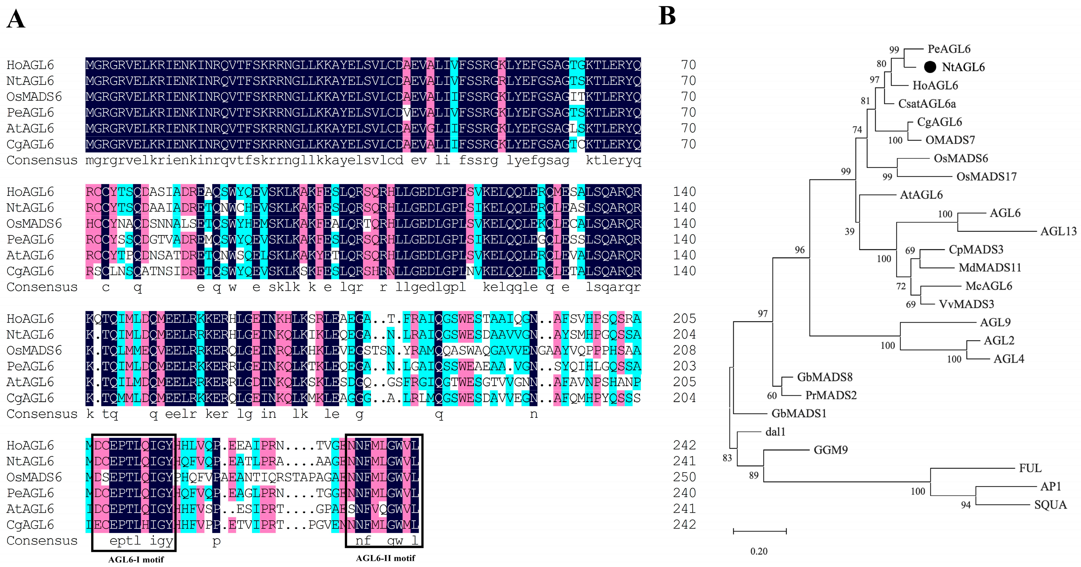
chinensis. The encoded protein contained an intact MADS domain, the
AGL6-I and
AGL6-II characteristic motifs, and an AGL2-like C-terminal sequence [
34]. Phylogenetic analysis assigned it to the AGL6-I-ZAG subclade [
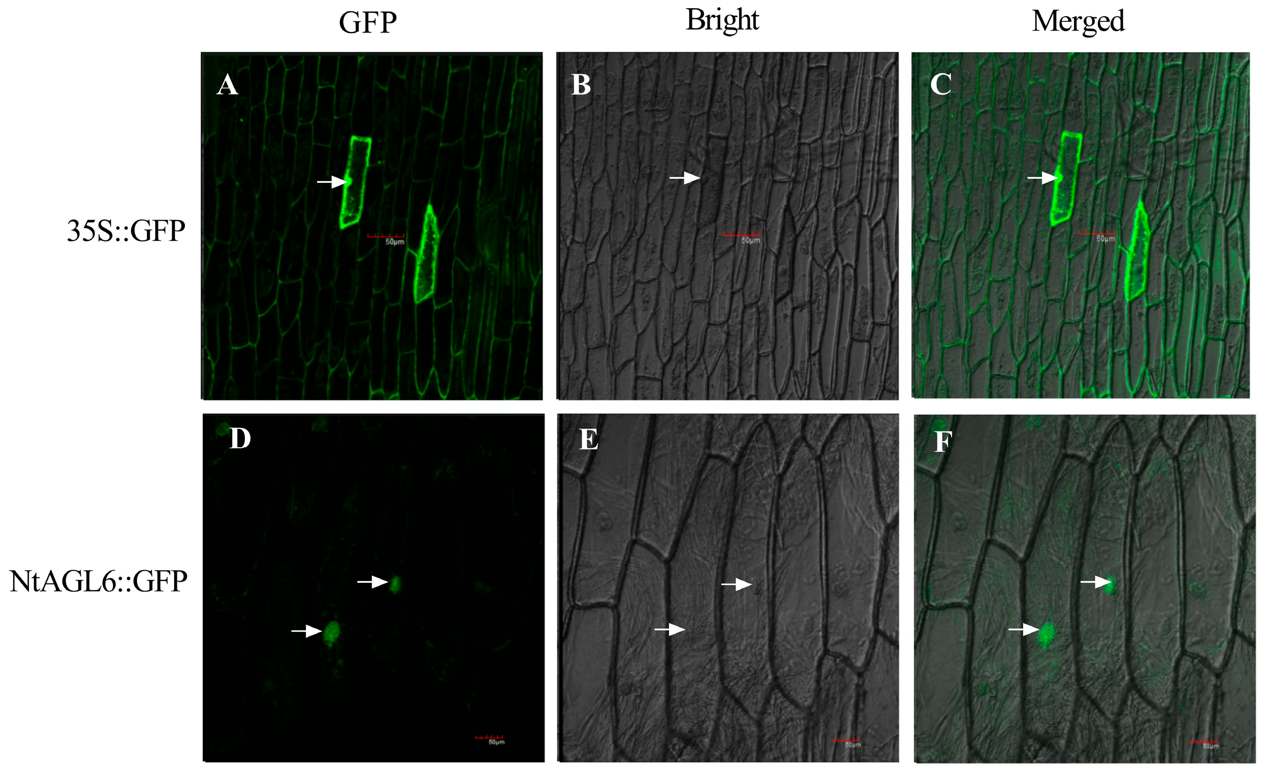
35], where it clustered with AGL6 proteins from other monocots. Subcellular localization confirmed that it is a nuclear-localized transcription factor. These features confirmed that
NtAGL6 is a typical
AGL6 subfamily member in
N. tazetta var.
chinensis.
AGL6 is one of the ancient subfamilies within the MADS-box family, with its origin predating the divergence of angiosperms and gymnosperms [
14,
15]. Two major clades, AGL6-I and AGL6-II, are commonly present in angiosperms, with members of the AGL6-I clade tending to be expressed in floral organs and involved in reproductive development [
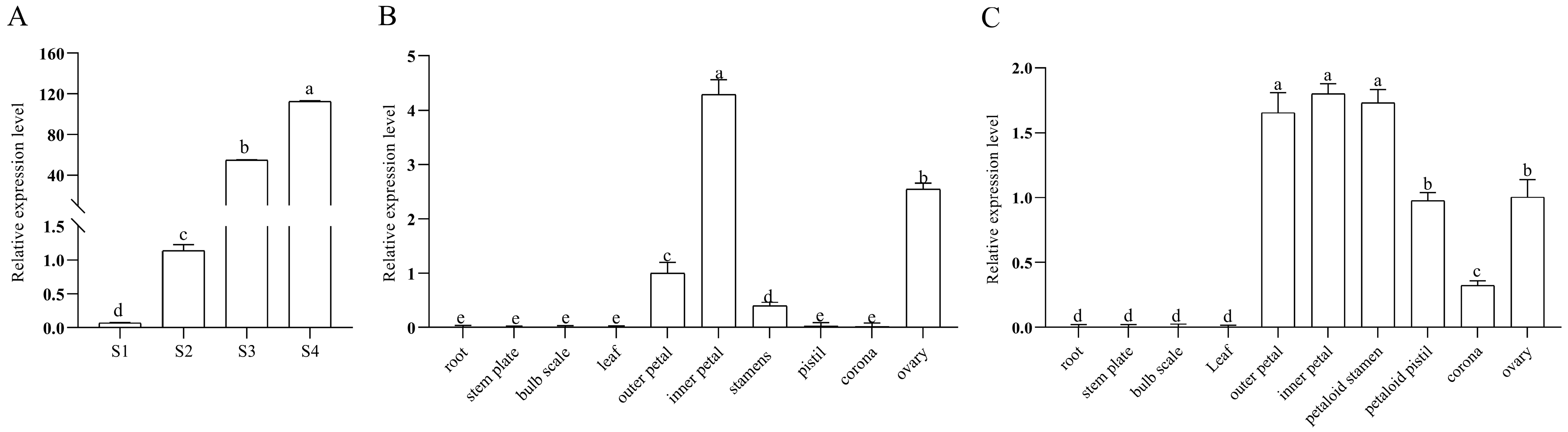
35]. The strictly floral organ-specific expression pattern of
NtAGL6 is consistent with the expression patterns of
AGL6 homologs previously reported in other monocots [
23,
24,
26,
36].
Thus, the sequence and expression characteristics of NtAGL6 are clearly consistent with those of AGL6-I clade members in monocots. However, whether its function follows a known pattern or exhibits lineage-specific divergence requires further analysis from three aspects: expression characteristics, heterologous activity, and regulatory mode.
3.2. Expression Characteristics of NtAGL6 and Its Potential Association with High Temperature-Induced Flowering
The expression of
NtAGL6 was strictly organ-specific, with no signal detected in vegetative organs, including root, basal plate, scale, and leaf, but with signals detected exclusively in floral organs. This pattern is highly conserved in monocots [
23,
24,
26,
36], further supporting the view that AGL6-I clade members have become highly specialized as floral development-related factors in monocots.
Within floral organs, the expression of NtAGL6 in the single-petal cultivar showed a gradient of outer petals > ovary > stamens > pistils ≈ corona. This gradient partially overlaps with, but is not completely identical to, the typical expression pattern of A-class genes (highly expressed in sepals and petals, absent in carpels). Its expression in the ovary suggested that its functional boundary may be broader than that of typical A-class genes. In the double-petal cultivar, the expression levels of NtAGL6 in petalized stamens and petalized pistils were significantly higher than those in the single-petal cultivar. However, this correlation alone cannot establish a causal relationship. Whether the petaloid phenotype is the cause or consequence of NtAGL6 upregulation, or whether both are co-regulated by upstream factors, requires further distinction through loss- or gain-of-function experiments in the endogenous Narcissus system.
At the transcriptional regulation level, the
NtAGL6 promoter contained GA
3-responsive elements (GARE-motif) and was significantly induced by exogenous GA
3, which is consistent with the known role of the GA pathway in flowering regulation [
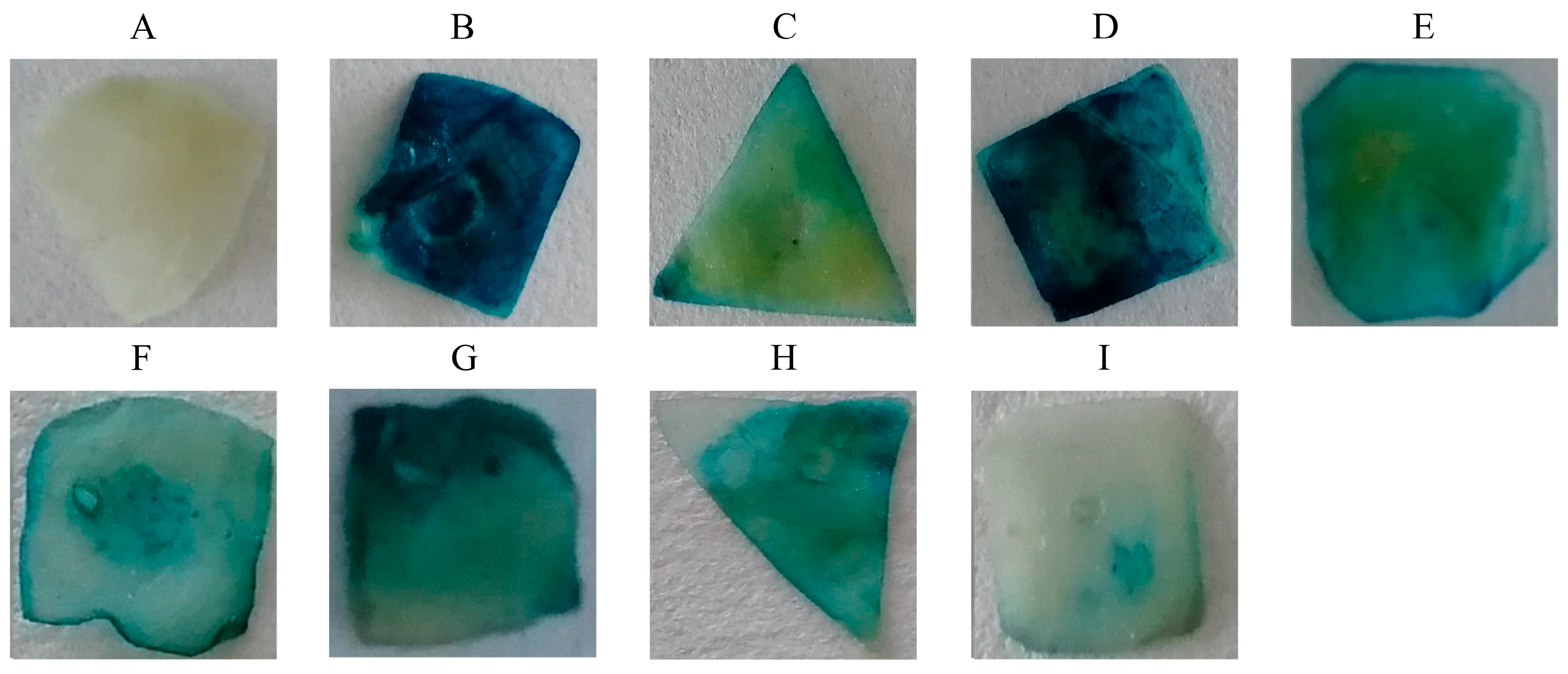
37]. More notably, the promoter was significantly induced by high temperature. Since floral bud differentiation in
N. tazetta var.
chinensis requires a temperature above 25 °C [
28], three independent lines of evidence from this study—the elevated expression of
NtAGL6 during flower bud differentiation, the presence of multiple HSE elements in its promoter, and the activation of GUS activity by high temperature—collectively point to a coherent working model: high temperature signals may promote floral transition by activating
NtAGL6 transcription. However, this inference remains indirect at present and awaits functional validation in the endogenous Narcissus system.
3.3. NtAGL6 Exhibits Flowering-Promoting Activity and Partial A-Class Functional Complementation in a Heterologous System
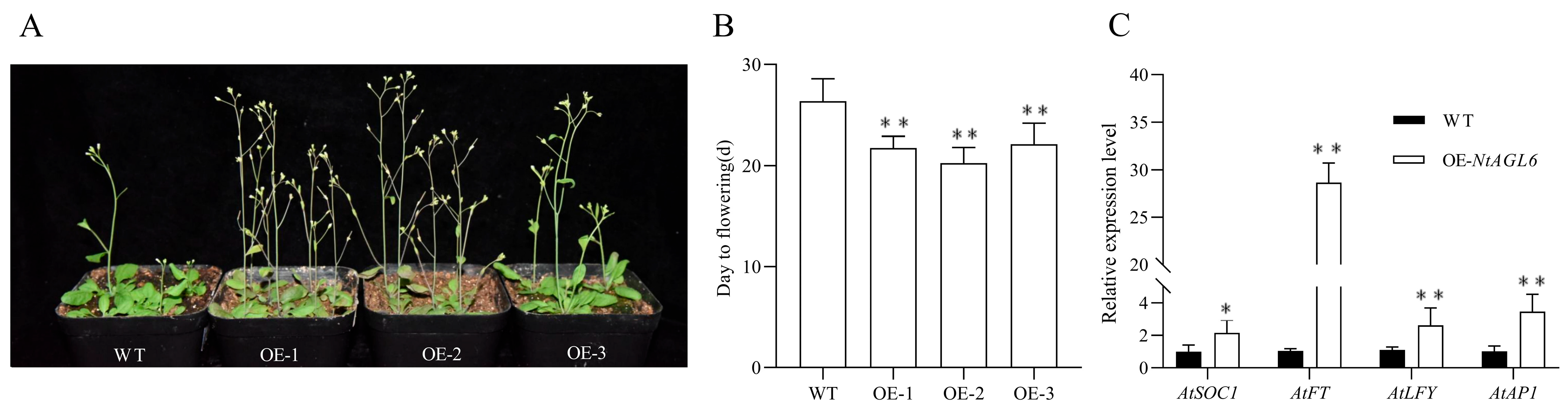
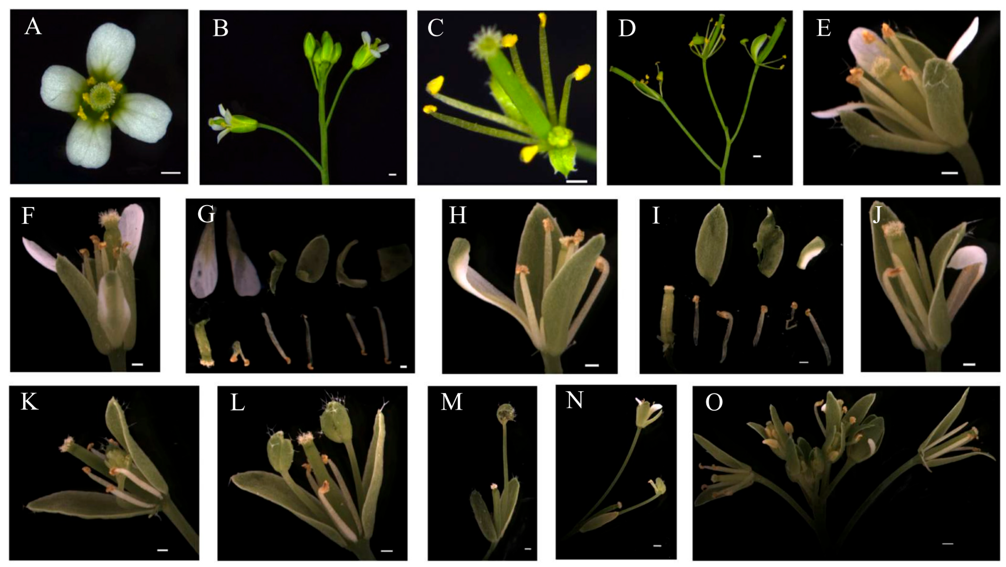
Ectopic expression of NtAGL6 in wild-type Arabidopsis led to early flowering, accompanied by upregulation of FT, SOC1, LFY, and AP1, with FT showing the most significant increase. In the ap1 mutant background, NtAGL6 partially restored petals and sepals and induced sepal petalody in some flowers. These phenotypes suggested that NtAGL6 can promote flowering and partially compensate for A-class floral organ identity determination when ectopically expressed in the heterologous Arabidopsis system.
This dual phenotype is not unique to
Narcissus; ectopic expression of
HoAGL6 from hyacinth [
32] and
PmAGL6 from mei flower [
33] also produced both early flowering and floral homeotic transformation phenotypes. In
Arabidopsis, endogenous
AGL6 primarily functions in flowering promotion [
16,
17], with A-class function being carried out by AP1 and AP2. In contrast,
AGL6 homologs from monocots such as
Narcissus and hyacinth exhibit dual activities in heterologous systems. One possible explanation is that AGL6 proteins possess the molecular potential to interact with multiple MADS-box proteins. In the endogenous network of
Arabidopsis, their interaction partners may be largely confined to the flowering time regulation pathway, whereas in plants such as
Narcissus, they may have retained the ability to interact with proteins involved in floral organ identity determination. The observation that the expression level of the A-class gene
NtAP1 in sepals and petals of
N. tazetta var.
chinensis is much lower than that of
AP1 in
Arabidopsis (our unpublished data) is consistent with this interpretation—when
AGL6 is still able to interact with floral organ identity proteins, the system’s demand for A-class gene expression may be relatively reduced.
However, a clear distinction must be made between “heterologous complementation capacity” and “endogenous function”. The restoration of petals and sepals in the ap1 mutant by NtAGL6 may result from heterologous interactions with Arabidopsis B-, C-, and E-class MADS-box proteins in the heterologous background, thereby compensating for the loss of AP1. This phenomenon indicates that NtAGL6 possesses the molecular potential to interact with proteins related to floral organ identity determination, but it does not directly prove that it performs A-class function endogenously in Narcissus.
Regarding flowering promotion, the significant upregulation of FT by NtAGL6 is particularly noteworthy. FT is a central integrator of flowering time regulation. The nearly 29-fold upregulation of FT upon NtAGL6 ectopic expression, far exceeding the upregulation of SOC1, LFY, and AP1, suggested that the early flowering phenotype may be mediated primarily through the FT pathway. This naturally raises the question: in what manner does NtAGL6 regulate FT?
3.4. Regulation of NtFT1 by NtAGL6 May Occur Through an Indirect Pathway
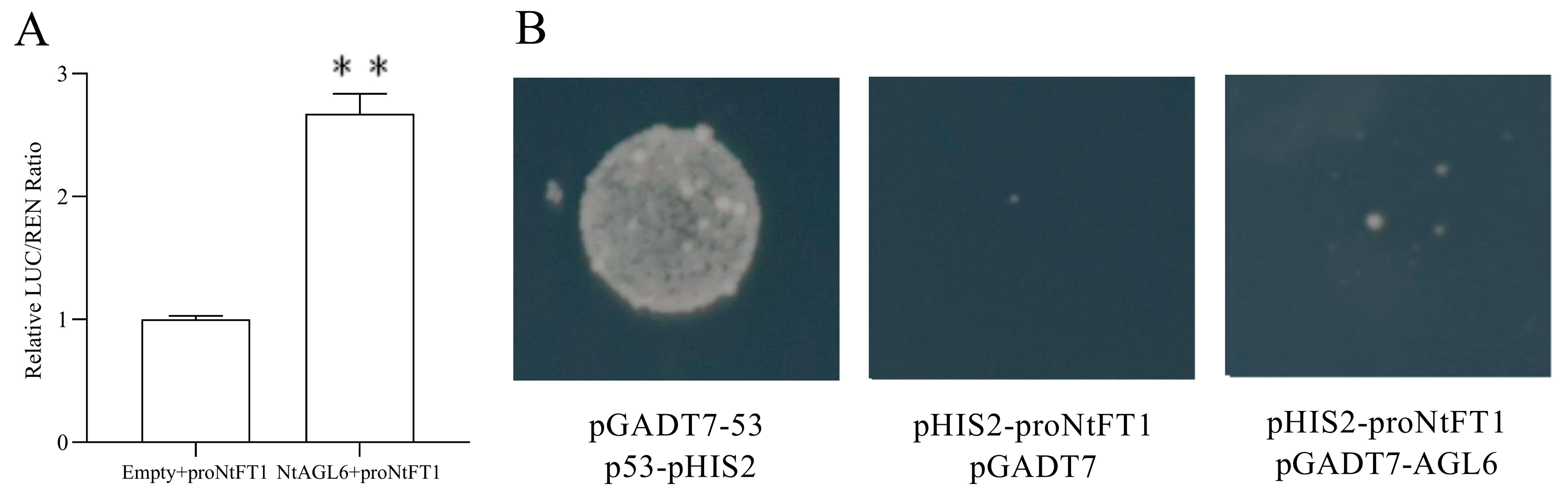
The direct answer to the above question is as follows: dual-luciferase assays showed that
NtAGL6 could increase the LUC activity driven by the
NtFT1 promoter by 2.57-fold, but yeast one-hybrid assays detected no direct binding of
NtAGL6 to the
NtFT1 promoter. This combined result of “activation without binding” suggested that the regulation of
NtFT1 by
NtAGL6 may occur through an indirect pathway. The
NtFT1 gene used in this study was previously identified as the functional
FT homolog in
N. tazetta var.
chinensis through sequence homology, phylogenetic analysis, and expression profiling [
38].
This finding directly addresses the core question raised in the Introduction. In
Arabidopsis,
AGL6 has been reported to promote flowering through positive regulation of
FT expression [
16], whereas
NtAGL6, although similarly activating the expression of an
FT homolog in a heterologous system, may employ a different molecular pathway. This suggests that the AGL6-FT regulatory module may have adopted different cis-regulatory logics between monocots and dicots. Interestingly, even in
Arabidopsis, recent studies have shown that
AGL6 can also regulate
FT expression indirectly through repression of
ELF3 [
19], indicating that the regulatory relationship between
AGL6 and
FT is more complex than simple direct binding. Our finding that
NtAGL6 activates
NtFT1 without direct promoter binding may thus represent an alternative mode within the
AGL6-
FT regulatory module.
Regarding the molecular basis of this indirect regulation, we propose three non-mutually exclusive hypotheses: (1)
NtAGL6 may need to form heterodimers with unknown MADS-box proteins to bind DNA effectively, and alone lacks sufficient affinity for the CArG-box elements in the NtFT1 promoter; (2)
NtAGL6 may indirectly upregulate
FT by repressing an unknown negative regulator of
FT, such as a member of the
TFL1 family or a specific miRNA. Although no typical FLC homolog was retrieved in our preliminary transcriptome analysis of
N. tazetta var.
chinensis, the possibility of functionally equivalent negative regulators of
FT cannot be excluded; (3)
NtAGL6 may recruit histone-modifying enzymes or chromatin remodeling complexes to alter the epigenetic state of the
FT chromatin region [
39,
40]. This mechanism does not require direct contact of
NtAGL6 with DNA but would not be detected in the yeast one-hybrid system. Among these three hypotheses, the first is particularly worthy of attention, as heterodimer formation among MADS-box proteins is a well-known phenomenon [
13,
41], and studies have shown that AGL6 proteins possess the structural basis for interacting with other MADS-box proteins.
Regardless of which mechanism is ultimately confirmed, the difference in FT regulatory mode between NtAGL6 and Arabidopsis AGL6 itself suggests that the connection mode of the AGL6-FT regulatory module is plastic during plant evolution. Although in vitro assays such as EMSA could provide further validation in the future, the current Y1H data, combined with the bioinformatic analysis of the promoter, strongly point to the dominance of an indirect regulatory pathway. It should also be noted that the dual-luciferase assay was performed in a heterologous system, which may not fully recapitulate the endogenous regulatory context.
3.5. Limitations and Future Directions
The main limitation of this study lies in the functional validation platform. Because a genetic transformation system for N. tazetta var. chinensis is not yet available, gene function validation relied entirely on heterologous expression in Arabidopsis and transient expression in tobacco. The fundamental limitation of this strategy is that the flowering-promoting and A-class complementary phenotypes observed for NtAGL6 in Arabidopsis may partially result from heterologous interactions with Arabidopsis MADS-box proteins; similarly, the promoter activation effects observed in tobacco leaves cannot be fully equated with the endogenous regulatory environment in Narcissus floral meristems. Therefore, the functional inferences regarding NtAGL6 in this study are preliminary and await verification in the endogenous Narcissus system. The 803 bp promoter fragment used in this study contains all core elements and key regulatory motifs relevant to our treatments and was experimentally confirmed to drive transcriptional activity. Nevertheless, we acknowledge that longer distal regions beyond this fragment may contain additional regulatory elements, although a longer fragment could not be obtained due to the current lack of a complete genome assembly for N. tazetta var. chinensis. Additionally, the specific molecular mediators involved in the indirect regulation of FT have not yet been identified, representing the most critical unresolved question at present.
Future research may be directed toward the following areas: (1) establishing a transient transformation system for Narcissus protoplasts or a VIGS (virus-induced gene silencing) system to validate the function of NtAGL6 and its regulatory relationship with FT in the native Narcissus background; (2) using yeast two-hybrid library screening or co-immunoprecipitation combined with mass spectrometry to systematically identify protein partners that interact with NtAGL6, with particular focus on bridging proteins that may mediate its indirect association with the FT promoter; and (3) integrating the promoter analysis results to further dissect how temperature signals are transmitted through the NtAGL6-FT module to the flowering decision center, thereby elucidating at the molecular level the unique mechanism of high temperature-induced flowering in N. tazetta var. chinensis.
4. Materials and Methods
4.1. Plant Materials and Growth Conditions
Three-year-old bulbs of
N. tazetta var.
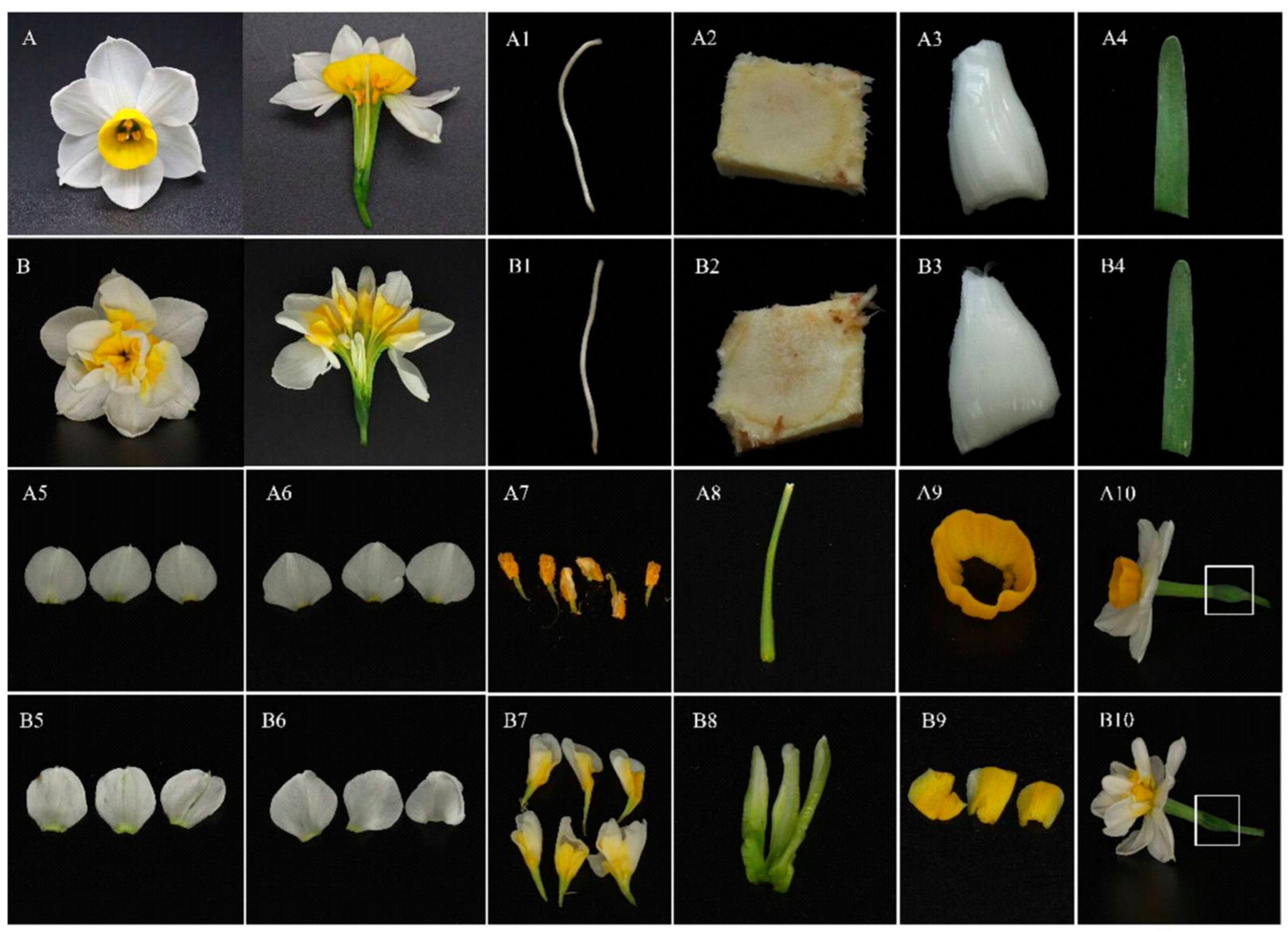
chinensis Roem cultivars ‘Jinzhan Yintai’ (single-petal) and ‘Yulinglong’ (double-petal) were purchased from Zhangzhou, Fujian, China. The bulbs were cleaned of old roots, disinfected, and then grown hydroponically in water at 25 °C under natural light conditions. The water was changed regularly during hydroponic culture. After roots and leaves had grown to appropriate stages, ten tissue types were collected: root, basal plate, scale, leaf, sepal (outer tepal), petal (inner tepal), stamen, pistil, corona, and ovary. These samples were used for tissue-specific expression analysis. The anatomical structures of the floral organs are shown in
Figure 8.
Samples for flower bud differentiation stages were collected from ‘Jinzhan Yintai’ bulbs stored at room temperature. The main buds were dissected periodically and classified into four stages based on morphological criteria: S1 (leaf bud stage, vegetative growth), S2 (inflorescence primordium formation stage), S3 (floral primordium formation stage, with individual flower bud primordia emerging within the spathe), and S4 (corolla formation stage, with floral organ primordia differentiating). Samples were collected at each stage. All collected samples were immediately frozen in liquid nitrogen and stored at −80 °C for subsequent RNA extraction.
Seeds of Arabidopsis thaliana wild-type Columbia (Col-0) and the mutant ap1 (SALK_151561C, Col-0 background) were obtained from the AraShare Arabidopsis Mutant Stock Center. Seeds were stratified at 4 °C for 3 d and then sown in nutrient soil (peat:vermiculite = 3:1, v/v). Plants were grown in a controlled environmental growth chamber under long-day conditions (16 h light/8 h dark) at 22 °C, with a light intensity of 100 μmol·m−2·s−1 and relative humidity of 60–70%.
Seeds of Nicotiana benthamiana were sown in nutrient soil and grown under the same long-day conditions (22 °C, 16 h light/8 h dark, 100 μmol·m−2·s−1) for dual-luciferase transient expression assays and promoter GUS activity detection.
4.2. Total RNA Extraction and cDNA Synthesis
Approximately 100 mg of each tissue sample stored at −80 °C was ground into a fine powder in liquid nitrogen. Total RNA was extracted using the FastPure Plant Total RNA Kit (TaKaRa Bio Inc., Dalian, China) following the manufacturer’s instructions. The quality and concentration of total RNA were assessed by 1.0% agarose gel electrophoresis and a NanoDrop 2000 spectrophotometer (Thermo Fisher Scientific, Waltham, MA, USA), respectively. For reverse transcription, 1 μg of total RNA was used with the HiScript® II 1st Strand cDNA Synthesis Kit (Vazyme Biotech Co., Ltd., Nanjing, China). The synthesized cDNA was stored at −20 °C for further use.
4.3. Cloning of the NtAGL6 Gene
Based on the transcriptome database of
N. tazetta var.
chinensis previously obtained by our research group (unpublished data),
AGL6 homologous sequences were screened through keyword searches and sequence alignment. Specific primers NtAGL6-F and NtAGL6-R (primer sequences listed in
Appendix A Table A1) were designed according to the open reading frame (ORF) of the target sequence. PCR amplification was performed using the high-fidelity DNA polymerase PrimeSTAR
® HS DNA Polymerase (TaKaRa) with cDNA from petals of ‘Jinzhan Yintai’ as template. The purified products were ligated into the pMD™18-T Vector (TaKaRa) and transformed into
Escherichia coli DH5α competent cells. After identification by colony PCR, positive clones were selected and sent to Sangon Biotech (Shanghai, China) for sequencing.
4.4. Sequence Alignment and Phylogenetic Analysis
The sequenced
NtAGL6 sequence was submitted to the NCBI database for BLAST homology searches (
https://blast.ncbi.nlm.nih.gov/Blast.cgi, accessed on 15 May 2025). AGL6 homologous protein sequences from representative species, including
Arabidopsis, rice, maize, wheat, orchid, magnolia, mei flower (
Prunus mume), and hyacinth, as well as protein sequences of other MADS-box subfamily members such as AP1 and SEP, were downloaded. Multiple sequence alignment was performed using the Clustal W (version 2.1) program. A phylogenetic tree was constructed using MEGA 7.0 software with the Neighbor-Joining (NJ) method. The p-distance model was selected, gaps were handled by pairwise deletion, and bootstrap analysis was performed with 1000 replicates.
4.5. Quantitative Real-Time PCR (qPCR)
Specific primers qNtAGL6-F and qNtAGL6-R were designed based on the specific region of the
NtAGL6 cDNA. The
Narcissus Actin gene (GenBank: JN204912) was used as the internal reference gene for
N. tazetta var.
chinensis, and
Arabidopsis ACT2 (GenBank: NM_001338358) was used for
Arabidopsis thaliana. Pre-experiments confirmed that the
Actin was stably expressed across all tested tissues. Primer sequences are listed in
Appendix A Table A1. qPCR reactions were performed using Hieff
® qPCR SYBR
® Green Master Mix (No Rox) (Yeasen Biotechnology Co., Ltd., Shanghai, China) on a Bio-Rad CFX96™ Real-Time System (Bio-Rad Laboratories, Hercules, CA, USA). Each sample included three biological replicates. Relative expression levels were calculated using the 2
−ΔΔCt method, and data are presented as means ± standard deviation (SD). Statistical significance among multiple groups was determined by one-way ANOVA followed by Duncan’s multiple range test (
p < 0.05). Different lowercase letters above the bars indicate significant differences.
4.6. Cloning and Analysis of the NtAGL6 Promoter
Genomic DNA was extracted from leaves of ‘Jinzhan Yintai’ using the DNAprep Pure Plant Kit (Tiangen Biotech Co., Ltd., Beijing, China). Based on the 5′-upstream sequence of the
NtAGL6 in the transcriptome database, nested PCR primers AGL6QDZ1, AGL6QDZ2, and AGL6QDZ3 (sequences listed in
Appendix A Table A1) were designed. After three rounds of PCR using the Genome Walking Kits (TaKaRa). The cloned promoter sequence was analyzed for cis-acting regulatory elements using the online promoter analysis tools PlantCARE (
http://bioinformatics.psb.ugent.be/webtools/plantcare/html/, accessed on 15 May 2025) and PLACE (
http://www.dna.affrc.go.jp/PLACE/signalscan.html, accessed on 15 May 2025).
4.7. Construction of the GUS Expression Vector and Agrobacterium Transformation
The cloned NtAGL6 promoter fragment was inserted into the plant expression vector pBI121 using the In-Fusion® HD Cloning Kit (TaKaRa), replacing the original CaMV 35S promoter to generate the recombinant vector pBI121-NtAGL6::GUS. The recombinant plasmid was extracted and introduced into Agrobacterium tumefaciens strain GV3101 by the freeze–thaw method. Positive transformants were selected on YEP plates supplemented with 50 mg·L−1 kanamycin and 50 mg·L−1 rifampicin. Transformants were verified by colony PCR and then preserved for further use.
4.8. Transient GUS Expression Analysis of the Promoter
A single colony of Agrobacterium GV3101 harboring the recombinant plasmid pBI121-NtAGL6::GUS was inoculated into 5 mL of YEP liquid medium and cultured overnight at 28 °C with shaking at 200 r·min−1. The next day, the culture was diluted 1:50 into fresh YEP medium and grown until OD600 reached 0.6–0.8. The cells were collected by centrifugation at 6000 rpm for 10 min, and the supernatant was discarded. The pellet was resuspended in infiltration buffer (10 mmol·L−1 MgCl2, 10 mmol·L−1 MES, 150 μmol·L−1 acetosyringone, pH 5.6) and adjusted to OD600 = 0.8. The suspension was then incubated at room temperature for 2–3 h. Agrobacterium GV3101 without the plasmid was used as a negative control, while Agrobacterium carrying the empty pBI121 vector (OD600 adjusted to 0.8) served as a positive control.
Fully expanded leaves from the middle part of Nicotiana benthamiana plants were selected for infiltration. The bacterial suspension was injected into the abaxial side of the leaves using a 1 mL syringe (without needle). For each treatment, at least five plants were infiltrated, with 2–3 leaves per plant. After infiltration, the plants were placed in a light incubator at 22 °C under normal growth conditions.
The treatment scheme consisted of four groups: (1) Abiotic stress group: after 24 h of normal culture post-infiltration, plants were subjected to 35 °C for 48 h or 4 °C for 48 h. (2) Hormone treatment group: after 24 h of normal culture post-infiltration, plants were sprayed with 100 μmol·L−1 GA3, 100 μmol·L−1 ABA, or 100 μmol·L−1 MeJA, respectively. The sprays were applied every 12 h for a total of four sprays (treatment duration: 48 h). (3) Normal temperature control group: plants were cultured normally and sprayed with an equal volume of sterile water. (4) Positive control group: plants were transformed with the empty pBI121 vector (OD600 = 0.8) and cultured normally. All treatments were sampled at 72 h post-infiltration (i.e., 48 h after treatment initiation). Leaf disks approximately 1 cm in diameter around the infiltration site were collected for GUS staining.
GUS staining was performed according to the method described by Jefferson et al. [
42]. Leaf disks were immersed in GUS staining solution (50 mmol·L
−1 sodium phosphate buffer pH 7.0, 10 mmol·L
−1 Na
2EDTA, 0.5 mmol·L
−1 K
3[Fe(CN)
6], 0.5 mmol·L
−1 K
4[Fe(CN)
6], 0.1% Triton X-100, and 1 mmol·L
−1 X-Gluc, freshly prepared) and incubated at 37 °C in the dark for 12–16 h. After staining, the disks were decolorized sequentially with 70% ethanol, 90% ethanol, and absolute ethanol until the green background was completely removed. The samples were observed and photographed under a stereomicroscope (Olympus SZX16). At least 15 leaf disks were observed for each treatment, and the experiment was independently repeated three times.
4.9. Subcellular Localization Vector Construction and Transient Expression in Onion Epidermal Cells
The complete ORF of NtAGL6 (without the stop codon) was inserted into the pCAMBIA1302 vector (containing an mGFP tag) using the In-Fusion method to construct the 35S::NtAGL6-GFP fusion expression vector. The recombinant plasmid was verified by sequencing and then transformed into Agrobacterium GV3101.
Fresh onion (Allium cepa L.) bulbs were peeled of the outer 3–4 layers of scales and surface-sterilized by soaking in 75% ethanol for 10 min, followed by four washes with sterile water. Using a sterile scalpel, approximately 1 cm × 1 cm squares were marked on the inner epidermis, which was then carefully peeled off with forceps. The epidermal strips were placed on MS solid medium (containing 3% sucrose and 0.8% agar, pH 5.8) and pre-cultured at 25 °C in the dark for 20 h.
Agrobacterium suspensions containing 35S::NtAGL6-GFP or the empty pCAMBIA1302 vector (OD
600 = 0.8) were prepared following the method described in
Section 4.8. The pre-cultured onion epidermal strips were immersed in the bacterial suspension for 15 min, then removed and blotted dry with sterile filter paper. The strips were placed on MS solid medium and cultured at 25 °C in the dark for 2 d. After culture, the epidermal strips were washed three times with sterile water, mounted on glass slides with coverslips, and observed under a laser scanning confocal microscope (Leica TCS SP8, Wetzlar, Germany) for GFP fluorescence signals. The excitation wavelength was 488 nm, and emission was detected at 505–530 nm. Onion epidermal cells transformed with the empty pCAMBIA1302 vector served as controls to observe the intracellular distribution of GFP. The experiment was independently repeated three times, and no fewer than 20 positive cells were observed per replicate.
4.10. Arabidopsis Transformation and Screening of Transgenic Lines
The recombinant vector 35S::NtAGL6 was transformed into
Agrobacterium GV3101 by the freeze–thaw method.
Arabidopsis transformation was performed using the floral dip method described by Clough and Bent [
43]. Wild-type Col-0 and
ap1 mutant
Arabidopsis plants were grown until bolting and flowering. Open flowers and mature siliques were removed to promote the growth of lateral inflorescences. When the lateral inflorescences were at full bloom, the
Agrobacterium suspension was prepared as described above to OD
600 = 0.8, and Silwet L-77 was added to a final concentration of 0.02%. The inflorescences were dipped into the bacterial suspension for 30 s with gentle agitation, then removed, covered with a plastic bag to maintain high humidity, and kept in the dark for 24 h, followed by normal culture. The transformation was repeated once after 7 d. Plants transformed with the empty pCAMBIA1302 vector served as controls.
T0 seeds were harvested, sterilized with 70% ethanol for 1 min and 2.5% sodium hypochlorite for 10 min, then rinsed five times with sterile water. The seeds were sown on MS selection medium supplemented with 20 mg·L−1 hygromycin, stratified at 4 °C for 3 d, and then transferred to long-day conditions at 22 °C. After 7–10 days of selection, resistant seedlings (with normal root elongation and green cotyledons) were transplanted into nutrient soil. T1 plants were further screened on hygromycin-containing medium, and lines showing a segregation ratio of approximately 3:1 were considered to carry a single-copy insertion. Homozygous transgenic lines were obtained by T3 generation. At least three independent homozygous lines per transformation event were used for subsequent phenotypic analysis. Transgenic plants were verified by PCR using NtAGL6-specific primers and by qRT-PCR for expression level analysis before use in experiments.
4.11. Phenotypic Observation and Flowering Time Measurement of Transgenic Arabidopsis
T3 homozygous transgenic Arabidopsis seeds (35S::NtAGL6/Col-0, 35S::NtAGL6/ap1, and their respective empty vector controls) were stratified at 4 °C for 3 d and then sown in nutrient soil. Plants were grown under long-day conditions at 22 °C. The number of days from sowing to bolting (when the inflorescence stem elongated to approximately 1 cm) was recorded daily for each plant. The experiment was independently repeated three times. Flowering time data are presented as means ± SD, and statistical significance was analyzed using Student’s t-test (* p < 0.05, ** p < 0.01).
At bolting stage, inflorescences of Arabidopsis were observed and photographed under a stereomicroscope (Olympus SZX16) to document floral organ morphology. Particular attention was paid to the rescue of petal and sepal phenotypes in transgenic plants of the ap1 mutant background, as well as any homeotic transformation phenotypes.
4.12. Expression Analysis of Flowering-Related Genes in Transgenic Arabidopsis
Rosette leaves and shoot apical meristem tissues were collected from 35S::NtAGL6/Col-0 transgenic
Arabidopsis and empty vector control plants. Total RNA was extracted and cDNA was synthesized following the method described in
Section 4.2. The
Arabidopsis Actin gene was used as the internal reference. The expression levels of key flowering regulatory genes
FT,
SOC1,
LFY, and
AP1 were examined. Primer sequences are listed in
Appendix A Table A1, and the qPCR system and program were the same as described in
Section 4.5. Three biological replicates were performed for each line, and the empty vector control was used as the calibrator sample to calculate the relative expression levels of each gene.
4.13. Dual-Luciferase Reporter Assay
The promoter fragment of the NtFT1 gene was inserted into the multiple cloning site of the pGreenII 0800-LUC vector using the In-Fusion method to construct the reporter vector NtFT1Pro-LUC, which contains the firefly luciferase gene (LUC) and the internal control Renilla luciferase gene (REN). Meanwhile, the complete ORF of NtAGL6 was inserted into the pGreenII 0800-62-SK vector to construct the effector vector 35S::NtAGL6. All recombinant plasmids were verified by sequencing and then transformed into Agrobacterium GV3101.
Agrobacterium strains harboring the reporter and effector vectors were inoculated separately into YEP liquid medium and cultured as described above to OD600 = 0.6–0.8. The cells were collected by centrifugation at 4000 rpm for 10 min, resuspended in infiltration buffer, and adjusted to OD600 = 0.8. The Agrobacterium cultures containing the reporter vector and the effector vector were mixed at a 1:1 volume ratio and then infiltrated into leaves of Nicotiana benthamiana plants. Co-infiltration of the reporter vector with Agrobacterium carrying the empty pGreenII 0800-62-SK vector served as the control.
After infiltration, the tobacco plants were grown under long-day conditions at 22 °C for 72 h. Leaf disks approximately 0.8 cm in diameter were collected from the edge of the infiltration zone (avoiding the central injection point) using a hole puncher and immediately frozen in liquid nitrogen. Samples were processed according to the protocol of the Dual Luciferase Reporter Gene Assay Kit (Promega, Madison, WI, USA). Firefly luciferase (LUC) and Renilla luciferase (REN) luminescence were measured sequentially using a microplate reader (SpectraMax iD3, Molecular Devices). The LUC/REN ratio was calculated to reflect promoter activity. Each treatment included four biological replicates (four leaves), and the experiment was independently repeated three times. Data are presented as means ± SD, and statistical significance was analyzed using Student’s t-test.
4.14. Yeast One-Hybrid Assay
The
NtFT1 promoter fragment (same as in
Section 4.13 ) was inserted into the pHIS2 vector using the In-Fusion method to construct the bait vector pHIS2-proNtFT1. The complete ORF of NtAGL6 was inserted into the pGADT7 vector to construct the prey vector pGADT7-AGL6. The recombinant plasmids were verified by sequencing and then co-transformed into
Saccharomyces cerevisiae strain Y187 using the PEG/LiAc method. Positive control (pGADT7-53 + p53-pHIS2) and negative control (pGADT7 + pHIS2-proNtFT1) were also included.
The transformed yeast cells were plated on SD/-Leu-Trp deficient solid medium and incubated at 30 °C for 3–5 d until colonies appeared. To determine the minimal inhibitory concentration of 3-amino-1,2,4-triazole (3-AT) required to suppress background autoactivation of the His3 reporter gene, yeast cells transformed with the empty pHIS2-proNtFT1 vector were first plated on SD/-Leu-Trp-His deficient medium containing 0, 5, 10, 15, 20, 25, 30, 35, 40, 45, or 50 mmol·L−1 3-AT. After incubation at 30 °C for 5 d, the lowest 3-AT concentration that completely inhibited yeast growth (30 mmol·L−1) was selected for subsequent screening assays. Single colonies of the co-transformed experimental and control groups were picked, resuspended in sterile water, adjusted to OD600 = 0.1, and 5 μL of each suspension was spotted onto SD/-Leu-Trp-His deficient solid medium containing 30 mmol·L−1 3-AT. The plates were incubated at 30 °C for 3–5 d, and yeast growth was monitored.