


Abstract
Handroanthus chrysanthus (Jacq.) S.O. Grose is a Neotropical tree species highly valued for its ornamental beauty, durable timber, and medicinal properties. However, overexploitation and the recalcitrant nature of its seeds have constrained propagation and conservation efforts, and the species has been listed as Vulnerable on the IUCN Red List since 2020. In this study, a direct adventitious shoot regeneration system was developed for H. chrysanthus by systematically evaluating explant type, basal medium, plant growth regulator combination, and light quality. Hypocotyls were identified as the most responsive explants for shoot induction, and the highest adventitious shoot induction frequency, 51.79%, was obtained on Murashige and Skoog medium (MS) supplemented with 5 mg·L−1 6-benzylaminopurine, 0.5 mg·L−1 indole-3-butyric acid, and 0.2 mg·L−1 thidiazuron under white fluorescent light. The highest shoot multiplication coefficient, 3.27, was obtained on MS medium containing 4 mg·L−1 6-benzylaminopurine and 0.3 mg·L−1 gibberellic acid. The maximum rooting frequency, 80%, was obtained on R14 medium after 30 days of culture. After acclimatization, 95% of the regenerated plantlets survived and grew vigorously under greenhouse conditions. To our knowledge, no direct adventitious shoot regeneration system has been reported for the Tabebuia alliance. This efficient regeneration protocol provides a practical platform for clonal propagation, germplasm conservation, and genetic improvement of H. chrysanthus and may also support regeneration and conservation in related endangered taxa within the Tabebuia alliance.
1. Introduction
Handroanthus chrysanthus (Jacq.) S.O. Grose (syn. Tabebuia chrysantha), commonly known as golden-bell wood, is a deciduous tree belonging to the Tabebuia alliance (Bignoniaceae). The alliance currently includes approximately 99 species across three genera, although these taxonomic numbers remain provisional because the classification of the Tabebuia alliance is still under revision based on ongoing molecular phylogenetic studies [1,2,3]. Many species, including H. chrysanthus, are renowned for their exceptionally durable timber (Ipê) and spectacular floral displays [4,5,6]. However, overexploitation has led to indiscriminate logging [7], and the species has been listed as Vulnerable on the IUCN Red List since 2020 [8].
H. chrysanthus was introduced into the subtropical regions of southern China over 40 years ago [5,6]. Currently, H. chrysanthus is primarily propagated by seeds. However, this approach encounters two major constraints. First, fruit maturation coincides with the rainy season in southern China, and the winged seeds are prone to dispersal and mold. Second, as a recalcitrant species, its seed viability declines rapidly after harvest—germination rates remain viable for approximately 60 days and drop to nearly zero after three months [9]. These limitations hinder large-scale propagation and ex situ conservation.
Plant tissue-culture technology offers many advantages—such as high reproduction efficiency, stable seedling growth, and seasonally unrestricted propagation—to effectively meet market demand. Moreover, in vitro micropropagation serves as a foundational technology in both conventional and modern breeding, enabling polyploid development, elite trait selection, and genetic transformation of recalcitrant tissues [10], thereby significantly accelerating the breeding process to improve undesirable traits such as short flowering period and cold sensitivity.
H. chrysanthus has also been used in traditional medicine for treating fever, inflammation, and pain [11,12,13,14]. Pharmacological studies have confirmed its anti-inflammatory, antimicrobial, and anticancer activities [15,16,17,18,19,20]. However, these medicinal properties are not the focus of the present study. In the Tabebuia alliance, the micropropagation protocols of H. heptaphyllus [21], H. impetiginosus [22,23,24,25], T. donnell-smithii [26], and H. guayacan [27] have been previously established using nodal segments from in vitro-germinated seedlings or adult plants. More recently, Héctor [7] developed a micropropagation protocol for H. chrysanthus using apical buds from seed-derived plants and achieved a maximum propagation coefficient of 3.35 on WPM (Woody Plant Medium) medium supplemented with 6.6 μM 6-BA.
However, to date, there have been no reports on the establishment of a direct adventitious shoot regeneration system within the Tabebuia alliance. Unlike nodal micropropagation, which is limited to clonal propagation, an adventitious shoot regeneration system provides a basis for genetic transformation (Agrobacterium-mediated transformation or CRISPR/Cas9), enabling targeted trait improvement such as flowering regulation or cold tolerance [10]. In this context, the main aim of this research was to study the direct organogenesis potential of H. chrysanthus. The specific objectives of this study were to investigate the effects of key factors on direct shoot regeneration in H. chrysanthus, including explant type (cotyledons and hypocotyls) and medium composition (basal culture media and plant growth regulators (PGRs)). This established regeneration protocol will facilitate large-scale propagation of H. chrysanthus and serve as an essential technical foundation for genetic improvement and accelerated breeding of new cultivars of H. chrysanthus. The optimized regeneration system described here also provides a technical reference for related species within the Tabebuia alliance, an economically significant lineage of Neotropical trees.
2. Results
2.1. Initial Aseptic Culture Establishment
In this study, the seeds of H. chrysanthus from six disinfection treatments were cultured on hormone-free MS basal medium. After 20 days, the contamination and germination rates recorded after 20 days are presented in Table 1. Among the six treatments evaluated, surface sterilization with 75% ethanol for 50 s followed by 0.1% HgCl2 for 10 min was the most effective for H. chrysanthus seeds, yielding a high germination rate of 86.32% with only 10.20% contamination. Longer exposure (16 min) reduced contamination but significantly decreased germination, consistent with the findings of Zha et al. [25], who observed similar HgCl2 toxicity in H. impetiginosus. Although a small number of seeds did not germinate, most disinfected seeds of H. chrysanthus germinated on hormone-free MS basal medium and developed into normal seedlings (Table 1). This finding contrasts with the previous report by Héctor [7], who suggested that MS medium was not suitable for the germination of H. chrysanthus seeds. The discrepancy may be related to differences in seed provenance. In Héctor’s study, seeds were collected from natural populations in Ecuador, whereas those used in the present study were obtained from cultivated trees in Guangzhou, China. These cultivated trees originated from populations introduced into subtropical China several decades ago [5,6], and long-term cultivation under local environmental conditions may have altered their seed germination characteristics and response to MS medium.
Table 1.
Effect of 0.1% HgCl2 sterilization duration on aseptic seedling establishment of Handroanthus chrysanthus.
2.2. Effect of Plant Growth Regulators on Shoot Regeneration from the Hypocotyls of H. chrysanthus
2.2.1. Effect of 6-BA and NAA on Shoot Regeneration
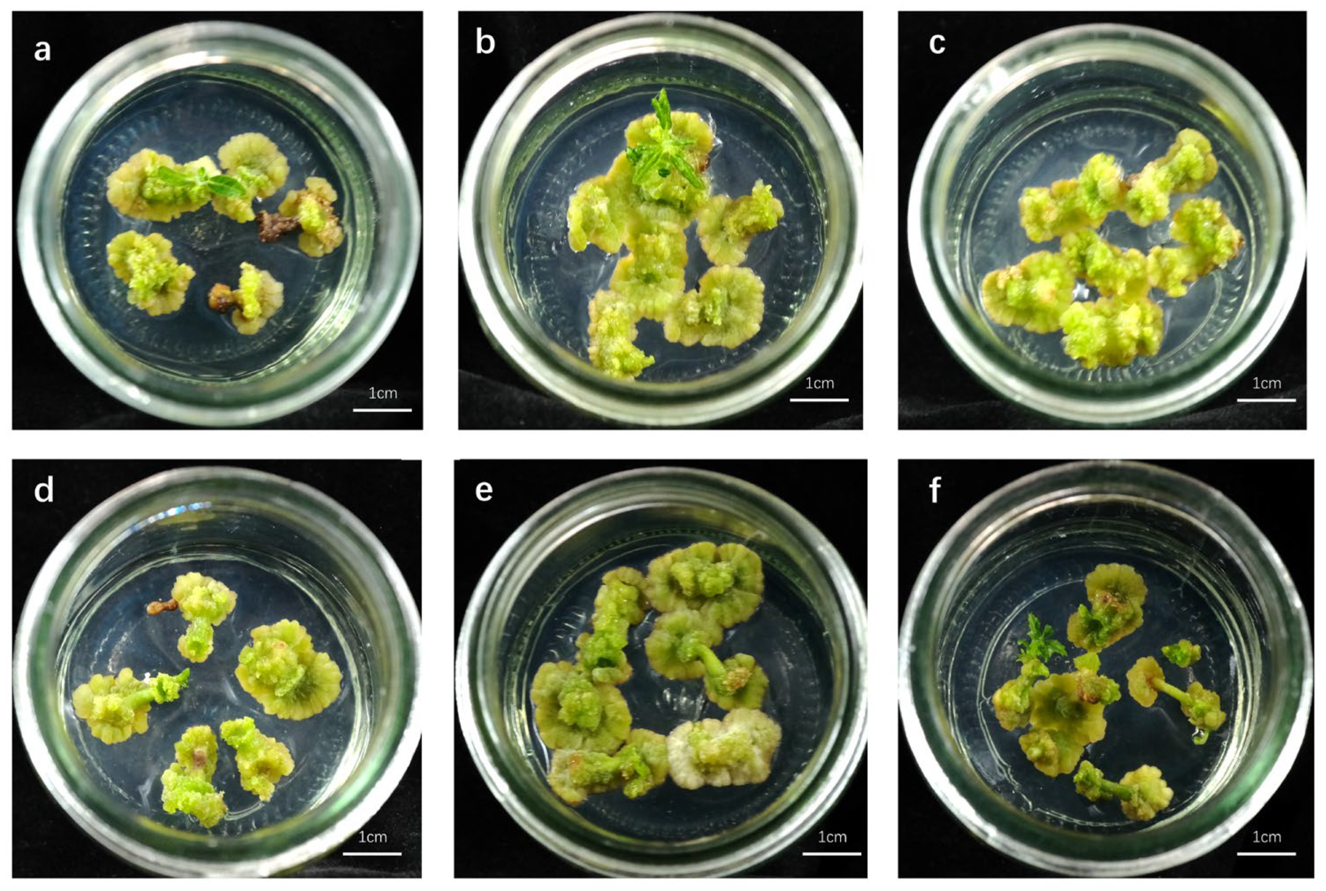
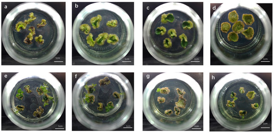
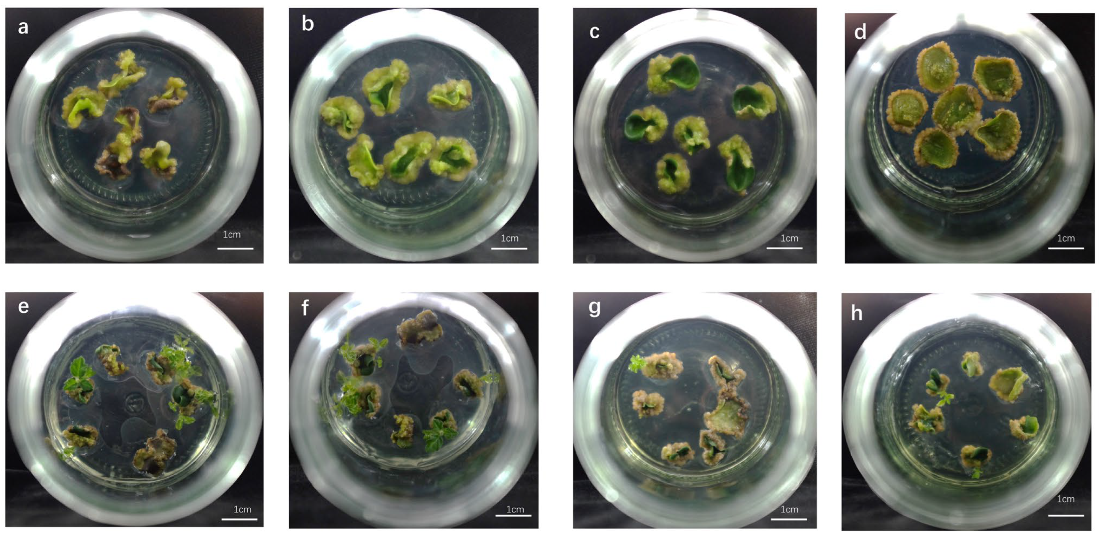
As shown in Table 2 and Figure 1, adventitious shoots were induced from hypocotyl explants cultured on MS basal medium supplemented with different concentrations of 6-BA and NAA (1-naphthaleneacetic acid). After seven days of culture, both ends of the hypocotyls began to swell, followed by the formation of compact green callus. The callus morphology, including growth rate, texture, and color, as well as the efficiency of shoot regeneration, varied significantly depending on the combination of 6-BA and NAA (Figure 1).
Table 2.
Effect of 6-BA and NAA combination on the induction of callus and shoot regeneration from the hypocotyls of Handroanthus chrysanthus after 45 days.
Figure 1.
In vitro culture of Handroanthus chrysanthus hypocotyls under different combinations of 6-BA and NAA after 35 days: (a–f) Callus and shoot regeneration from hypocotyls in treatments SR2, SR5, SR8, SR9, SR10, and SR11, respectively. Scale bar 1 cm.
Adventitious shoot formation was first observed at 20 days of culture in SR6 medium (MS + 5.0 mg·L−1 6-BA + 0.1 mg·L−1 NAA), with shoots emerging from one end of the hypocotyl explants. In SR5, SR10, and SR12 media, shoots appeared around day 25, whereas in the remaining formulations, the induction took 35 to 45 days. After 45 days, MS medium containing 5.0 mg·L−1 6-BA with 0.05–0.1 mg·L−1 NAA (SR5 and SR6) produced adventitious shoots at a frequency of approximately 20% (Table 2).
2.2.2. Effect of TDZ, 6-BA, and IBA Combinations on Shoot Regeneration
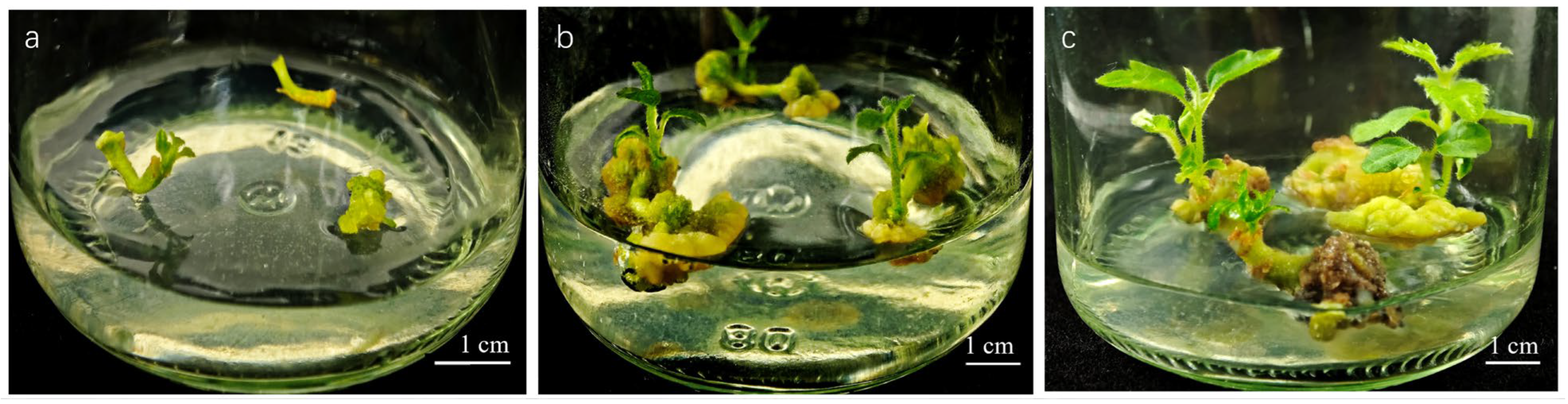
Thidiazuron (TDZ) is a phenylurea-derived compound widely recognized as a highly effective cytokinin for inducing adventitious shoot regeneration in recalcitrant woody plant species [28,29]. To improve the regeneration efficiency of H. chrysanthus, we evaluated the effect of TDZ, 6-BA, and IBA (indole-3-butyric acid) combinations on adventitious shoot regeneration. Initial morphological changes were observed within 5 days of culture, with explants showing swelling at both ends or throughout their length, followed by callus formation at the cut. Shoot initiation first occurred in medium SI3 at 15 days, followed by media SI10, SI12, and SI13 at 20 days, while the remaining media showed shoot formation between 30 and 40 days. Among these, medium SI13 (MS supplemented with 0.2 mg·L−1 TDZ, 5.0 mg·L−1 6-BA, and 0.5 mg·L−1 IBA) achieved the highest regeneration frequency of 51.79% (Table 3). Figure 2 illustrates the adventitious bud regeneration process from H. chrysanthus hypocotyls in SI13 media. Notably, at constant IBA (0.5 mg·L−1) and TDZ (0.2 mg·L−1) concentrations, increasing 6-BA levels enhanced both shoot regeneration and multiplication. However, when 6-BA (1.5 mg·L−1) and IBA (0.5 mg·L−1) concentrations were maintained, elevated TDZ levels progressively reduced shoot regeneration rates, indicating that excessive TDZ concentrations negatively impact regeneration efficiency in H. chrysanthus hypocotyl cultures.
Table 3.
Effects of 6-BA, TDZ, and IBA combinations on the regeneration of adventitious shoots from the hypocotyls of Handroanthus chrysanthus.
Figure 2.
Adventitious bud regeneration process from Handroanthus chrysanthus hypocotyls in SI13 media: (a–c) The SI13 culture media contained 5 mg⋅L−1 6-BA + 0.2 mg⋅L−1 TDZ + 0.5 mg⋅L−1 IBA. Hypocotyls cultured in Medium SI13 for 20 d, 45 d, and 60 d, respectively. Scale bar 1 cm.
2.3. Effect of Light Spectra on Shoot Regeneration from the Hypocotyls of H. chrysanthus
To investigate the effects of light spectra on the adventitious bud regeneration rate of hypocotyls, H. chrysanthus hypocotyl explants were cultured horizontally on MS medium supplemented with 5 mg·L−1 6-BA, 0.2 mg·L−1 TDZ, and 0.5 mg·L−1 IBA under white, red, green, or blue light-emitting diode (LED) light. White LED light proved most effective, yielding the highest shoot induction rate (51.79%) and the greatest number of shoots per explant (2.20). In contrast, green light completely inhibited shoot organogenesis, with no adventitious shoots observed throughout the culture period (Table 4).
Table 4.
Effect of light spectrum on the regeneration of adventitious shoots from hypocotyl explants of Handroanthus chrysanthus.
2.4. Effects of Basal Media and Plant Growth Regulators on Shoot Regeneration from the Cotyledons of H. chrysanthus
In preliminary experiments, cotyledon explants of H. chrysanthus were excised and cultured with their abaxial side in contact with either MS [30] or Douglas-fir Cotyledon Revised (DCR) [31] basal medium, each supplemented with various concentrations of 6-BA and NAA. However, adventitious shoot regeneration was observed only in cultures on DCR-based media; all MS-based treatments failed to induce adventitious shoot generation from cotyledon explants. Therefore, Table 5 only presents the results using DCR as the basal medium. With its lower inorganic salt content and relatively mild osmotic environment, DCR medium effectively supports adventitious bud formation in various woody plants through either indirect organogenesis or direct regeneration pathways [31,32,33]. After 60 days of culture, the frequencies of callus and adventitious bud induction, as well as the number of induced buds, were determined, and the results are shown in Figure 3 and Table 5.
Table 5.
Adventitious bud induction of Handroanthus chrysanthus cotyledons in different culture media.
Figure 3.
Adventitious bud induction and growth of Handroanthus chrysanthus cotyledons in different culture media. Scale bar = 1 cm: (a) DC1; (b) DC3; (c) DC5; (d) DC7; (e) DC2; (f) DC4; (g) DC8; (h) DC9.
After 9 days of culture, the cotyledon explants began to swell and formed the callus at the wounded cuts. Adventitious bud differentiation was first observed around day 35 in five media: DC2, DC4, DC7, DC8, and DC9. The highest bud induction rate of 25.00% was achieved in the DCR medium containing 5 mg·L−1 6-BA and 0.05 mg·L−1 NAA (DC4). As illustrated in Figure 3e, explants cultured on this medium produced yellow-green callus at the cut, from which clusters of robust, green shoots with well-expanded leaves emerged. The relatively high variability in shoot induction from cotyledon explants (e.g., DC4: 25.00% ± 5.88) may be attributed to explant heterogeneity, including the differences in physiological state and endogenous hormone levels among individual cotyledons from different seeds. Interestingly, increasing the 6-BA concentration (as in medium DC7) resulted in a significantly lower shoot induction rate compared to DC4, indicating that higher cytokinin levels are not beneficial for cotyledon regeneration in H. chrysanthus (Table 5).
To our knowledge, this study represents the first successful report of in vitro adventitious shoot regeneration for the Tabebuia alliance. Our results demonstrated that H. chrysanthus hypocotyl-derived cultures consistently outperformed cotyledon explants, including induction frequency (51.79% versus 25.00%) and response time (15–20 days versus 30–40 days). Explant-dependent variation in regeneration potential is well documented among woody species, including Eucalyptus [34] and a range of other taxa [28]. Specifically, hypocotyl segments have consistently demonstrated greater regenerative capacity compared to cotyledonary explants.
2.5. Effect of 6-BA and GA3 on Shoot Proliferation
Following the establishment of an adventitious bud induction system for H. chrysanthus hypocotyls, we further investigated a rapid micropropagation protocol using the shoots. Ideally, the adventitious shoots obtained from the induction experiment (Section 2.2) should be used for proliferation optimization. However, the optimal induction treatment (SI13) yielded a regeneration rate of only 51.79%, and the number of available adventitious shoots was insufficient to support the multi-treatment, multi-replicate proliferation experiments. Therefore, as an alternative, we used seedling-derived nodal segments (with axillary buds) as explants for proliferation optimization, a strategy commonly employed in similar studies [7,21,22,23,24,25,26,27]. Although we have developed a micropropagation system for seedling-derived explants of H. impetiginosus, this protocol did not achieve satisfactory multiplication rates when applied to H. chrysanthus. Moreover, using the medium formulation (WPM supplemented with 6.6 μM 6-BA) reported by Héctor [7], we could only obtain a proliferation coefficient of 1.8. To optimize the rapid propagation protocol of H. chrysanthus, we employed MS basal medium supplemented with various combinations of 6-BA (2, 3, and 4 mg·L−1) and GA3 (0.1, 0.3, and 0.5 mg·L−1) based on the study by Cardoso and Silva [35] on the micropropagation of Zeyheria montana (Bignoniaceae).
As shown in Table 6, all media formulations yielded a proliferation coefficient exceeding 2.1, indicating that the combination of 6-BA and GA3 is beneficial for the rapid propagation of H. chrysanthus. The optimal medium for shoot proliferation was MS basal medium containing 4 mg·L−1 6-BA and 0.3 mg·L−1 GA3 (SP6), which resulted in a proliferation coefficient of 3.27 and an adventitious bud induction rate of 91.67%. The proliferated shoots grew vigorously with well-expanded leaves (Figure 4 and Figure 5).
Table 6.
The effects of 6-BA and GA3 combinations on shoot proliferation of Handroanthus chrysanthus.
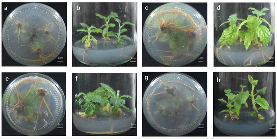
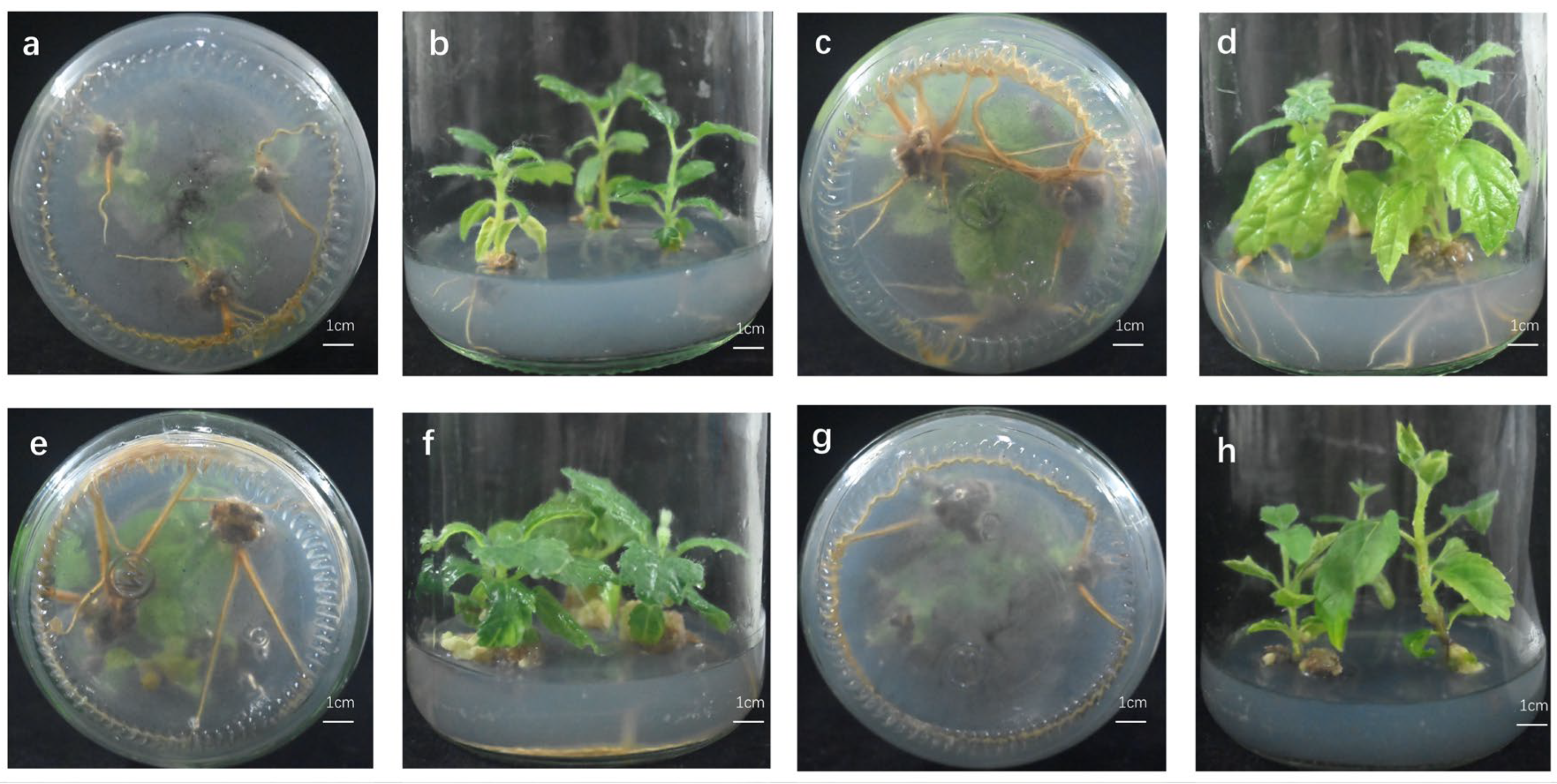
Figure 4.
Effects of different hormone concentrations on rooting and growth of Handroanthus chrysanthus micropropagated seedlings. Representative shoot and root growth of H. chrysanthus micropropagated seedlings at 30 days in R1 (a,b), R11 (c,d), R14 (e,f), and R16 (g,h).
Figure 5.
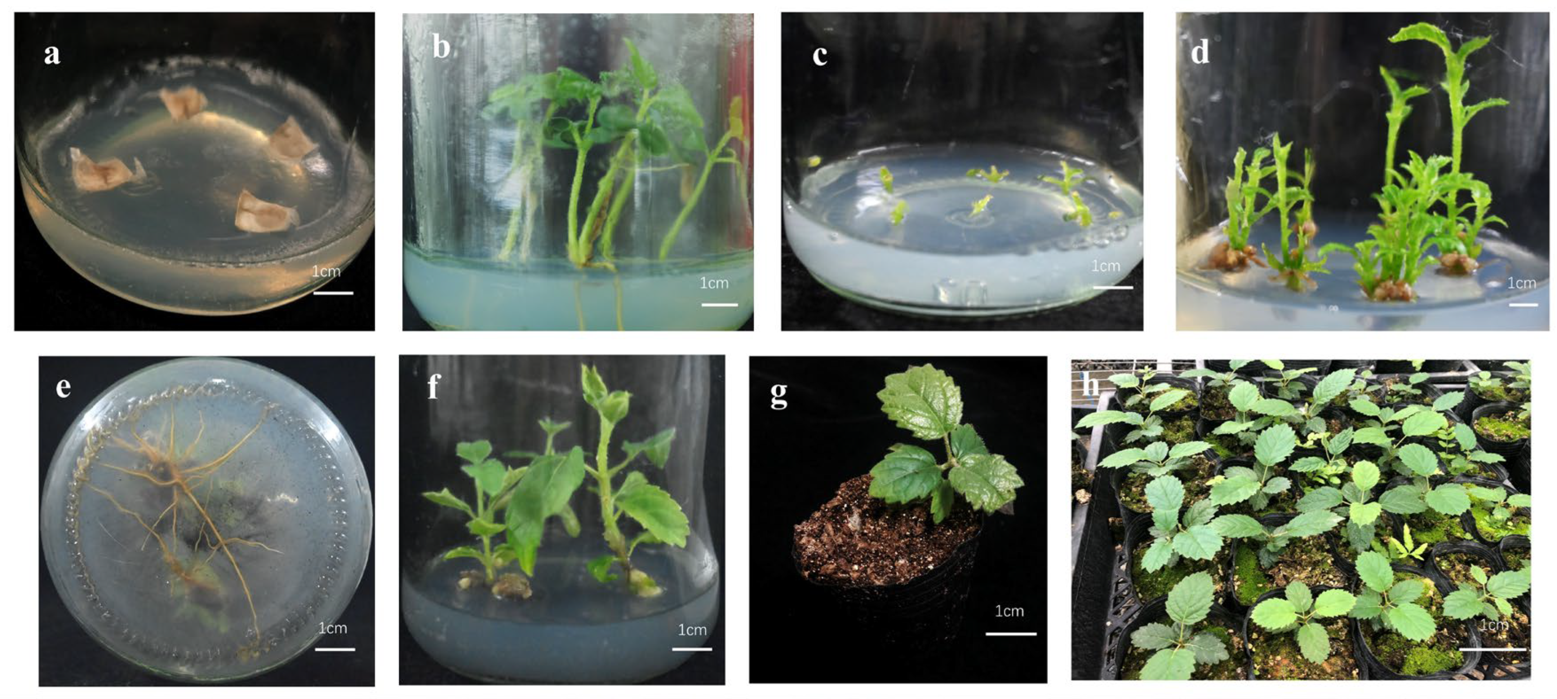
Micropropagation procedure of Handroanthus chrysanthus: (a) seed surface sterilization and inoculation in vitro; (b) regenerated shoots after 90 days of culture; (c) induction stage of stem segments with axillary buds on MS medium supplemented with 4 mg⋅L−1 6-BA and 0.3 mg⋅L−1 GA3 (SP6); (d) shoot proliferation on SP6 medium after 45 days; (e,f) rooting of regenerated shoots after 30 days of culture; (g) acclimatization of rooted plantlets at 10 days after transplantation; (h) growth condition after transplantation for 60 days.
2.6. Rooting and Acclimatization of Micropropagated Seedlings
Half-strength MS basal media supplemented with IBA and/or NAA were evaluated for their potential in inducing root formation (Table 7). When IBA was used alone, the rooting percentage was only approximately 30% after 30 days, whereas the combination of NAA and IBA significantly promoted the rooting process (Figure 4). The highest rooting rate of seedlings reached 80% after 30 days in medium R14 (1/2 MS + IBA 5 mg·L−1 + NAA 0.5 mg·L−1 + activated charcoal (A.C.) 0.2 g·L−1). Based on a comprehensive evaluation of rooting percentage and the average root number per plant, R11 (1/2 MS + IBA 10 mg·L−1 + NAA 0.05 mg·L−1 + A.C. 0.2 g·L−1) represented another preferable medium.
Table 7.
Effect of NAA and IBA on root induction and growth of Handroanthus chrysanthus.
The vigorous plantlets with 2–3 cm root length were transferred to room temperature under natural light for 3–4 days. The planting substrate consisted of a sterilized mixture of peat soil, vermiculite, and perlite (3:1:1). During transplantation, plantlets were gently removed from containers, and the planting trays were covered with transparent lids and maintained in a greenhouse for 7 days before removing the covers. Under natural conditions, the survival rate reached 95% after one month. Figure 5 illustrates the successful micropropagation process of H. chrysanthus.
3. Discussion
In the present study, medium containing 6-BA combined with NAA (SR5 and SR6) yielded a regeneration frequency of approximately 20%, whereas the combination of TDZ, 6-BA, and IBA (SI13) significantly enhanced regeneration to 51.79% from H. chrysanthus hypocotyls. TDZ exhibits potent cytokinin-like activity while also modulating endogenous auxin metabolism, thereby stimulating the accumulation of natural cytokinins and promoting morphogenic responses [36,37]. In contrast, 6-BA directly provides adenine-type cytokinin activity that supports shoot organogenesis and subsequent proliferation [38]. The synergistic effect of combining TDZ and 6-BA has been recognized as a critical factor in optimizing tissue culture systems for diverse woody species, including mulberry (Morus spp.) [39] and others [28,40]. In mulberry, TDZ was shown to rearrange adenine-type cytokinin, downregulate endogenous cytokinin signaling, and enhance auxin signaling by upregulating related genes without altering auxin biosynthesis [39]. It should be noted that a negative control (PGR-free medium) was not included in the present experiment for shoot regeneration from hypocotyls, which limits the ability to assess baseline shoot regeneration. Future studies should include such controls to validate the necessity of exogenous PGRs.
Light plays a pivotal role in regulating plant growth and morphogenic processes, including in vitro regeneration. The explants of plant species complete their regeneration process by responding to different photoreceptors under various light spectra [41,42]. Photoreceptors transmit light signals to downstream regulatory factors, such as PHYTOCHROME-INTERACTING FACTORS (PIFs), ELONGATED HYOCOTYL 5 (HY5), and CONSTITUTIVE PHOTOMORPHOGENIC 1 (COP1) [43]. These conserved light-responsive signaling factors then regulate downstream genes and proteins involved in plant regeneration, including the cytokinin-responsive factor ARABIDOPSIS RESPONSE REGULATOR 12 (ARR12) and WUSCHEL (WUS) of Arabidopsis [44]; auxin synthesis genes, such as AMIDASE 1 (AMI1); and jasmonic acid (JA)-responsive genes, such as DE-ETIOLATED-2 (DET2) [45,46]. Studies have shown that blue [47], red [48], or a combination of red and blue light [49] can significantly promote shoot regeneration efficacy for different plant species. Our results showed that white LED light was most effective for adventitious shoot regeneration in H. chrysanthus, while green light completely suppressed organogenesis. This finding is consistent with previous reports in other woody species. For instance, white or yellow light promoted adventitious shoot regeneration in Populus alba × P. berolinensis, whereas green light inhibited this process [50].
Hyperhydricity represents one of the most pervasive physiological disorders in plant tissue culture [51,52]. Hyperhydricity is also a common challenge during in vitro propagation of Handroanthus species [53,54]. During the in vitro regeneration of H. chrysanthus, approximately 10–15% of the regenerated shoots exhibited varying degrees of hyperhydricity, as shown in Figure 2b. Anatomical studies on H. impetiginosus revealed that hyperhydric shoots exhibit severe structural abnormalities at the proliferation stage, including disorganized cortex, epidermal discontinuity, collapsed cells, and stomatal malformation—defects that directly compromise survival during acclimatization [53]. High concentrations of cytokinins—commonly used to maximize shoot proliferation—are well-established inducers of hyperhydricity, whereas reducing cytokinin levels in the medium can effectively alleviate this disorder [55]. The moderate multiplication coefficient of 3.27 achieved in our study represents a pragmatic compromise. In a recent study, Grira [54] reduced the hyperhydricity of H. guayacan by increasing gelling agent (agar or Gelrite) concentration, optimizing sucrose and cytokinin levels, and supplementing with calcium, silicon, and anti-ethylene compounds (AgNO3). Meanwhile, alternative cytokinin types—particularly meta-topolin (mT) and its riboside (mTR)—have also shown promise in reducing hyperhydricity while maintaining efficient shoot multiplication in H. guayacan, T. rosea, Tectona grandis, and wild olive. Collectively, these strategies offer a promising approach for refining regeneration systems in Handroanthus species.
4. Materials and Methods
4.1. Plant Material and Explant Preparation
Winged seeds of H. chrysanthus were collected from mature trees located at South China Agricultural University, Guangzhou, China. Seeds were initially cut off the wings and washed with a mild detergent solution for 15 min, followed by thorough rinsing under running tap water for 30 min. Surface sterilization was conducted by immersing the seeds in 75% (v/v) ethanol for 50 s, followed by three rinses with sterile distilled water. Subsequently, seeds were treated with 0.1% (w/v) HgCl2 for varying durations (6, 8, 10, 12, 14, 16 min) and rinsed five times with sterile distilled water. Sterilized seeds were placed on hormone-free Murashige and Skoog (MS) basal medium for germination. After 20 days, hypocotyls and cotyledons were excised from aseptic seedlings and used as explants for regeneration experiments.
4.2. Culture Media and Conditions
The basal media used in this study included MS [25], 1/2 MS, and DCR [33] (Douglas-fir Cotyledon Revised Medium). MS medium (HB8469-5) and 1/2 MS medium (HB8469-12) were supplied by Qingdao Hopebio Bio-Technology Co., Ltd. (Qingdao, China), whereas DCR medium (PM1631) was obtained from Coolaber Science and Technology Co., Ltd. (Beijing, China). All media were supplemented with 30 g·L−1 (for micropropagation) or 20 g·L−1 (for regeneration) sucrose and solidified with 6.0 g·L−1 agar (for micropropagation) or 2.4 g·L−1 phytagel (for regeneration). The pH was adjusted to 5.8 prior to autoclaving at 121 °C for 20 min. Cultures were maintained in a growth room at 25 ± 2 °C under a 16 h photoperiod provided by white LED light (50 µmol·m−2·s−1 photosynthetic photon flux density). In the light quality experiment, the photosynthetic photon flux density (PPFD) was maintained constant at 50 µmol·m−2·s−1 under all light spectra tested (white LED, red, green, and blue). LED tubes were purchased from iGrowLite (Guangzhou Zhiui Guangtian Agricultural Technology Co., Ltd., Guangzhou, China), model IGL-T5-10W-S, with the following peak wavelengths: white (400–700 nm), red (660 nm), green (520 nm), and blue (450 nm).
Preliminary experiments were conducted to select the optimal basal medium for each explant type. For hypocotyl explants, MS, 1/2 MS, WPM, and DCR were compared. Based on the results of previous studies on the related species H. impetiginosa ‘Zi Xiuqiu’ in our laboratory [30], MS medium was found to be the most suitable for hypocotyl regeneration and was therefore used for all formal hypocotyl experiments. For cotyledon explants, preliminary tests showed that explants failed to grow on MS medium, while shoot formation was observed only on DCR medium. Consequently, DCR medium was used exclusively for cotyledon regeneration experiments.
4.3. Adventitious Shoot Regeneration from Hypocotyls
Hypocotyl segments (approximately 0.8–1.0 cm in length) were excised and placed horizontally on MS basal medium supplemented with different concentrations of TDZ (thidiazuron), 6-BA (6-benzylaminopurine), NAA (1-naphthaleneacetic acid), and IBA (indole-3-butyric acid). For the 6-BA + NAA growth regulator combination, the concentration of 6-BA was 3, 5, or 7 mg·L−1, while NAA was tested at 0, 0.05, 0.1, 0.2, 0.4, or 0.5 mg·L−1. These treatments were designated as SR1 to SR13, with the specific concentrations detailed in Table 2. For the TDZ + 6-BA + IBA combination, a two-factor factorial design was used. TDZ concentrations tested were 0.2, 0.5, 1.0, 2.0, and 3.0 mg·L−1; 6-BA concentrations were 0, 0.5, 1.0, 1.5, 3.0, and 5.0 mg·L−1; IBA was tested at 0.3 and 0.5 mg·L−1. Selected combinations (SI1–SI13) representing the full range of responses are presented in Table 3. Each treatment consisted of 30 explants. Data on callus formation, shoot regeneration frequency, and the number of shoots per explant were recorded after 20, 45, and 60 days of culture. BAP (DH038-2), IBA (II172) and NAA (IN211) were purchased from Beijing Dingguo Chang-sheng Biotechnology Co., Ltd. (Beijing, China), and TDZ (P6186) was purchased from Sigma-Aldrich (St. Louis, MO, USA).
4.4. Adventitious Shoot Regeneration from Cotyledons
Cotyledons from 20-day-old seedlings were placed abaxially on MS or DCR medium containing 6-BA (3, 5, or 7 mg·L−1) and NAA (0, 0.05, or 0.5 mg·L−1). Each treatment comprised 15 explants with three replicates. After 60 days, data on callus formation, shoot regeneration rate, and induced shoot number per explant were recorded and analyzed.
4.5. Shoot Proliferation
Regenerated shoots (1.5–2.0 cm in length) were transferred to MS medium supplemented with 6-BA (2, 3, or 4 mg·L−1) and gibberellic acid (GA3; 0.1, 0.3, or 0.5 mg·L−1) for proliferation. Each treatment involved 15 explants and was repeated three times. After 45 days of subculture, the proliferation coefficient was calculated as the number of newly formed adventitious shoots (≥0.5 cm in height) per explant, excluding any shoots that were already present on the original explant.
4.6. Rooting and Acclimatization
In vitro-derived shoots (2–3 cm long) were transferred to half-strength MS medium supplemented with different concentrations of IBA (0, 0.01, 0.05, and 0.5 mg·L−1) and NAA (3, 5, and 10 mg·L−1), with or without 0.2 g·L−1 of activated charcoal (A.C.). Each treatment involved 15 explants and was repeated three times. Rooting percentage, root number, and root length were recorded after 30 days. Well-rooted plantlets were acclimatized in a greenhouse using a sterilized substrate mixture of peat, vermiculite, and perlite (3:1:1 v/v/v). The survival rate was assessed after one month.
4.7. Statistical Analysis
All experiments were arranged in a completely randomized design with three replicates per treatment. Before one-way or two-way ANOVA, data were tested for normality (Shapiro–Wilk test, p > 0.05) and homogeneity of variances (Levene’s test, p > 0.05). Percentage data (e.g., shoot regeneration rate, rooting percentage) were arcsine square-root transformed when necessary to satisfy ANOVA assumptions, whereas untransformed means are presented in the tables. Data were subjected to analysis of variance (ANOVA) using SPSS 26.0 software. Mean separations were performed using Duncan’s multiple-range test at a significance level of p < 0.05. All explants were randomly assigned to each treatment. Three replicates refer to three independent biological replicates.
5. Conclusions
In this study, we established a reliable and efficient direct adventitious shoot regeneration system for H. chrysanthus using hypocotyl explants. The key findings can be summarized as follows: (1) Explant type is critical—hypocotyls showed significantly higher regenerative capacity than cotyledons. (2) TDZ is a key regulator—its combination with 6-BA and IBA synergistically enhanced shoot induction. (3) Light quality matters—white LED light was optimal for shoot regeneration. (4) Ex vitro acclimatization is achievable—over 95% of regenerated plantlets survived in the greenhouse.
This regeneration system provides a technical platform for several future applications. First, it enables the mass propagation of H. chrysanthus to meet horticultural demand and support ex situ conservation efforts for this vulnerable species. Second, it serves as a foundation for genetic improvement—the efficient adventitious shoot induction is a prerequisite for Agrobacterium-mediated transformation and CRISPR/Cas9 gene editing, which could be used to improve undesirable traits such as a short flowering period and cold sensitivity. Third, the protocols developed here offer a technical reference for other endangered species within the Tabebuia alliance (e.g., H. heptaphyllus, H. impetiginosus), facilitating their regeneration and conservation.
Collectively, this work transforms a species-specific regeneration protocol into a broader strategy for combining biotechnology with conservation and breeding of non-model woody plants.
Author Contributions
Conceptualization, X.Z. and C.P.; methodology, H.F., J.Z. (Junjie Zhang) and J.Z. (Jiwen Zha); investigation, H.F., B.C., J.Z. (Jiwen Zha) and X.X.; formal analysis, H.F.; data curation, H.F. and Y.L.; writing—original draft preparation, H.F.; writing—review and editing, X.Z. and C.P.; visualization, H.F.; supervision, X.Z.; project administration, C.P.; funding acquisition, C.P. All authors have read and agreed to the published version of the manuscript.
Funding
This research was funded by the Guangdong Provincial Science and Technology Special Fund Project for Zhongshan City (major special project + task list management mode), grant number 2021sdr003.
Data Availability Statement
The original contributions presented in the study are included in the article; further inquiries can be directed to the corresponding author.
Acknowledgments
During the preparation of this manuscript, the authors used ChatGPT (OpenAI) (https://chatgpt.com) from 1 May 2026 to 30 June 2026 for language polishing and grammar checking. The authors have reviewed and edited the output and take full responsibility for the content of this publication.
Conflicts of Interest
The authors declare the following financial interests/personal relationships, which may be considered as potential competing interests: Changcao Peng reports financial support from the Guangdong Provincial Science and Technology Special Fund Project for Zhongshan City. Patents: Changcao Peng has a patent issued to “A method for obtaining complete regenerated plants from in vitro hypocotyl explants of Handroanthus chrysanthus”. ZL 2023 1 0767393.8). The other authors declare no known competing financial interests or personal relationships that could have appeared to influence the work reported in this paper.
References
- Shang, X.; Liu, G.; Wu, Z.; Zhang, P. Combined transcriptomic and metabolomic analysis reveals the mechanism of flavonoid biosynthesis in Handroanthus chrysanthus (Jacq.) S.O.Grose. Forests 2022, 13, 1285. [Google Scholar] [CrossRef] [Scilit]
- Gentry, A.H. Bignoniaceae: Part II (tribe tecomeae). Flora Neotrop. 1992, 25, 1–370. [Google Scholar]
- Grose, S.O.; Olmstead, R.G. Taxonomic revisions in the polyphyletic genus Tabebuia s. l. (Bignoniaceae). Syst. Bot. 2007, 32, 660–670. [Google Scholar] [CrossRef] [Scilit]
- Campos, T.S.; Pereira, V.M.; El Merzougui, S.; Beleski, D.; Pérez, H.E.; Pivetta, K.F.L.; Vendrame, W.A. Cryopreservation of Lavender Trumpet Tree (Handroanthus impetiginosus) Seeds. Horticulturae 2024, 10, 1256. [Google Scholar] [CrossRef] [Scilit]
- Meng, J.; Zhang, Y.; Wei, Y.; Li, R.; Li, Z.; Zhong, C. Identification of Commercial Cultivars in the Tabebuia Alliance Using Genotyping-by-Sequencing. Forests 2023, 14, 271. [Google Scholar] [CrossRef] [Scilit]
- Zhang, J.; Liao, S.B.; Sun, B.; Luo, S.X.; Chen, Y.; Shi, G.Z.; Zhang, K. Flowering phenology and flower morphology of Handroanthus chrysantha. J. Zhejiang A F Univ. 2017, 34, 759–764. [Google Scholar] [CrossRef]
- Héctor, M. Development of a protocol for the micropropagation of two forest species threatened with extinction in Ecuador. Plant Cell Tiss. Organ Cult. 2024, 159, 2. [Google Scholar] [CrossRef] [Scilit]
- IUCN. The IUCN Red List of Threatened Species 2021. Available online: https://www.iucnredlist.org/species/146784568/146784570 (accessed on 20 April 2026).
- Huang, X.M.; Yang, C.Q.; Chen, Z.H.; Chen, M.X.; Huang, Q.C.; Chen, K.W. Rapid loss of seed vigor and polyembryony phenomenon in Tabebuia chrysantha. Guangdong Agric. Sci. 2012, 39, 50–52. [Google Scholar] [CrossRef]
- Zhang, X.; Xu, G.; Cheng, C.; Lei, L.; Sun, J.; Xu, Y.; Deng, C.; Dai, Z.; Yang, Z.; Chen, X.; et al. Establishment of an Agrobacterium-mediated genetic transformation and CRISPR/Cas9-mediated targeted mutagenesis in Hemp (Cannabis sativa L.). Plant Biotechnol. J. 2021, 19, 1979–1987. [Google Scholar] [CrossRef] [Scilit] [PubMed]
- Koyama, J.; Morita, I.; Kino, A.; Tagahara, K. Micellar Electrokinetic Chromatography (MEKC) separation of furanonaphthoquinones from Tabebuia impetiginosa. Chem. Pharm. Bull. 2000, 48, 873–875. [Google Scholar] [CrossRef] [Scilit]
- Reis, F.P.; Bonfa, I.M.S.; Cavalcante, R.B.; Okoba, D.; Vasconcelos, S.B.S.; Candeloro, L.; Filiu, W.F.O.; Monreal, A.C.D.; Silva, V.J.; Santa Rita, P.H.; et al. Tabebuia aurea decreases inflammatory, myotoxic and hemorrhagic activities induced by the venom of Bothrops neuwiedi. J. Ethnopharmacol. 2014, 158, 352–357. [Google Scholar] [CrossRef] [Scilit] [PubMed]
- de Sousa, A.C.; de Oliveira, M.L.; Ferreira, J.L.; Rocha, N.H.; Honda, E.H.; Bogo, M.R. Effects of a Tabebuia avellanedae extract and lapachol on the labeling of blood constituents with technetium-99m. Sci. Res. Essays 2015, 10, 97–104. [Google Scholar] [CrossRef] [Scilit]
- Kim, J.H.; Lee, S.M.; Myung, C.H.; Lee, K.R.; Hyun, S.M.; Lee, J.E.; Park, Y.S.; Jeon, S.R.; Park, J.I.; Chang, S.E.; et al. Melanogenesis inhibition of β-lapachone, a natural product from Tabebuia avellanedae, with effective in vivo lightening potency. Arch. Dermatol. Res. 2015, 307, 229–238. [Google Scholar] [CrossRef] [Scilit] [PubMed]
- El-Hawary, S.S.; Taher, M.A.; Amin, E.; AbouZid, S.F.; Mohammed, R. Genus Tabebuia: A comprehensive review journey from past achievements to future perspectives. Arab. J. Chem. 2021, 14, 103046. [Google Scholar] [CrossRef] [Scilit]
- Freitas, A.E.; Moretti, M.; Budni, J.; Balen, G.O.; Fernandes, S.C.; Veronezi, P.O.; Heller, M.; Micke, G.A.; Pizzolatti, M.G.; Rodrigues, A.L.S. NMDA receptors and the L-arginine–nitric oxide–cyclic guanosine monophosphate pathway are implicated in the antidepressant-like action of the ethanolic extract from Tabebuia avellanedae in mice. J. Med. Food 2013, 16, 1030–1038. [Google Scholar] [CrossRef] [Scilit] [PubMed]
- Iwamoto, K.; Fukuda, Y.; Tokikura, C.; Noda, M.; Yamamoto, A.; Yamamoto, M.; Yamashita, M.; Zaima, N.; Iida, A.; Moriyama, T. The anti-obesity effect of taheebo (Tabebuia avellanedae Lorentz ex Griseb) extract in ovariectomized mice and the identification of a potential anti-obesity compound. Biochem. Biophys. Res. Commun. 2016, 478, 1136–1140. [Google Scholar] [CrossRef] [Scilit]
- Miranda, F.G.G.; Vilar, J.C.; Alves, I.A.N.; Cavalcanti, S.C.H.; Antoniolli, A.R. Antinociceptive and antiedematogenic properties and acute toxicity of Tabebuia avellanedae Lor. ex Griseb. inner bark aqueous extract. BMC Pharmacol. 2001, 1, 6. [Google Scholar] [CrossRef] [Scilit] [PubMed]
- Panda, S.P.; Panigrahy, U.P.; Panda, S.; Jena, B.R. Stem extract of Tabebuia chrysantha induces apoptosis by targeting sEGFR in Ehrlich Ascites carcinoma. J. Ethnopharmacol. 2019, 235, 219–226. [Google Scholar] [CrossRef] [Scilit] [PubMed]
- Pires, T.C.S.P.; Dias, M.I.; Calhelha, R.C.; Carvalho, A.M.; Queiroz, M.J.R.P.; Barros, L.; Ferreira, I.C.F.R. Bioactive properties of Tabebuia impetiginosa-based phytopreparations and phytoformulations: A comparison between extracts and dietary supplements. Molecules 2015, 20, 22863–22871. [Google Scholar] [CrossRef] [Scilit] [PubMed]
- Duarte, E.; Sansberro, P.; Luna, C. Micropropagation of Handroanthus heptaphyllus (Vell.) Mattos from seedling explants. Afr. J. Biotechnol. 2016, 15, 1292–1298. [Google Scholar] [CrossRef] [Scilit]
- Larraburu, E.E.; Apóstolo, N.M.; Llorente, B.E. In Vitro Propagation of Pink Lapacho: Response Surface Methodology and Factorial Analysis for Optimisation of Medium Components. Int. J. For. Res. 2012, 318258. [Google Scholar] [CrossRef] [Scilit]
- Larraburu, E.E.; Yarte, M.E.; Llorente, B.E. Azospirillum brasilense inoculation, auxin induction and culture medium composition modify the profile of antioxidant enzymes during in vitro rhizogenesis of pink lapacho. Plant Cell Tiss. Organ Cult. 2016, 127, 381–392. [Google Scholar] [CrossRef] [Scilit]
- Máximo, W.P.; Santos, B.R.; Martins, J.P.R.; Beijo, L.A.; Barbosa, S. Multiplication and in vitro rooting of Handroanthus impetiginosus (Mart. ex DC.) Mattos. Ciênc. Florest. 2020, 30, 658–668. [Google Scholar] [CrossRef] [Scilit]
- Zha, J.W.; Fang, H.T.; Deng, J.; Qin, F.Y.; Zhang, J.J.; Peng, C.C.; Zhao, X.L. Establishment of a rapid propagation system for Handroanthus impetiginosa ‘Zi Xiuqiu’. Mol. Plant Breed. 2023, 1–15. [Google Scholar]
- González-Rodríguez, J.A.; Ramírez-Garduza, F.; Robert, M.L.; O’Connor-Sánchez, A.; Peña-Ramírez, Y.J. Adventitious shoot regeneration from adult tissues of the tropical timber tree yellow Ipé primavera (Tabebuia donnell-smithii Rose [Bignoniaceae]). Vitr. Cell. Dev. Biol.-Plant 2010, 46, 411–421. [Google Scholar] [CrossRef] [Scilit]
- Grira, M.; Prinsen, E.; Werbrouck, S.P.O. The Effect of Topophysis on the In Vitro Development of Handroanthus guayacan and on Its Metabolism of Meta-Topolin Riboside. Plants 2023, 12, 3577. [Google Scholar] [CrossRef] [Scilit] [PubMed]
- Novikova, T.I.; Zaytseva, Y.G. TDZ-Induced Morphogenesis Pathways in Woody Plant Culture. In Thidiazuron: From Urea Derivative to Plant Growth Regulator; Ahmad, N., Faisal, M., Eds.; Springer: Singapore, 2018; pp. 25–44. [Google Scholar] [CrossRef] [Scilit]
- Elmoreigi, R.A. Effect of explant type and growth regulators on in vitro regeneration of apricot (Prunus armeniaca L.) Al-Amar rootstock. Plant Cell Tiss. Organ Cult. 2024, 160, 1. [Google Scholar] [CrossRef] [Scilit]
- Murashige, T.; Skoog, F. A revised medium for rapid growth and bio-assays with tobacco tissue cultures. Physiol. Plant. 1962, 15, 473–497. [Google Scholar] [CrossRef] [Scilit]
- Gupta, P.K.; Durzan, D.J. Shoot multiplication from mature trees of Douglas-fir (Pseudotsuga menziesii) and sugar pine (Pinus lambertiana). Plant Cell Rep. 1985, 4, 177–179. [Google Scholar] [CrossRef] [Scilit] [PubMed]
- Huang, H.; Wei, Y.; Zhai, Y.; Ouyang, K.; Chen, X.; Bai, L. High frequency regeneration of plants via callus-mediated organogenesis from cotyledon and hypocotyl cultures in a multipurpose tropical tree (Neolamarckia Cadamba). Sci. Rep. 2020, 10, 4558. [Google Scholar] [CrossRef] [Scilit] [PubMed]
- Dai, Q.C.; Li, N.; Lin, S.X.; Zhang, J.X. Differences in Callus Induction and Plant Regeneration of Neolamarckia cadamba from Different Geographical Provenances. Mol. Plant Breed. 2022, 1–13. Available online: https://link.cnki.net/urlid/46.1068.S.20220321.1327.011 (accessed on 20 April 2026).
- Wang, Y.; Wu, Z.; Li, X.; Shang, X. Regeneration and Genetic Transformation in Eucalyptus Species, Current Research and Future Perspectives. Plants 2024, 13, 2843. [Google Scholar] [CrossRef] [Scilit] [PubMed]
- Cardoso, J.C.; Teixeira da Silva, J.A. Micropropagation of Zeyheria montana Mart. (Bignoniaceae), an endangered endemic medicinal species from the Brazilian cerrado biome. Vitr. Cell. Dev. Biol. Plant 2013, 49, 710–716. [Google Scholar] [CrossRef] [Scilit]
- Erland, L.; Giebelhaus, R.; Victor, J.; Murch, S.; Saxena, P. The morphoregulatory role of thidiazuron: Metabolomics-guided hypothesis generation for mechanisms of activity. Biomolecules 2020, 10, 1253. [Google Scholar] [CrossRef] [Scilit] [PubMed]
- Liu, Z.; Dai, X.; Li, J.; Liu, N.; Liu, X.; Li, S.; Xiang, F. The Type-B Cytokinin Response Regulator ARR1 Inhibits Shoot Regeneration in an ARR12-Dependent Manner in Arabidopsis. Plant Cell 2020, 32, 2271–2291. [Google Scholar] [CrossRef] [Scilit] [PubMed]
- Jones, M.; Cao, J.; O’Brien, R.; Murch, S.; Saxena, P. The mode of action of thidiazuron: Auxins, indoleamines, and ion channels in the regeneration of Echinacea purpurea L. Plant Cell Rep. 2007, 26, 1481–1490. [Google Scholar] [CrossRef] [Scilit] [PubMed]
- Luo, Y.; Han, Y.; Wei, W.; Han, Y.; Yuan, J.; He, N. Transcriptome and metabolome analyses reveal the efficiency of in vitro regeneration by TDZ pretreatment in mulberry. Sci. Hortic. 2023, 310, 111678. [Google Scholar] [CrossRef] [Scilit]
- Ouyang, Y.; Chen, Y.; Lü, J.; Teixeira da Silva, J.A.; Zhang, X.; Ma, G. Somatic embryogenesis and enhanced shoot organogenesis in Metabriggsia ovalifolia W.T. Wang. Sci. Rep. 2016, 6, 24662. [Google Scholar] [CrossRef] [Scilit] [PubMed]
- Han, J.; Li, Y.; Zhao, Y.; Sun, Y.; Li, Y.; Peng, Z. How does light regulate plant regeneration? Front. Plant Sci. 2025, 15, 1474431. [Google Scholar] [CrossRef] [Scilit] [PubMed]
- Su, P.; Wang, D.; Wang, P.; Gao, Y.; Jia, H.; Hou, J.; Wu, L. In vitro regeneration, photomorphogenesis and light signaling gene expression in Hydrangea quercifolia cv. ‘Harmony’ under different LED environments. Planta 2024, 259, 71. [Google Scholar] [CrossRef] [Scilit] [PubMed]
- Bhatnagar, A.; Singh, S.; Khurana, J.P.; Burman, N. HY5-COP1: The central module of light signaling pathway. J. Plant Biochem. Biotechnol. 2020, 29, 590–610. [Google Scholar] [CrossRef] [Scilit]
- Dai, X.; Wang, J.; Wang, L.; Liu, Z.; Li, Q.; Cai, Y.; Li, S.; Xiang, F. HY5 inhibits in vitro shoot stem cell niches initiation via directly repressing pluripotency and cytokinin pathways. Plant J. 2022, 110, 781–801. [Google Scholar] [CrossRef] [Scilit] [PubMed]
- Chan, A.; Stasolla, C. Light induction of somatic embryogenesis in Arabidopsis is regulated by PHYTOCHROME E. Plant Physiol. Biotech. 2023, 195, 163–169. [Google Scholar] [CrossRef] [Scilit] [PubMed]
- Mira, M.M.; Day, S.; Ibrahim, S.; Hill, R.D.; Stasolla, C. The Arabidopsis Phytoglobin 2 mediates phytochrome B (phyB) light signaling responses during somatic embryogenesis. Planta 2023, 257, 88. [Google Scholar] [CrossRef] [Scilit] [PubMed]
- Assou, J.; Bethge, H.; Wamhoff, D.; Winkelmann, T. Effect of cytokinins and light quality on adventitious shoot regeneration from leaflet explants of peanut (Arachis hypogaea). J. Hortic. Sci. Biotech. 2023, 98, 508–525. [Google Scholar] [CrossRef] [Scilit]
- Burritt, D.J.; Leung, D.W.M. Adventitious shoot regeneration from Begonia × erythrophylla petiole sections is developmentally sensitive to light quality. Physiol. Plant. 2003, 118, 289–296. [Google Scholar] [CrossRef] [Scilit]
- Loshyna, L.; Bulko, O.; Kuchuk, M. Adventitious regeneration of blackberry and raspberry shoots and the assessment of the LED-lighting impact. Zemdirb.-Agric. 2022, 109, 49–54. [Google Scholar] [CrossRef] [Scilit]
- Wang, H.; Liu, H.; Wang, W.; Zu, Y. Effects of Thidiazuron, basal medium and light quality on adventitious shoot regeneration from in vitro cultured stem of Populus alba × P. berolinensis. J. For. Res. 2008, 19, 257–259. [Google Scholar] [CrossRef] [Scilit]
- Polivanova, O.B.; Bedarev, V.A. Hyperhydricity in Plant Tissue Culture. Plants 2022, 11, 3313. [Google Scholar] [CrossRef] [Scilit] [PubMed]
- Werbrouck, S.P.O. Meta-topolin and Related Cytokinins as a Solution to Some In Vitro Problems. In Meta-Topolin: A Growth Regulator for Plant Biotechnology and Agriculture; Ahmad, N., Strnad, M., Eds.; Springer: Singapore, 2021; pp. 85–91. [Google Scholar] [CrossRef] [Scilit]
- Jausoro, V.; Llorente, B.E.; Apóstolo, N.M. Structural differences between hyperhydric and normal in vitro shoots of Handroanthus impetiginosus (Mart. ex DC) Mattos (Bignoniaceae). Plant Cell Tiss. Organ Cult. 2010, 101, 183–191. [Google Scholar] [CrossRef] [Scilit]
- Grira, M. Strategies Towards Improved Micropropagation Protocols of Woody Plants: A Focus on Hyperhydricity Control, Topophysis, Culture Density and Cytokinin Metabolism. Ph.D. Thesis, Ghent University, Ghent, Belgium, 2025. Available online: http://hdl.handle.net/1854/LU-01K5V9WVXR19MA79VDNCSPAA1V (accessed on 23 September 2025).
- Barua, R.; Igamberdiev, A.U.; Debnath, S.C. Hyperhydricity Syndrome in In Vitro Plants: Mechanisms, Physiology, and Control. Plants 2025, 14, 3721. [Google Scholar] [CrossRef] [Scilit] [PubMed]

Disclaimer/Publisher’s Note: The statements, opinions and data contained in all publications are solely those of the individual author(s) and contributor(s) and not of MDPI and/or the editor(s). MDPI and/or the editor(s) disclaim responsibility for any injury to people or property resulting from any ideas, methods, instructions or products referred to in the content. |
© 2026 by the authors. Licensee MDPI, Basel, Switzerland. This article is an open access article distributed under the terms and conditions of the Creative Commons Attribution (CC BY) license.