Abstract
The genus Plantago (Plantaginaceae) is widely distributed worldwide. The Plantago species are used in traditional medicine as wound healers, anti-inflammatory agents, antipyretics, and analgesics. This study aimed to investigate the phytochemical composition from the aerial parts of Plantago indica L. and to evaluate its biological activities. Isolation studies and in vitro investigations were conducted on an aqueous phase of 80% EtOH extract of Plantago indica. In addition, in vivo studies were carried out using the MeOH, 80% EtOH, and water extracts. Plantarenaloside (1), 3-oxo-α-ionol β-glucoside (2), martynoside (3), acteoside (4), feruloyl gardoside (5), and ursolic acid (6) were isolated from the extract. The structures of the compounds were elucidated using 1D- and 2D-NMR and ESI-MS analyses. The extract, fractions, and pure compounds were tested in vitro for cytotoxicity (MTT), anti-inflammatory activity (NO, IL-6, and TNF-α production), wound healing (scratch test), and antioxidant capacity (DPPH, ABTS, SO). Feruloyl gardoside (20.11–58.27%) significantly reduced NO levels at concentrations of 25–100 µM. It significantly reduced IL-6 levels (40.17%) at 100 µM. Additionally, the in vivo anti-inflammatory (acetic acid-induced vascular permeability) and wound healing (incision and excision models) effects of the extracts were investigated. The findings suggest that P. indica may be considered to be a potential therapeutic option for managing inflammation and for promoting wound healing.
1. Introduction
The genus Plantago, belonging to the Plantaginaceae family, includes 246 species distributed globally [1]. In Türkiye, there are a total of 23 species, among which the endemic species P. anatolica and P. euphratica are also included [2].
Various Plantago species are commonly utilized in traditional medicine due to their antimicrobial, antidiabetic, antispasmodic, antiviral, anti-inflammatory, wound healing, and diuretic properties [3,4]. Ethnobotanical research in Türkiye has demonstrated the extensive use of these plants in folk medicine for the treatment of various conditions, such as abdominal pain, abscesses, wounds, burns, diabetes, cough, bronchitis, constipation, hemorrhoids, sore throat, and rheumatism [5,6,7]. Specifically, in traditional Turkish medicine, the seeds of P. indica are favored for their laxative properties [8]. On a global scale, the seeds are also recognized for their ability to stimulate blood circulation due to their high soluble fiber content [9]. In addition, it has been reported that the Plantago species are consumed as food by adding them to salads, soups, cakes, and bread, and are used in the preparation of tea, fruit juice, wine, cereals, and ice cream [10].
Phytochemical analyses have indicated that the Plantago species contain diverse secondary metabolites, including iridoids, phenylethanoid glycosides, flavonoids, tannins, triterpenes, saponins, and sterols [4,11]. Aucubin and catalpol in the iridoid structure, and acteoside and plantamajoside in the phenylethanoid glycoside structure are very important for the chemotaxonomic identification of the Plantago species [9].
The pharmacological activities of the Plantago species have been supported by numerous in vitro and in vivo studies, confirming their antioxidant, antimicrobial, cytotoxic, antidiarrheal, immunomodulatory, and antihypertensive effects [12,13,14,15,16,17]. Many studies have also highlighted the significant role of the Plantago species in anti-inflammatory and wound healing effects [18,19,20,21,22,23,24].
Studies specifically focusing on P. indica have been relatively limited. One study analyzed 14 iridoid compounds across 14 different Plantago species, revealing their taxonomic relationships and identifying plantarenaloside and aucubin in P. indica, which belongs to the Psyllium subgenus [25]. In another study, lipid and fatty acid profiles in the seeds from six plants, including P. indica, were evaluated. This study concluded that P. indica seeds have a low total oil content (4.36 g/100 g) but a high γ-linolenic acid content (13.13% of unsaturated fatty acids) [26].
The anti-inflammatory and wound healing properties of the genus Plantago are well-documented in the literature. In our previous studies, we conducted comprehensive investigations on these biological activities in various Plantago species [19,27]. In this context, the present study is a continuation of our previous studies. Wound healing is a multifactorial process involving ROS species, inflammatory response, and tissue regeneration [28,29]. Therefore, the aim of this study was to investigate the antioxidant, anti-inflammatory, and wound-healing activities together. For this purpose, the phytochemical characterization from an aqueous phase of the 80% EtOH extract of the plant was contributed and its antioxidant activity was evaluated using 2,2-diphenyl-1-picrylhydrazyl (DPPH), 2,2′-azino-bis(3-ethylbenzothiazoline-6-sulfonic acid) (ABTS), and superoxide (SO) radical scavenging assays. To determine the anti-inflammatory effect, the nitric oxide (NO), interleukin-6 (IL-6), and tumor necrosis factor alpha (TNF-α) levels were measured in lipopolysaccharide (LPS)-stimulated RAW 264.7 macrophage cells in vitro and an acetic acid-induced vascular permeability test was performed in vivo. The wound healing potential was investigated using an in vitro scratch assay and in vivo linear incision and circular excision wound models.
2. Results
2.1. Characterization of the Isolated Compounds
The method for preparing the 80% EtOH extract from the aerial parts of P. indica is described in detail in Section 4.3. The water-soluble fraction of the 80% EtOH extract was subjected to various chromatographic methods. As a result, six compounds were isolated. The structures of the isolated compounds were determined by comparing 1D- and 2D-NMR (1H, 13C, HMQC, HMBC) and ESI-MS data (Supplementary Files) with data from the literature. The compounds were identified as plantarenaloside (1), 3-oxo-α-ionol β-glucoside (2), martynoside (3), acteoside (4), feruloyl gardoside (5), and ursolic acid (6) [19,30,31,32,33,34]. The chemical structures of all compounds isolated from P. indica are presented in Figure 1.
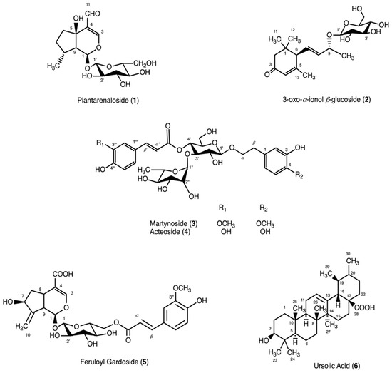
Figure 1.
Chemical structures of compounds isolated from Plantago indica.
2.2. In Vivo Biological Activities
2.2.1. Acetic Acid-Induced Capillary Permeability Results
The results of the present study revealed that the MeOH and 80% EtOH extracts prepared from the aerial parts of P. indica exhibited remarkable anti-inflammatory activity. As presented in Table 1, the administration of 80% EtOH extract at a dose of 100 mg/kg to mice resulted in 35.3% inhibition, while the MeOH extract at a dose of 100 mg/kg caused 26.6% inhibition in the acetic acid-induced capillary permeability test.

Table 1.
Inhibitory effect of the test materials on inflammation with acetic acid-induced increased capillary permeability.
2.2.2. Linear Incision Wound Model
The resistance of the repaired tissue to breaking under tension is referred to as tensile strength. Tensile strength is determined in the linear incision wound model. The repaired tissue is removed and its tensile strength is measured [35]. It was concluded that the 80% EtOH extract of P. indica promoted wound healing in rats at a statistically significant level, with a value of 26.5% (Table 2).
Table 2.
Effect of the test materials on wound healing using a linear incision model.
2.2.3. Circular Excision Wound Model
A circular excision wound model was used to measure the percentage reduction in the wound area [36]. In mice, the 80% EtOH extract demonstrated statistically significant wound healing on days 6, 8, 10, and 12, with a wound contraction rate of 37.7% on day 12. The MeOH extract also showed a significant wound healing effect on days 6, 8, and 10, reaching 33.7% wound contraction on day 10 (Table 3).
Table 3.
Effect of the test materials on wound healing using the circular excision model.
2.3. In Vitro Biological Activities
2.3.1. Cell Viability Assay (MTT)
An MTT assay was performed to determine the non-cytotoxic concentrations of the extract, fractions, and isolated pure compounds on the L929 and RAW 264.7 cell lines. In both cell lines, the extract was applied at concentrations of 20, 100, 200, and 400 μg/mL, while the fractions were tested at 10, 50, 100, and 200 μg/mL (Figure 2A and Figure 3A). The pure compounds were prepared in six different concentrations ranging from 100 to 3.125 μM using a two-fold serial dilution. It was concluded that the extract and fractions did not exhibit any cytotoxic effect on both cell lines at the tested concentrations. For pure compounds, cell viability in the L929 and RAW 264.7 cell lines was found to be above 80% at almost all concentrations except for ursolic acid at 50 and 100 μM (Figure 2B and Figure 3B).
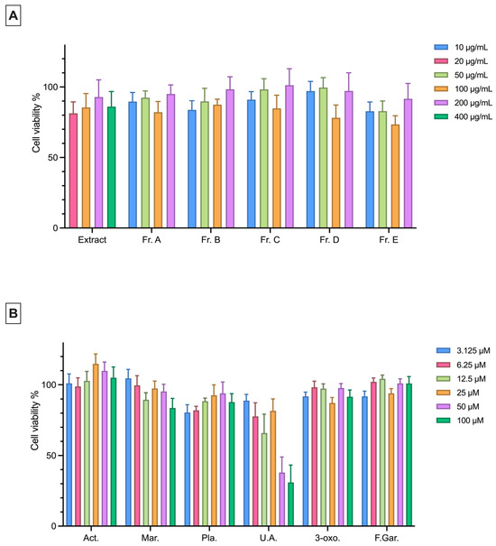
Figure 2.
Cytotoxic effects of the extract, fractions (A) and pure compounds (B) at different concentrations on L929 cells (105 cells/mL) as determined by the MTT assay. Data are presented as the mean ± SEM from three independent experiments. Act: Acteoside; Mar: Martynoside; Pla: Plantarenaloside; U.A.: Ursolic Acid; 3-oxo: 3-oxo-α-ionol β-glucoside; F.Gar.: Feruloyl Gardoside.
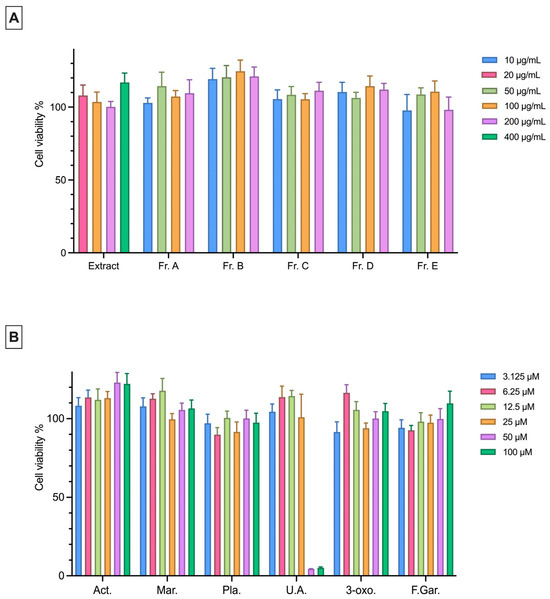
Figure 3.
Cytotoxic effects of the extract, fractions (A) and pure compounds (B) at different concentrations on RAW 264.7 cells (5 × 10⁵ cells/mL) as determined by the MTT assay. Data are presented as the mean ± SEM from three independent experiments. Act: Acteoside; Mar: Martynoside; Pla: Plantarenaloside; U.A.: Ursolic Acid; 3-oxo: 3-oxo-α-ionol β-glucoside; F.Gar.: Feruloyl Gardoside.
2.3.2. Effects on LPS-Induced NO, IL-6, and TNF-α Cytokine Production
The extract did not reduce the levels of NO, IL-6, and TNF-α significantly in LPS-stimulated RAW 264.7 macrophages compared to the LPS (+) group (Figure 4 and Figure 5). Among the fractions, Fr. A (31.14% inhibition) and Fr. B (83.31%) significantly reduced NO production at a concentration of 100 μg/mL compared to the LPS (+) group (Figure 4B). Fr. B (23.93%) was found to significantly reduce the TNF-α levels at a concentration of 100 μg/mL (Figure 5B). Ursolic acid (16.47–31.51%) and 3-oxo-α-ionol β-glucoside (10.77–63.71%) significantly inhibited NO production at all the tested concentrations (Figure 4C). All other isolated compounds significantly reduced NO production at 25, 50, and 100 μM (13.62–64.45%) (Figure 4C). Additionally, plantarenaloside significantly inhibited NO production even at 3.125 µM, with an inhibition rate of 11.27% (Figure 4C).
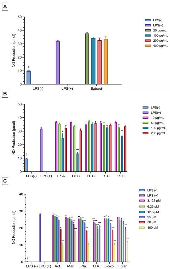
Figure 4.
Anti-inflammatory effects of the extract (A), fractions (B), and pure compounds (C) at different concentrations on RAW 264.7 cells (5 × 10⁵ cells/mL) as determined by NO production. Data are presented as the mean ± SEM from three independent experiments. LPS (+), positive control; LPS (−), negative control. # p < 0.05 compared to the LPS (−) group; *: p < 0.05; **: p < 0.01; ***: p < 0.001. The sample materials and the reference material were compared to LPS (+). Act: Acteoside; Mar: Martynoside; Pla: Plantarenaloside; U.A.: Ursolic Acid; 3-oxo: 3-oxo-α-ionol β-glucoside; F.Gar.: Feruloyl Gardoside.
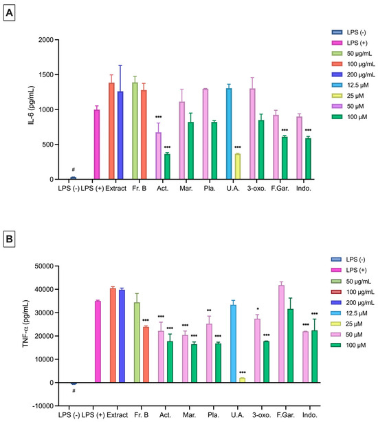
Figure 5.
Anti-inflammatory effects of the extract, fractions, and pure compounds at different concentrations on RAW 264.7 cells (5 × 10⁵ cells/mL) as determined by IL-6 (A) and TNF-α (B) production. Data are presented as the mean ± SEM from three independent experiments. LPS (+), positive control; LPS (−), negative control. # p < 0.05 compared to the LPS (−) group; *: p < 0.05; **: p < 0.01; ***: p < 0.001. The sample materials and the reference material were compared to LPS (+). Act: Acteoside; Mar: Martynoside; Pla: Plantarenaloside; U.A.: Ursolic Acid; 3-oxo: 3-oxo-α-ionol β-glucoside; F.Gar.: Feruloyl Gardoside; Indo: Indomethacin.
Acteoside significantly inhibited IL-6 cytokine production at 50 and 100 μM (33.57–65.66%) concentrations, while feruloyl gardoside (40.17%) and indomethacin (42.12%) showed a similar inhibition at 100 μM (Figure 5A). Ursolic acid (65.42%) significantly inhibited IL-6 production at 25 μM (Figure 5A). All compounds, except for feruloyl gardoside, significantly reduced the TNF-α levels at 50 and 100 μM (21.37–52.00%) (Figure 5B). Additionally, ursolic acid significantly inhibited TNF-α production at 25 μM concentrations (Figure 5B).
2.3.3. Scratch Assay Results
The wound healing effects of the extract, fractions, and isolated pure compounds on the L929 cell line were evaluated using the scratch assay method. The wound area was measured at 0 and 24 h. The extract was tested at concentrations of 20, 100, 200, and 400 μg/mL, while the fractions were tested at 10, 50, 100, and 200 μg/mL. The pure compounds were examined at six different concentrations ranging from 100 to 3.125 μM through two-fold serial dilution. The in vitro wound healing effect of the samples were evaluated statistically by comparing them with the FBS (−) control group. Among the fractions, Fr. B showed the highest effect at a concentration of 200 μg/mL (Closure %: 90.83%; p < 0.001). Fr. B, C, and D showed significant effects at almost all concentrations (Table 4).
Table 4.
Effect of extract and fractions on scratch wound model in the L929 fibroblast (105 cells/mL).
Plantarenaloside, martynoside, acteoside, and feruloyl gardoside were found to be significantly effective at all concentrations. Among the pure compounds, 3-oxo-α-ionol β-glucoside exhibited the highest wound healing activity at a concentration of 12.5 μM (Closure %: 75.2%; p < 0.001) (Table 5).
Table 5.
Effect of pure compounds on a scratch wound model in the L929 fibroblast (105 cells/mL).
2.3.4. Antioxidant Activity
The P. indica extract and the fractions were evaluated for their scavenging activities against the DPPH, ABTS, and SO radicals at concentrations of 50, 100, 200, and 400 µg/mL for the extract and at concentrations of 25, 50, 100, and 200 μg/mL for the fractions. The extracts and fractions showed a radical scavenging potential on the DPPH, ABTS, and SO radicals, with an increasing effect as the concentration increased. Quercetin, a natural compound known for its antioxidant activity, was used as the reference compound in the experiments.
The inhibitory concentration 50% (IC50) value of the extract against the DPPH radical was measured as 160.62 μg/mL. Among the fractions, Fr. C (IC50: 15.13 μg/mL) and Fr. D (IC50: 12.93 μg/mL) exhibited a higher DPPH radical scavenging activity compared to the other fractions (Figure 6A).
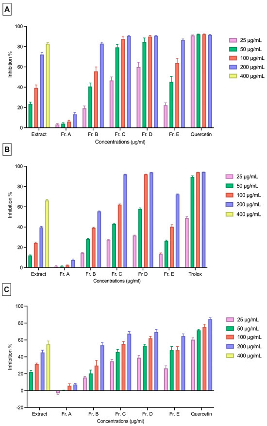
Figure 6.
DPPH (A), ABTS (B), and SO (C) radical scavenging activities of fractions of P. indica and the reference compound. Data are expressed as the mean ± SEM of three independent experiments.
The extract showed an IC50 value of 283.40 μg/mL against the ABTS radical. Among the fractions, Fr. C (IC50: 76.69 μg/mL) and Fr. D (IC50: 44.85 μg/mL) demonstrated a notably stronger ABTS radical scavenging activity than the other fractions (Figure 6B).
The extract exhibited an IC50 value of 228.80 μg/mL against the SO radical. Similar to the results observed with the DPPH and ABTS radical scavenging activities, Fr. C (88.82 μg/mL) and Fr. D (55.24 μg/mL) showed the strongest SO radical scavenging effects (Figure 6C).
3. Discussion
The antidiabetic, antimicrobial, and cytotoxic effects of the Plantago species, as well as their anti-inflammatory and wound healing activities, have been demonstrated using various in vitro and in vivo experimental models [14,19,37,38]. In the present investigation, phytochemical studies were performed on the aerial parts of P. indica and its anti-inflammatory and wound healing effects were demonstrated through both in vitro and in vivo experiments.
In our research, fractionation was performed considering the chemical properties of the major secondary metabolites found in the Plantago species. An aqueous phase of 80% EtOH extract was fractionated using a polyamide column. This allowed for the separation of phenolic and terpenic compounds according to their polarity. Fractions were selected based on their TLC profiles and biological activities were investigated according to this chemical content.
Wound healing is a multifaceted process involving anti-inflammatory and radical-scavenging activities [39]. Because healing, inflammation, and oxidative stress are closely interconnected, the combined evaluation of these three processes was deemed to be important and included in our study. Reactive oxygen species (ROS) are important signaling molecules that play a role in the early stages of wound healing, but excessive ROS production can negatively affect the healing process. Therefore, a close relationship between antioxidant activity and wound healing is known. In this study, the results of the DPPH, ABTS, and SO radical scavenging tests were evaluated together with the in vitro scratch test findings. The results show that fractions with high antioxidant capacity (Fr. C, Fr. D, and Fr. E) exhibit more pronounced wound-healing activity. Previous studies have shown that the Plantago species possess significant antioxidant activity. Research on the extracts obtained from different Plantago species and plant parts has reported significant free radical scavenging effects using various in vitro methods, such as DPPH, ABTS, and NO [12,40,41]. Consistent with the data reported in the literature, the present study also demonstrates that P. indica possesses antioxidant activity using different in vitro methods.
Among the obtained compounds, plantarenaloside (1) and feruloyl gardoside (5) are iridoid glycosides, while 3-oxo-α-ionol β-glucoside (2) is a megastigmane glycoside, martynoside (3) and acteoside (4) are phenylethanoid glycosides, and ursolic acid (6) is a triterpene [19,30,31,32,33,34]. The predominance of iridoid and phenylethanoid glycosides among the isolated compounds is consistent with the literature reporting that these groups of compounds are characteristic of the Plantago species. Furthermore, upon review of the literature, it was understood that 3-oxo-α-ionol β-glucoside (2) and feruloyl gardoside (5) were isolated from a Plantago species for the first time with this study.
The anti-inflammatory and wound healing effects of three different extracts were also evaluated using in vivo models. In the acetic acid-induced capillary permeability assay, both the MeOH and 80% EtOH extracts exhibited significant activity. In the linear incision wound model, only the 80% EtOH extract was found to be effective, while in the circular excision model, both the MeOH and 80% EtOH extracts demonstrated notable wound healing activity. By contrast, the aqueous extract showed minimal anti-inflammatory and wound healing effects. Considering the TLC profiles of P. indica, it was concluded that the alcoholic extracts are richer in secondary metabolites compared to the aqueous extract. These findings for P. indica are consistent with other in vivo studies in the literature demonstrating the wound healing and anti-inflammatory effects of the Plantago species [42,43,44].
The cytotoxic effects of the extract, its fractions, and its isolated compounds were tested on the L929 and RAW 264.7 cell lines. Except for ursolic acid, no cytotoxic effects were observed on either cell line. Ursolic acid exhibited cytotoxicity at 50 and 100 μM concentrations in both cell lines, consistent with previous reports [45].
In LPS-stimulated RAW 264.7 cells, the levels of NO, IL-6, and TNF-α were measured. Fr. A and Fr. B significantly reduced the NO levels. Previous studies have demonstrated the anti-inflammatory activities of iridoids [46]. Considering the TLC profiles and phytochemical analysis results, Fr. A and Fr. B fractions were determined to be rich in iridoid compounds, and it is thought that the observed anti-inflammatory effects may be related to this group of compounds. Similarly, plantarenaloside significantly reduced the NO and TNF-α levels at nearly all concentrations tested, likely due to its classification as an iridoid glycoside. In a study conducted on Penstemon gentianoides, a species belonging to the Plantaginaceae family, plantarenaloside was isolated and its effect on nitric oxide (NO) production was evaluated in LPS-stimulated RAW 264.7 macrophage cells. Plantarenaloside was studied at a concentration of 25 and 50 μg/mL. The results demonstrated that plantarenaloside inhibited NO production, which may be attributed to the suppression of iNOS enzyme activity or the down regulation of its gene expression. These findings support the anti-inflammatory potential of plantarenaloside [47]. In our present study, plantarenaloside significantly reduced NO levels (11.27–63.57%) in LPS-stimulated RAW 264.7 cells at all concentrations tested except 6.25 μM. These findings are consistent with the NO production inhibitory effect of plantarenaloside reported in the literature.
Ursolic acid significantly reduced the levels of NO, IL-6, and TNF-α at non-cytotoxic concentrations. A previous study showed that ursolic acid reduced the release of pro-inflammatory mediators, such as TNF-α, IL-6, and IL-1β, in LPS-stimulated macrophage cells and that this effect was associated with the inhibition of the TLR4/MyD88 signaling pathway. Furthermore, ursolic acid has been reported to modulate the inflammatory response by inducing autophagy in macrophages [48]. In another study, the effect of ursolic acid at 5- and 10-μM concentrations on reducing the NO, IL-6, and TNF-α levels in LPS-stimulated RAW 264.7 cells were investigated. It was concluded that ursolic acid significantly reduced the NO and IL-6 levels at both concentrations and the TNF-α level at 10 μM concentration [49]. In our study, ursolic acid (16.47–31.51%) significantly inhibited the NO levels in LPS-stimulated RAW 264.7 cells at all concentrations tested (3.125–25 μM). It also significantly reduced the IL-6 and TNF-α levels at 25 μM. These findings are consistent with the results reported in the literature.
The compound 3-oxo-α-ionol β-glucoside was found to exert a suppressive effect on inflammatory mediators, such as NO, IL-6, and TNF-α, in LPS-stimulated RAW 264.7 cells. In one study, the effect of 3-oxo-α-ionol β-glucoside on NO production in LPS-stimulated RAW 264.7 cells were investigated. The compound was tested at concentrations of 12, 25, 50, and 100 μM, and it was found to significantly inhibit NO production at 100 and 50 μM concentrations [50]. In our current study, 3-oxo-α-ionol β-glucoside significantly reduced NO production (10.77–63.71%) at all doses in the concentration range of 3.125–100 µM, and the obtained data are consistent with the existing literature.
Acteoside and martynoside significantly reduced the levels of NO with the ratio of 12.58–64.47% and 13.63–43.50% at the concentration of 25–100 μM, respectively. Additionally, these compounds decreased the production of TNF-α by 35.95–48.39% and 40.91–52.00% at the concentration of 50–100 μM, respectively. In addition, acteoside significantly inhibited IL-6 levels by 33.57 to 65.66% at concentrations of 50–100 μM, respectively. In our previous study investigating the anti-inflammatory effect of acteoside, the compound was tested at concentrations of 1, 10, 25, 50, and 100 μg/mL to evaluate its effect on NO production in LPS-stimulated RAW 264.7 cells. Significant inhibition of NO production was observed at 1 and 50 μg/mL concentrations. Additionally, the effects of the compound on the PGE2 and TNF-α levels were investigated at concentrations of 10, 50, and 100 μg/mL, and it was found to be effective at all tested concentrations. When evaluated together with our current study, it was concluded that the anti-inflammatory effects of these compounds may be associated with their phenolic structures [19].
In the scratch assay, Fr. B (47.07–90.83% wound closure) showed a greater effect compared to the other fractions. This was attributed to the high phenolic content of Fr. B. Among the isolated compounds, 3-oxo-α-ionol β-glucoside (30.85–75.20% wound closure) exhibited a more pronounced wound healing effect than the others. Additionally, martynoside (14.71–66.13% wound closure) also demonstrated a strong effect. In one study, water and EtOH extracts were prepared separately from the dried leaves of P. major and a combined extract was obtained by mixing these two extracts in equal volumes. Additionally, a water extract was prepared from the fresh leaves. Using a scratch assay, the wound healing effects of these four extracts were evaluated on oral epithelial cells at concentrations of 0.1, 1.0, and 10.0 mg/mL. As a result, while all extracts were found to be effective at most of the tested concentrations, the EtOH extract at 10.0 mg/mL showed the highest activity [24]. In another study, a 70% EtOH extract standardized with acteoside was prepared from the leaves of P. australis. The wound healing effects of the extract and acteoside were investigated using a scratch assay on HaCaT cells. At a concentration of 25 μg/mL, the extract showed 81.06% wound closure, while acteoside exhibited 58.70–57.77% wound closure at 5 and 10 μg/mL, respectively [20]. In the present study, an 80% EtOH extract prepared from P. indica provided significant wound closure of 30.83% at a concentration 100 μg/mL. On the other hand, acteoside showed significant wound closure (19.95–62.58%) at all concentrations in the range of 3.125–100 μM. The results obtained are consistent with the literature for acteoside. The studies investigating the in vitro effects of the Plantago species using a scratch assay are in agreement with our current findings. The wound healing activity of the Plantago species has been scientifically demonstrated, thereby supporting their traditional use in wound treatment.
The results of in vitro and in vivo biological effect experiments conducted on P. indica scientifically support the use of the Plantago genus in traditional medicine. Both P. indica and its isolated compounds appear to hold therapeutic potential for the treatment of wounds and inflammation.
4. Materials and Methods
4.1. Plant Material
Aerial parts of P. indica L. were collected in July 2021 at the flowering stage from Incesu Beach, Atakum, Samsun, Türkiye. The plant was identified by Assistant Professor Fergan Karaer (Ondokuz Mayıs University, Faculty of Education, Department of Science). A voucher specimen has been deposited in the herbarium of the Faculty of Pharmacy at Hacettepe University (HUEF 21022). The plant material was dried under shade and powdered prior to extraction.
4.2. Chemicals, Reagents and Instruments
MeOH (≥99.7%, purity), EtOH (≥99.8%, purity), CHCl3 (≥99.8%, purity), and petroleum ether (40–70 °C, analytical grade) were purchased from Sigma-Aldrich (St. Louis, MO, USA). Methanol-d4 (≥99.8%, %D) and Pyridine-d5 (≥99.5%, %D) were obtained from Isotec, Inc. (Miamisburg, OH, USA).
Open column chromatography was carried out using various stationary phases, including silica gel [Merck, Kieselgel 60, 70–230 mesh (Darmstadt, Germany) and Fuji BW-200 (Fuji Silysia Chemical, Kasugai, Japan)], octadecylsilyl silica [Merck, 40–63 μm (Darmstadt, Germany) and Cosmosil 140C18-OPN (Nacalai Tesque, Inc., Kyoto, Japan)], polyamide [Fluka, polyamide 6, 50–160 μm (Sigma-Aldrich, St. Louis, MO, USA)], and Sephadex [LH-20 (Pharmacia, Uppsala, Sweden)]. Aluminium (Silica gel 60 F254, 0.2 mm, Merck, Darmstadt, Germany) and glass plates were used in thin-layer chromatography (TLC) (Silica gel 60 F254, 9.5–11.5 µm, Merck, Darmstadt, Germany). Dulbecco’s Modified Eagle Medium (DMEM), LPS, Fetal bovine serum (FBS), and 3-(4,5-dimethylthiazol-2-yl)-2,5-diphenyltetrazolium bromide (MTT) were purchased from Sigma-Aldrich Chem. Co. (St. Louis, MO, USA). Dulbecco’s phosphate-buffered saline was purchased from Pan-Biotech (Aidenbach, Germany), and penicillin-streptomycin was purchased from Gibco Invitrogen Life Technologies (Waltham, MA, USA).
Compounds were concentrated by vacuum rotary evaporators [OSB-2200 (Eyela, Tokyo Rikakikai Co., Ltd., Tokyo, Japan) and Buchi R-210 (Büchi Labortechnik AG, Flawil, Switzerland)]. Microplate readings were performed using a BioTek μQuant microplate reader (MQX200, Winooski, VT, USA). The HPLC (high-performance liquid chromatography) system was equipped with an EYELA (Tokyo Rikakikai Co., Ltd., Tokyo, Japan) Micro Feeder MP-Σ pump, a UV-Vis detector (Jasco UV-2075 Plus, Jasco Co., Tokyo, Japan), and a Develosil RPAQUEOUS column [5 µm; 20 mm × 250 mm] (Nomura Chemical Co., Ltd., Aichi, Japan). Nuclear magnetic resonance (NMR) spectra were measured using a JEOL JMN-ECA600 (600 MHz) spectrometer (Jeol Ltd., Tokyo, Japan) with JEOL Delta software (version 6.4). Wound closure in the scratch assay was determined using a Leica DM IL LED (Wetzlar, Germany) light microscope and the Leica ICC50 W/E digital camera system integrated into this microscope, with the wound area directly determined by the measurement tools on the microscope screen.
4.3. Extraction, Fractionation, and Isolation Process
The starting point of this study was the strong ethnobotanical evidence indicating the traditional use of the Plantago species for wound healing and anti-inflammatory purposes. In line with this traditional knowledge, the first aim was to demonstrate the efficacy of P. indica through in vivo models. Subsequently, in vitro biological assays and phytochemical analyses were conducted to better understand the observed biological activity at the molecular level. Isolation studies and in vitro experiments were performed using an 80% EtOH extract, while in vivo experiments were conducted using MeOH, 80% EtOH, and aqueous extracts.
Approximately 10 g of powdered plant material was used for the MeOH and 80% EtOH extracts in the in vivo experiments. The extracts were obtained by extracting the plant material with the respective solvents (100 mL; 1:10 w/v ratio) 3 times at 40 °C for 8 h. The obtained filtrates were combined and the solvent was removed under vacuum to obtain the MeOH (2.26 g) and 80% EtOH (2.53 g) extracts. For the water extract, 6.62 g of plant material was weighed and boiled with 500 mL of water for 30 min, then filtered [8]. The water in the resulting extract was evaporated and removed. Thus, the water extract (1.37 g) was obtained. These three extracts were used in the in vivo experiments. Due to the higher efficacy of the 80% EtOH extract and its similar TLC profile to the MeOH extract, this extract was selected for phytochemical and in vitro studies.
Phytochemical analyses and in vitro biological activity studies were carried out on the 80% EtOH extract. For this purpose, 460.3 g of powdered plant material was extracted five times with 3 L of 80% EtOH at 40 °C for 8 h and filtered. The obtained filtrates were combined and the solvent was removed under vacuum to obtain a total of 89.65 g (19.5% w/w) of crude extract. The crude extract was dissolved in water and then the lipophilic compounds and chlorophylls were removed using petroleum ether. The obtained aqueous phase (50 g) was fractionated using a polyamide column. Elution was started with 100% water and was carried out with 75:25, 50:50, 25:75 H2O/MeOH and 100% MeOH, respectively. In this way, 5 fractions were obtained from the polyamide column (Frs. A-E).
Fr. A (1.51 g) was subjected to silica gel column chromatography (SCC) (150 g) using CHCl3/MeOH (100:0 to 75:25) as the solvent system to give Frs. A1–19. Fr. A13 (150 mg) was applied to a vacuum liquid chromatography column filled with Silicagel-C18 (20 g) using H2O/MeOH (90:10 to 75:25) and thereby Compound 1 (87.5 mg) was isolated. Fr. A7 was applied to HPLC system and the flow rate was 4 mL/min. Compound 2 (6.4 mg) was purified with an H2O/MeOH (55:45) solvent system.
Fr. D (1.4 g) was subjected to SCC (150 g) using CHCl3/MeOH (100:0 to 75:25) as the solvent system to give Fr. D1–19. Fr. D10 was applied to the HPLC system at a flow rate of 4 mL/min. Compound 3 (7.3 mg) was purified with the solvent system H2O/MeOH (45:55). Fr. D14 (150 mg) was applied to Sephadex LH-20 CC (50 g, MeOH) and obtained 6 subfraction, D14a–e. D14d was applied to the HPLC system. The flow rate was set at 4 mL/min. Compound 4 (4.5 mg) was obtained with the solvent system H2O/MeOH (55:45). Fr. D19 (600 mg) was applied to SCC (150 g) using CHCl3/MeOH (100:0 to 70:30) and obtained 13 subfractions, D19a–m. D19l was applied to the HPLC system at a flow rate of 4 mL/min. The solvent system used was H2O/MeOH (50:50). Compound 5 (3.2 mg) was purified.
Fr. E (650 mg) was subjected to SCC (75 g) using CHCl3/MeOH (100:0 to 85:15) as the solvent system to give Fr. E1–22. Fr. E7 (100 mg) was applied to Sephadex LH-20 CC (65 g, MeOH) and obtained 7 subfractions, E7a–g. E7e (59 mg) was applied to SCC (30 g) with CHCl3/MeOH (100:0 to 92:8) and Compound 6 (16.1 mg) was purified.
4.4. In Vivo Assays
4.4.1. Animals
In vivo biological activity studies were performed using male Swiss albino mice and Sprague-Dawley rats. Animals were obtained from Kobay laboratory animal production (Ankara, Türkiye). During this time, the animals were housed in polysulfone cages under controlled conditions at room temperature (20–25 °C) and constant humidity (40–50%), with a 12:12-h light–dark cycle and were provided with standard feed and water. Seven animals were used in each group. Animal experiments were conducted in accordance with the European ethical guidelines for animal experimentation and internationally accepted ethical standards for the care and use of laboratory animals’ approval. This study was carried out in accordance with the approval of the Local Ethics Committee for Animal Experimentation (protocol no. 786).
4.4.2. Preparation of Test Samples
For the evaluation of anti-inflammatory activity, test materials were suspended in a mixture of distilled water and 0.5% sodium carboxymethyl cellulose (CMC) and administered orally to the animals. Animals were divided into 5 groups: a control group, a reference drug group, MeOH extract-treated groups, 80% EtOH extract-treated groups, and aqueous extract-treated groups. Animals in the control group were given only 0.5% CMC, while those in the reference drug group were given indomethacin (10 mg/kg) prepared in 0.5% CMC.
To evaluate the wound healing activity, an ointment base containing 1% glycol stearate/1,2 propylene glycol/liquid paraffin (3:6:1) was used as a carrier. Each test sample was homogeneously mixed with this ointment base and applied topically to the wound area at a dose of 0.5 g. Animals were divided into 6 groups: negative control group, vehicle group, reference drug group, MeOH extract-treated groups, 80% EtOH extract-treated groups, and aqueous extract-treated groups. No treatment was applied to animals in the negative control group; only the ointment base was applied to animals in the vehicle group. Madecassol® (Bayer, Istanbul, Türkiye) ointment containing 1% Centella asiatica extract was applied to the animals in the reference drug group.
Wound healing activity was performed using linear incision and circular excision wound models. In the examination of anti-inflammatory activity, the capillary permeability increase model created with acetic acid was used.
4.4.3. Acetic Acid-Induced Capillary Permeability
In order to evaluate the effect of test samples on acetic acid-induced increased vascular permeability in mice, the Whittle (1964) method was applied with some modifications [51]. The test sample was administered orally to the mice at a dose of 0.2 mL per 20 g of body weight. Indomethacin was used as a reference compound at a dose of 10 mg/kg. An amount of 0.1 mL of 4% Evans Blue Dye solution was injected into the tail vein of each mouse 30 min after administration. Following this injection, 10 min later, 0.4 mL of the 0.5% acetic acid solution was administered intraperitoneally. The animals were euthanized by cervical dislocation 20 min after the application. The peritoneum of each animal was opened, the internal organs were washed with distilled water, and peritoneal fluid was collected. Using glass wool, the peritoneal fluid was transferred into 10 mL bottles containing 0.1 M NaOH. The bottle was filled up to 10 mL with distilled water. The absorbance of the dye was measured spectrophotometrically at 590 nm. The degree of extravasation of Evans Blue Dye was measured spectrophotometrically at a wavelength of 590 nm [52,53].
4.4.4. Linear Incision Wound Model
Anesthesia was administered to the rats using 0.15 cc of Ketalar® (Pfizer, New York, NY, USA) and the hair on their backs was shaved. On each side of the dorsal midline, at a distance of 1.5 cm, linear paravertebral wounds measuring 5 cm in length were created using a scalpel. The wounds were closed with three surgical sutures placed 1 cm apart. All animals except the negative control group were treated with topical ointment to the wound area once a day for 9 days. The sutures were removed at the end of the ninth day and the animals were euthanized under anesthesia on the tenth day. To determine the rate of wound healing, the tensile strength of the skin tissue was measured using a tensiometer (Zwick/Roell Z0.5, ZwickRoll GmbH & Co. KG, Ulm, Germany) [54].
4.4.5. Circular Excision Wound Model
Anesthesia was administered to the mice using 0.01 cc of Ketalar® and the hair on their dorsal regions was shaved. Two full-thickness circular excision wounds of 5 mm diameter were created bilaterally on the back of each animal with the help of a biopsy drill. The prepared ointments were topically applied to the wound area every day until complete healing was achieved. The wound healing process was monitored daily with a camera (Fuji, S20 Pro, Fujifilm Co., Tokyo, Japan) and the wound area was measured and evaluated using the images obtained with AutoCAD software (Autodesk, Inc., San Rafael, CA, USA). Wound healing was calculated by taking into account the percentage of reduction compared to the initial wound area. At the end of the tenth day, a tissue samples were taken from each mouse and stored appropriately for histopathological analysis [55].
4.5. In Vitro Assays
4.5.1. Cell Culture
The RAW 264.7 cells were kindly provided by Prof. Dr. Hasan Kırmızıbekmez (Department of Pharmacognosy, Faculty of Pharmacy, Yeditepe University, Istanbul, Türkiye). The L929 cells were purchased from the Biota Lab, Istanbul, Türkiye. Both cell lines were cultured in a DMEM medium containing 10% FBS and 1% penicillin-streptomycin.
4.5.2. Cell Viability
For the cytotoxicity evaluation, an adapted version of the MTT assay described by Mossman was used [56]. Cell suspensions were prepared at densities of 5 × 10⁵ cells/mL for the RAW 264.7 cell line and 105 cells/mL for the L929 cell line. Cells were seeded into 96-well plates at 100 µL per well. Plates were incubated at 37 °C for 24 h in an environment containing 5% CO2 and 95% humidity. After the incubation period, the culture medium was removed and 100 µL of the test solutions at different concentrations prepared in DMEM were added to each well. The cells were incubated for an additional 48 h. Subsequently, the medium was removed and a fresh medium (100 µL) was added to each well. Next, 10 µL of MTT (5 mg MTT/1 mL PBS) was added to each well and the plates were incubated for 4 h. Following this incubation, the medium was aspirated and 100 µL dimethyl sulfoxide (DMSO) was added to each well to dissolve the formed formazan crystals. The absorbance was measured spectrophotometrically at 577 nm with a reference wavelength of 655 nm [57].
4.5.3. LPS-Induced NO, IL-6, and TNF-α Production in the RAW 264.7 Macrophages
Cells were seeded into 96-well plates at a density of 5 × 10⁵ cells/mL. The plates were incubated at 37 °C in a humidified incubator containing 5% CO2 for 24 h. At the end of the incubation period, the medium in each well was aspirated and replaced with 100 μL of the sample solutions at various concentrations. Subsequently, 100 μL of the LPS solution (0.2 μg/mL) was added to each well. The plates were then incubated under the same conditions for another 24 h. After incubation, 100 μL of the medium above the cells from each well was transferred to a new 96-well plate. Nitric oxide (NO) production was determined using the Griess reaction [58], by adding 100 μL of Griess reagent to each well [57]. The levels of IL-6 and TNF-α were measured using ELISA kits in accordance with the manufacturer’s instructions, based on the collected medium above the cells [57,59,60]. Indomethacin was used as the reference anti-inflammatory compound in IL-6 and TNF-α production experiments.
4.5.4. Scratch Assay
The wound healing effect was determined on the L929 fibroblast cell line using the scratch method. The L929 cells were seeded in 96-well plates at 105 concentrations with 100 μL per well and incubated for 24 h. Afterwards, the cells were scratched with a 200 μL pipette tip, thus creating a wound model in the cells. The medium in the wells was discarded and replaced with 100 μL of fresh medium containing the samples at various concentrations. DMEM without FBS was used as the negative control and DMEM with 10% FBS was used as the positive control. The degree of wound closure in the cells was measured at 0 and 24 h [61].
4.5.5. Antioxidant Activity
The antioxidant activity of the P. indica extract and fractions were evaluated by measuring their scavenging effects against the DPPH, ABTS, and SO radicals.
Determination of the DPPH Radical Scavenging Activity
DPPH is a stable free radical with an unpaired valence electron at a nitrogen atom bridge. The DPPH radicals are reduced in the presence of antioxidant compounds, allowing for a determination of the radical scavenging activity [62]. This assay is frequently employed for an antioxidant evaluation because it is valid, inexpensive, rapid, and straightforward [63].
Sample solutions were prepared at various concentrations in MeOH. Then, 200 μL of each sample and the MeOH solution (blank) were transferred into a 96-well plate, followed by the addition 50 μL of the 1 mM DPPH solution. After waiting in the dark for 30 min, the absorbance was measured at 520 nm. Quercetin, known for its antioxidant properties, was used as a reference compound. The DPPH radical scavenging activity was assessed by comparing the absorbance of the control solution with that of the sample solutions [27].
Determination of the ABTS Radical Scavenging Activity
The ABTS solution (7 mM) prepared in distilled water and in a 2.45 mM potassium persulfate solution were mixed in equal volumes and kept in the dark for 12–16 h. In this way, the ABTS radical (ABTS+) was formed and the solution was activated. Dilution was made until the absorbance value of the ABTS+ solution at 734 nm was 0.700 ± 0.050. A total of 80% EtOH was used for dilution. Sample solutions were prepared in 24-well plates with 500 μL of 80% EtOH at various concentrations. A total of 130 μL of each solution was transferred to a 96-well plate and 50 μL of ABTS+ (absorbance = 0.700 ± 0.050) was added to the wells. Absorbance measurements were performed at 734 nm. Trolox (6-hydroxy-2,5,7,8-tetramethylchroman-2-carboxylic acid) was used as a standard compound in the experiment.
The radical scavenging activity was determined by comparing the absorbance of the solution (blank) containing 80% EtOH and ABTS+ radical with the absorbances of the test solutions.
Determination of the SO Radical Scavenging Activity
This assay is based on the reduction of the nitro blue tetrazolium (NBT) reagent by superoxide anion radicals generated by DMSO, forming a blue formazan compound. In the presence of antioxidant compounds, the concentration of superoxide radicals decreases, resulting in a change in the amount of the colored diformazan compound. The radical scavenging activity is measured by determining this change using a spectrophotometer.
Sample solutions were prepared at various concentrations in DMSO. A total of 30 μL of the solutions were transferred to 96-well plates followed by the addition of 10 μL of the NBT (nitro blue tetrazolium) solution. Finally, 100 µL of alkaline DMSO, prepared by mixing 0.9 mL of DMSO with 0.1 mL of 5 mM NaOH, was added to each well. An absorbance measurement was made at 560 nm. Quercetin was used as the reference compound.
The SO radical scavenging activity was determined by comparing the absorbance of the control solution (containing DMSO, NBT, and alkaline DMSO) with the absorbance of the sample solutions [27].
4.6. Statistical Analysis
Results are presented as the mean of three repeated measurements ± the standard error of the mean (SEM). GraphPad Prism 10.0 program was used for all statistical evaluations. Differences between groups were analyzed using Dunnett’s multiple comparison test after a one-way ANOVA test. A p value of less than 0.05 was considered statistically significant.
5. Conclusions
This study includes detailed phytochemical and biological activity studies on the aerial parts of P. indica. Plantarenaloside (1), 3-oxo-α-ionol β-glucoside (2), martynoside (3), acteoside (4), feruloyl gardoside (5), and ursolic acid (6) were isolated from an aqueous phase of an 80% EtOH extract of the above-ground parts of P. indica. Of these, 3-oxo-α-ionol β-glucoside (2) and feruloyl gardoside (5) were determined to have been obtained from a Plantago species for the first time. In vitro studies showed that the 80% EtOH extract and the isolated compounds have significant anti-inflammatory and wound-healing potential. Acteoside inhibited NO (65.66%) and IL-6 (64.45%) production to the highest degree, while ursolic acid was the most effective compound in TNF-α inhibition (92.43%). In the scratch test, 3-oxo-α-ionol β-glucoside (12.5 μM; 72.2%) showed the highest wound closure activity. In vivo experiments were conducted on the MeOH, 80% EtOH, and water extracts. These included acetic acid-induced capillary permeability, linear incision wound model, and circular excision wound model experiments. In all three experiments, the 80% EtOH extract showed the strongest effect (35.3%, 26.5%, and 37.7%, respectively). These findings reveal that P. indica has considerable potential in terms of anti-inflammatory and wound-healing activities.
Supplementary Materials
The following supporting information can be downloaded at: https://www.mdpi.com/article/10.3390/plants15010141/s1, Figures S1–S36: NMR (1H, 13C, COSY, HMQC, HMBC) and chemical structures of compounds 1–6 (plantarenaloside, 3-oxo-α-ionol β-glucoside, martynoside, acteoside, feruloyl gardoside, ursolic acid).
Author Contributions
Conceptualization, I.S.; methodology, H.B. and I.S.; software, H.B.; validation, Z.D., E.K.A., and A.N.; formal analysis, E.K.A. and A.N.; investigation, H.B. and Z.D.; resources, H.B.; data curation, E.K.A. and A.N.; writing—original draft preparation, E.K.A. and H.B.; writing—review and editing, E.K.A., Z.D., and I.S.; visualization, H.B.; supervision, I.S.; project administration, I.S.; funding acquisition, Z.D. and I.S. All authors have read and agreed to the published version of the manuscript.
Funding
This work was funded by the 2214/A International Research Fellowship Programme provided by the Scientific and Technological Research Council of Turkey (TUBITAK) (1059B142300168) and the Hacettepe University Scientific Research Projects Coordination Units (Project No: THD-2023-20875).
Institutional Review Board Statement
The animal study protocol was approved by the Ethics Committee of the Local Animal Experiments Ethics Committee of (Kobay), Türkiye (protocol No: 786, Date: March 2025) for studies involving animals.
Data Availability Statement
The data presented in this study are available upon request from the corresponding author. The data are not publicly available due to privacy and ethical restrictions.
Conflicts of Interest
The authors declare no conflicts of interest.
Abbreviations
The following abbreviations are used in this manuscript:
| ABTS | 2,2′-azino-bis(3-ethylbenzothiazoline-6-sulfonic acid) |
| CMC | Carboxymethyl Cellulose |
| DMEM | Dulbecco’s Modified Eagle Medium |
| DMSO | Dimethyl Sulfoxide |
| DPPH | 2,2-Diphenyl-1-picrylhydrazyl |
| ELISA | Enzyme Linked Immunosorbent Assay |
| EtOH | Ethanol |
| FBS | Fetal Bovine Serum |
| HPLC | High-Performance Liquid Chromatography |
| IC50 | Inhibitory Concentration 50% |
| IL-1β | Interleukin-1 beta |
| IL-6 | Interleukin-6 |
| LPS | Lipopolysaccharides |
| MeOH | Methanol |
| MTT | 3-(4,5-dimethylthiazol-2-yl)-2,5-diphenyl tetrazolium bromide |
| NaOH | Sodium Hydroxide |
| NBT | Nitro Blue Tetrazolium |
| NMR | Nuclear Magnetic Resonance |
| NO | Nitric oxide |
| PBS | Phosphate Buffered Saline |
| SCC | Silica Gel Column Chromatography |
| SEM | Standard Error of the Mean |
| SO | Superoxide |
| TLC | Thin-Layer Chromatography |
| TNF-α | Tumor Necrosis Factor alpha |
References
- WFO. Plantago L. POWO. Available online: https://powo.science.kew.org/taxon/urn:lsid:ipni.org:names:30001135-2 (accessed on 2 December 2025).
- Tutel, B.; Kandemir, İ.; Kus, S.; Kence, A. Classification of Turkish Plantago L. species using numerical taxonomy. Turk. J. Bot. 2005, 29, 51–61. [Google Scholar]
- Zhakipbekov, K.; Turgumbayeva, A.; Issayeva, R.; Kipchakbayeva, A.; Kadyrbayeva, G.; Tleubayeva, M.; Akhayeva, T.; Tastambek, K.; Sainova, G.; Serikbayeva, E.; et al. Antimicrobial and other biomedical properties of extracts from Plantago major, Plantaginaceae. Pharmaceuticals 2023, 16, 1092. [Google Scholar] [CrossRef]
- Dong, Y.; Hou, Q.; Sun, M.; Sun, J.; Zhang, B. Targeted isolation of antioxidant constituents from Plantago asiatica L. and in vitro activity assay. Molecules 2020, 25, 1825. [Google Scholar] [CrossRef] [PubMed]
- Gurbuz, I.; Gencler Ozkan, A.M.; Akaydin, G.; Salihoglu, E.; Gunbatan, T.; Demirci, F.; Yesilada, E. Folk medicine in Duzce Province (Turkey). Turk. J. Bot. 2019, 43, 769–784. [Google Scholar] [CrossRef]
- Karakaya, S.; Polat, A.; Aksakal, O.; Sumbullu, Y.Z.; Incekara, U. Ethnobotanical study of medicinal plants in Aziziye district (Erzurum, Turkey). Turk. J. Pharm. Sci. 2020, 17, 211–220. [Google Scholar] [CrossRef] [PubMed]
- Kilic, M.; Yildiz, K.; Kilic, F.M. Traditional uses of medicinal plants in Artuklu, Turkey. Hum. Ecol. Rev. 2020, 48, 619–632. [Google Scholar] [CrossRef]
- Baytop, T. Türkiye’de Bitkiler ile Tedavi Geçmişten Günümüze, 2nd ed.; Nobel Tıp Kitapevleri: İstanbul, Türkiye, 1999; p. 380. [Google Scholar]
- Goncalves, S.; Romano, A. The medicinal potential of plants from the genus Plantago (Plantaginaceae). Ind. Crops Prod. 2016, 83, 213–226. [Google Scholar] [CrossRef]
- Hammami, S.; Debbabi, H.; Jlassi, I.; Joshi, R.K.; Mokni, R.E. Chemical composition and antimicrobial activity of essential oil from the al parts of Plantago afra L. (Plantaginaceae) growing wild in Tunisia. S. Afr. J. Bot. 2020, 132, 410–414. [Google Scholar] [CrossRef]
- Budzianowska, A.; Toton, E.; Romaniuk-Drapala, A.; Kikowska, M.; Budzianowski, J. Cytotoxic effect of phenylethanoid glycosides isolated from Plantago lanceolata L. Life 2023, 13, 556. [Google Scholar] [CrossRef]
- Farcas, A.D.; Zagrean-Tuza, C.; Vlase, L.; Gheldiu, A.M.; Parvu, M.; Mot, A.C. Epr fingerprinting and antioxidant response of four selected Plantago species. Stud. Univ. Babes-Bol. Chem. 2020, 65, 209–220. [Google Scholar] [CrossRef]
- Pesantes-Sangay, S.J.; Calla-Poma, R.D.; Requena-Mendizabal, M.F.; Alvino-Vales, M.I.; Millones-Gomez, P.A. Chemical composition and antibacterial effect of Plantago major extract on periodontal pathogens. Pesqui. Bras. Odontopediatria Clin. Integr. 2020, 20, e0012. [Google Scholar] [CrossRef]
- Alsaraf, K.M.; Mohammad, M.H.; Al-Shammari, A.M.; Abbas, I.S. Selective cytotoxic effect of Plantago lanceolata L. against breast cancer cells. J. Egypt. Natl. Cancer Inst. 2019, 31, 10. [Google Scholar] [CrossRef] [PubMed]
- Dong, C.L.; Qin, Y.; Ma, J.X.; Cui, W.Q.; Chen, X.R.; Hou, L.Y.; Chen, X.Y.; God’spower, B.O.; Eliphaz, N.; Qin, J.J.; et al. The active ingredients identification and antidiarrheal mechanism analysis of Plantago asiatica L. superfine powder. Front. Pharmacol. 2021, 11, 612478. [Google Scholar] [CrossRef]
- Zhao, H.; Wang, Q.H.; Sun, Y.P.; Yang, B.Y.; Wang, Z.B.; Chai, G.F.; Guan, Y.Z.; Zhu, W.G.; Shu, Z.P.; Lei, X.; et al. Purification, characterization and immunomodulatory effects of Plantago depressa polysaccharides. Carbohydr. Polym. 2014, 112, 63–72. [Google Scholar] [CrossRef]
- Fu, Y.S.; Lue, S.I.; Lin, S.Y.; Luo, C.L.; Chou, C.C.; Weng, C.F. Plantago asiatica seed extracts alleviated blood pressure in phase I-spontaneous hypertension rats. Molecules 2019, 24, 1734. [Google Scholar] [CrossRef] [PubMed]
- Farcas, A.D.; Mot, A.C.; Parvu, A.E.; Al Toma, V.; Popa, M.A.; Mihai, M.C.; Sevastre, B.; Roman, I.; Vlase, L.; Parvu, M. In vivo pharmacological and anti-inflammatory evaluation of Xerophyte Plantago sempervirens Crantz. Oxid. Med. Cell. Longev. 2019, 2019, 5049643. [Google Scholar] [CrossRef]
- Genc, Y.; Harput, U.S.; Saracoglu, I. Active compounds isolated from Plantago subulata L. via wound healing and antiinflammatory activity guided studies. J. Ethnopharmacol. 2019, 241, 112030. [Google Scholar] [CrossRef] [PubMed]
- Sperotto, N.D.D.; Steffens, L.; Verissimo, R.M.; Henn, J.G.; Peres, V.F.; Vianna, P.; Chies, J.A.B.; Roehe, A.; Saffi, J.; Moura, D.J. Wound healing and anti-inflammatory activities induced by a Plantago australis hydroethanolic extract standardized in verbascoside. J. Ethnopharmacol. 2018, 225, 178–188. [Google Scholar] [CrossRef]
- Barua, C.C.; Pal, S.K.; Roy, J.D.; Buragohain, B.; Talukdar, A.; Barua, A.G.; Borah, P. Studies on the anti-inflammatory properties of Plantago erosa leaf extract in rodents. J. Ethnopharmacol. 2011, 134, 62–66. [Google Scholar] [CrossRef]
- Hussan, F.; Mansor, A.S.; Hassan, S.N.; Tengku Nor Effendy Kamaruddin, T.N.; Budin, S.B.; Othman, F. Anti-inflammatory property of Plantago major leaf extract reduces the inflammatory reaction in experimental acetaminophen-induced liver injury. Evid. Based Complement. Altern. Med. 2015, 2015, 347861. [Google Scholar] [CrossRef]
- Majkic, T.; Bekvalac, K.; Beara, I. Plantain (Plantago L.) species as modulators of prostaglandin E2 and thromboxane A2 production in inflammation. J. Ethnopharmacol. 2020, 262, 113140. [Google Scholar] [CrossRef]
- Zubair, M.; Ekholm, A.; Nybom, H.; Renvert, S.; Widen, C.; Rumpunen, K. Effects of Plantago major L. leaf extracts on oral epithelial cells in a scratch assay. J. Ethnopharmacol. 2012, 141, 825–830. [Google Scholar] [CrossRef]
- Taskova, R.; Evstatieva, L.; Handjieva, N.; Popov, S. Iridoid patterns of genus Plantago L. and their systematic significance. Z. Naturforsch C J. Biosci. 2002, 57, 42–50. [Google Scholar] [CrossRef] [PubMed]
- Ozcan, T. Fatty acid composition of seed oils in some sand dune vegetation species from Turkey. Chem. Nat. Compd. 2014, 50, 804–809. [Google Scholar] [CrossRef]
- Harput, U.S.; Genc, Y.; Saracoglu, I. Cytotoxic and antioxidative activities of Plantago lagopus L. and characterization of its bioactive compounds. Food Chem. Toxicol. 2012, 50, 1554–1559. [Google Scholar] [CrossRef] [PubMed]
- Ukaegbu, K.; Allen, E.; Svoboda, K.K.H. Reactive oxygen species and antioxidants in wound healing: Mechanisms and therapeutic totential. Int. Wound J. 2025, 22, e70330. [Google Scholar] [CrossRef]
- Hunt, M.; Torres, M.; Bachar-Wikstrom, E.; Wikstrom, J.D. Cellular and molecular roles of reactive oxygen species in wound healing. Commun. Biol. 2024, 7, 1534. [Google Scholar] [CrossRef]
- Andrzejewska-Golec, E.; Ofterdinger-Daegel, S.; Calis, I.; Swiatek, L. Chemotaxonomic aspects of iridoids occurring in Plantago subg. psyllium (Plantaginaceae). Plant Syst. Evol. 1993, 185, 85–89. [Google Scholar] [CrossRef]
- Pabst, A.; Barron, D.; Semon, E.; Schreier, P. Two diastereomeric 3-oxo-α-ionol β-D-glucosides from raspberry fruit. Phytochemistry 1992, 31, 1649–1652. [Google Scholar] [CrossRef]
- Calıs, I.; Saracoglu, I.; Shizuka, K.; Sansei, N. Phenylpropanoid glycosides isolated from Rhynchocorys stricta Scrophulariaceae. Turk. J. Med. Sci. 1988, 12, 234–238. [Google Scholar]
- Chen, X.; Cao, Y.G.; Ren, Y.J.; Liu, Y.L.; Fan, X.L.; He, C.; Li, X.D.; Ma, X.Y.; Zheng, X.K.; Feng, W.S. Ionones and lignans from the fresh roots of Rehmannia glutinosa. Phytochemistry 2022, 203, 113423. [Google Scholar] [CrossRef] [PubMed]
- Seebacher, W.; Simic, N.; Weis, R.; Saf, R.; Kunert, O. Complete assignments of 1H and 13C NMR resonances of oleanolic acid, 18α-oleanolic acid, ursolic acid and their 11-oxo derivatives. Magn. Reson. Chem. 2003, 41, 636–638. [Google Scholar] [CrossRef]
- Küpeli Akkol, E.; Renda, G.; İlhan, M.; Yazıcı Bektaş, N. Wound healing acceleration and anti-inflammatory potential of Prunella vulgaris L.: From conventional use to preclinical scientific verification. J. Ethnopharmacol. 2022, 295, 115411. [Google Scholar] [CrossRef] [PubMed]
- Küpeli Akkol, E.; Subaş, T.; Özgen, U.; Süntar, I.; Ilhan, M.; Keleş, H. Effects of aphthoquinones from the roots of Onosma armeniacum Klokov on wound healing. Chem. Biodivers. 2024, 21, e202301946. [Google Scholar] [CrossRef]
- Ferrazzano, G.F.; Cantile, T.; Roberto, L.; Ingenito, A.; Catania, M.R.; Roscetto, E.; Palumbo, G.; Zarrelli, A.; Pollio, A. Determination of the in vitro and in vivo antimicrobial activity on salivary streptococci and lactobacilli and chemical characterisation of the phenolic content of a Plantago lanceolata infusion. Biomed. Res. Int. 2015, 2015, 286817. [Google Scholar] [CrossRef]
- Tinkov, A.A.; Nemereshina, O.N.; Popova, E.V.; Polyakova, V.S.; Gritsenko, V.A.; Nikonorov, A.A. Plantago maxima leaves extract inhibits adipogenic action of a high-fat diet in female Wistar rats. Eur. J. Nutr. 2014, 53, 831–842. [Google Scholar] [CrossRef]
- Patil, K.R.; Mahajan, U.B.; Unger, B.S.; Goyal, S.N.; Belemkar, S.; Surana, S.J.; Ojha, S.; Patil, C.R. Animal models of inflammation for screening of anti-inflammatory drugs: Implications for the discovery and development of phytopharmaceuticals. Int. J. Mol. Sci. 2019, 20, 4367. [Google Scholar] [CrossRef]
- Mesquita, L.M.S.; Da Rocha, C.Q.; Affonso, L.H.L.; Cerulli, A.; Piacente, S.; Tangerina, M.M.P.; Martins, M.B.G.; Vilegas, W. Phenolic isomers from Plantago catharinea leaves: Isolation, identification, quantification and in vitro antioxidant activity. Nat. Prod. Commun. 2017, 12, 409–412. [Google Scholar] [CrossRef]
- Pereira, C.G.; Custodio, L.; Rodrigues, M.J.; Neng, N.R.; Nogueira, J.M.F.; Carlier, J.; Costa, M.C.; Varela, J.; Barreira, L. Profiling of antioxidant potential and phytoconstituents of Plantago coronopus. Braz. J. Biol. 2017, 77, 632–641. [Google Scholar] [CrossRef] [PubMed]
- Kuranel, E.; Akkol Kupeli, E.K.; Suntar, İ.; Gursoy, S.; Keles, H.; Aktay, G. Investigating biological activity potential of Plantago lanceolata L. in healing of skin wounds by a preclinical research. Turk. J. Pharm. Sci. 2016, 13, 135–144. [Google Scholar] [CrossRef]
- Kovac, I.; Durkac, J.; Holly, M.; Jakubcova, K.; Perzel’ova, V.; Mucaji, P.; Svajdlenka, E.; Sabol, F.; Legath, J.; Belak, J.; et al. Plantago lanceolata L. water extract induces transition of fibroblasts into myofibroblasts and increases tensile strength of healing skin wounds. J. Pharm. Pharmacol. 2015, 67, 117–125. [Google Scholar] [CrossRef] [PubMed]
- Thome, R.G.; dos Santos, H.B.; dos Santos, F.V.; Oliveira, R.J.D.; de Camargos, L.F.; Pereira, M.N.; Longatti, T.R.; Souto, C.M.; Franco, C.S.; Schüffner, R.D.A.; et al. Evaluation of healing wound and genotoxicity potentials from extracts hydroalcoholic of Plantago major and Siparuna guianensis. Exp. Biol. Med. 2012, 237, 1379–1386. [Google Scholar] [CrossRef]
- Mazumder, K.; Tanaka, K.; Fukase, K. Cytotoxic activity of ursolic acid derivatives obtained by isolation and oxidative derivatization. Molecules 2013, 18, 8929–8944. [Google Scholar] [CrossRef] [PubMed]
- Viljoen, A.; Mncwangi, N.; Vermaak, I. Anti-inflammatory iridoids of botanical origin. Curr. Med. Chem. 2012, 19, 2104–2127. [Google Scholar] [CrossRef]
- Domingues, M.; Keck, S.A.; Jeffery, E.; Cespedes, C.L. Effects of extracts, flavonoids and iridoids from Penstemon gentianoides (Plantaginaceae) on inhibition of inducible nitric oxide synthase (iNOS), cyclooxygenase-2 (COX-2) in LPS-Activated RAW 264.7 macrophage cells and their antioxidant activity. Bol. Latinoam. Caribe Plantas Med. Aromat. 2010, 9, 397–413. [Google Scholar]
- Zhao, J.; Zheng, H.; Sui, Z.; Jing, F.; Quan, X.; Zhao, W.; Liu, G. Ursolic acid exhibits anti-inflammatory effects through blocking TLR4-MyD88 pathway mediated by autophagy. Cytokine 2019, 123, 154726. [Google Scholar] [CrossRef] [PubMed]
- Sołtys, A.; Galanty, A.; Grabowska, K.; Paśko, P.; Zagrodzki, P.; Podolak, I. Multidirectional effects of terpenoids from Sorbus intermedia (EHRH.) PERS fruits in cellular model of benign prostate hyperplasia. Pharmaceuticals 2023, 16, 965. [Google Scholar] [CrossRef]
- Yang, J.-R.; Zhu, Y.-T.; Zeng, Y.-Q.; Li, H.-Q.; Li, C.-H.; Gao, J.-M. Three new ionone glycosides from Rhododendron capitatum Maxim. Molecules 2024, 29, 2462. [Google Scholar] [CrossRef]
- Whittle, B.A. The use of changes in capillary permeability in mice to distinguish between narcotic and nonnarcotic alalgesics. Br. J. Pharmacol. 1964, 22, 246–253. [Google Scholar] [CrossRef]
- Ayaz, F.; Küpeli Akkol, E.; Gören Nezhun, G.; Calıs, I.; Gürağaç Dereli, F.; Duman, H.; Choudhary, M.I.; Küçükboyacı, N. Bioassay-guided isolation and identification of anti-inflammatory sesquiterpene lactones from Chrysophthalmum montanum (DC.) Boiss. Rec. Nat. Prod. 2019, 14, 48–56. [Google Scholar] [CrossRef]
- Ilhan, M.; Gürağaç Dereli, F.T.; Tümen, I.; Küpeli Akkol, E. Anti-inflammatory and antinociceptive features of Bryonia alba L.: As a possible alternative in treating rheumatism. Open Chem. 2019, 17, 23–30. [Google Scholar] [CrossRef]
- Suntar, I.; Kupeli Akkol, E.; Senol, F.S.; Keles, H.; Orhan, I.E. Investigating wound healing, tyrosinase inhibitory and antioxidant activities of the ethanol extracts of Salvia cryptantha and Salvia cyanescens using in vivo and in vitro experimental models. J. Ethnopharmacol. 2011, 135, 71–77. [Google Scholar] [CrossRef]
- Kupeli Akkol, E.; Koca, U.; Pesin, I.; Yilmazer, D. Evaluation of the wound healing potential of Achillea biebersteinii Afan. (Asteraceae) by in vivo excision and incision models. Evid. Based Complement. Altern. Med. 2011, 2011, 474026. [Google Scholar] [CrossRef] [PubMed]
- Mossman, T. Rapid colorimetric assay for cellular growth and survival: Application to proliferation and cytotoxicity assays. J. Immunol. Methods 1983, 65, 55–63. [Google Scholar] [CrossRef] [PubMed]
- Dogan, Z.; Telli, G.; Tel, B.C.; Saracoglu, I. Scutellaria brevibracteata Stapf and active principles with anti-inflammatory effects through regulation of NF-kappa B/COX-2/iNOS pathways. Fitoterapia 2022, 158, 105159. [Google Scholar] [CrossRef] [PubMed]
- Green, L.C.; Wagner, D.A.; Glogowski, J.; Skipper, P.L.; Wishnok, J.S.; Tannenbaum, S.R. Analysis of nitrate, nitrite, and [15N] nitrate in biological fluids. Anal. Biochem. 1982, 126, 131–138. [Google Scholar] [CrossRef]
- Li, H.; Feng, J.X.; Liu, C.; Hou, S.T.; Meng, J.L.; Liu, J.Y.; Zilong, S.; Chang, M.C. Polysaccharides from an edible mushroom, Hericium erinaceus, alleviate ulcerative colitis in mice by inhibiting the NLRP3 inflammasomes and reestablish intestinal homeostasis. Int. J. Biol. Macromol. 2024, 267, 131251. [Google Scholar] [CrossRef]
- Xu, F.J.; Yang, L.; Qu, A.L.; Li, D.B.; Yu, M.F.; Zheng, S.J.; Ruan, X.; Wang, Q. A design of Tetrastigma hemsleyanum’s flavonoid loaded polyvinylpyrrolidone nanoparticles for improving its bioavailability and biological activities. Food Chem. 2025, 473, 143099. [Google Scholar] [CrossRef]
- Jagiello, K.; Uchanska, O.; Matyja, K.; Jackowski, M.; Wiatrak, B.; Kubasiewicz-Ross, P.; Karuga-Kuzniewska, E. Supporting the wound healing process-curcumin, resveratrol and baicalin in in vitro wound healing studies. Pharmaceuticals 2023, 16, 82. [Google Scholar] [CrossRef]
- Bakeshlouy Afshar, M.; Poursattar Marjani, A.; Gozali Balkanloo, P. Introducing graphene quantum dots in decomposable wheat starch-gelatin based nano-biofilms. Sci. Rep. 2024, 14, 2069. [Google Scholar] [CrossRef]
- Baliyan, S.; Mukherjee, R.; Priyadarshini, A.; Vibhuti, A.; Gupta, A.; Pandey, R.P.; Chang, C.M. Determination of antioxidants by DPPH radical scavenging activity and quantitative phytochemical analysis of Ficus religiosa. Molecules 2022, 27, 1326. [Google Scholar] [CrossRef] [PubMed]


Disclaimer/Publisher’s Note: The statements, opinions and data contained in all publications are solely those of the individual author(s) and contributor(s) and not of MDPI and/or the editor(s). MDPI and/or the editor(s) disclaim responsibility for any injury to people or property resulting from any ideas, methods, instructions or products referred to in the content. |
© 2026 by the authors. Licensee MDPI, Basel, Switzerland. This article is an open access article distributed under the terms and conditions of the Creative Commons Attribution (CC BY) license.