Article

Salinity as an Inducer of Antioxidant Activity Exerted by Mangrove Species from Campeche, Mexico

by
Carlos A. Chan-Keb
1,
José L. Aragón-Gastélum
1,
Claudia M. Agraz-Hernández
2,
Román A. Pérez-Balan
1,
Eduardo J. Gutiérrez Alcántara
1,
Marco A. Popoca-Cuaya
1,
Mónica A. Guillen-Poot
3,
Emanuel Hernández-Núñez
4 and
Francisco J. Aguirre-Crespo
1,*
1
Facultad de Ciencias Químico-Biológicas, Universidad Autónoma de Campeche, San Francisco de Campeche, Av. Agustín Melgar S/N entre Calle 20 y Juan de la Barrera, Col. Buenavista, Campeche CP 24039, México
2
Instituto EPOMEX, Universidad Autónoma de Campeche, San Francisco de Campeche, Av. Agustín Melgar S/N entre Calle 20 y Juan de la Barrera, Col. Buenavista, Campeche CP 24039, México
3
Independent Researcher, Mérida CP 97215, México
4
Departamento de Estudios de Posgrado e Investigación del Instituto Tecnológico Superior del Calkiní en, el Estado de Campeche (ITESCAM), Av. AH Canun S/N San Felipe, Calkiní, Campeche CP 24900, México
*
Author to whom correspondence should be addressed.
Plants 2025, 14(5), 800; https://doi.org/10.3390/plants14050800
Submission received: 26 October 2024 / Revised: 5 February 2025 / Accepted: 20 February 2025 / Published: 4 March 2025

Abstract

The mangrove ecosystem is reported to have a large diversity of species that develop in environments with high salinity levels. Plant species from mangroves are used in traditional medicine and are potential sources of chemicals entities with therapeutic applications. The present work aims to assess and document the influence of salinity on the antioxidant activity exerted by extracts of mangrove species through spectroscopic and chemical analysis. The highest salinity is recorded in Río Verde (RV) in Petén Neyac (PN), an LPBR site. The leaves of Laguncularia racemosa (from RV and PN) recorded the highest extraction yield (35.29 ± 0.45%). Phytochemical analysis indicated the presence of several families of secondary metabolites in the leaves of Rhizophora mangle, Avicennia germinans, and L. racemosa collected in PN and RV, and the chromatographic profile confirms the complexity of the extracts, especially in L. racemosa-RV. The highest content of chlorophylls, carotenoids, and simple phenols was recorded in R. mangle (in RV and PN); flavonoids were high in A. germinans (RV), and the highest antioxidant activity was recorded in L. racemosa (RV) using the DPPH model (EC50: 39.74 ± 0.91 μg/mL; Emax: 67.82 ± 1.00%). According to HPLC, gallic acid (GA), and quercetin (Q) are important metabolites in L. racemosa. FTIR spectra can identify several chemical groups and fingerprint regions in complex mixtures, such as methanolic extracts of the species under study. In this context, this is the first report on chemical changes resulting from species collected at sites with different degrees of salinity. GA is the main metabolite affected by salinity and participates in the antioxidant activity exerted by the original extract, which could explain the physiological adaptations of L. racemosa and its traditional uses. L. racemosa (RV) is ideal for a bioguided phytochemical study that would yield valuable knowledge about its medicinal properties, support ecological conservation, and drive innovation across multiple industries. Further analytical studies are needed to corroborate the impact of salinity on the biosynthesis of secondary metabolites.

1. Introduction

Mangroves are communities of global distribution [1], are reported on the coastline of several countries, and their forest plays a crucial role in protecting against erosion and saline intrusion; they are a natural barrier against hurricanes and are a feeding area and refuge for endemic and migratory birds and fish [2]. Mangroves are a salt-tolerant group, mainly arboreal, and salinity plays a role in their growth and survival. Diverse studies document that salinity delays propagule growth, and development [3,4].
Taxonomic, morphological, ethnobotanical, phytochemical, and pharmacological aspects of 84 mangrove species have been documented, and 27 species have traditional medicinal uses that require pharmacological validation [5]. Some mangrove species are being studied in the search for molecules with health applications [6]. In Mexico, few studies have investigated the effects of salinity on the metabolic profile or the pharmacological activity induced by crude mangrove extracts. This research becomes particularly relevant due to the significant variability of salinity in Mexican mangroves, which is influenced by the heterogeneity of hydro-sedimentary conditions, topography, and geomorphology of coastal areas, as well as by the diversity of climates [7].
In Mexico, 25% (194,190 ha) of the mangrove coverage is in Campeche state. This coverage is distributed across the “Área Natural de Protección de Flora y Fauna Laguna de Términos”, and “Río Champotón e Icahao”, and the “Reserva de la Biosfera Los Petenes (RBLP)”. The RBLP is in the northwest coastal strip of the Yucatan Peninsula (Figure 1), and includes “Reserva de la Biosfera Ría Celestún” and the “Área Natural Protegida Estatal El Palmar”. The RBLP is highly significant due to the presence of the Petenes ecosystem, characterized by semi-deciduous forests and mangroves, or solely mangroves, along with its high biodiversity. As a result, it was designated a RAMSAR site in 1999 [8].
The red (Rhizophora mangle L.; Rhizophoraceae), black (Avicennia germinans L. (L.); Acanthaceae) and white mangrove (Laguncularia racemosa (L.) Gaertn f.; Combretaceae), as well as the buttonwood (Conocarpus erectus (L.); Combretaceae), are considered the dominant species of the mangrove ecosystem [9]. These species are affected by the modification of water flow, deforestation, organic pollution, solid waste, and chemical pollution, among other factors [10].
R. mangle, A. germinans, and L. racemosa are recognized as resources for the Mayan community settled in the RBLP [11]. However, few studies estimate the physiological, ecological, economic, or medicinal value of secondary metabolites derived from mangrove species [12]. From an ethnomedical perspective, the roots, bark, and leaves of R. mangle are used to treat diarrhea, dysentery, leprosy, tuberculosis, and fungal infections as an antiseptic, toothache, bone fractures, and diabetes; pharmacological studies indicate antiulcer and antioxidant activity. Tannins, triterpenes, flavonoid glycosides, quercetin, myricetin, and kaempferol diglycosides have been reported in the R. mangle leaves [5,12]. The bark, leaves, and fruits of A. germinans are used as an astringent and in the treatment of rheumatism, swelling, throat ailments, diarrhea, bleeding, tumors, swellings, incontinence, sore throats, chest pain, and mouth ulcers; pharmacological studies indicate antimicrobial effects against Escherichia coli, Klebsiella sp., Proteus sp., and Staphylococcus aureus. Finally, only the presence of glycosides has been reported [5]. L. racemosa has been used as a healing agent, soap, herbicide, and antihypertensive agent [12,13]. Amino acids, polysaccharides, sugars, triterpenes [14], flavonoids, hydrolyzable tannins, condensed tannins, hydrocarbons and fatty acids, sterols, triterpenes, and anthraquinones, as well as simple and complex polyphenols, have been reported in these species [15,16,17,18,19,20,21,22].
In this context, the present work aims to generate chemical, phytochemical, and pharmacological basis to support prospective studies of the leaves of R. mangle, A. germinans, and L. racemosa grown in the Río Verde and Petén Neyac areas of the RBLP. These sites are subject to different environmental conditions that could influence the biosynthesis of the secondary metabolites that are of interest in relation to health.

2. Materials and Methods

In Campeche, Mexico, the mangrove ecosystem of the RBLP exhibits the highest degree of conservation. Río Verde (RV; 19°57′27.932″ N; 90°27′4.265″ W) features a coastal forest dominated by R. mangle, with L. racemosa; beyond this lies a monospecific forest of A. germinans (inland forest). Petén Neyac (PN; 20°19′48.745″ N; 90°29′23.164″ W) is also dominated by R. mangle along the coastal fringe, with a mixed forest further inland dominated by R. mangle, L. racemosa, and A. germinans. Both sites were selected for their differing hydrological behaviors and health conditions (Figure 1).

2.1. Soil Interstitial Water Salinity

The interstitial water salinity of the mangrove forest was measured in situ and obtained from piezometers (PVC; h1 = 1.5 m; h2: 0.5 m depth; h3: 30 cm (maximum root biomass); 0.1 m i.d.; h2: 30 cm; slot 1 cm i.d.c/0.04 × 0.03 m) with stabilized drainage. For this, a refractometer (ATAGO, Inc., Bellevue, WA, USA; 1 mL; range: 0 to 100 Practical Salinity Units (PSU: 1 g/Kg) was used [13].

2.2. Collection of Plant Material

Specimens weighing 500 g each of R. mangle, A. germinans, and L. racemosa leaves from mature trees were collected from the RV and PN (September 2018) sites. The specimens were identified as described by Agraz-Hernández et al. [23]. The aerial part of each species was dried at room temperature; subsequently, the dried material was ground and stored [24].

2.3. Extraction

Two grams of ground leaves of R. mangle, A. germinans, and L. racemosa (from RV and PN, respectively) were extracted three times (for 24 h each) by cold maceration with 40 mL of methanol at 25 °C [24]. Subsequently, the extracts were filtered, dried under reduced pressure, and stored at 5 °C. Variations in yield were determined by gravimetry.

2.4. Qualitative Phytochemistry

The presence of the families of secondary metabolites was assessed as follows: polyphenols (FeCl3 test), flavonoids (Shinoda test), tannins (gelatin precipitation), sterols, pentacyclic triterpenes (Liebermann–Burchard test), saponins (foam test), coumarins (fluorescence), alkaloids (Meyer and Dragendorff test), cyanogenic glycosides (Guignard test), cardiotonic glycosides (Keller–Kilani test), and anthraquinones (Bontrager test) [25,26]. For these tests, a stock solution (1 mg/mL) was employed,

2.5. Spectroscopic Analysis

Methanolic extracts (1 mg/mL) were examined by UV-Vis spectroscopy (λ = 300–700 nm; Δ = 10 nm) [27]. In addition, 5 mg of extract in 195 mg of KI was employed to develop the FTIR (λ = 4000–500 cm−1) spectrum [28]. Functional group and fingerprint regions analyses were used to identify changes in the extracts’ metabolic content and to identify secondary metabolites.

2.6. Chromatographic Profile

Under certain modifications, phenols and flavonoids were identified via HPLC according to the method described by Cu-Quiñones [29]. For this, Dionex UltiMate 3000 (obtained from RS HPLC Systems) was used with a mobile phase solution-A (H2O-acetic acid 1%), and solution-B (ACN 100%); the chromatography methodology was as follows: 10–40% (0–28 min): 40–60% (28–39 min); 60–90% (39–50 min), and UV detector (λ = 272 nm [30]). Automatic sample injection (20 μL loop), and Chromeleon as a system manager were employed. Separation was achieved by phase reverse column (Alltima HP, C-18 HL, 5 μm p.s., i.d. 4.6 × 250 mm). The gallic acid (GA), quercetin (Q), and sample stock solutions (1 mg/mL) were prepared with HPLC-grade H2O, followed by sonication for 10 min, and the resulting volume was made up to 1 mL with Sol A. All samples were filtered through a 0.45 μm PVDF-syringe filter. The mobile phase was degassed before injection.

2.7. In Vitro Test

From a stock solution (1 mg/mL), chlorophylls and carotenoid content were estimated by spectrophotometry [31]. Simple phenols equivalent to GA were estimated using Folin–Ciocalteu reagent [32]. Flavonoids equivalent to Q were measured using AlCl3 10% solution [33]. The antioxidant activities induced by R. mangle, A. germinans, and L. racemosa extracts (10–560 μg/mL), as well as Q, GA, and caffeic acid (CA) (0.032–32 μg/mL), were determined using the DPPH model [34]. The methanolic extract of Camellia sinensis (1–56 μg/mL) was used as a positive control.

2.8. Statistical Analysis

In all cases, three independent experiments were carried out with three replicates. The normality of the variables was evaluated with the Shapiro–Wilk method and Levene’s test of homogeneity of variance (α = 0.05). In cases of non-compliance with the assumption of normal distribution, the Box–Cox transformation method was used [35]. The variables were evaluated using a 2-way ANOVA. Finally, a simple linear regression analysis was performed between salinity (independent variable) and phenol concentration (dependent variable). Analyses were performed with STATISTICA V.12 (©Copyright StatSoft, Inc., Palo Alto, CA, USA, 1984–2014) and SPSS 15.0 for Windows (Copyright © 2006 of SPSS Inc., Chicago, IL, USA).

3. Results

Higher salinity was recorded in RV than in PN. Salinity increases from the coastal fringe forest where R. mangle develops (42.12 and 42.25 PSU) towards the inland forest that houses A. germinans (74.87 and 58.93 PSU). Salinity decreases at both sites in forests where L. racemosa trees are present (43.25 and 40.59 PSU). Regarding the leaf extraction with methanol, the qualitative phytochemistry of A. germinans, R. mangle, and L. racemosa extracts indicated the presence of alkaloids, saponins, cardiac and cyanogenic glycosides, simple polyphenols, anthraquinones, and flavonoids; likewise, it was recorded that the presence of these metabolic families does not vary according to the collection site. The extracts from leaves of L. racemosa collected in RV and PN recorded the highest extraction yields (35.2 ± 0.8 and 35.4 ± 0.1%), compared to R. mangle and A. germinans, showing significant differences between species (F2,13 = 29.08; p < 0.0001).
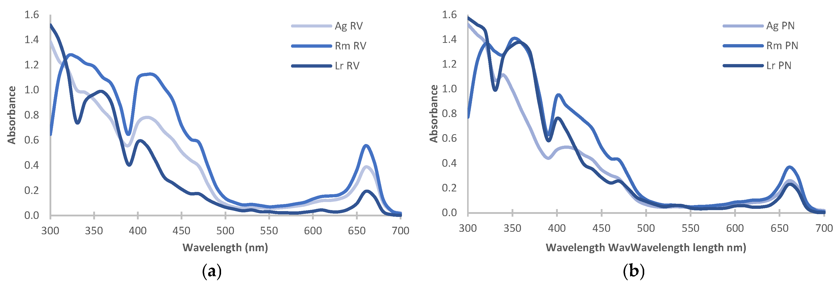
The estimation of chlorophylls (chlorophyll a, chlorophyll b, and total chlorophyll) and carotenoids by UV-Vis spectroscopy is presented in Table 1; the highest pigment content was recorded in R. mangle. Similarly, UV-Vis spectroscopy allowed us to establish that RV extracts register hyperchromic effects in the λ: 640–690 nm region, and hypochromic effects (λ: 400–450 nm) were documented in PN extracts (Figure 2).
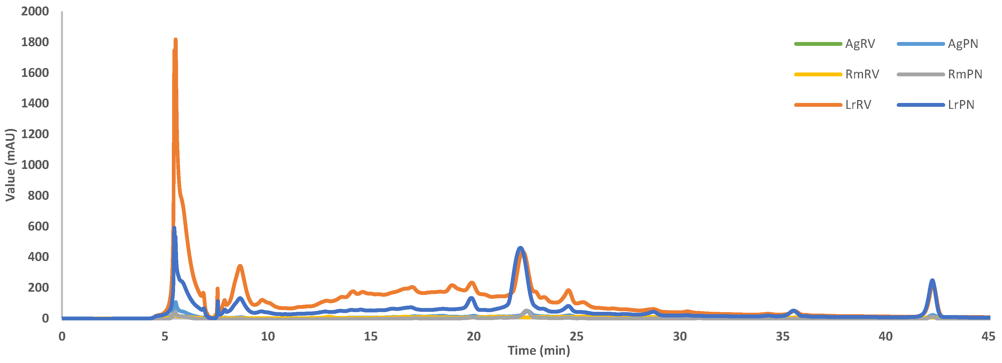
To expand the information about the metabolic content of the species, the chromatographic profile indicated that the extracts of L. racemosa showed the greatest metabolic diversity, recording the highest number of peaks (n = 17; AUC = 2253.26; 50.61%); the most intense peaks were at 5.48, 8.64, 17, and 22.32 min (33.52% AUC; Figure 3). The chromatograms of A. germinans and R. mangle demonstrated a lower abundance of peaks and low-intensity peaks. The HPLC analysis (λ = 272 nm) indicated that GA and Q peaked at 5.48 and 24.6 min, respectively (Figure S1); however, the presence of other flavonoids was not ruled out [30]. The AUC showed that GA was 8.45 times more abundant than Q (AUC = 665.14 vs. 78.7) in L. racemosa-RV extract; this sample had 3.17 and 2.27 times more GA and Q than L. racemosa-PN extract. Finally, caffeic acid was related to the peaks located between 15 and 17 min [30].
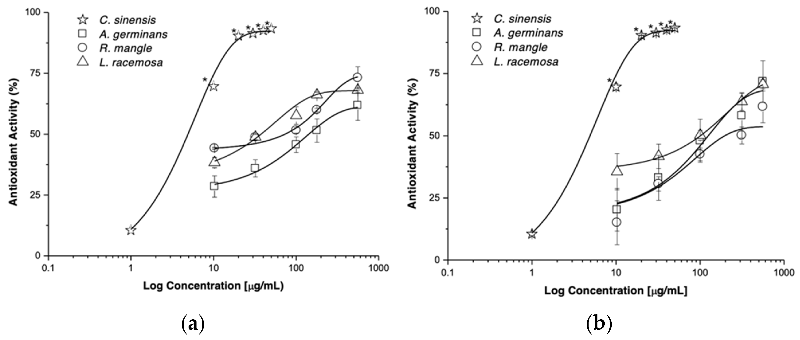
Additionally, the presence of phenols and flavonoids did not register significant variations between species or between collection sites (Table 2). On the other hand, a positive correlation was recorded between phenols content and salinity at both the PN (Y = −14.56 + 1.091x; R2 = 0.99; p = 0.012) and RV (Y = 7.43 + 0.33x; R2 = 0.99; p = 0.046) sites. The antioxidant activity exerted by the extracts of A. germinans, R. mangle, and L. racemosa varied in a concentration-dependent manner (Table 2, Figure 4), and L. racemosa-RV extract (EC50: 39.74 ± 0.91 μg/mL) presented the highest antioxidant potency.
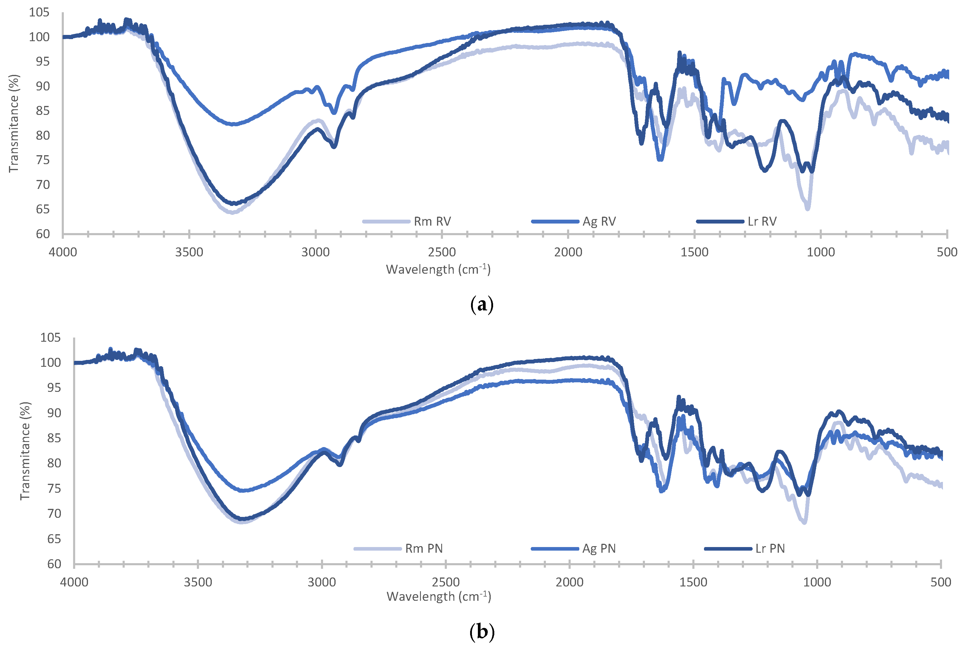
FTIR analysis of methanolic extracts of R. mangle, A. germinans, and L. racemosa was used to record variations in the banding pattern in the mid-infrared region (Figure 5). The bands at 3328.78 ± 2.49 cm−1 were related to R-OH narrowing in water, alcohols, phenols, flavonoids, and amines were corroborated with the bands at 1370 and 1221 cm−1 assigned to phenolic hydroxyls [36,37]. Bands at 2929.38 ± 2.71 and 2851.52 ± 1.32 cm−1 corresponded to narrowing and deforming vibrations of methyl and methylene groups [38,39], the band at 1471 cm−1 to the bending of methylene [40], and the band at 1402.5 ± 1.36 cm−1 to the symmetrical scissor and rocking bending of methyl groups [41]; these bands suggest the presence of lipids in the extracts. Bands at ±1700, 1617, and 1230 cm−1 indicated the presence of carbonyl carbons, aliphatic amines (asymmetric stretching of N-H), and esters, respectively [42]. Bands at 1035.8, 1052.2, and 1071.8 cm−1 correspond to vibrations of C-O-C bonds, which are related to the presence of heterosides. The band at 982.6 was related to -C = C-H narrowing vibrations [43].
The presence of chlorophylls was confirmed with bands at 1657, 1640, and 1549 cm−1, which are related to carbonyl carbons (C13) of the E ring, to the aldehyde of the B ring and the C = C bonds of the macrocycle [44]. Carotenoids were identified with the bands at 2924, 2854, 1643, and 1510 cm−1 [45]. Quercetin was identified with bands at 3248, 1670, 1500, 1000, and 650 cm−1 [46], Gallic acid with bands at 1654, 1100, 1025, 763, and 669 cm−1 [47], and caffeic acid with bands at 1645, 1625, 1530, 1450, 1280, 1217, 1120, 815, and 648 cm−1 [48].
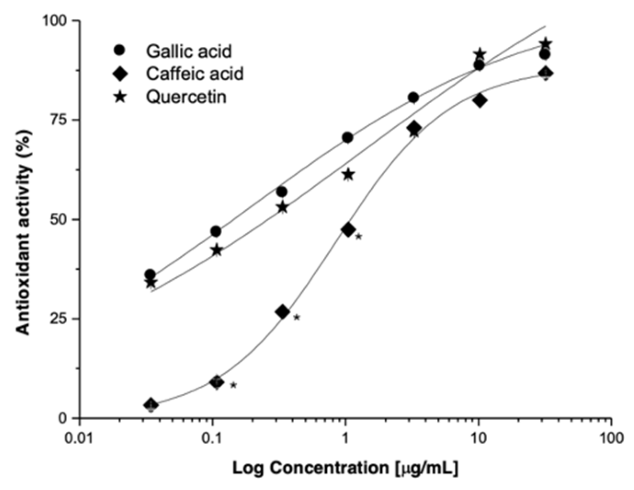
The participation of caffeic acid (CA), gallic acid (GA), and quercetin (Q) in the antioxidant activity was evaluated in the DPPH model. CA, GA, and Q exert an antioxidant activity in a concentration-dependent manner (Figure 6, Table 3). The potency of Q was >300 times higher than L. racemosa-RV extract, and the efficacy increased by ±25%. It is important to highlight the antioxidant activity exerted by GA and CA (p < 0.05).

4. Discussion

Previous studies indicate that methanolic extracts from different mangrove species (R. mangle, A. germinans, and L. racemosa) exert a concentration-dependent antioxidant effect [49]. However, the mangrove is a community susceptible to changes in precipitation, temperature, and salinity, among other factors, and these are reflected in photosynthetic responses [50], in the production of leaf litter, hypocotyls [51], propagules, leaves, and flowers [2], among other phenotypic manifestations. Factors such as phenological development, seasonality, time, temperature, polarity of solvents, and extraction methods, among other aspects, influence the raw materials obtained from wild species.
In Río Verde (RV), mesohaline and hypoxic–oxic conditions are observed in the forest along the coastline. Further inland, the forest exhibits euhaline and hypoxic–anoxic conditions. In Petén Neyac (PN), mesohaline and oxic environments are recorded in the coastal forest, while further inland, euhaline and hypoxic conditions are observed. These conditions regulate the physiological processes that allow adaptation to salinity and capture atmospheric oxygen, that are reflected in changes to the forest’s structure and functionality [52]. Hence, it is important to understand the plants’ metabolism, before and after their harvest.
The gravimetric analysis of the extracts derived from samples collected in RV and PN allows the measurement of changes in the amount of extractable material from the leaves of A. germinans, R. mangle, and L. racemosa. Qualitative phytochemical analysis is used to characterize phytochemicals [53] to identify various families of secondary metabolites. However, chemical reactions do not achieve chemical differentiation.
UV-Vis spectroscopy showed absorption changes in the regions of λ: 400–450 and λ: 640–690 nm, which are related to carotenoids and chlorophylls [54]. The peak between 410–430 nm is related to chlorophyll (Abs: 410, 430, and 662 nm) and to degradation products such as chlorophyllide (Abs: 412, 431, and 662 nm; loss of phytol) and pheophytin (Abs: 410 nm; Mg2+ loss) [55]. The absence of the shoulder at λ: 430 nm in the L. racemosa spectrum supports differences in the chlorophyll degradation process that can be extrapolated to the metabolic content. In this context, analysis is required under other experimental conditions that avoid degradation and guarantee the presence of pigments [56], among other chemical entities.
The chromatographic profile confirmed the complexity of the extracts. L. racemosa presented the greatest metabolic diversity, compared to A. germinans and R. mangle. Gallic acid, caffeic acid, and quercetin were detected at 272 nm. Other phenolic acids (methyl gallate, syringic acid, p-coumaric acid, synaptic acid, and ferulic acid) and flavonoids (catechin, rutin, myricetin, apigenin, and kaempferol) have also been detected at that wavelength and showed different retention times [30]. In this context, salinity had a favorable impact on the biosynthesis of gallic acid, among other metabolites of a phenolic nature (L. racemosa-RV vs. L. racemosa-PN; Figure 3). Future analytical experiments (e.g., HPLC-MS) are required to confirm the presence and abundance of these metabolites. The analysis of the functional group and fingerprint region of the FTIR spectrum allows us to confirm the variation of the chemical content of the samples under study.
The variations in extraction yield and the estimation of chlorophyll a/b are related to the stomatal system and photorespiration, due to the differences in the photosynthetic adaptations of R. mangle, A. germinans, and L. racemosa. The leaves of L. racemosa are amphistomatic, with high stomatal density (86.1 and 55.3/mm2 upper and lower) and structural and organizational differences in the guard cells [57,58], in hypostomatic leaves of R. mangle (74.2 stomata/mm2) and A. germinans (86.1 stomata/mm2), suggesting better management of salinity through control of stomatal opening, carbon gain and instantaneous water use efficiency (WUE) [59]. Situations that modify the flow of O2, CO2, water, the Calvin cycle, and production of 3-phosphoglycerate to be used in central metabolic pathways [60].
Salinity is an abiotic stressor and activator of the shikimate pathway, example of a metabolic defense pathway in the presence of reactive oxygen species [61]. Aromatic amino acids, cinnamic acids, and their derivatives result from the shikimate pathway, metabolites recognized for their antioxidant properties, among other health benefits [62]. An increase in the content of phenols and flavonoids has been reported in plants (e.g., Lepidium sativum, Crocus sativus, and Schizonepeta tenuifolia) subjected to salt stress conditions [63,64,65]. In the context of the Campeche mangrove, it is important to establish the experimental basis regarding the impact of salinity on the metabolic profile, to understand the physiological, ecological, and phytochemical mechanisms that are applicable to the search for new drugs. Pharmacological effects will depend on chemical content, the abundance of metabolites, dosage, and frequency of use.
Many effects induced by natural products (NP) are consistent with Concentration-Response Curves (CRC). This model allows the determination of the pharmacological parameters of potency (EC50) and efficacy (Emax) induced by the extracts, metabolites, and positive controls. Extracts and NP derived from mangrove species induce concentration-dependent antioxidant effects, and the antioxidant effect of L. racemosa-RV is three times more potent than that of L. racemosa-PN (Table 2). The antioxidant effect of NP is proportional to NP-DPPH complex concentration. The maximum effect is reached when an NP occupies DPPH molecules. The shape and shift of CRCs are related to the disparity in the affinities of the NP to donate electrons to neutralize the DPPH radical. CA and Q can be used as biomarkers of L. racemosa-RV standardized extracts and will contribute to the safe and effective use of the region’s plants.
Changes in antioxidant activity are related to biological variability, the collection site, post-harvest processes (e.g., drying), handling (light, temperature, extraction, and storage), and other oxidative conditions [66]. Antioxidant activity depends on hydroxylation patterns in aromatic systems, carbonyl carbons (aromatic acids, esters, or lactones), and methyl esters of phenolic acids’ presence [67]. Also, the Folin–Ciocalteu reaction (FCR) is not specific to phenolic compounds, as the FCR reduces and reacts with various non-phenolic compounds [68]. Gallic acid was found to be one of the most abundant and important metabolites in the induction of the antioxidant effect by methanolic extract of L. racemosa-RV. However, other metabolites with antioxidant activity have also been observed. This opens the opportunity for the identification of other active metabolites from L. racemosa-RV, as well as evaluation of the synergy in the induction of antioxidant activity. Therefore, more information is required regarding the analytical phytochemical profile of mangrove species for later use in the physiological, ecological, and pharmacological contexts, among others.

5. Conclusions

Through an experimental matrix (two sites, three species) that generates chemical, chromatographic, and spectroscopic information, the present work documents for the first time the influence of salinity on R. mangle, A. germinans, and L. racemosa grown in Río Verde and Petén Neyac, RBLP, Campeche, México. In this context, MeOH extracts of L. racemosa (RV) leaves are well-suited for a bioguided phytochemical study to provide valuable insights into their medicinal properties, promote ecological conservation, and stimulate innovation across various industries. GA is the main metabolite affected by salinity and participates in antioxidant activity, which could explain the physiological adaptations of L. racemosa and its traditional uses. However, other metabolites could act synergistically in situations of physiological stress. Further analytical studies are needed to confirm the impact of salinity on the biosynthesis of secondary metabolites, as well as phytochemical and pharmacological studies that allow the development of standardized extracts from L. racemosa-RV, among other value products.

Supplementary Materials

The following supporting information can be downloaded at: https://www.mdpi.com/article/10.3390/plants14050800/s1, Figure S1 Chromatogram profile obtained by HPLC of gallic acid (GA; t = 5.48 min) and quercetin (Q; t = 24.6 min) at λ = 272 nm. Mobile phase: Sol A (H2O-acetic acid 1%), Sol B (ACN 100%); 10–40% (0–28 min): 40–60% (28–39 min); 60–90% (39–50 min). Flux: 0.5 mL/min.

Author Contributions

Conceptualization: F.J.A.-C. and C.A.C.-K. Data curation: C.A.C.-K., M.A.G.-P., M.A.P.-C. and J.L.A.-G. Formal analysis: F.J.A.-C., E.H.-N. and C.A.C.-K. Funding acquisition: F.J.A.-C. Investigation: E.H.-N., M.A.P.-C., E.J.G.A., M.A.G.-P., C.A.C.-K. and F.J.A.-C. Methodology: F.J.A.-C., E.H.-N. and C.A.C.-K. Project administration: F.J.A.-C., C.M.A.-H. and E.J.G.A. Resources: F.J.A.-C., C.M.A.-H. and E.J.G.A. Supervision. F.J.A.-C., E.H.-N. and R.A.P.-B. Validation: R.A.P.-B., M.A.G.-P. and M.A.P.-C. Visualization: C.A.C.-K., M.A.P.-C., M.A.G.-P. and J.L.A.-G. Writing—original draft: C.A.C.-K., F.J.A.-C., J.L.A.-G., C.M.A.-H., R.A.P.-B., E.J.G.A., M.A.P.-C., M.A.G.-P. and E.H.-N. Writing—review and editing: C.A.C.-K., F.J.A.-C., J.L.A.-G., C.M.A.-H., R.A.P.-B., E.J.G.A., M.A.P.-C., M.A.G.-P. and E.H.-N. All authors have read and agreed to the published version of the manuscript.

Funding

This research received no external funding.

Data Availability Statement

All data generated and analyzed in this work are included in this manuscript.

Acknowledgments

All authors would like to thank the “Facultad de Ciencias Químico-Biológicas and the Instituto de Ecología, Pesquería del Golfo de México” from “Universidad Autónoma de Campeche” for the laboratory facilities, equipment, leaf collection support, and buildings used to develop this work (Project: 035/UAC/2023). We thank Iliana G. Osorio Horta (FCQB-UAC) and Julio Enrique Oney Montalvo (ITESCAM-Calkiní) for their technical support and the analysis via HPLC, respectively.

Conflicts of Interest

The authors declare no conflicts of interest.

References

  1. Kathiresan, K.; Bingham, B.L. Biology of Mangroves and Mangrove Ecosystems. Adv. Mar. Bio. 2001, 40, 81–251. [Google Scholar] [CrossRef]
  2. Chan-Keb, C.A.; Agraz-Hernández, C.M.; Muñiz-Salazar, R.; Posada-Vanegas, G.; Osti-Sáenz, J.; Reyes Castellano, J.E.; Conde-Molina, K.P.; Vega-Serratos, B.E. Ecophysiological response of Rhizophora mangle to the variation in hydrochemistry during five years along the coast of Campeche, México. Diversity 2018, 10, 9. [Google Scholar] [CrossRef]
  3. Mitra, A.; Chowdhury, R.; Sengupta, K.; Banerjee, K. Impact of salinity on mangroves of Indian Sundarbans. J. Coast. Environ. 2010, 1, 71–82. [Google Scholar]
  4. Mariam, H.; Alamgir, A.N.M. Flowering and Fruiting Behavior, Seed Germination, and Survival Status of Heritiera fomes Buch-Ham and Excoecaria agallocha L. Species in the Sundarban Mangrove Forest of Bangladesh. Open Access Libr. J. 2022, 9, 1–11. [Google Scholar] [CrossRef]
  5. Nabeelah Bibi, S.; Fawzi, M.M.; Gokhan, Z.; Rajesh, J.; Nadeem, N.; Kannan, R.R.R.; Albuquerque, R.D.D.G.; Pandian, S.K. Ethnopharmacology, Phytochemistry, and Global Distribution of Mangroves―A Comprehensive Review. Mar. Drugs 2019, 17, 231. [Google Scholar] [CrossRef] [PubMed]
  6. Pastene, E.R. Current state of the search for plants with antioxidant activity. Bol. Latinoam. Caribe Plant. Med. Aromat. 2009, 8, 449–455. [Google Scholar]
  7. Flores-Verdugo, F.; González-Farías, F.; Ramírez-Flores, O.; Amezcua-Linares, F.; Yáñez-Arancibia, A.; Day, J.W. Mangrove ecology, aquatic primary productivity, and fish community dynamics in the Teacapán-Agua Brava lagoon-estuarine system (Mexican Pacific). Estuaries 1997, 20, 276–290. [Google Scholar] [CrossRef]
  8. Acosta-Lugo, E.; Alonso-Parra, D.; Andrade-Hernández, M.; Castillo-Tzab, D.; Chablé-Santos, J.; Durán-García, R.; Espadas-Manrique, C.; Fernández-Stohanzlova, I.; Fraga-Berdugo, J.; Galicia, E.; et al. Plan de Conservación de la eco-región Petenes-Celestún-Palmar. Universidad Autónoma de Campeche, Pronatura Peninsula de Yucatan AC: Campeche, México, 2010. Available online: http://etzna.uacam.mx/epomex/pdf/Eco-region.pdf#page=9 (accessed on 19 February 2025).
  9. Zamora-Crescencio, P.; Mas, J.F.; Rico-Gray, V.; del Rosario Domínguez-Carrasco, M.; Villegas, P.; Gutiérrez-Báez, C.; Barrientos-Medina, R.C. Arboreal composition and structure of the petenes of the Petenes Biosphere Reserve, Campeche, Mexico. Polibotánica 2015, 39, 1–19. Available online: http://www.polibotanica.mx/pdf/pb39/compo.pdf (accessed on 19 February 2025).
  10. Oliva, M.; Montiel, S. Stakeholder linkage in conservation strategies: A qualitative tool for improving the management of a biosphere reserve in the Yucatan Peninsula, Mexico. Trop. Conserv. Sci. 2016, 9, 423–438. [Google Scholar] [CrossRef]
  11. Méndez-Cabrera, F.; Montiel, S. Preliminary assessment of the wild fauna and flora used by the Mayan populations of two coastal communities in Campeche, Mexico. Univ. Cienc. 2007, 23, 127–139. [Google Scholar]
  12. Agraz-Hernández, C.M. Estudio de caso: Usos y beneficios ecológicos, económicos y sociales que proporcionan los ecosistemas de manglar en el estado de Campeche. In La Biodiversidad en Campeche: Estudio de Estado; Villalobos-Zapata, G.J., Mendoza-Vega, J., Eds.; Comisión Nacional Para el Conocimiento y Uso de la Biodiversidad, Gobierno del Estado de Campeche, Universidad Autónoma de Campeche, El Colegio de la Frontera Sur: Campeche, México, 2010; pp. 470–475. [Google Scholar]
  13. Agraz-Hernández, C.M.; García-Zaragoza, C.; Iriarte-Vivar, S.; Flores-Verdugo, F.J.; Moreno-Casasola, P. Forest structure, productivity and species phenology of mangroves in the La Mancha lagoon in the Atlantic coast of Mexico. Wetl. Ecol. Manag. 2011, 19, 273–293. [Google Scholar] [CrossRef]
  14. Bandaranayake, W.M. Bioactivities, bioactive compounds, and chemical constituents of mangrove plants. Wetl. Ecol. Manag. 2002, 10, 421–452. [Google Scholar] [CrossRef]
  15. Rafii, Z.A.; Dodd, R.S.; Fromard, F. Biogeographic variation in foliar waxes of mangrove species. Biochem. Syst. Ecol. 1996, 24, 341–345. [Google Scholar] [CrossRef]
  16. Neves-Costa, F.; Jerz, G.; Hewitson, P.; de Souza-Figueiredo, F.; Ignatova, S. Laguncularia racemosa phenolics profiling by three-phase solvent system step-gradient using high-performance countercurrent chromatography with off-line electrospray mass-spectrometry detection. Molecules 2021, 26, 2284. [Google Scholar] [CrossRef]
  17. Glasenapp, Y.; Korth, I.; Nguyen, X.V.; Papenbrock, J. Sustainable use of mangroves as sources of valuable medicinal compounds: Species identification, propagation and secondary metabolite composition. S. Afr. J. Bot. 2019, 121, 317–328. [Google Scholar] [CrossRef]
  18. Koch, B.P.; Rullkötter, J.; Lara, R.J. Evaluation of triterpenols and sterols as organic matter biomarkers in a mangrove ecosystem in northern Brazil. Wetl. Ecol. Manag. 2003, 11, 257–263. [Google Scholar] [CrossRef]
  19. Koch, B.P.; Harder, J.; Lara, R.J.; Kattner, G. The effect of selective microbial degradation on the composition of mangrove derived pentacyclic triterpenols in surface sediments. Org. Geochem. 2005, 36, 273–285. [Google Scholar] [CrossRef]
  20. He, D.; Ladd, S.N.; Sachs, J.P.; Jaffé, R. Inverse relationship between salinity and 2H/1H fractionation in leaf wax n-alkanes from Florida mangroves. Org. Geochem. 2017, 110, 1–12. [Google Scholar] [CrossRef]
  21. Wu, J.; Xiao, Q.; Xu, J.; Li, M.Y.; Pan, J.Y.; Yang, M.H. Natural products from true mangrove flora: Source, chemistry and bioactivities. Nat. Prod. Rep. 2008, 25, 955–981. [Google Scholar] [CrossRef]
  22. Rodrigues, C.F.B.; Gaeta, H.H.; Belchor, M.N.; Ferreira, M.J.P.; Pinho, M.V.T.; de Oliveira Toyama, D.; Toyama, M.H. Evaluation of potential thrombin inhibitors from the white mangrove (Laguncularia racemosa (L.) CF Gaertn.). Mar. Drugs 2015, 13, 4505–4519. [Google Scholar] [CrossRef]
  23. Agraz-Hernández, C.; Noriega-Trejo, R.; López-Portillo, J.; Flores-Verdugo, F.; Jiménez-Zacarías, J. Guía de Campo. Identificación de los Manglares en México; Centro EPOMEX-UAC: Campeche, México, 2006. [Google Scholar]
  24. Comisión Permanente de la Farmacopea de los Estados Unidos Mexicanos. Farmacopea Herbolaria de los Estados Unidos Mexicanos. 3.0; Secretaría de Salud CDMX México: Ciudad de México, México, 2021. [Google Scholar]
  25. Robles-García, M.A.; Aguilar-Antonio, J.; Gutiérrez-Lomelí, M.; Rodríguez-Félix, F.; Morales-Del-Río, J.A.; Guerrero-Medina, P.J.; Madrigal-Pulido, J.A.; Del-Toro-Sánchez, C.L. Qualitative identification of secondary metabolites and cytotoxicity determination of tempisque extracts (Sideroxylum capiri pittier). Biotecnia 2016, 18, 3–8. [Google Scholar] [CrossRef]
  26. García-Granados, R.U.; Cruz-Sosa, F.; Alarcón-Aguilar, F.J.; Nieto-Trujillo, A.; Gallegos-Martínez, M.E. Qualitative phytochemical analysis of the aqueous extracts of Thalassia testudinum Banks ex Köning et Sims from the area of Champoton, Campeche, Mexico, during the annual cycle 2016–2017. Polibotánica 2019, 48, 151–168. [Google Scholar] [CrossRef]
  27. Mohy El-Din, S.M.; Mohyeldin, M.M. Component analysis and antifungal activity of the compounds extracted from four brown seaweeds with different solvents at different seasons. J. Ocean Univ. China 2018, 17, 1178–1188. [Google Scholar] [CrossRef]
  28. Aguirre-Crespo, F.J.; Cerino-Pérez, E.; Valdovinos-Estrella, J.D.C.; Maldonado-Velázquez, M.G.; Ortega-Morales, B.O.; Zamora-Crescencio, P.; Hernandez-Nuñez, E.; Estrada-Soto, S.E. Vasorelaxant and Antioxidant Activity of Some Medicinal Plants from Campeche, Mexico. Pharmacogn. Mag. 2021, 17, 23–30. [Google Scholar] [CrossRef]
  29. Cu-Quiñones, L.D. Desarrollo de un Prototipo de Forma Farmacéutica a Partir de Las Hojas de Jatropha gaumeri Greenm (pomol che). Master’s Thesis, Universidad Autónoma de Campeche, Campeche, México, 2021. [Google Scholar]
  30. Seal, T. Quantitative HPLC analysis of phenolic acids, flavonoids and ascorbic acid in four different solvent extracts of two wild edible leaves, Sonchus arvensis and Oenanthe linearis of North-Eastern region in India. J. Appl. Pharm. Sci. 2016, 6, 157–166. [Google Scholar] [CrossRef]
  31. Lichtenthaler, H.K.; Buschmann, C. Chlorophylls and carotenoids: Measurement and characterization by UV-VIS spectroscopy. Curr. Protoc. Food Anal. Chem. 2001, 1, F4-3. [Google Scholar] [CrossRef]
  32. Mediani, A.; Abas, F.; Tan, C.P.; Khatib, A. Effects of different drying methods and storage time on free radical scavenging activity and total phenolic content of Cosmos caudatus. Antioxidants 2014, 3, 358–370. [Google Scholar] [CrossRef]
  33. Aguirre-Crespo, F.J.; Aragón-Gastélum, J.L.; Gutiérrez-Alcántara, E.J.; Zamora-Crescencio, P.; Gómez-Galicia, D.L.; Alatriste-Kurzel, D.R.; Alvarez, G.; Hernández-Núñez, E. β-Sitosterol Mediates Gastrointestinal Smooth Muscle Relaxation Induced by Coccoloba uvifera via Muscarinic Acetylcholine Receptor Subtype 3. Sci. Pharm. 2024, 92, 19. [Google Scholar] [CrossRef]
  34. Stratil, P.; Klejdus, B.; Kubáň, V. Determination of total content of phenolic compounds and their antioxidant activity in vegetables evaluation of spectrophotometric methods. J. Agric. Food Chem. 2006, 54, 607–616. [Google Scholar] [CrossRef]
  35. Zar, J.H. Biostatistical Analysis, 3rd ed.; Prentice Hall: Upper Anhe, NJ, USA, 1996; p. 662. [Google Scholar]
  36. Castorena-García, J.H.; Rojas-López, M.; Delgado-Macuil, R.; Robles de la Torre, R.R. Análisis de pulpa y aceite de aguacate con espectroscopia infrarroja. Concienc. Tecnológica 2011, 42, 5–10. [Google Scholar]
  37. Zhang, J.; Kamdem, D. FTIR Characterization of Copper Ethanolamine—Wood Interaction for Wood Preservation. Holzforschung 2000, 54, 119–122. [Google Scholar] [CrossRef]
  38. Hu, Y.; Pan, Z.J.; Liao, W.; Li, J.; Gruget, P.; Kitts, D.D.; Lu, X. Determination of antioxidant capacity and phenolic content of chocolate by attenuated total reflectance-Fourier transformed-infrared spectroscopy. Food Chem. 2016, 202, 254–261. [Google Scholar] [CrossRef]
  39. Ng, S.; Lasekan, O.; Muhammad, K.; Sulaiman, R.; Hussain, N. Effect of roasting conditions on color development and Fourier transform infrared spectroscopy (FTIR-ATR) analysis of Malaysian-grown tropical almond nuts (Terminalia catappa L.). Chem. Cent. J. 2014, 8, 55. [Google Scholar] [CrossRef] [PubMed]
  40. Rivera, W.; Velasco, X.; Rincón, C.A. TGA and FTIR Evaluation of Composition Changes Produced by Roasting of Coffee Beans. Rev. Colomb. Fís. 2013, 45, 205–208. [Google Scholar]
  41. Movasaghi, Z.; Rehman, S.; ur Rehman, I. Fourier Transform Infrared (FTIR) Spectroscopy of Biological Tissues. Appl. Spectrosc. Rev. 2008, 43, 134–179. [Google Scholar] [CrossRef]
  42. Nandiyanto, A.B.D.; Oktiani, R.; Ragadhita, R. How to read and interpret FTIR spectroscope of organic material. Indones. J. Sci. Technol. 2019, 4, 97–118. [Google Scholar] [CrossRef]
  43. Mondragon-Cortez, P. Espectroscopía de Infrarrojo Para Todos…y 51 Espectros de Alimentos Consumidos en México; Centro de Investigación y Asistencia en Tecnología y Diseño del Estado de Jalisco, AC.: Jalisco, México, 2017; pp. 115–162. Available online: https://ciatej.mx/files/el_ciatej/comunicacion/divulgacion/espectroscopia.pdf (accessed on 19 February 2025).
  44. Lewis, N.H.; Fleming, G.R. Two-dimensional electronic-vibrational spectroscopy of chlorophyll a, and b. J. Phys. Chem. Lett. 2016, 7, 831–837. [Google Scholar] [CrossRef]
  45. Singh, A.; Ahmad, S.; Ahmad, A. Green extraction methods and environmental applications of carotenoids-a review. RSC Adv. 2015, 5, 62358–62393. [Google Scholar] [CrossRef]
  46. Guzansky-Milanezi, F.; Martins-Meireles, L.; Malavazi de Christo-Scherer, M.; de Oliveira, J.P.; Romero da Silva, A.; Lamas de Araujo, M.; Coutinho-Endringer, D.; Fronza, M.; Cunegundes-Guimarães, M.C.; Scherer, R. Antioxidant, antimicrobial and cytotoxic activities of gold nanoparticles capped with quercetin. Saudi Pharm. J. 2019, 27, 968–974. [Google Scholar] [CrossRef]
  47. Vijayalakshmi, R.; Ravindhran, R. Comparative fingerprint and extraction yield of Diospyrus ferrea (willd.) Bakh. root with phenol compounds (gallic acid), as determined by uv–vis and ft–ir spectroscopy. Asian Pac. J. Trop. Biomed. 2012, 2, S1367–S1371. [Google Scholar] [CrossRef]
  48. Catauro, M.; Barrino, F.; Dal Poggetto, G.; Crescente, G.; Piccolella, S.; Pacifico, S. New SiO2/caffeic acid hybrid materials: Synthesis, spectroscopic characterization, and bioactivity. Materials 2020, 13, 394. [Google Scholar] [CrossRef] [PubMed]
  49. Aguirre-Crespo, F.J.; Cu-Quiñones, L.D.; Popoca-Cuaya, M.A.; Maldonado-Velázquez, M.G.; Chan-Keb, C.; Graz-Hernández, C.M.; Hernández-Núñez, E. Análisis fitoquímico preliminar y valoración de la actividad antioxidante de tres especies de Mangle de Campeche, México. Quim. Hoy 2022, 10, 1–5. [Google Scholar] [CrossRef]
  50. Lopes, D.M.S.; Tognella, M.M.P.; Falqueto, A.R.; Soares, M.L.G. Salinity variation effects on photosynthetic responses of the mangrove species Rhizophora mangle L. growing in natural habitats. Photosynthetica 2019, 57, 1142–1155. [Google Scholar] [CrossRef]
  51. Agraz Hernández, C.M.; Chan Keb, C.A.; Iriarte-Vivar, S.; Posada Venegas, G.; Vega Serratos, B.; Osti Sáenz, J. Phenological variation of Rhizophora mangle and ground water chemistry associated to changes of the precipitation. Hidrobiológica 2015, 25, 49–61. [Google Scholar]
  52. Agraz-Hernández, C.M.; Chan-Keb, C.A.; Muñiz-Salazar, R.; Pérez-Balan, R.A.; Vanegas, G.P.; Manzanilla, H.G.; Osti-Sáenz, J.; del Río Rodríguez, R. Pore water chemical variability and its effect on phenological production in three mangrove species under drought conditions in southeastern Mexico. Diversity 2022, 14, 668. [Google Scholar] [CrossRef]
  53. Diaz-Villagómez, D.G.; Hernández-Ramírez, R.E.; Cruz-Cruz, D.; Villegas-Gómez, C. Análisis fitoquímico: Una visión integral de los métodos de extracción de productos naturales. Nat. Tecnol. 2023, 10, 22–35. Available online: http://repositorio.ugto.mx/bitstream/20.500.12059/10156/1/473-1463-1-PB.pdf (accessed on 19 February 2025).
  54. Grassino, M.; Batstone, D.J.; Yong, K.W.; Capson-Tojo, G.; Hülsen, T. Method development for PPB culture screening, pigment analysis with UPLC-UV-HRMS vs. spectrophotometric methods, and spectral decomposition-based analysis. Talanta 2022, 246, 123490. [Google Scholar] [CrossRef]
  55. Milenković, S.M.; Zvezdanović, J.B.; Anđelković, T.D.; Marković, D.Z. The identification of chlorophyll and its derivatives in the pigment mixtures: HPLC-chromatography, visible and mass spectroscopy studies. Adv. Technol. 2012, 1, 16–24. [Google Scholar]
  56. Eckardt, N.A.A. A new chlorophyll degradation pathway. Plant Cell 2009, 21, 700. [Google Scholar] [CrossRef]
  57. Pelozo, A.; T-Boeger, M.R.; Sereneski-de-Lima, C.; Soffiatti, P. Leaf morphological strategies of seedlings and saplings of Rhizophora mangle (Rhizophoraceae), Laguncularia racemosa (Combretaceae) and Avicennia schaueriana (Acanthaceae) from Southern Brazil. Rev. Biol. Trop. 2016, 64, 305–317. [Google Scholar] [CrossRef]
  58. Erickson, A.A.; Saltis, M.; Bell, S.S.; Dawes, C.J. Herbivore feeding preferences as measured by leaf damage and stomatal ingestion: A mangrove crab example. J. Exp. Mar. Biol. Ecol. 2003, 289, 123–138. [Google Scholar] [CrossRef]
  59. Sobrado, M. Leaf characteristics and gas exchange of the mangrove Laguncularia racemosa as affected by salinity. Photosynthetica 2005, 43, 217–221. [Google Scholar] [CrossRef]
  60. Taiz, L.; Zeiger, E. Plant Physiology; Sinauer Associates: Sindiran, MA, USA, 2010. [Google Scholar]
  61. Lee, S.J.; Jeong, E.M.; Ki, A.Y.; Oh, K.S.; Kwon, J.; Jeong, J.H.; Chung, N.J. Oxidative defense metabolites induced by salinity stress in roots of Salicornia herbacea. J. Plant Physiol. 2016, 206, 133–142. [Google Scholar] [CrossRef] [PubMed]
  62. Quiñones, M.; Miguel, M.; Aleixandre, A. Los polifenoles, compuestos de origen natural con efectos saludables sobre el sistema cardiovascular. Nutr. Hosp. 2012, 27, 76–89. [Google Scholar] [CrossRef]
  63. Aarland, R.; Castellanos-Hernandez, O.; Rodriguez-Sahagun, A.; Acevedo-Hernandez, G. Efecto del estrés salino sobre la morfología y fitoquímica de orégano mexicano (Lippia graveolens Kunth) cultivado in vitro. Biotecnia 2020, 22, 131–137. [Google Scholar] [CrossRef]
  64. Mzabri, I.; Legsayer, M.; Kouddane, N.; Boukroute, A.; Berrichi, A. Salt Stress Effects on Some Morphological, Physiological and Biochemical Parameters of Saffron Plant (Crocus sativus L.) in Eastern Morocco. J. Mater. 2017, 8, 4894–4901. Available online: http://www.jmaterenvironsci.com/Document/vol8/vol8_NS/519-JMES-3824-Mzabri.pdf (accessed on 21 June 2024).
  65. Zhou, Y.; Tang, N.; Huang, L.; Zhao, Y.; Tang, X.; Wang, K. Effects of Salt Stress on Plant Growth, Antioxidant Capacity, Glandular Trichome Density, and Volatile Exudates of Schizonepeta tenuifolia Briq. Int. J. Mol. Sci. 2018, 19, 252. [Google Scholar] [CrossRef]
  66. Escribano-Bailón, M.T.; Rivas-Gonzalo, J.C.; García-Estévez, I. Wine color evolution and stability. In Red Wine Technology; Academic Press: Cambridge, MA, USA, 2019; pp. 195–205. [Google Scholar] [CrossRef]
  67. Foti, M.C.; Daquino, C.; Geraci, C. Electron-transfer reaction of cinnamic acids and their methyl esters with the DPPH· radical in alcoholic solutions. J. Org. Chem. 2004, 69, 2309–2314. [Google Scholar] [CrossRef]
  68. Gülçin, İ. Antioxidant activity of food constituents: An overview. Arch. Toxicol. 2012, 86, 345–391. [Google Scholar] [CrossRef]
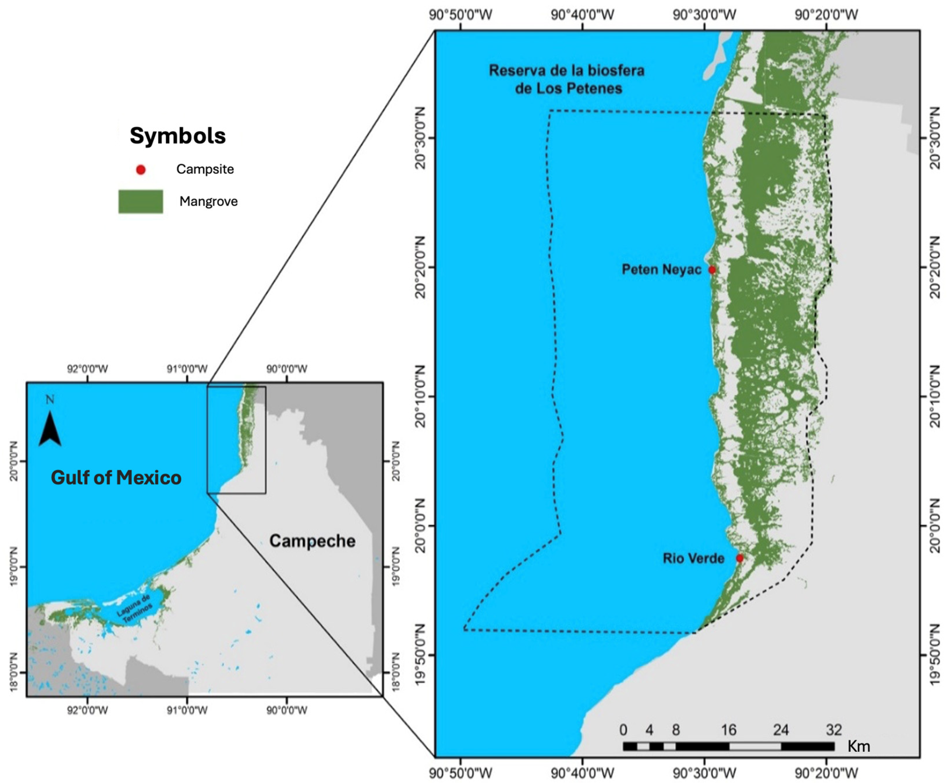
Figure 1. RBLP, Campeche, Mexico. Río Verde (RV; 19°57′27.932″ N; 90°27′4.265″ W) and Petén Neyac (PN; 20°19′48.745″ N; 90°29′23.164″ W) site colect are marked in .
Plants 14 00800 g001
Figure 2. UV-Vis spectra of the methanolic extracts from leaves of A. germinans, R. mangle, and L. racemosa collected in Río Verde (a) and Petén Neyac (b).
Plants 14 00800 g002
Figure 3. Chromatogram profile obtained by HPLC of the methanolic extracts from L. racemosa (Lr), R. mangle (Rm), and L. racemosa (Lr) leaves collected in Los Petenes Biosphere Reserve. (RBLP; Río Verde (RV) and Petén Neyac (PN)) at λ = 272 nm. Mobile phase: Sol A (H2O-acetic acid 1%), Sol B (ACN 100%); 10–40% (0–28 min): 40–60% (28–39 min); 60–90% (39–50 min). Flux: 0.5 mL/min.
Plants 14 00800 g003
Figure 4. Antioxidant activity by extracts of L. racemosa, R. mangle, and A. germinans collected in Río Verde (a) and Petén Neyac (b) of the RBLP. The results are the average ± SD of three experiments with three replicates. (* p < 0.05).
Plants 14 00800 g004
Figure 5. FTIR spectra of the methanolic extracts of the leaves of R. mangle, A. germinans, and L. racemosa collected in Río Verde (a) and Petén Neyac (b) in the RBLP.
Plants 14 00800 g005
Figure 6. Antioxidant activity induced by quercetin (Q), gallic acid (GA), and caffeic acid (CA); results expressed as the average ± SD of three experiments with three replicates each. (* p < 0.05 vs. Q).
Plants 14 00800 g006
Table 1. Extraction yield and pigment content in the methanolic extracts of leaves of R. mangle (Rm), L. racemose (Lr), and A. germinans (Ag).
SpeciesSiteSalinity (PSU)Yield (%)ChlorophyllCarotenoids (mg/g)
A
(mg/g)
B
(mg/g)
Ratio a/bTotal (mg/g)
AgRV74.87 ± 521.98 ± 0.940.16 ± 0.060.07 ± 0.032.30.23 ± 0.050.01 ± 0.0030
PN58.93 ± 318.04 ± 2.070.12 ± 0.030.05 ± 0.012.40.17 ± 0.040.008 ± 0.0010
RmRV42.12 ± 417.46 ± 0.460.26 ± 0.020.10 ± 0.012.60.35 ± 0.020.018 ± 0.0004
PN42.25 ± 221.52 ± 2.220.17 ± 0.020.06 ± 0.012.80.23 ± 0.030.013 ± 0.0010
LrRV43.25 ± 435.21 ± 0.760.09 ± 0.030.01 ± 0.0190.10 ± 0.040.005 ± 0.0040
PN40.59 ± 235.38 ± 0.140.11 ± 0.040.02 ± 0.015.50.13 ± 0.050.009 ± 0.0020
Table 2. Phenols, flavonoids, and antioxidant activity (DPPH) induced by extracts from A. germinans (Ag), R. mangle (Rm), L. racemosa (Lr). RV: Río Verde; PN: Petén Neyac.
SpeciesSitePhenols (mg/g)Flavonoids (mg/g)Potency
EC50 (μg/mL)
Efficacy
Emax (%)
AgRV0.42 ± 0.20.26 ± 0.08157.83 ± 5.4461.44 ± 3.45
PN0.63 ± 0.30.20 ± 0.08194.76 ± 16.9868.75 ± 12.5
RmRV0.64 ± 0.20.14 ± 0.0459.81 ± 5.3574.42 ± 8.95
PN0.86 ± 0.30.23 ± 0.07294.47 ± 14.1953.69 ± 9.17
LrRV0.54 ± 0.30.15 ± 0.0239.74 ± 0.9167.82 ± 1.00
PN0.75 ± 0.20.16 ± 0.13123.47 ± 4.8773.27 ± 4.56
C. sinensis---8.69 ± 0.34 *92.36 ± 0.87 *
* p < 0.05 vs. C. sinensis.
Table 3. Antioxidant activity induced by quercetin, caffeic acid, and gallic acid in the DPPH model.
ActivityCaffeic AcidGallic AcidQuercetinL. racemosa-RV
EC50 (μg/mL)1.09 ± 0.060.22 ± 0.010.12 ± 0.01 *39.74 ± 0.91
Emax (%)86.75 ± 0.2091.35 ± 0.1494.15 ± 0.11 *67.82 ± 1.00
Relative Potency−9.08−1.831−331.2
Relative Efficacy91.197.010072.0
* p < 0.05 vs. L. racemosa (RV).
Disclaimer/Publisher’s Note: The statements, opinions and data contained in all publications are solely those of the individual author(s) and contributor(s) and not of MDPI and/or the editor(s). MDPI and/or the editor(s) disclaim responsibility for any injury to people or property resulting from any ideas, methods, instructions or products referred to in the content.