Article

Phenolic-Rich Extracts of Galenia africana and Tulbaghia violacea Accelerate Keratinocyte Migration and Mitigate Oxidative Stress to Enhance Wound Healing

by
Banele Ndlovu
1,2,
Randall Fisher
2 and
Farzana Fisher (née Rahiman)
1,*
1
Skin Research Lab, Department of Medical Biosciences, University of the Western Cape, Cape Town 7535, South Africa
2
Radiation Biophysics Division, Separated Sector Cyclotron Lab., NRF iThemba LABS, Old Faure Rd, Eerste River, Cape Town 7100, South Africa
*
Author to whom correspondence should be addressed.
Plants 2025, 14(22), 3523; https://doi.org/10.3390/plants14223523
Submission received: 10 October 2025 / Revised: 10 November 2025 / Accepted: 13 November 2025 / Published: 19 November 2025

Abstract

The rising prevalence of chronic burn wounds in South Africa places a significant burden on healthcare, driving the search for cost-effective, alternative treatments. Despite their long history of traditional use for skin ailments, the specific wound healing mechanisms of South African species Galenia africana L. and Tulbaghia violacea Harv. remain scientifically unvalidated, representing a critical knowledge gap and a novel area of inquiry. This study sought to evaluate the physiological and cellular effects of these plant extracts, specifically investigating their influence on keratinocyte function and oxidative stress. Initial analysis of crude ethanolic extracts confirmed the presence of key phenolic compounds like ferulic acid and rutin in both plants, with G. africana exhibiting a stronger overall radical-scavenging activity. In vitro assays were performed on the human keratinocyte (HaCaT) cell line. Both G. africana and T. violacea extracts significantly enhanced cell viability (at 40–80 μg/mL) and demonstrably reduced intracellular reactive oxygen species (ROS) levels, indicating a protective antioxidant effect. Critically, in the scratch wound assay, both extracts significantly accelerated cell migration and wound closure, achieving 76% (G. africana) and 88% (T. violacea) closure within 24 h. These findings suggest that G. africana and T. violacea extracts act to support keratinocyte proliferation and migration while simultaneously mitigating oxidative cellular damage. This work provides an important mechanistic basis supporting the traditional use of these specific, regionally important plants and highlights their high therapeutic potential as novel, cost-effective agents to accelerate skin repair and recovery.

1. Introduction

A wound is defined as a disruption to the skin’s integrity, characterised by damage not only to the epidermis and dermis, but also extending to the underlying subcutaneous tissue, including bone and muscle [1]. Acute superficial abrasions are wounds that typically heal within a predictable timeframe, while chronic wounds fail to progress through normal healing stages and often require medical intervention [2]. Chronic wounds present a significant challenge to healthcare systems, particularly in South Africa (SA), where there is a high prevalence of burn wounds resulting from urban and rural fires [3]. Wildfires are a frequent occurrence in SA, often exacerbated by informal housing structures constructed from highly flammable materials such as wood and plastics [4]. In addition, the social and economic conditions in certain communities, such as overcrowding and limited access to fire prevention resources, contribute to the high incidence of burn injuries. The increasing costs of dermatological care and medication make hospital treatments unaffordable for many, leading them to seek alternative treatment options or delayed treatment [5]. Consequently, a delay in medical attention often results in severe infections, which can lead to conditions such as hypovolemia and septicaemia [6]. Therefore, there is an urgent need to explore alternative, cost-effective treatment options that promote faster healing.
Traditional and complementary medicine remains an essential resource, particularly in Africa, where it contributes substantially to primary healthcare services [7]. Li et al. [8] reported that the use of leaves as a wound dressing was one of the earliest forms of treatment to prevent infection, reduce pain and increase the rate of wound closure. In SA, approximately 27 million people use herbal medicine, with a significant portion of these remedies prescribed by traditional practitioners [9]. The Western Cape (WC) province has over 8650 medicinal plant species, 65% of which are endemic to the region [10]. Ethnobotanical studies have reported that traditional herbal practitioners in the WC region regularly incorporate these medicinal plants into their healing practices [11].
Some of the medicinal plants used by WC herbal practitioners include Aizoon africanum (L.) Klak (syn. Galenia africana L.) and Tulbaghia violacea Harv. [12,13]. G. africana L. is commonly known as “kraalbos”, belongs to the Aizoaceae family and is found in the WC, Eastern Cape, Northern Cape and Free State provinces [12,14]. Traditional healers have topically applied different pastes, decoctions and lotions from G. africana to treat wounds [15,16]. The ethanolic extracts of G. africana have demonstrated selective cytotoxicity in cancerous cells by significantly reducing their viability while exerting minimal effects on normal cells [16,17,18]. T. violacea, or “wild garlic”, belongs to the Amaryllidaceae family and is widely distributed in the WC, Eastern Cape and KwaZulu-Natal; it has significant medicinal and culinary applications [19,20]. Traditionally, T. violacea is used to flavour food and to treat a range of ailments, including fever, colds, hypertension and stomach problems [21]. This plant emits a strong garlic-like odour and can be crushed on the skin as an insect repellent without causing irritation [19]. Steroidal saponins isolated from T. violacea have demonstrated dermatological effects, including anti-inflammatory and antioxidant properties, which support its traditional use in wound care products [22,23].
Although ethnobotanical studies have documented the traditional cosmetic applications of G. africana and T. violacea, these reports mostly rely on anecdotal evidence, and there is limited scientific information available on their wound healing properties [24,25,26]. More rigorous scientific investigations are required to substantiate the therapeutic efficacy and elucidate the mechanisms of action of these plants as wound healing agents. Therefore, this study aimed to evaluate the wound healing and antioxidant effects of G. africana and T. violacea as potential alternative therapies.

2. Materials and Methods

2.1. Materials and Reagents

Fluorescein sodium salt (Cat no. F6377), AAPH (2,2-Azobis (2-methylpropionamidine) dihydrochloride (Cat no: 440914), Perchloric acid, Trolox (6-Hydrox-2,5,7,8-tetramethylchroman-2-carboxylic acid) (Cat no. 238831), TPTZ (2,4,6-tri [2-pyridyl]-s-triazine) (Cat no. T1253), L-Ascorbic acid (Cat no: A5960), Iron (III) chloride hexahydrate, di-sodium hydrogen orthophosphate dehydrate (Na2HPO4·2H2O) (Cat no. 5822880EM), Folin–Ciocalteus phenol reagent (Cat no. 109001), Sodium Carbonate (Cat no. 223530), gallic acid (Cat no. G7384), rutin (Cat no. PHL89270), chlorogenic acid (Cat no. C3878), Coumaric acid (Cat no. C9008), caffeic acid (Cat no: C0625), ferulic acid (Cat no: PHR1791), quercetin (Cat no. Q4951), gallic acid (Cat no. 27645), catechin (Cat no. C1251), epicatechin (Cat no. E1753) and protocatechuic acid (Cat no. 03930590) were obtained from Sigma-Aldrich (St. Louis, Mo, USA). Acetate Buffer (Cat no. 1021000), Hydrochloric acid (Cat no: 100319 LP), Hexane (Cat no. 2868040 LC), Acetone (Cat no: 1022040 LC), were purchased from Saarchem Laboratory Chemicals.

2.2. Plant Material and Extract Preparation

Commercially prepared ethanolic extracts of G. africana and T. violacea ethanolic extracts were purchased from Brenn-O-Kem (PTY) Ltd. (Wolseley, South Africa) and Zuplex Botanicals (Pty) Ltd. (Amanzimtoti, South Africa), respectively. The suppliers provided the following specifications and preparation methods for each extract:
G. africana extract: The plant material (Batch no: IBTKB01/2023) was harvested in Komaggas, Namaqua District, Northern Cape Province and identified at the Herbarium, Department of Botany, University of the Western Cape, Bellville, by Mr F. Weitz (Vries 1; herb no 6595). The leaves and shoots of G. africana were air-dried, ground and macerated in 60% ethanol at a 20% (w/v) ratio for 12 h at room temperature. The resulting 60% ethanolic extract was supplied as a stock solution with a concentration of 20 mg/mL.
T. violacea extract: The plant material (Batch no: UWCTVT-09/24) was cultivated by Muthi Futhi at its site in Dakeni, KwaZulu-Natal. The supplier confirmed the plant’s taxonomic identity. The whole plant, including aerial parts and bulbs, was chopped, sun-dried and ground into a fine powder using a hammer mill. The extract was prepared by macerating 100 g of the powder in 50% ethanol (Alco NCP 96.4%, Durban, South Africa) at room temperature for 48 h, followed by filtration through a 25 μm filter. The extract was supplied as a 100 mg/mL solution, representing a 10% (w/v) preparation.
For the phytochemical and antioxidant assays, the extracts were diluted to a final concentration of 1 mg/mL, consistent with a previous study [27]. For the cell culture experiments, the extracts were further diluted to 0.1 mg/mL, a concentration considered physiologically relevant [28].

2.3. Phytochemical Analysis

2.3.1. Total Polyphenolic Content

The total polyphenolic content (TPC) of G. africana and T. violacea ethanolic extracts were determined spectrophotometrically using the Folin–Ciocalteu method Siddiqui et al. [29]. Briefly, 25 μL of the samples and standards were mixed with 125 μL of the Folin–Ciocalteu reagent (10%) in a 96-well plate and left to stand for 5 min. Sodium carbonate solution at 100 μL (20% w/v) was then mixed with the samples, incubated for 2 h at room temperature and then read at 765 nm. Gallic acid (800 mg/mL in 10% ethanol) was used to set up the standard curve, and the total polyphenolic content of the samples was expressed as gallic acid equivalents (GAEs) in mg per gram dry weight. All the samples were analysed in triplicate.

2.3.2. Determination of Total Flavonoid Content

The flavanol content was determined using the method previously described Tyavambiza et al. [27], with minor modification. Briefly, 50 μL of the G. africana and T. violacea extracts (1 mg/mL) were mixed with 250 μL of a 50 g/L 4-Dimethylamino-cinnamaldehyde in a 96-well plate and incubated for 30 min at room temperature. The absorbance readings were read at 640 nm. Results were expressed as milligram catechin equivalent per gram (mg CE/g). The flavonol content was determined according to the method in [30] where 12.5 μL of 0.1% HCl in 95% ethanol and 225 μL of 2% Hydrochloric acid was added to 12.5 μL of the G. africana and T. violacea extracts (1 mg/mL), incubated for 30 min at room temperature. The absorbance was read at 360 nm. Quercetin (1.0 mM) was used as a standard and results were expressed as milligram quercetin equivalent per gram (mg QE/g). Total flavonoid content was determined as the sum of the quantified flavanol and flavonol fractions.

2.4. Quantitative Analysis of Extracts by High-Performance Liquid Chromatography (HPLC)

High-performance liquid chromatography (HPLC) has been used to isolate phytochemicals from plant extracts to purify, identify and quantify individual compounds [31]. These identified compounds may possess therapeutic properties, including antioxidant and anti-inflammatory activities [32]. HPLC analysis was performed using an Agilent 1200 series HPLC system (Santa Clara, CA, USA) with a UV detector. The compound separation and analysis were conducted using an Agilent 1200 Series Quaternary Pump (Cat. No G1311A, Agilent Technologies, Santa Clara, CA, USA) column: Phenomenex Luna 5-micron C18, 150 × 4.6 mm. The flow rate was 1 mL/min, and the injection volume was 20 μL. The mobile phase consisted of A: water containing 0.1% acetic acid and B: methanol containing 0.1% acetic acid. For equilibration, the gradient conditions included time 0 min = 95% A to time 35 min = 80% B, with detection at wavelengths of 280, 320 and 350 nm. Quantification was performed using external calibration curves. The commercially available standards (>99%), namely, rutin, quercetin, kaempferol, gallic acid, catechin, epicatechin, protocatechuic acid, ferulic acid, caffeic acid, coumaric acid and chlorogenic acid, were used. The compounds in G. africana and T. violacea were identified by comparing their retention times with those of the standards. Individual stock solutions and the calibration curves were constructed from five serial dilutions in the range of 1–50 mg/L, all showing excellent linearity (R2 > 0.999). All analyses were performed in triplicate.

2.5. Antioxidant Studies

The antioxidant activity of the G. africana and T. violacea ethanolic extracts were determined using common colorimetric assays, namely, DPPH, FRAP and ORAC.

2.5.1. DPPH Radical Scavenging Assay

The antioxidant activity of G. africana and T. violacea ethanolic extracts were evaluated using the DPPH (2,2-diphenyl-1-picrylhydrazyl) (Sigma-Aldrich, Darmstadt, Germany; M.W. 394.32) assay. DPPH is a free radical with a deep violet colour that loses its colour when it is reduced in the presence of an antioxidant molecule [33]. Therefore, the more antioxidants are present, the more colour the sample loses. The DPPH activity was measured using the method adapted from Chaves et al. [34]. Briefly, the solution of the radical was prepared by dissolving 2.4 mg DPPH in 100 mL of methanol. 0.1 mL of the samples was added to 3.9 mL of DPPH solution in a 96-well plate (Greiner Bio-One, Kremsmunster, Austria), then incubated in the dark for 30 min at room temperature. The absorbance was read at 515 nm using a SpectraMax i3X multi-well reader (Molecular Devices, San Jose, CA, USA). Trolox (1.0 mM in ethanol) was used as a standard and a calibration curve was plotted with % DPPH scavenged versus concentration of Trolox. The experiment was carried out in triplicate. The absorbance of the samples was compared to a standard curve and the results were expressed as mol TE/g. Free radical scavenging activity was calculated using the following equation [35]:
% DPPH radical-scavenging = [(Absorbance of control − Absorbance of test Sample)/(Absorbance Of control)] × 100

2.5.2. Oxygen Radical Absorbance Capacity Assay

The total antioxidant capacity of G. africana and T. violacea ethanolic extracts were evaluated using Oxygen Radical Absorbance Capacity (ORAC). The assay evaluates the radical-scavenging activity of compounds against peroxyl radicals generated by 2,2′-azobis(2-amidino-propane) dihydrochloride (AAPH) using a fluorescent protein as oxidation probe [36]. The assay was adapted from López et al. [37], briefly, 12 μL of the extract samples were mixed with 138 μL of the fluorescein (μM), which served as the target for free radicals in a 96-well plate. 50 μL of AAPH (768 μM) was added to the plate and fluorescence (emission 538 nm, excitation 485 nm) was analysed every 1 min for 2 h using a Fluorescence plate reader. Trolox (1.0 mM in ethanol) was used as a standard. The absorbance of the samples was compared to a standard curve, and the results were expressed as mol TE/g.

2.5.3. Ferric Reducing Antioxidant Power Assay (FRAP) Assay

The colorimetric Ferric Reducing Antioxidant Power (FRAP) assay was used to measure the reduction of ferric ion (Fe3+) complex by the extract according to the method performed by Gohari et al. [38]. The assay is based on the reduction of Fe3+ 2,4,6-Tri(2-pyridyl)-s-triazine (TPTZ) complex (colourless complex) to Fe2+-tripyridyltriazine (blue-coloured complex) formed by the action of electron-donating antioxidants at low pH. The FRAP reagent was prepared by mixing 10 mL of 300 mM acetate buffer, 10 mL TPTZ in 40 mM HCl and 20 mM FeCl3.6H2O. The working solutions of the FRAP reagent (1–5 mL) were mixed with the sample (G. africana and T. violacea) in a 96-well plate to a final concentration of 20 μL and incubated at room temperature for 30 min in the dark. The assay was detected by the reduction of Fe3+ TPTZ to ferrous (Fe2+) in the presence of an electron-donating antioxidant. The plate was read at 593 nm. Ascorbic acid (1.0 mM in distilled water) was used in the preparation of the standard solutions. The absorbance of the samples was compared to a standard curve, and the values were expressed as mol AAE/g.

2.6. Cell Culture Studies

To investigate the wound healing potential of G. africana and T. violacea ethanolic extracts in vitro, the cell viability, scratch and intracellular ROS assays were conducted.

2.6.1. Cell Line

The immortalised human keratinocytes (HaCaT) cell line was purchased from Cell Line Service GmbH (Cat no. 300493, Eppelheim, Germany). For the experiments, the ethanolic extracts were diluted in to 0.1 mg/mL Dulbecco’s Modified Eagle’s Medium (DMEM) (Cat no. BE12-709F, Lonza, Cape Town, South Africa) supplemented with 5% heat-inactivated foetal bovine serum (HI-FBS, Hyclone, Little Chalfont, UK) and 1% of antibiotics such as 250 μg/mL amphotericin B (Cat no. 15290018, GIBCO BRL, Grand Island, NY, USA), penicillin 10,000 Units/mL and streptomycin 10,000 μg/mL (Cat no. 15140122, GIBCO BRL, Grand Island, NY, USA). The treatment concentrations for the extracts were 20, 40, 60, 80, and 100 μg/mL.

2.6.2. Cell Viability and Proliferation

The effects of the plant extracts on HaCaT cells were evaluated using a colorimetric cell viability assay to quantify viable cell number in multi-well microplates, with minor modifications to a previously described protocol [39]. Cell viability was determined using 3-(4, 5-Dimethylthiazol-2-yl)-2, 5-Diphenyltetrazolium (MTT) (Bromide, Sigma Aldrich, St. Louis, MI, USA), a positively charged tetrazolium salt that readily penetrates viable cells and is reduced to an insoluble, coloured formazan. This assay was also used to determine the optimal concentrations used to treat the cells for wound healing. Briefly, 5 × 10³ cells/well HaCaT cells were seeded into 96-well tissue culture plates (Greiner Bio-one, Kremsmunster, Austria) and allowed to adhere overnight. Subsequently, the cells were treated with 20, 40, 60, 80, and 100 μg/mL concentrations of the plant extracts and incubated for 24 and 48 h. Following incubation, 10 μL of MTT solution (5 mg/mL) was added to each well and the cells were incubated at 37 °C for 3 h. Dimethyl sulfoxide (DMSO) (Cat. no D2650, Sigma, Deutschland GmbH, Schnelldorf, Germany) was used to solubilize the formazan product and absorbance was measured at 570 nm using a microplate reader. Cells without extract treatments served as a negative control, while cells treated with 1% Triton-X 100 were used as a positive control.

2.6.3. Scratch Assay

A scratch wound assay was used to assess the migration rate of HaCaT cells following treatment with G. africana and T. violacea. Cells were seeded in serum-free media at a density of 2.5 × 10⁵ cells/well in Culture-Inserts (Cat. 81176, ibidi GmbH, Gräfelfing, Germany) inside 24-well plates (Cat. no. 662160, Greiner Bio-one, Kremsmunster, Austria) and allowed to adhere. The cells were then treated with serum-free culture media containing the optimum concentrations of the extracts for 12 h. After incubation, the ibidi inserts were removed to create an artificial linear wound with a 500 µm diameter. Images of each wound were captured at 0, 4, 8, 12, 16, 20 and 24 h using an inverted microscope (Zeiss PrimoVert, Carl Zeiss Microscopy GmbH, Jena, Germany). To measure the percentage area of cell migration and gap closure, ImageJ software version 1.8.0 (National Institutes of Health (NIH), Bethesda, MD, USA) was used for analysis and the area of the scratch wound was quantified using the MiToBo plug-in for ImageJ [40].

2.6.4. Dichlorofluorescin Diacetate (DCFDA) Cellular Antioxidant Detection Assay

Intracellular reactive oxygen species (ROS) levels are integral to the wound healing processes by regulating cellular responses; however, due to excess ROS levels, inflammation can cause tissue damage and impede proper healing [41]. Cellular ROS production was determined using the DCFDA (2′,7′-dichlorodihydrofluorescein diacetate) probe as an indicator of ROS generation after pre-treated HaCaT cells were exposed to tert-butyl hydroperoxide (tBHP) an exogenous inducer of oxidative stress [42]. DCF-DA is a non-fluorescent compound, taken up by cells and deacetylated by intracellular esterases. ROS then oxidises the resulting deacetylated product to 2′,7′-dichlorofluorescein, a fluorescent product [43]. Following the protocols previously described [44,45] with minor modifications, HaCaT cells were seeded at a density of 5 × 10³ cells/well in 96-well black plates (Greiner Bio-One, Kremsmunster, Austria) and allowed to adhere overnight. The cells were then treated with the different concentrations of the extracts and incubated for 24 and 48 h. Following incubation, the cells were washed with Hanks’ Balanced Salt Solution (HBSS) (Cat. 14025050, Gibco, Waltham, MA, USA) and exposed to 200 µM tBHP (458139, Sigma-Aldrich, St. Louis, MO, USA) solution and incubated for 1 h. The cells were then loaded with 10 µM DCF-DA probe in HBSS and incubated for 30 min in the dark at 37 °C. The Fluorescence of the cells was then measured using a microplate reader (Glomax Multi Detection System, Promega, WI, USA) (λex = 495 nm/λem = 525 nm). Cells, treated with 200 µM tBHP only were used as a positive control. To analyse ROS levels, the fluorescence values for all treatment groups, including the positive control were normalised to the negative control (untreated cells). All subsequent statistical analysis was conducted using these values.

2.7. Data Analysis

Results were reported as mean ± standard error of the mean (SEM) of three independent variables. GraphPad Prism version 8.0.1 (GraphPad Software, Inc., San Diego, CA, USA) was used to assess the statistical significance and differences among groups, which were evaluated using one-way analysis of variance (ANOVA). Tukey’s test was performed for the comparison of means for the corresponding results. Differences between values with a p < 0.05 were considered statistically significant.

3. Results and Discussion

3.1. Phytochemical Analysis and Antioxidant Activity

Table 1 shows that G. africana displayed significantly higher (p < 0.05) polyphenolic content (106.39 ± 3.51 mg GAE/g) compared to T. violacea (78.57 ± 4.16 mg GAE/g), representing 13.3% and 9.8% of the gallic acid standard (800 mg GAE/g), respectively. G. africana contained (39.79 ± 0.27 mg QE/g) flavonoids, corresponding to 4% of the quercetin standard, while T. violacea showed no detectable flavonoid content. According to Piluzza and Bullitta [46], the total polyphenolic content generally correlates with antioxidant assays such as DPPH and FRAP, suggesting that phenolic compounds contribute to antioxidant activity. Correspondingly, the DPPH assay revealed that both extracts demonstrated substantial radical scavenging activity, with G. africana found to be significantly higher (187.42 ± 11.09 µmol TE/g; p < 0.05) than T. violacea (117.60 ± 10.97 µmol TE/g), representing 18.7% and 11.8% of Trolox standard activity, respectively. These results are consistent with similar findings reported in a previous study on Alkanna corcyrensis extracts, which showed high scavenging ability (227.01 ± 2.15) [47]. In the FRAP, G. africana (195.91 ± 7.38 µmol AAE/g) showed significantly (p < 0.05) higher ferric reducing activity than T. violacea (116.25 ± 21.77 µmol AAE/g), equivalent to 19.6% and 11.6% of the ascorbic acid standard’s activity, respectively. The ORAC assay further corroborated these findings, with G. africana demonstrating a notably higher antioxidant capacity (1247.83 ± 21.70 µmol TE/g; p < 0.05) compared to T. violacea (172.82 ± 21.47 µmol TE/g). Most notably, G. africana’s ORAC activity exceeded the Trolox standard by 25%, while T. violacea showed only 17.3% of the standard’s activity. This suggests that G. africana possesses stronger peroxyl radical scavenging activity relative to T. violacea. A similar study reported that Tulbaghia species had relatively low antioxidant capacity, suggesting that their bioactivities may rely on individual compounds being more effective when isolated in pure form [48]. These results show that G. africana has stronger antioxidant potential, likely due to its higher polyphenolic content compared to T. violacea. Previous studies have demonstrated a correlation between phenolic compounds and antioxidant activity, as phenolics are effective reducing agents and free radical scavengers, which significantly contribute to antioxidant capacity [27].

3.2. HPLC Chromatographic Profiles of the Plant Extracts

The ethanolic extracts were subjected to HPLC analysis to further identify and quantify the phytochemicals present that contribute to the antioxidant properties of the plant extract (Table 2). Figure 1 displays the chromatograms of compounds identified from G. africana including ferulic acid (1.88 mg/mL) (Figure 1A) and rutin (35.87) (Figure 1B). Figure 2 shows compounds identified from the chromatograms of T. violacea such as ferulic acid (1.88 mg/mL), coumaric acid (13.58 mg/mL) (Figure 2A) and rutin (1.18 mg/mL) (Figure 2B). Various studies have widely reported on the use of these compounds in treatments for dermatological conditions [49,50,51]. Literature has shown that compounds such as rutin, coumaric acid and ferulic acid had strong free radical scavenging activity, phenolic content and are potent antioxidants [52,53,54]. Studies have found that ferulic acid and rutin possess antioxidant activity, are well absorbed by the skin and can effectively protect the skin from UVB radiation [50,51]. According to Choi et al. [55], rutin inhibits collagenase, an enzyme that degrades collagen, which plays a role in maintaining skin elasticity and reducing wrinkles. Reports on the natural phenolic compounds, such as coumaric acid, found that it can be used in skincare and wound healing due to its ability to reduce oxidative stress and mediate inflammation [49]. These results suggest that the phenolic compounds present in these plants could be associated with their traditional use for skin conditions such as wound healing and skin infections [56,57].

3.2.1. The Effects of Ethanolic Extracts on Cell Viability in HaCaT Cells

To assess the effect of G. africana and T. violacea on cell viability, HaCaT cells were exposed to the ethanolic extracts at concentrations ranging from 20 to 100 μg/mL. Figure 3 shows that neither extract had an adverse effect on the viability of cells within these concentration ranges. However, a significant increase (p < 0.05) in cell proliferation was noted at concentrations between 40–80 μg/mL after 24 h of exposure to the G. africana extract, compared to the negative control (Figure 3A). The optimum concentration of G. africana for promoting cell proliferation was determined at 40 μg/mL as a significant 25% increase (p < 0.05) in cell growth after 24 h, compared to the negative control. A similar study on the toxicity potential of G. africana in human fibroblast cells reported that Galenia-gold nanoparticles did not significantly reduce cell viability after 24 h [58].
T. violacea induced a significant increase in cell proliferation at concentrations between 40–60 μg/mL after 24 h of exposure and between 20–100 μg/mL after 48 h compared to the negative control (Figure 3B). The optimum concentration of T. violacea for enhancing cell proliferation was determined to be 60 μg/mL after 24 h of exposure, due to a significant increase (p < 0.05) in cell viability compared to the negative control. Studies have shown that rutin, a compound isolated from T. violacea, demonstrated a concentration-dependent efficacy on HaCaT cell viability, exhibiting non-cytotoxic effects [59,60]. The cell viability results suggest that G. africana and T. violacea extracts could promote the cell proliferation phase of wound healing, leading to accelerated wound closure [61,62].

3.2.2. The Effect of Ethanolic Extracts on Cell Migration

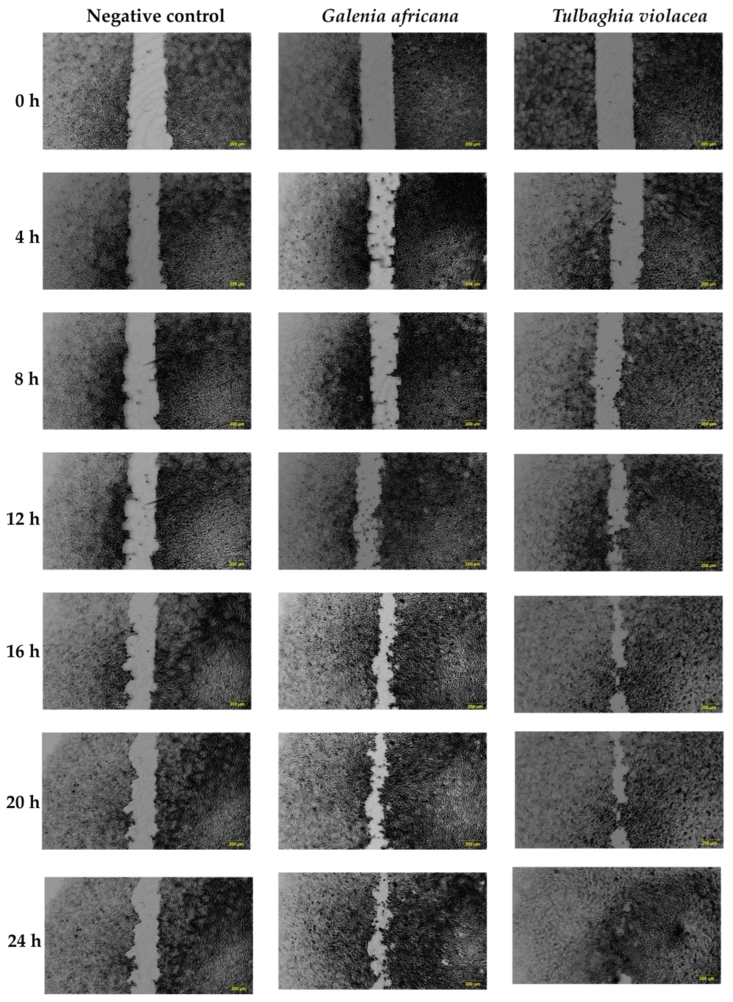
In response to a disruption caused to the skin barrier, keratinocyte epidermal cells secrete cytokines such as interleukin 1 (IL-1) and growth factors, namely, vascular endothelial growth factor (VEGF) and platelet-derived growth factor (PDGF), which induce endothelial cell migration and angiogenesis in the wound bed [63]. Understanding the cell migration process is therefore an essential part of wound healing and was studied using a scratch wound assay (Figure 4). The negative control showed a slower gap closure compared to the extract-treated cells. After 24 h of exposure to the optimum concentrations of G. africana (40 μg/mL) and T. violacea (60 μg/mL), increased wound closure was observed. Both plant extracts promoted cell migration, with T. violacea achieving an 88% reduction in wound gap after 24 h, while G. africana had a 76% gap closure at 24 h compared to the initial scratch (baseline, 0 h). The accelerated wound healing effects observed may be associated with the modulation of signalling pathways that contribute to cell proliferation, migration and extracellular matrix remodelling. Previous studies have reported that rutin, ferulic acid and coumaric acid promote healing by modulating key factors crucial for tissue regeneration, including the upregulation of VEGF and collagen, while also influencing the activity of TGF-β3 pathway to regulate inflammation [52,64,65]. A study by Ghaisas et al. [65] reported that ferulic acid could inhibit lipid peroxidation, resulting in the restoration of normal VEGF levels, thereby promoting wound healing. The oral consumption and topical application of ferulic acid significantly increased hydroxyproline levels, an important amino acid used as a marker for collagen synthesis and repairing damaged skin cells [64]. Additionally, compounds such as coumaric acid and rutin have demonstrated wound healing properties by decreasing the duration of the inflammatory phase of wound healing while promoting angiogenesis, reducing oxidative stress and enhancing epidermal regeneration [59,66].
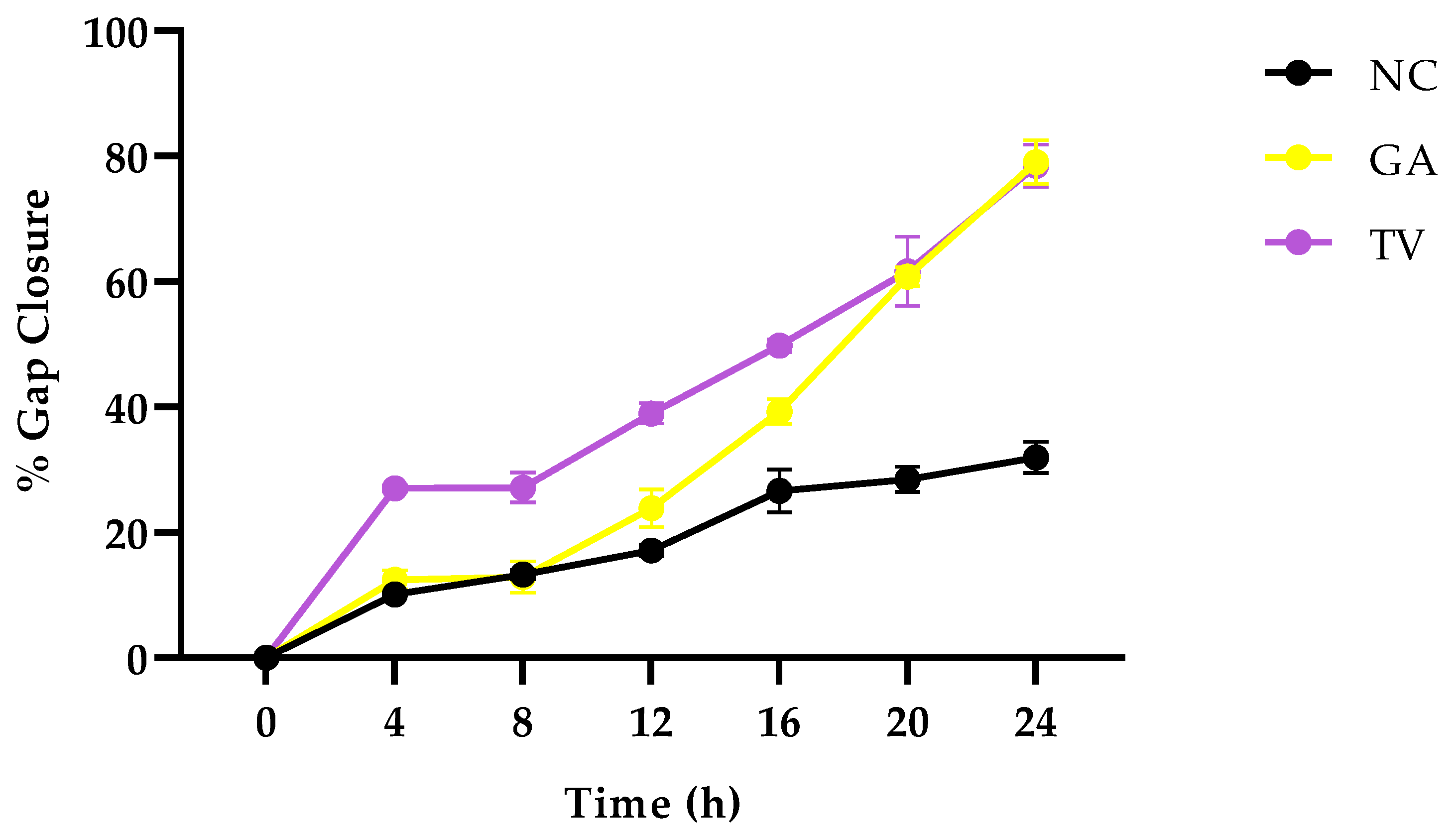
Figure 5 represents the percentage of wound closure over 24 h varied significantly between treatments. T. violacea promoted the most rapid healing, while G. africana resulted in a moderate speed but notably faster closure compared to the negative control, which showed a minimal change throughout the observed period. These results indicate that both plants effectively promote wound closure in vitro, which could be attributed to their ability to suppress inflammatory cytokines, resulting in a rapid progression from the inflammatory phase to the later stages of wound healing [67]. Previous studies have reported that compounds found in these plants, such as rutin, ferulic acid and coumaric acid, possess anti-inflammatory effects that suppress proinflammatory cytokines such as IL-6 and Tumor Necrosis Factor alpha (TNF-α), promoting angiogenesis and tissue regeneration [68,69,70]. T. violacea and G. africana could potentially increase the production of anti-inflammatory cytokines to modulate the inflammatory response, promoting rapid cell migration; however, further molecular experiments are required to corroborate this.

3.2.3. Cellular Antioxidant Assay (CAA)

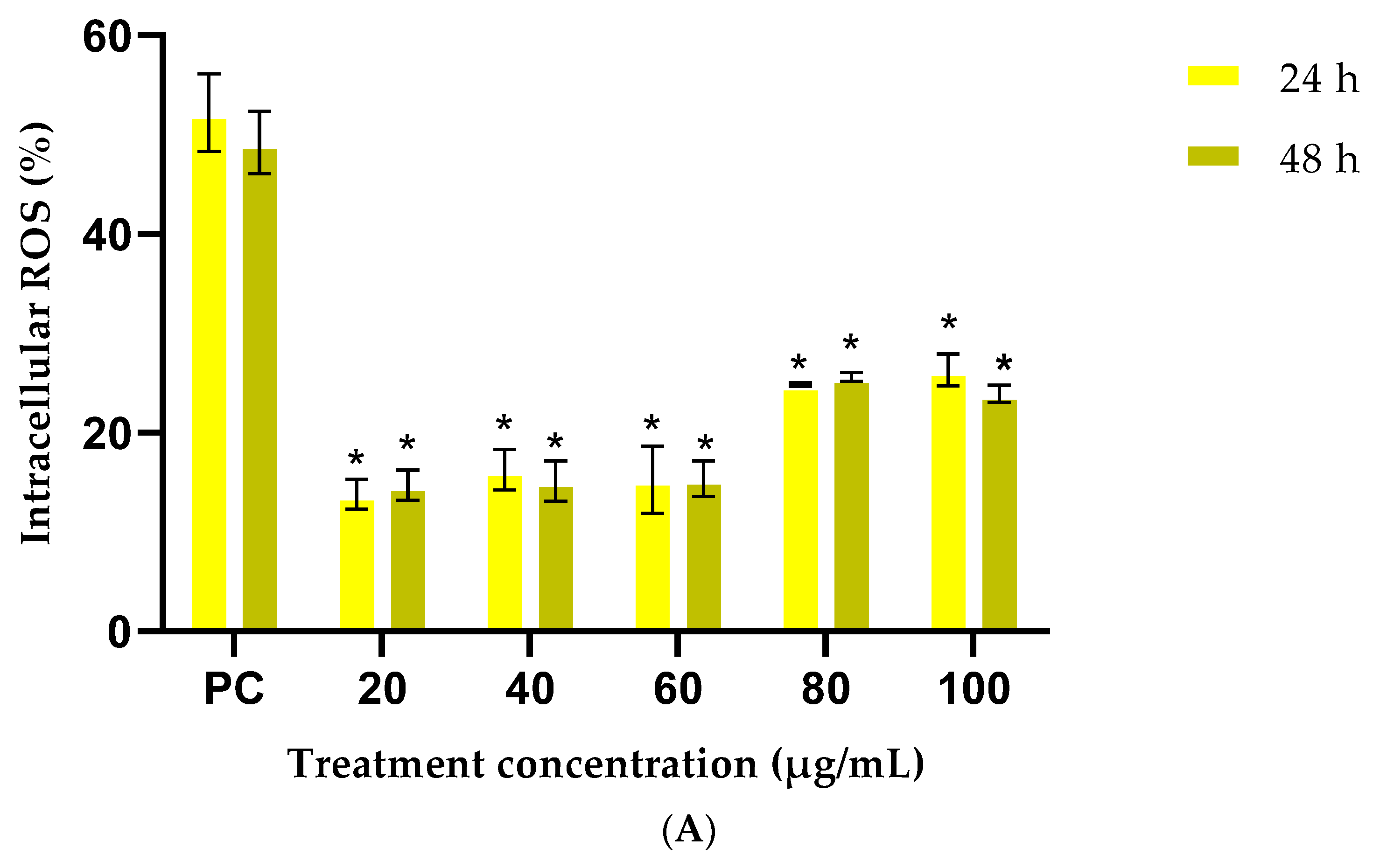
Oxidative stress is a major contributing factor to delayed wound healing, since it causes an increase in ROS production that adversely affects cellular lipids, proteins and DNA, leading to cellular and tissue dysfunction [71]. For the intracellular ROS assay, cells exposed to the positive control showed a significant increase in ROS activity, indicating the assay’s sensitivity and responsiveness for detecting intracellular ROS (Figure 6). Results show that cells stimulated with tBHP and pre-treated with different concentrations (20–100 μg/mL) of G. africana (Figure 6A) showed a significant decrease (p < 0.05) in intracellular ROS levels compared to the positive control. These results suggest that G. africana could potentially penetrate cells and act with strong radical scavenging potency in a stressed environment [72]. Several studies have corroborated the antioxidant effects of G. africana, with reports that it is effective in mitigating oxidative stress-related conditions such as cancer [17,18].
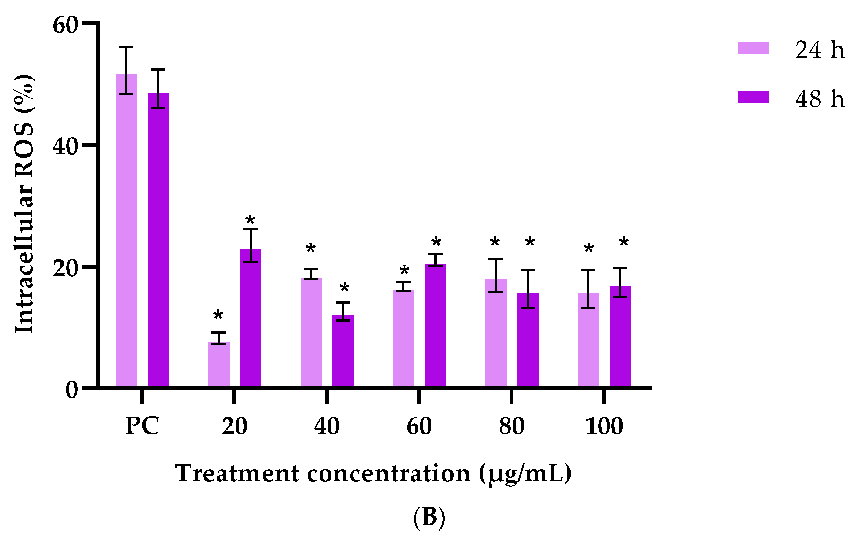
Stimulated tBHP cells pretreated with T. violacea at concentrations between 20–100 μg/mL (Figure 6B) had a significant reduction (p < 0.05) in ROS activity across all exposure times compared to the positive control. According to Afzal et al. [73], as part of their metabolic processes, healthy cells can maintain a certain baseline level of ROS. These results suggest that T. violacea could prevent the increase in ROS while also actively maintaining baseline ROS levels, suggesting that it can enhance the cells’ antioxidant defences.
Previous studies have reported that medicinal plants have compounds, such as ferulic acid can inhibit enzymes that catalyse free radical generation, while concurrently enhancing scavenger enzyme activity [52,74]. The observed antioxidant activity in HaCaT cells after exposure to the plant extracts could also be attributed to rutin, a potent antioxidant found in both G. africana and T. violacea. A previous study by Lang and Han [75] corroborates these results by showing that rutin can inhibit HaCaT cell oxidative stress by modulating the nuclear factor erythroid 2-related factor 2 (Nrf2), an important factor in the endogenous antioxidant defence system. The ability of these extracts to reduce intracellular ROS highlights their potential as protective agents against oxidative-induced cellular damage. The wound healing activities demonstrated by G. africana and T. violacea are likely supported by their antioxidant effects, which reduce oxidative stress at the site of injury and promote cellular repair.

4. Conclusions

The results of the bioactive assays conducted in the present study provide scientific validation for the traditional use of G. africana and T. violacea as wound healing agents, as both extracts demonstrated no cytotoxic effects and enhanced epidermal cell migration. G. africana demonstrated greater phenolic content and antioxidant activity, while T. violacea showed better wound migration. This study provides scientific evidence that supports the traditional use of G. africana and T. violacea, suggesting that these plants could serve as alternative agents that accelerate wound healing. HPLC analysis identified known phytoconstituents such as ferulic acid and coumaric acid in the plant extracts, all of which are known to possess antioxidant, anti-inflammatory and wound-healing properties. Future studies will evaluate the biological activity of these key compounds at their detected concentrations to determine their individual contributions to the wound healing effects observed in vitro. This will provide insight into the active constituents responsible for the bioactivity and form the basis for standardisation of the extract in future therapeutic applications. Additional research will include a broader, non-targeted phytochemical analysis using Liquid Chromatography–Mass Spectrometry (LC–MS) or Gas Chromatography–Mass Spectrometry (GC–MS) to identify other active compounds contributing to the plants’ wound-healing activity. Future studies will investigate the molecular mechanisms of the extracts, focusing on their specific signalling pathways and key protein markers. This research is essential to fully validate their safety, efficacy and potential as wound-healing agents.

Author Contributions

Conceptualization, F.F.; investigation, B.N.; writing—original draft preparation, B.N.; writing—review and editing, F.F., B.N., R.F.; supervision, F.F., R.F.; funding acquisition, F.F. All authors have read and agreed to the published version of the manuscript.

Funding

This research was funded by the National Research Foundation (NRF) of South Africa (Grant Number: JINR23031382195). Any opinion, findings, and conclusions or recommendations reported in this paper are those of the authors and therefore the NRF does not accept any liability in regard thereto.

Data Availability Statement

No new research data were created. All data was retrieved from published articles available to the public from various databases.

Acknowledgments

The authors wish to thank Lisa Joorst and Chenické Matthys for their assistance with the wound healing assay.

Conflicts of Interest

The authors declare no conflicts of interest.

References

  1. Kirwan, H.; Pignataro, R. The skin and wound healing. Pathol. Interv. Musculoskelet. Rehabil. 2015, 25, 125–129. [Google Scholar]
  2. Ferreira, M.C.; Júnior, P.T.; Carvalho, V.F.; Kamamoto, F. Complex wounds. Clinics 2006, 61, 571–578. [Google Scholar] [CrossRef]
  3. Madubula, N.; van Eeden, E. An analysis of the vulnerability of informal and formal households to disaster risks in the Rand West City region. Jàmbá-J. Disaster Risk Stud. 2024, 16, 1589. [Google Scholar] [CrossRef]
  4. Davies, B.; du Toit, C.; Hlela, M.B.K.M. Fire deaths in Cape Town, South Africa: A retrospective review of medico-legal and toxicological findings (2006–2018). Burns 2024, 50, 1011–1023. [Google Scholar] [CrossRef] [PubMed]
  5. Asong, J.; Ndhlovu, P.; Khosana, N.; Aremu, A.; Otang-Mbeng, W. Medicinal plants used for skin-related diseases among the Batswanas in Ngaka Modiri Molema District Municipality, South Africa. South Afr. J. Bot. 2019, 126, 11–20. [Google Scholar] [CrossRef]
  6. Karan, A.; Amado, V.; Vitorino, P.; Kulber, D.; Taela, A.; DeUgarte, D.A. Evaluating the socioeconomic and cultural factors associated with pediatric burn injuries in Maputo, Mozambique. Pediatr. Surg. Int. 2015, 31, 1035–1040. [Google Scholar] [CrossRef] [PubMed]
  7. World Health Organization. WHO Global Report on Traditional and Complementary Medicine 2019; World Health Organization: Geneva, Switzerland, 2019. [Google Scholar]
  8. Li, M.; Xia, W.; Khoong, Y.M.; Huang, L.; Huang, X.; Liang, H.; Zhao, Y.; Mao, J.; Yu, H.; Zan, T. Smart and versatile biomaterials for cutaneous wound healing. Biomater. Res. 2023, 27, 87. [Google Scholar] [CrossRef]
  9. Hughes, G.D.; Aboyade, O.M.; Okonji, C.O.; Clark, B.; Mabweazara, S.Z. Comparison of the prevalence of non-communicable diseases and traditional herbal medicine use in urban and rural communities in South Africa. Adv. Integr. Med. 2021, 8, 136–143. [Google Scholar] [CrossRef]
  10. Goldblatt, P. Floristic diversity in the Cape flora of South Africa. Biodivers. Conserv. 1997, 6, 359–377. [Google Scholar] [CrossRef]
  11. Philander, L.A. An ethnobotany of Western Cape Rasta bush medicine. J. Ethnopharmacol. 2011, 138, 578–594. [Google Scholar] [CrossRef]
  12. Klak, C.; Hanáček2, P.; Bruyns, P.V. Disentangling the Aizooideae: New generic concepts and a new subfamily in Aizoaceae. Taxon 2017, 66, 1147–1170. [Google Scholar] [CrossRef]
  13. Moodley, K.; Joseph, K.; Naidoo, Y.; Islam, S.; Mackraj, I. Antioxidant, antidiabetic and hypolipidemic effects of Tulbaghia violacea Harv. (wild garlic) rhizome methanolic extract in a diabetic rat model. BMC Complement. Altern. Med. 2015, 15, 408. [Google Scholar] [CrossRef]
  14. Van Wyk, B.-E. A review of Khoi-San and Cape Dutch medical ethnobotany. J. Ethnopharmacol. 2008, 119, 331–341. [Google Scholar] [CrossRef]
  15. Heredia, D.; Green, I.; Klaasen, J.; Rahiman, F. Importance and relevance of phytochemicals present in Galenia africana. Scientifica 2022, 2022, 5793436. [Google Scholar] [CrossRef] [PubMed]
  16. Ng’uni, T.; Klaasen, J.A.; Fielding, B.C. Acute toxicity studies of the South African medicinal plant Galenia africana. Toxicol. Rep. 2018, 5, 813–818. [Google Scholar] [CrossRef]
  17. Mohamed, L.; Chakraborty, S.; ArulJothi, K.; Mabasa, L.; Sayah, K.; Costa-Lotufo, L.V.; Jardine, A.; Prince, S. Galenia africana plant extract exhibits cytotoxicity in breast cancer cells by inducing multiple programmed cell death pathways. Saudi Pharm. J. 2020, 28, 1155–1165. [Google Scholar] [CrossRef]
  18. Ndlovu, B.; De Kock, M.; Klaasen, J.; Rahiman, F. In vitro comparison of the anti-proliferative effects of Galenia africana on human skin cell lines. Sci. Pharm. 2021, 89, 12. [Google Scholar] [CrossRef]
  19. Styger, G.; Aboyade, O.M.; Gibson, D.; Hughes, G. Tulbaghia—A Southern African Phytomedicine. J. Altern. Complement. Med. 2016, 22, 255–261. [Google Scholar] [CrossRef] [PubMed]
  20. Krstin, S.; Sobeh, M.; Braun, M.S.; Wink, M. Tulbaghia violacea and Allium ursinum extracts exhibit anti-parasitic and antimicrobial activities. Molecules 2018, 23, 313. [Google Scholar] [CrossRef]
  21. Raji, I.; Obikeze, K.; Mugabo, P. Potential beneficial effects of Tulbaghia violacea William Henry Harvey (Alliaceae) on cardiovascular system-A review. Trop. J. Pharm. Res. 2015, 14, 1111–1117. [Google Scholar] [CrossRef]
  22. Bungu, L.; van de Venter, M.; Frost, C. Evidence for an in vitro anticoagulant and antithrombotic activity in Tulbaghia violacea. Afr. J. Biotechnol. 2008, 7, 681–688. [Google Scholar]
  23. Iwalewa, E.; McGaw, L.; Naidoo, V.; Eloff, J. Inflammation: The foundation of diseases and disorders. A review of phytomedicines of South African origin used to treat pain and inflammatory conditions. Afr. J. Biotechnol. 2007, 6, 2868–2885. [Google Scholar] [CrossRef]
  24. Hulley, I.; Van Wyk, B.-E. Quantitative medicinal ethnobotany of Kannaland (western Little Karoo, South Africa): Non-homogeneity amongst villages. South Afr. J. Bot. 2019, 122, 225–265. [Google Scholar] [CrossRef]
  25. Van Wyk, B.-E.; De Wet, H.; Van Heerden, F. An ethnobotanical survey of medicinal plants in the southeastern Karoo, South Africa. South Afr. J. Bot. 2008, 74, 696–704. [Google Scholar] [CrossRef]
  26. Thring, T.; Weitz, F. Medicinal plant use in the Bredasdorp/Elim region of the Southern Overberg in the Western Cape Province of South Africa. J. Ethnopharmacol. 2006, 103, 261–275. [Google Scholar] [CrossRef] [PubMed]
  27. Tyavambiza, C.; Meyer, M.; Wusu, A.D.; Madiehe, A.M.; Meyer, S. The antioxidant and in vitro wound healing activity of cotyledon orbiculata aqueous extract and the synthesized biogenic silver nanoparticles. Int. J. Mol. Sci. 2022, 23, 16094. [Google Scholar] [CrossRef] [PubMed]
  28. Lim, H.S.; Yo, S.R.; Lee, M.Y.; Seo, C.S.; Shin, H.K.; Jeong, S.J. Potential inhibitory effects of the traditional herbal prescription Hyangso-san against skin inflammation via inhibition of chemokine production and inactivation of STAT1 in HaCaT keratinocytes. Mol. Med. Rep. 2018, 17, 2515–2522. [Google Scholar] [CrossRef] [PubMed]
  29. Siddiqui, N.; Rauf, A.; Latif, A.; Mahmood, Z. Spectrophotometric determination of the total phenolic content, spectral and fluorescence study of the herbal Unani drug Gul-e-Zoofa (Nepeta bracteata Benth). J. Taibah Univ. Med. Sci. 2017, 12, 360–363. [Google Scholar] [CrossRef]
  30. Okaiyeto, K.; Kerebba, N.; Rautenbach, F.; Singh, S.K.; Dua, K.; Oguntibeju, O.O. UPLC-ESI-QTOF-MS phenolic compounds identification and quantification from ethanolic extract of: In vitro antioxidant and antidiabetic potentials. Arab. J. Chem. 2022, 16, 104447. [Google Scholar] [CrossRef]
  31. Alahmad, A.; Alghoraibi, I.; Zein, R.; Kraft, S.; Dräger, G.; Walter, J.-G.; Scheper, T. Identification of major constituents of Hypericum perforatum L. extracts in Syria by development of a rapid, simple, and reproducible HPLC-ESI-Q-TOF MS analysis and their antioxidant activities. ACS Omega 2022, 7, 13475–13493. [Google Scholar] [CrossRef]
  32. Kumar, B.R. Application of HPLC and ESI-MS techniques in the analysis of phenolic acids and flavonoids from green leafy vegetables (GLVs). J. Pharm. Anal. 2017, 7, 349–364. [Google Scholar] [CrossRef]
  33. Gulcin, İ.; Alwasel, S.H. DPPH radical scavenging assay. Processes 2023, 11, 2248. [Google Scholar] [CrossRef]
  34. Chaves, N.; Santiago, A.; Alías, J.C. Quantification of the antioxidant activity of plant extracts: Analysis of sensitivity and hierarchization based on the method used. Antioxidants 2020, 9, 76. [Google Scholar] [CrossRef]
  35. Yen, G.C.; Duh, P.D. Scavenging effect of methanolic extracts of peanut hulls on free-radical and active-oxygen species. J. Agric. Food Chem. 1994, 42, 629–632. [Google Scholar]
  36. Kumar, G.; Gautam, G.K.; Kumar, R.; Rana, H. The pharmacological and phytochemical study of Adansonia digitata. Curr. Trends Pharm. Pharm. Chem. 2022, 4, 54–58. [Google Scholar] [CrossRef]
  37. López, V.; Cásedas, G.; Petersen-Ross, K.; Powrie, Y.; Smith, C. Neuroprotective and anxiolytic potential of green rooibos (Aspalathus linearis) polyphenolic extract. Food Funct. 2022, 13, 91–101. [Google Scholar] [CrossRef]
  38. Gohari, A.; Hajimehdipoor, H.; Saeidnia, S.; Ajani, Y.; Hadjiakhoondi, A. Antioxidant activity of some medicinal species using FRAP assay. J. Med. Plants 2011, 10, 54–60. [Google Scholar]
  39. Ilaghi, M.; Sharifi, I.; Sharififar, F.; Sharifi, F.; Oliaee, R.T.; Babaei, Z.; Meimamandi, M.S.; Keyhani, A.; Bamorovat, M. The potential role and apoptotic profile of three medicinal plant extracts on Leishmania tropica by MTT assay, macrophage model and flow cytometry analysis. Parasite Epidemiol. Control 2021, 12, e00201. [Google Scholar] [CrossRef] [PubMed]
  40. Schindelin, J.; Arganda-Carreras, I.; Frise, E.; Kaynig, V.; Longair, M.; Pietzsch, T.; Preibisch, S.; Rueden, C.; Saalfeld, S.; Schmid, B. Fiji: An open-source platform for biological-image analysis. Nat. Methods 2012, 9, 676–682. [Google Scholar] [CrossRef]
  41. Li, R.; Liu, K.; Huang, X.; Li, D.; Ding, J.; Liu, B.; Chen, X. Bioactive materials promote wound healing through modulation of cell behaviors. Adv. Sci. 2022, 9, 2105152. [Google Scholar] [CrossRef]
  42. Kučera, O.; Endlicher, R.; Roušar, T.; Lotková, H.; Garnol, T.; Drahota, Z.; Červinková, Z. The effect of tert-butyl hydroperoxide-induced oxidative stress on lean and steatotic rat hepatocytes in vitro. Oxidative Med. Cell. Longev. 2014, 2014, 752506. [Google Scholar] [CrossRef]
  43. Skóra, B.; Piechowiak, T.; Szychowski, K.A.; Gmiński, J. Entrapment of silver nanoparticles in L-α-phosphatidylcholine/cholesterol-based liposomes mitigates the oxidative stress in human keratinocyte (HaCaT) cells. Eur. J. Pharm. Biopharm. 2021, 166, 163–174. [Google Scholar] [CrossRef]
  44. Vasarri, M.; Bergonzi, M.C.; Leri, M.; Castellacci, R.; Bucciantini, M.; De Marchi, L.; Degl’Innocenti, D. Protective Effects of Oleanolic Acid on Human Keratinocytes: A Defense Against Exogenous Damage. Pharmaceuticals 2025, 18, 238. [Google Scholar] [CrossRef]
  45. Lee, B.M.; Woo, H.J.; Jang, B.J.; Shin, J.A.; Ham, Y.M.; Jang, E.B.; Kim, S.C.; Kim, J.M.; Shin, H.S. Assessment of Damnacanthus major Siebold & Zucc callus for antioxidative and moisturizing capacities using an artificial skin alternative. Biotechnol. Bioprocess Eng. 2024, 29, 689–698. [Google Scholar] [CrossRef]
  46. Piluzza, G.; Bullitta, S. Correlations between phenolic content and antioxidant properties in twenty-four plant species of traditional ethnoveterinary use in the Mediterranean area. Pharm. Biol. 2011, 49, 240–247. [Google Scholar] [CrossRef]
  47. Panou, E.; Tsafantakis, N.; Zengin, G.; Graikou, K.; Ganos, C.; Fokialakis, N.; Chinou, I. Untargeted Metabolomic Profiling and Bioactivity Insights into Alkanna corcyrensis. Sci. Pharm. 2025, 93, 45. [Google Scholar] [CrossRef]
  48. Takaidza, S.; Mtunzi, F.; Pillay, M. Analysis of the phytochemical contents and antioxidant activities of crude extracts from Tulbaghia species. J. Tradit. Chin. Med. 2018, 38, 272–279. [Google Scholar] [CrossRef]
  49. Boo, Y.C. p-Coumaric acid as an active ingredient in cosmetics: A review focusing on its antimelanogenic effects. Antioxidants 2019, 8, 275. [Google Scholar] [CrossRef]
  50. Saleem, U.; Khalid, S.; Zaib, S.; Anwar, F.; Ahmad, B.; Ullah, I.; Zeb, A.; Ayaz, M. Phytochemical analysis and wound healing studies on ethnomedicinally important plant Malva neglecta Wallr. J. Ethnopharmacol. 2020, 249, 112401. [Google Scholar] [CrossRef]
  51. Gęgotek, A.; Ambrożewicz, E.; Jastrząb, A.; Jarocka-Karpowicz, I.; Skrzydlewska, E. Rutin and ascorbic acid cooperation in antioxidant and antiapoptotic effect on human skin keratinocytes and fibroblasts exposed to UVA and UVB radiation. Arch. Dermatol. Res. 2019, 311, 203–219. [Google Scholar] [CrossRef]
  52. Zduńska, K.; Dana, A.; Kolodziejczak, A.; Rotsztejn, H. Antioxidant properties of ferulic acid and its possible application. Ski. Pharmacol. Physiol. 2018, 31, 332–336. [Google Scholar] [CrossRef]
  53. Enogieru, A.B.; Haylett, W.; Hiss, D.C.; Bardien, S.; Ekpo, O.E. Rutin as a potent antioxidant: Implications for neurodegenerative disorders. Oxidative Med. Cell. Longev. 2018, 2018, 6241017. [Google Scholar] [CrossRef] [PubMed]
  54. Choi, S.-S.; Park, H.-R.; Lee, K.-A. A comparative study of rutin and rutin glycoside: Antioxidant activity, anti-inflammatory effect, effect on platelet aggregation and blood coagulation. Antioxidants 2021, 10, 1696. [Google Scholar] [CrossRef]
  55. Choi, S.J.; Lee, S.-N.; Kim, K.; Joo, D.H.; Shin, S.; Lee, J.; Lee, H.K.; Kim, J.; Kwon, S.B.; Kim, M.J. Biological effects of rutin on skin aging. Int. J. Mol. Med. 2016, 38, 357–363. [Google Scholar] [CrossRef]
  56. Fisher, F.; Africa, C.; Klaasen, J.; Fisher, R. South African Medicinal Plants Traditionally Used for Wound Treatment: An Ethnobotanical Systematic Review. Plants 2025, 14, 818. [Google Scholar] [CrossRef]
  57. Ndlovu, B.; Klaasen, J.; Hughes, G.; Fisher, F. An ethnobotanical survey investigating medicinal plants used by Cape bush doctors to treat dermatophyte infections. Sci. Afr. 2024, 24, e02156. [Google Scholar] [CrossRef]
  58. Elbagory, A.M.; Meyer, M.; Cupido, C.N.; Hussein, A.A. Inhibition of bacteria associated with wound infection by biocompatible green synthesized gold nanoparticles from South African plant extracts. Nanomaterials 2017, 7, 417. [Google Scholar] [CrossRef]
  59. Dehelean, C.A.; Coricovac, D.; Pinzaru, I.; Marcovici, I.; Macasoi, I.G.; Semenescu, A.; Lazar, G.; Cinta Pinzaru, S.; Radulov, I.; Alexa, E. Rutin bioconjugates as potential nutraceutical prodrugs: An in vitro and in ovo toxicological screening. Front. Pharmacol. 2022, 13, 1000608. [Google Scholar] [CrossRef] [PubMed]
  60. Wu, P.; Liu, Y.; Zhai, H.; Wu, X.; Liu, A. Rutin alleviates psoriasis-related inflammation in keratinocytes by regulating the JAK2/STAT3 signaling. Ski. Res. Technol. 2024, 30, e70011. [Google Scholar] [CrossRef]
  61. Albahri, G.; Badran, A.; Hijazi, A.; Daou, A.; Baydoun, E.; Nasser, M.; Merah, O. The therapeutic wound healing bioactivities of various medicinal plants. Life 2023, 13, 317. [Google Scholar] [CrossRef]
  62. Oguntibeju, O.O. Medicinal plants and their effects on diabetic wound healing. Vet. World 2019, 12, 653. [Google Scholar] [CrossRef] [PubMed]
  63. Wojtowicz, A.M.; Oliveira, S.; Carlson, M.W.; Zawadzka, A.; Rousseau, C.F.; Baksh, D. The importance of both fibroblasts and keratinocytes in a bilayered living cellular construct used in wound healing. Wound Repair Regen. 2014, 22, 246–255. [Google Scholar] [CrossRef] [PubMed]
  64. Singh, H.; Ali, S.; Khan, N.; Mishra, A.; Mishra, A. Wound healing potential of Cleome viscosa Linn. seeds extract and isolation of active constituent. South Afr. J. Bot. 2017, 112, 460–465. [Google Scholar] [CrossRef]
  65. Ghaisas, M.M.; Kshirsagar, S.B.; Sahane, R.S. Evaluation of wound healing activity of ferulic acid in diabetic rats. Int. Wound J. 2014, 11, 523–532. [Google Scholar] [CrossRef]
  66. Selvakumar, G.; Lonchin, S. A bio-polymeric scaffold incorporated with p-Coumaric acid enhances diabetic wound healing by modulating MMP-9 and TGF-β3 expression. Colloids Surf. B Biointerfaces 2023, 225, 113280. [Google Scholar] [CrossRef]
  67. Mahmoud, N.N.; Hamad, K.; Al Shibitini, A.; Juma, S.; Sharifi, S.; Gould, L.; Mahmoudi, M. Investigating inflammatory markers in wound healing: Understanding implications and identifying artifacts. ACS Pharmacol. Transl. Sci. 2024, 7, 18–27. [Google Scholar] [CrossRef] [PubMed]
  68. Liu, J.; Guan, Y.; Yang, L.; Fang, H.; Sun, H.; Sun, Y.; Yan, G.; Kong, L.; Wang, X. Ferulic Acid as an Anti-Inflammatory Agent: Insights into Molecular Mechanisms, Pharmacokinetics and Applications. Pharmaceuticals 2025, 18, 912. [Google Scholar] [CrossRef]
  69. Ganeshpurkar, A.; Saluja, A.K. The pharmacological potential of rutin. Saudi Pharm. J. 2017, 25, 149–164. [Google Scholar] [CrossRef] [PubMed]
  70. Abd Rashid, N.; Abd Halim, S.A.S.; Teoh, S.L.; Budin, S.B.; Hussan, F.; Ridzuan, N.R.A.; Jalil, N.A.A. The role of natural antioxidants in cisplatin-induced hepatotoxicity. Biomed. Pharmacother. 2021, 144, 112328. [Google Scholar] [CrossRef]
  71. Muzammil, S.; Neves Cruz, J.; Mumtaz, R.; Rasul, I.; Hayat, S.; Khan, M.A.; Khan, A.M.; Ijaz, M.U.; Lima, R.R.; Zubair, M. Effects of drying temperature and solvents on in vitro diabetic wound healing potential of Moringa oleifera leaf extracts. Molecules 2023, 28, 710. [Google Scholar] [CrossRef]
  72. Barzegar, A.; Moosavi-Movahedi, A.A. Intracellular ROS protection efficiency and free radical-scavenging activity of curcumin. PLoS ONE 2011, 6, e26012. [Google Scholar] [CrossRef]
  73. Afzal, S.; Abdul Manap, A.S.; Attiq, A.; Albokhadaim, I.; Kandeel, M.; Alhojaily, S.M. From imbalance to impairment: The central role of reactive oxygen species in oxidative stress-induced disorders and therapeutic exploration. Front. Pharmacol. 2023, 14, 1269581. [Google Scholar] [CrossRef] [PubMed]
  74. Yahaya, E.S.; Cordier, W.; Steenkamp, P.; Steenkamp, V. Effect of ethnomedicinal extracts used for wound healing on cellular migration and intracellular reactive oxygen species release in SC-1 fibroblasts. South Afr. J. Bot. 2018, 118, 11–17. [Google Scholar] [CrossRef]
  75. Lang, G.-P.; Han, Y.-Y. Rutin ameliorates H2O2-induced oxidative stress injury in HaCaT cells via the Nrf2-regulated pathway. J. Evol. Biochem. Physiol. 2022, 58, 1389–1400. [Google Scholar] [CrossRef]
Figure 1. HPLC chromatograms of the bioactive compounds from G. africana ethanolic extracts at wavelengths of (A): 320 and (B): 350 nm. Peaks indicated by arrows and numbers represent 1: Ferulic acid and 2: Rutin identified by comparison with commercially available standards. Chromatographic profiles: phenolic acids monitored at 320 nm (blue line) and flavonols monitored at 360 nm (pink line). The unlabelled peaks represent other components of the complex extract that were not identified in this targeted analysis.
Plants 14 03523 g001
Figure 2. HPLC chromatograms of the bioactive compounds from T. violacea ethanolic extracts at wavelengths of (A): 320 and (B): 350 nm. Peaks indicated by arrows and numbers represent 1: Ferulic acid, 2: Rutin and 3: Coumaric acid identified by comparison with authentic standards. Chromatographic profiles: phenolic acids monitored at 320 nm (blue line) and flavonols monitored at 360 nm (pink line). The unlabelled peaks represent other components of the complex extract that were not identified in this targeted analysis.
Plants 14 03523 g002
Figure 3. Cell viability and proliferation of normal skin (HaCaT) cells treated with 20, 40, 60, 80 and 100 µg/mL concentrations of Galenia africana (A) and Tulbaghia violacea (B) ethanolic extracts for 24 and 48 h were determined using an MTT assay. NC: negative control—untreated cells with media, PC: positive control- cells treated with 1% Triton-X 100. Data represent the mean ± SEM, n = 3, where * indicates statistical significance (p < 0.05) compared to the negative control.
Plants 14 03523 g003
Figure 4. HaCaT cell migration percentage was determined using a scratch assay. Images show the wound area at 0 h (initial scratch) and the progression of wound closure at 4, 8, 12, 16, 20 and 24 h after treatment with the optimum concentration of 40 μg/mL for Galenia africana and 60 μg/mL Tulbaghia violacea. The negative control shows untreated cells with serum-free media. Scale bar: 200 µm.
Plants 14 03523 g004
Figure 5. Percentage of wound gap closure over time in HaCaT cells pretreated with Galenia africana (GA) (40 μg/mL) and Tulbaghia violacea (TV) (60 μg/mL). NC: negative control—untreated cells with serum-free media. Data represent the mean percentage of the original wound area that has closed at each time point over the 24 h.
Plants 14 03523 g005
Figure 6. The effects of Galenia africana (A) and Tulbaghia violacea (B) on intracellular ROS formation in HaCaT cells exposed to the pro-oxidative agent tBHP for 24 and 48 h. Data represents normalised ROS production, expressed relative to the negative control (untreated cells). Cells treated with tBHP were used as a positive control (PC). Data are shown as mean ± SEM, n = 3, where * indicates statistical significance (p < 0.05) compared to the PC.
Plants 14 03523 g006aPlants 14 03523 g006b
Table 1. Phytochemical composition and antioxidant activity of Galenia africana and Tulbaghia violacea extracts.
Treatment (1 mg/mL)G. africana Ethanolic ExtractT. violacea Ethanolic Extract
Polyphenols (mg GAE/g)106.39 ± 2.0378.57 ± 2.40
Flavonoid content (mg QE/g)39.79 ± 0.27-
DPPH μmol TE/g187.42 ± 6.40117.60 ± 6.33
ORAC μmol TE/g1247.83 ± 12.53172.82 ± 12.39
FRAP μmol AAE/g195.91 ± 4.26116.25 ± 12.57
Abbreviations: TPC, total phenolic content; TFC, total flavonoid content; GAE, gallic acid equivalents; CE, catechin equivalents; AAE, ascorbic acid equivalents; QE, quercetin equivalent; DPPH,2,2-diphenyl-1-picrylhydrazyl. Values represent mean ± standard error of mean (n = 3).
Table 2. Identification and quantification of key bioactive compounds in Galenia africana and Tulbaghia violacea ethanolic extracts.
Plant ExtractPhytochemicalsQuantity (mg/L)Retention Time (min)Wavelength (nm)
G. africanaFerulic acid1.8822.227320
Rutin35.8725.313350
T. violaceaRutin1.1824.003350
Coumaric acid13.5821.008320
Ferulic acid1.8822.185320
Disclaimer/Publisher’s Note: The statements, opinions and data contained in all publications are solely those of the individual author(s) and contributor(s) and not of MDPI and/or the editor(s). MDPI and/or the editor(s) disclaim responsibility for any injury to people or property resulting from any ideas, methods, instructions or products referred to in the content.