Article

Characterization of Caleosin Genes in Carica papaya and Insights into Lineage-Specific Family Evolution in Brassicales

1
National Key Laboratory for Tropical Crop Breeding/Hainan Key Laboratory for Biosafety Monitoring and Molecular Breeding in Off-Season Reproduction Regions, Institute of Tropical Biosciences and Biotechnology/Sanya Research Institute of Chinese Academy of Tropical Agricultural Sciences, Haikou 571101, China
2
College of Biology and Food Engineering, Guangdong University of Petrochemical Technology, Maoming 525000, China
*
Authors to whom correspondence should be addressed.
Plants 2025, 14(21), 3296; https://doi.org/10.3390/plants14213296
Submission received: 28 September 2025 / Revised: 23 October 2025 / Accepted: 27 October 2025 / Published: 29 October 2025

Abstract

Caleosins (CLOs) or peroxygenases (PXGs), a class of structural proteins of lipid droplets (LDs), comprise a small family of multifunctional proteins widely involved in oil accumulation, organ development, and stress responses. Despite the proposal of two clades termed H and L in Arabidopsis thaliana, their evolution in the order Brassicales has not been well established. In this study, the first genome-wide analysis of the caleosin family was conducted in papaya (Carica papaya), a Caricaceae plant without any recent whole-genome duplication (WGD). A high number of five members representing both H and L clades were identified from the papaya genome. Further identification and comparison of 68 caleosin genes from 14 representative plant species revealed seven orthogroups, i.e., H1–4 and L1–3, where H1 and L1 have already appeared in the basal angiosperm Amborella trichopoda, supporting their early divergence before angiosperm radiation. Five CpCLO genes belong to H1 (1) and L1 (4), and extensive expansion of the L1 group was shown to be contributed to by species-specific tandem and transposed duplications, which may contribute to environmental adaptation. Orthologous and syntenic analyses uncovered that lineage-specific expansion of the caleosin family in Brassicales relative to A. trichopoda was largely contributed to by tandem duplication and recent WGDs, as well as the ancient γ whole-genome triplication (WGT) shared by all core eudicots. Independent gain or loss of certain introns and apparent expression divergence of caleosin genes were also observed. Tissue-specific expression analysis showed that CpCLO2 and −5 are constitutively expressed, whereas others appear to be tissue-specific, implying function divergence. Interestingly, the H-forms CpCLO1 and RcCLO1 were shown to exhibit similar expression profiles to most oleosin genes that are preferentially expressed oil-rich tissues such as seeds/endosperms, shoots, and calluses, implying their putative involvement in oil accumulation, as observed in Arabidopsis. The results obtained from this study provide a global view of CpCLO genes and insights into lineage-specific family evolution in Brassicales, which facilitates further functional studies in papaya and other non-model species.

1. Introduction

Caleosins (CLOs)/peroxygenases (PXGs) comprise a small family of multifunctional proteins that are involved not only in the formation, stabilization, and degradation of lipid droplets (LDs), but also in a wide range of plant development and stress responses [1,2,3,4]. Though they were first identified as a class of minor LD proteins in sesame (Sesamum indicum) seeds [5], other intracellular locations have also been reported, e.g., endoplasmic reticulum (ER), vacuole, plasmalemma, and chloroplast envelope fractions [6,7,8]. Additionally, they are also abundant in several non-seed tissues, e.g., megagametophytes, pollen, fruits, leaves, stems, and roots [6,9,10,11,12,13,14,15]. Structurally, caleosins exhibit some similarities to oleosins (OLEs), the major LD proteins that include three main domains, i.e., an N-terminal hydrophilic domain, a central hydrophobic domain, and a C-terminal hydrophilic domain [2,16,17]. Compared with oleosins, the central hydrophobic domain of caleosins is relatively shorter (i.e., 20 vs. 72 residues) and usually features a proline-knot pattern of PX3PSX3P (X represents a nonpolar residue) that is somewhat different from PX5SPX3P, as observed in oleosins [16,18,19,20,21,22,23]. Moreover, the N-terminal hydrophilic domain of caleosins typically possesses a single EF-hand calcium binding motif, whereas the C-terminal hydrophilic domain usually harbors several kinase phosphorylation sites [1,2,16,24,25]. Ca2+ is essential for their peroxygenase activity, which is dependent on a heme group coordinated by the two invariant His residues located in N- and C-terminal regions [16,24,25,26]. The distribution of caleosins is also wider, which are present not only in land plants but also in some fungi and green algae [27,28,29,30].
Genome-wide surveys showed that the caleosin family in land plants is highly diverse, varying from two to 22 members [18,19,27,28,29,30,31,32]. Interestingly, two members were not only reported in the basal angiosperm Amborella trichopoda, but also in three core eudicots, i.e., castor bean (Ricinus communis), flax (Linum usitatissimum), and cucumber (Cucumis sativus), in stark contrast to eight as described in the model plant Arabidopsis thaliana [18,19,30]. According to phylogenetic analysis, these caleosins could be divided into two main clades, i.e., H and L, with high and low molecular weights (MWs), respectively, where the L clade was supposed to evolve from the H clade due to the fragment deletion at the N-terminal [19,32]. The presence of both H- and L-forms in A. trichopoda implies early divergence of these two clades before angiosperm radiation. It has been established that after the split with A. trichopoda, the last common ancestor of core eudicots (including castor bean, flax, cucumber, and A. thaliana) underwent one so-called γ whole-genome triplication (WGT), whereas A. thaliana further experienced two lineage-specific whole-genome duplications (WGDs) known as β and α [33,34]. A. thaliana was shown to harbor four H-forms (i.e., AtCLO1, −2, −3, and −8) and four L-forms (i.e., AtCLO47), where two pairs (i.e., AtCLO4/−6 and AtCLO5/−7) are organized in tandem repeats and another two pairs (i.e., AtCLO1/−2 and AtCLO5/−6) were characterized as WGD repeats [19]. Nevertheless, the exact mechanisms contributing to the extensive expansion of the AtCLO family and lineage-specific evolution in Brassicales are yet to be studied.
Papaya (Carica papaya L.) belongs to the Caricaceae family within the order Brassicales, which includes 17 families and approximately 400 genera [35]. Though multiple independent recent WGDs have been described in Brassicales [36], no additional WGD after the γ event was detected in papaya, which was estimated to have diverged with A. thaliana about 72 million years ago (Mya) [37]. From an evolutionary perspective, analyzing papaya genes could provide insights into lineage-specific family evolution in this economically and ecologically important plant order [23,38,39]. In this study, a systemic analysis of caleosin genes was conducted in papaya, including sequence characteristics, gene structures, chromosomal localizations, evolutionary relationships, and expression profiles, as well as a comprehensive comparison with representative plant species such as A. thaliana, castor bean, Aquilegia coerulea, and A. trichopoda. Significantly, 1:1 orthologous relationships were observed between castor bean and A. trichopoda, whereas 1:1 and 4:1 were found between papaya and castor bean/A. trichopoda. Moreover, our results showed that the expansion of the CpCLO family was contributed by tandem, transposed, and dispersed duplications, in contrast to a more important role of WGDs in Brassicaceae plants. Apparent expression divergence of caleosin genes was also observed, implying putative function divergence. Herein, we report our findings.

2. Results

2.1. Identification, Chromosomal Localization, and Duplication Event Analysis of Five Caleosin Genes in Papaya

The search of the papaya genome resulted in five caleosin genes, which are slightly less than the six members as reported for the oleosin family [23]. Without any exception, all deduced CpCLO proteins were shown to harbor a single caleosin domain (under the Pfam accession number of PF05042) (Table 1). The peptide size varies from 204 (CpCLO2) to 240 (CpCLO1) residues, and the average length of 213 residues is relatively longer than 153 residues as observed for six CpOLEs [23]. Correspondingly, a higher MW with an average of 24.21 kilodalton (kDa) was found for CpCLOs relative to CpOLEs (16.10 kDa). The isoelectric point (pI) of CpCLOs varies from 5.05 (CpCLO1) to 9.57 (CpCLO5), where 60.00% of them (CpCLO1–3) have a pI value of less than 7, in contrast to only CpOLE6 harboring an acidic pI [23]. Interestingly, all CpCLOs possess a grand average of hydropathicity (GRAVY) value of less than 0, varying from −0.329 to −0.509, in stark contrast to the highly hydrophobic feature of CpOLEs [23]. Correspondingly, the ProtScale analysis showed that all CpCLOs exhibit similar hydropathicity scales and only the central region (more precisely the amphipathic α-helix and the proline-knot motif, see below) is hydrophilic (Figure 1A). Distinct amino acid (AA) composition and secondary structure were also observed between CpCLOs and CpOLEs. As shown in Figure 1B, except for the absence of the Cys in CpCLO4 and –5, other members contain all 20 residues, rich in Leu, Gly, Ala, and Val, as observed in CpOLEs. Compared with CpOLEs, CpCLOs usually contain more Glu, Lys, Phe, Ser, Asp, Asn, and Tyr, but less Leu, Gly, Ala, and Val, Gln, Thr, and Pro; they always harbor more hydrophilic but less hydrophobic residues and they also have more random coils but less alpha helixes and extended strands. Interestingly, the cluster analyses of amino acid and secondary structure percentages revealed that these proteins were grouped in families, though subgroups were also observed within each family (Figure 1B,C).
Sequence alignment of five CpCLOs is shown in Figure 1D. In contrast to the conservation of the caleosin domain, N- and C-terminal regions are highly diverse. CpCLO1 differs from other members with a 29-residue insertion, which was defined as the H insertion in A. thaliana [19]. This insertion mainly contributes to the longer peptide size and higher MW in CpCLO1 (Table 1). The caleosin domain is composed of a Ca2+ binding motif, an amphipathic α-helix, a proline-knot motif, two heme-binding sites, and four main phosphorylation sites, i.e., one for Tyr kinase and three for casein kinase II (CK II) (Figure 1D). The identified proline-knot pattern P/AX3P/FSX3P is somewhat different from PX5SPX3P, found in CpOLEs [23]. The heme-binding sites are composed of two invariable His residues, which were shown to be essential for peroxygenase activity [24], implying that they have similar functions. The first CK II phosphorylation site is usually a Thr residue, but it is placed by an Ile residue in CpCLO4, implying its divergence. Despite the presence of two Cys residues in CpCLO1–3, their positions are usually different, and the one just before the third CK II phosphorylation site is only found in CpCLO1 (Figure 1D).
Chromosome localization indicates that five CpCLO genes are unevenly distributed over three chromosomes (Chrs). Whereas CpCLO5 is located on Chr6, CpCLO1/–4 and CpCLO2/–3 are co-located on Chr3 and Chr2, respectively (Figure 1E). Among them, CpCLO2 and –3 were defined as tandem repeats for their neighboring locations, where CpCLO2 was characterized as the dispersed repeat of CpCLO1. Moreover, both CpCLO4 and –5 were characterized as transposed repeats of CpCLO2. A similar role of tandem and transposed duplications on the expansion of the caleosin family was also observed in A. thaliana, where AtCLO4/–6 and AtCLO5/–7 were characterized as tandem repeats and AtCLO3/–8 were characterized as transposed repeats. The main difference is that A. thaliana harbors two α WGD repeats, i.e., AtCLO1/–2 and AtCLO5/–6 (Figure S1), which is consistent with a previous study [19].
The evolutionary rates of four duplicate pairs identified in the CpCLO family are summarized in Table 2. In accordance with early divergence of CpCLO1 and –2, their Ks (synonymous substitution rate) value was not calculated due to high sequence divergence, whereas others vary from 0.8983 to 1.2871. The relatively higher Ks value observed between CpCLO2 and –4 implies the early origin of CpCLO4 compared to CpCLO3 and –5. Since the Ka (nonsynonymous substitution rate)/Ks values of all duplicate pairs are less than one, purifying selection may play a key role in their evolution.

2.2. Comparison of Caleosin Genes in Papaya and A. thaliana Revealed Species-Specific Evolution

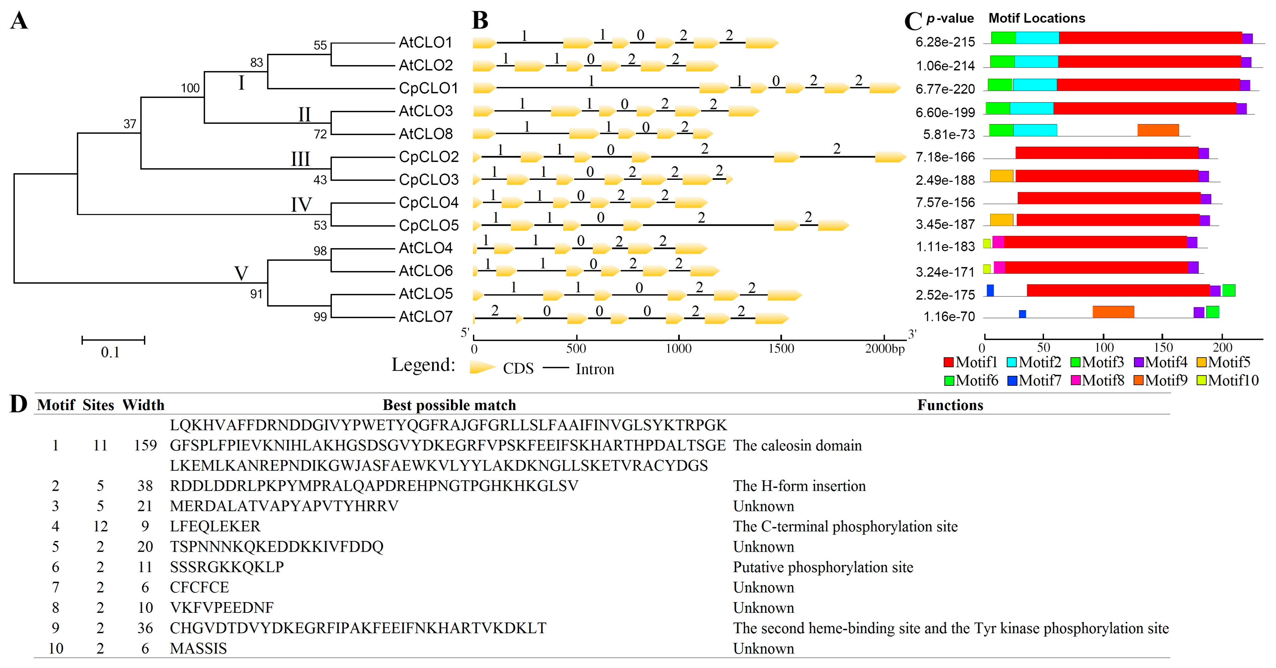
Despite residing in the same order of Brassicales, there are five CpCLO genes identified in papaya, which is relatively less than the eight present in A. thaliana [19], reflecting the occurrence of two additional WGDs in the latter [33]. To uncover their evolutionary relationships, an unrooted phylogenetic tree was constructed using full-length peptides. As shown in Figure 2A, 13 caleosins were clustered into five groups: Group I includes three members from two species, i.e., CpCLO1, AtCLO1, and AtCLO2; Group II and V are A. thaliana-specific, including AtCLO3/–8 and AtCLO4/–5/–6/–7, respectively; and Group III and IV are papaya-specific, including CpCLO2/–3 and CpCLO4/–5, respectively. Among them, Groups I and II belong to the H clade, as described in A. thaliana [19], where CpCLO1 exhibits 84.21% and 83.33% sequence similarities with AtCLO1 and AtCLO2, which are relatively higher than the similarities of 76.86% and 57.68% with AtCLO3 and AtCLO8, respectively. Groups III, IV, and V belong to the L clade, where 68.37–74.76% sequence similarities were observed among CpCLO2–5, usually higher than those of among AtCLO4–7 (Table S1). Interestingly, comparative sequence similarities of 70.33–74.04% were observed between AtCLO4 and CpCLO2–5 (Table S1), implying their close relationships.
Exon–intron structures of Cp/AtCLO genes were further investigated. As shown in Figure 2B, most genes feature five introns with the phase pattern of 1, 1, 0, 2, and 2, where “0”, “1”, and “2” indicate that an intron is located between codons, the first and second bases of a codon, and the second and third bases of a codon, respectively. Nevertheless, CpCLO3 was shown to have gained one more intron in phase 2 at the 3′ end; AtCLO8 has lost the last intron and exhibits the phases 1, 1, 0, and 2; and AtCLO7 harbors the unusual intron phases of 2, 0, 0, 0, 2, and 2, implying their divergence.
The conserved motifs of Cp/AtCLOs were also compared. Among ten motifs identified using MEME, Motifs 1 and 4 are widely distributed (Figure 2C,D), which were characterized as the caleosin domain and the C-terminal phosphorylation site, as shown in Figure 1D, respectively. By contrast, other motifs appear to be group or sequence-specific, i.e., Motifs 2 and 3 for H-forms, Motif 9 for AtCLO7 and –8, Motif 5 for CpCLO3 and –5, Motifs 6 and 7 for AtCLO5 and –7, Motifs 8 and 10 for AtCLO4 and –6 (Figure 2C). Among them, Motif 2 was characterized as the H-form insertion (Figure 2D), whereas Motif 9 was also characterized as the caleosin domain, more precisely the region including the second heme-binding site and the Tyr kinase phosphorylation site, as shown in Figure 1D. These results support the divergence of H- and L-forms and imply possible functional divergence even within the five groups identified in this study.

2.3. Characterization of Caleosin Genes from Representative Plant Species and Insights into Lineage-Specific Family Evolution in Brassicales

Distinct evolution patterns observed between papaya and A. thaliana impelled us to investigate whether the identified duplication events were species or lineage-specific. For the purpose, caleosin family genes were further identified from representative plant species, including A. trichopoda (the basal angiosperm in Amborellales), Aquilegia coerulea (a Ranunculales member of the basal-most eudicot clade that did not share the γ WGT), castor bean (an Euphorbiaceae plant in Malpighiales that did not experience any recent WGD), and nine Brassicales plants, i.e., horseradish tree (Moringa oleifera, a Moringaceae plant that did not experience any recent WGD), Bretschneidera sinensis (an Akaniaceae plant that experienced one recent WGD known as Bs-α), caperbush (Capparis spinosa, a Capparaceae plant that experienced the β WGD and one so-called Cs-α WGD), Cleome violacea (a Cleomaceae plant that experienced the β WGD), acaya (Gynandropsis gynandra, a Cleomaceae plant that experienced the β WGD and one so-called Gg-α WGD), spider flower (Tarenaya hassleriana, a Cleomaceae plant that experienced the β WGD, the Gg-α WGD, and one so-called Th-α WGD), saltwater cress (Eutrema salsugineum, a Brassicaceae plant that experienced the β and α WGDs), A. lyrata (a Brassicaceae plant that experienced the β and α WGDs), and A. Ahalleri (a Brassicaceae plant that experienced the β and α WGDs) [40,41,42,43,44,45,46,47]. In accordance with previous studies [18,30], two members that include one H-form and one L-form were identified from both updated genomes of both A. trichopoda and castor bean [42,44]. By contrast, three to seven members were found in other species (Table S2). Interestingly, though the majority of caleosin genes identified in this study possess five introns with the conserved phase pattern of 1, 1, 0, 2, and 2, RcCLO2 was shown to contain six introns with the phases of 1, 1, 0, 2, 2, and 2, as observed in CpCLO3 (Table S2), implying independent gain of such an intron.
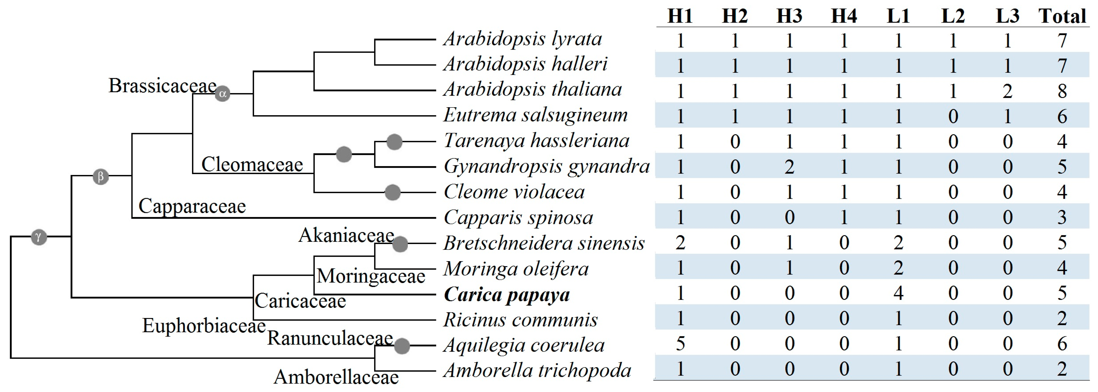
To infer lineage-specific evolution, orthogroups were identified using Orthofinder. As shown in Figure 3, a total of seven orthogroups were obtained. Among them, H1 and L1 are widely present, including A. trichopoda and castor bean, whereas others appear to be lineage-specific. Unlike papaya and caperbush, all other Brassicales species examined in this study contain at least one H3 member, whereas species sharing the β WGD possess one H4 member. Moreover, all tested Brassicaceae species harbor at least one L3 member, whereas three Arabidopsis plants contain one L2 member (Figure 3). Notably, compared with other H4 members, the short peptide length of AtCLO8 was shown to result from a “T” base in the fourth exon (Figure S2). Moreover, compared with A. thaliana, both A. lyrata and A. Ahalleri only retain gene fragments homologous to AtCLO7, implying Arabidopsis-specific tandem duplication followed by pseudogenization in these two species.
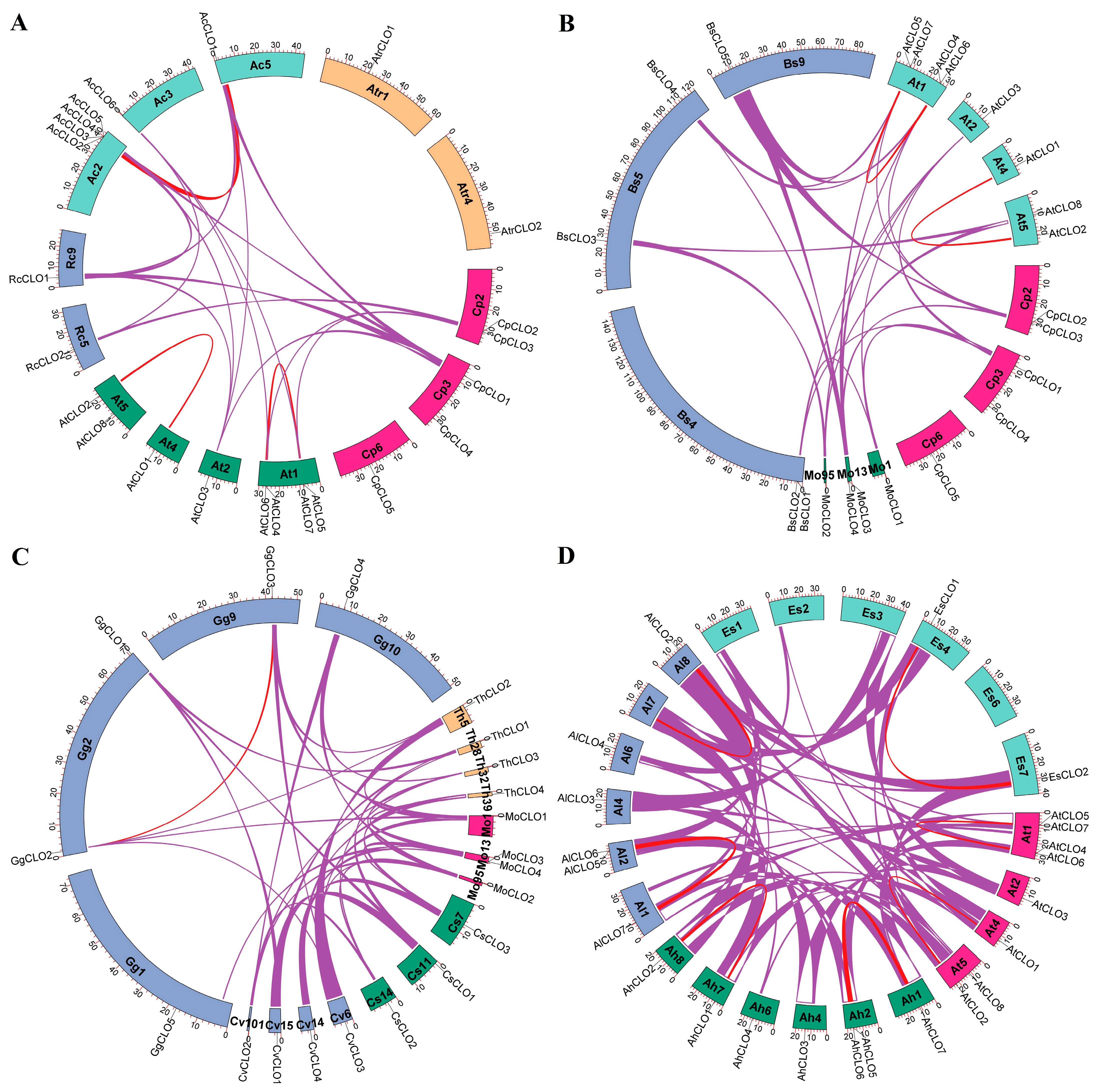
To learn more about the origin and evolution of caleosin genes, species-specific duplication events and interspecific syntenic analyses were further conducted. Notably, the H clade has extensively expanded in A. coerulea via WGD, proximal, and tandem duplications (Table S2), resulting in five members in H1, in stark contrast to species-specific expansion of the L1 group in papaya (four members) (Figure 3). Similarly to papaya, no WGD repeat was found in A. trichopoda, castor bean, horseradish tree, caperbush, C. violacea, or spider flower. By contrast, one to two WGD repeats were identified in other species, i.e., A. coerulea (AcCLO1/–2), B. sinensis (BsCLO1/–3 and BsCLO4/–5), acaya (GgCLO2/–3), saltwater cress (EsCLO1/–2 and EsCLO5/–6), A. lyrata (AlCLO1/–2 and AlCLO5/–7), and A. Ahalleri (AhCLO1/–2 and AhCLO5/–7) (Table S2), reflecting the occurrence of one or more recent WGDs [42,46,47]. Interestingly, though AtrCLO2 was characterized as a transposed repeat of AtrCLO1, L1 (e.g., RcCLO2) was usually characterized as a dispersed repeat of H1 (e.g., RcCLO1) (Table S2). Since no syntelog was identified for either AtrCLO1 or –2 in all other species examined in this study, species-specific chromosome rearrangement or translocation in A. trichopoda could be speculated. By contrast, two out of five CpCLO genes, i.e., CpCLO1 and –2, were shown to have syntelogs in most tested species, with the exception of A. trichopoda, implying a conserved evolution of H1 and L1. Notably, though AtCLO3 was characterized as a dispersed repeat of AtCLO1 residing in H3, it was shown to be located within syntenic blocks with CpCLO1, RcCLO1, AcCLO1, and AcCLO2 (Figure 4A), implying WGD-derivation of H3 from H1 followed by chromosome rearrangement or translocation. Moreover, despite the fragmented status of the horseradish tree genome in 33,332 scaffolds, besides MoCLO1 (H1) and –3 (L1), MoCLO2 (H3) also harbors syntelogs in B. sinensis (BsCLO3), A. thaliana (AtCLO3 and –8) (Figure 4B), caperbush (CsCLO2), C. violacea (CvCLO2 and –3), acaya (GgCLO4), spider flower (ThCLO2), A. coerulea (AcCLO1 and –2) (Figure 4C), castor bean (RcCLO1), and A. coerulea (AcCLO1 and –2) (Figure S3), implying early origin of H3. Since horseradish tree and castor bean did not undergo any recent WGD after the γ WGT shared by all core eudicots [34], the syntenic relationships of 2:1 and 2:2 observed between horseradish tree (MoCLO1 and –2) and castor bean (RcCLO1)/A. coerulea (AcCLO1 and –2) (Figure S3) imply the birth of H3 from the γ WGT, followed by species-specific gene loss in papaya, caperbush, and castor bean (Figure 3). The location of MoCLO2 (H3), CvCLO2 (H3), CvCLO3 (H4), AtCLO3 (H3), and AtCLO8 (H4) within syntenic blocks (Figure 4B,C) provides direct evidence of H4 from H3, most likely as a result of the β WGD, though transposition was observed in four Brassicaceae plants (Table S2). By contrast, H2 and L3, which are specific to Brassicaceae plants and are all located within syntenic blocks with H1 and L1, respectively (Figure 4D), are most likely to arise from the α WGD. Moreover, the majority of caleosin genes present in four tested Brassicaceae plants are still located within syntenic blocks, exhibiting 1:1 and 2:2 relationships (Figure 4D), which is in accordance with the relatively short time of their divergence.

2.4. Caleosin Genes in Castor Bean and Papaya Underwent Apparent Expression Divergence

Species-specific expansion of the caleosin family in papaya propelled us to study their expression profiles and possible function divergence. For the purposes, expression profiles of RcCLO genes (each for two clades) were first examined in leaves, male flowers, II/III endosperms, V/VI endosperms, and germinating seeds. As shown in Figure S4A, similar to RcOLE genes, RcCLO1 is preferentially expressed in endosperms. By contrast, RcCLO2 is predominantly expressed in male flowers, germinating seeds, and leaves (Figure S4A), implying expression divergence of H and L forms. Notably, the total transcripts of the RcCLO family in male flowers and leaves are relatively more than those of the RcOLE family, but considerably less than those in endosperms and germinating seeds (Figure S4A).
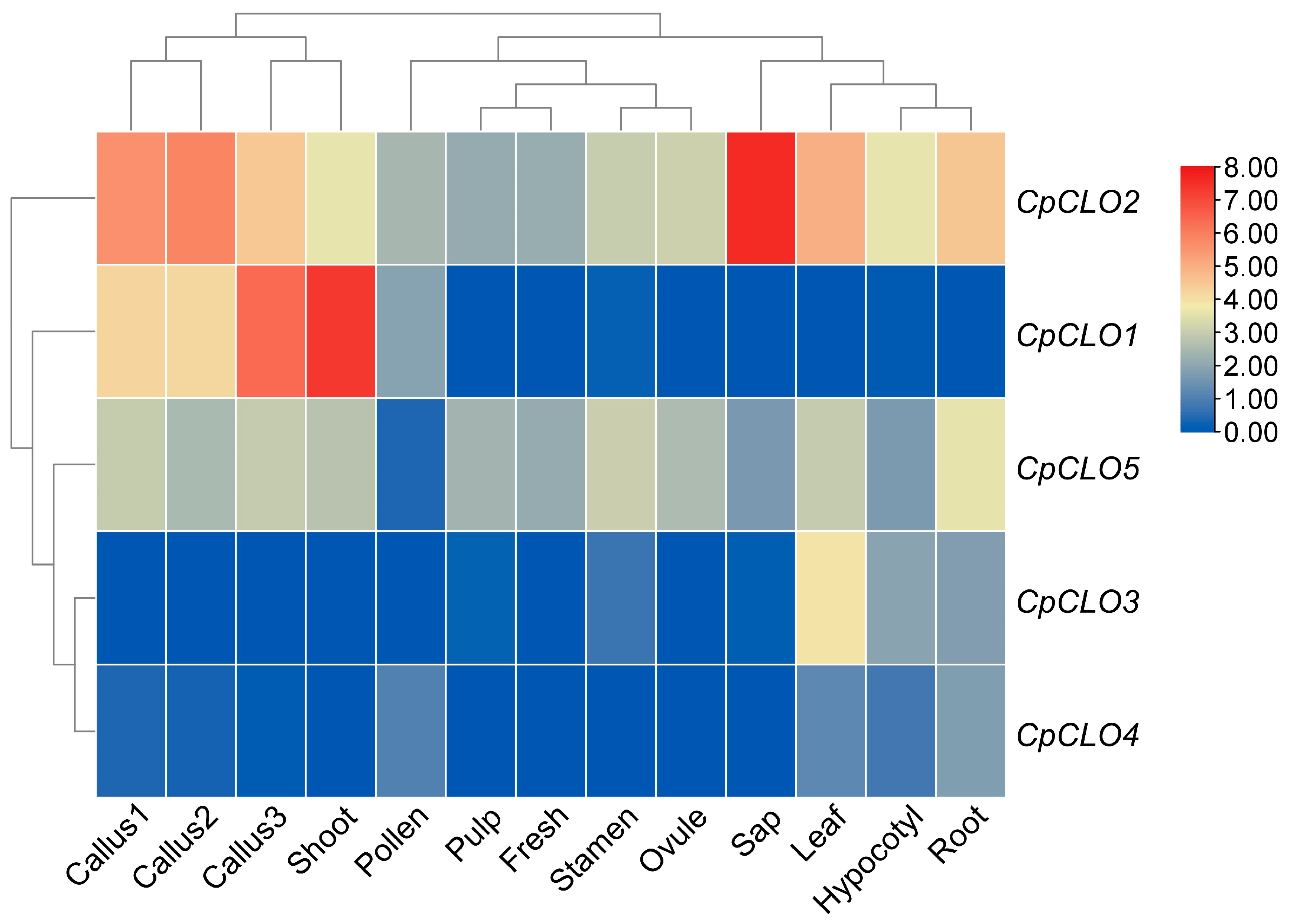
Tissue-specific expression profiles of five CpCLO genes are shown in Figure 5. Though their transcripts were detected in at least one of 13 samples examined in this study, i.e., callus (three stages termed I, II, and III), shoot, hypocotyl, root, leaf, sap, stamen, pollen, ovule, fresh, and pulp, distinct expression patterns were observed. Similarly to the CpOLE family, total transcripts of the CpCLO family are highly abundant in shoot and three stages of callus, though the most expressed tissue is the sap (Figure S4B). Interestingly, the majority of CpCLO transcripts in the sap are contributed by CpCLO2, a constitutively expressed member occupying 43.48–71.85% of total transcripts in pulp, stamen, fresh, pollen, leaf, root, ovule, hypocotyl, callus1, and callus2. By contrast, CpCLO1 contributes to 74.69% and 90.34% of total transcripts in callus3 and shoot samples, respectively. CpCLO5 is also constitutively expressed, though its abundances in most tested samples are considerably lower than those of CpCLO2. The expression of CpCLO24 was shown to be tissue-specific. CpCLO2 is highly abundant in shoot, callus3, callus1, and callus2 and has low abundance in pollen, but is rarely expressed in other tissues. CpCLO3 is predominantly expressed in leaf, hypocotyl, and root, but rarely in other tissues, whereas CpCLO4 is preferentially expressed in leaf, root, and pollen, but rarely in other tissues (Figure 5). Correspondingly, the shoot tissue is clustered with three stages of callus with high abundances of CpOLE and CpCLO transcripts. Moreover, CpCLO1 is clustered with CpOLE25, which are highly abundant in shoot and three stages of callus; CpCLO2 is clustered with CpOLE6, the unique CpOLE member that is constitutively expressed in all tested tissues; and CpCLO35 are clustered with CpOLE1 (Figure S4B). These results imply apparent expression divergence of CpCLO genes even between four L forms (i.e., CpOLE25). Additionally, similar to those observed in castor bean, the total transcripts of the CpCLO family in leaves, roots, and sap are relatively larger than those of the CpOLE family (Figure S4B).

3. Discussion

LDs are lipid storage compartments that are enclosed by a monolayer of phospholipids and several structural proteins such as oleosins, caleosins, and steroleosins [17]. Among them, oleosins and caleosins are typical for the presence of a 12-residue proline-knot motif, which is necessary for budding-LDs to enter the cytosol [48]. In oilseeds, the LD size is largely determined by the amounts of oleosins and caleosins [25,49]. Oleosins first appeared in the single-celled algae and have diverged into at least seven clades known as P (primitive), U (universal), SL (seed low MW), SH (seed high MW), T (tapetum), M (mesocarp), and N (novel) during later evolution [23,50]. By contrast, only two clades of caleosins known as H and L have been described in higher plants [19]. In papaya, six oleosin genes representing five clades (i.e., U, SL, SH, M, and N) were identified, and the comparison with representative plant species revealed that the T clade is limited to the Brassicaceae family, appearing sometime after the split with Cleomaceae [23]. To fill the gap of research on CpCLO genes, in this study, we first conducted a genome-wide analysis of the caleosin family in papaya, and further addressed lineage-specific family evolution in the whole Brassicales order.
In contrast to only two members representing two clades that were found in castor bean and A. trichopoda ([18,30], this study), an unexpectedly high number of five caleosin genes were identified in papaya, though the family amounts were lower than the 7 to 13 reported in A. thaliana and other Brassicales plants [19,20]. Phylogenetic analysis and sequence comparison revealed that, like A. thaliana, papaya also includes two clades, i.e., one H-form and four L-forms, where the H clade features an extra N-terminal insertion of 29 residues that has been named H insertion [19]. Notably, the composition is different from A. thaliana, which contains four H-forms and four L-forms [19], implying species- or lineage-specific expansion.
To gain insights into the origin and evolution of caleosin genes, a total of 63 members were further identified from 13 representative plant species, which belong to eight plant families, i.e., Amborellaceae (A. trichopoda), Ranunculaceae (A. coerulea), Euphorbiaceae (castor bean), Moringaceae (horseradish tree), Akaniaceae (B. sinensis), Capparaceae (caperbush), Cleomaceae (C. violacea, acaya, and spider flower), and Brassicaceae (saltwater cress, A. Ahalleri, A. lyrata, and A. thaliana). Among them, castor bean, A. coerulea, and A. trichopoda were adopted as out-groups of Brassicales plants, core eudicots, and core angiosperms, respectively. Interestingly, despite the occurrence of one or more additional WGDs after the split with papaya, comparative or a relatively smaller number of caleosin family genes were found in caperbush (3), C. violacea (4), spider flower (4), B. sinensis (5), and acaya (5). Moreover, only four members were identified in horseradish tree, another Brassicales plant without a recent WGD [41]. Nevertheless, four MoCLO genes belong to three out of the seven orthogroups identified in this study, i.e., H1, H3, and L1, in contrast with only two (i.e., H1 and L1) for five CpCLO genes. Since H1 and L1 were shown to be shared by A. trichopoda, their early origin before angiosperm radiation could be speculated. Though H3 is absent from castor bean, papaya, and caperbush, it is present in all other Brassicales plants examined in this study. An interesting result is that RcCLO1 (H1), MoCLO1 (H1), and MoCLO2 (H3) are located within syntenic blocks, supporting WGD-derivation of H3 from H1, probably the γ WGT occurred in the last common ancestor of core eudicots after the split with A. coerulea [34,42]. Compared with papaya and horseradish tree, other tested Brassicales plants were proven to have experienced at least one recent WGD, which resulted in several duplicate pairs, i.e., one in B. sinensis (Bs-α), one in acaya (Gg-α), and two in four Brassicaceae plants (α). Two WGD repeats shared by saltwater cress, A. Ahalleri, A. lyrata, and A. thaliana formed two Brassicaceae-specific groups, i.e., H2 and L3, as defined in this study. Notably, though H4 was characterized as the transposed repeat of H3 in all four tested Brassicaceae plants, they were all shown to be located within syntenic blocks with the H3 member in both horseradish tree (MoCLO2) and C. violacea (CvCLO2), supporting WGD-derivation of H4 from H3—probably the β WGD shared by several families within Brassicales, e.g., Capparaceae, Cleomaceae, and Brassicaceae [45,47]. Additionally, since this transposed event is not found in other species beyond Brassicaceae, its appearance in the last common ancestor of Brassicaceae plants could be speculated. Taken together, possible evolution routes of the caleosin family in Brassicales are as follows: the family first diverged into H1 and L1 before angiosperm radiation, and the γ WGT gave birth to H3, though it has been lost in castor bean, papaya, and caperbush; in papaya, the L clade underwent extensive expansion via species-specific tandem and transposed duplications; in horseradish tree, the L clade also underwent species-specific expansion via tandem duplication; in B. sinensis, H1 and L1 underwent species-specific expansion via tandem duplication and Bs-α WGD, respectively; the β WGD gave rise to H4 from H3, though H3 was lost in caperbush during latter evolution; in acaya, H3 was further expanded via the Gg-α; in Brassicaceae, the α WGD resulted in H2 and L3, whereas L1 in Arabidopsis further gave birth to L2 via tandem duplication; L3 was also expanded via tandem duplication, which was retained in A. thaliana but underwent pseudogenization in both A. lyrata and A. Ahalleri.
In addition to local duplication and species-specific retention of repeats after WGDs, structural and expression divergences were also shown to play key roles in the evolution of the caleosin family. Generally, caleosin genes feature five introns in the phase pattern of 1, 1, 0, 2, and 2. However, independent gain or loss of certain introns was also observed in CpCLO3, RcCLO2, AtCLO8, and AtCLO7. Moreover, unusual intron phases of 2, 0, 0, 0, 2, and 2 were also observed in AtCLO7. Lineage or species-specific gain of certain introns was also reported for the oleosin family, e.g., CpOLE3 [20,21,23,48], though their biological significances have not been clarified.
Over more than 200 million years of evolution, apparent expression divergence was observed between two clades of caleosin genes. A good example is castor bean, an oil seed crop of the Euphorbiaceae family [44] possessing a single H-form and one L-form. Whereas the L-form RcCLO2 is constitutively expressed, the H-form RcCLO1 has evolved to be preferentially expressed in seeds/endosperms, which are consistent with a previous study [18]. Though no seed transcriptome is available in papaya, analyzing 13 tissue samples showed that the H-form CpCLO1 is predominantly expressed in shoots and the last stage of callus development, when CpOLE genes are highly abundant [23]. As for four L-forms, CpCLO2 and –5 were shown to be constitutively expressed, whereas CpCLO3 and –4 exhibit apparent tissue-specific expression patterns. Moreover, CpCLO2 has evolved into the dominant member, whose transcripts are usually more than other three L-forms in most tissues, implying its putatively key roles. In A. thaliana, the situation is more complex, as extensive expansion was observed in both clades, i.e., four H-forms and four L-forms [19]. Among the four H-forms, AtCLO1 and –2 were shown to exhibit the seed-preferential expression pattern similar to RcCLO1, whereas AtCLO3 appears to be constitutively expressed [19,51]. Among the four L-forms, AtCLO4 is also constitutively expressed, whereas the other three members seem to be predominantly expressed in flowers [19]. Moreover, the involvement of AtCLO1, –2, –4, and –6 in oil accumulation and embryo development was supported by mutant analyses, which were shown to have overlapping functions [51]. AtCLO1 was also shown to be involved in the degradation of storage lipids during seed germination [3], whereas AtCLO2 was proven to be associated with seed dormancy [52]. The PXG activity, which is Ca2+-dependent, has been reported for AtCLO1–4 [7,24,53]. AtCLO3 was functionally characterized in the generation of oxidized fatty acids in the stress-related ABA and salicylic acid signaling pathways [7], whereas AtCLO4 was characterized as a negative regulator of the ABA response [53].

4. Conclusions

This study presents the first genome-wide analysis of the caleosin family in papaya, resulting in a high number of five members representing two clades, i.e., H (1) and L (4). Comparison of 68 caleosin genes from 14 representative plant species revealed seven orthogroups, i.e., H1–4 and L1–3, two of which (i.e., H1 and L1) have appeared before angiosperm radiation. Five CpCLO genes belong to H1 and L1, and extensive expansion of the L1 group appears to be contributed by species-specific tandem and transposed duplications, which may contribute to environmental adaptation. By contrast, WGDs were shown to play a more important role in family expansion in Brassicaceae plants such as A. thaliana. Structural (e.g., gain or loss of certain introns) and apparent expression divergence of caleosin genes was also observed. These findings provide a global view of CpCLO genes and insights into lineage-specific family evolution in Brassicales, which facilitates further functional studies in papaya and other nonmodel species.

5. Materials and Methods

5.1. Databases and Identification of Caleosin Family Genes

Genomic sequences of representative plant species, i.e., A. trichopoda (v2.1; Amborellaceae, Amborellales), A. coerulea (v3.1; Ranunculaceae, Ranunculales), R. communis (WT05; Euphorbiaceae, Malpighiales), C. papaya (Sunset v1; Caricaceae, Brassicales), M. oleifera (v1; Moringaceae, Brassicales), B. sinensis (v1; Akaniaceae, Brassicales), C. spinosa (v1; Capparaceae, Brassicales), C. violacea (v2.1; Cleomaceae, Brassicales), G. gynandra (v1; Cleomaceae, Brassicales), T. hassleriana (v1; Cleomaceae, Brassicales), E. salsugineum (v1.0; Brassicaceae, Brassicales), A. halleri (v2.1; Brassicaceae, Brassicales), A. lyrata (v2.1; Brassicaceae, Brassicales), and A. thaliana (Araport11; Brassicaceae, Brassicales), were downloaded from NGDC (http://bigd.big.ac.cn/gsa, accessed on 31 January 2025), Phytozome (v13, https://phytozome.jgi.doe.gov/pz/portal.html, accessed on 31 January 2025), and NCBI (https://www.ncbi.nlm.nih.gov/, accessed on 31 January 2025). The caleosin domain profile (PF05042) was obtained from Pfam 33.1 (https://pfam.xfam.org/, accessed on 31 January 2025), which was used for HMMER (v3.3, http://hmmer.janelia.org/, accessed on 31 January 2025) searches, as described before [54]. Gene models of candidates were manually curated with available mRNAs, and presence of the conserved caleosin domain in deduced peptides was confirmed using MOTIF Search (https://www.genome.jp/tools/motif/, accessed on 31 January 2025). Pseudogenes and/or homologous fragments were identified with coding sequences (CDSs) of obtained caleosin genes using BLASTN (v2.17.0) as previously described [55]. Exon–intron structures were displayed using GSDS2.0 (http://gsds.cbi.pku.edu.cn/, accessed on 31 January 2025), whereas protein properties, hydropathicity scales, and secondary structures were determined using ProtParam (http://web.expasy.org/protparam/, accessed on 31 January 2025), ProtScale (v1) (https://web.expasy.org/protscale/, accessed on 31 January 2025), and SOPMA (v1) (https://npsa-prabi.ibcp.fr/cgi-bin/npsa_automat.pl?page=/NPSA/npsa_sopma.html, accessed on 31 January 2025), respectively.

5.2. Multiple Sequence Alignment, Phylogenetic, and Conserved Motif Analyses

Sequence alignment of nucleotides and proteins was performed using ClustalW (v2.1, www.megasoftware.net, accessed on 31 January 2025) and MUSCLE (v5, http://www.drive5.com/muscle/, accessed on 31 January 2025), respectively, and alignment display was carried out using Boxshade (v1) (https://embnet.vital-it.ch/software/http://www.bio-soft.net/sms/, accessed on 31 January 2025). A phylogenetic tree was constructed using RAxML (v2) (http://www.phylo.org/portal2/home.action#, accessed on 31 January 2025) as previously described [56], which implemented the maximum likelihood method and bootstrap of 1000 replicates. Conserved motifs were identified using MEME (v5.4.1, http://meme-suite.org/tools/meme, accessed on 31 January 2025) with the following parameters: any number of repetitions; the maximum number of motifs, 10; and the optimum width of each motif, between 5 and 200 residues.

5.3. Definition of Orthogroups, Chromosomal Localization, Synteny Analysis, and Calculation of Evolutionary Rate

Orthologous genes were clustered using Orthofinder (v2.3.8) [57], whereas chromosomal localization was conducted using TBtools-II (v2.210) [58]. For synteny analysis, duplicate pairs were identified using the all-to-all BLASTp (v2.17.0) method with the E-value cutoff of 1 × 10⁻¹⁰, and gene colinearity was inferred using MCScanX (v2.0) with the cutoff of five BLAST hits as previously described [56]. Duplication modes such as tandem, proximal, transposed, dispersed, and WGD were identified using the DupGen_finder pipeline [59], and the Ks and Ka of duplicate pairs were calculated using codeml [60].

5.4. Gene Expression Analysis

Global expression profiles of RcCLO and CpCLO genes were analyzed on the basis of Illumina RNA-seq samples, as shown in Table S3. Five samples examined in castor been are the cotyledons of germinating seeds, expanding true leaves that appear after the first cotyledons and leaf-pair, developing male flowers, endosperm II/III (free-nuclear stage of developing seeds), and endosperm V/VI (onset of cellular endosperm development). The thirteen samples tested in papaya are hypocotyl (7 days on MS medium), callus I (21 days on callus induction medium K5), callus II (21 days on callus induction medium M13), callus III (21 days on callus induction medium CI), shoot (28 days on shoot induction medium), root, leaf, phloem sap, stamen, pollen, and ovule. Quality control of raw reads was carried out using Trimmomatic (v1) [61], and read mapping was performed using HISAT2 [62]. Relative gene expression levels were normalized using the FPKM (fragments per kilobase of exon per million fragments mapped) method [63], whereas differential analysis was conducted using DESeq2 (v1.22.1) [64] under the criteria of |log2 fold change (FC)| ≥ 1 and false discovery rate (FDR) < 0.05. Unless specifically stated, the tools in this study were used with default parameters.

Supplementary Materials

The following supporting information can be downloaded at https://www.mdpi.com/article/10.3390/plants14213296/s1, Figure S1: Chromosome localization and duplication events detected in A. thaliana. Chromosome localization was conducted using TBtools-II, whereas different modes of gene duplication were identified using the DupGen_finder pipeline. Serial numbers are indicated at the top of each chromosome, and the scale is in Mb. Duplicate pairs identified in this study are connected using lines in different colors, i.e., WGD (red), tandem (green), transposed (purple), and dispersed (gold). (At: A. thaliana; CLO: caleosin). Figure S2: Sequence alignment of AlCLO4, AhCLO4, and AtCLO8. Sequence alignment was performed using ClustalX. Broken lines in the sequences represent gaps introduced for best alignment, whereas identical nucleotides are indicated using asterisks. (Ah: A. halleri; Al: A. lyrata; At: A. thaliana; CLO: caleosin). Figure S3: Synteny analyses of caleosin genes within and between M. oleifera, R. communis, and A. coerulea. Syntenic blocks were inferred using MCScanX (E-value ≤ 1 × 10⁻¹⁰; BLAST hits ≥ 5). Shown are caleosin gene-encoding chromosomes/scaffolds and only syntenic blocks containing caleosin genes are marked, where red and purple lines indicate intra- and inter-species, respectively. (Ac: A. coerulea; CLO: caleosin; Mo: M. oleifera; Rc: communis). Figure S4: Expression profiles of caleosin and oleosin genes in R. communis (A) and C. papaya (B). The heatmaps were generated using the R package, where color scales represent FPKM normalized Log2 transformed counts. Blue and red indicate low and high expression, respectively. (CLO: caleosin; Cp: C. papaya; FPKM: fragments per kilobase of exon per million fragments mapped). Table S1: Percent similarity within and between CpCLOs and AtCLOs. (At: A. thaliana; CLO: caleosin). Table S2: List of caleosin genes identified in A. thaliana and representative plant species. (Ac: A. coerulea; Ah: A. halleri; Al: A. lyrata; At: A. thaliana; Atr: A. trichopoda; Bs: B. sinensis; CLO: caleosin; Cs: C. spinosa; Cv: C. violacea; Es: E. salsugineum; Gg: G. gynandra; Mo: M. oleifera; Rc: R. communis; Th: T. hassleriana). Table S3: Detailed information of transcriptome data used in this study.

Author Contributions

Z.Z. conceived the idea and designed the project outline. Z.Z., X.F., X.Y., C.L. and Y.Z. performed the experiments and analyzed the data. Z.Z. and Y.Z. prepared and refined the manuscript. All authors have read and agreed to the published version of the manuscript.

Funding

This work was supported by the Hainan Province Science and Technology Special Fund (ZDYF2024XDNY171) and the National Natural Science Foundation of China (32460342 and 31971688). The funders had no role in study design, data collection and analysis, decision to publish, or preparation of the manuscript.

Data Availability Statement

The datasets analyzed during the current study are available in the NCBI SRA repository (https://www.ncbi.nlm.nih.gov/sra/ (accessed on 31 January 2025)), whose accession numbers are shown in Table S3.

Acknowledgments

The authors appreciate those contributors who make the related genome and transcriptome data accessible in public databases. They also appreciate two reviewers for helpful suggestions.

Conflicts of Interest

The authors declare no conflicts of interest.

References

  1. Frandsen, G.; Müller-Uri, F.; Nielsen, M.; Mundy, J.; Skriver, K. Novel plant Ca2+-binding protein expressed in response to abscisic acid and osmotic stress. J. Biol. Chem. 1996, 271, 343–348. [Google Scholar] [CrossRef]
  2. Takahashi, S.; Katagiri, T.; Yamaguchi-Shinozaki, K.; Shinozaki, K. An Arabidopsis gene encoding a Ca2+-binding protein is induced by abscisic acid during dehydration. Plant Cell Physiol. 2000, 41, 898–903. [Google Scholar] [CrossRef]
  3. Poxleitner, M.; Rogers, S.W.; Lacey Samuels, A.; Browse, J.; Rogers, J.C. A role for caleosin in degradation of oil-body storage lipid during seed germination. Plant J. 2006, 47, 917–933. [Google Scholar] [CrossRef] [PubMed]
  4. Jiang, P.L.; Chen, J.C.; Chiu, S.T.; Tzen, J.T. Stable oil bodies sheltered by a unique caleosin in cycad megagametophytes. Plant Physiol. Biochem. 2009, 47, 1009–1016. [Google Scholar] [CrossRef] [PubMed]
  5. Chen, E.C.; Tai, S.S.; Peng, C.C.; Tzen, J.T. Identification of three novel unique proteins in seed oil bodies of sesame. Plant Cell Physiol. 1998, 39, 935–941. [Google Scholar] [CrossRef]
  6. Hernandez-Pinzon, I.; Patel, K.; Murphy, D.J. The Brassica napus calcium-binding protein, caleosin, has distinct endoplasmic reticulum- and lipid body-associated isoforms. Plant Physiol. Biochem. 2001, 39, 615–622. [Google Scholar] [CrossRef]
  7. Partridge, M.; Murphy, D.J. Roles of a membrane-bound caleosin and putative peroxygenase in biotic and abiotic stress responses in Arabidopsis. Plant Physiol. Biochem. 2009, 47, 796–806. [Google Scholar] [CrossRef] [PubMed]
  8. Miklaszewska, M.; Zienkiewicz, K.; Klugier-Borowska, E.; Rygielski, M.; Feussner, I.; Zienkiewicz, A. CALEOSIN 1 interaction with AUTOPHAGY-RELATED PROTEIN 8 facilitates lipid droplet microautophagy in seedlings. Plant Physiol. 2023, 193, 2361–2380. [Google Scholar] [CrossRef]
  9. Naested, H.; Frandsen, G.I.; Jauh, G.Y.; Hernandez-Pinzon, I.; Nielsen, H.B.; Murphy, D.J.; Rogers, J.C.; Mundy, J. Caleosins: Ca2+-binding proteins associated with lipid bodies. Plant Mol. Biol. 2000, 44, 463–476. [Google Scholar] [CrossRef]
  10. Jiang, P.L.; Jauh, G.Y.; Wang, C.S.; Tzen, J.T. A unique caleosin in oil bodies of lily pollen. Plant Cell Physiol. 2008, 49, 1390–1395. [Google Scholar] [CrossRef]
  11. Zienkiewicz, K.; Castro, A.J.; Alché Jde, D.; Zienkiewicz, A.; Suárez, C.; Rodríguez-García, M.I. Identification and localization of a caleosin in olive (Olea europaea L.) pollen during in vitro germination. J. Exp. Bot. 2010, 61, 1537–1546. [Google Scholar] [CrossRef] [PubMed]
  12. Pasaribu, B.; Chung, T.Y.; Chen, C.S.; Wang, S.L.; Jiang, P.L.; Tzen, J.T. Identification of caleosin and two oleosin isoforms in oil bodies of pine megagametophytes. Plant Physiol. Biochem. 2014, 82, 142–150. [Google Scholar] [CrossRef]
  13. Pasaribu, B.; Chen, C.S.; Liao, Y.K.; Jiang, P.L.; Tzen, J.T. Identification of caleosin and oleosin in oil bodies of pine pollen. Plant Physiol. Biochem. 2017, 111, 20–29. [Google Scholar] [CrossRef]
  14. Pasaribu, B.; Fu, J.H.; Jiang, P.L. Identification and characterization of caleosin in Cycas revoluta pollen. Plant Signal Behav. 2020, 15, 1779486. [Google Scholar] [CrossRef]
  15. Lu, J.Y.; Xiong, S.X.; Yin, W.; Teng, X.D.; Lou, Y.; Zhu, J.; Zhang, C.; Gu, J.N.; Wilson, Z.A.; Yang, Z.N. MS1, a direct target of MS188, regulates the expression of key sporophytic pollen coat protein genes in Arabidopsis. J. Exp. Bot. 2020, 71, 4877–4889. [Google Scholar] [CrossRef] [PubMed]
  16. Chen, J.C.; Tsai, C.C.; Tzen, J.T. Cloning and secondary structure analysis of caleosin, a unique calcium-binding protein in oil bodies of plant seeds. Plant Cell Physiol. 1999, 40, 1079–1086. [Google Scholar] [CrossRef]
  17. Huang, A.H. Plant lipid droplets and their associated proteins: Potential for rapid advances. Plant Physiol. 2018, 176, 1894–1918. [Google Scholar] [CrossRef]
  18. Hyun, T.K.; Kumar, D.; Cho, Y.Y.; Hyun, H.N.; Kim, J.S. Computational identification and phylogenetic analysis of the oil-body structural proteins, oleosin and caleosin, in castor bean and flax. Gene 2013, 515, 454–460. [Google Scholar] [CrossRef]
  19. Shen, Y.; Xie, J.; Liu, R.D.; Ni, X.F.; Wang, X.H.; Li, Z.X.; Zhang, M. Genomic analysis and expression investigation of caleosin gene family in Arabidopsis. Biochem. Biophys. Res. Commun. 2014, 448, 365–371. [Google Scholar] [CrossRef]
  20. Zou, Z.; Zhao, Y.; Zhang, L. Genomic insights into lineage-specific evolution of the oleosin family in Euphorbiaceae. BMC Genom. 2022, 23, 178. [Google Scholar] [CrossRef] [PubMed]
  21. Zou, Z.; Zheng, Y.J.; Zhang, Z.T.; Xiao, Y.H.; Xie, Z.N.; Chang, L.L.; Zhang, L.; Zhao, Y.G. Molecular characterization oleosin genes in Cyperus esculentus, a Cyperaceae plant producing oil in underground tubers. Plant Cell Rep. 2023, 42, 1791–1808. [Google Scholar] [CrossRef] [PubMed]
  22. Zou, Z.; Fu, X.W.; Huang, J.Q.; Zhao, Y.G. Molecular characterization of CeOLE6, a diverged SH oleosin gene, preferentially expressed in Cyperus esculentus tubers. Planta 2024, 260, 122. [Google Scholar] [CrossRef]
  23. Zou, Z.; Zhang, L.; Zhao, Y. Integrative analysis of oleosin genes provides insights into lineage-specific family evolution in Brassicales. Plants 2024, 13, 280. [Google Scholar] [CrossRef]
  24. Hanano, A.; Burcklen, M.; Flenet, M.; Ivancich, A.; Louwagie, M.; Garin, J.; Blee, E. Plant seed peroxygenase is an original heme-oxygenase with an EF-hand calcium binding motif. J. Biol. Chem. 2006, 281, 33140–33151. [Google Scholar] [CrossRef]
  25. Chen, J.C.; Tzen, J.T. An in vitro system to examine the effective phospholipids and structural domain for protein targeting to seed oil bodies. Plant Cell Physiol. 2001, 42, 1245–1252. [Google Scholar] [CrossRef]
  26. Blée, E.; Flenet, M.; Boachon, B.; Fauconnier, M.L. A non-canonical caleosin from Arabidopsis efficiently epoxidizes physiological unsaturated fatty acids with complete stereoselectivity. FEBS J. 2012, 279, 3981–3995. [Google Scholar] [CrossRef]
  27. Song, W.; Qin, Y.; Zhu, Y.; Yin, G.; Wu, N.; Li, Y.; Hu, Y. Delineation of plant caleosin residues critical for functional divergence, positive selection and coevolution. BMC Evol. Biol. 2014, 14, 124. [Google Scholar] [CrossRef] [PubMed]
  28. Rahman, F.; Hassan, M.; Hanano, A.; Fitzpatrick, D.A.; McCarthy, C.P.; Murphy, D.J. Evolutionary, structural and functional analysis of the caleosin/peroxygenase gene family in the Fungi. BMC Genom. 2018, 19, 976. [Google Scholar] [CrossRef] [PubMed]
  29. Rahman, F.; Hassan, M.; Rosli, R.; Almousally, I.; Hanano, A.; Murphy, D.J. Evolutionary and genomic analysis of the caleosin/peroxygenase (CLO/PXG) gene/protein families in the Viridiplantae. PLoS ONE 2018, 13, e0196669. [Google Scholar] [CrossRef]
  30. Hanano, A.; Blée, E.; Murphy, D.J. Caleosin/peroxygenases: Multifunctional proteins in plants. Ann. Bot. 2023, 131, 387–409. [Google Scholar] [CrossRef]
  31. Khalil, H.B.; Brunetti, S.C.; Pham, U.M.; Maret, D.; Laroche, A.; Gulick, P.J. Characterization of the caleosin gene family in the Triticeae. BMC Genom. 2014, 15, 239. [Google Scholar] [CrossRef]
  32. Shen, Y.; Liu, M.; Wang, L.; Li, Z.; Taylor, D.C.; Li, Z.; Zhang, M. Identification, duplication, evolution and expression analyses of caleosins in Brassica plants and Arabidopsis subspecies. Mol. Genet. Genom. 2016, 291, 971–988. [Google Scholar] [CrossRef]
  33. Bowers, J.E.; Chapman, B.A.; Rong, J.; Paterson, A.H. Unravelling angiosperm genome evolution by phylogenetic analysis of chromosomal duplication events. Nature 2003, 422, 433–438. [Google Scholar] [CrossRef] [PubMed]
  34. Jiao, Y.; Leebens-Mack, J.; Ayyampalayam, S.; Bowers, J.E.; McKain, M.R.; McNeal, J.; Rolf, M.; Ruzicka, D.R.; Wafula, E.; Wickett, N.J.; et al. A genome triplication associated with early diversification of the core eudicots. Genome Biol. 2012, 13, R3. [Google Scholar] [CrossRef] [PubMed]
  35. Cardinal-McTeague, W.M.; Sytsma, K.J.; Hall, J.C. Biogeography and diversification of Brassicales: A 103 million year tale. Mol. Phylogenet. Evol. 2016, 99, 204–224. [Google Scholar] [CrossRef]
  36. Mabry, M.E.; Brose, J.M.; Blischak, P.D.; Sutherland, B.; Dismukes, W.T.; Bottoms, C.A.; Edger, P.P.; Washburn, J.D.; An, H.; Hall, J.C.; et al. Phylogeny and multiple independent whole-genome duplication events in the Brassicales. Am. J. Bot. 2020, 107, 1148–1164. [Google Scholar] [CrossRef]
  37. Yue, J.; VanBuren, R.; Liu, J.; Fang, J.; Zhang, X.; Liao, Z.; Wai, C.M.; Xu, X.; Chen, S.; Zhang, S.; et al. SunUp and Sunset genomes revealed impact of particle bombardment mediated transformation and domestication history in papaya. Nat. Genet. 2022, 54, 715–724. [Google Scholar] [CrossRef]
  38. Zou, Z.; Li, M.Y.; Jia, R.Z.; Zhao, H.; He, P.P.; Zhang, Y.L.; Guo, A.P. Genes encoding light-harvesting chlorophyll a/b-binding proteins in papaya (Carica papaya L.) and insight into lineage-specific evolution in Brassicaceae. Gene 2020, 748, 144685. [Google Scholar] [CrossRef] [PubMed]
  39. Zou, Z.; Zheng, Y.J.; Xie, Z.N. Analysis of Carica papaya informs lineage-specific evolution of the aquaporin (AQP) family in Brassicales. Plants 2023, 12, 3847. [Google Scholar] [CrossRef]
  40. Cheng, S.; van den Bergh, E.; Zeng, P.; Zhong, X.; Xu, J.; Liu, X.; Hofberger, J.; de Bruijn, S.; Bhide, A.S.; Kuelahoglu, C.; et al. The Tarenaya hassleriana genome provides insight into reproductive trait and genome evolution of crucifers. Plant Cell 2013, 25, 2813–2830. [Google Scholar] [CrossRef]
  41. Tian, Y.; Zeng, Y.; Zhang, J.; Yang, C.; Yan, L.; Wang, X.; Shi, C.; Xie, J.; Dai, T.; Peng, L.; et al. High quality reference genome of drumstick tree (Moringa oleifera Lam.), a potential perennial crop. Sci. China Life Sci. 2015, 58, 627–638. [Google Scholar] [CrossRef]
  42. Filiault, D.L.; Ballerini, E.S.; Mandáková, T.; Aköz, G.; Derieg, N.J.; Schmutz, J.; Jenkins, J.; Grimwood, J.; Shu, S.; Hayes, R.D.; et al. The Aquilegia genome provides insight into adaptive radiation and reveals an extraordinarily polymorphic chromosome with a unique history. eLife 2018, 7, e36426. [Google Scholar] [CrossRef]
  43. Käfer, J.; Bewick, A.; Andres-Robin, A.; Lapetoule, G.; Harkess, A.; Caïus, J.; Fogliani, B.; Gâteblé, G.; Ralph, P.; dePamphilis, C.W.; et al. A derived ZW chromosome system in Amborella trichopoda, representing the sister lineage to all other extant flowering plants. New Phytol. 2022, 233, 1636–1642. [Google Scholar] [CrossRef]
  44. Lu, J.; Pan, C.; Fan, W.; Liu, W.; Zhao, H.; Li, D.; Wang, S.; Hu, L.; He, B.; Qian, K.; et al. A Chromosome-level genome assembly of wild castor provides new insights into its adaptive evolution in tropical desert. Genom. Proteom. Bioinf. 2022, 20, 42–59. [Google Scholar] [CrossRef]
  45. Wang, L.; Fan, L.; Zhao, Z.; Zhang, Z.; Jiang, L.; Chai, M.; Tian, C. The Capparis spinosa var. herbacea genome provides the first genomic instrument for a diversity and evolution study of the Capparaceae family. Gigascience 2022, 11, giac106. [Google Scholar] [CrossRef]
  46. Zhang, H.; Du, X.; Dong, C.; Zheng, Z.; Mu, W.; Zhu, M.; Yang, Y.; Li, X.; Hu, H.; Shrestha, N.; et al. Genomes and demographic histories of the endangered Bretschneidera sinensis (Akaniaceae). Gigascience 2022, 11, giac050. [Google Scholar] [CrossRef]
  47. Zhao, W.; Li, J.; Sun, X.; Zheng, Q.; Liu, J.; Hua, W.; Liu, J. Integrated global analysis in spider flowers illuminates features underlying the evolution and maintenance of C4 photosynthesis. Hortic. Res. 2023, 10, uhad129. [Google Scholar] [CrossRef]
  48. Huang, M.D.; Huang, A.H. Bioinformatics reveal five lineages of oleosins and the mechanism of lineage evolution related to structure/function from green algae to seed plants. Plant Physiol. 2015, 169, 453–470. [Google Scholar] [CrossRef] [PubMed]
  49. Siloto, R.M.P.; Findlay, K.; Lopez, V.A.; Yeung, E.C.; Nykifork, C.L.; Moloney, M.M. The accumulation of oleosins determines the size of seed oil bodies in Arabidopsis. Plant Cell 2006, 18, 1961–1974. [Google Scholar] [CrossRef] [PubMed]
  50. Huang, C.Y.; Huang, A.C. Unique motifs and length of hairpin in oleosin target the cytosolic side of endoplasmic reticulum and budding lipid droplet. Plant Physiol. 2017, 174, 2248–2260. [Google Scholar] [CrossRef] [PubMed]
  51. Liu, X.; Yang, Z.; Wang, Y.; Shen, Y.; Jia, Q.; Zhao, C.; Zhang, M. Multiple caleosins have overlapping functions in oil accumulation and embryo development. J. Exp. Bot. 2022, 73, 3946–3962. [Google Scholar] [CrossRef]
  52. Toorop, P.E.; Barroco, R.M.; Engler, G.; Groot, S.P.; Hilhorst, H.W. Differentially expressed genes associated with dormancy or germination of Arabidopsis thaliana seeds. Planta 2005, 221, 637–647. [Google Scholar] [CrossRef]
  53. Kim, Y.Y.; Jung, K.W.; Yoo, K.S.; Jeung, J.U.; Shin, J.S. A stress-responsive caleosin-like protein, AtCLO4, acts as a negative regulator of ABA responses in Arabidopsis. Plant Cell Physiol. 2011, 52, 874–884. [Google Scholar] [CrossRef]
  54. Zou, Z.; Yang, J.H. Genomic analysis of Dof transcription factors in Hevea brasiliensis, a rubber-producing tree. Ind. Crops Prod. 2019, 134, 271–283. [Google Scholar] [CrossRef]
  55. Zou, Z.; Gong, J.; An, F.; Xie, G.S.; Wang, J.K.; Mo, Y.Y.; Yang, L.F. Genome-wide identification of rubber tree (Hevea brasiliensis Muell. Arg.) aquaporin genes and their response to ethephon stimulation in the laticifer, a rubber-producing tissue. BMC Genom. 2015, 16, 1001. [Google Scholar] [CrossRef] [PubMed]
  56. Zou, Z.; Fu, X.; Li, C.; Yi, X.; Huang, J.; Zhao, Y. Integrative analysis provides insights into genes encoding LEA_5 domain-containing proteins in tigernut (Cyperus esculentus L.). Plants 2025, 14, 762. [Google Scholar] [CrossRef] [PubMed]
  57. Emms, D.M.; Kelly, S. OrthoFinder: Phylogenetic orthology inference for comparative genomics. Genome Biol. 2019, 20, 238. [Google Scholar] [CrossRef]
  58. Chen, C.; Wu, Y.; Li, J.; Wang, X.; Zeng, Z.; Xu, J.; Liu, Y.; Feng, J.; Chen, H.; He, Y.; et al. TBtools-II: A “one for all, all for one” bioinformatics platform for biological big-data mining. Mol. Plant 2023, 16, 1733–1742. [Google Scholar] [CrossRef]
  59. Qiao, X.; Li, Q.; Yin, H.; Qi, K.; Li, L.; Wang, R.; Zhang, S.; Paterson, A.H. Gene duplication and evolution in recurring polyploidization-diploidization cycles in plants. Genome Biol. 2019, 20, 38. [Google Scholar] [CrossRef] [PubMed]
  60. Yang, Z. PAML 4: Phylogenetic analysis by maximum likelihood. Mol. Biol. Evol. 2007, 24, 1586–1591. [Google Scholar] [CrossRef]
  61. Bolger, A.M.; Lohse, M.; Usadel, B. Trimmomatic: A flexible trimmer for Illumina sequence data. Bioinformatics 2014, 30, 2114–2120. [Google Scholar] [CrossRef]
  62. Kim, D.; Langmead, B.; Salzberg, S.L. HISAT: A fast spliced aligner with low memory requirements. Nat. Methods 2015, 12, 357–360. [Google Scholar] [CrossRef] [PubMed]
  63. Mortazavi, A.; Williams, B.A.; McCue, K.; Schaeffer, L.; Wold, B. Mapping and quantifying mammalian transcriptomes by RNA-seq. Nat. Methods 2008, 5, 621–628. [Google Scholar] [CrossRef] [PubMed]
  64. Love, M.I.; Huber, W.; Anders, S. Moderated estimation of fold change and dispersion for RNA-seq data with DESeq2. Genome Biol. 2014, 15, 550. [Google Scholar] [CrossRef] [PubMed]
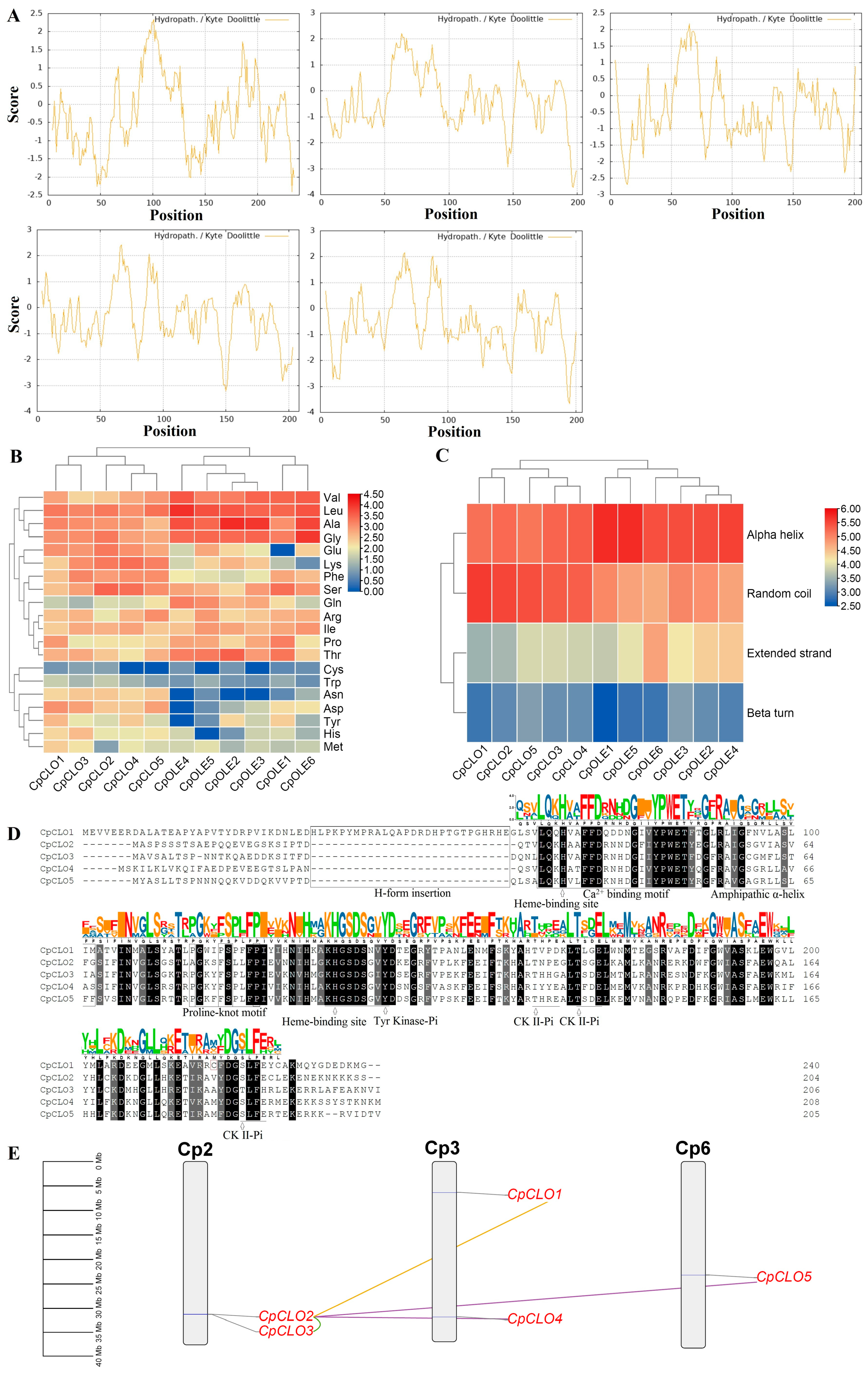
Figure 1. Sequence and duplication event analyses of caleosin family genes in C. papaya. (A) Kyte–Doolittle hydrophobicity plots of five CpCLOs analyzed using ProtScale. (B) Comparison of amino acid composition between CpCLOs and CpOLEs, which was calculated using ProtParam. (C) Comparison of secondary structure between CpCLOs and CpOLEs, which was identified using the SOPMA method. (D) Multiple sequence alignment of CpCLOs performed using MUSCLE. Broken lines in the sequences represent gaps introduced for best alignment, whereas identical and similar amino acids are highlighted in black and dark gray, respectively. The SeqLogo of the caleosin domain generated using WebLogo is shown above the alignment, and the positions of the H-form insertion, an EF-hand calcium binding motif, the amphipathic α-helix, and the proline knot-like motif (PX3P/FSX3P) are boxed in black and are indicated at the bottom of the sequences. Two invariable His residues for heme-binding and four phosphorylation sites (one for Tyr kinase and three for CK II) are underlined and pointed to by arrows. The Cys residue prior to the last CK II phosphorylation site is boxed in purple. (E) Chromosome localization and duplication events detected in papaya. Chromosome localization was conducted using TBtools-II, whereas different modes of gene duplication were identified using the DupGen_finder pipeline. Serial numbers are indicated at the top of each chromosome, and the scale is in Mb. Duplicate pairs identified in this study are connected using lines in different colors, i.e., tandem (green), transposed (purple), and dispersed (gold). (Cp: C. papaya; CK II: casein kinase II; CLO: caleosin; Mb: megabase; OLE: oleosin).
Plants 14 03296 g001
Figure 2. Structural and phylogenetic analyses of caleosin family genes in C. papaya and A. thaliana. (A) Shown is an unrooted phylogenetic tree resulting from full-length caleosins with RAxML (maximum likelihood method and bootstrap of 1000 replicates), where the distance scale denotes the number of amino acid substitutions per site and the name of each clade is indicated next to the corresponding group. (B) Shown are the exon–intron structures. “0”, “1”, and “2” indicate that an intron is located between codons, the first and second bases of a codon, and the second and third bases of a codon, respectively. (C) Shown is the distribution of conserved motifs identified using MEME, where different motifs are represented by different color blocks as indicated and the same color block in different proteins indicates a certain motif. (At: A. thaliana; Cp: C. papaya; CLO: caleosin). (D) Shown is the detailed information of ten motifs identified in this study.
Plants 14 03296 g002
Figure 3. Species-specific distribution of seven caleosin orthogroups in 14 representative plant species. The species tree refers to NCBI Taxonomy (https://www.ncbi.nlm.nih.gov/taxonomy, accessed on 31 January 2025) and well-established recent WGDs are marked, which include the γ WGT shared by all core eudicots, the β WGD shared by the majority of Brassicales plants, and the α WGD specific to Brassicaceae. Names of tested plant families are indicated next to the corresponding branches. (H: high molecular weight; L: low molecular weight).
Plants 14 03296 g003
Figure 4. Synteny analyses within and between C. papaya and representative plant species. (A) Synteny analysis within and between C. papaya, A. thaliana, R. communis, A. coerulea, and A. trichopoda. (B) Synteny analysis within and between C. papaya, M. oleifera, B. sinensis, and A. thaliana. (C) Synteny analysis within and between M. oleifera, C. spinosa, C. violacea, G. gynandra, and T. hassleriana. (D) Synteny analysis within and between A. thaliana, A. halleri, A. lyrata, and E. salsugineum. Syntenic blocks were inferred using MCScanX (v2.0) (E-value ≤ 1 × 10⁻¹⁰; BLAST hits ≥ 5), and the circos diagram was conducted using TBtools-II. Shown are caleosin gene-encoding chromosomes/scaffolds and only syntenic blocks containing caleosin genes are marked, where red and purple lines indicate intra- and inter-species, respectively. The scale is in Mb. (Ac: A. coerulea; Ah: A. halleri; Al: A. lyrata; At: A. thaliana; Atr: A. trichopoda; Bs: B. sinensis; CLO: caleosin; Cp: C. papaya; Cs: C. spinosa; Cv: C. violacea; Es: E. salsugineum; Gg: G. gynandra; Mo: M. oleifera; Rc: R. communis; Th: T. hassleriana).
Plants 14 03296 g004
Figure 5. Expression profiles of CpCLO genes. The heatmap was generated using the R package (v3.5.0), where color scales represent FPKM normalized Log2 transformed counts. Blue and red indicate low and high expression, respectively. (CLO: caleosin; Cp: C. papaya; FPKM: fragments per kilobase of exon per million fragments mapped).
Plants 14 03296 g005
Table 1. Caleosin genes identified in C. papaya. (AA: amino acid; Chr: chromosome; CLO: caleosin; Cp: C. papaya; GRAVY: grand average of hydropathicity; kDa: kilodalton; MW: molecular weight; pI: isoelectric point).
Gene NameLocusPositionAAMW (kDa)pIGRAVYCaleosin LocationClade
CpCLO1sunset03G0007900Chr3:6306411..6308488(+)24027.245.05−0.32962..228H
CpCLO2sunset02G0020020Chr2:31227224..31229504(+)20422.676.09−0.41925..192L
CpCLO3sunset02G0020030Chr2:31241163..31242710(+)20623.576.80−0.40225..192L
CpCLO4sunset03G0022160Chr3:31772679..31773742(+)20823.899.41−0.40127..194L
CpCLO5sunset06G0015560Chr6:23171674..23173752(+)20523.679.57−0.50926..192L
Table 2. Caleosin duplicates identified in C. papaya. Ks and Ka were calculated using PAML. (CLO: caleosin; Cp: C. papaya; Ka: nonsynonymous substitution rate; Ks: synonymous substitution rate).
Gene1Gene2Identity (%)KaKsKa/Ks
CpCLO1CpCLO246.80.5152--
CpCLO2CpCLO371.20.23781.20200.1978
CpCLO2CpCLO467.50.26461.28710.2056
CpCLO2CpCLO569.60.27570.89830.3069
Disclaimer/Publisher’s Note: The statements, opinions and data contained in all publications are solely those of the individual author(s) and contributor(s) and not of MDPI and/or the editor(s). MDPI and/or the editor(s) disclaim responsibility for any injury to people or property resulting from any ideas, methods, instructions or products referred to in the content.