Article

Activity-Based Profiling of Papain-like Cysteine Proteases During Late-Stage Leaf Senescence in Barley

by
Igor A. Schepetkin
* and
Andreas M. Fischer
*
Department of Plant Sciences and Plant Pathology, Montana State University, Bozeman, MT 59717, USA
*
Authors to whom correspondence should be addressed.
Plants 2025, 14(20), 3132; https://doi.org/10.3390/plants14203132
Submission received: 28 June 2025 / Revised: 26 September 2025 / Accepted: 9 October 2025 / Published: 11 October 2025
(This article belongs to the Section Plant Development and Morphogenesis)

Abstract

Leaf senescence is a developmental process that allows nutrients to be remobilized and transported to sink organs. Previously, papain-like cysteine proteases (PLCPs) have been found to be highly expressed during leaf senescence in different plant species. In this study, we analyzed active PLCPs in barley (Hordeum vulgare L.) leaves during the terminal stage of natural senescence. Anion exchange chromatography of protein extracts from barley leaves, harvested six weeks after anthesis, followed by activity assays using the substrates Z-FR-AMC and Z-RR-AMC, revealed a single prominent peak corresponding to active PLCPs. This hydrolytic activity was completely inhibited by E-64, a potent and irreversible inhibitor of cysteine proteases. Fractions enriched for PLCP activity were affinity-labeled with DCG-04 and subjected to SDS-PAGE fractionation, separating two major bands at 43 and 38 kDa. These bands were analyzed using tandem mass spectrometry, allowing the identification of eleven PLCPs. Identified enzymes belong to eight PLCP subfamilies, including CTB/cathepsin B-like (HvPap-19 and -20), RD19/cathepsin F-like (HvPap-1), ALP/cathepsin H-like (HvPap-12 or aleurain), SAG12/cathepsin L-like A (HvPap-17), CEP/cathepsin L-like B (HvPap-14), RD21/cathepsin L-like D (HvPap-6 and -7), cathepsin L-like E (HvPap-13 and -16), and XBCP3 (HvPap-8). Among the identified PLCPs, HvPap-6 was the most abundant. Peptides corresponding to HvPap-6 were identified in both the 43 kDa and 38 kDa bands in approximately the same quantity based on total spectral count. Thus, our results indicate that two active HvPap-6 isoforms can be isolated from barley leaves at late senescence.

1. Introduction

Barley is a global crop, ranking fourth among cereals in the world in terms of production after maize, rice, and wheat [1]. It is a versatile crop used for human food, animal feed, and for malting, especially in the brewing industry [2].
During leaf senescence, nutrients are mobilized and translocated from the senescing tissues to young leaves, storage organs, or developing seeds [3,4,5]. During this process, protein breakdown involves several classes of proteases, including serine proteases, metalloproteases, aspartic proteases, cysteine proteases, and the proteasome [6,7]. Among these enzymes, cysteine proteases are the most strongly associated with leaf senescence in various species [8]. Papain-like cysteine proteases (PLCPs; family C1A in the MEROPS classification system) are one of the most abundant groups of cysteine proteases [9,10]. These enzymes are synthesized in an inactive form to prevent their premature activity. This inactive state is achieved by a prodomain, an N-terminal extension that blocks the active site and prevents binding with substrates [11]. The protease domain contains in its active center a nucleophilic cysteine residue which, in combination with histidine and aspartic acid, forms a catalytic triad [9].
In plants, PLCPs play a crucial role in nitrogen remobilization, programmed cell death, plant immunity, and the initiation of signaling pathways [8,12,13,14,15]. They are involved in breaking down proteins in senescing leaves, releasing amino acids for translocation to other parts of the plant where they are needed, such as for seed filling [16,17,18]. During plant senescence, high expression was found for many PLCP genes in different plant species [19,20]. In barley, PLCPs have been classified as HvPap-1 to HvPap-42 [8]. Significant upregulation of HvPap genes (including HvPap-1, 2, 4, 6-8, 12-15, 17, 19, 20, and 22) has been previously reported during barley leaf senescence [8,21,22,23,24]. Understanding the functional roles of specific PLCPs during leaf senescence provides foundational knowledge for the improvement of crop traits, including seed/grain protein content and nitrogen use efficiency [25,26,27]. The overlapping activities and functional redundancy of PLCPs make it difficult to isolate and study the contribution of specific proteases to the overall process of plant senescence [28,29]. On the other hand, gene expression profiles do not always correspond to protein-level changes and do not reflect many functional features of proteomes [29].
Mass spectrometry (MS) is a powerful tool that can assist breeding aimed at developing superior barley varieties [30]. Counting of peptide spectra from MS datasets allows for label-free quantification, where the number of spectra assigned to a protein is used as an indicator of its relative abundance [31,32,33]. Combining activity-based protein profiling (ABPP) with tandem MS analysis is a powerful approach for studying protease function [34].
We have previously characterized PLCP activities at different stages of barley leaf senescence, using fluorogenic substrates, specific inhibitors, DCG-04 labeling, and immunoblotting [35]. Since PLCP activities are maximal during late senescence, while ribulose-1,5-bisphosphate carboxylase/oxygenase (Rubisco) is largely depleted [35] (hence, not a problem for tandem MS identification of low-abundance proteins), we chose that developmental stage to identify PLCPs critical for natural barley leaf senescence.

2. Results and Discussion

2.1. Isolation of PLCPs Using Ion-Exchange and Affinity Chromatography

Previous studies showed that cysteine proteases can be purified from plant protein extracts by anion-exchange chromatography (e.g., [36,37,38]). In the present study, the total protein extract from fully senesced leaves collected from barley shoots at 6 weeks after anthesis was loaded on a DEAE-Sepharose column, followed by washing with loading buffer. When a linear gradient of NaCl (0.0–1.0 M) was used with the elution buffer, a single peak of enzyme activity was identified, based on assays performed with the fluorogenic substrates Z-FR-AMC and Z-RR-AMC (Figure 1).
The activity of the peak fractions measured with these substrates was completely inhibited by E-64, a specific inhibitor of cysteine proteases [39]. The R-AMC cleaving aminopeptidase activity eluted earlier than the Z-FR-AMC/Z-RR-AMC cleaving activities and was insensitive to E-64 even at high concentration (up to 25 µM), but was inhibited by bestatin, an aminopeptidase inhibitor, and 1,10-phenanthroline, a metallopeptidase inhibitor (Table 1).
Because the activity of aleurain (a cysteine aminopeptidase) was inhibited by E-64 [40], we suggest that barley PLCPs can be enriched in one step using DEAE-Sepharose. It should be noted that fractionation of PLCPs from maize leaves by ion-exchange chromatography also produced a single Z-FR-AMC cleavage activity peak, which is sensitive to E-64 inhibition [37].
The active fractions (based on the Z-FR-AMC/Z-RR-AMC enzymatic assays) were pooled and labeled with DCG-04, a biotinylated epoxide probe that binds covalently and irreversibly to the active site of PLCPs [41]. This probe has previously been used to identify cysteine proteases involved in senescence and abiotic stress responses in maize, wheat, and other plants [37,42,43].
The labeled proteins were captured on streptavidin beads; the column was washed with 1% SDS/1% NP-40 and eluted with Laemmli reducing sample buffer with excess biotin (25 mM) and heat [44]. Proteins were separated by SDS-PAGE. The protein bands, visualized by Coomassie Blue staining, showed that DCG-04-labeled proteases are separated into two bands with molecular weights of 43 and 38 kDa, similar to those found in our previous experiments using SDS-PAGE and streptavidin-horseradish peroxidase to detect DCG-04-labeled barley proteases [35]. A typical SDS-PAGE gel image with these two bands is shown in the Supplementary Materials (Figure S1). The bands were then excised and digested with trypsin, followed by peptide extraction and analysis by tandem MS.

2.2. Semiquantitative Tandem MS Analysis of Barley PLCPs
Based on tandem MS analysis [32], eleven PLCPs, including HvPap-1, -6, -7, -8, -12, -13, -14, -16, -17, -19, and -20, were identified in the protein extract from barley leaves harvested six weeks after anthesis (Figure 2). Total spectral counts for all identified PLCPs from one representative experiment are shown in Table 2. In addition, Supplementary Tables S1–S11 provide MS data showing peptide identification, their Mascot ion scores, as well as m/z (mass-to-charge ratio) and charge data for selected samples from one representative experiment. Four active PLCPs, including HvPap-6, -12, -13, and -14, have been previously identified in senescing barley leaves, although without semi-quantitative tandem MS analysis based on total spectral count [45].
Peptides corresponding to HvPap-6 and HvPap-7 were identified in both 43 kDa and 38 kDa gel bands in approximately the same quantity based on total spectral counts (Figure 2A). The total number of peptide spectra associated with each PLCP in both gel bands was calculated, and their proportion among all eleven identified PLCPs was estimated. Based on this analysis, HvPap-6 is the most abundant PLCP in late-senescence barley leaves (Figure 2B). Using immunoblotting, Cohen et al. [23] also found a significant increase in HvPap-6 protein levels during barley leaf senescence. Our findings are consistent with recent results by Havé et al. [43] showing that triticain α, an ortholog of HvPap-6, is the major active PLCP in naturally senescent wheat leaves. Furthermore, Sekhon et al. [46] demonstrated the functional importance of Mir3 (a maize HvPap-6 ortholog) for leaf senescence, using a genome-wide association study (GWAS), gene expression, and protease activity assays. Those authors also showed that knockout of Arabidopsis RD21A, an ortholog of HvPap-6, triticain α, and Mir3, leads to a delayed-senescence phenotype. A delay of whole-plant senescence was also demonstrated by Pružinská et al. [47] in rd21a/aalp (cathepsin H) double mutants, but not in rd21a (single) mutants. Thus, evidence from several plant species, including barley, strongly points to the functional relevance of RD21-like PLCPs for leaf senescence, but their participation in protein degradation/nitrogen remobilization, senescence regulation, or biotic/abiotic stress tolerance of senescing tissues remains to be established. Phylogenetic relationships between PLCPs are discussed in the next section.
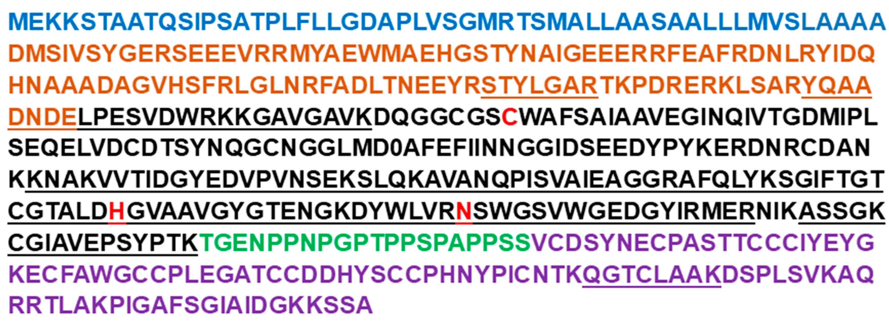
In Arabidopsis, RD21A is known to exist as two forms, an intermediate form containing the C-terminal granulin domain and a mature form from which the granulin domain has been removed [48]. Our results suggest that HvPap-6 could also be heterogeneous, which explains the presence of active isoforms differing by approximately 5 kDa (molecular weights of ~43 and 38 kDa). Thus, the 43 kDa band is likely to be a longer-chain form of HvPap-6, implying partial cleavage of the prodomain and/or granulin domain. Indeed, although the STYLGAR peptide from the prodomain of HvPap-6 was detected only once in the 43 kDa band with a relatively low peptide identification probability (19%), another peptide (YQAADNDELPESVDWR), which covers the end of the prodomain and the beginning of the protease domain, was detected multiple times with a high peptide identification probability (91–100%). Thus, HvPap-6 may require only partial cleavage within the prodomain for activation. On the other hand, the granulin peptide QGTCLAAK of HvPap-6 was detected in both 43 kDa and 38 kDa bands with high probability (99%) (Figure 3).
Figure 3. Primary full-length sequence of the HvPap-6 protein identified by tandem MS as the predominant active PLCP during late-stage leaf senescence in barley. Peptide sequences detected by tandem MS are underlined. The amino acid coverage is based on 3 independent samples. Amino acids marked in blue belong to the signal peptide; brown—inhibitor domain; black—protease domain; green—proline-rich domain; violet—granulin domain; catalytic residues (C, H, N) are shown in red. The amino acids were attributed to the different domains based on [48].
Plants 14 03132 g003
It should be noted that no peptides were detected that cover a conserved region around the catalytic cysteine. This was found previously in other studies on cysteine proteases using DCG-04-based proteomics (e.g., [29]) and may be explained by the fact that irreversible binding of DCG-04 to the catalytic cysteine changes the molecular weight of peptide ions. However, because this conserved region exhibits high amino acid sequence similarity across PLCPs, its identification by MS analysis provides limited value in distinguishing closely related proteins [49].

2.3. Phylogenetic Analysis of Barley PLCPs

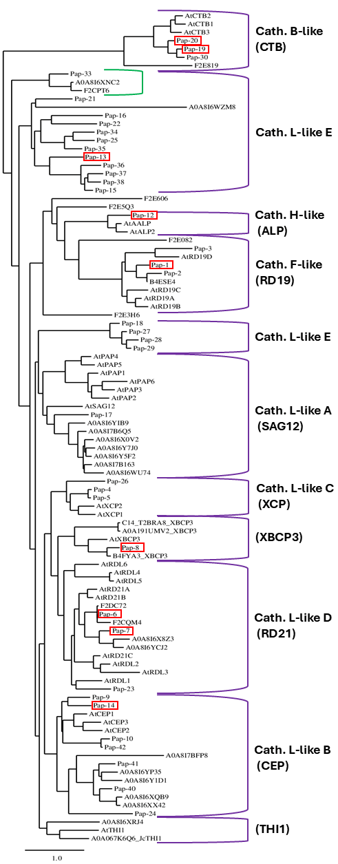
Previously, 42 barley PLCPs (designated HvPap-1 to HvPap-42) have been reported and classified into 8 subfamilies, including cathepsin B-like, cathepsin F-like, cathepsin H-like, and 5 subgroups (from A to E) of cathepsin L-like [8]. To identify previously not characterized barley PLCPs, we explored the UniProt database (https://www.uniprot.org) and found 64 barley PLCPs, including 22 proteases with identity/similarity < 90% as compared to the previously described HvPaps (Table 3). To gain insights into the subfamily classification of the barley PLCPs and to investigate the relationship of new members with known PLCPs, we constructed a phylogenetic tree using these 64 barley PLCPs, together with 32 PLCPs from Arabidopsis thaliana and four PLCPs from other plant species to increase the number of members of the XBCP3 and THI1 groups. PLCPs with >90% identity/similarity in comparison with sequences of the previously reported barley PLCPs were not included in the analysis because (a) they were considered to represent minor genetic variations based on the pangenome concept [50] and (b) PLCPs with high identity/similarity will not form branches on the phylogenetic tree, creating noise that can complicate the analysis.
As a result, the PLCPs were clustered into ten subfamilies (Figure 4). Most barley PLCPs could be classified in the subfamilies described by Richau et al. [13] and/or Díaz-Mendoza et al. [8]. The exceptions are HvPap-31, -32, and -39, which were not included in any subfamilies, confirming the previous phylogenetic tree analysis [8]. Although Diaz-Mendoza et al. [8] did not classify HvPap-24 as belonging to any cluster, our analysis suggests that this protease belongs to the L-like B subfamily (CEP cluster according to Richau et al. [13]). One barley PLCP (UniProt ID: A0A8I6XRJ4) is represented in the THI1 subfamily together with protease AtTHI1 from A. thaliana (UniProt ID: Q9LNC1) and one member from Jatropha curcas (UniProt ID: A0A067K6Q6). Based on the present analysis and the phylogenetic tree analysis by Liu et al. [54], HvPap-8 is located in the XBCP3 cluster, not in the cathepsin L-like D subfamily (RD21 cluster) as previously reported [8]. Both the RD21 and XBCP3 clusters include PLCPs with a granulin domain, although proteases without the granulin domain could also be present in these subfamilies [37].
The eleven PLCPs identified by tandem MS analysis in the present study are scattered across eight PLCP subfamilies (indicated by red rectangles in Figure 4), including CTB/cathepsin B-like (HvPap-19 and -20), RD19/cathepsin F-like (HvPap-1), ALP/cathepsin H-like (HvPap-12 or aleurain), SAG12/cathepsin L-like A (HvPap-17), CEP/cathepsin L-like B (HvPap-14), RD21/cathepsin L-like D (HvPap-6 and -7), cathepsin L-like E (HvPap-13 and -16), and XBCP3 (HvPap-8). As HvPap-6 was the most prominent PLCP identified in this study and is located in the RD21/cathepsin L-like D cluster, Table 4 specifically compares HvPap-6 and HvPap-7 with orthologs from wheat (triticain α), maize (Mir3), and Arabidopsis (RD21A). All these proteases are active in senescing leaves, and for two of them (RD21A and Mir3), available data point to a functional involvement in the senescence process [46,47].
Based on previously reported PLCP classifications by [8,13,37] and the present work, the distribution of PLCPs into ten subfamilies for H. vulgare, A. thaliana, and Zea mays is shown in Table 5. It should be noted that 18 barley PLCPs belong to the cathepsin L-like E subfamily, forming the largest cluster, which has no related members in Arabidopsis and maize.
To find orthologs of the cathepsin L-like E family members in other plant species, we extracted proteins with similarity > 50% to these eighteen barley PLCPs from the UniProt database and found 326 orthologs. All identified genes except one (A0A8J5GYP2) belong to the Poaceae family, mainly to the BOP clade (Bambusoideae, Oryzoideae, and Pooideae), and have no related family members in maize and sorghum (see Supplementary Table S12), suggesting that cathepsin L-like E proteases are mostly restricted to the BOP clade of the Poaceae family with the C3 photosynthetic pathway.
Using tandem MS analysis, we identified only two barley proteases from the cathepsin L-like E subfamily, HvPap-13 and HvPap-16, with relatively low abundance (Figure 2B). Thus, PLCPs of this subfamily may be more important at other stages of plant development. Moreover, organ-specific PLCP activity has recently been found in maize roots [37]. Some PLCPs also play a crucial role in hydrolyzing storage proteins during seed germination [55,56]. Further studies are needed to identify cathepsin L-like E proteases at different stages of leaf development as well as in different plant organs, such as during seed germination or root development.

3. Materials and Methods

3.1. Materials and Reagents

E-64 (trans-epoxysuccinyl-L-leucylamido-(4-guanidino)butane), Z-Phe-Arg-7-amino-4-methylcoumarin (Z-FR-AMC), 1,10-phenanthroline, bestatin, DEAE-Sepharose CL-6B, and dithiothreitol (DTT) were from Millipore-Sigma (St. Louis, MO, USA). CAA0225 was from EMD Millipore Corporation (Burlington, MA, USA). A color-coded prestained protein marker was obtained from Cell Signaling Technologies (Danvers, MA, USA). Z-Arg-Arg-7-amino-4-methylcoumarin (Z-RR-AMC) was from Echelon Biosciences (Salt Lake City, UT, USA), and Arg-7-amido-4-methylcoumarin (R-AMC) was obtained from AK Scientific (Palo Alto, CA, USA). Dimethyl sulfoxide (DMSO) was from Acros Organics (Fair Lawn, NJ, USA). DCG-04, a biotinylated E-64 derivative [41], was obtained from Psyclo Peptide Inc. (Shanghai, China). High-capacity streptavidin agarose resin was from Thermo Scientific (Rockford, IL, USA).

3.2. Plant Material

Barley plants (Hordeum vulgare L. var. ‘GemCraft’) [57] were grown as described previously [35]. Briefly, barley plants (three plants per 4 L pot) were grown in potting soil in a greenhouse bay of the Plant Growth Center at Montana State University (Bozeman, MT, USA) between August and November 2023, with a 22 °C/18 °C day/night temperature cycle. Days were extended to a 16 h photoperiod using Son-Agro 430 W high-pressure sodium lamps (Philips, Somerset, NJ, USA). The photosynthetic photon flux density (PPFD) from artificial light was 120–180 μmol m−2 s−1 and 600–800 μmol m−2 s−1 from natural light at the leaf level. Plants were fertilized once per week until flowering with Peter’s Professional General-Purpose fertilizer (250 mL per pot; 4 g/L; Scotts-Sierra Horticultural Products Company, Marysville, OH, USA). Fully senesced leaves from 40 to 50 plants were collected six weeks after anthesis and stored at −20 °C until protein extraction.

3.3. Ion-Exchange and Affinity Purification of PLCPs

Soluble proteins from barley leaves were extracted by homogenizing 10 g of leaf powder in 200 mL of ice-cold 50 mM Tris-HCl buffer (pH 7.4). Homogenates were filtered through one layer of Miracloth (Calbiochem, San Diego, CA, USA), centrifuged at 20,000× g for 20 min at 4 °C, and the supernatants were then filtered through a 0.45 μm filter (Avantor, Radnor, PA, USA). PLCPs were separated using anion-exchange chromatography on a DEAE-Sepharose CL-6B column (volume 6 mL) equilibrated with 0.05 M Tris-HCl buffer (pH 7.4). The column was washed with equilibration buffer, and bound material was eluted with a NaCl gradient (0.0–1.0 M). Collected fractions (1 mL each) were kept in an ice bath and used for measurements of peptidase activity and protein content. Fractions with active cysteine proteases were pooled. To reduce the NaCl concentration and exchange the buffer to 0.1 M Na-citrate (pH 5.5), three sequential filtration steps using 10 kDa ultrafiltration 15 mL units (Merck Millipore Ltd., Carrigtwohill, County Cork, Ireland) were utilized, and the samples were incubated with gentle shaking with 10 µM DCG-04 in the presence of 2 mM DTT for 3 h at 37 °C. No remaining PLCP activity was found after three hours, as confirmed using Z-FR-AMC and Z-RR-AMC fluorogenic substrates. The intensity of DCG-04-labeled barley cysteine proteases was determined in our previous experiments using streptavidin-horseradish peroxidase [35]. After the incubation, proteins were rapidly precipitated using 80% acetone under inclusion of 100 mM NaCl [58].
Affinity enrichment of DCG-04-reactive PLCPs was performed using streptavidin-agarose. Each sample was incubated with 150 μL bed volume streptavidin-agarose for 60 min at room temperature. To reduce nonspecific binding, the column was washed with 1% SDS and 1% NP-40 in 0.05 M Tris-HCl buffer (pH 7.4). After washing, bound polypeptides were eluted by adding 100 µL of Laemmli reducing sample buffer with excess biotin (25 mM) and boiling for 7 min [44].
DCG-04-reactive proteins were then separated on ready-made ExpressPlusTM 4–12% acrylamide gels (GenScript Inc., Piscataway, NJ, USA) in Tris-MOPS SDS-PAGE Running Buffer (GenScript). The gels were fixed for 45 min, stained with Coomassie Brilliant Blue R-250 overnight, and destained with 50% methanol, 10% acetic acid, and 40% water. Visible bands from the Coomassie-stained gels were excised and shipped to the IDeA National Resource for Quantitative Proteomics at the University of Arkansas (Little Rock, AR, USA).

3.4. Enzymatic Assays and Protein Determination

Fluorogenic substrates Z-FR-AMC to determine cathepsin B- and L-like activity, and Z-RR-AMC to determine cathepsin B-like activity, were used. These substrates show fluorescence when AMC is released as a consequence of the hydrolysis of the Arg-AMC bond. The emitted fluorescence was detected with a SpectraMax M2 microplate reader (Molecular Devices, San Jose, CA, USA) with λex = 360 nm and λem = 460 nm. Before use, the substrates were dissolved in DMSO at 10 mM and stored at −20 °C. The measurements were made in 96-well black microplates (PerkinElmer Inc., Waltham, MA, USA), and each well contained a 100 μL final volume of 0.1 M Na-citrate buffer (pH 5.5) with DTT (2 mM), an aliquot of extract or chromatographic fraction, and fluorogenic substrate (25 μM). The reaction was initiated by the addition of the substrate. The final concentration of DMSO in microplate wells was 1% in all assays. The assays were conducted at room temperature, and relative fluorescence readings were recorded over a period of 10 min.
For the inhibition assays, an inhibitor (1 µL of stock solution of E-64 or bestatin in DMSO, or stock solution of 1,10-phenanthroline in ethanol) at different concentrations was added to the reaction mixture. For all inhibitors tested, the concentration of an inhibitor that caused 50% inhibition of the enzymatic reaction (IC50) was calculated by plotting percent inhibition against the logarithm of inhibitor concentration (at least six data points). The data are presented as the mean values of at least three independent experiments.
Protein concentration was determined using Bradford reagent (Thermo Scientific, Rockford, IL, USA) and bovine serum albumin as a standard.

3.5. Gel-Based Tandem MS Analysis

Each SDS-PAGE gel band was subjected to in-gel trypsin digestion [59]. Gel segments were destained in 50% methanol with 50 mM ammonium bicarbonate, followed by reduction in 10 mM Tris[2-carboxyethyl]phosphine (Pierce, Rockford, IL, USA) and alkylation in 50 mM iodoacetamide (Sigma-Aldrich, St. Louis, MO, USA). Gel slices were then dehydrated in acetonitrile (Thermo Fisher Scientific, Rockford, IL, USA), followed by the addition of 100 ng porcine sequencing grade modified trypsin (Promega, Madison, WI, USA) in 50 mM ammonium bicarbonate (Sigma-Aldrich) and incubation at 37 °C for 12–16 h. Peptide products were then acidified in 0.1% formic acid (Pierce).
Tryptic peptides were separated by reverse phase XSelect CSH C18 2.5 µm resin (Waters, Milford, MA, USA) on an in-line 150 × 0.075 mm column using a nanoAcquity UPLC system (Waters). Peptides were eluted using a 60 min gradient from 98:2 to 65:35 buffer A–B ratio (buffer A = 0.1% formic acid, 0.5% acetonitrile; buffer B = 0.1% formic acid, 99.9% acetonitrile). Eluted peptides were ionized by electrospray (2.4 kV) followed by tandem MS analysis using higher-energy collisional dissociation (HCD) on an Orbitrap Fusion Tribrid mass spectrometer (Thermo Fisher Scientific) in top-speed data-dependent mode. MS data were acquired using the FTMS analyzer in profile mode at a resolution of 240,000 over a range of 375 to 1500 m/z. Following HCD activation, tandem MS data were acquired using the ion trap analyzer in centroid mode and normal mass range with precursor mass-dependent normalized collision energy between 28.0 and 31.0.

3.6. Database Searching and Criteria for Protein Identification

Proteins were identified by database search using Mascot (Matrix Science, London, UK; version 2.6.2) with a parent ion tolerance of 3 ppm and a fragment ion tolerance of 0.5 Da. Mascot was set up to search the 2024_06 UniProt Hordeum_vulgare database (34,528 entries), assuming the digestion enzyme trypsin. Scaffold (version 5.3.3, Proteome Software Inc., Portland, OR, USA) was used to validate tandem MS-based peptide and protein identifications. Peptide identifications were accepted if they could be established at greater than 6.0% probability to achieve a false discovery rate (FDR) < 1.0% by the Peptide Prophet algorithm [60] with Scaffold delta-mass correction. Protein identifications were accepted if they could be established at >92.0% probability to achieve an FDR < 1.0% and contained at least two identified peptides. Protein probabilities were assigned by the Protein Prophet algorithm [61].

3.7. Computational Methods

For the generation of a phylogenetic tree, 64 barley PLCP amino acid sequences were retrieved from the UniProt open-access resource [51,52] were used. Additionally, 32 PLCPs of A. thaliana were included [13], as well as three members of the XBCP3 subfamily from other plant species (T2BRA8, a cysteine protease CP14 from Nicotiana tabacum; A0A191UMV2, a PLCP from Nicotiana benthamiana; B4FYA3, a xylem bark cysteine peptidase 3 from Zea mays), and one member of the THI1 subfamily from Jatropha curcas (A0A067K6Q6). The Phylogeny.fr platform was used for the construction of the tree [62].

4. Conclusions

In conclusion, eleven active PLCPs were identified in protein extracts from late-senescence barley leaves using tandem MS analysis. The identified PLCPs were distributed across eight PLCP subfamilies. Among the identified PLCPs, HvPap-6 was the most abundant and was present in barley leaves in at least two active isoforms. This finding may facilitate functional comparisons with PLCPs from other plant species and will contribute to the elucidation of their specific roles during barley leaf senescence. Results obtained by other researchers in Arabidopsis, wheat, and maize suggest that HvPap-6 and its orthologs (RD21-like proteases) are functionally important during leaf senescence, but their exact role in nitrogen remobilization, senescence regulation, and/or abiotic/biotic stress tolerance of senescing tissues remains to be established using transgenic approaches or profiting from natural variation. Exploration of the barley pangenome (https://panbarlex.ipk-gatersleben.de/; [53]) suggests the absence of HvPap-6 from one accession (HOR 7385), providing an entry point for HvPap-6 functional studies.
Further studies are needed to fully establish the classification of barley PLCPs, especially with respect to the cathepsin L-like E subfamily.

Supplementary Materials

The following supporting information can be downloaded at: https://www.mdpi.com/article/10.3390/plants14203132/s1. Figure S1. PLCPs from barley leaves were enriched using DEAE-Sepharose ion-exchange and affinity chromatography of DCG-04-labeled barley proteases, then separated on 4–12% SDS-PAGE and stained with Coomassie Blue. To demonstrate the specificity of DCG-04 labeling, the cathepsin inhibitor CAA0225 (30 μM) was added 30 min prior to labeling to block the binding of DCG-04 to PLCPs. Bands with molecular weights around 43 and 38 kDa (indicated with dotted rectangles) were excised and analyzed by tandem MS. Tables S1–S11. Peptide sequences of barley PLCPs, identified by tandem MS analysis. The samples were prepared as described in Section 3.5 (Gel-Based Tandem MS Analysis). Modification (+57) indicates a carbamidomethylation; (+16) indicates an oxidation; “Delta” is “Actual” minus “Calculated” peptide mass (Da); m/z is the mass-to-charge ratio. Table S12. Distribution of barley L-like E cathepsin orthologs across plant species.

Author Contributions

Conceptualization, A.M.F. and I.A.S.; methodology, I.A.S. and A.M.F.; formal analysis, I.A.S.; investigation, I.A.S.; resources, A.M.F.; writing—original draft preparation, I.A.S.; writing—review and editing, I.A.S. and A.M.F.; supervision, A.M.F.; funding acquisition, A.M.F. All authors have read and agreed to the published version of the manuscript.

Funding

This research was funded by the United States Department of Agriculture (USDA), the National Institute of Food and Agriculture (NIFA) award 2023-67014-39568, and by the Montana Agricultural Experiment Station (MAES), project MONB00248.

Data Availability Statement

Data are contained within the article and Supplementary Materials.

Acknowledgments

We thank Samuel MacKintosh at the IDeA National Resource for Quantitative Proteomics for performing tandem MS analyses.

Conflicts of Interest

The authors declare no conflicts of interest.

References

  1. Newton, A.C.; Flavell, A.J.; George, T.S.; Leat, P.; Mullholland, B.; Ramsay, L.; Revoredo-Giha, C.; Russell, J.; Steffenson, B.J.; Swanston, J.S.; et al. Crops that feed the world 4. Barley: A resilient crop? Strengths and weaknesses in the context of food security. Food Secur. 2011, 3, 141–178. [Google Scholar] [CrossRef]
  2. Izydorczyk, M.S.; Edney, M. Wrigley, C., Batey, I., Miskelly, D., Eds.; Barley: Grain-Quality Characteristics and Management of Quality Requirements. In Cereal Grains: Assessing and Managing Quality, Woodhead Publishing Series in Food Science Technology and Nutrition, 2nd ed.; Woodhead Publishing: Sawston, UK, 2017; pp. 195–234. [Google Scholar]
  3. Quirino, B.F.; Noh, Y.S.; Himelblau, E.; Amasino, R.M. Molecular aspects of leaf senescence. Trends Plant Sci. 2000, 5, 278–282. [Google Scholar] [CrossRef]
  4. Lee, S.; Masclaux-Daubresse, C. Current understanding of leaf senescence in rice. Int. J. Mol. Sci. 2021, 22, 4515. [Google Scholar] [CrossRef]
  5. Lei, P.; Yu, F.; Liu, X.Y. Recent advances in cellular degradation and nuclear control of leaf senescence. J. Exp. Bot. 2023, 74, 5472–5486. [Google Scholar] [CrossRef]
  6. Diaz-Mendoza, M.; Velasco-Arroyo, B.; Santamaria, M.E.; González-Melendi, P.; Martinez, M.; Diaz, I. Plant senescence and proteolysis: Two processes with one destiny. Genet. Mol. Biol. 2016, 39, 329–338. [Google Scholar] [CrossRef]
  7. Roberts, I.N.; Caputo, C.; Criado, M.V.; Funk, C. Senescence-associated proteases in plants. Physiol. Plant. 2012, 145, 130–139. [Google Scholar] [CrossRef] [PubMed]
  8. Díaz-Mendoza, M.; Velasco-Arroyo, B.; González-Melendi, P.; Martínez, M.; Díaz, I. C1A cysteine protease-cystatin interactions in leaf senescence. J. Exp. Bot. 2014, 65, 3825–3833. [Google Scholar] [CrossRef]
  9. Rawlings, N.D.; Barrett, A.J.; Bateman, A. MEROPS: The peptidase database. Nucleic Acids Res. 2010, 38, D227–D233. [Google Scholar] [CrossRef]
  10. Rawlings, N.D.; Barrett, A.J.; Thomas, P.D.; Huang, X.; Bateman, A.; Finn, R.D. The MEROPS database of proteolytic enzymes, their substrates and inhibitors in 2017 and a comparison with peptidases in the PANTHER database. Nucleic Acids Res. 2018, 46, D624–D632. [Google Scholar] [CrossRef]
  11. Verma, S.; Dixit, R.; Pandey, K.C. Cysteine proteases: Modes of activation and future prospects as pharmacological targets. Front. Pharmacol. 2016, 7, 107. [Google Scholar] [CrossRef] [PubMed]
  12. Liu, P.; Shi, C.N.; Liu, S.; Lei, J.J.; Lu, Q.S.; Hu, H.C.; Ren, Y.; Zhang, N.; Sun, C.W.; Chen, L.; et al. A papain-like cysteine protease-released small signal peptide confers wheat resistance to wheat yellow mosaic virus. Nat. Commun. 2023, 14, 7773. [Google Scholar] [CrossRef]
  13. Richau, K.H.; Kaschani, F.; Verdoes, M.; Pansuriya, T.C.; Niessen, S.; Stüber, K.; Colby, T.; Overkleeft, H.S.; Bogyo, M.; Van der Hoorn, R.A.L. Subclassification and biochemical analysis of plant papain-like cysteine proteases displays subfamily-specific characteristics. Plant Physiol. 2012, 158, 1583–1599. [Google Scholar] [CrossRef] [PubMed]
  14. Martinez, M.; Diaz, I. The origin and evolution of plant cystatins and their target cysteine proteinases indicate a complex functional relationship. BMC Evol. Biol. 2008, 8, 198. [Google Scholar] [CrossRef]
  15. Misas-Villamil, J.C.; van der Hoorn, R.A.L.; Doehlemann, G. Papain-like cysteine proteases as hubs in plant immunity. New Phytol. 2016, 212, 902–907. [Google Scholar] [CrossRef]
  16. Masclaux-Daubresse, C.; Daniel-Vedele, F.; Dechorgnat, J.; Chardon, F.; Gaufichon, L.; Suzuki, A. Nitrogen uptake, assimilation and remobilization in plants: Challenges for sustainable and productive agriculture. Ann. Bot. 2010, 105, 1141–1157. [Google Scholar] [CrossRef]
  17. Hajibarat, Z.; Saidi, A. Senescence-associated proteins and nitrogen remobilization in grain filling under drought stress condition. J. Genet. Eng. Biotechnol. 2022, 20, 101. [Google Scholar] [CrossRef]
  18. Havé, M.; Marmagne, A.; Chardon, F.; Masclaux-Daubresse, C. Nitrogen remobilization during leaf senescence: Lessons from Arabidopsis to crops. J. Exp. Bot. 2017, 68, 2513–2529. [Google Scholar] [CrossRef] [PubMed]
  19. Vatov, E.; Ludewig, U.; Zentgraf, U. Disparate dynamics of gene body and cis-regulatory element evolution illustrated for the senescence-associated cysteine protease gene SAG12 of plants. Plants 2021, 10, 1380. [Google Scholar] [CrossRef] [PubMed]
  20. Wagstaff, C.; Leverentz, M.K.; Griffiths, G.; Thomas, B.; Chanasut, U.; Stead, A.D.; Rogers, H.J. Cysteine protease gene expression and proteolytic activity during senescence of Alstroemeria petals. J. Exp. Bot. 2002, 53, 233–240. [Google Scholar] [CrossRef]
  21. Parrott, D.L.; McInnerney, K.; Feller, U.; Fischer, A.M. Steam-girdling of barley (Hordeum vulgare) leaves leads to carbohydrate accumulation and accelerated leaf senescence, facilitating transcriptomic analysis of senescence-associated genes. New Phytol. 2007, 176, 56–69. [Google Scholar] [CrossRef]
  22. Jukanti, A.K.; Heidlebaugh, N.M.; Parrott, D.L.; Fischer, I.A.; McInnerney, K.; Fischer, A.M. Comparative transcriptome profiling of near-isogenic barley (Hordeum vulgare) lines differing in the allelic state of a major grain protein content locus identifies genes with possible roles in leaf senescence and nitrogen reallocation. New Phytol. 2008, 177, 333–349. [Google Scholar] [CrossRef]
  23. Cohen, M.; Hertweck, K.; Itkin, M.; Malitsky, S.; Dassa, B.; Fischer, A.M.; Fluhr, R. Enhanced proteostasis, lipid remodeling, and nitrogen remobilization define barley flag leaf senescence. J. Exp. Bot. 2022, 73, 6816–6837. [Google Scholar] [CrossRef]
  24. Hollmann, J.; Gregersen, P.L.; Krupinska, K. Identification of predominant genes involved in regulation and execution of senescence-associated nitrogen remobilization in flag leaves of field grown barley. J. Exp. Bot. 2014, 65, 3963–3973. [Google Scholar] [CrossRef]
  25. Hu, J.J.; Rampitsch, C.; Bykova, N.V. Advances in plant proteomics toward improvement of crop productivity and stress resistance. Front. Plant Sci. 2015, 6, 209. [Google Scholar] [CrossRef]
  26. Gregersen, P.L.; Holm, P.B.; Krupinska, K. Leaf senescence and nutrient remobilisation in barley and wheat. Plant Biol. 2008, 10, 37–49. [Google Scholar] [CrossRef]
  27. Distelfeld, A.; Avni, R.; Fischer, A.M. Senescence, nutrient remobilization, and yield in wheat and barley. J. Exp. Bot. 2014, 65, 3783–3798. [Google Scholar] [CrossRef] [PubMed]
  28. Nägler, D.K.; Ménard, R. Family C1 cysteine proteases:: Biological diversity or redundancy? Biol. Chem. 2003, 384, 837–843. [Google Scholar] [CrossRef] [PubMed]
  29. Kocks, C.; Maehr, R.; Overkleeft, H.S.; Wang, E.W.; Iyer, L.K.; Lennon-Duménil, A.M.; Ploegh, H.L.; Kessler, B.M. Functional proteomics of the active cysteine protease content in drosophila S2 cells. Mol. Cell. Proteom. 2003, 2, 1188–1197. [Google Scholar] [CrossRef]
  30. Bahmani, M.; O’Lone, C.E.; Juhász, A.; Nye-Wood, M.; Dunn, H.; Edwards, I.B.; Colgrave, M.L. Application of mass spectrometry-based proteomics to barley research. J. Agric. Food Chem. 2021, 69, 8591–8609. [Google Scholar] [CrossRef] [PubMed]
  31. Washburn, M.P.; Wolters, D.; Yates, J.R. Large-scale analysis of the yeast proteome by multidimensional protein identification technology. Nat. Biotechnol. 2001, 19, 242–247. [Google Scholar] [CrossRef]
  32. Bantscheff, M.; Schirle, M.; Sweetman, G.; Rick, J.; Kuster, B. Quantitative mass spectrometry in proteomics: A critical review. Anal. Bioanal. Chem. 2007, 389, 1017–1031. [Google Scholar] [CrossRef] [PubMed]
  33. Gerster, S.; Kwon, T.; Ludwig, C.; Matondo, M.; Vogel, C.; Marcotte, E.M.; Aebersold, R.; Bühlmann, P. Statistical approach to protein quantification. Mol. Cell. Proteom. 2014, 13, 666–677. [Google Scholar] [CrossRef]
  34. Fonovic, M.; Bogyo, M. Activity-based probes as a tool for functional proteomic analysis of proteases. Expert Rev. Proteom. 2008, 5, 721–730. [Google Scholar] [CrossRef]
  35. Schepetkin, I.A.; Fischer, A.M. Cathepsin B- and L-like protease activities are induced during developmental barley leaf senescence. Plants 2024, 13, 3009. [Google Scholar] [CrossRef]
  36. Tsuji, A.; Tsukamoto, K.; Iwamoto, K.; Ito, Y.; Yuasa, K. Enzymatic characterization of germination-specific cysteine protease-1 expressed transiently in cotyledons during the early phase of germination. J. Biochem. 2013, 153, 73–83. [Google Scholar] [CrossRef]
  37. Hüynck, J.S.; Kaschani, F.; van der Linde, K.; Ziemann, S.; Müller, A.N.; Colby, T.; Kaiser, M.; Villamil, J.C.M.; Doehlemann, G. Proteases underground: Analysis of the maize root apoplast identifies organ specific papain-like cysteine protease activity. Front. Plant Sci. 2019, 10, 473. [Google Scholar] [CrossRef]
  38. Jinka, R.; Ramakrishna, V.; Rao, S.K.; Rao, R.P. Purification and characterization of cysteine protease from germinating cotyledons of horse gram. BMC Biochem. 2009, 10, 28. [Google Scholar] [CrossRef]
  39. Barrett, A.J.; Kembhavi, A.A.; Hanada, K. E-64 [L-trans-epoxysuccinyl-leucyl-amido(4-guanidino)butane] and related epoxides as inhibitors of cysteine proteinases. Acta Biol. Med. Ger. 1981, 40, 1513–1517. [Google Scholar] [PubMed]
  40. Holwerda, B.C.; Galvin, N.J.; Baranski, T.J.; Rogers, J.C. In vitro processing of aleurain, a barley vacuolar thiol protease. Plant Cell 1990, 2, 1091–1106. [Google Scholar] [CrossRef] [PubMed]
  41. Greenbaum, D.; Medzihradszky, K.F.; Burlingame, A.; Bogyo, M. Epoxide electrophiles as activity-dependent cysteine protease profiling and discovery tools. Chem. Biol. 2000, 7, 569–581. [Google Scholar] [CrossRef]
  42. Poret, M.; Chandrasekar, B.; van der Hoorn, R.A.L.; Coquet, L.; Jouenne, T.; Avice, J.C. Proteomic investigations of proteases involved in cotyledon senescence: A model to explore the genotypic variability of proteolysis machinery associated with nitrogen remobilization efficiency during the leaf senescence of oilseed rape. Proteomes 2017, 5, 29. [Google Scholar] [CrossRef]
  43. Havé, M.; Espinasse, C.; Cottyn-Boitte, B.; Puga-Freitas, R.; Bagard, M.; Balliau, T.; Zivy, M.; Ganeshan, S.; Chibbar, R.N.; Castell, J.F.; et al. Triticain alpha represents the major active papain-like cysteine protease in naturally senescing and ozone-treated leaves of wheat. Plant Physiol. Biochem. 2025, 219, 109380. [Google Scholar] [CrossRef]
  44. Cheah, J.S.; Yamada, S. A simple elution strategy for biotinylated proteins bound to streptavidin conjugated beads using excess biotin and heat. Biochem. Biophys. Res. Commun. 2017, 493, 1522–1527. [Google Scholar] [CrossRef]
  45. Frank, S.; Hollmann, J.; Mulisch, M.; Matros, A.; Carrión, C.C.; Mock, H.P.; Hensel, G.; Krupinska, K. Barley cysteine protease PAP14 plays a role in degradation of chloroplast proteins. J. Exp. Bot. 2019, 70, 6057–6069. [Google Scholar] [CrossRef]
  46. Sekhon, R.S.; Saski, C.; Kumar, R.; Flinn, B.S.; Luo, F.; Beissinger, T.M.; Ackerman, A.J.; Breitzman, M.W.; Bridges, W.C.; de Leon, N.; et al. Integrated genome-scale analysis identifies novel genes and networks underlying senescence in maize. Plant Cell 2019, 31, 1968–1989. [Google Scholar] [CrossRef] [PubMed]
  47. Pruzinská, A.; Shindo, T.; Niessen, S.; Kaschani, F.; Tóth, R.; Millar, A.H.; van der Hoorn, R.A.L. Major Cys protease activities are not essential for senescence in individually darkened Arabidopsis leaves. BMC Plant Biol. 2017, 17, 4. [Google Scholar] [CrossRef]
  48. Huang, J.; van der Hoorn, R.A.L. RD21-like proteases: Key effector hubs in plant-pathogen interactions. J. Exp. Bot. 2025, 76, 4212–4219. [Google Scholar] [CrossRef] [PubMed]
  49. Kim, M.S.; Zhong, J.; Pandey, A. Common errors in mass spectrometry-based analysis of post-translational modifications. Proteomics 2016, 16, 700–714. [Google Scholar] [CrossRef]
  50. Schreiber, M.; Jayakodi, M.; Stein, N.; Mascher, M. Plant pangenomes for crop improvement, biodiversity and evolution. Nat. Rev. Genet. 2024, 25, 563–577. [Google Scholar] [CrossRef] [PubMed]
  51. Ahmad, S.; Gonzales, L.J.D.; Bowler-Barnett, E.H.; Rice, D.L.; Kim, M.; Wijerathne, S.; Luciani, A.; Kandasaamy, S.; Luo, J.; Watkins, X.; et al. The UniProt website API: Facilitating programmatic access to protein knowledge. Nucleic Acids Res. 2025, 53, W547–W553. [Google Scholar] [CrossRef]
  52. Bateman, A.; Martin, M.J.; Orchard, S.; Magrane, M.; Adesina, A.; Ahmad, S.; Bowler-Barnett, E.H.; Bye-A-Jee, H.; Carpentier, D.; Denny, P.; et al. UniProt: The universal protein knowledgebase in 2025. Nucleic Acids Res. 2024, 52, D609–D617. [Google Scholar] [CrossRef]
  53. Jayakodi, M.; Lu, Q.X.; Pidon, H.; Rabanus-Wallace, M.T.; Bayer, M.; Lux, T.; Guo, Y.; Jaegle, B.; Badea, A.; Bekele, W.; et al. Structural variation in the pangenome of wild and domesticated barley. Nature 2024, 636, 654–662. [Google Scholar] [CrossRef] [PubMed]
  54. Liu, H.J.; Hu, M.H.; Wang, Q.; Cheng, L.; Zhang, Z.B. Role of papain-like cysteine proteases in plant development. Front. Plant Sci. 2018, 9, 1717. [Google Scholar] [CrossRef]
  55. Szewinska, J.; Siminska, J.; Bielawski, W. The roles of cysteine proteases and phytocystatins in development and germination of cereal seeds. J. Plant Physiol. 2016, 207, 10–21. [Google Scholar] [CrossRef]
  56. Martinez, M.; Gómez-Cabellos, S.; Giménez, M.J.; Barro, F.; Diaz, I.; Diaz-Mendoza, M. Plant proteases: From key enzymes in germination to allies for fighting human gluten-related disorders. Front. Plant Sci. 2019, 10, 721. [Google Scholar] [CrossRef]
  57. Hu, G.S.; Evans, C.P.; Satterfield, K.; Ellberg, S.; Marshall, J.M.; Schroeder, K.L.; Obert, D.E. Registration of ‘GemCraft’ spring malting barley cultivar. J. Plant Regist. 2024, 18, 11–16. [Google Scholar] [CrossRef]
  58. Nickerson, J.L.; Doucette, A.A. Rapid and quantitative protein precipitation for proteome analysis by mass spectrometry. J. Proteome Res. 2020, 19, 2035–2042. [Google Scholar] [CrossRef] [PubMed]
  59. Wojtkiewicz, M.; Berg Luecke, L.; Kelly, M.I.; Gundry, R.L. Facile preparation of peptides for mass spectrometry analysis in bottom-up proteomics workflows. Curr. Protoc. 2021, 1, e85. [Google Scholar] [CrossRef] [PubMed]
  60. Keller, A.; Nesvizhskii, A.I.; Kolker, E.; Aebersold, R. Empirical statistical model to estimate the accuracy of peptide identifications made by MS/MS and database search. Anal. Chem. 2002, 74, 5383–5392. [Google Scholar] [CrossRef]
  61. Nesvizhskii, A.I.; Keller, A.; Kolker, E.; Aebersold, R. A statistical model for identifying proteins by tandem mass spectrometry. Anal. Chem. 2003, 75, 4646–4658. [Google Scholar] [CrossRef]
  62. Dereeper, A.; Guignon, V.; Blanc, G.; Audic, S.; Buffet, S.; Chevenet, F.; Dufayard, J.F.; Guindon, S.; Lefort, V.; Lescot, M.; et al. Phylogeny.fr: Robust phylogenetic analysis for the non-specialist. Nucleic Acids Res. 2008, 36, W465–W469. [Google Scholar] [CrossRef] [PubMed]
Figure 1. Anion exchange chromatography of late-senescence barley leaf PLCPs. Fractions eluted with a gradient of NaCl (0.0 to 1.0 M NaCl) from DEAE-Sepharose were tested for PLCP activity using 25 µM of the substrates Z-FR-AMC (blue line) and Z-RR-AMC (dark red line); aminopeptidase activity was tested using 25 µM of the substrate R-AMC (green line). Total protein (yellow line) was determined using Bradford reagent. Abbreviation: RFU, relative fluorescence units.
Plants 14 03132 g001
Figure 2. Tandem MS analysis of late-senescence barley leaf PLCPs. The distribution (in %) of PLCPs between gel bands with molecular masses of 38 and 43 kDa, based on total spectral counts, is presented in panel (A), while panel (B) shows the relative abundance (in %) of the identified PLCPs (based on both gel bands). In both panels, the data represent the mean value ± S.D. of three independent experiments. Statistically significant differences (* p < 0.05) by t-test in the distribution of PLCPs between the two gel bands are shown in panel (A).
Plants 14 03132 g002
Figure 4. Phylogenetic tree of barley PLCPs in which proteins are clustered based on full-length amino acid sequence comparisons. PLCPs present in late-senescence barley leaves, as identified by tandem MS in the present study, are indicated by red rectangles. The green bracket denotes a distinct subcluster within the cathepsin L-like E subfamily.
Plants 14 03132 g004
Table 1. Effect of protease inhibitors on cleavage of Z-RR-AMC, Z-FR-AMC, and R-AMC substrates by fractions obtained by ion-exchange chromatography from barley leaf extract. Extracts were obtained from senescing leaves collected six weeks after anthesis.
FractionSubstrateEffect/IC50
E-64
Fr. No 26–27R-AMCNo inhibition up to 25 µM
Fr. No 35–36Z-RR-AMC9.1 ± 2.2 nM
Z-FR-AMC15.3 ± 4.1 nM
Bestatin
Fr. No 26–27R-AMC247 ± 38 µM
Fr. No 35–36Z-RR-AMCNo inhibition up to 1 mM
Z-FR-AMCNo inhibition up to 1 mM
1,10-Phenanthroline
Fr. No 26–27R-AMC68.5 ± 12.2 µM
Fr. No 35–36Z-RR-AMCNo inhibition up to 5 mM
Z-FR-AMCNo inhibition up to 5 mM
IC50 values are based on three technical replicates. For the inhibition analysis, fractions Nos. 26–27 and Nos. 35–36 were pooled and evaluated for cleavage of R-AMC or Z-RR-AMC/Z-FR-AMC substrates, respectively.
Table 2. The total number of peptide spectra identified as belonging to a specific PLCP and the proportion of amino acid sequence (% coverage) represented by the identified peptides detected by tandem MS analysis in two protein bands excised from SDS-PAGE.
NameUniProt IDTotal Spectral Count (% of Coverage) *
43 kDa Band38 kDa Band
HvPap-1F2DDC95 (13)47 (24)
HvPap-6A0A8I6WYU4270 (29)257 (25)
HvPap-7F2E6V28 (15)6 (17)
HvPap-8B4ESF05 (9.4)13 (22)
HvPap-12P051678 (14)83 (26)
HvPap-13A0A8I6XJP47 (16)40 (44)
HvPap-14B4ESF23 (9.5)73 (37)
HvPap-16B4ESF42 (4.9)10 (21)
HvPap-17A0A8I6Y6A56 (17)12 (21)
HvPap-19A0A8I7BBL87 (17)40 (33)
HvPap-20A0A8I7B6S75 (8.2)17 (18)
* Data from one representative experiment.
Table 3. Classification of barley PLCPs, based on previously reported and the present phylogenetic tree analysis.
Name 1Subfamily 2Gene Identifier 3UniProt IDnAA 4MW, Da 5
HvPap-1F-like (RD19)MOREX.r3.5HG0499760F2DDC937741,016
HvPap-2F-like (RD19)MOREX.r3.2HG0159420A0A8I6WSN337841,885
HvPap-3F-like (RD19)MOREX.r3.2HG0131510B4ESE536838,969
B4ESE4F-like (RD19)Akashinriki.Proj.2HG00143120B4ESE438141,931
F2E082F-like (RD19)N.F.F2E08234137,057
HvPap-12H-like (ALP)MOREX.r3.5HG0480100P0516736239,122
HvPap-19B-like (CTB)MOREX.r3.4HG0339740A0A8I7BBL834437,222
HvPap-20B-like (CTB)MOREX.r3.4HG0339730A0A8I7B6S735338,423
HvPap-30B-like (CTB)MOREX.r3.4HG0339750A0A8I6X9J334737,752
F2E819B-like (CTB)N.F.F2E81947150,283
HvPap-17L-like A (SAG12)MOREX.r3.5HG0511140A0A8I6Y6A534936,895
A0A8I6YIB9L-like A (SAG12)MOREX.r3.6HG0570590A0A8I6YIB934336,737
A0A8I7B6Q5L-like A (SAG12)MOREX.r3.4HG0336140A0A8I7B6Q534136,789
A0A8I7B163L-like A (SAG12)MOREX.r3.1HG0061980A0A8I7B16333936,752
A0A8I6Y7J0L-like A (SAG12)MOREX.r3.7HG0673530A0A8I6Y7J034036,792
A0A8I6Y5F2L-like A (SAG12)MOREX.r3.6HG0623260A0A8I6Y5F233937,020
A0A8I6X0V2L-like A (SAG12)MOREX.r3.2HG0103220A0A8I6X0V234037,030
A0A8I6WU74L-like A (SAG12)MOREX.r3.1HG0061970A0A8I6WU7434037,333
HvPap-9L-like B (CEP)MOREX.r3.4HG0342040A0A8I6XX7036540,033
HvPap-10L-like B (CEP)MOREX.r3.3HG0308010A0A8I6XAH737340,574
HvPap-14L-like B (CEP)MOREX.r3.3HG0304500B4ESF236739,790
HvPap-24L-like B (CEP)MOREX.r3.2HG0211390A0A8I6WKU732036,267
HvPap-40L-like B (CEP)MOREX.r3.6HG0548850A0A8I6XXP537441,375
HvPap-41L-like B (CEP)MOREX.r3.6HG0543660A0A8I6XQ4635739,752
HvPap-42L-like B (CEP)MOREX.r3.3HG0308000A0A8I7BAT337940,979
A0A8I6YP35L-like B (CEP)MOREX.r3.6HG0545100A0A8I6YP3535839,836
A0A8I6XQB9L-like B (CEP)MOREX.r3.6HG0545330A0A8I6XQB923124,863
A0A8I6XX42L-like B (CEP)MOREX.r3.6HG0545210A0A8I6XX4236640,642
A0A8I6Y1D1L-like B (CEP)MOREX.r3.6HG0545130A0A8I6Y1D133837,546
A0A8I7BFP8L-like B (CEP)MOREX.r3.7HG0747000A0A8I7BFP835238,442
HvPap-4L-like C (XCP)MOREX.r3.5HG0462580B4ESE635638,651
HvPap-5L-like C (XCP)MOREX.r3.1HG0004220B4ESE735138,212
HvPap-26L-like C (XCP)MOREX.r3.6HG0609400A0A287UKW836439,959
HvPap-6L-like D (RD21)MOREX.r3.2HG0204520A0A8I6WYU446350,226
HvPap-7L-like D (RD21)MOREX.r3.2HG0212170F2E6V247350,677
HvPap-23L-like D (RD21)N.F.B4ESF819021,595
F2CQM4L-like D (RD21)N.F.F2CQM443647,292
A0A8I6X8Z3L-like D (RD21)MOREX.r3.2HG0211940A0A8I6X8Z346950,348
F2DC72L-like D (RD21)N.F.F2DC7228931,994
A0A8I6YCJ2L-like D (RD21)MOREX.r3.7HG0685270A0A8I6YCJ247751,115
HvPap-13L-like EMOREX.r3.5HG0506230A0A8I6XJP436639,825
HvPap-15L-like EMOREX.r3.6HG0545500M0YYX840644,371
HvPap-16L-like EMOREX.r3.7HG0749670B4ESF438942,091
HvPap-18L-like EAkashinriki.Proj.4HG00376280F2CR4336539,599
HvPap-21L-like EMOREX.r3.2HG0205780A0A8I6WU7935538,776
HvPap-22L-like EMOREX.r3.1HG0004940A0A8I6WPU134537,350
HvPap-25L-like EMOREX.r3.1HG0085150A0A8I7B43035538,295
HvPap-27L-like EMOREX.r3.3HG0245120A0A8I6WQD435738,641
HvPap-28L-like EMOREX.r3.5HG0424540A0A8I6XSH639643,119
HvPap-29L-like EMOREX.r3.3HG0245250A0A8I7B9F235938,974
HvPap-33L-like EMOREX.r3.2HG0104490A0A8I6W9L735537,217
HvPap-34L-like EMOREX.r3.2HG0106320F2EC7336039,066
HvPap-35L-like EMOREX.r3.2HG0212960A0A8I6WXF834937,351
HvPap-36L-like EMOREX.r3.6HG0539010A0A8I6YPN038341,925
HvPap-37L-like EMOREX.r3.4HG0418460F2DVR538942,527
HvPap-38L-like EMOREX.r3.3HG0230090A0A8I6XUD239042,342
A0A8I6XNC2L-like EMOREX.r3.3HG0271470A0A8I6XNC235037,378
F2CPT6L-like E10TJ18.Proj.3HG00173970F2CPT622523,480
HvPap-8(XBCP3)MOREX.r3.1HG0076400B4ESF045748,431
A0A8I6XRJ4(THI1)MOREX.r3.5HG0492390A0A8I6XRJ441844,588
HvPap-31N.S.N.F.F2E60632935,447
HvPap-32N.S.N.F.F2E3H636539,351
HvPap-39N.S.N.F.F2E5Q333337,013
A0A8I6WZM8N.S.MOREX.r3.2HG0208910A0A8I6WZM833036,201
1 HvPap number as introduced by Díaz-Mendoza et al. [8], or UniProt identifier [51,52]. 2 Subfamily name based on the phylogenetic tree analysis of Díaz-Mendoza et al. [8], Richau et al. [13] and the present study. 3 Gene identifier is based on cultivar ‘Morex’ v3 gene IDs (beginning with HORVU.MOREX.r3) or pangenome gene IDs (see https://panbarlex.ipk-gatersleben.de/; [53]). 4 Number of amino acids (nAA) in the predicted full-length protein sequence. 5 Molecular weight of the full-length protein sequence. N.S., the subfamily was not specified. N.F., the gene identifier has not been found among ‘Morex’ v3 gene models, or in the pangenome database.
Table 4. Comparison of HvPap-6 orthologs by pairwise sequence alignment.
NameSpeciesUniProt IDnAAM.W., DaComparison with HvPap-6
Identity (%)Similarity (%)
HvPap-6Hordeum vulgare subsp. vulgareA0A8I6WYU446350,226--
HvPap-7Hordeum vulgare subsp. vulgareF2E6V247350,67756.266.3
Triticain αTriticum aestivumQ0WXG846150,40790.292.3
Mir3Zea maysO2250048051,78771.577.8
RD21AArabidopsis thalianaP4329746250,96662.273.6
In the pairwise sequence alignments, identity refers to the percentage of identical amino acids at corresponding positions in two aligned sequences; similarity considers not only identical amino acids but also those with similar physicochemical properties.
Table 5. Distribution of PLCPs over subfamilies for H. vulgare, A. thaliana, and Zea mays.
No.Subfamily of PLCPsA. thaliana
by [13]
H. vulgare
(Present Study)
Z. mays
By [13]By [8]By [37]By [13]
1RD21Cathepsin L-like D971213
2CEPCathepsin L-like B31273
3XCPCathepsin L-like C2332
4XBCP3N.S.1111
5THI1N.S.11108
6SAG12Cathepsin L-like A68115
7RD19Cathepsin F-like4452
8ALPCathepsin H-like2125
9CTBCathepsin B-like3421
 N.S.Cathepsin L-like EN.F.18N.F.N.F.
The distribution is presented as the number of PLCPs by subfamilies for plant species with more than 20 sequenced PLCPs. N.S., not specified; N.F., not found. The numbering of subfamilies for the first nine clusters is given as in [13].
Disclaimer/Publisher’s Note: The statements, opinions and data contained in all publications are solely those of the individual author(s) and contributor(s) and not of MDPI and/or the editor(s). MDPI and/or the editor(s) disclaim responsibility for any injury to people or property resulting from any ideas, methods, instructions or products referred to in the content.