Article

Perovskia atriplicifolia Benth (Russian Sage), a Source of Diterpenes Exerting Antioxidant Activity in Caco-2 Cells

1
Dipartimento di Farmacia, Università degli Studi di Salerno, Via Giovanni Paolo II n. 132, 84084 Fisciano, SA, Italy
2
PhD Program in Drug Discovery and Development, Dipartimento di Farmacia, Università degli Studi di Salerno, Via Giovanni Paolo II n. 132, 84084 Fisciano, SA, Italy
3
Unità di Patologia Sperimentale, Dipartimento di Scienze Biomediche, Università degli Studi di Cagliari, Cittadella Universitaria SS 554, 09042 Monserrato, CA, Italy
*
Author to whom correspondence should be addressed.
Plants 2025, 14(17), 2795; https://doi.org/10.3390/plants14172795
Submission received: 4 August 2025 / Revised: 26 August 2025 / Accepted: 31 August 2025 / Published: 6 September 2025
(This article belongs to the Section Phytochemistry)

Abstract

Perovskia atriplicifolia Benth., a perennial aromatic plant widespread in Iran’s Sistan and Baluchestan region, is known for its essential oil composition, rich in aromatic and non-aromatic sesquiterpenes. To the best of our knowledge, limited information exists on the composition of its non-volatile extracts. Herein, the phytochemical investigation of the EtOH extract of P. atriplicifolia aerial parts was performed, guided by an analytical approach based on LC-(-)ESI/QExactive/MS/MS. This led to the identification of phenolics, flavonoids, diterpenes (mainly carnosic acid derivatives), and triterpenes. Structural elucidation was performed via NMR and HRMSMS analysis. Furthermore, considering the occurrence of diterpenes closely related to carnosic acid and carnosol, known for their antioxidant properties, the antioxidant activity of the extract (0.5–5.0 μg/mL) and selected pure compounds (0.5–25 μM; compounds 5, 7, 9, 10, 12, 16) was evaluated in Caco-2 intestinal cells, showing significant reduction in free radical levels. The quantitative results highlighted that the above cited compounds occurred in concentrations ranging from 1.73 to 520.21 mg/100 g aerial parts, with carnosol (12) exhibiting the highest concentration (520.21 mg/100 g aerial parts), followed by 1α-hydroxydemethylsalvicanol (9) (91.73 mg/100 g aerial parts) and carnosic acid (16) (88.16 mg/100 g aerial parts).

1. Introduction

Perovskia is a small genus within the Lamiaceae family, comprising nine species primarily distributed in rocky regions of Central Asia, including Iran, Pakistan, and Afghanistan [1]. P. atriplicifolia Benth., also known as Russian sage, is one of the three native Perovskia spp. in Iran, particularly widespread in the Sistan and Baluchestan provinces [2,3]. Known locally in Persian as “Brazambal balochil,” this plant has been traditionally used in Iranian folk medicine to treat fevers [4], leishmaniasis [5], headache [6], rheumatic pains [7], and toothache [4].
Phytochemical studies on Perovskia species have identified the presence of rosmarinic acid, other hydroxycinnamic acids, monoterpenes (in essential oils), and various diterpenoids—mainly from the abietane class—including carnosol and rosmanol in the aerial parts, and tanshinones in the roots [8,9]. These compounds are associated with a range of pharmacological activities, including antioxidant, cytotoxic, neuroprotective, anti-inflammatory, and antimicrobial effects [10,11,12].
Previous investigations on P. atriplicifolia were mainly focused on the chemical composition of its essential oil, characterized using GC-MS [13]. To the best of our knowledge, little is known about the chemical composition of the non-volatile extracts of P. atriplicifolia. Herein, the phytochemical investigation of the ethanol (EtOH) extract of P. atriplicifolia aerial parts was performed, starting with an analytical approach based on LC-(-)ESI/QExactive/MS/MS. Based on the accurate masses, characteristic fragmentation patterns, retention times, and comparison with the literature data on Perovskia spp., 19 compounds (diterpenes, flavonoids, phenolics, and triterpenes) were tentatively identified. To unambiguously characterize these compounds and to distinguish among structural isomers, isolation and structure elucidation by 1D- and 2D-NMR experiments of isolated compounds were carried out. Considering the well-documented antioxidant properties of diterpenes related to carnosic acid and carnosol [14,15], the antioxidant activity of the EtOH extract and more representative diterpenes was evaluated in Caco-2 intestinal cells. With the aim of correlating the activity of the P. atriplicifolia aerial parts EtOH extract with the amount of the compounds present, the quantitative determination of the main constituents was carried out by an analytical approach based on LC-(-)ESI/QExactive/MS/MS.

2. Results

2.1. Chemical Investigation of the P. atriplicifolia Aerial Parts EtOH Extract

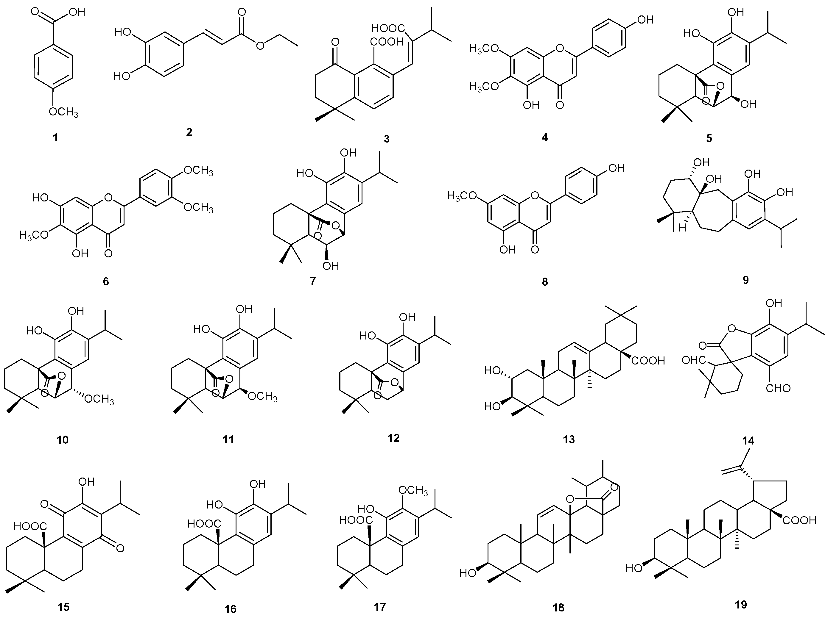
An analytical approach based on LC-(-)ESI/QExactive/MS/MS, in negative-ion mode, was carried out to obtain a chemical profile of the EtOH extract of P. atriplicifolia aerial parts (Figure 1). The LC-ESI/HRMS analysis of the EtOH extract showed the presence of 19 compounds. Initially, some peaks were tentatively identified based on their accurate masses, on the characteristic fragmentation pattern, and by comparing the results obtained with the data reported in the literature [16] (Figure 1). The careful analysis of the multistage mass spectra suggested the presence of phenolics (1, 2), diterpenes (3, 5, 7, 912, 1417), and flavonoids (4, 6, 8), as well as triterpenes (13, 18, 19) (Table 1).
Compounds 1 and 2 showed typical molecular formulae and fragmentation patterns observed for compounds belonging to the phenolic class. Compound 1 showed an ion at m/z 151.0392, supporting the molecular formula C8H8O3, and it was tentatively identified as p-methoxybenzoic acid, previously reported in P. artiplicifolia [17]. Compound 2 was identified as caffeic acid ethyl ester, reported in Satureja bachtiarica [18].
The MS/MS spectra of compounds 3, 5, 7, 1012, and 1417 showed a main fragment ion [(M − CO2) − H] corresponding to a typical fragment generated by decarboxylation, in agreement with the literature data for carnosic acid derivatives [19]. Also, compound 3 showed a fragmentation pattern typical of abietane derivatives, molecules previously reported in P. artemisioides roots [20]. In addition, compounds 5, 7, 10, and 11 were characterized by deprotonated molecular ions derived by loss of a water molecule and a subsequent loss of a methyl radical. All these data suggested that compounds 5, 10, and 11 were structurally related to rosmanol, a diterpenoid differing from carnosic acid in the occurrence of a lactone function between the carboxylic acid group and the hydroxy function at C-6 and in a hydroxy group at C-7; compound 7 corresponded to epi-isorosmanol, characterized by lactone function between the carboxylic acid group and the hydroxy function at C-7 and in a hydroxy group at C-6. Compound 9 was assigned to the icetexane derivative previously identified in P. artemisioides aerial parts [14]. Compounds 4, 6, and 8 were recognized as flavonoids and identified as scrophulein, eupatilin, and genkwanin, respectively, previously reported in P. atriplicifolia [21,22,23]. Compounds 13, 18, and 19 were identified as the triterpenes maslinic acid, urs-11-en-28,13β-olide, and betulinic acid, respectively, previously reported in the literature [14,24].

2.2. Isolation and Identification of Compounds 1–19

To unambiguously assign compounds corresponding to these peaks and to discriminate among structural isomers or stereoisomers, isolation and structure elucidation by 1D and 2D NMR experiments of isolated compounds were carried out. The EtOH extract of P. atriplicifolia aerial parts was fractionated by column chromatography on silica gel, and the fractions were purified by semipreparative HPLC-UV, as described in the Experimental Section, affording compounds reported in Table 1. The structures of the isolated compounds were established by 1D and 2D NMR spectroscopy in combination with mass spectrometry (Figure 2). The isolated compounds were identified by analysis of their NMR spectroscopic data in comparison to those reported in the literature as p-methoxybenzoic acid (1) [17], caffeic acid ethyl ester (2) [18], perovskin A (3) [20], scrophulein (4) [22], epirosmanol (5) [25], eupatilin (6) [23], epiisorosmanol (7) [14], genkwanin (8) [26], 1α-hydroxydemethylsalvicanol (9) [14], 7-O-methylrosmanol (10) [14], 7-epi-7-O-methylrosmanol (11) [26], carnasol (12) [14], maslinic acid (13) [26], rosmadial (14) [27], royleanonic acid (15) [28], carnosic acid (16) [14], 12-O-methylcarnosic acid (17) [14], urs-11-en-28,13β-olide (18) [29], and betulinic acid (19) [14] (Figure 2) (Figures S1–S4, S6, S7, S9, S10, S12, S13, S15–S18, S20–S25, S27 and S28, Tables S1–S3). To our knowledge, this is the first report of compounds 3, 7, 919 in P. atricipifolia. In addition, herein, compounds 15 and 18 are described for the first time in the genus Perovskia.

2.3. Evaluation of the Antioxidant Activity of EtOH Extract of P. atriplicifolia and Pure Compounds

Considering the ability of carnosol (12) and carnosic acid (16) to exert antioxidant effects by reducing ROS, NO, and lipid peroxides, as well as their involvement in activating the Nrf2/HO-1/NQO-1 pathway, where Nrf2 dissociates from Keap1, translocates to the nucleus, and binds to ARE sequences to upregulate antioxidant genes [10,30], the eventual cytotoxic and antioxidant activity of the extracts and the main abietane derivatives in the aerial part extract of P. atriplicifolia were evaluated.

2.3.1. Cell Viability

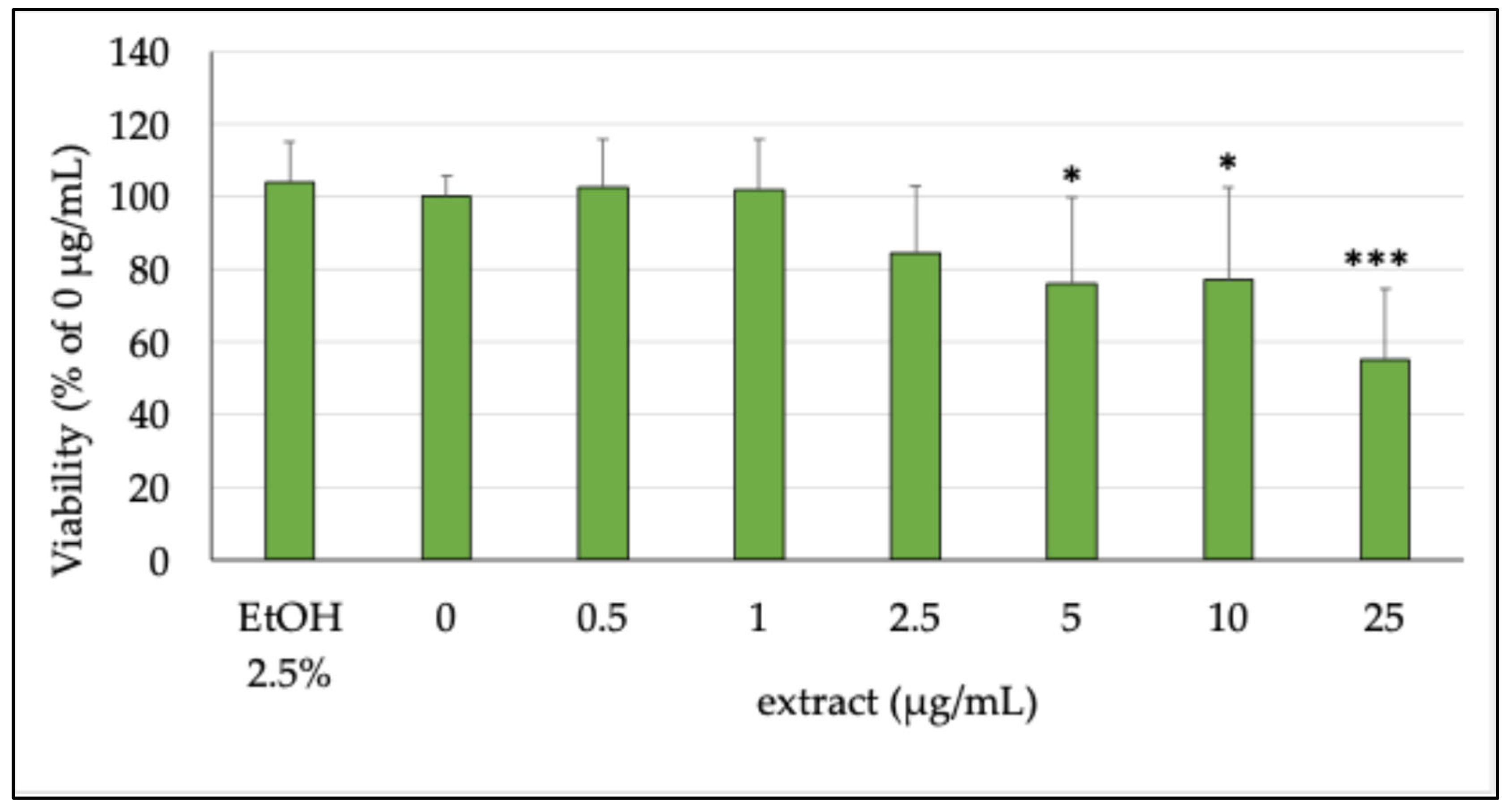
The potential cytotoxic effect of the extract was assessed on differentiated Caco-2 cells, which are usually employed to simulate differentiated intestinal epithelium in vitro [31] at concentrations ranging from 0.5 to 25 µg/mL. Figure 3 reports the cell viability results obtained on differentiated cells. As shown, the tested extract caused a decrease in cellular viability of Caco-2 cells with respect to untreated cells (0 μg/mL, 100% viable cells), starting from 5 µg/mL, resulting in the lowest significantly cytotoxic concentration. This result was likely due to the very high concentration of polyphenols and certainly not from the medium used to dissolve the extract (EtOH 2.5%), as shown in Figure 3. A recent study has indeed shown that some compounds isolated from sage species, such as carnosol (12) and carnosic acid (16), can lead to toxic events at high concentrations in both cancer and normal cells [32]. Given the results obtained in this model, we then decided to use 2.5 µg/mL as the maximum concentration, because, among those tested in our experiments, it was the highest non-toxic concentration.
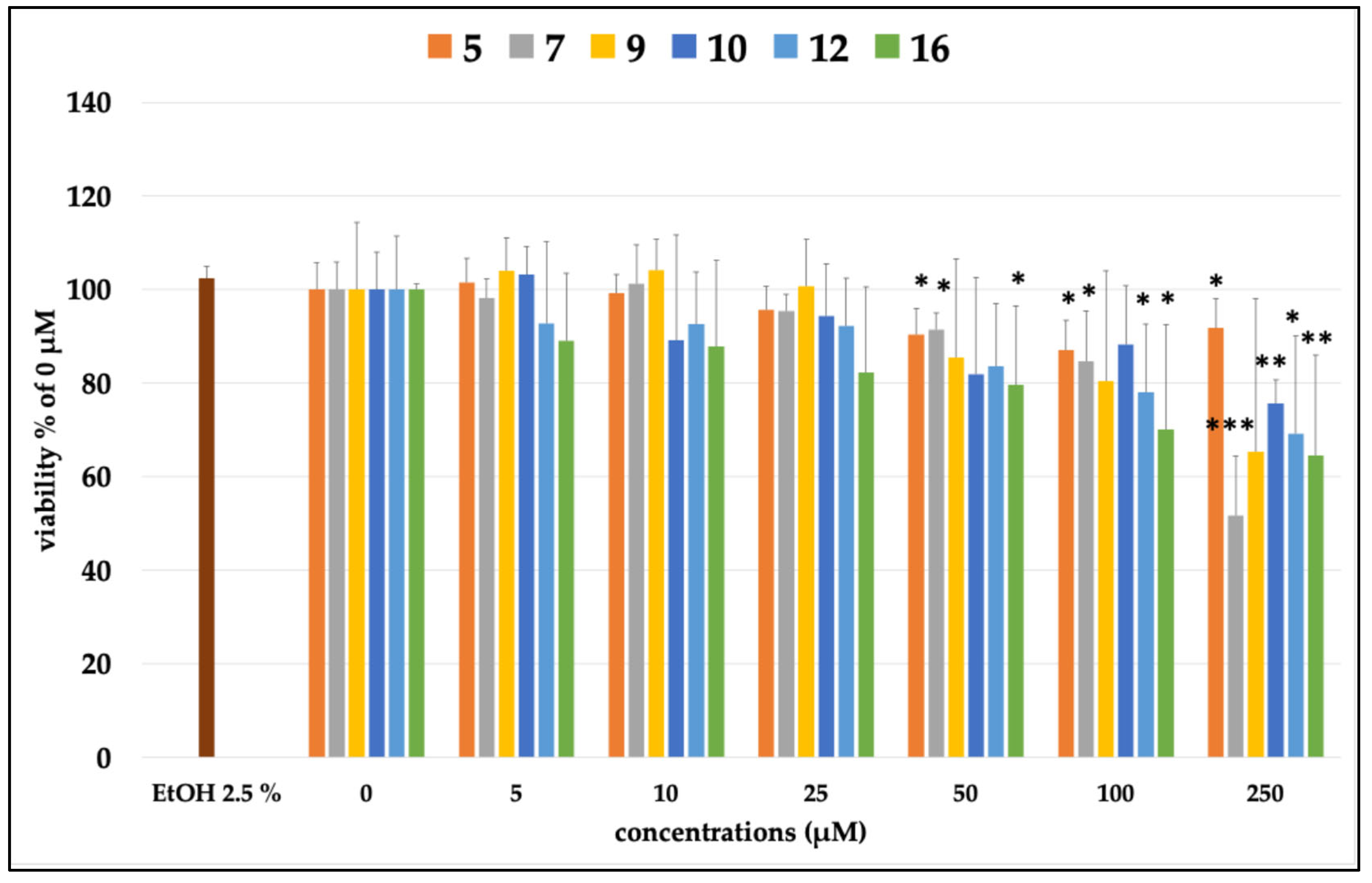
In this context, we also tested six of the most concentrated compounds in the extract, namely compounds 5, 7, 9, 10, 12, and 16. As shown in Figure 4, some of the tested compounds were found to be toxic starting at 50 µM (es, 5, 7, and 16), and then increased in toxicity in a dose-dependent fashion. For this reason, in the subsequent study, only concentrations below the toxic dose were tested.

2.3.2. Inhibition of ROSs Production

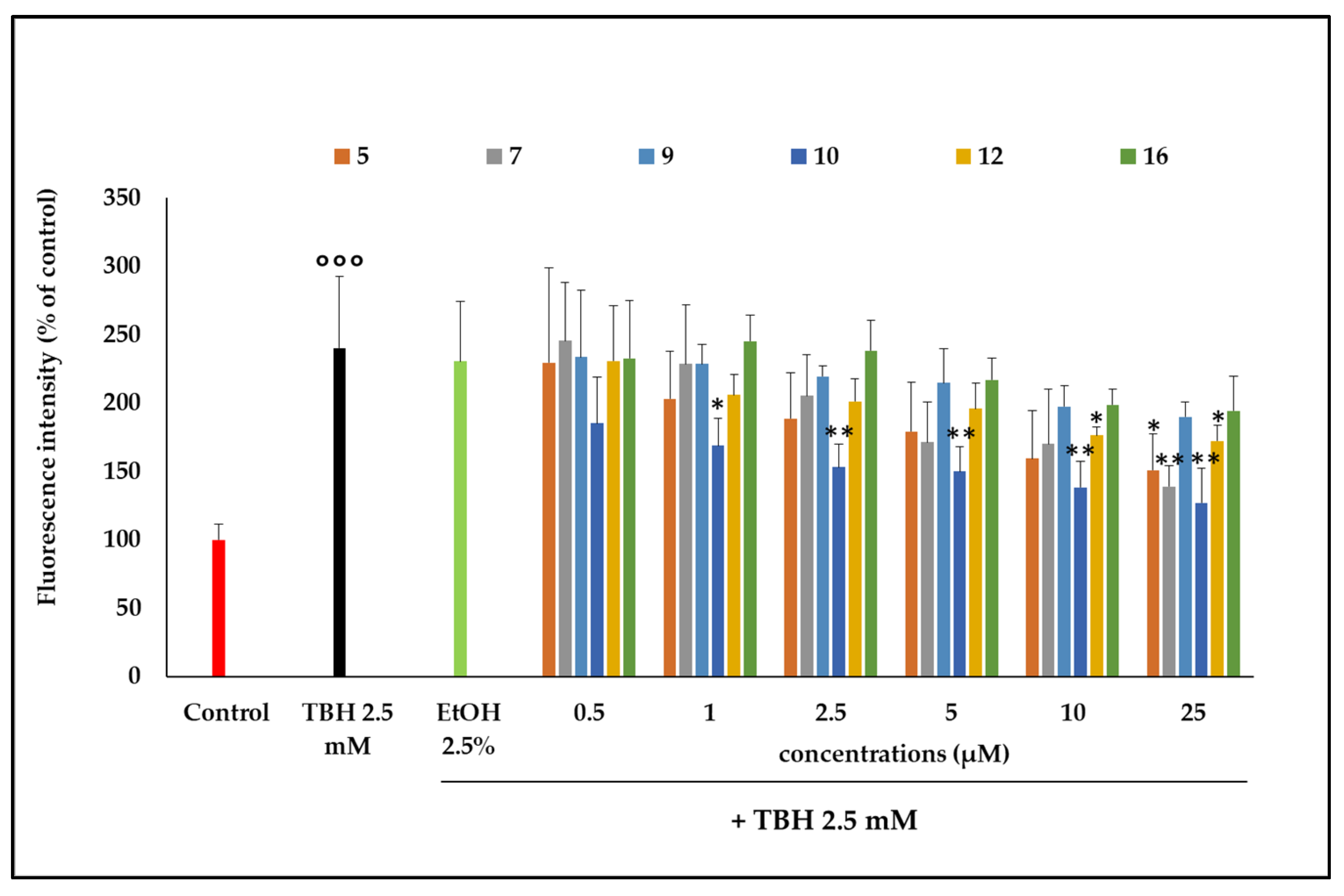
TBH induced determination of intracellular ROSs production and consequent oxidative damage to cell phospholipids at the concentration of 2.5 mM, compared with untreated cells (Control, Figure 5). In cells pretreated with the ethanol extract, a significant inhibition of ROSs was detected. Although very low concentrations of extract were tested, it was able to limit the production of ROSs induced by TBH after the first 60 min of incubation. This inhibition was observed at 2.5 and 5 µg/mL after 60 min of incubation (p < 0.05). This outcome shows that the substances present in the extract are active in counteracting the oxidizing action of TBH, since the ethanol present in the incubation environment (EtOH 2.5%) did not show any antioxidant activity when compared with the action of TBH alone. Among the compounds involved in this activity, there could be some of the more highly represented molecules in the extract, like those already studied in the previous test. In this regard, it was seen how 7-O-methylrosmanol (10) showed antioxidant activity starting from low concentrations (1 μM) and then increasing in a dose-dependent manner (Figure 6). Other compounds, such as 12, 5, and 7, showed antioxidant capacity in a dose-dependent manner starting from lower concentrations and leading to significant inhibition of TBH-induced oxidative stress at the highest concentrations tested (10 and 25 μM) (Figure 6).

2.4. Quantitative Analysis of the Constituents of P. atriplicifolia EtOH Extract

Quantitative analysis was conducted using an LC-(-)ESI/QExactive/MS/MS system in parallel reaction monitoring (PRM) mode, a high-resolution, high-accuracy technique for targeted quantification using quadrupole-Orbitrap hybrid instruments. In PRM, precursor ions are selected in the quadrupole, fragmented in the HCD cell, and analyzed in the Orbitrap via the C-trap, which enhances signal-to-noise by extending ion accumulation [33]. The amount (mg/100 g dry aerial parts) of the most representative abietane derivatives (5, 7, 9, 10, 12, and 16) in the EtOH extract of P. artemisioides aerial parts (Table 2) was determined. The quantitative results highlighted that the above-cited compounds occurred in the extract in concentration ranging from 1.73 to 520.21 mg/100 g aerial parts with carnosol (12) exhibiting the highest concentration (520.21 mg/100 g aerial parts) followed by 1α-hydroxydemethylsalvicanol (9) (91.73 mg/100 g aerial parts) and carnosic acid (16) (88.16 mg/100 g aerial parts) (Table 2).

3. Discussion

3.1. Quantitative Analysis of Carnosol (12) and Carnosic Acid (16)

Results of quantitative analysis highlighting that carnosol (12) exhibited a concentration of 520.21 mg/100 g aerial parts and carnosic acid (16) a concentration of 88.16 mg/100 g aerial parts (Table 2) are consistent with literature reports describing carnosic acid and carnosol as key diterpenes found in the aerial parts of Perovskia genus, as well as in other genera within the Lamiaceae family, such as Salvia spp. and Rosmarinus officinalis. Specifically, our previous research highlighted the presence of carnosic acid and carnosol in the aerial parts of Perovskia artemisioides, with concentrations of 855.00 mg/100 g and 435.60 mg/100 g of dry aerial parts, respectively. Similarly, in the aerial parts of Salvia officinalis, these diterpenoids were also abundant, with concentrations of 829.40 mg/100 g and 115.50 mg/100 g of dry aerial parts, respectively [14]. Finally, in R. officinalis, during the flowering stage—which typically occurs in spring under mild weather conditions—a notable accumulation of secondary metabolites was observed, including caffeic acid, ferulic acid, and carnosic acid (729.34 mg/100 g dry aerial parts) [34]. In contrast, during the fruit maturation stage in summer, characterized by higher temperatures, the plant showed increased production of rosmarinic acid, hesperidin, and carnosol (987.01 mg/100 g dry aerial parts) [35].

3.2. Bioactivity of Carnosol (12) and Carnosic Acid (16)

Carnosol (12) and carnosic acid (16), the main abietane diterpenes found in the aerial parts of Perovskia spp., are reported to mitigate oxidative stress by reducing levels of reactive oxygen species (ROSs), nitric oxide (NO), and lipid peroxides. They also enhance antioxidant defenses by increasing glutathione (GSH) and the activity of enzymes such as superoxide dismutase (SOD), catalase (CAT), and glutathione peroxidase (GPx) [31]. These effects are associated with activation of the Nrf2/HO-1/NQO-1 pathway, wherein the compounds promote Nrf2 release from Keap1, allowing its nuclear translocation. In the nucleus, Nrf2 binds to antioxidant response element (ARE) sequences, inducing the expression of antioxidant genes [30].
Carnosic acid (16) and carnosol (12), both found also in rosemary and other sage species, have already been studied for their antioxidant effects in intestinal cells. Both compounds showed clear antioxidant activity in intestinal cells, primarily by reducing oxidative stress and enhancing cellular defense mechanisms. In particular, in pig intestinal epithelial cells, carnosol pretreatment reduced oxidative stress markers (ROSs, MDA, NO) and increased antioxidant enzyme levels (SOD). It also promoted cell survival under oxidative stress by activating the Nrf2 pathway, which upregulates antioxidant and protective genes [36]. Furthermore, studies in animal models (mice and poultry) showed that carnosic acid enhanced antioxidant capacity in the intestine. It reduced oxidative stress and inflammation, improved gut barrier function, and modulated the expression of key antioxidant proteins such as Nrf2 and SOD. These effects were observed in both chemically-induced colitis and inflammation models [37,38,39].
Oxidative stress is a major contributing factor to neurodegenerative disorders. Therefore, the neuroprotective effects observed for carnosol and carnosic acid in neuronal cells may be linked to the antioxidant properties of reported abietane derivatives [40]. Carnosic acid has been shown to exert neuroprotective activity against ischemia/hypoxia-induced injury by scavenging or reducing reactive oxygen species (ROSs) and nitric oxide (NO), and by inhibiting the COX-2 and MAPK pathways [41]. Furthermore, carnosic acid has been reported to help prevent amyloid-β (Aβ)-induced neurotoxicity, suggesting a potential role in the prevention of Alzheimer’s disease [40].
In addition, in cancer cells, carnosol inhibits proliferation, survival, migration, and invasion while promoting apoptosis. These effects are linked to the inhibition of key signaling pathways, including extracellular signal-regulated kinase (ERK), p38, c-Jun N-terminal kinase (JNK), Akt, mechanistic target of rapamycin (mTOR), and cyclooxygenase-2 (COX-2). Finally, carnosol and carnosic acid exhibit anti-inflammatory effects by activating PPAR-γ and suppressing NF-κB and COX-2 pathways [42]. They also reduce LPS-induced inflammation by inhibiting pro-inflammatory cytokines (e.g., TNF-α, IL-6) and blocking NF-κB and MAPK signaling [43].

4. Materials and Methods

4.1. Plant Material and Extraction

The aerial parts of P. atriplicifolia were collected from the Taftan region of Tamin, Sistan and Baluchestan Province, Iran (latitude: 28°69′ N; longitude: 61°16′ E; altitude: 2000 m above sea level), in July 2022. The plant species was identified by Dr. Mehdi Rahimmalek at the Botany Department of Isfahan University of Technology (IUT), Isfahan, Iran, using Flora Iranica [44]. The air-dried aerial parts (380 g) were ground and extracted by maceration at room temperature with EtOH (Merck, Milan, Italy) (3 × 3.6 L for 72 h).

4.2. LC-(-)ESI/QExactive/MS/MS Analysis

P. atriplicifolia EtOH extract was analyzed by hybrid quadrupole-Orbitrap mass spectrometer, using negative electrospray ionization mode (UHPLC-(-)ESI/QExactive MS/MS, Thermo Fisher Scientific, Waltham, MA, USA). An injection of 3 μL of EtOH extract (1 mg/mL) was used. LC-HRMS/MS conditions and parameters are detailed in the Supplementary Materials.

4.3. Isolation Procedure

The dried EtOH extract (8.15 g) was obtained by filtration and vacuum evaporation; 3 g of extract was fractionated via silica gel CC (Sephadex LH-20, GE Healthcare, Sigma-Aldrich, Milan, Italy; 100 × 5 cm) using MeOH (Merck, Milan, Italy) as the mobile phase, yielding 41 fractions (10 mL), monitored by TLC. Similar fractions (per TLC) were pooled into 8 main groups. Purification was carried out using RP-HPLC-UV, resulting in the isolation of compounds 119 (see Supplementary Materials). The purity of the tested compounds (95–99%) was determined by LC-HRMS analysis (Figures S5, S8, S11, S14, S19, and S26).

4.4. Cell Culture

The Caco-2 cell line was sourced from ECACC (Salisbury, UK) and handled according to the procedures described in the Supplementary Materials.

4.5. MTT Viability Test

To evaluate the cytotoxic effects of the extract and pure compounds on differentiated Caco-2 cells (21 days post-seeding; ECACC, Salisbury, UK), cell viability was assessed using the MTT assay, as previously described [45] (see Supplementary Materials).

4.6. Determination of Intracellular Reactive Oxygen Species (ROS) Production

ROSs production in Caco-2 cells was assessed using the fluorescent probe H2-DCF-DA, following the method of Gil et al. [46] with minor modifications (see Supplementary Materials). Extracts and pure compounds were evaluated at concentrations of 0.5–5 μg/mL and 0.5–25 μM, respectively (see Supplementary Materials).

4.7. Statistical Analyses

Statistical analysis was performed using GraphPad Prism 5 (GraphPad Software, San Diego, CA, USA). One-way ANOVA followed by Tukey’s post hoc test was applied, with p-values < 0.05 considered statistically significant.

4.8. Quantitative Analysis of Main Diterpenoid Derivatives (5, 7, 9, 10, 12, 16) by LC-(-)ESI/QExactive/MS/MS

Quantitative analysis was conducted using an LC-(-)ESI/QExactive/MS/MS system in Parallel Reaction Monitoring (PRM) mode, a high-resolution, high-accuracy technique for targeted quantification using quadrupole-Orbitrap hybrid instruments.
For LC-ESI/HRMS, mobile phases consisted of water (Deltek, Naples, Italy) (A) and acetonitrile (Deltek, Naples, Italy) (B), both with 0.1% formic acid, at 0.3 mL/min. Samples (5 µL, 0.25 mg/mL) were analyzed in negative-ion mode using a Luna Omega C18 column (1.6 µm, 100 × 2.1 mm; Phenomenex, Aschaffenburg, Germany) maintained at 30 °C. A linear gradient was applied, starting at 45% B and held at this % for 2.20 min; then increased to 85% B in 8.9 min, to 100% B in 0.9 min, held at 100% for 3 min, and finally returned to 45% B over 3 min.
Precursor/product ions and normalized collision energies (NCE) considered in PRM are listed in Table S1. Data were acquired in centroid mode (17,500 resolution, AGC target 1 × 10⁵, max IT 50 ms) and processed using Xcalibur 2.2 (Thermo Fisher Scientific, Waltham, MA, USA). A stock solution (1 mg/mL) of external standards was diluted in methanol to prepare six concentrations (0.5–20.0 µg/mL) for calibration. The collision cell exit potential (CXP) was 30 eV. Limits of detection (LOD) and quantification (LOQ) were determined based on signal-to-noise ratios of 3 and 10, respectively [47], as reported in Table S1 (Figure S4).

5. Conclusions

This work presents the first comprehensive investigation of the EtOH extract of Perovskia atriplicifolia aerial parts. An analytical approach based on LC-(-)ESI/QExactive/MS/MS, combined with NMR characterization of isolated compounds, enabled the identification of 19 constituents, including diterpenes, flavonoids, phenolics, and triterpenes. Among these, compounds 3, 7, 919 were reported for the first time in P. atriplicifolia. In addition, herein, compounds 15 and 18 were described for the first time in the genus Perovskia. The results indicate that the EtOH extract is a rich source of abietane diterpenes related to carnosic acid and carnosol. Furthermore, the extract and carnosic acid derivatives (5, 7, 9, 10, 12, and 16) exhibited significant antioxidant activity, demonstrated by their ability to reduce free radical production in the Caco-2 cell line.
In particular, 7-O-methylrosmanol (10) showed antioxidant activity from as low as 1 μM in a dose-dependent manner; compounds 12, 5, and 7 also exhibited antioxidant activity in a dose-dependent manner beginning at lower concentrations and resulting in significant inhibition of TBH-induced oxidative stress at the highest concentrations tested (10 and 25 μM). To correlate the activity of the ethanol extract from P. atriplicifolia aerial parts with the levels of its constituents, a quantitative analysis of the main compounds was performed using an LC-(-)ESI/QExactive/MS/MS-based analytical approach, revealing that the identified compounds (5, 7, 9, 10, 12, and 16) occurred in concentrations ranging from 1.73 to 520.21 mg per 100 g of aerial parts. Carnosol (12) was the most abundant (520.21 mg/100 g), followed by 1α-hydroxydemethylsalvicanol (9) (91.73 mg/100 g), and carnosic acid (16) (88.16 mg/100 g). These findings evidence the chemical richness of P. atriplicifolia ethanol extract, which contains some antioxidant properties, like carnosic acid and carnosol, in high amounts, highlighting its potential as an ingredient in antioxidant nutraceutical and pharmaceutical formulations.

Supplementary Materials

The following supporting information can be downloaded at: https://www.mdpi.com/article/10.3390/plants14172795/s1, Figures S1, S3, S6, S9 S12 S15, S17, S20, S22, S24, S27: 1H NMR Spectra (600 MHz, CD3OD) of compounds 3, 5, 7, 9, 10, 11, 12, 14, 15, 16, 17, respectively; Figures S2, S4, S7, S10, S13, S16, S18, S21, S23, S25, S28, HRMS Spectra of compounds 3, 5, 7, 9, 10, 11, 12, 14, 15, 16, 17, respectively; Figures S5, S8, S11, S14, S19, S26: LC-MS Spectra of compounds 5, 7, 9, 10, 12, 16, respectively; Table S1: NMR Spectroscopic Data (600 MHz, CD3OD) of Compounds 3, 5, 7, 10, 11; Table S2: NMR Spectroscopic Data (600 MHz, CD3OD) of Compounds 12, 15–17; Table S3: NMR Spectroscopic Data (600 MHz, CD3OD) of Compounds 9, 14; Table S4: LC–HRMS/MS conditions for quantitation of compounds 5, 7, 9, 10, 12, 16.

Author Contributions

Conceptualization, A.C., M.M. and S.P.; software, M.R.S., A.C., M.P.M. and G.S.; validation, M.M. and M.D.; formal analysis, M.R.S., A.C., M.P.M. and G.S.; investigation, M.R.S., M.P.M. and G.S.; resources, M.D. and S.P.; data curation, M.R.S., A.C., M.M. and M.D.; writing—original draft preparation, A.C., M.R.S., M.P.M. and G.S.; writing—review and editing, M.M., M.D. and S.P.; visualization, M.M.; supervision, S.P.; funding acquisition, S.P. All authors have read and agreed to the published version of the manuscript.

Funding

This research received no external funding.

Data Availability Statement

The original contributions presented in this study are included in the article/Supplementary Materials. Further inquiries can be directed to the corresponding author.

Conflicts of Interest

The authors declare no conflict of interest.

References

  1. Hashemifar, Z.; Rahimmalek, M. Genetic structure and variation in Perovskia abrotanoides Karel and P. atriplicifolia as revealed by molecular and morphological markers. Sci. Hortic. 2018, 230, 169–177. [Google Scholar] [CrossRef]
  2. Tabefam, M.; Farimani, M.M.; Danton, O.; Ramseyer, J.; Kaiser, M.; Ebrahimi, S.N.; Salehi, P.; Batooli, H.; Potterat, O.; Hamburger, M. Antiprotozoal diterpenes from Perovskia abrotanoides. Planta Med. 2018, 84, 913–919. [Google Scholar] [CrossRef]
  3. Ahmad, I.; Fozia; Waheed, A.; Tahir, N.B.; Rais, A.K. Anti-inflammatory constituents from Perovskia atriplicifolia. Pharm. Biol. 2015, 53, 1628–1631. [Google Scholar] [CrossRef]
  4. Pourhosseini, S.H.; Hadian, J.; Sonboli, A.; Nejad Ebrahimi, S.; Mirjalili, M.H. Genetic and Chemical Diversity in Perovskia abrotanoides Kar. (Lamiaceae) Populations Based on ISSRs Markers and Essential Oils Profile. Chem. Biodivers. 2018, 15, e1700508. [Google Scholar] [CrossRef]
  5. Duke, J.; Bogenschuts, J.; Cellier, D. Handbook of Medicinal Herbs; CRC Press: Boca Raton, FL, USA, 2002. [Google Scholar]
  6. Tareen, R.B.; Bibi, T.; Khan, M.A.; Ahmad, M.; Zafar, M.; Hina, S. Indigenous knowledge of folk medicine by the women of Kalat and Khuzdar regions of Balochistan, Pakistan. Pak. J. Bot. 2010, 42, 1465–1485. [Google Scholar]
  7. Alizadeh, Z.; Farimani, M.M.; Parisi, V.; Marzocco, S.; Ebrahimi, S.N.; De Tommasi, N. Nor-abietane Diterpenoids from Perovskia abrotanoides Roots with Anti-inflammatory Potential. J. Nat. Prod. 2021, 84, 1185–1197. [Google Scholar] [CrossRef] [PubMed]
  8. Afshari, M.; Rahimmalek, M.; Sabzalian, M.R.; Bielecka, M.; Matkowski, A.; Talebi, M. Changes in physiological, phytochemical traits and gene expression of two Perovskia species in response to water deficit. Sci. Hortic. 2022, 293, 110747. [Google Scholar] [CrossRef]
  9. Zaker, A.; Sykora, C.; Gössnitzer, F.; Abrishamchi, P.; Asili, J.; Mousavi, S.H.; Wawrosch, C. Effects of some elicitors on tanshinone production in adventitious root cultures of Perovskia abrotanoides Karel. Ind. Crop. Prod. 2015, 67, 97–102. [Google Scholar] [CrossRef]
  10. Miroliaei, M.; Aminjafari, A.; Slusarczyk, S.; Nawrot-Hadzik, I.; Rahimmalek, M.; Matkowski, A. Inhibition of Glycation-induced Cytotoxicity, Protein Glycation, and Activity of Proteolytic Enzymes by Extract from Perovskia atriplicifolia Roots. Pharmacogn. Mag. 2017, 13, s676–s683. [Google Scholar] [CrossRef] [PubMed]
  11. Ciobotaru, G.V.; Goje, I.-D.; Dehelean, C.A.; Danciu, C.; Magyari-Pavel, I.Z.; Moacă, E.-A.; Muntean, D.; Imbrea, I.M.; Sărățeanu, V.; Pop, G. Analysis of the Antioxidant and Antimicrobial Activity, Cytotoxic, and Anti-Migratory Properties of the Essential Oils Obtained from Cultivated Medicinal Lamiaceae Species. Plants 2025, 14, 846. [Google Scholar] [CrossRef]
  12. Johnson, J.J. Carnosol: A promising anti-cancer and anti-inflammatory agent. Cancer Lett. 2011, 305, 1–7. [Google Scholar] [CrossRef] [PubMed]
  13. Dabiri, M.; Sefidkon, F. Analysis of the essential oil from aerial parts of Perovskia atriplicifolia Benth. at different stages of plant growth. Flavour Fragr. J. 2001, 16, 435–438. [Google Scholar] [CrossRef]
  14. Sadeghi, Z.; Masullo, M.; Cerulli, A.; Nazzaro, F.; Farimani, M.; Piacente, S. Terpenoid Constituents of Perovskia artemisioides Aerial Parts with Inhibitory Effects on Bacterial Biofilm Growth. J. Nat. Prod. 2020, 84, 26–36. [Google Scholar] [CrossRef]
  15. Loussouarn, M.; Krieger-Liszkay, A.; Svilar, L.; Bily, A.; Birtić, S.; Havaux, M. Carnosic Acid and Carnosol, Two Major Antioxidants of Rosemary, Act through Different Mechanisms. Plant Physiol. 2017, 175, 1381–1394. [Google Scholar] [CrossRef]
  16. Hosseini, S.H.; Masullo, M.; Cerulli, A.; Martucciello, S.; Ayyari, M.; Pizza, C.; Piacente, S. Antiproliferative Cardenolides from the Aerial Parts of Pergularia tomentosa. J. Nat. Prod. 2019, 82, 74–79. [Google Scholar] [CrossRef]
  17. Ali, M.S.; Saleem, M.; Erian, A.W. A new acylated steroid glucoside from Perovskia atriplicifolia. Fitoterapia 2001, 72, 712–714. [Google Scholar] [CrossRef] [PubMed]
  18. Rahmani Samani, M.; D’Urso, G.; Nazzaro, F.; Fratianni, F.; Masullo, M.; Piacente, S. Phytochemical Investigation and Biofilm-Inhibitory Activity of Bachtiari Savory (Satureja bachtiarica Bunge) Aerial Parts. Plants 2024, 13, 67. [Google Scholar] [CrossRef] [PubMed]
  19. Pacifico, S.; Piccolella, S.; Lettieri, A.; Nocera, P.; Bollino, F.; Catauro, M. A metabolic profiling approach to an Italian sage leaf extract (SoA541) defines its antioxidant and anti-acetylcholinesterase properties. J. Funct. Foods. 2017, 29, 1–9. [Google Scholar] [CrossRef]
  20. Sadeghi, Z.; Cerulli, A.; Marzocco, S.; Moridi Farimani, M.; Masullo, M.; Piacente, S. Anti-inflammatory Activity of Tanshinone-Related Diterpenes from Perovskia artemisioides Roots. J. Nat. Prod. 2023, 86, 812–821. [Google Scholar] [CrossRef]
  21. Perveen, S.; Al-Taweel, A.; Fawzy, G.; Ibrahim, T.; Malik, A.; Khan, A. Cholinesterase inhibitory triterpenes from Perovskia atriplicifolia. Asian J. Chem. 2014, 26, 6163. [Google Scholar] [CrossRef]
  22. Khaliq, S.; Volk, F.-J.; Frahm, A. Phytochemical Investigation of Perovskia abrotanoides. Planta Med. 2007, 73, 77–83. [Google Scholar] [CrossRef]
  23. Tarawneh, A.; León, F.; Pettaway, S.; Elokely, K.M.; Klein, M.L.; Lambert, J.; Mansoor, A.; Cutler, S.J. Flavonoids from Perovskia atriplicifolia and Their in Vitro Displacement of the Respective Radioligands for Human Opioid and Cannabinoid Receptors. J. Nat. Prod. 2015, 78, 1461–1465. [Google Scholar] [CrossRef]
  24. Siddiqui, B.S.; Sultana, I.; Begum, S. Triterpenoidal constituents from Eucalyptus camaldulensis var. obtusa leaves. Phytochemistry 2000, 54, 861–865. [Google Scholar] [CrossRef]
  25. Jiang, Z.-Y.; Li, Z.-Q.; Huang, C.-G.; Zhou, J.; Hu, Q.-F.; Liu, W.-X.; Huang, X.-Z.; Wang, W.; Zhang, L.-Z.; Xia, F.-T. Abietane Diterpenoids from Perovskia atriplicifolia and Their Anti-HBV Activities. Bull. Korean Chem. Soc. 2015, 36, 623–627. [Google Scholar] [CrossRef]
  26. Marrero, J.G.; Moujir, L.; Andrés, L.S.; Montaño, N.P.; Araujo, L.; Luis, J.G. Semisynthesis and Biological Evaluation of Abietane-Type Diterpenes. Revision of the Structure of Rosmaquinone. J. Nat. Prod. 2009, 72, 1385–1389. [Google Scholar] [CrossRef]
  27. Jiang, Z.-Y.; Zhou, J.; Huang, C.-G.; Hu, Q.-F.; Huang, X.-Z.; Wang, W.; Zhang, L.-Z.; Li, G.-P.; Xia, F.-T. Two novel antiviral terpenoids from the cultured Perovskia atriplicifolia. Tetrahedron 2015, 71, 3844–3849. [Google Scholar] [CrossRef]
  28. Mahmoud, A.A.; Al-Shihry, S.S.; Son, B.W. Diterpenoid quinones from Rosemary (Rosmarinus officinalis L.). Phytochemistry 2005, 66, 1685–1690. [Google Scholar] [CrossRef] [PubMed]
  29. Hongcheng, W.; Fujimotot, Y. Triterpene esters from Eucalyptus tereticornis. Phytochemistry 1993, 33, 151–153. [Google Scholar] [CrossRef]
  30. Liang, H.; Guan, M.; Li, T.; Li, S.; Ma, C.; Li, C. Critical review on biological effect and mechanisms of diterpenoids in Rosmarinus officinalis. Food Med. Homol. 2025, 2, 9420021. [Google Scholar] [CrossRef]
  31. Santoru, M.L.; Piras, C.; Murgia, F.; Spada, M.; Tronci, L.; Leoni, V.P.; Serreli, G.; Deiana, M.; Atzori, L. Modulatory Effect of Nicotinic Acid on the Metabolism of Caco-2 Cells Exposed to IL-1beta and LPS. Metabolites 2020, 10, 204. [Google Scholar] [CrossRef] [PubMed]
  32. Irtegun Kandemir, S.; Fidan, H.S.; Yener, I.; Mete, N.; Ertas, A.; Topcu, G.; Kolak, U. Investigation of cytotoxic and apoptotic effects of 63 compounds obtained from Salvia species: Promising anticancer agents. J. Food Biochem. 2022, 46, e14226. [Google Scholar] [CrossRef]
  33. Rauniyar, N. Parallel Reaction Monitoring: A Targeted Experiment Performed Using High Resolution and High Mass Accuracy Mass Spectrometry. Int. J. Mol. Sci. 2015, 16, 28566–28581. [Google Scholar] [CrossRef]
  34. Šulniūtė, V.; Pukalskas, A.; Venskutonis, P.R. Phytochemical composition of fractions isolated from ten Salvia species by supercritical carbon dioxide and pressurized liquid extraction methods. Food Chem. 2017, 224, 37–47. [Google Scholar] [CrossRef]
  35. Jordán, M.J.; Lax, V.; Rota, M.C.; Lorán, S.; Sotomayor, J.A. Effect of the phenological stage on the chemical composition, and antimicrobial and antioxidant properties of Rosmarinus officinalis L. essential oil and its polyphenolic extract. Ind. Crop. Prod. 2013, 48, 144–152. [Google Scholar] [CrossRef]
  36. Jiang, Z.; Li, L.; Hou, L.; Zhou, Z.; Wang, C. Mechanism of protective effect of carnosol on pig intestinal epithelial cells. Int. J. Clin. Exp. Pathol. 2020, 13, 447–455. [Google Scholar] [PubMed]
  37. Zhang, J.; Shen, M.; Yin, Y.; Chen, Y.; Deng, X.; Mo, J.; Zhou, X.; Lin, J.; Chen, X.; Xie, X.; et al. Carnosic acid reduces lipid content, enhances gut health, and modulates microbiota composition and metabolism in diet-induced obese mice. Food Funct. 2025, 16, 1888–1902. [Google Scholar] [CrossRef] [PubMed]
  38. Du, C.; Li, Z.; Zhang, J.; Yin, N.; Tang, L.; Li, J.; Sun, J.; Yu, X.; Chen, W.; Xiao, H.; et al. The protective effect of carnosic acid on dextran sulfate sodium-induced colitis based on metabolomics and gut microbiota analysis. Food Sci. Hum. Well. 2023, 12, 1212–1223. [Google Scholar] [CrossRef]
  39. Wan, S.S.; Li, X.Y.; Liu, S.R.; Tang, S. The function of carnosic acid in lipopolysaccharides-induced hepatic and intestinal inflammation in poultry. Poult. Sci. 2024, 103, 103415. [Google Scholar] [CrossRef]
  40. Mirza, F.; Zahid, S.; Holsinger, R.M.D. Neuroprotective Effects of Carnosic Acid: Insight into Its Mechanisms of Action. Molecules 2023, 28, 2306. [Google Scholar] [CrossRef]
  41. Hou, C.-W.; Lin, Y.-T.; Chen, Y.-L.; Wang, Y.-H.; Chou, J.-L.; Ping, L.-Y.; Jeng, K.-C. Neuroprotective effects of carnosic acid on neuronal cells under ischemic and hypoxic stress. Nutr. Neurosci. 2012, 15, 257–263. [Google Scholar] [CrossRef]
  42. Poeckel, D.; Greiner, C.; Verhoff, M.; Rau, O.; Tausch, L.; Hörnig, C.; Steinhilber, D.; Schubert-Zsilavecz, M.; Werz, O. Carnosic acid and carnosol potently inhibit human 5-lipoxygenase and suppress pro-inflammatory responses of stimulated human polymorphonuclear leukocytes. Biochem. Pharmacol. 2008, 76, 91–97. [Google Scholar] [CrossRef] [PubMed]
  43. Kim, J.-Y.; Hong, H.-L.; Kim, G.M.; Leem, J.; Kwon, H.H. Protective Effects of Carnosic Acid on Lipopolysaccharide-Induced Acute Kidney Injury in Mice. Molecules 2021, 26, 7589. [Google Scholar] [CrossRef] [PubMed]
  44. Rechinger, K.H.W. Flora Iranica; Akademic Druck-u. Verlagsanstalt: Graz, Austria, 1982; Volume 150, pp. 749–781. [Google Scholar]
  45. Spada, M.; Piras, C.; Diana, G.; Leoni, V.P.; Frau, D.V.; Serreli, G.; Simbula, G.; Loi, R.; Noto, A.; Murgia, F.; et al. Glutamine Starvation Affects Cell Cycle, Oxidative Homeostasis and Metabolism in Colorectal Cancer Cells. Antioxidants 2023, 12, 683. [Google Scholar] [CrossRef] [PubMed]
  46. Gil, K.A.; Nowicka, P.; Wojdylo, A.; Serreli, G.; Deiana, M.; Tuberoso, C.I.G. Antioxidant Activity and Inhibition of Digestive Enzymes of New Strawberry Tree Fruit/Apple Smoothies. Antioxidants 2023, 12, 805. [Google Scholar] [CrossRef]
  47. Montoro, P.; Molfetta, I.; Maldini, M.; Ceccarini, L.; Piacente, S.; Pizza, C.; Macchia, M. Determination of six steviol glycosides of Stevia rebaudiana (Bertoni) from different geographical origin by LC-ESI-MS/MS. Food Chem. 2013, 141, 745–753. [Google Scholar] [CrossRef]
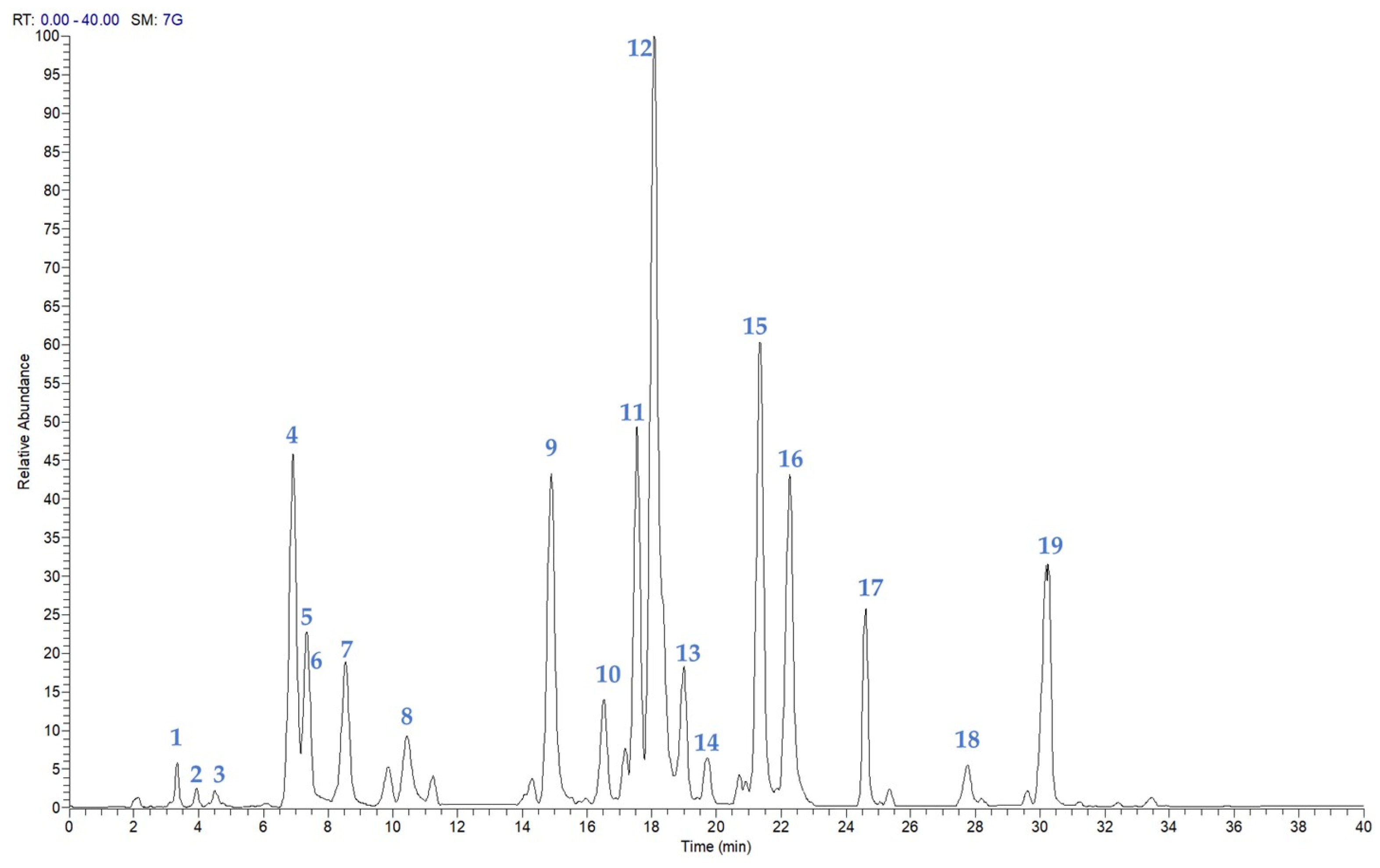
Figure 1. LC-ESI/HRMS analysis of EtOH extract of P. atriplicifolia aerial parts in negative ion mode.
Plants 14 02795 g001
Figure 2. Specialized metabolites isolated from EtOH extract of P. atriplicifolia.
Plants 14 02795 g002
Figure 3. Percentage of cell viability compared with the untreated cells (0 μg/mL, 100% viable cells) of differentiated Caco-2 cells incubated for 24 h with different concentrations (0.5–25 μg/mL) of the ethanolic extract. EtOH 2.5% was tested alone as the vehicle of the extract. Each column represents the mean ± SD of the independent experiments (n = 12). *** = p < 0.001 conc. vs. 0 μg/mL; * = p < 0.05 conc. vs. 0 μg/mL.
Plants 14 02795 g003
Figure 4. Percentage of cell viability compared with the control (0 μM, 100% viable cells) of differentiated Caco-2 cells incubated for 24 h with different concentrations (5–250 μM) of the compounds. EtOH 2.5% was tested alone as the vehicle of the compounds. Each column represents the mean ± SD of the independent experiments (n = 12). *** = p < 0.001 conc. vs. 0 μM; ** = p < 0.01 conc. vs. 0 μM; * = p < 0.05 conc. vs. 0 μM.
Plants 14 02795 g004
Figure 5. ROSs levels, measured using H2-DCF-DA fluorescence and expressed as a percentage of the untreated samples (Control), were assessed in Caco-2 cells after 60 min of incubation with different concentrations of the ethanol extract (0.5–5 µg/mL) in co-incubation with TBH 2.5 mM. EtOH 2.5% was tested alone as the vehicle for the compounds. Each column represents the mean ± SD of the independent experiments (n = 16). °° = p < 0.01 TBH 2.5 mM vs. Control; * = p < 0.05 extracts + TBH 2.5 mM vs. TBH 2.5 mM.
Plants 14 02795 g005
Figure 6. ROSs levels, measured using H2-DCF-DA fluorescence and expressed as a percentage of the untreated samples (Control), were assessed in Caco-2 cells after 60 min of incubation with varying concentrations of the extract (0.5–25 µM) in co-incubation with TBH 2.5 mM. EtOH 2.5% was tested alone as the vehicle for the compounds. Each column represents the mean ± SD of the independent experiments (n = 16). °°° = p < 0.001 TBH 2.5 mM vs. Control; ** = p < 0.01 compounds vs. TBH 2.5 mM; * = p < 0.05 compounds vs. TBH 2.5 mM.
Plants 14 02795 g006
Table 1. Compounds identified in P. atriplicifolia aerial parts by LC-(-)ESI/QExactive/MS/MS analysis in negative ion mode.
Rt[M-H]Molecular FormulaΔ ppmMS/MSCompound
13.42151.0392C8H8O31.32136.01p-methoxybenzoic acid
23.96207.0659C11H12O43.30162.98, 179.03, 135.04caffeic acid ethyl ester
34.79329.1398C19H22O54.50242.15, 285.14perovskin A
46.93313.0718C17H14O63.56283.02, 298.05scrophulein
57.33345.1708C20H26O53.27283.17, 301.18epirosmanol
67.52343.0830C18H16O75.19328.05eupatilin
78.53345.1708C20H26O53.36283.17, 301.18epiisorosmanol
810.42283.0613C16H12O54.38268.04genkwanin
914.89333.2073C20H30O43.91179.11, 191.11, 315.201α-hydroxydemethylsalvicanol
1016.47359.1868C21H28O54.09283.17, 329.18, 315.167-O-methylrosmanol
1117.54359.1867C21H28O53.84283.17, 329.18, 315.167-epi-7-O-methylrosmanol
1218.07329.1760C20H26O43.93285.19carnasol
1318.99471.3486C30H48O43.64453.34maslinic acid
1419.70343.1555C20H24O54.43219.06, 299.16, 315.16, 325.14rosmadial
1521.33345.1710C20H26O53.88286.19, 301.22royleanonic acid
1622.28331.1916C20H28O43.82287.20carnosic acid
1724.61345.2072C21H30O43.31283.17, 286.19, 301.1812-O-methylcarnosic acid
1827.73453.3377C30H46O33.13407.33urs-11-en-28,13β-olide
1930.21455.3534C30H48O33.05189.14, 206.99betulinic acid
Table 2. Quantitative results of compounds 5, 7, 9, 10, 12, and 16 (mg/g extract ± SD) in P. atriplicifolia EtOH Extract.
Compoundmg/100 g Dry Plant ± SD
520.73 ± 0.14
711.74 ± 0.39
991.73 ± 1.87
101.73 ± 0.40
12520.21 ± 23.29
1688.16 ± 7.26
Disclaimer/Publisher’s Note: The statements, opinions and data contained in all publications are solely those of the individual author(s) and contributor(s) and not of MDPI and/or the editor(s). MDPI and/or the editor(s) disclaim responsibility for any injury to people or property resulting from any ideas, methods, instructions or products referred to in the content.