Review

Chelidonium majus L.: A Current Perspective on Isoquinoline Alkaloids, Emerging Phytochemicals, Alkaloid Biosynthesis, and Biological Activities

1
Doctoral School Engineering of Plant and Animal Resources, University of Life Sciences “King Mihai I” from Timișoara, Aradului Street No. 119, 300645 Timisoara, Romania
2
Research Center for Pharmaco-Toxicological Evaluation, “Victor Babeș” University of Medicine and Pharmacy Timisoara, Eftimie Murgu Square No. 2, 300041 Timișoara, Romania
3
Faculty of Chemical Engineering, Biotechnologies and Environmental Protection, Politehnica University Timișoara, Vasile Pârvan No. 6, 300223 Timișoara, Romania
4
Faculty of Engineering and Applied Technologies, University of Life Sciences “King Mihai I” from Timișoara, Calea Aradului No. 119, 300645 Timișoara, Romania
5
Research Institute for Biosecurity and Bioengineering, University of Life Sciences “King Mihai I” from Timisoara, 300645 Timișoara, Romania
6
Faculty of Medicine, “Victor Babeș” University of Timișoara, 2 Eftimie Murgu Square, 300041 Timisoara, Romania
7
University Clinic of Toxicology, Drug Industry, Management and Legislation, Faculty of Pharmacy, “Victor Babeș” University of Medicine and Pharmacy Timișoara, Eftimie Murgu Square No. 2, 300041 Timișoara, Romania
*
Authors to whom correspondence should be addressed.
Plants 2025, 14(17), 2627; https://doi.org/10.3390/plants14172627 (registering DOI)
Submission received: 21 July 2025 / Revised: 20 August 2025 / Accepted: 20 August 2025 / Published: 23 August 2025
(This article belongs to the Collection Bioactive Compounds in Plants)

Abstract

Phytochemistry serves as a vital bridge between traditional medicinal knowledge and modern scientific research, with important implications for pharmaceutical and industrial applications. This review offers an updated and integrated perspective on Chelidonium majus (greater celandine), focusing on its isoquinoline alkaloids—the principal bioactive constituents—alongside emerging phytochemicals (e.g., lignanamides, polyphenols). Detailed biosynthetic pathways of isoquinoline alkaloids are described, tracing their formation from the shikimate pathway to multiple structural subclasses. Reported biological activities include anticancer, antioxidant, anti-inflammatory, antimicrobial, antiviral, and immunomodulatory effects. A bibliometric analysis was conducted using VOSviewer software (Scopus dataset, 2015–2025), enabling the identification of major research themes and temporal trends. These research tools supported a structured and data-driven overview of the current scientific landscape. However, additional studies are needed to optimize its therapeutic use while ensuring efficacy and safety.

1. Introduction

Modern drug discovery is increasingly integrating traditional knowledge with cutting-edge scientific methodologies to identify and optimize plant-based therapeutics, due to advances in analytical techniques, molecular biology, and computational approaches [1,2,3].
Nowadays, herbal therapy is widely recognized as an alternative and adjuvant treatment method for various acute and chronic diseases, alongside the use of plant-derived drugs [4]. Among the many medicinal herbs, greater celandine stands out due to its diverse bioactive profile and historical use in both traditional and modern herbal medicine.
Based on APG IV, the greater celandine belongs to the Papaveraceae family, which is part of the Ranunculales order within the eudicot group of vascular plants. This species is the only member of the Chelidonium genus. It has a wide distribution across temperate regions in Europe and Asia. It is endemic to Europe and Western Asia, but it has been introduced to North America, where it has become naturalized in numerous areas. Central and eastern Europe is where it is most prevalent, but it can be found as far east as China and Korea. In North America, it is frequently regarded as an invasive species. The plant thrives in habitats that are moist and shaded, such as woodland edges, hedgerows, and disturbed areas [5,6].
Chelidonium majus L. (Papaveraceae) is an herbaceous perennial that can reach heights of 30–100 cm, with a branched, erect, and slightly pubescent stem that, when broken, exudes a distinctive yellow-orange latex rich in isoquinoline alkaloids, traditionally used in folk medicine for the topical treatment of warts, eczema, fungal infections, and other skin disorders (Figure 1A,B) [7].
The leaves have deep lobes and are pinnately divided, resulting in a fern-like appearance. Their upper surface is bluish-green and paler underneath, with fine hairs often covering them (Figure 1B). The leaves are arranged in an alternating pattern on the stem, with the basal leaves being bigger and petiolate and the upper leaves being smaller and more sessile. The flowers are bright yellow, have an approximate diameter of 1.5–2.5 cm, and have four rounded petals, two falling off sepals, and numerous stamens (Figure 1C). The arrangement of these flowers is umbel-like inflorescences, with inflorescences located at the tips of the stems. The fruit’s capsule, a dehiscent silique, which is 3–5 cm long and has two valves, splits open to release numerous small, black seeds [8,9,10]. Elaiosomes found in these seeds attract ants and help in seed dispersion through myrmecochory (ant-mediated dispersion) [11].
Ecologically, C. majus thrives in semi-shaded habitats, often along hedgerows, woodland edges, disturbed soils, and moist wastelands [12]. In traditional medicine, the plant has a long ethnopharmacological history dating back to Greco-Roman times, when it was used for treating warts, skin diseases, and eye conditions. In European folk medicine, fresh latex was applied topically for wart removal, while aerial parts and roots were prepared as infusions or tinctures for liver and gallbladder ailments, digestive disorders, and as a mild sedative [13,14,15].
Despite greater celandine’s long-standing application in folk medicine, accumulating clinical evidence has raised concerns regarding its safety profile, particularly its hepatotoxic potential [16,17,18]. These adverse reactions are believed to result from idiosyncratic mechanisms, possibly involving oxidative stress, mitochondrial dysfunction, and immune-mediated responses triggered by the greater celandine’s alkaloid constituents. Regulatory committees in several European countries have already issued safety warnings or imposed restrictions on the use of greater celandine, particularly in products intended for long-term or unsupervised use [19].
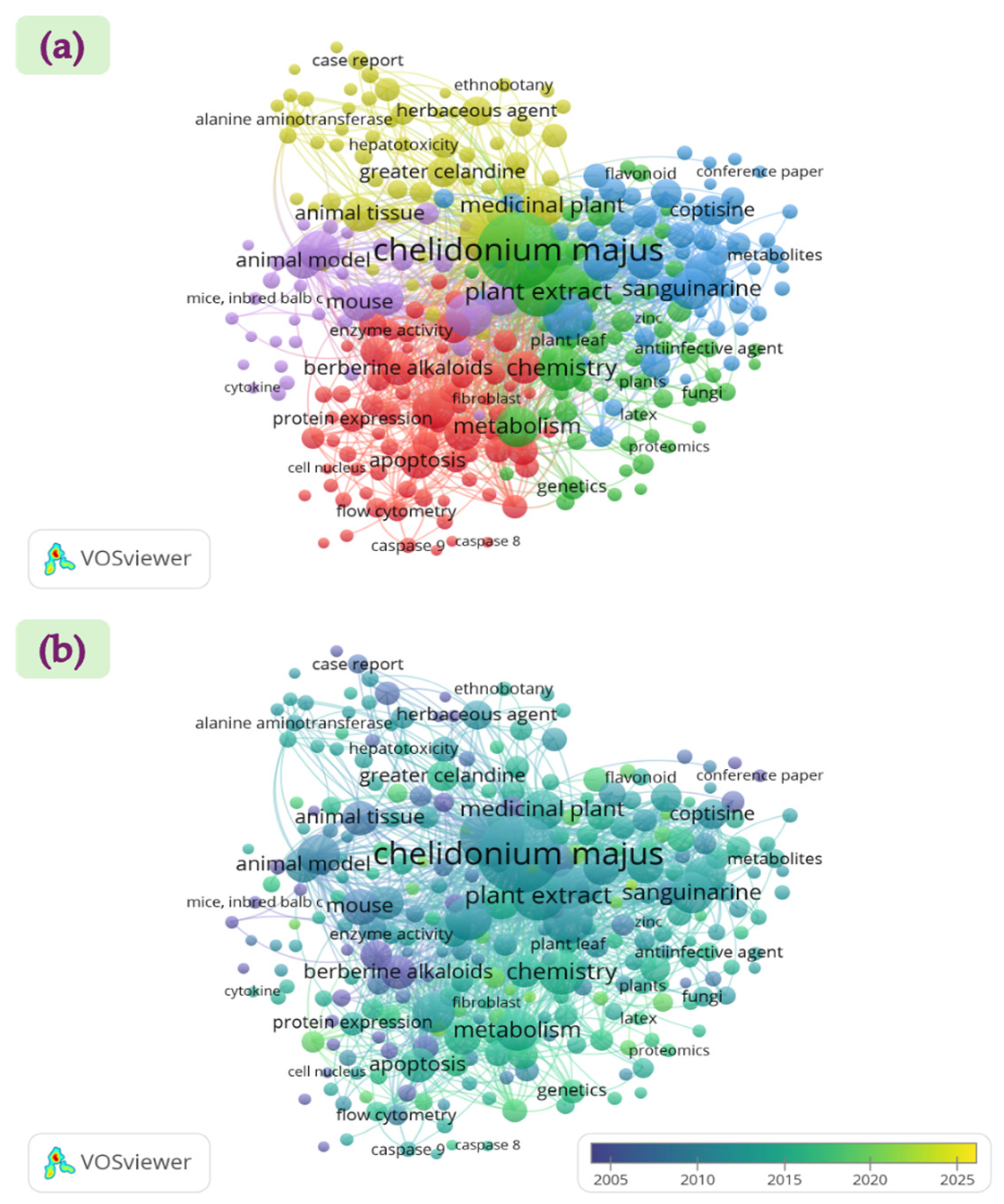
A keyword co-occurrence analysis was conducted to identify thematic structures and research trends in scientific publications related to C. majus. Terms were extracted from the titles, abstracts, and author keywords of all selected documents. The visualization maps (Figure 2) were generated in VOSviewer software (Version 1.6.20) [20], from a Scopus dataset comprising 1518 publications. In the network visualization (Figure 2a), curved lines represent relationships between terms, and circle size reflects the frequency of keyword occurrence. A minimum occurrence threshold of five was applied, resulting in five distinct keyword clusters. The overlay visualization (Figure 2b) indicates that research activity on greater celandine has been relatively limited during the 2015–2025 period.
This review addresses these developments by (i) integrating well-known isoquinoline alkaloids with emerging classes such as lignanamides and polyphenols; (ii) presenting the most detailed, step-by-step account of alkaloid biosynthesis in C. majus to date; (iii) conducting a bibliometric co-occurrence analysis of publications (2005–2025 period) using VOSviewer to identify thematic clusters and research trends; and (iv) embedding phytochemical and bioactivity data findings within a broader scientific context.

2. Emerging Phytochemicals and Biological Functions

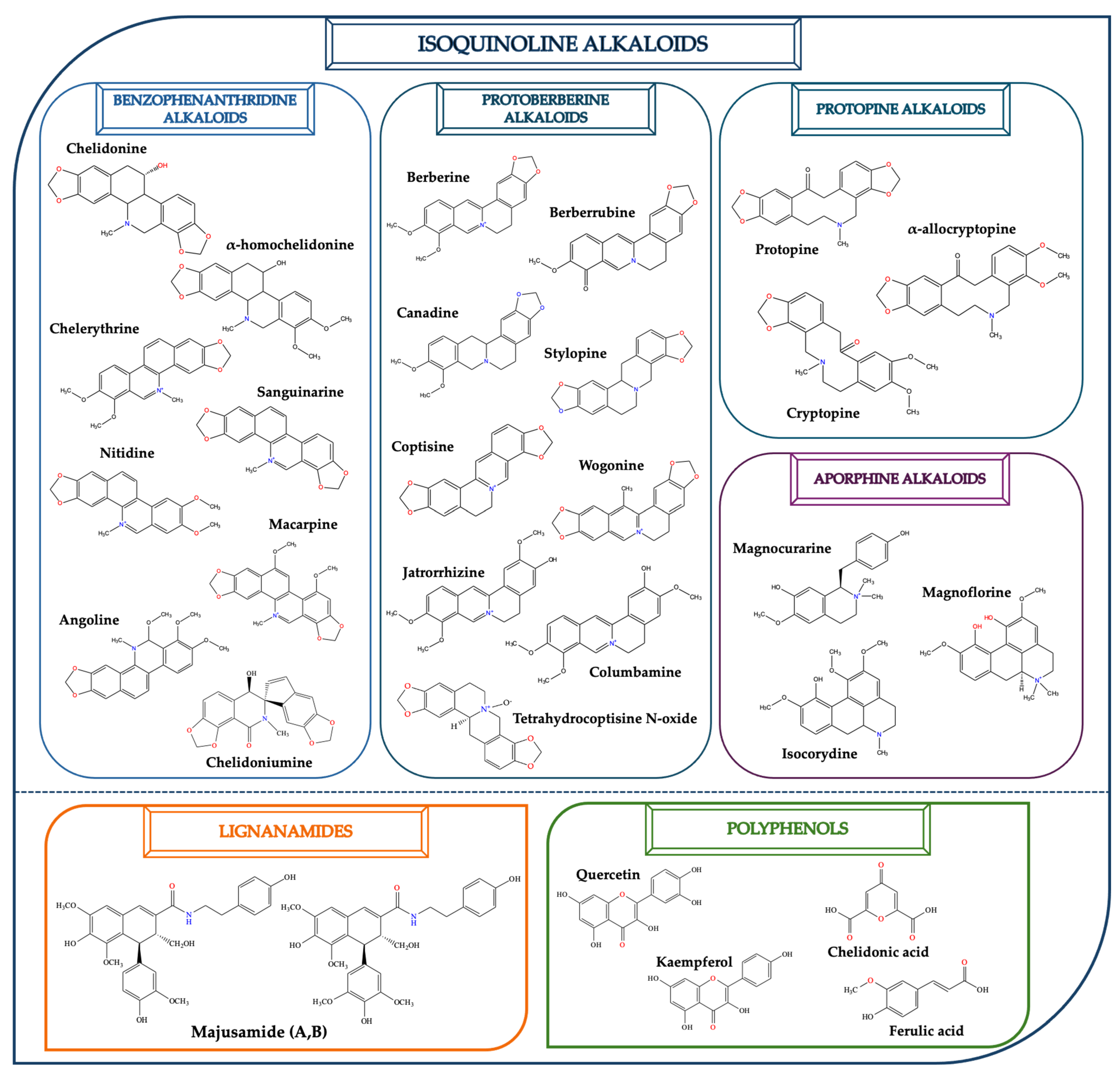
C. majus possesses a complex chemical profile, with isoquinoline alkaloids representing the principal biologically active constituents. A concise overview of these phytochemicals is presented below (Figure 3).

2.1. Isoquinoline Alkaloids

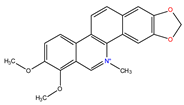
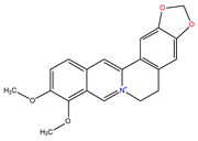
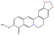
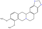
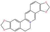
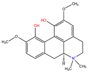
In greater celandine, four major structural groups of isoquinoline-derived alkaloids have been reported: (i) phenanthridine or 3,4-benzophenanthridine alkaloids (e.g., chelidonine, chelerythrine, sanguinarine, chelidoniumine, nitidine, macarpine, angoline, and their derivatives); (ii) protoberberine alkaloids (e.g., berberine, berberrubine, canadine, stylopine, wogonine, coptisine, jatrorrhizine, columbamine, tetrahydrocoptisine N-oxide); (iii) protopine alkaloids (e.g., protopine, cryptopine); and (iv) aporphine alkaloids (e.g., magnocurarine, magnoflorine, isocorydine) [21,22].

2.2. Lignanamides

In addition to the well-studied isoquinoline alkaloids previously described, C. majus has also been shown to contain a variety of other bioactive compound classes, such as lignanamides. Lignanamides, a distinct class of secondary metabolites characterized by the presence of amide functional groups within lignan frameworks, also referred to as bis-alkaloids, are presumed to arise via peroxide-mediated coupling reactions of phenylpropanoid amide monomers or dimers [23]. A comprehensive phytochemical study by Huang et al. led to the isolation of two novel lignanamides (majusamides A and B), as well as two new alkaloids (chelidoniumine and tetrahydrocoptisine N-oxide), from a 75% ethanol extract of the aerial parts of the greater celandine. Pharmacological testing revealed that several of these compounds exhibited moderate inhibitory activity against nitric oxide production in lipopolysaccharide (LPS)-stimulated BV-2 macrophage cells, suggesting notable anti-inflammatory potential [24].

2.3. Polyphenols and Other Phytochemicals

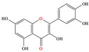
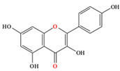
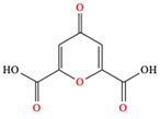
In addition to these isoquinoline alkaloids and lignanamides, greater celandine also contains other different constituents, such as polyphenols (e.g., chelidonic acid, ferulic acid, caffeic acid, quercetin, kaempherol, rutoside) [25,26], phytosterols (e.g., ergosterol), biogenic amines (e.g., histamine, tyramine), and non-essential and essential amino acids (e.g., tyrosine, alanine, serin, isoleucine, valine, threonine) [13,21,22].
It has also been reported to accumulate a wide range of macro- and microelements, including calcium, potassium, magnesium, sodium, iron, manganese, copper, zinc, lead, cadmium, chromium, and nickel, which reflect both its physiological requirements and its ability to absorb elements from the surrounding urban environment [12,27,28].
The following table will briefly introduce some emergent phytochemicals from greater celandine, which have significant biological functions (Table 1).

3. Biosynthesis of Isoquinoline Alkaloids

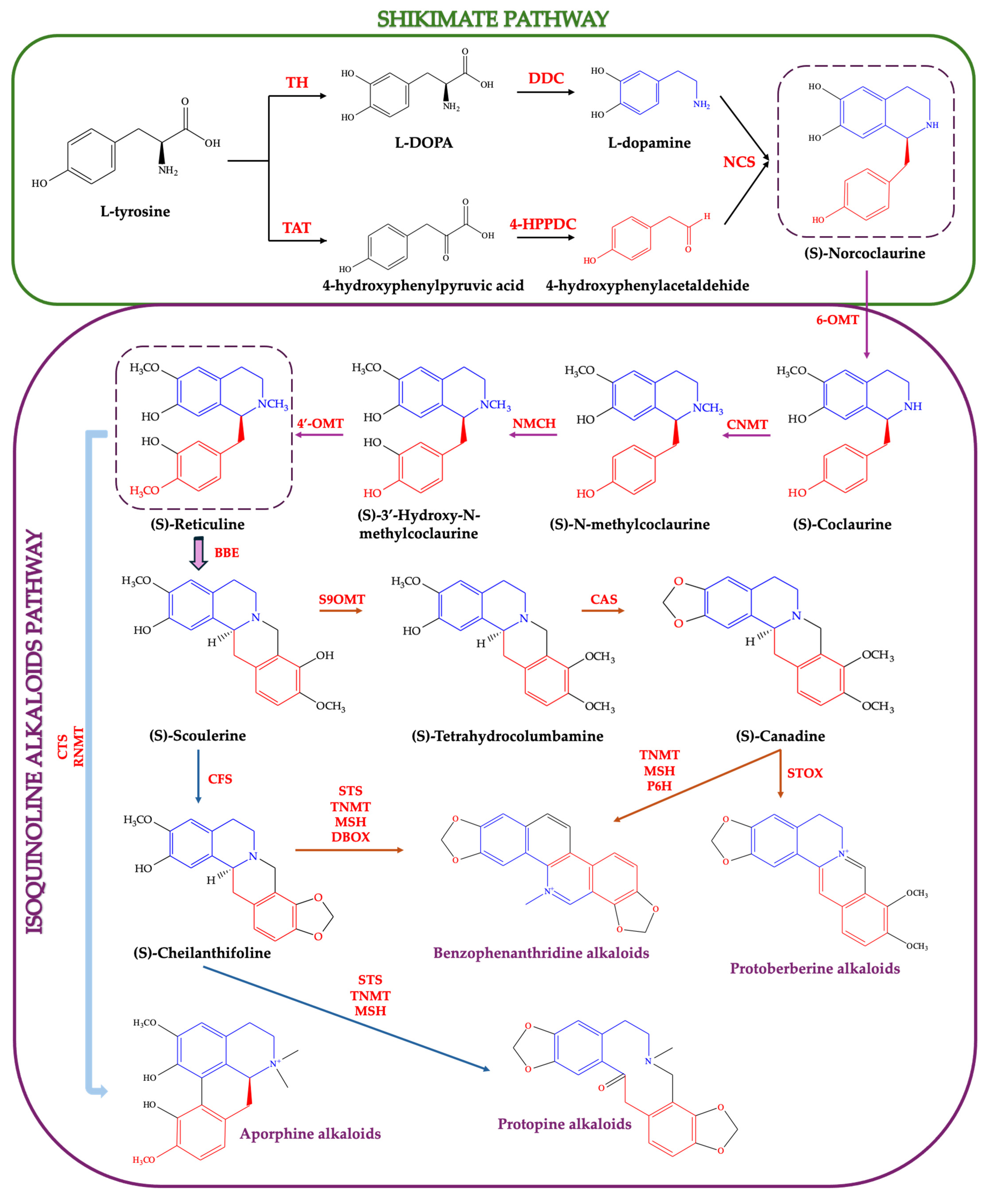
Isoquinoline alkaloids constitute a large and structurally diverse class of nitrogen-containing secondary metabolites, predominantly found in plants. Greater celandine is particularly rich in these compounds, which represent its principal biologically active constituents. From a chemical point of view, these alkaloids are derived from the shikimate pathway, and characterized by a benzylisoquinoline skeleton, which consists of a benzyl group (−CH2C6H5) attached to an isoquinoline core. In one route, L-tyrosine is hydroxylated by tyrosine hydroxylase (TH) to L-DOPA and then decarboxylated by DOPA decarboxylase (DDC) to form dopamine. In the other route, tyrosine undergoes transamination by tyrosine aminotransferase (TAT) to 4-hydroxyphenylpyruvic acid, followed by decarboxylation by 4-hydroxyphenylpyruvate decarboxylase (4-HPPDC) to yield 4-hydroxyphenylacetaldehyde. These two intermediates are then condensed by norcoclaurine synthase (NCS) to form (S)-norcoclaurine, the core scaffold for isoquinoline alkaloid molecules biosynthesis (Figure 4) [87,88,89,90,91,92,93]. Subsequent enzyme modifications, including methylation, oxidation, hydroxylation, and rearrangements, generate a vast array of benzylisoquinoline derivatives [88].
Between the shikimate and isoquinoline alkaloid biosynthetic pathways lies an intermediate sequence in which (S)-norcoclaurine undergoes successive methylation and hydroxylation steps to yield (S)-reticuline [89,90,93]. Initially, (S)-norcoclaurine is methylated at the 6-hydroxyl position by norcoclaurine 6-O-methyltransferase (6-OMT) to produce (S)-coclaurine. This is followed by N-methylation via coclaurine N-methyltransferase (CNMT), generating (S)-N-methylcoclaurine. Subsequent hydroxylation at the 3′ position by the cytochrome P450-dependent (S)-N-methylcoclaurine 3′-hydroxylase (NMCH) produces (S)-3′-hydroxy-N-methylcoclaurine, which is then methylated at the 4′-hydroxyl group by 3′-hydroxy-N-methylcoclaurine 4′-O-methyltransferase (4′-OMT) to form (S)-reticuline. The committed step in isoquinoline alkaloid biosynthesis (e.g., benzophenanthridine, protoberberine, protopine alkaloids) is the stereoselective conversion of (S)-reticuline to (S)-scoulerine, catalyzed by the berberine bridge enzyme (BBE) [93].
In the protoberberine and benzophenanthridine biosynthetic pathways, (S)-scoulerine undergoes O-methylation by scoulerine 9-O-methyltransferase (S9OMT) to produce (S)-tetrahydrocolumbamine. This intermediate is subsequently converted to (S)-canadine via methylenedioxy bridge formation catalyzed by canadine synthase (CAS). (S)-Canadine serves as a central precursor for multiple downstream alkaloid classes, including benzophenanthridines and protoberberines, through a series of oxidative and methylation reactions mediated by enzymes (e.g., TNMT, tetrahydroprotoberberine-cis-N-methyltransferase; MSH, N-methylstylopine-14-hydroxylase; P6H, protopine-6-hydroxylase; STOX, (S)-tetrahydroprotoberberine oxidase) [87,88,89,90,91,92,93].
In another branch of the biosynthetic pathway, (S)-scoulerine is converted to (S)-cheilanthifoline via methylenedioxy bridge formation catalyzed by cheilanthifoline synthase (CFS). The introduction of a second methylenedioxy bridge, followed by oxidative rearrangement catalyzed by a series of enzymes including stylopine synthase (STS), tetrahydroprotoberberine-N-methyltransferase (TNMT), and methylstylopine hydroxylase (MSH), leads to the formation of protopine. Protopine serves as a direct precursor to various protopine-derived alkaloids, and to benzophenanthridine alkaloids (e.g., sanguinarine and chelerythrine) via the sequential action of dihydrobenzophenanthridine oxidase (DBOX) [90,91,92,93].
(S)-Reticuline also functions as a key precursor in the biosynthesis of aporphine alkaloids. In this branch, (S)-reticuline undergoes oxidative phenol coupling catalyzed by corytuberine synthase (CTS) to produce (S)-corytuberine. Subsequent methylation and oxidation reactions, mediated by reticuline N-methyltransferase (RNMT) and related enzymes, yield a variety of aporphine-type alkaloids, including magnoflorine and isocorydine [87,88,89,90,91,92,93]. These compounds are characterized by a tetracyclic aporphine skeleton derived from the intramolecular coupling of the benzyl and isoquinoline moieties of reticuline.

4. Recent Data on the Bioactivity of Chelidonium majus

Greater celandine has been used in traditional medicine since ancient times. Celandine was thought by Dioscorides to have the most potent medicinal properties in treating visual disorders, eye diseases, and relieving dental inflammation in the first century [94,95]. Today, celandine is known to possess a range of therapeutic benefits, primarily because of its complex phytochemistry, but it also has some toxicological risks [13,18,22,96]. Below, we will outline the most recent data concerning the biological actions of greater celandine.

4.1. Anticancer Activity

Using multiple hyphenated instrumental techniques including HPLC-DAD, Petruczynik et al. investigate the anticancer activity and measure specific isoquinoline alkaloids in extracts from C. majus. They evaluated the cytotoxic effects of this extract on human cancer cell lines: (i) pharyngeal squamous carcinoma (FaDu), (ii) tongue squamous carcinoma (SCC-25), (iii) breast adenocarcinoma (MCF-7), and (iv) triple-negative breast adenocarcinoma (MDA-MB-231). According to the results, all of the tested extracts demonstrated significant cytotoxicity against these cancer cell lines. Notably, the root extract of C. majus showed the highest cytotoxic activity against FaDu and MDA-MB-231 cells, with IC50 values of 2.52 ± 0.25 µg/mL and 2.61 ± 0.33 µg/mL, respectively. These findings indicate that the celandine extract studied has potent anticancer properties, which require further in vivo studies to investigate its potential as therapeutic agents [97].
Tuzimsky et al. investigated the chemical composition and anticancer properties of extracts from C. majus, Mahonia aquifolium and Sanguinaria canadensis, focusing on their isoquinoline alkaloid content and cytotoxic effects on various human cancer cell lines (e.g., FaDu, SCC-25, MCF-7, MDA-MB-231). The researchers used liquid chromatography to identify and quantify several isoquinoline alkaloids in the plant extracts, with a particular focus on celandine, such as chelidonine, sanguinarine, chelerythrine, and berberine. The results revealed that this time the root extracts showed significant cytotoxicity, particularly against melanoma cell lines. The root extract had an IC50 of 12.65 µg/mL against A375 cells and 1.93 µg/mL against SK-MEL-3 cells. Furthermore, the celandine root extract was tested on a Danio rerio (zebrafish) larvae xenograft model and demonstrated a significant reduction in the number of cancer cells in vivo, confirming its potent anticancer activity. According to these findings, the celandine extract is a potential candidate for further research and development as natural anticarcinogenic agents [98].
In another study, Nile et al. evaluate the cytotoxic effect against the HeLa and CaSki human cervical cancerous cells, using extracts made from different celandine parts (e.g., leaf, stem, flower, pod, and root). According to their findings, the pod and flower extracts exhibited the most potent cytotoxic effects after 48 h of exposure at 1000 μg/mL, with the pod extracts showing an IC50 value of 6.1% (HeLa) and 0.72% (CaSki), while the flower extracts showed an IC50 value of 7.9% (HeLa) and 9.5% (CaSki), respectively [99]. In another example, Nawrot et al. investigated whether C. majus latex’s proteins and alkaloids had a combined effect on human cervical carcinoma cells. A novel major uncharacterized nucleic acid binding latex protein, CmMLP1, which contains 147 amino acids, was isolated by them, and they discovered its association with alkaloids like berberine, 8-hydroxycheleritrine, and dihydroberberine. Using molecular docking analyses, it was found that CmMLP1 has a hydrophobic cavity that has a high affinity for these alkaloids. In vitro cytotoxicity assays demonstrated that fractions containing both CmMLP1 and the associated alkaloids significantly reduced the viability of human cervical cancer cell lines, including both HPV-positive (HeLa) and HPV-negative (C33A) cells. It was observed that the combination had a stronger cytotoxic effect on HPV-positive cells, which suggests a potential synergistic interaction between CmMLP1 and the alkaloids. These findings shed light on the molecular interactions within C. majus latex and its potential use in cervical cancer treatment [100].
Terzic et al. investigated the cytotoxic effect of three extracts obtained from greater celandine with different solvents, such as ethyl acetate, methanol, and water. The effect of C. majus extracts on cancer cell viability was assessed across a range of cancer cell lines, including HGC-27 (gastric cancer), DU-145 (prostate carcinoma), HT-29 (colon adenocarcinoma), MDA-MB-231 (breast adenocarcinoma), HELA (cervix adenocarcinoma), and A549 (lung cancer), and normal cell line HEK-293 (kidney epithelia). The ethyl extract demonstrated favorable IC50 values across multiple cancer cell lines, including MDA-MB-231, HGC-27, HT-29, and DU, with IC50 values of 8.108 μg/mL, 8.27 μg/mL, 9.078 μg/mL, and 22.1 μg/mL, respectively. The methanolic extract showed efficacy against HT, HGC-27, and HELA with IC50 values of 4.525 μg/mL, 8.538 μg/mL, and 18.55 μg/mL, respectively. The water extract, on the other hand, generally displayed less favorable IC50 values, which suggests a weaker inhibitory effect [101].
Natanzi et al. evaluated the impact of alkaloids extracted from C. majus on the Daudi Burkitt lymphoma cell line. Their study focused on assessing cytotoxicity, apoptosis induction, and the expression of the vascular endothelial growth factor (VEGF) gene, a key regulator of angiogenesis in cancer progression. The results revealed that the alkaloid extract exhibited dose-dependent cytotoxicity against Daudi cells, with a 50% cytotoxic concentration (CC50) determined to be 56.35 µg/mL after 48 h. According to flow cytometry analysis, CC50 concentration treatment caused 1.4% early apoptosis and 37.94% late apoptosis in Daudi cells. VEGF gene expression was downregulated in Daudi cells treated with the alkaloid extract in dose-dependent manner, as demonstrated by real-time PCR results. Their study results provide a foundation for further in vivo studies to explore the therapeutic potential of C. majus alkaloids as natural anticancer agents [102].
In a study conducted by Salehi et al., the anticancer potential of isoquinoline alkaloids extracted from C. majus was investigated, with a specific focus on their ability to bind G-quadruplex (G4) DNA structures. Among the screened compounds, berberine and chelerythrine emerged as the most potent G4 binders, showing high affinity for telomeric and oncogene–promoter G-quadruplexes. These compounds demonstrated significant stabilization of G4 structures, suggesting their potential as transcriptional repressors of cancer-associated genes. In vitro cytotoxicity assays revealed that both compounds significantly reduced cell viability in MCF-7 (breast cancer) and HCT116 (colorectal cancer) cell lines, with IC50 values ranging from 2.5 to 5.0 μM, depending on the cell type and compound [103].

4.2. Anti-Inflammatory and Analgesic Activities

Mikołajczak et al. studied the analgesic and anti-inflammatory effects of C. majus herb extracts by evaluating three fractions: full water extract (FWE), protein-enriched fraction (PEF), and non-protein fraction (NPF). In the “hot plate” analgesia test, FWE and PEF administered at a dose of 10 mg/kg (intraperitoneally) exhibited significant analgesic activity three hours post-administration, with responses comparable to morphine (5 mg/kg) (p < 0.05). Interestingly, FWE also induced a pro-inflammatory effect at the same time point, as indicated by elevated cytokine levels. Furthermore, all three fractions significantly reduced peripheral IL-1 and IL-4 levels, suggesting systemic anti-inflammatory effects [104].
Zielinska et al. investigated the effects of C. majus root extract and its alkaloids (e.g., coptisine, berberine, chelidonine, chelerythrine, and sanguinarine) on cytokine secretion in lipopolysaccharide-stimulated human neutrophils. Berberine, chelidonine, and chelerythrine significantly reduced TNF-α secretion in a concentration-dependent manner, particularly at 10 µM, with chelidonine and chelerythrine exhibiting minimal cytotoxicity at this level. Sanguinarine was identified as the most potent inhibitor of IL-1β production, showing significant effects at 1 µM. However, it also induced an increase in IL-8 and TNF-α secretion at lower concentrations (e.g., 0.1 µM) and displayed high cytotoxicity at concentrations ≥ 5 µM. These results suggest selective, dose-dependent immunomodulatory effects of the individual alkaloids, with differing safety profiles [105].
In in vitro studies, chelerythrine extracted from C. majus significantly suppressed the production of key inflammatory mediators, such as nitric oxide (NO), tumor necrosis factor-alpha (TNF-α), interleukin-6 (IL-6), and IL-1β in LPS-stimulated RAW264.7 macrophages. It also downregulated the expression of inducible nitric oxide synthase (iNOS) and cyclooxygenase-2 (COX-2) in a dose-dependent manner, without inducing cytotoxicity at concentrations ≤ 10 µM. Functionally, it inhibited GAPDH enzymatic activity and decreased pyruvate levels, indicating suppression of glycolytic flux [106].

4.3. Antiviral Activity

Musidlak et al. provides an in-depth analysis of how different components of C. majus latex (e.g., specifically its crude form, protein fraction, alkaloid-rich fraction) interact with the human papillomavirus (HPV) replication cycle. Their results showed that crude latex exhibited the most antiviral effect, decreasing infectivity by approximately 72.5%. The protein fraction and alkaloid-rich fraction reduced infectivity by 35.6% and 22.9%, respectively. Increased TNF-α secretion was associated with the protein fraction of the latex, which suggests a potential for inducing inflammatory responses. In contrast, the latex components did not impact the attachment of HPV pseudovirions to host cells or the degradation of viral capsid proteins (L1 and L2), suggesting that their antiviral activity does not relate to the initial stages of infection. Both alkaloids and proteins found in C. majus latex are responsible for its antiviral properties against HPV, as evidenced by the study. The viral replication cycle is impeded by these components at post-entry but before nuclear translocation stages without affecting viral attachment or capsid integrity [107].
In their observational case series, Gardin and Braga evaluated 20 outpatients diagnosed with COVID-19 who were treated with a 10% mother tincture of C. majus L. The tincture was administered orally at a dose of 20–30 drops, three times daily, over a treatment period ranging from 3 to 12 days. The patient cohort included individuals aged 14–71 years, and the initiation of treatment occurred between 1 and 19 days after the onset of symptoms. Notably, complete or near-complete symptom resolution was observed within 1–9 days of initiating treatment. Although the observational nature and small sample size of their study limit its generalizability, the study suggests that C. majus may contribute to symptom alleviation in mild COVID-19 cases [108].

4.4. Antimicrobial and Antifungal Activities

The study of Hong et al. investigated the antioxidant and antimicrobial activities of C. majus L. against oral microorganisms. According to the DPPH and ABTS radical scavenging assay, the extract demonstrated significant antioxidant activity and the most significant levels of phenolic and flavonoid content (212.00 mg CAE/g, 135.70 mg NE/g). Furthermore, the antimicrobial assay demonstrated strong antimicrobial activity against oral microorganisms (e.g., S. mutans, S. gordonii, S. sobrinus, S. sanguinis), and it was found that the most inhibitive is the ethyl acetate fraction [81].
Qi et al. investigated the alkaloid impact of methicillin-resistant Staphylococcus aureus (MRSA) and methicillin-sensitive Staphylococcus aureus (MSSA) on the motility, longevity, and reactive oxygen species levels of wild-type worms (N2 strain). The results indicated that the concentration of total alkaloids, which was between 0.625 – 10 mg/mL, had a significant effect on MRSA. Increased concentration correlates with an increase in antibacterial activity, suggesting that total alkaloids exert antimicrobial effects at specific doses [109]. In another study conducted by Krzyzek et al., they investigated the antimicrobial efficacy of plant extracts (C. majus and Corydalis cheilanthifolia) against a multidrug-resistant strain of Helicobacter pylori. The findings revealed that the C. majus extract shows impressive antibacterial properties, with an MIC of 128 µg/mL and an MBC of 256 µg/mL, respectively [110].
In the study by Lu et al., the nematicidal activities of two isoquinoline alkaloids—berberine and sanguinarine—isolated from C. majus were assessed against the root-knot nematode Meloidogyne incognita. In vitro assays demonstrated strong dose-dependent lethality, with both compounds significantly inhibiting second-stage juvenile (J2) viability and egg hatching. At concentrations of 100 μg/mL, berberine and sanguinarine achieved over 90% J2 mortality within 48 h [111].
In a study conducted by Sadecki et al., aerial tissues of C. majus were investigated for the presence of antimicrobial peptides (AMPs). Using a PepSAVI-MS pipeline, the team identified a novel, cysteine-rich peptide, CM-AMP1. The synthetic peptide exhibited potent membrane-lytic activity, achieving near-complete inhibition of E. coli ATCC 25922 and MRSA USA300 at micromolar concentrations [112].

4.5. Antioxidant Activity

Segneanu et al. investigated the development of a novel carrier system that combines extracts from Romanian wild-grown greater celandine with gold nanoparticles. The extract–nanoparticle system exhibited more antioxidant activity, resulting in a 3.8% increase in flavonoid content and a 24.6% increase in FRAP results, compared to the herb extract alone [113]. Khodabande et al. evaluated greater celandine leaf extracts across three distinct phenological stages (e.g., vegetative, flowering, fruiting) indicating that the plant’s biochemical composition and antioxidant potential vary significantly with developmental phase. The flowering stage was identified as the most phytochemically active, exhibiting the highest concentrations of total phenols (17.8 ± 1.59 mg/g DW) and total flavonoids (69.7 ± 0.86 mg/g DW). These compounds are known for their strong free radical scavenging properties, suggesting that extracts collected during this period may offer the greatest therapeutic benefit. The vegetative stage demonstrated the highest carotenoid content (2.083 mg/g DW) and protein content (0.27 ± 0.034 mg/g DW), indicating intense metabolic and structural activity during early growth. The growth stage (vegetative to early flowering) also showed the maximum DPPH radical scavenging activity at 408.88 ± 24.83 g/g DW, reflecting a potent ability to neutralize reactive oxygen species (ROS). Conversely, the FRAP assay revealed that ferric-reducing antioxidant power peaked during the fruiting stage (1.75 ± 0.04 mg/g FW), possibly due to the accumulation of secondary metabolites necessary for reproductive tissue protection. Additionally, the soluble sugar content reached its maximum during flowering (0.338 ± 0.009 mg/g DW), supporting the energetic requirements of the plant during this intensive phase of development. These findings highlight the critical role of harvest timing in optimizing the bioactivity of C. majus extracts [114].
In a study conducted by Dostemessova et al., the antioxidant properties of C. majus were systematically evaluated using methanolic extracts obtained from both aerial and root parts of plants collected at three distinct ecological sites in the Kungei-Alatau region. The study demonstrated that root extracts exhibited significantly higher antioxidant activity compared to aerial parts. Notably, the Kolsay (N1) root sample showed the most pronounced antioxidant potential, with DPPH (1735.85 ± 14.28 µmol TE/mL), ABTS (917.97 ± 18.06 µmol TE/mL), and PFRAP (18.93 ± 0.24 mmol AAE/mL) values. Additionally, the highest total phenolic content (2617.62 ± 9.59 µg GAE/mL) was observed in the root extract from the Kayindy (N2) site. The findings suggest that the antioxidant activity of greater celandine is primarily linked to its phenolic content rather than alkaloid concentration. The authors also emphasized that environmental conditions, such as soil pH and composition, play a critical role in modulating the phytochemical profile of the plant [115].

4.6. Immunomodulatory Activity

Zielinska et al. carried out a study to examine how greater celandine extracts and their individual alkaloids impact the production of cytokines in human neutrophils stimulated with lipopolysaccharide. In the root extract of celandine, 25 compounds were found, with chelerythrine being the most abundant (1761.22 µg/g), followed by sanguinarine (1373.80 µg/g), chelidonine (1181.41 µg/g), and coptisine (1077.06 µg/g). Furthermore, five individual alkaloids (e.g., coptisine, berberine, chelidonine, chelerythrine, sanguinarine) were evaluated on the secretion of pro-inflammatory cytokines IL-1β, IL-8, and TNF-α. The results showed that celandine root extract itself increased the secretion of all three cytokines in a concentration-dependent manner (1.25–12.5 µg/mL). The study concluded that while alkaloids from celandine (e.g., chelidonine, chelerythrine) can regulate cytokine production, other alkaloids (e.g., coptisine and sanguinarine) have significant cytotoxic effects, which limits their therapeutic applications [105].

4.7. Insecticidal Activity

In a study conducted by Zou et al., greater celandine was shown to exhibit potent insecticidal activity against Lymantria dispar larvae, primarily due to its rich content of isoquinoline alkaloids. Both the crude extract (CECm) and the total alkaloid fraction (TACm) demonstrated significant larvicidal effects, with coptisine identified as the most abundant compound. Treatment with TACm significantly reduced larval food intake, growth rate, and assimilation efficiency, indicating pronounced antifeedant and growth-inhibitory effects. Enzymatic assays revealed that CECm and TACm strongly inhibited the activity of key detoxification and antioxidant enzymes (e.g., acetylcholinesterase (AChE), carboxylesterase (CarE), glutathione S-transferase (GST), superoxide dismutase (SOD), catalase (CAT)) in both in vitro and in vivo experiments [116].
Given the diversity of experimental approaches and outcomes reported, a comprehensive summary table was added to consolidate key information on plant part, extraction method, and biological activity (Table 2).

5. Conclusions and Future Perspectives

Chelidonium majus L. remains a phytochemically rich species of high pharmacological interest, characterized by a predominance of isoquinoline alkaloids and complemented by emerging classes of bioactive constituents such as lignanamides and polyphenols. Advances in analytical chemistry have refined our understanding of its complex alkaloid biosynthetic pathways, from shikimate precursors through multiple structural subclasses, including protoberberine, benzophenanthridine, aporphine, and protopine alkaloids.
Biological studies consistently support the plant’s anticancer, antioxidant, anti-inflammatory, antimicrobial, antiviral, and immunomodulatory activities, yet the translation of these effects into clinical practice is hindered by variability in chemical composition, gaps in pharmacokinetic data, and unresolved safety concerns, particularly regarding hepatotoxicity. The balance between therapeutic potential and toxic risk remains a critical research priority, and further controlled clinical trials and mechanistic toxicology studies are essential to delineate its therapeutic window and establish evidence-based safety guidelines.

Author Contributions

Conceptualization, C.A.D. and T.C.; methodology, investigation, R.R.; resources, D.I.; data curation, S.L.; writing—original draft preparation: R.R.; writing—review and editing, S.L.; visualization, M.R.T. and F.H.; supervision, C.A.D. and T.C.; funding acquisition, T.C. All authors have read and agreed to the published version of the manuscript.

Funding

The publication of this research paper was supported by the University of Life Sciences “King Mihai I” from Timișoara.

Data Availability Statement

Data are contained within the article.

Conflicts of Interest

The authors declare no conflicts of interest.

Abbreviations

APG IVAngiosperm Phylogeny Group fourth version
CC50Cytotoxic concentration 50%
CDK1Cyclin-dependent kinase 1
COX2Cyclooxygenase-2
EGFREpidermal growth factor receptor
GPVIPlatelet glycoprotein VI
HEI-OC1House Ear Institute-Organ of Corti 1
HPLC-DADHigh-performance liquid chromatography with photodiode-array detection
HPVHuman papillomavirus
IL-8Interleukine-8
JNKc-Jun N-terminal kinases
LSD1Lysine-specific demethylase 1A
MBCMinimum Bactericidal Concentration
MICMinimum Inhibitory Concentration
NF-kBNuclear factor kappa-light-chain-enhancer of activated B cells
p21, p53Cyclin-dependent kinase inhibitor
PGE2Prostaglandin E2
SARS-CoV-2Severe acute respiratory syndrome coronavirus 2
STAT3Signal transducer and activator of transcription 3
TNF-αTumor necrosis factor α
TRPA1Transient receptor potential ankyrin 1
VEGFVascular endothelial growth factor
ZIKVZika virus

References

  1. Sadybekov, A.V.; Katritch, V. Computational Approaches Streamlining Drug Discovery. Nature 2023, 616, 673–685. [Google Scholar] [CrossRef]
  2. Naithani, U.; Guleria, V. Integrative Computational Approaches for Discovery and Evaluation of Lead Compound for Drug Design. Front. Drug Discov. 2024, 4, 1362456. [Google Scholar] [CrossRef]
  3. Southey, M.W.Y.; Brunavs, M. Introduction to Small Molecule Drug Discovery and Preclinical Development. Front. Drug Discov. 2023, 3, 1314077. [Google Scholar] [CrossRef]
  4. Salm, S.; Rutz, J.; van den Akker, M.; Blaheta, R.A.; Bachmeier, B.E. Current State of Research on the Clinical Benefits of Herbal Medicines for Non-Life-Threatening Ailments. Front. Pharmacol. 2023, 14, 1234701. [Google Scholar] [CrossRef]
  5. Zare, G.; Diker, N.Y.; Arituluk, Z.C.; Tatli Cankaya, I.I. Chelidonium majus L. (Papaveraceae) Morphology, Anatomy and Traditional Medicinal Uses in Turkey. İstanbul J. Pharm. 2021, 51, 123–132. [Google Scholar] [CrossRef]
  6. Samatadze, T.E.; Yurkevich, O.Y.; Hazieva, F.M.; Konyaeva, E.A.; Morozov, A.I.; Zoshchuk, S.A.; Amosova, A.V.; Muravenko, O.V. Agro-Morphological, Microanatomical and Molecular Cytogenetic Characterization of the Medicinal Plant Chelidonium majus L. Plants 2020, 9, 1396. [Google Scholar] [CrossRef]
  7. Borges, A.; Calvo, M.L.M.; Vaz, J.A.; Calhelha, R.C. Enhancing Wound Healing: A Comprehensive Review of Sericin and Chelidonium majus L. as Potential Dressings. Materials 2024, 17, 4199. [Google Scholar] [CrossRef] [PubMed]
  8. Tutin, T.G. (Ed.) Flora Europaea: Plantaginaceae to Compositae (and Rubiaceae), Vol. 4; Cambridge University Press: Cambridge, UK, 1964. [Google Scholar]
  9. Colombo, M.L.; Tomè, F. Chelidonium majus L. (Greater Celandine): In Vitro Culture and the Production of Sanguinarine, Coptisine, and Other Isoquinoline Alkaloids. In Medicinal and Aromatic Plants VIII; Bajaj, Y.P.S., Ed.; Biotechnology in Agriculture and Forestry; Springer: Berlin/Heidelberg, Germany, 1995; Volume 33. [Google Scholar] [CrossRef]
  10. Achilova, S.S.; Rabbimov, A. Some Biological Characteristics of the Plant of the Large Blood Pump—Chelidonium majus L. in the Conditions of the Zarafshon Oasis. Int. J. Virol. Mol. Biol. 2024, 13, 37–38. [Google Scholar] [CrossRef]
  11. Wu, J.; Peng, L.; Dong, S.; Xia, X.; Zhao, L. Transcriptome Analysis of Chelidonium majus Elaiosomes and Seeds Provide Insights into Fatty Acid Biosynthesis. PeerJ 2019, 7, e6871. [Google Scholar] [CrossRef] [PubMed]
  12. Rahmonov, O.; Srodek, D.; Pytel, S.; Kupka, T.; Makieieva, N. Mineral Composition of Chelidonium majus L. and Soils in Urban Areas. Appl. Sci. 2025, 15, 4718. [Google Scholar] [CrossRef]
  13. Zielińska, S.; Jezierska-Domaradzka, A.; Wójciak-Kosior, M.; Sowa, I.; Junka, A.; Matkowski, A.M. Greater Celandine’s Ups and Downs—21 Centuries of Medicinal Uses of Chelidonium majus From the Viewpoint of Today’s Pharmacology. Front. Pharmacol. 2018, 9, 299. [Google Scholar] [CrossRef]
  14. Gilca, M.; Tiplica, G.S.; Salavastru, C.M. Traditional and ethnobotanical dermatology practices in Romania and other Eastern European countries. Clin. Dermatol. 2018, 36, 338–352. [Google Scholar] [CrossRef]
  15. Gilca, M.; Gaman, L.; Panait, E.; Stoian, I.; Atanasiu, V. Chelidonium majus—An Integrative Review: Traditional Knowledge versus Modern Findings. Forsch. Komplementmed. 2010, 17, 241–248. [Google Scholar] [CrossRef] [PubMed]
  16. Power, S.; Barritt, A. A Yellow Flower With Jaundice Power: Liver Injury Attributed to Greater Celandine. ACG Case Rep. J. 2024, 11, e01005. [Google Scholar] [CrossRef] [PubMed]
  17. Moro, P.A.; Cassetti, F.; Giugliano, G.; Falce, M.T.; Mazzanti, G.; Menniti-Ippolito, F.; Raschetti, R.; Santuccio, C. Hepatitis from Greater Celandine (Chelidonium majus L.): Review of Literature and Report of a New Case. J. Ethnopharmacol. 2009, 124, 328–332. [Google Scholar] [CrossRef]
  18. Pantano, F.; Mannocchi, G.; Marinelli, E.; Gentili, S.; Graziano, S.; Busardò, F.P.; di Luca, N.M. Hepatotoxicity Induced by Greater Celandine (Chelidonium majus L.): A Review of the Literature. Eur. Rev. Med. Pharmacol. Sci. 2017, 21, 46–52. [Google Scholar]
  19. Maggini, V.; Lombardi, N.; Crescioli, G.; Gallo, E.; Sivelli, F.; Gensini, G.F.; Vannacci, A.; Firenzuoli, F. Chelidonium majus: Relevant safety aspects of a hepatotoxic plant, trawling the web. Phytother. Res. 2019, 33, 2465–2469. [Google Scholar] [CrossRef]
  20. VOSviewer Software. Version 1.6.20. Available online: www.vosviewer.com (accessed on 11 July 2025).
  21. Maji, A.K.; Banerji, P. Chelidonium majus L. (Greater Celandine)—A Review on Its Phytochemical and Therapeutic Perspectives. Int. J. Herb. Med. 2015, 3, 10–27. [Google Scholar] [CrossRef]
  22. Li, X.L.; Sun, Y.P.; Wang, M.; Wang, Z.B.; Kuang, H.X. Alkaloids in Chelidonium majus L: A Review of Its Phytochemistry, Pharmacology, and Toxicology. Front. Pharmacol. 2024, 15, 1440979. [Google Scholar] [CrossRef] [PubMed]
  23. Sun, J.; Tong, L.-T.; Tu, P.-F.; Chen, L.-L.; Xu, X.; Song, Y.; Yang, X.-X.; Guo, Z.-B.; Zou, X.; Sun, C.-X.; et al. Lignanamides: A Comprehensive Review of Chemical Constituents, Biological Activities, Extraction Methods and Synthetic Pathway. Food Chem. 2024, 460, 140459. [Google Scholar] [CrossRef]
  24. Huang, X.-Y.; Shao, Z.-X.; An, L.-J.; Xue, J.-J.; Li, D.-H.; Li, Z.-L.; Hua, H.-M. New Lignanamides and Alkaloids from Chelidonium majus and Their Anti-Inflammation Activity. Fitoterapia 2019, 139, 104359. [Google Scholar] [CrossRef] [PubMed]
  25. Grosso, C.; Ferreres, F.; Gil-Izquierdo, A.; Valentão, P.; Sampaio, M.; Lima, J.; Andrade, P.B. Box–Behnken Factorial Design to Obtain a Phenolic-Rich Extract from the Aerial Parts of Chelidonium majus L. Talanta 2014, 130, 128–136. [Google Scholar] [CrossRef] [PubMed]
  26. Krizhanovska, V.; Sile, I.; Kronberga, A.; Nakurte, I.; Mezaka, I.; Dambrova, M.; Pugovics, O.; Grinberga, S. The Cultivation of Chelidonium majus L. Increased the Total Alkaloid Content and Cytotoxic Activity Compared with Those of Wild-Grown Plants. Plants 2021, 10, 1971. [Google Scholar] [CrossRef]
  27. Rahmonov, O.; Środek, D.; Pytel, S.; Makieieva, N.; Kupka, T. Relationships between Heavy Metal Concentrations in Greater Celandine (Chelidonium majus L.) Tissues and Soil in Urban Parks. Int. J. Environ. Res. Public Health 2023, 20, 3887. [Google Scholar] [CrossRef] [PubMed]
  28. Rahmonov, O.; Środek, D.; Pytel, S.; Kupka, T.; Makieieva, N. Accumulation of heavy metals in soil and Chelidonium majus L. in the urban environment. J. Elem. 2023, 28, 1329–1351. [Google Scholar] [CrossRef]
  29. Li, M.; Zhu, Y.; Shao, J.; Wang, C.; Dong, B.; Cui, H.; Dai, D. Chelidonine Reduces IL-1β-Induced Inflammation and Matrix Catabolism in Chondrocytes and Attenuates Cartilage Degeneration and Synovial Inflammation in Rats. Braz. J. Med. Biol. Res. 2023, 56, e12604. [Google Scholar] [CrossRef]
  30. Jang, H.J.; Yang, J.H.; Hong, E.; Jo, E.; Lee, S.; Lee, S.; Choi, J.S.; Yoo, H.S.; Kang, H. Chelidonine Induces Apoptosis via GADD45a-p53 Regulation in Human Pancreatic Cancer Cells. Integr. Cancer Ther. 2021, 20, 15347354211006191. [Google Scholar] [CrossRef]
  31. Xie, Y.J.; Gao, W.N.; Wu, Q.B.; Yao, X.J.; Jiang, Z.B.; Wang, Y.W.; Wang, W.J.; Li, W.; Hussain, S.; Liu, L.; et al. Chelidonine Selectively Inhibits the Growth of Gefitinib-Resistant Non-Small Cell Lung Cancer Cells through the EGFR-AMPK Pathway. Pharmacol. Res. 2020, 159, 104934. [Google Scholar] [CrossRef]
  32. Chen, P.; Ji, X.-Y.; Feng, J.-T.; Wang, X.-Q.; Zhang, B. The Synergistic Mechanism of Chelidonium majus Alkaloids on Melanoma Treatment via a Multi-Strategy Insight. Molecules 2024, 29, 5412. [Google Scholar] [CrossRef]
  33. Hao, J.; Qin, X.; Guan, L.; Chen, S.; Hao, X.; Zhang, P.; Bai, H.; Zhao, W.; Huang, Z.; Chu, S.; et al. Chelerythrine Inhibits NR2B NMDA Receptor Independent of PKC Activity. Biochem. Biophys. Res. Commun. 2024, 739, 150914. [Google Scholar] [CrossRef]
  34. Loe, M.W.C.; Lee, R.C.H.; Chin, W.X.; Min, N.; Teo, Z.Y.; Ho, S.X.; Yi, B.; Chu, J.J.H. Chelerythrine Chloride Inhibits Zika Virus Infection by Targeting the Viral NS4B Protein. Antivir. Res. 2023, 219, 105732. [Google Scholar] [CrossRef] [PubMed]
  35. Qian, W.; Yang, M.; Li, X.; Sun, Z.; Li, Y.; Wang, X.; Wang, T. Anti-Microbial and Anti-Biofilm Activities of Combined Chelerythrine-Sanguinarine and Mode of Action Against Candida albicans and Cryptococcus neoformans In Vitro. Colloids Surf. B Biointerfaces 2020, 191, 111003. [Google Scholar] [CrossRef] [PubMed]
  36. Gong, Y.; Li, S.; Wang, W.; Li, Y.; Ma, W.; Sun, S. In Vitro and In Vivo Activity of Chelerythrine Against Candida albicans and Underlying Mechanisms. Future Microbiol. 2019, 14, 1545–1557. [Google Scholar] [CrossRef]
  37. He, H.; Zhuo, R.; Dai, J.; Wang, X.; Huang, X.; Wang, H.; Xu, D. Chelerythrine Induces Apoptosis via ROS-Mediated Endoplasmic Reticulum Stress and STAT3 Pathways in Human Renal Cell Carcinoma. J. Cell. Mol. Med. 2020, 24, 50–60. [Google Scholar] [CrossRef] [PubMed]
  38. Valipour, M.; Zarghi, A.; Ebrahimzadeh, M.A.; Irannejad, H. Therapeutic Potential of Chelerythrine as a Multi-Purpose Adjuvant for the Treatment of COVID-19. Cell Cycle 2021, 20, 2321–2336. [Google Scholar] [CrossRef]
  39. Mao, X.; Zhao, G.; Wang, Q.; He, J.; Liu, Y.; Liu, T.; Li, W.; Peng, Y.; Zheng, J. Chelerythrine Chloride is an Affinity-Labeling Inactivator of CYP3A4 by Modification of Cysteine239. J. Med. Chem. 2024, 67, 2802–2811. [Google Scholar] [CrossRef]
  40. Havelek, R.; Seifrtova, M.; Kralovec, K.; Habartova, K.; Cahlikova, L.; Rezacova, M. Chelidonine and Homochelidonine Induce Cell Death through Cell Cycle Checkpoints and MAP Kinase Pathways. Nat. Prod. Commun. 2017, 12, 1934578X1701200910. [Google Scholar] [CrossRef]
  41. Lu, C.; Zhang, N.; Kou, S.; Gao, L.; Peng, B.; Dai, Y.; Zheng, J. Sanguinarine Synergistically Potentiates Aminoglycoside-Mediated Bacterial Killing. Microb. Biotechnol. 2022, 15, 2055–2070. [Google Scholar] [CrossRef]
  42. Chi, H.; Zhang, X.; Chen, X.; Fang, S.; Ding, Q.; Gao, Z. Sanguinarine is an Agonist of TRPA1 Channel. Biochem. Biophys. Res. Commun. 2021, 534, 226–232. [Google Scholar] [CrossRef]
  43. Zhang, S.; Leng, T.; Zhang, Q.; Zhao, Q.; Nie, X.; Yang, L. Sanguinarine Inhibits Epithelial Ovarian Cancer Development via Regulating Long Non-Coding RNA CASC2-EIF4A3 Axis and/or Inhibiting NF-κB Signaling or PI3K/AKT/mTOR Pathway. Biomed. Pharmacother. 2018, 102, 302–308. [Google Scholar] [CrossRef]
  44. Shu, D.; Zhu, Y.; Lu, M.; He, A.-D.; Chen, J.-B.; Ye, D.-S.; Liu, Y.; Zeng, X.-B.; Ma, R.; Ming, Z.-Y. Sanguinarine Attenuates Collagen-Induced Platelet Activation and Thrombus Formation. Biomedicines 2021, 9, 444. [Google Scholar] [CrossRef] [PubMed]
  45. Qi, X.; Chen, Y.; Liu, S.; Liu, L.; Yu, Z.; Yin, L.; Fu, L.; Deng, M.; Liang, S.; Lü, M. Sanguinarine Inhibits Melanoma Invasion and Migration by Targeting the FAK/PI3K/AKT/mTOR Signalling Pathway. Pharm. Biol. 2023, 61, 696–709. [Google Scholar] [CrossRef]
  46. Mengzhe, Y.; Beibei, Z.; Zhenqiang, L.; Nannan, C.; Anqiao, L.; Jianyu, Y.; Xingzhe, G.; Xianyu, B.; Yuanjiao, H.; Aijun, J.; et al. Sanguinarine Suppresses Cell Proliferation, Migration, and Invasion in Nasopharyngeal Carcinoma Inhibiting mTOR Signaling. J. Tradit. Chin. Med. 2022, 42, 687–692. [Google Scholar] [CrossRef]
  47. Lu, Q.; Luo, S.; Shi, Z.; Yu, M.; Guo, W.; Li, C. Nitidine Chloride, a Benzophenanthridine Alkaloid from Zanthoxylum nitidum (Roxb.) DC., Exerts Multiple Beneficial Properties, Especially in Tumors and Inflammation-Related Diseases. Front. Pharmacol. 2022, 13, 1046402. [Google Scholar] [CrossRef]
  48. Khan, H.; Hadda, T.B.; Touzani, R. Diverse Therapeutic Potential of Nitidine, A Comprehensive Review. Curr. Drug Metab. 2018, 19, 986–991. [Google Scholar] [CrossRef]
  49. Shen, C.; Kuang, Y.; Xu, S.; Li, R.; Wang, J.; Zou, Y.; Wang, C.; Xu, S.; Liang, L.; Lin, C.; et al. Nitidine Chloride Inhibits Fibroblast Like Synoviocytes-Mediated Rheumatoid Synovial Inflammation and Joint Destruction by Targeting KCNH1. Int. Immunopharmacol. 2021, 101 Pt A, 108273. [Google Scholar] [CrossRef]
  50. Lin, C.; Ge, L.; Tang, L.; He, Y.; Moqbel, S.A.A.; Xu, K.; Ma, D.; Zhou, X.; Ran, J.; Wu, L. Nitidine Chloride Alleviates Inflammation and Cellular Senescence in Murine Osteoarthritis Through Scavenging ROS. Front. Pharmacol. 2022, 13, 919940. [Google Scholar] [CrossRef]
  51. Lian, C.; Huang, Y.; Hu, P.; Cao, Y.; Zhang, Z.; Feng, F.; Zhang, J. Nitidine Chloride Triggers Autophagy and Apoptosis of Ovarian Cancer Cells through Akt/mTOR Signaling Pathway. Curr. Pharm. Des. 2023, 29, 1524–1534. [Google Scholar] [CrossRef] [PubMed]
  52. Song, D.; Hao, J.; Fan, D. Biological Properties and Clinical Applications of Berberine. Front. Med. 2020, 14, 564–582. [Google Scholar] [CrossRef]
  53. Och, A.; Podgórski, R.; Nowak, R. Biological Activity of Berberine—A Summary Update. Toxins 2020, 12, 713. [Google Scholar] [CrossRef] [PubMed]
  54. Wang, K.; Yin, J.; Chen, J.; Ma, J.; Si, H.; Xia, D. Inhibition of Inflammation by Berberine: Molecular Mechanism and Network Pharmacology Analysis. Phytomedicine 2024, 128, 155258. [Google Scholar] [CrossRef]
  55. Xiong, R.-G.; Huang, S.-Y.; Wu, S.-X.; Zhou, D.-D.; Yang, Z.-J.; Saimaiti, A.; Zhao, C.-N.; Shang, A.; Zhang, Y.-J.; Gan, R.-Y.; et al. Anticancer Effects and Mechanisms of Berberine from Medicinal Herbs: An Update Review. Molecules 2022, 27, 4523. [Google Scholar] [CrossRef]
  56. Gasmi, A.; Asghar, F.; Zafar, S.; Oliinyk, P.; Khavrona, O.; Lysiuk, R.; Peana, M.; Piscopo, S.; Antonyak, H.; Pen, J.J.; et al. Berberine: Pharmacological Features in Health, Disease and Aging. Curr. Med. Chem. 2024, 31, 1214–1234. [Google Scholar] [CrossRef]
  57. Wang, K.; Feng, X.; Chai, L.; Cao, S.; Qiu, F. The Metabolism of Berberine and Its Contribution to the Pharmacological Effects. Drug Metab. Rev. 2017, 49, 139–157. [Google Scholar] [CrossRef] [PubMed]
  58. Rao, J.; Wang, T.; Yu, L.; Wang, K.; Qiu, F. Inactivation of CYP2D6 by Berberrubine and the Chemical Mechanism. Biochemistry 2024, 63, 3078–3089. [Google Scholar] [CrossRef] [PubMed]
  59. He, X.; Cui, J.; Ma, H.; Abuduaini, N.; Huang, Y.; Tang, L.; Wang, W.; Zhang, Y.; Wang, Y.; Lu, W.; et al. Berberrubine Is a Novel and Selective IMPDH2 Inhibitor That Impairs the Growth of Colorectal Cancer. Biochem. Pharmacol. 2023, 218, 115868. [Google Scholar] [CrossRef]
  60. Miao, Z.; Chang, D.; Du, X.; Sun, C. Berberrubine Protects Against Cisplatin-Induced Ototoxicity by Promoting Folate Biosynthesis. Front. Pharmacol. 2025, 15, 1496917. [Google Scholar] [CrossRef]
  61. Chu, Y.; Nie, Q.; Zhou, X.; Yang, J.; Fang, J.; Zhang, J. Berberrubine as a Novel TrxR Inhibitor Enhances Cisplatin Sensitivity in the Treatment of Non-Small Cell Lung Cancer. Bioorg. Chem. 2025, 158, 108329. [Google Scholar] [CrossRef]
  62. Ma, Y.; Yang, Q.Q.; Gu, D.M.; Yuan, X.; Wang, Y.H.; Guo, L.C. Canadine Inhibits Epithelial Mesenchymal Transformation of HPV-Negative Cervical Cancer. Tissue Barriers 2024, 12, 2256641. [Google Scholar] [CrossRef] [PubMed]
  63. Chen, Y.; Zhang, M.; He, Y.; Li, S.; Feng, S.; Liu, Z.; Zhang, N.; Liu, M.; Wang, Q. Canadine Platinum(IV) Complexes Targeting Epithelial-Mesenchymal Transition as Antiproliferative and Antimetastatic Agents. J. Med. Chem. 2024, 67, 12868–12886. [Google Scholar] [CrossRef]
  64. Anjum, F.; Sulaimani, M.N.; Shafie, A.; Mohammad, T.; Ashraf, G.M.; Bilgrami, A.L.; Alhumaydhi, F.A.; Alsagaby, S.A.; Yadav, D.K.; Hassan, M.I. Bioactive Phytoconstituents as Potent Inhibitors of Casein Kinase-2: Dual Implications in Cancer and COVID-19 Therapeutics. RSC Adv. 2022, 12, 7872–7882. [Google Scholar] [CrossRef]
  65. Velayutham, N.K.; Thamaraikani, T.; Wahab, S.; Khalid, M.; Ramachawolran, G.; Abullais, S.S.; Wong, L.S.; Sekar, M.; Gan, S.H.; Ebenezer, A.J.; et al. Stylopine: A Potential Natural Metabolite to Block Vascular Endothelial Growth Factor Receptor 2 (VEGFR2) in Osteosarcoma Therapy. Front. Pharmacol. 2023, 14, 1150270. [Google Scholar] [CrossRef]
  66. Liu, Y.; Gong, S.; Li, K.; Wu, G.; Zheng, X.; Zheng, J.; Lu, X.; Zhang, L.; Li, J.; Su, Z.; et al. Coptisine Protects Against Hyperuricemic Nephropathy through Alleviating Inflammation, Oxidative Stress and Mitochondrial Apoptosis via PI3K/Akt Signaling Pathway. Biomed. Pharmacother. 2022, 156, 113941. [Google Scholar] [CrossRef] [PubMed]
  67. Zhai, J.; Li, Z.; Zhang, H.; Lu, Z.; Zhang, Y.; Li, M.; Kang, J.; Yang, Z.; Ma, L.; Ma, L.; et al. Coptisine Mitigates Diabetic Nephropathy via Repressing the NRLP3 Inflammasome. Open Life Sci. 2023, 18, 20220568. [Google Scholar] [CrossRef]
  68. Meng, J.; Song, X.; Xing, X.; Chen, J.; Lou, D. Coptisine Prevents Angiotensin II-Induced Endothelial Cell Injury and Senescence via the lncRNA SNHG12/miR-603/NAMPT Pathway. Exp. Ther. Med. 2023, 27, 68. [Google Scholar] [CrossRef]
  69. Zhou, Y.; Zhou, C.; Zhang, X.; Vong, C.T.; Wang, Y.; Cheang, W.S. Coptisine Attenuates Diabetes—Associated Endothelial Dysfunction through Inhibition of Endoplasmic Reticulum Stress and Oxidative Stress. Molecules 2021, 26, 4210. [Google Scholar] [CrossRef] [PubMed]
  70. He, M.-F.; Liang, J.-H.; Shen, Y.-N.; Zhang, C.-W.; Yang, K.-Y.; Liu, L.-C.; Xie, Q.; Hu, C.; Song, X.; Wang, Y. Coptisine Inhibits Influenza Virus Replication by Upregulating p21. Molecules 2023, 28, 5398. [Google Scholar] [CrossRef]
  71. Yang, J.; Zhang, M.; Luo, Y.; Xu, F.; Gao, F.; Sun, Y.; Yang, B.; Kuang, H. Protopine Ameliorates OVA-Induced Asthma through Modulating TLR4/MyD88/NF-κB Pathway and NLRP3 Inflammasome-Mediated Pyroptosis. Phytomedicine 2024, 126, 155410. [Google Scholar] [CrossRef]
  72. Nie, C.; Wang, B.; Wang, B.; Lv, N.; Yu, R.; Zhang, E. Protopine Triggers Apoptosis via the Intrinsic Pathway and Regulation of ROS/PI3K/Akt Signalling Pathway in Liver Carcinoma. Cancer Cell Int. 2021, 21, 396. [Google Scholar] [CrossRef] [PubMed]
  73. Li, J.; Xu, Z.; OuYang, C.; Wu, X.; Xie, Y.; Xie, J. Protopine Alleviates Lipopolysaccharide-Triggered Intestinal Epithelial Cell Injury through Retarding the NLRP3 and NF-κB Signaling Pathways to Reduce Inflammation and Oxidative Stress. Allergol. Immunopathol. 2022, 50, 84–92. [Google Scholar] [CrossRef]
  74. Zhang, B.; Zeng, M.; Li, M.; Kan, Y.; Li, B.; Xu, R.; Wu, Y.; Wang, S.; Zheng, X.; Feng, W. Protopine Protects Mice against LPS-Induced Acute Kidney Injury by Inhibiting Apoptosis and Inflammation via the TLR4 Signaling Pathway. Molecules 2020, 25, 15. [Google Scholar] [CrossRef]
  75. Huang, W.; Kong, L.; Cao, Y.; Yan, L. Identification and Quantification, Metabolism and Pharmacokinetics, Pharmacological Activities, and Botanical Preparations of Protopine: A Review. Molecules 2022, 27, 215. [Google Scholar] [CrossRef]
  76. Xu, T.; Kuang, T.; Du, H.; Li, Q.; Feng, T.; Zhang, Y.; Fan, G. Magnoflorine: A Review of Its Pharmacology, Pharmacokinetics and Toxicity. Pharmacol. Res. 2020, 152, 104632. [Google Scholar] [CrossRef]
  77. Zhong, L.; Qin, Y.; Liu, M.; Sun, J.; Tang, H.; Zeng, Y.; Zhang, J.; Wang, W.; Liang, G.; Zhao, X. Magnoflorine Improves Cognitive Deficits and Pathology of Alzheimer’s Disease via Inhibiting of JNK Signaling Pathway. Phytomedicine 2023, 112, 154714. [Google Scholar] [CrossRef]
  78. Liang, H.; Chang, X.; Xia, R.; Wu, W.; Guo, H.; Yang, M. Magnoflorine Attenuates Cerebral Ischemia-Induced Neuronal Injury via Autophagy/Sirt1/AMPK Signaling Pathway. Evid. Based Complement. Alternat. Med. 2022, 2022, 2131561. [Google Scholar] [CrossRef] [PubMed]
  79. Wang, L.; Li, P.; Zhou, Y.; Gu, R.; Lu, G.; Zhang, C. Magnoflorine Ameliorates Collagen-Induced Arthritis by Suppressing the Inflammation Response via the NF-κB/MAPK Signaling Pathways. J. Inflamm. Res. 2023, 16, 2271–2296. [Google Scholar] [CrossRef] [PubMed]
  80. Cheng, Y.; Lu, Z.; Mao, T.; Song, Y.; Qu, Y.; Chen, X.; Chen, K.; Liu, K.; Zhang, C. Magnoflorine Ameliorates Chronic Kidney Disease in High-Fat and High-Fructose-Fed Mice by Promoting Parkin/PINK1-Dependent Mitophagy to Inhibit NLRP3/Caspase-1-Mediated Pyroptosis. J. Agric. Food Chem. 2024, 72, 12775–12787. [Google Scholar] [CrossRef]
  81. Hong, S.; Park, J.; Park, M.; Park, J.M.; Lee, H. Exploring the Antioxidant and Antimicrobial Potential of Chelidonium majus Extracts against Oral Microorganisms. Biocatal. Agric. Biotechnol. 2024, 59, 103231. [Google Scholar] [CrossRef]
  82. Bilušić, T.; Šola, I.; Čikeš Čulić, V. Identification of Flavonoids, Antioxidant and Antiproliferative Activity of Aqueous Infusions of Chelidonium majus L., Lamium album L., Leonurus cardiaca L. and Lysimachia nummularia L. Food Technol. Biotechnol. 2024, 62, 49–58. [Google Scholar] [CrossRef]
  83. Farid, M.; Alia, H.F.M.; Abdelgayed, S.S. Chemical Characterization of Chelidonium majus Flowers with Hepatotoxicity Protective Study in Male Albino Rats. Int. J. Vet. Sci. 2019, 8, 218–223. [Google Scholar]
  84. Kim, D.-S.; Kim, S.-J.; Kim, M.-C.; Jeon, Y.-D.; Um, J.-Y.; Hong, S.-H. The Therapeutic Effect of Chelidonic Acid on Ulcerative Colitis. Biol. Pharm. Bull. 2012, 35, 666–671. [Google Scholar] [CrossRef]
  85. Makieieva, N.; Kupka, T.; Spaleniak, G.; Korchowiec, J.; Rachiy, B.; Lis, T.; Błażejowski, J.; Grech, E.; Zawada, A.; Korchowiec, B. Experimental and Theoretical Characterization of Chelidonic Acid Structure. Struct. Chem. 2022, 33, 2133–2145. [Google Scholar] [CrossRef]
  86. Gomaa, H.F.; Fadl, N.N.; Elmashad, W.M.A.; Abouelfadl, D.M.; Mannaa, F.A.; Abdel-Wahhab, K.G. Protective Efficiency of Chelidonium majus Extract against Hepatoimmune and DNA Changes Induced by Aflatoxin B1. J. Appl. Pharm. Sci. 2022, 12, 140–149. [Google Scholar]
  87. Tian, Y.; Kong, L.; Li, Q.; Wang, Y.; Wang, Y.; An, Z.; Ma, Y.; Tian, L.; Duan, B.; Sun, W.; et al. Structural Diversity, Evolutionary Origin, and Metabolic Engineering of Plant Specialized Benzylisoquinoline Alkaloids. Nat. Prod. Rep. 2024, 41, 1787–1810. [Google Scholar] [CrossRef] [PubMed]
  88. Zhao, W.; Liu, J.; Chen, Y. Advances in the Biosynthesis of Naturally Occurring Benzylisoquinoline Alkaloids. Front. Plant Sci. 2025, 16, 1548471. [Google Scholar] [CrossRef]
  89. Wang, Y.; Subrizi, F.; Carter, E.M.; Sheppard, T.D.; Ward, J.M.; Hailes, H.C. Enzymatic Synthesis of Benzylisoquinoline Alkaloids Using a Parallel Cascade Strategy and Tyrosinase Variants. Nat. Commun. 2022, 13, 5436. [Google Scholar] [CrossRef]
  90. Minami, H.; Kim, J.; Ikezawa, N.; Takemura, T.; Katayama, T.; Kumagai, H.; Sato, F. Microbial Production of Plant Benzylisoquinoline Alkaloids. Proc. Natl. Acad. Sci. USA 2008, 105, 7393–7398. [Google Scholar] [CrossRef]
  91. Bradley, S.A.; Zhang, J.; Jensen, M.K. Deploying Microbial Synthesis for Halogenating and Diversifying Medicinal Alkaloid Scaffolds. Front. Bioeng. Biotechnol. 2020, 8, 594126. [Google Scholar] [CrossRef] [PubMed]
  92. Yun, D.; Yoon, S.Y.; Park, S.J.; Park, Y.J. The Anticancer Effect of Natural Plant Alkaloid Isoquinolines. Int. J. Mol. Sci. 2021, 22, 1653. [Google Scholar] [CrossRef]
  93. Jiao, X.; Fu, X.; Li, Q.; Gao, S.; Zhao, C.; Wang, J.; Wu, J.; Xu, Y.; Li, Z.; Yan, Y. De Novo Production of Protoberberine and Benzophenanthridine Alkaloids through Metabolic Engineering of Yeast. Nat. Commun. 2024, 15, 8759. [Google Scholar] [CrossRef]
  94. Barton, B.H.; Castle, T. The British Flora Medica, or History of the Medicinal Plants of Great Britain; E. Cox: London, UK, 1845. [Google Scholar]
  95. Mayer, J.G.; Uehleke, B.; Saum, K. Handbuch der Klosterheilkunde; Verlag Zabert Sandmann: München, Germany, 2003. [Google Scholar]
  96. LiverTox: Clinical and Research Information on Drug-Induced Liver Injury; Greater Celandine; National Institute of Diabetes and Digestive and Kidney Diseases: Bethesda, MD, USA, 2012. Available online: https://www.ncbi.nlm.nih.gov/books/NBK548684/ (accessed on 11 July 2025).
  97. Petruczynik, A.; Tuzimski, T.; Plech, T.; Misiurek, J.; Szalast, K.; Szymczak, G. Comparison of Anticancer Activity and HPLC-DAD Determination of Selected Isoquinoline Alkaloids from Thalictrum foetidum, Berberis sp. and Chelidonium majus Extracts. Molecules 2019, 24, 3417. [Google Scholar] [CrossRef]
  98. Tuzimski, T.; Petruczynik, A.; Plech, T.; Kaproń, B.; Makuch-Kocka, A.; Szultka-Młyńska, M.; Misiurek, J.; Buszewski, B.; Waksmundzka-Hajnos, M. Determination of Selected Isoquinoline Alkaloids from Chelidonium majus, Mahonia aquifolium and Sanguinaria canadensis Extracts by Liquid Chromatography and Their In Vitro and In Vivo Cytotoxic Activity against Human Cancer Cells. Int. J. Mol. Sci. 2023, 24, 6360. [Google Scholar] [CrossRef]
  99. Nile, S.H.; Wang, H.; Nile, A.; Lin, X.; Dong, H.; Venkidasamy, B.; Sieniawska, E.; Enkhtaivan, G.; Kai, G. Comparative Analysis of Metabolic Variations, Antioxidant Potential and Cytotoxic Effects in Different Parts of Chelidonium majus L. Food Chem. Toxicol. 2021, 156, 112483. [Google Scholar] [CrossRef]
  100. Nawrot, R.; Warowicka, A.; Rudzki, P.J.; Musidlak, O.; Dolata, K.M.; Musijowski, J.; Stolarczyk, E.U.; Goździcka-Józefiak, A. Combined Protein and Alkaloid Research of Chelidonium majus Latex Reveals CmMLP1 Accompanied by Alkaloids with Cytotoxic Potential to Human Cervical Carcinoma Cells. Int. J. Mol. Sci. 2021, 22, 11838. [Google Scholar] [CrossRef]
  101. Terzic, M.; Fayez, S.; Fahmy, N.M.; Eldahshan, O.A.; Uba, A.I.; Ponniya, S.K.M.; Selvi, S.; Nilofar; Koyuncu, I.; Yüksekdağ, Ö.; et al. Chemical Characterization of Three Different Extracts Obtained from Chelidonium majus L. (Greater Celandine) with Insights into Their In Vitro, In Silico and Network Pharmacological Properties. Fitoterapia 2024, 174, 105835. [Google Scholar] [CrossRef]
  102. Sadeghpour Natanzi, M.; Parsania, M.; Ashrafi, F.; Pouriayevali, M.H. Evaluation of Chelidonium majus L. Alkaloid Effect on VEGF Gene Expression and Cell Apoptosis in the Burkitt Lymphoma Cell Line. Iran J. Ped. Hematol. Oncol. 2025, 15, 347–357. [Google Scholar]
  103. Salehi, S.; Schallmayer, E.; Bandomir, N.; Kärcher, A.; Güth, J.F.; Heitel, P. Screening of Chelidonium majus isoquinoline alkaloids reveals berberine and chelidonine as selective ligands for the nuclear receptors RORβ and HNF4α, respectively. Arch. Pharm. 2024, 357, e2300756. [Google Scholar] [CrossRef]
  104. Mikołajczak, P.Ł.; Kędzia, B.; Ożarowski, M.; Kujawski, R.; Bogacz, A.; Bartkowiak-Wieczorek, J.; Białas, W.; Gryszczyńska, A.; Buchwald, W.; Szulc, M.; et al. Evaluation of Anti-Inflammatory and Analgesic Activities of Extracts from Herb of Chelidonium majus L. Cent.-Eur. J. Immunol. 2015, 40, 400–410. [Google Scholar] [CrossRef]
  105. Zielińska, S.; Czerwińska, M.E.; Dziągwa-Becker, M.; Dryś, A.; Kucharski, M.; Jezierska-Domaradzka, A.; Płachno, B.J.; Matkowski, A. Modulatory Effect of Chelidonium majus Extract and Its Alkaloids on LPS-Stimulated Cytokine Secretion in Human Neutrophils. Molecules 2020, 25, 842. [Google Scholar] [CrossRef] [PubMed]
  106. He, Y.; Fan, T.; Zhuge, R.; Li, H.; Li, G.; Zhou, L.; Xu, L.; Hao, X.; Gu, W.; Wang, J. Chelerythrine Chloride Alleviated Lipopolysaccharide-Induced Acute Lung Injury by Inhibiting Glycolytic Pathway Through Targeting Glyceraldehyde-3-Phosphate Dehydrogenase. Molecules 2025, 30, 2572. [Google Scholar] [CrossRef]
  107. Musidlak, O.; Warowicka, A.; Broniarczyk, J.; Adamczyk, D.; Goździcka-Józefiak, A.; Nawrot, R. The Activity of Chelidonium majus L. Latex and Its Components on HPV Reveal Insights into the Antiviral Molecular Mechanism. Int. J. Mol. Sci. 2022, 23, 9241. [Google Scholar] [CrossRef]
  108. Gardin, N.E.; Braga, A.J. Greater Celandine (Chelidonium majus L.) for COVID-19: A Twenty-Case Series. Phytother. Res. 2021, 35, 3792–3798. [Google Scholar] [CrossRef]
  109. Qi, J.; Zhang, X.; Guo, X.; Yang, Y.; Fan, X.; Han, Y.; Liu, Y. Evaluation of the In Vitro and In Vivo Antimicrobial Activity of Alkaloids Prepared from Chelidonium majus L. Using MRSA-Infected C. elegans as a Model Host. Fitoterapia 2024, 175, 105944. [Google Scholar] [CrossRef]
  110. Krzyżek, P.; Junka, A.; Słupski, W.; Dołowacka-Jóźwiak, A.; Płachno, B.J.; Sobiecka, A.; Matkowski, A.; Chodaczek, G.; Płusa, T.; Gościniak, G.; et al. Antibiofilm and Antimicrobial-Enhancing Activity of Chelidonium majus and Corydalis cheilanthifolia Extracts against Multidrug-Resistant Helicobacter pylori. Pathogens 2021, 10, 1033. [Google Scholar] [CrossRef] [PubMed]
  111. Lu, Q.; Li, Y.; Zhang, M.; Wang, Z. Nematicidal Potential of Berberine and Sanguinarine from Chelidonium majus against Meloidogyne incognita. Asian Pac. J. Trop. Biomed. 2025, 15, 102384. [Google Scholar] [CrossRef]
  112. Sadecki, P.; Mangal, M.; Chintapenta, L.K.; Novakowski, S.K.; Massefski, W.W.; van der Donk, W.A. The Greater Celandine: Identification and Characterization of an Antimicrobial Peptide from Chelidonium majus. J. Nat. Prod. 2024, 87, 544–553. [Google Scholar] [CrossRef] [PubMed]
  113. Segneanu, A.-E.; Vlase, G.; Vlase, T.; Ciocalteu, M.-V.; Bejenaru, C.; Buema, G.; Bejenaru, L.E.; Boia, E.R.; Dumitru, A.; Boia, S. Romanian Wild-Growing Chelidonium majus—An Emerging Approach to a Potential Antimicrobial Engineering Carrier System Based on AuNPs: In Vitro Investigation and Evaluation. Plants 2024, 13, 734. [Google Scholar] [CrossRef] [PubMed]
  114. Khodabande, Z.; Jafarian, V.; Sariri, R. Antioxidant Activity of Chelidonium majus Extract at Phenological Stages. Appl. Biol. Chem. 2017, 60, 497–503. [Google Scholar] [CrossRef]
  115. Dostemessova, A.; Canpolat, Ş.; Kurmanbayeva, M.; Sadykova, D.; Yürümez Canpolat, E.; Ildız, E.; Izbastina, K.; İşlek, C. Biological Activity and Alkaloid Composition of Chelidonium majus L. Growing at Three Sites in the Eastern Part of Kungey Alatau Ridge (Kazakhstan). Appl. Biol. Res. 2025, 27, 80–93. [Google Scholar] [CrossRef]
  116. Zou, C.; Lv, C.; Wang, Y.; Cao, C.; Zhang, G. Larvicidal activity and insecticidal mechanism of Chelidonium majus on Lymantria dispar. Pestic. Biochem. Physiol. 2017, 142, 123–132. [Google Scholar] [CrossRef]
Figure 1. (AC) A photograph of C. majus plant.
Plants 14 02627 g001
Figure 2. Keyword co-occurrence analysis of publications on greater celandine retrieved from the Scopus database (n = 1518): (a) Network visualization showing relationships between terms (curved lines) and frequency of occurrence (circle size); and (b) Overlay visualization indicating relatively limited research activity during the 2015–2025 period.
Plants 14 02627 g002
Figure 3. The main active biologically constituents of C. majus.
Plants 14 02627 g003
Figure 4. Overview of the main steps of the isoquinoline alkaloids biosynthesis pathway [87,88,89,90,91,92,93].
Plants 14 02627 g004
Table 1. Biological functions of some emerging phytochemicals isolated from C. majus.
Phytochemical GroupPhytoconstituentBiological FunctionsReferences
BenzophenanthridinesPlants 14 02627 i001
Chelidonine
Anti-inflammatory activity: suppress the IL-1β-mediated catabolism and inflammation of chondrocytes, and the NF-kB pathway activation
Anticancer activity: induce G2/M cell cycle arrest by downregulating CDK1, S cell cycle arrest by upregulating p21 and p53 with GADD45a increase, and finally triggered apoptotic cell death with caspase-3 cleavage in BxPC-3 and MIA PaCa-2 cells; regulate EGFR-AMPK signaling pathway, and reduce EGFR phosphorylation; exhibit a pronounced inhibitory effect on the proliferation of B16F10 cells
[29,30,31,32]
Plants 14 02627 i002
Chelerythrine
Anti-inflammatory activity: inhibitory effects on the GluN2A and GluN2B NMDAR receptor in primary cultured cortical neurons
Antiviral activity: potent inhibitor of ZIKV infection that targets the ZIKV NS4B protein; inhibitory activity suitable for cerebral vasospasm prevention and eryptosis reduction, as well as beneficial effects in suppressing pulmonary inflammation (e.g., SARS-CoV-2)
Antifungal activity: inhibition on biofilm formation of the pathogens (e.g., Candida albicans and Cryptococcus neoformans)
Anti-tumor activity against renal cells carcinoma is mainly induced by activation of the ER stress pathway and inhibition of STAT3 phosphorylation
Affinity-labeling inactivator of CYP3A4
Inhibits Aβ aggregation, and induces Aβ disaggregation in Alzheimer’s disease
[33,34,35,36,37,38,39]
Plants 14 02627 i003
α-homochelidonine
Antiproliferative activity
[40]
Plants 14 02627 i004
Sanguinarine
Antimicrobial activity: effectively potentiated aminoglycoside killing on diverse bacterial pathogens (e.g., Escherichia coli, Acinetobacter baumannii, Klebsiella pneumonia and Pseudomonas aeruginosa)
Agonist of TRPA1 channel
Anti-inflammatory activity
Antiplatelet and antithrombotic activities: reduce phosphorylation of the downstream signaling pathways in collagen specific receptor GPVI; inhibit integrin αIIbβ3 outside-in signaling via reducing β3 and Src (Tyr-416) phosphorylation
Anticancer activity
[41,42,43,44,45,46]
Plants 14 02627 i005
Nitidine
Anticancer activity
Anti-inflammatory activity: inhibit fibroblast-like synoviocytes-mediated rheumatoid synovial inflammation and joint destruction; therapeutic potential for rheumatoid arthritis
Neuroprotective activity
[47,48,49,50,51]
ProtoberberinesPlants 14 02627 i006
Berberine
Anticancer activity: arrests human cancer cells in the G1 and G2/M phases; significantly increase the expression of caspase genes CASP3, CASP8 and CASP9, and proapoptotic genes BAK1, BAX and BIK
Anti-inflammatory activity: reduce prostaglandin E2 (PGE2) production, and inhibit COX2 expression
Suppress the NF-κB signaling pathway by stabilizing IκBα and inhibiting its degradation
Inhibit the MAPK signaling cascade, particularly by suppressing MEK and downstream kinases (e.g., JNK, p38)
Inhibit protein tyrosine phosphatases (PTPs)
Promote the activation of the JAK/STAT1 axis
Antidiabetic activity
Neuroprotective activity
[52,53,54,55,56]
Plants 14 02627 i007
Berberrubine
CYP2D6 inactivator
Anticancer activity: inosine monophosphate dehydrogenase inhibitor, thioredoxin reductase inhibitor
Auditory protective effect: promote folate biosynthesis, improve JC-1 signals in HEI-OC1 cells and cochlear hair cells
Promote autophagy in liver macrophages
suppress the STING (stimulator of interferon genes) signaling
Reduce macrophage infiltration and the production of pro-inflammatory cytokines (e.g., IL-6, TNF-α)
[57,58,59,60,61]
Plants 14 02627 i008
Canadine
Antiproliferative and antimetastatic activities
[62,63]
Plants 14 02627 i009
Stylopine
Antiviral activity: casein kinase-2 inhibitor
Anticancer activity on osteosarcoma cells
[64,65]
Plants 14 02627 i010
Coptisine
Anti-inflammatory activity
Anticancer activity
Antidiabetic activity
Antiviral activity: regulate p21 signaling pathway to inhibit viral replication
Suppress the NLRP3 inflammasome through downregulation expression and decrease levels of cleaved caspase-1, IL-1β, and IL-18 in renal tissue
Decreased serum creatinine and urea nitrogen levels
Downregulate miR-21-5p, a microRNA implicated in fibrogenesis
[66,67,68,69,70]
ProtopinesPlants 14 02627 i011
Protopine
Reduce inflammatory cell infiltration, decrease levels of pro-inflammatory mediators (e.g., IL-6, IL-13, TNF-α, COX-2, iNOS)
Suppress NLRP3 inflammasome activation
Inhibit the TLR4/MyD88/NF-κB/NLRP3 axis
Anticancer activity by suppressing the PI3K/Akt survival pathway
Antioxidant activity
[71,72,73,74,75]
AporphinePlants 14 02627 i012
Magnoflorine
Neuroprotective activity: inhibit JNK signaling pathway; exhibit an antidepressant effect through the LSD1 target; attenuate the cerebral ischemia-induced neuronal damage
Antidiabetic activity
Anti-inflammatory activity: inhibit the production of nitric oxide, protect the murine macrophage cells from lipopolysaccharide-induced apoptosis
[76,77,78,79,80]
PolyphenolsPlants 14 02627 i013
Quercetin
Antibacterial, antioxidant, and anticancer activities
[13,81,82]
Plants 14 02627 i014
Kaempferol
Antioxidant, protective effect against hepatotoxicity
[13,82,83]
Plants 14 02627 i015
Chelidonic acid
Mild analgesic and antimicrobial activities
Regulate levels of interleukin-6 and tumor necrosis factor-α in serum
Attenuate increases in COX-2 and HIF-1α levels
[84,85]
Plants 14 02627 i016
Ferulic acid
Antioxidant, immunomodulatory and anti-inflammatory effects
[86]
Table 2. Overview of recent experimental bioactivity data for Chelidonium majus: plant parts, extraction methods, and biological functions.
Biological ActivitiesPlant PartSolvent/Extract TypeExtraction MethodReferences
Anticancer activityAerial part
Ethanol extract
Maceration (72 h), ultrasonic extraction, acid–base liquid–liquid extraction
[97]
Aerial part
Ethanol extract, chloroform alkaloid fraction
Maceration (72 h), ultrasonic extraction (5 h), acid–base liquid–liquid extraction
[98]
Whole plant
Methanol: water: chloroform extraction (molar ratio 2.5:1:1)
Solvent extraction, centrifugation, derivatization, vacuum drying
[99]
Different parts (e.g., leaf, stem, flower, pod and root)
Methanol extract
Sequential solvent extraction, centrifugation, vacuum drying
Whole plant tissue
Tris-HCl buffer (0.1 M, pH 8.0, with 10% glycerol)
Grinding in liquid nitrogen, buffer dissolution, centrifugation for protein extract
[100]
Milky sap (latex)
Tris-HCl buffer (0.1 M, pH 8.0, with 10% glycerol, 1:2 ratio)
Direct sap collection, buffer dissolution, centrifugation for protein fraction
Aerial parts
Ethyl acetate
Methanol extract
Ethanol extract
Traditional extraction methods (e.g., maceration at room temperature, decoction), solvent evaporation, lyophilization
[101]
Whole plant
Methanol extract
Triple maceration (80% MeOH), acid-base treatment (HCl/NaCl, NaOH), chloroform extraction
[102]
Anti-inflammatory and analgesic activitiesWhole plant
Full water extract (FWE)
Protein-enriched fraction (PEF)
Non-protein fraction (NPF)
Percolation (3 h, 25 L/min), vacuum concentration, acetone fractionation (4:1)
Hot water extraction (90 °C), vacuum concentration, cold acetone precipitation, centrifugation, protein recovery, freeze-drying
Hot water extraction (90 °C), vacuum concentration, acetone precipitation (supernatant fraction), freeze-drying
[104]
Roots and root collars
Methanol extract
Ultrasonic-assisted extraction (2 × 2 h), solvent-to-solid 1:20 (v/w)
[105]
Antiviral activityLatex
Crude latex (S1)
Protein fraction (S2)
Alkaloid-rich fraction (S3)
Centrifugation (14,000 rpm, 20 min, 4 °C); acetone precipitation (1:4 v/v, −20 °C, overnight); vacuum concentration; PBS resuspension
[107]
Antimicrobial and antifungal activitiesWhole plant
Crude 80% MeOH extract
Fractions: n-hexane, EtOAc, BuOH, aqueous
Maceration (80% MeOH, 12 h, 25 °C, 200 rpm); evaporation + freeze-drying; partitioning in water with n-hexane, EtOAc, BuOH
[81]
Whole plant
Total alkaloid extract (80% acidic ethanol, purified via resin adsorption and reflux with dichloromethane:methanol)
Reflux extraction (95 °C, 80% acidic ethanol, 1:15, 2 cycles), filtration, evaporation
Reflux extraction (DCM:MeOH, 20 h)
[109]
Aerial and belowground parts
Aqueous extract
Ultrasound-assisted extraction (50 °C, 50 min, 360 W, 1:10 ratio)
[111]
Antioxidant activityRoots, aerial parts
Methanol extract
Sonication extraction
[115]
Immunomodulatory
activity
Roots and root collars
Methanol extract
Ultrasonic-assisted extraction (2 × 2 h), solvent-to-solid 1:20 (v/w)
[105]
Insecticidal activityAerial parts
Hydroalcoholic extract
Supersonic extraction
[116]
Disclaimer/Publisher’s Note: The statements, opinions and data contained in all publications are solely those of the individual author(s) and contributor(s) and not of MDPI and/or the editor(s). MDPI and/or the editor(s) disclaim responsibility for any injury to people or property resulting from any ideas, methods, instructions or products referred to in the content.