Article

Edible Flowers as Bioactive Food Ingredients with Antidiabetic Potential: A Study on Paeonia officinalis L., Forsythia × intermedia, Gomphrena globosa L., and Clitoria ternatea L.

by
Maciej Książkiewicz
1,
Michalina Karczewska
1,
Filip Nawrot
1,
Karolina Grabowska
2,
Marcin Szymański
3,
Judyta Cielecka-Piontek
4 and
Elżbieta Studzińska-Sroka
4,*
1
The Student Scientific Society of Poznan University of Medical Sciences, Section “Pharmacognosy”, Poznan University of Medical Sciences, 3 Rokietnicka Str., 60-806 Poznan, Poland
2
Chair of Pharmacognosy, Jagiellonian University Medical College, 9 Medyczna Str., 30-688 Cracow, Poland
3
Center for Advanced Technologies, Adam Mickiewicz University in Poznań, 10 Uniwersytetu Poznańskiego Str., 61-614 Poznan, Poland
4
Department of Pharmacognosy and Biomaterials, Poznan University of Medical Sciences, 3 Rokietnicka Str., 60-806 Poznan, Poland
*
Author to whom correspondence should be addressed.
Plants 2025, 14(16), 2603; https://doi.org/10.3390/plants14162603
Submission received: 5 July 2025 / Revised: 18 August 2025 / Accepted: 19 August 2025 / Published: 21 August 2025
(This article belongs to the Special Issue Bioactive Plants, Phytocompounds and Plant-Derived Food)

Abstract

Type 2 diabetes is a serious public health problem in the 21st century. To find new substances supporting diabetes therapy, researchers are increasingly paying attention to the biological potential of edible flowers. This study assessed the antidiabetic potential of ethanol, 50% ethanol, and water extracts from Paeonia officinalis L., Forsythia × intermedia, Gomphrena globosa L., and Clitoria ternatea L. flowers. Extracts were tested for antioxidant activity (DPPH, ABTS, FRAP, CUPRAC, and Fe2+ chelation), enzyme inhibition (α-glucosidase, α-amylase, hyaluronidase, and cholinesterases), and anti-inflammatory effects (NO inhibition in LPS-stimulated RAW264.7 macrophages). Phytochemical composition was also analysed. Extracts of P. officinalis stood out with the highest total phenolic content (50% ethanol extract of P. officinalis 178.49 mg GAE/g) and total flavonoid content (aqueous extracts of P. officinalis 4.27 mg QE/g), high gallic acid level, and the effective inhibition of α-glucosidase and α-amylase (α-glucosidase inhibition 98–99% for all P. officinalis extracts, and α-amylase inhibition ~ 100% for ethanolic extract). Strong hyaluronidase (76.9–95.5%) and cholinesterase inhibition was also observed. F. × intermedia extracts were rich in rutin and chlorogenic acid and showed potent inhibitory effects on α-glucosidase (50% ethanol extract 91.59%), α-amylase (aqueous extract 89.35%), and hyaluronidase (aqueous extract 73.8%). Ethanol extracts of G. globosa exhibited a high α-amylase inhibition (93–95%). Although C. ternatea showed moderate antioxidant activity, it showed an apparent anti-inflammatory effect, effectively reducing NO production in activated macrophages for 50% ethanol extract. In summary, P. officinalis and F. × intermedia flowers are promising sources of extracts with antioxidant, antidiabetic, and anti-inflammatory effects supporting their use in further research on type 2 diabetes therapy.

Graphical Abstract

1. Introduction

Type 2 diabetes mellitus (T2DM) is one of the most significant public health challenges worldwide, affecting approximately 1 in 11 adults [1]. Current estimates indicate that the number of people with diabetes, currently around 445 million, is expected to nearly double by 2050 [2]. The main risk factors contributing to T2DM development include obesity, a sedentary lifestyle, and diets high in processed foods, particularly those rich in sugars and refined grains [1]. T2DM is a complex metabolic disorder characterised by chronic hyperglycaemia, which is closely linked to oxidative stress and inflammation [3,4]. Oxidative damage has also been implicated in the foetal origins of adult metabolic diseases [5]. Inflammation plays a critical role in diabetes pathogenesis, with enzymes such as hyaluronidase contributing to vascular complications, including diabetic nephropathy [6,7,8]. Additionally, elevated acetylcholinesterase and butyrylcholinesterase plasma levels have been reported in T2DM patients, suggesting their involvement in disease progression [9,10]. Current diabetes management includes first-line therapy with metformin; however, its use is often limited by side effects and insufficient efficacy in some patients, necessitating the combination with newer agents such as DPP-4 inhibitors, GLP-1 receptor agonists, and SGLT-2 inhibitors [11]. Despite their therapeutic benefits, these drugs are also associated with adverse effects, including gastrointestinal discomfort and an increased infection risk [12,13,14]. Given these limitations, there is a growing interest in complementary strategies, including lifestyle changes and the use of functional foods—foods containing bioactive compounds, which are often plant polyphenols, with health benefits [15].
Polyphenols are a diverse group of plant secondary metabolites, including several biologically active subclasses such as flavonoids, phenolic acids, anthocyanins, and proanthocyanidins—all of which have been investigated for their potential antidiabetic effects. Flavonoids, the most abundant polyphenols in the human diet, can modulate glucose metabolism by inhibiting carbohydrate-digesting enzymes (e.g., α-glucosidase and α-amylase), enhancing insulin sensitivity, and improving pancreatic β-cell function [16]. Phenolic acids such as chlorogenic, caffeic, or gallic acid contribute to glycemic regulation by protecting β-cells from apoptosis, supporting their function, and exhibiting potent antioxidant activity [17,18]. Similarly, anthocyanins and proanthocyanidins, abundant in colourful fruits and flowers, have been shown to reduce insulin resistance, mitigate oxidative stress, and prevent diabetes-related complications such as nephropathy and retinopathy [19,20]. Both preclinical and clinical studies have demonstrated that polyphenol-rich plant extracts improve glycemic control and modulate inflammatory pathways implicated in diabetes pathogenesis [21,22]. Plant-based dietary components with hypoglycemic, antioxidant, and anti-inflammatory properties have shown promise in animal models for improving diabetes and its complications by modulating oxidative stress and inflammation [23]. In recent years, there has been a growing interest in plant ingredients and natural products supporting glycemic control in people with type 2 diabetes. An example is Gymnema sylvestre extracts in lozenges that reduce the perception of sweet tastes and may reduce sugar intake and body weight [24]. Research shows that β-glucan has found a use in functional foods, e.g., Coprinus comatus in candy bars, an ingredient supporting glycemic control [25]. EFSA-approved microalgae such as Arthrospira and Chlorella provide prebiotic polysaccharides that support gut health and may beneficially influence glucose metabolism [26], and inositol, naturally present in legumes and whole grains, among others, has also been shown to improve insulin sensitivity and fasting glucose in clinical trials [27]. Among edible flowers, many of which are rich in polyphenols, several have demonstrated promising antidiabetic effects in vitro and in vivo. For example, Hibiscus sabdariffa has shown efficacy in both preclinical and clinical settings [28]. Other edible flowers, such as acacia, daisy, chamomile, cornflower, lavender, marigold (Tagetes erecta), and pumpkin, have been reported to inhibit α-glucosidase and/or α-amylase in vitro, and also exhibit antioxidant, anti-inflammatory, and cholinesterase-inhibiting properties [29,30]. These multifaceted properties may synergistically contribute to their antidiabetic potential. It therefore seems reasonable to assume that edible flowers can serve as bioactive food ingredients for diabetes prevention. Including them in daily diets—as infusions, edible dish decorations, or in formulated products—may offer health benefits beyond basic nutrition, and provide valuable support for conventional therapies in chronic diseases, including diabetes, by alleviating inflammation, reducing oxidative stress, and improving glycemic control.
Many edible flowers also have traditional medicinal uses that suggest biological activity. For instance, Clitoria ternatea is used in Indonesia, India, and Cuba for various ailments, including fever, eye diseases, and liver problems [31]. Gomphrena globosa is traditionally employed in India, Bangladesh, and Latin America for hypertension, jaundice, and urinary issues [32]. In Chinese medicine, Paeonia lactiflora flowers are used for menstrual irregularities, depression, and skin health [33]. In contrast, Forsythia × intermedia is primarily known as an ornamental plant in Europe, with limited medicinal data [34]. The bioactivity of these plants is often attributed to their diverse phytochemical content, including polyphenols, terpenes, and essential oils. However, their culinary use and rich phytochemical content make them especially attractive for development as novel functional foods targeting metabolic disorders. Polyphenols, in particular, have demonstrated antidiabetic potential through the inhibition of carbohydrate-digesting enzymes such as α-glucosidase and α-amylase, as well as anti-inflammatory and antioxidant effects [35,36]. Despite traditional uses and preliminary data, a comprehensive evaluation of these four species regarding their antidiabetic potential and phytochemical profiles is lacking. Moreover, there is little information linking their chemical composition to bioactivity.
Therefore, this study aims to assess the biological activities—including antidiabetic, antioxidant, neuroprotective, and anti-inflammatory effects—of the extracts obtained from the selected edible flowers (Paeonia officinalis L., Forsythia × intermedia, Gomphrena globosa L., and Clitoria ternatea L.), alongside detailed phytochemical characterisation, to identify promising candidates for functional food development targeting T2DM prevention.

2. Results and Discussion

2.1. Phytochemical Studies of the Extracts

2.1.1. Total Polyphenol Content (TPC) and Total Flavonoid Content (TFC)

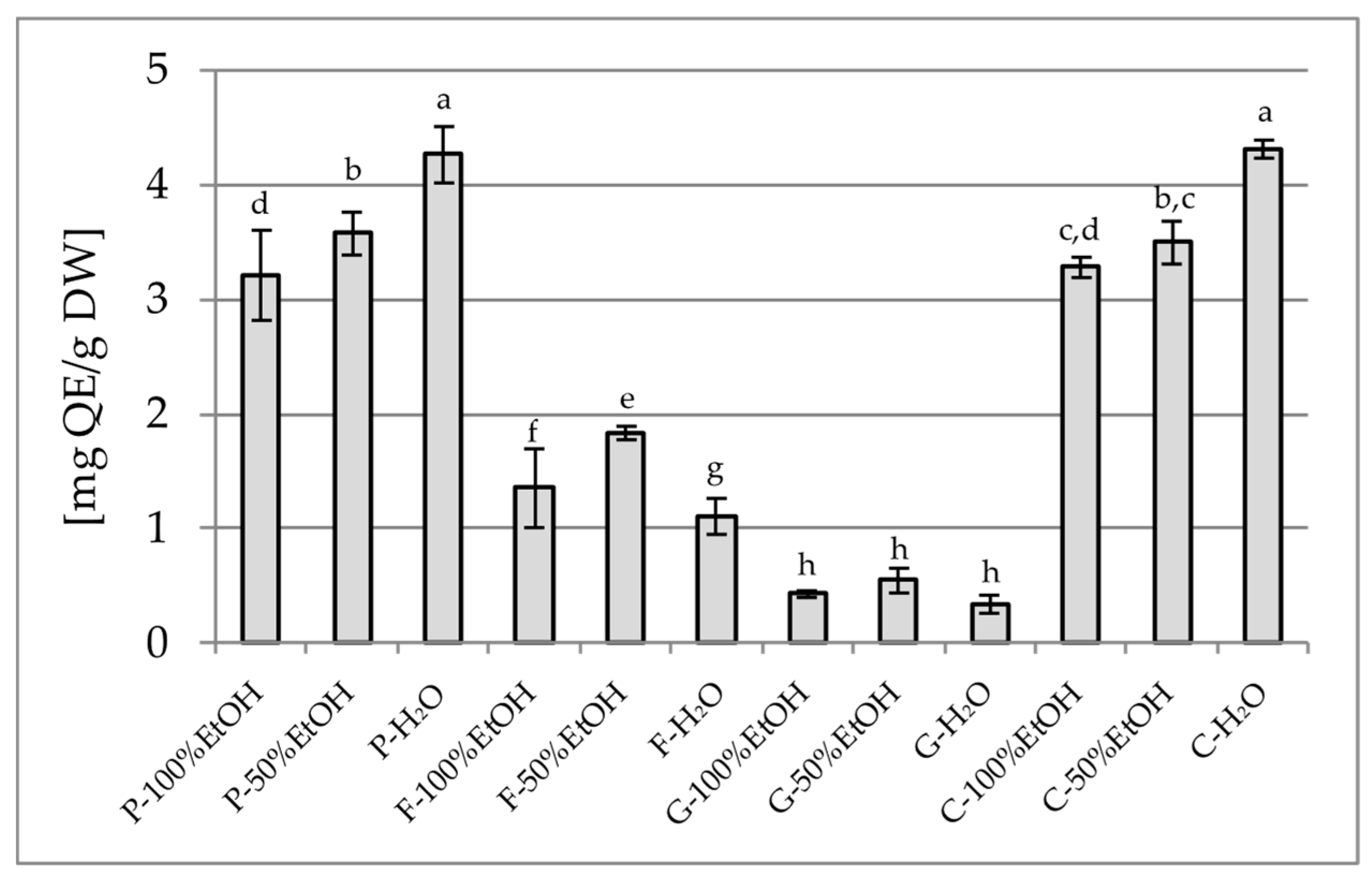
The analysis of the total polyphenol content (TPC) and the total flavonoid content (TFC) revealed clear differences depending on both the plant species and the type of solvent used. The highest TPC was observed for the 50% ethanol extract of P. officinalis (178.49 mg GAE/g) (Figure 1), which significantly exceeded the values noted for the other extracts of this species and also the other plants included in the study. In contrast, the lowest TPC values were obtained for aqueous extracts of G. globosa (1.12 mg GAE/g). It is worth noting that aqueous extracts often showed a lower phenolic content than the 50% ethanol extracts, which may be due to the limited solubility of certain polyphenols in water. In terms of TFC, the highest values were found in aqueous extracts of P. officinalis (4.27 mg QE/g) and C. ternatea (4.31 mg QE/g) (Figure 2), while the lowest values were recorded in all extracts of G. globosa (0.34–0.56 mg QE/g).

2.1.2. Standardisation of the Extract Using High-Performance Liquid Chromatography (HPLC)

In the HPLC analysis, the content of four phenolic compounds was determined: chlorogenic acid, gallic acid, p-coumaric acid, ferulic acid, and quercetin-3-O-rutinoside (rutin) in ethanol, water, and 50% ethanol extracts from the flowers of P. officinalis, F. × intermedia, G. globosa, and C. ternatea. The obtained results (Table 1) showed significant qualitative and quantitative differences in the composition of phenolic compounds, depending on both the plant species and the type of solvent used. Moreover, it should be emphasised that the concentration of secondary metabolites varies depending on factors such as plant part, age, geographical origin, and extraction method [37,38].
The highest rutin content was found in the ethanol and 50% ethanol extracts from F. × intermedia, amounting to 2828.71 mg/100 g dry weight (DW) and 4657.71 mg/100 g DW, respectively. In the aqueous extract of F. × intermedia, rutin was present only in trace amounts (0.90 mg/100 g DW). The results show that the justification in TPC results was that water F. × intermedia extracts demonstrated a lower content of polyphenols and flavonoids. These results may indicate the presence of factors limiting its solubility or a specific form of this flavonoid in the plant material. Studies by various authors indicate that rutin is a flavonoid present in the flowers of different Forsythia species. Kicel et al. reported that rutin is one of the dominant compounds in F. × intermedia, with an even higher content noted in F. suspensa flowers. The rutin content in F. × intermedia flowers was similar to our analyses, amounting to 42.30 mg/g [39]. Moderate rutin concentrations were also detected in extracts from P. officinalis (167.65–239.59 mg/100 g DW) and the 50% ethanol extract from C. ternatea (160.35 mg/100 g DW). Although reports on rutin content in P. officinalis flowers are limited, Lee et al. indicated the presence of this compound in Paeonia suffruticosa flowers (334.26 µg/g) [40]. In contrast, Paz Gonçalves reported rutin in C. ternatea flowers [41]. Extracts from G. globosa contained rutin, but in small amounts. Chlorogenic acid was found in varying concentrations across the analysed flower extracts, with the highest content noted in the 50% ethanol extract from F. × intermedia, reaching 796.03 mg/100 g DW. A high amount was also detected in the ethanol extract of this species (319.79 mg/100 g DW), while in the aqueous extract, the content was negligible (0.31 mg/100 g DW). The presence of chlorogenic acid in the species F. × intermedia has been previously described [39]. Among the other species, G. globosa and C. ternatea extracts contained only trace amounts of chlorogenic acid (0.70–2.83 mg/100 g DW and 0.17–1.93 mg/100 g DW, respectively), which is consistent with previous reports [42,43]. In contrast, in P. officinalis flowers, chlorogenic acid was not detected in any of the tested extracts. Gallic acid was the dominant compound in P. officinalis flowers, with a content ranging from 390.27 ± 15.48 to 603.67 ± 10.10 mg/100 g DW, depending on the extract. The presence of gallic acid in the flowers of various Paeonia species has already been reported in the literature [44,45]. However, these studies are scarce. In the other examined flowers, gallic acid levels were trace or undetectable. According to the literature, ferulic acid presence was confirmed in extracts from C. ternatea [46] and in small amounts in G. globosa [47]. The ferulic acid content in F. × intermedia flowers was low and has not been previously reported in the literature. Meanwhile, p-coumaric acid was present in small amounts in all analysed flower species except for P. officinalis. The results clearly emphasise the significant influence of solvent type on the extraction efficiency of individual compounds. Generally, 50% ethanol extracts provided higher yields of rutin and ferulic acid than aqueous or pure ethanol extracts, which can be attributed to the optimal polarity of this solvent for a broad spectrum of phenolic compounds. The aqueous extract, however, was most effective in extracting gallic acid from P. officinalis flowers.
The results of our studies indicate that the compounds particularly abundant in the extracts studied are gallic acid in P. officinalis and rutin and chlorogenic acid in F. × intermedia. The scientific data available in the literature suggest that chlorogenic acid, gallic acid, and rutin have a multifaceted effect that may support treating type 2 diabetes through mechanisms related to improving insulin sensitivity and protecting pancreatic β cells. Chlorogenic acid has been shown to lower fasting blood glucose levels in clinical trials [48,49] and in in vivo studies [50]. In turn, gallic acid increases PPARγ expression. It activates the PI3K/Akt pathway, which leads to GLUT4 translocation to the plasma membrane, increased glucose uptake, and improved insulin sensitivity in a rat model of a high-fat diet and streptozotocin-induced diabetes [51,52,53]. Furthermore, gallic acid reduces fasting glycemia, improves hepatic glucose metabolism, and protects pancreatic β cells from oxidative stress through antioxidant mechanisms and the modulation of the Nrf2 pathway [54]. Rutin, on the other hand, has antihyperglycemic effects by stimulating insulin secretion, increasing glucose uptake, and protecting Langerhans β cells from degeneration [55,56,57]. At the same time, it reduces oxidative stress and the formation of advanced glycation end products (AGEs), which prevents the vascular damage typical of diabetic complications [58]. Furthermore, rutin ameliorates diabetic neuropathy by lowering plasma glucose and decreasing oxidative stress via the Nrf2 signalling pathway in rats [59].

2.1.3. GC-MS Analysis

The GC-MS analysis of flowers from four plant species revealed the presence of several lipophilic compounds, including long-chain hydrocarbons (e.g., pentacosane, tetratetracontane, and hexatriacontane), fatty acid esters, alcohols, and steroid compounds. The dominant constituents included dihydrofuranone in F. × intermedia (79.67%), tetratetracontane in G. globosa (41.74%), and octadecane, 3-ethyl-5-(2-ethylbutyl)- in C. ternatea (49.10%) (Table 2). Data from the literature suggest that the identified substances show potential significance in the context of antidiabetic therapy. In the three analysed samples (P. officinalis, F.× intermedia, and G. globosa), oleic acid and its ester derivatives belonging to monounsaturated fatty acids were detected. These compounds have anti-inflammatory effects, increase tissue sensitivity to insulin, and improve the lipid profile, which has been confirmed in clinical and experimental studies [60]. In the P. officinalis and G. globosa samples, steroid derivatives (e.g., androstatriene or spirost-8-en-11-one) were also identified. The available evidence indicates that this class of compounds may protect pancreatic β-cells from oxidative stress and inhibit carbohydrate-digesting enzymes such as α-glucosidase, thereby potentially limiting postprandial blood glucose spikes [61]. Although many of the saturated alkanes detected in the studied edible flowers do not show direct pharmacological activity, their presence may affect the lipophilicity of the plant metabolite mixture. Increased lipophilicity may facilitate the transport of active substances in the body and stabilise their action. Studies on semifluorinated alkanes have shown that similar compounds act as effective carriers for pharmaceuticals, increasing their permeability and stability [62].

2.2. Studies of the Biological Activity of Extracts

Diabetes is a complex disease, involving not only glycemic disturbances but also dysfunctions of other organs, especially in elderly patients. Its prevention and control—particularly in the early stages—require a multidimensional approach. This includes lowering blood glucose levels and reducing oxidative stress, alleviating inflammation, and supporting cognitive functions. In light of these findings, and recognising the need for a multifaceted approach, our study evaluates the antidiabetic potential of aqueous, ethanol–aqueous, and ethanol extracts using a range of in vitro experimental methods.

2.2.1. Antioxidant Activity Assay

Antioxidant activity plays a key role in maintaining human health, particularly in preventing and managing type 2 diabetes. Excessive reactive oxygen species (ROS) production and impaired antioxidant defences can damage pancreatic β-cells, contributing to insulin resistance and hyperglycemia [63]. Reducing oxidative stress is thus a justified complementary strategy. Epidemiological studies support this view, showing that diets rich in antioxidants (e.g., vitamin C) may reduce the risk of type 2 diabetes, particularly in adult women [64]. Moreover, reducing oxidative stress may help prevent diabetic complications by protecting the vascular endothelium, thus lowering the risk of retinopathy and nephropathy [65,66].
Our study demonstrated a high antioxidant potential in several edible flower extracts (Table 3). P. officinalis extracts (both water and ethanol-based) showed powerful antiradical activity in DPPH (90–98%) and ABTS (99–100%) tests. F. × intermedia extracts, especially the 50% ethanol variant, also showed considerable activity. In contrast, C. ternatea and G. globosa displayed lower antioxidant activity in both tests (DPPH < 30%, ABTS up to 54%). Interestingly, Fe2+ chelation was most effective in aqueous extracts, while the ethanolic ones showed no chelating ability at the tested concentrations. This suggests different mechanisms of antioxidant action: water extracts likely act via metal ion complexation (inhibiting the Fenton reaction), while ethanol extracts act through radical scavenging [62,67]. For a more comprehensive assessment of antioxidant potential, tests were also conducted to determine the ability of antioxidants to reduce metal ions (CUPRAC—Cu2+ ions to Cu+, FRAP—Fe3+ ions to Fe2+). These tests confirmed the strong antioxidant potential of P. officinalis, especially its ethanolic extracts (175.06 and 144.06 mg TE/g DW). Similar trends, with high activity observed for ethanol-based extracts, were observed for F. × intermedia, which is consistent with the results of our phytochemical studies indicating a lower chlorogenic acid and rutin content in the aqueous extract of F. × intermedia compared with alcoholic extracts. In contrast, both C. ternatea and G. globosa had consistently a low reducing capacity across all assays. These findings confirm that P. officinalis and F. × intermedia are promising sources of antioxidants and may aid in preventing oxidative stress-related diseases such as type 2 diabetes. Overall, the variation in the results between antioxidant assays highlights the importance of using a wide array of tests (DPPH, ABTS, Fe2+ chelation, CUPRAC, and FRAP) to assess the antioxidant activity [67] fully.
Reports in the literature support our findings. Different parts of P. officinalis (leaves, roots) have shown strong antioxidant properties [68], while Paeonia ostii flower extracts were effective in DPPH and ABTS tests [69]. Forsythia koreana inhibited superoxide radicals [70], and Forsythia suspensa activated antioxidant pathways (Nrf2/HO-1) in inflammatory models [71]. Our study’s low antioxidant activity of C. ternatea aligns with reports showing higher activity in aqueous than ethanol-based extracts. Similarly, though data on G. globosa is limited, some studies confirm the antioxidant properties of ethanolic macerates.

2.2.2. Inhibition of Carbohydrate-Hydrolysing Enzymes

The inhibition of digestive enzymes such as α-amylase and α-glucosidase is a well established strategy in controlling postprandial hyperglycemia in type 2 diabetes [72]. In this study, we evaluated the ability of selected edible flower extracts to inhibit these enzymes, using standard in vitro assays to assess their potential for modulating carbohydrate digestion. The first enzyme tested was α-glucosidase (Figure 3). Nearly complete inhibition was observed among the tested samples for all P. officinalis extracts (98–99%). Potent inhibition was also observed for the 100% ethanol extract of C. ternatea (98.7%), which is consistent with studies by Escher et al. that demonstrated the high α-glucosidase inhibition by lyophilised extracts of C. ternatea [73]. F. × intermedia and G. globosa exhibited potent inhibition only in selected extracts (91.59% in F-50% EtOH and 84.10% in G-100% EtOH, respectively). In comparison, aqueous extracts of F. × intermedia and G. globosa showed minimal activity (<10%), indicating that the active glucosidase inhibitors are primarily ethanol-soluble. Moreover, Tang et al. mentioned that significant inhibitory effects on α-glucosidase were observed with the phenolic compounds from G. globosa [74].
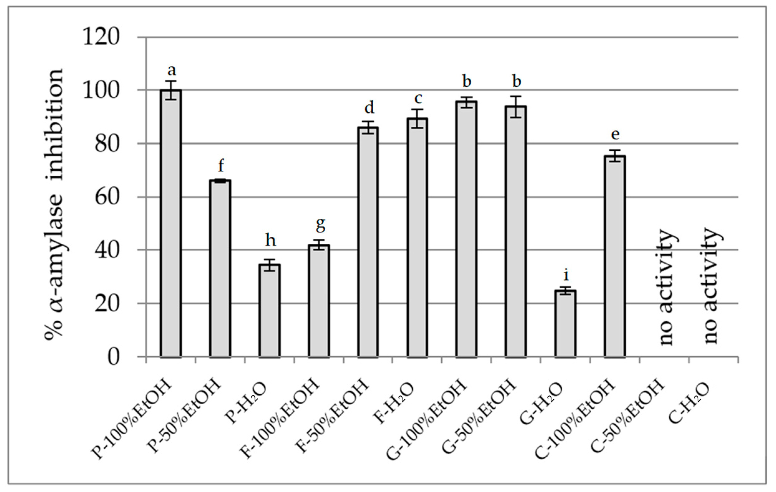
In the case of α-amylase (Figure 4), the ethanolic extract of P. officinalis demonstrated the strongest activity (~100%), followed by ethanol-based extracts of G. globosa (93–95%). Interestingly, F. × intermedia exhibited high activity in the aqueous (89.35%) and 50% ethanol (85.90%) extracts, while the 100% ethanol extract was notably weaker (41.93%), suggesting that the active constituents may be more water-soluble. C. ternatea hydroalcoholic and aqueous extracts showed no measurable α-amylase inhibition.
Our findings complement other studies showing a high α-amylase-inhibitory effect for the root and leaf aqueous and methanol extracts, respectively, from P. officinalis, which inhibited α-amylase in a dose-dependent manner [68]. The results for F. × intermedia, including different species and parts of the plant, demonstrated that the leaves of F. suspensa showed high inhibition on α-glucosidase. Significantly, the inhibitory profiles differed substantially between the two enzymes, underscoring the need for dual-enzyme assays when evaluating the antidiabetic potential of plant extracts. Moreover, these results are particularly noteworthy as they suggest that P. officinalis and F. × intermedia flower extracts may exhibit stronger enzyme inhibitory activity than those of C. ternatea and G. globosa, which have documented but comparatively moderate effects [74,75,76,77,78], although the 100% ethanolic extract of G. globosa also shows considerable potential. Thus, our data point to the flower extracts of P. officinalis and F. × intermedia as promising candidates that are potentially superior to these previously studied species in terms of enzyme inhibition relevant to diabetes risk reduction.

2.2.3. Anti-Inflammatory Activity Assay

Chronic inflammation plays a key role in the pathogenesis of type 2 diabetes and its complications [79]. Persistent low-grade inflammation contributes to insulin resistance, pancreatic β-cell dysfunction, and the progression of vascular complications. It has been proven that compounds of natural origin can alleviate ongoing inflammation [80]; hence, expanding knowledge in the biological activity of natural substances is an important research direction.
Hyaluronidase Inhibition Assay
One of the mechanisms recognised as an indicator of anti-inflammatory potential is the inhibition of hyaluronidase. High-molecular-weight hyaluronic acid stimulates anti-inflammatory responses. It tends to act as a physical barrier, preventing immune cell infiltration into tissues [81], while lower-molecular-weight molecules induce inflammation [82]. Moreover, hyaluronic acid also possesses antioxidative properties, and it is believed that the high-molecular-weight form of hyaluronic acid can protect against the effects of reactive oxygen species [83].
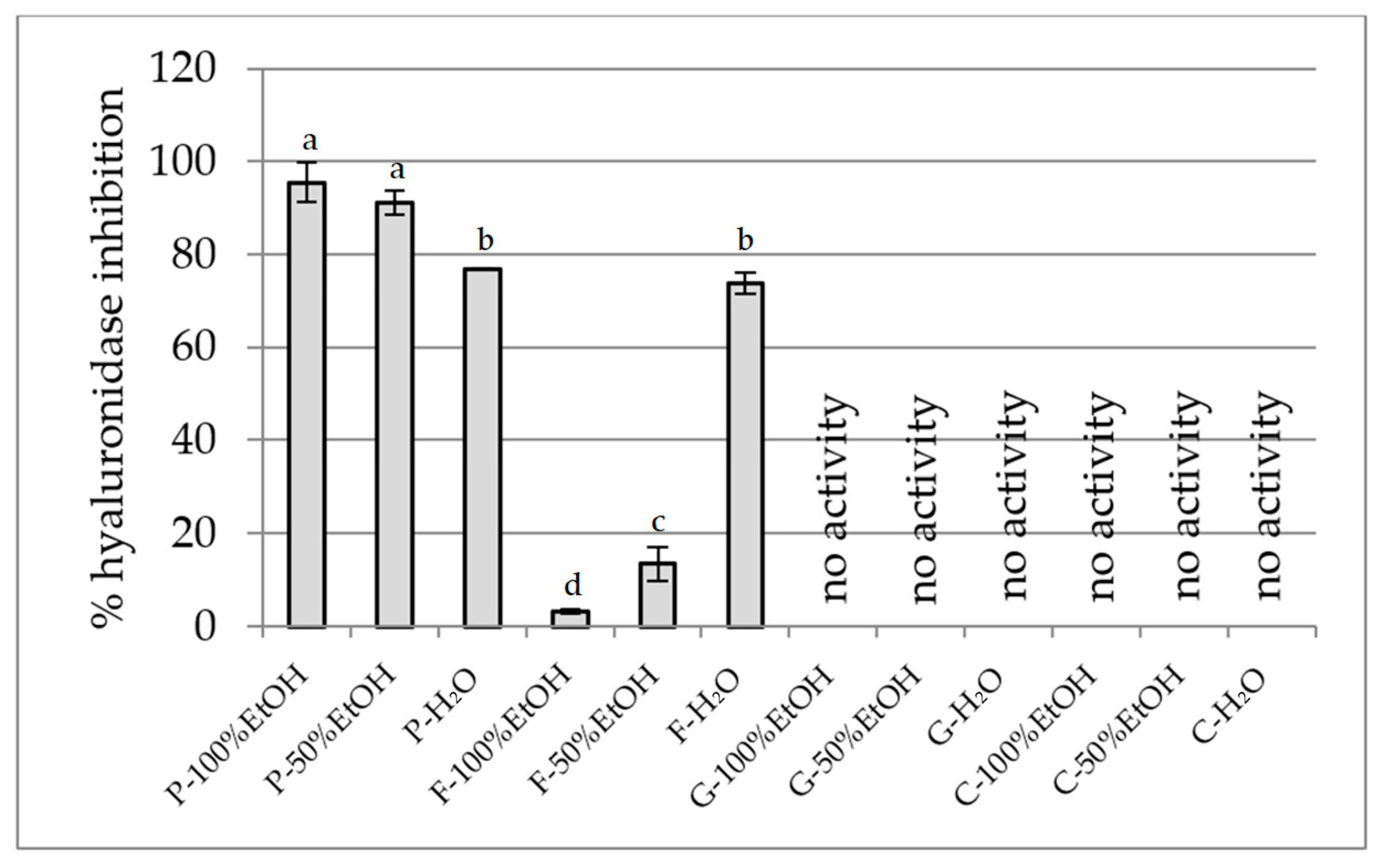
Our findings highlight that among the edible flowers tested, P. officinalis and F. × intermedia possess a promising anti-hyaluronidase effect (Figure 5). P. officinalis extracts exhibited the strongest hyaluronidase inhibition, with values reaching 95.5% for the 100% ethanol extract, 91.1% for the 50% ethanol extract, and 76.9% for the aqueous extract. This high activity suggests the presence of potent anti-hyaluronidase compounds. In contrast, F. × intermedia showed a varied profile: low inhibition in the 100% ethanol extract (3.3%) and moderate to high in the 50% ethanol (13.4%) and aqueous extract (73.8%), indicating that water-soluble constituents are primarily responsible for its inhibitory effect. Notably, G. globosa and C. ternatea extracts demonstrated no hyaluronidase inhibition across all solvent extracts, suggesting limited potential in this anti-inflammatory pathway.
Determination of Anti-Inflammatory Potential in RAW 264.7 Model
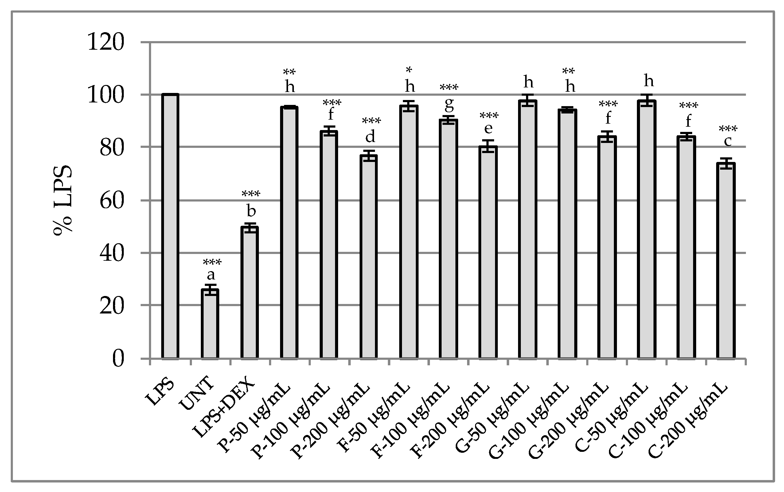
The anti-inflammatory potential of the tested extracts was evaluated using the LPS-stimulated RAW267.4 murine macrophage model by measuring nitric oxide (NO) release. None of the tested samples were toxic to macrophages at the tested concentration range (up to 200 μg/mL), with cell viability comparable to untreated control cells. Thus, the anti-inflammatory assay was conducted across the entire concentration range. However, no anti-inflammatory activity was noted for the tested extracts at lower concentrations. Weak to moderate effects were noted at 100 and 200 μg/mL. The greatest inhibition of nitric oxide release was achieved with the C. ternatea and P. officinalis samples (Figure 6). The observed effect of the inhibition of NO release by the two extracts was significantly higher than that of LPS-stimulated cells.
Importantly, our analyses demonstrated that flower extracts from P. officinalis and C. ternatea contained the highest levels of polyphenols, including flavonoids, among all tested species. While no data have yet described NO inhibition by P. officinalis, the acetate fraction of C. ternatea flower extracts significantly and dose-dependently suppressed NO production [84]. The observed anti-inflammatory effect may be due to gallic acid and rutin present in the extracts, compounds that have been reported in the literature as inhibitors of NO synthesis [85,86]. Although flowers of F. × intermedia have not been studied in the RAW 264.7 murine macrophage model, data from the literature suggest that the butanol fraction from Forsythia koreana flowers inhibits NO production [87]. In contrast, G. globosa flowers have not been studied in this model before. Notably, the observed anti-inflammatory activity did not correlate with the antioxidant potential of the tested extracts. While P. officinalis flower extracts exhibited strong antioxidant activity, those from C. ternatea showed considerably weaker antioxidant effects.

2.2.4. Anti-Cholinesterase Activity Assay

Type 2 diabetes is associated with numerous complications, including the dysfunction of both the central and peripheral nervous systems. Epidemiological studies indicate the increased risk of developing dementia and Alzheimer’s disease in people with diabetes [88], which is associated, among others, with the deficiency of the neurotransmitter acetylcholine (ACh) in the brain. The elevated activity of the enzymes acetylcholinesterase (AChE) and butyrylcholinesterase (BChE), responsible for breaking down ACh, contributes to the deterioration of cholinergic transmission in the nervous system [89]. Therefore, the inhibition of AChE and BChE represents a promising therapeutic strategy to protect cognitive functions and support metabolic control in patients with diabetes.
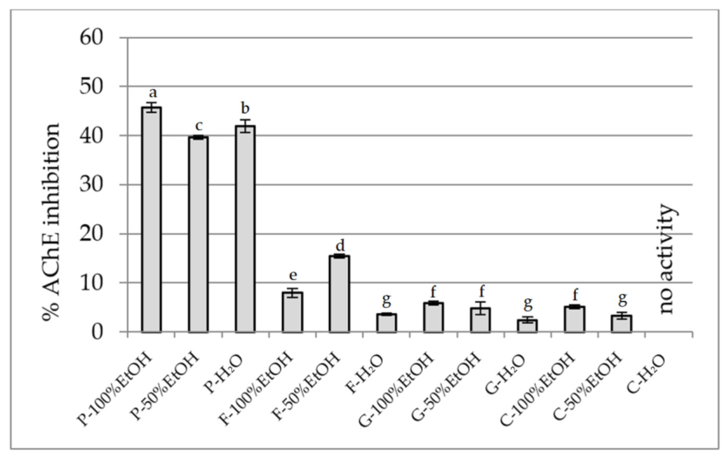
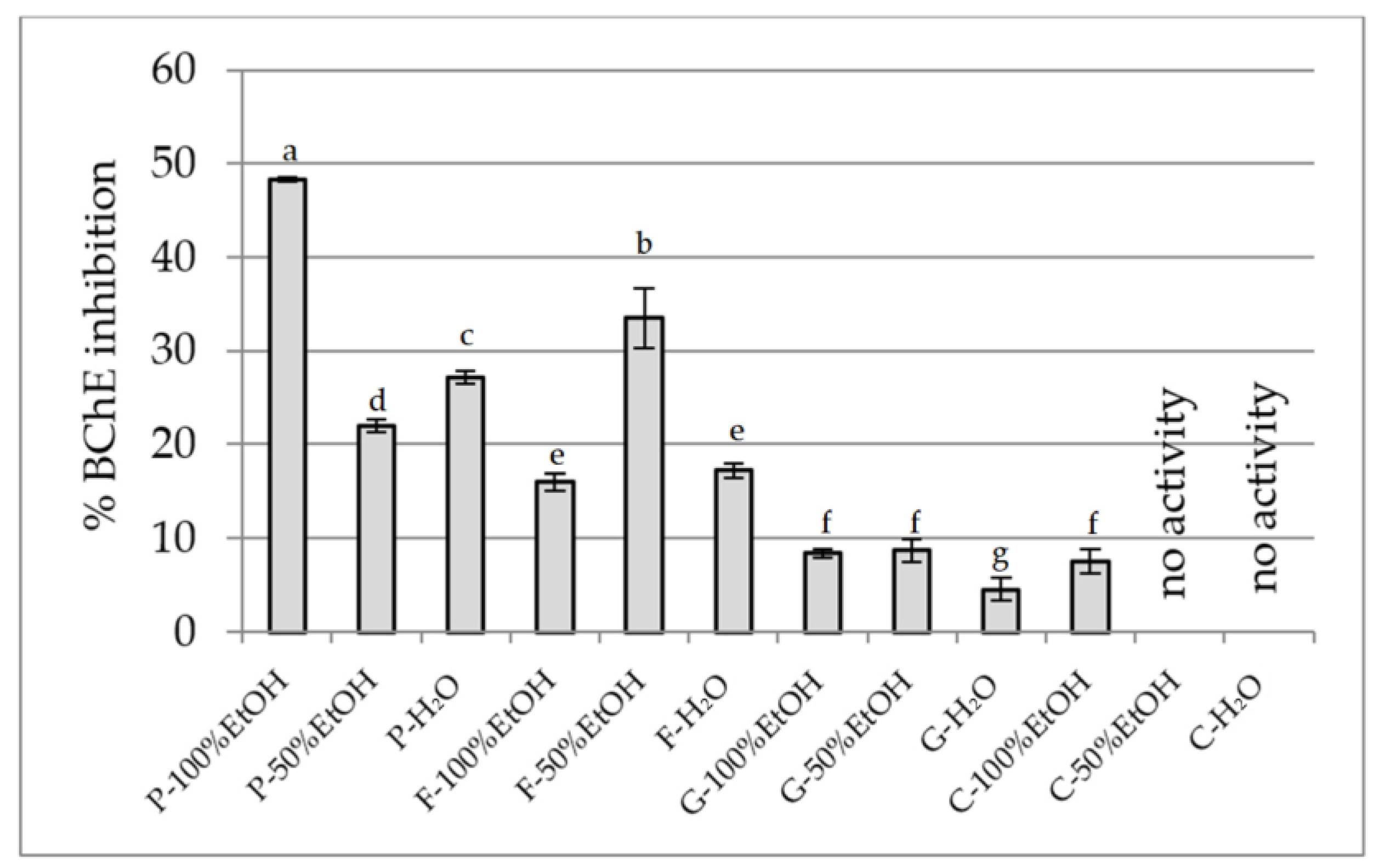
Based on this, the inhibitory potential of extracts from the flowers of P. officinalis, Forsythia × intermedia, G. globosa, and C. ternatea to inhibit AChE and BChE was assessed (Figure 7 and Figure 8, respectively). The highest activity against AChE was demonstrated by ethanol extracts of P. officinalis (~46%), as well as hydroalcoholic and aqueous extracts of this species (~40%). Considerably lower activity was observed for the extracts from F. × intermedia and G. globosa (usually <16%), while C. ternatea showed very weak activity (3–5%), with no effect in the aqueous extract. Similar inhibition patterns were found for BChE. The strongest activity was demonstrated by the ethanol extract from P. officinalis (48.3%), while the other extracts of this species inhibited the enzyme within a range of 22–27%. Moderate activity was observed for extracts from F. × intermedia (up to 33%), with these extracts being more effective. Extracts from G. globosa and C. ternatea showed low inhibitory activity (<10%). The different activity between the extracts is explained by their different chemical composition [34,41,44,90]. The chemical composition of P. officinalis flowers is not well known, but our studies indicate the presence of significant amounts of gallic acid. This phenolic acid has the ability to inhibit AChE and has a relatively strong effect on BChE, as demonstrated in the studies of Orhan et al. [91]. Also, rutin and chlorogenic acid, present in extracts from F. × intermedia, show activity against both enzymes, but inhibit AChE more strongly than BChE [92]. Other compounds present in extracts from the remaining species did not significantly contribute to cholinesterase inhibition.

3. Materials and Methods

3.1. Chemical Reagents

Sodium carbonate, sodium hydroxide, DMSO, formic acid, methanol, ammonium acetate, and copper (II) chloride were purchased from Avantor Performance Materials Poland S.A. (Gliwice, Poland). The Folin–Ciocalteu phenol reagent and HPLC-grade acetonitrile were from Merck (Darmstadt, Germany). All other chemicals were from the Sigma–Aldrich Chemical Co. (Taufkirchen, Germany or St. Louis, MO, USA). High-quality pure water and ultra-high-quality pure water were prepared using a Direct-Q 3 UV Merck Millipore purification system (Merck, Darmstadt, Germany).

3.2. Plant Material

The plant material tested was the dried flowers of 4 different species: P. officinalis, F. × intermedia, G. globosa, and C. ternatea. The plant material from F. × intermedia came from the Botanical Garden of Adam Mickiewicz University in Poznan. The rest of the plant material was purchased from Nanga, Poland.
All plant samples are stored at the Department of Pharmacognosy and Biomaterials, Poznan University of Medical Sciences. The sample codes are as follows: PO_fl_1_2023, FI_fl_1_2023, CG_fl_1_2023, and CI_fl_1_2023. The F. × intermedia sample was identified by Dr. Mateusz Sowelo (Botanical Garden of Adam Mickiewicz University in Poznan). The samples of G. globosa and C. ternatea were commercially available and purchased.

3.3. Extraction Process

The experiments were performed on the flowers of four different plant species. The weighed portions (2.5 g) and the solvent (water, ethanol, or 50% ethanol) were placed in a glass Erlenmeyer flask sealed with a glass cork and parafilm. The extraction was performed in an ultrasonic bath, heated to 60 °C. Then, the extracts were filtered through cotton wool into a beaker, in which the filtrate from each repetition stage was combined. Due to the fourfold repetition of the extraction (20 min each), 200 mL samples were obtained. In the next stage, the volume of the extracts was decreased using a rotary evaporator. Lastly, extracts were supplemented with appropriate solvents, and their volume was equalised to 25 mL each.

3.4. Phytochemical Studies of the Extracts

3.4.1. Total Polyphenol Content

The procedure was conducted following the method outlined by Studzińska-Sroka et al. with modifications [93]. Briefly, 25.0 µL of either the test extract or standard, followed by 200.0 µL of distilled water, 15.0 µL of the Folin–Ciocalteu reagent, and 60.0 µL of a 20% calcium carbonate solution, were successively added to the wells. A control sample containing only the reagents (excluding the test extract or standard) was included. The plate was then shaken in the dark for 30 min at 350 rpm at room temperature. Absorbance was recorded at 760 nm using a Multiskan GOx1510 microplate reader (Thermo-Scientific, Waltham, MA, USA). The experiment was carried out in duplicate. The results are the average of n = 6 measurements for the extract samples and n = 3 measurements for the reference. The results were reported as milligrams of gallic acid equivalents (GAE) per gram of dry plant material (DW), along with the standard deviation (SD).

3.4.2. Total Flavonoid Content

The analysis was carried out according to the method described by Studzińska-Sroka et al. [93]. In short, 100.0 µL of the tested extract or reference compound was combined with 100.0 µL of a 2% methanolic aluminium chloride solution. After shaking the plate for 1 min, the mixture was incubated for 10 min in the dark at room temperature. Absorbance was measured at 415 nm using a Multiskan GO 1510 microplate reader (Thermo Fisher Scientific, Vantaa, Finland). The blank sample consisted of the extract mixed with methanol instead of the aluminium chloride solution. The experiment was carried out in duplicate. The results are the average of n = 6 measurements for the extract samples and of n = 3 measurements for the reference. The results are expressed as milligrams of quercetin equivalent (QE) per gram of dry plant material (DW), with standard deviation (SD).

3.4.3. Standardisation of the Extract Using High-Performance Liquid Chromatography (HPLC)

The compounds selected for quantification were chosen based on their clear presence in HPLC profiles and their occurrence in the flowers of the studied species, as reported in the literature. Due to limited access to LC-MS, we focused on reliably detectable compounds with available standards. A high-performance liquid chromatography system (Dionex Thermoline Fisher Scientific, Dreieich, Germany) equipped with a high-pressure pump (UltiMate 3000), an autosampler (UltiMate 3000), and a DAD detector (UltiMate 3000) with Chromeleon software version 7.0 from Dionex Thermoline Fisher Scientific (Dreieich, Germany) was used to assess the presence and content of chlorogenic acid, ferulic acid, gallic acid, p-coumaric acid, and rutin in different flower extracts from P. officinalis, F. × intermedia, G. globosa, and C. ternatea. The analysis was performed according to the method described by Paczkowska-Walendowska et al. [94], and the procedure was validated to ensure the reliability of the results. The chromatographic conditions included a detection wavelength of 240 nm for chlorogenic acid and ferulic acid, 265 nm for gallic acid and p-coumaric acid, and 360 nm for rutin; a mobile phase flow rate of 1 mL/min; a column temperature of 40 °C; and a mobile phase consisting of 0.1% formic acid (solvent A) and acetonitrile (solvent B). The gradient elution was as follows: 0–35 min: 2–20% B; 35–55 min: 20–70% B; 55–60 min: 2% B. Standard solutions were prepared in HPLC-grade methanol at the following concentrations: gallic acid—0.1 and 0.01 mg/mL, ferulic acid—0.001, 0.01, and 0.1 mg/mL, p-coumaric acid—0.001, 0.01, and 0.1 mg/mL, and rutin—0.5 mg/mL. All solutions were filtered through a 0.45 µm membrane filter before injection. A series of injection volumes was applied for each compound and concentration to construct calibration curves. Injection volumes were as follows: for chlorogenic acid—4, 6, 8, and 10 µL (0.1 mg/mL), and 1, 2, 4, 6, 8, 10, and 12 µL (1.0 mg/mL); for gallic acid—2, 5, 10, 20, and 50 µL (0.01 mg/mL), and 10, 20, and 50 µL (0.1 mg/mL); for ferulic acid—5 µL (0.001 mg/mL), 1, 10, and 40 µL (0.01 mg/mL), 8, 16, and 32 µL (0.1 mg/mL); for p-coumaric acid—5 µL (0.001 mg/mL), 1, 10, and 40 µL (0.01 mg/mL), 8, 16, and 32 µL (0.1 mg/mL); rutin—5, 10, 20, 40, 60, and 80 µL (0.5 mg/mL). Calibration curves were established, and the method was validated by evaluating parameters such as linearity, direct and indirect precision, and the limits of detection (LOD) and quantification (LOQ). The results are expressed as milligrams of compound per 100 g of dry plant material (DW), with standard deviation (SD).

3.4.4. GC-MS Analysis

Chromatographic studies were performed on a GC-MS chromatograph (SCION TQ, BRUKER). Approximately 1.15 ± 0.01 g of plant material (each flower tested) was finely ground using a mortar and pestle. Subsequently, 10 mL of chloroform (Thermo Scientific, spectroscopy grade) was added to the sample, and grinding was continued until a homogeneous solution was obtained. The resulting extract was then filtered through a 0.20 µm syringe filter. The filtrate was subjected to gas chromatography–mass spectrometry (GC-MS) analysis. For this step, 1.0 µL of the solution was injected onto the column. The chromatograph was equipped with a VF-5 ms Crawford Scientific silica column. The electron energy was 70 eV, and the ion source was 200 °C. Helium was used as the carrier gas at a 1.0 mL/min flow rate. Temperature programme: enable coolant at 50.0 °C, coolant timeout 20.00 min, stabilisation time 0.50 min; temperature 60.0 °C, hold 3.00 min, total 3.00 min; temperature 280.0 °C, rate 10.0 °C/min, hold 35.00 min, total 60.00 min. The compounds’ identification was based on comparing their retention time and mass spectra with NIST standards.

3.5. Studies of the Biological Activity of Extracts

3.5.1. Antioxidant Activity Assay

DPPH Assay
A DPPH assay was employed to assess antiradical activity, following the procedure outlined in reference [95]. In brief, 25.0 µL of the test extract—prepared at a concentration of 2.5 mg of dry plant material per mL—was mixed with 175.0 µL of DPPH solution (3.9 mg in 50 mL of methanol). The mixture was shaken in the dark at 350 rpm for 5 min at room temperature and then incubated in the dark for an additional 25 min. Absorbance was recorded at 517 nm using a microplate reader (Multiskan GO 1510, Thermo Fisher Scientific, Vantaa, Finland). All analyses were performed in duplicate, and the final results represent the mean of five measurements (n = 5). Results are expressed as % ± standard deviation (SD).
ABTS Assay
An ABTS assay was also employed to assess antiradical activity. It followed the procedure outlined in reference [96]. In short, 10.0 µL of the test extract (2.5 mg of dry plant material per mL) was mixed with 200.0 µL of ABTS solution (0.1920 g ABTS-(NH4)2 in 50 mL of 0.3311 mg K2S2O8 in 500 mL of distilled water). Before use, the basic ABTS solution was diluted so that the absorbance was ~0.77 with a 734 nm wavelength. The mixture was shaken in the dark at 500 rpm for 30 min at room temperature. Absorbance was recorded at 734 nm using a microplate reader (Multiskan GO 1510, Thermo Fisher Scientific, Vantaa, Finland). All analyses were performed in duplicate, and the final results represent the mean of five measurements (n = 5). Results are expressed as % ± standard deviation (SD).
CUPRAC Assay
A CUPRAC assay was performed following a previously described method [97]. The CUPRAC reagent was prepared by combining equal volumes of neocuproine (7.5 mM), copper (II) chloride (10 mM), and ammonium acetate buffer (1 M, pH 7.0). Subsequently, 50 µL of the test extract (2.5 mg dry plant material/mL) was mixed with 150 µL of the reagent. After incubating the mixture in the dark with shaking (350 rpm) for 30 min at room temperature, absorbance was measured at 450 nm. A blank sample was prepared by substituting the extract with an equal volume of the extraction solvent. The experiment was conducted in duplicate, and results are expressed as the mean of four measurements (n = 4). The test results were expressed as mg of Trolox per gram of dry weight ± standard deviation (SD).
FRAP Assay
A FRAP assay was carried out based on the method described by Tiveron et al. [98] with slight modifications. The stock FRAP reagent was composed of a 300 mM acetate buffer (pH 3.6), a 10 mM TPTZ solution prepared in 40 mM HCl, and a 20 mM FeCl3·6H2O solution. The working FRAP reagent was freshly made by combining 25 mL of acetate buffer, 2.5 mL of the TPTZ solution, and 2.5 mL of the FeCl3·6H2O solution, and then preheated to 37 °C before use. For the assay, 20 µL of the test extract (2.5 mg dry plant material/mL) was mixed with 180 µL of the FRAP reagent and incubated at 37 °C for 30 min in the dark. For the blank FRAP reagent, TPTZ solution was used instead. Absorbance was measured at 593 nm in four replicates (n = 4). The test results were expressed as mg of Trolox per gram of dry weight. The test results were expressed as mg of Trolox per gram of dry weight ± standard deviation (SD).
Chelation Power on Ferrous (Fe2+) Ions Assay
The Fe2+ chelating ability of the extracts was assessed based on the method by Dinis et al. [99], with modifications. In summary, 200.0 μL of the sample solution at a concentration of 2.5 mg of dry plant material per mL was mixed with 10.0 μL of FeCl2·4H2O (1 mM) and pre-incubated in a 96-well plate at room temperature for 10 min (with shaking, 500 rpm). Then, 40.0 μL of ferrozine (2.5 mM) was added, and the mixture was incubated for 30 min at room temperature (with shaking, 500 rpm). The absorbance of the Fe2+–ferrozine complex was recorded at 562 nm in five replicates (n = 5). The chelating activity, corresponding to the final sample concentration, was calculated and expressed as % of chelating activity ± standard deviation (SD).

3.5.2. Inhibition of Carbohydrate-Hydrolysing Enzymes

α-Glucosidase Inhibition Assay
The α-glucosidase inhibitory activity of the extracts was assessed following the method of Studzińska-Sroka et al. [93], with slight modifications. In short, 50.0 µL of the extract (25.0 mg dry plant material/mL) was pre-incubated with 50.0 µL of 0.1 M phosphate buffer (pH 6.8) and 30.0 µL of α-glucosidase solution (0.5 U/mL) in a 96-well plate at 37 °C for 15 min. Afterwards, 20.0 µL of a 5 mM p-nitrophenyl-α-D-glucopyranoside (pNPG) solution in 0.1 M phosphate buffer was added, and the mixture was incubated for 20 min at 37 °C. The reaction was terminated by adding 100.0 µL of 0.2 M sodium carbonate. Absorbance was read at 405 nm using a Multiskan GO 1510 microplate reader (Thermo Fisher Scientific, Vantaa, Finland), 2 min after stopping the reaction. Control measurements were performed without extract, while blanks consisted of extract or standard solutions without enzyme. Results represent the mean of three replicates (n = 3) and are expressed as % of inhibition ± standard deviation (SD).
α-Amylase Inhibition Assay
The α-amylase inhibitory activity of the extracts was evaluated following the protocol described by Studzińska-Sroka et al. [93], with slight modifications. Briefly, 50.0 µL of the extract (25.0 mg dry plant material/mL) was preincubated with 20 µL of α-amylase solution (2.0 U/mL, prepared in phosphate buffer at pH 6.9) at 37 °C for 20 min. Subsequently, 20.0 µL of a 0.5% starch solution in phosphate buffer was added, and the mixture was incubated again at 37 °C for another 20 min. Then, 60 µL of a colour reagent (containing 96 mM 3,5-dinitrosalicylic acid, 5.31 M potassium sodium tartrate in 2 M sodium hydroxide, and deionised water) was added, and the plate was heated at 90 °C. After 45 min, 100 µL of distilled water was added, and the absorbance was measured at 540 nm using a microplate reader (Multiskan GO 1510, Thermo Fisher Scientific, Vantaa, Finland). The control sample included 20 µL of distilled water with 20 µL of amylase solution, while the blank consisted of 20 µL of distilled water and 20 µL of phosphate buffer. Results represent the average of four replicates (n = 4), and are expressed as % of inhibition ± standard deviation (SD).

3.5.3. Anti-Inflammatory Activity Assay

Hyaluronidase Inhibition Assay
The anti-hyaluronidase activity assay followed a previously reported procedure [100], with adjustments. In short, 25.0 µL of incubation buffer, 25.0 µL of hyaluronidase enzyme solution (30 U/mL), 10.0 µL of the test extract (25 mg of dry plant material per mL), and 15.0 µL of acetate buffer were combined in a well. The mixture was incubated at 37 °C for 15 min with shaking at 200 rpm. Subsequently, 25.0 µL of hyaluronic acid (HA) solution was added, followed by a second incubation for 45 min under the same conditions. Afterwards, 200.0 µL of cetyltrimethylammonium bromide (CTAB) solution in 2% NaOH was introduced, and the mixture was incubated at room temperature for 10 min without shaking. Absorbance was measured at 600 nm using a microplate reader (Multiskan GO 1510, Thermo Fisher Scientific, Vantaa, Finland). The blank composition was as previously described [101]. The assay was performed in duplicate, and values were calculated from four measurements (n = 4). Results are expressed as % of inhibition ± standard deviation (SD).
Determination of Anti-Inflammatory Potential in RAW 264.7 Model
Murine RAW 264.7 macrophages were cultured at 37 °C, in a 5% CO2 atmosphere, with relative humidity, using DMEM high glucose, supplemented with 10% foetal bovine serum as the culture medium. Before the anti-inflammatory experiment, the toxic potential of the tested samples to the macrophages was evaluated using MTT assay, in the concentration range of 10–200 μg/mL, as described previously [97]. For the determination of anti-inflammatory potential, the macrophages were seeded onto 96-well plates (1.5 × 10⁵ cells/well) and pre-treated with the tested samples (at the concentration 10–200 μg/mL) or dexamethasone (0.5 μg/mL) as a reference drug, for 1 h. Next, inflammation was induced by adding 10 ng/mL of LPS to each well, as previously described [97], and the incubation was performed for 24 h. Cells treated with LPS alone were used as a positive control, while cells not treated with either tested samples or LPS were included as the untreated control. The values were calculated from three measurements (n = 3). Nitric oxide level was determined in cell culture supernatants with Griess Reagent Kit (Promega Corporation, Madison, Winooski, VT, USA), according to the manufacturer’s protocol. The results were expressed as a % of the LPS control.

3.5.4. Anti-Cholinesterase Activity Assay

AChE and BChE Inhibition Activity Assays
The studied extracts were dissolved in an appropriate solvent to a concentration of 25 mg/mL. The cholinesterase inhibitory activity was assessed using Ellman’s spectrophotometric method [102], incorporating modifications previously described by Studzińska-Sroka et al. [103]. In brief, 5.0 µL of each test sample was combined with 60.0 µL of TRIS-HCl buffer (50 mM, pH 8.0) and 30.0 µL of AChE or BChE solution (0.2 U/mL). The mixture was incubated for 5 min at 25 °C with shaking at 500 rpm. Following this, 30.0 µL of acetylthiocholine iodide (1.5 mM) and 125.0 µL of 5,5′-dithiobis(2-nitrobenzoic acid) solution (0.3 mM, containing 10 mM NaCl and 2 mM MgCl2·6H2O) were added, and incubation continued for 30 min under the same conditions. Sample blanks were prepared by replacing the enzyme with buffer. The control sample included an appropriate solvent instead of the test substance, and its blank replaced both the enzyme and the sample with buffer and an appropriate solvent, respectively. Absorbance was recorded at 405 nm using a Multiskan GO 1510 microplate reader (Thermo Fisher Scientific, Vantaa, Finland). Each extract was tested in two independent experiments, and the results represent the mean of five measurements (n = 5).

3.6. Statistical Analysis

Statistical calculations were performed using Microsoft Excel (Microsoft Office LTSC 2021 Professional Plus, Microsoft Corp., Redmond, WA, USA). Data are presented as mean ± standard deviation (SD). One-way analysis of variance (ANOVA) was used to evaluate differences between groups. Post hoc comparisons were performed using the Newman–Keuls test to assess differences between all experimental groups and the Dunnett test to compare each treatment group with the LPS-treated control. Statistical significance was considered at p < 0.05.

4. Conclusions

The present study shows that edible flower extracts—especially those from P. officinalis and F. × intermedia—possess interesting biological potential that may contribute to the prevention or adjuvant treatment of type 2 diabetes and its complications. Among the species studied, P. officinalis extracts, especially those obtained with 50% ethanol, showed the highest total polyphenol content and strong antioxidant activity in many tests performed (DPPH, ABTS, CUPRAC, and FRAP), together with the strong inhibition of α-glucosidase and α-amylase, suggesting a broad spectrum of bioactivity. High levels of gallic acid and rutin in this species probably contribute to these effects. Similarly, F. × intermedia extracts showed high rutin and chlorogenic acid contents and selective enzyme inhibition, with aqueous extracts showing the potent inhibition of α-amylase and hyaluronidase. This result further emphasised that the type of extract may be crucial for the direction of action shown. Although C. ternatea extracts showed a moderate polyphenol content, the ethanol–water extract of the raw material showed a noticeable inhibition of NO release in macrophage assays, indicating promising anti-inflammatory properties. In contrast, G. globosa extracts generally showed a low polyphenol content and weak antioxidant and anti-inflammatory activity, although ethanol extracts showed significant α-amylase inhibition. In conclusion, this study highlighted the value of edible flowers—especially P. officinalis and F. × intermedia—as a rich source of bioactive phytochemicals with antioxidant, antidiabetic, and anti-inflammatory properties. These findings may be useful for further studies on their therapeutic potential for alleviating oxidative stress, hyperglycemia, and chronic inflammation associated with metabolic disorders.

Author Contributions

Conceptualization, E.S.-S.; methodology, E.S.-S., K.G. and M.S.; software, E.S.-S. and M.K. (Maciej Książkiewicz); validation, E.S.-S., K.G. and M.S.; formal analysis, E.S.-S., K.G., M.K. (Maciej Książkiewicz), M.K. (Michalina Karczewska), F.N. and M.S.; investigation, M.K. (Maciej Książkiewicz), M.K. (Michalina Karczewska), F.N., K.G., M.S. and E.S.-S.; resources, J.C.-P.; data curation, M.K. (Maciej Książkiewicz), M.K. (Michalina Karczewska), F.N., K.G., M.S. and E.S.-S.; writing—original draft preparation, E.S.-S. and M.K. (Maciej Książkiewicz); writing—review and editing, E.S.-S., M.K. (Maciej Książkiewicz), and J.C.-P.; visualisation, M.K. (Maciej Książkiewicz) and E.S.-S.; supervision, E.S.-S.; project administration, M.K. (Maciej Książkiewicz) and E.S.-S.; funding acquisition, E.S.-S. All authors have read and agreed to the published version of the manuscript.

Funding

This research received no external funding.

Data Availability Statement

All data supporting reported results can be found within the manuscript.

Acknowledgments

The authors would like to thank Mateusz Sowelo from the Botanical Garden Adam Mickiewicz University in Poznan for collecting the plant material used in this study.

Conflicts of Interest

The authors declare no conflicts of interest.

References

  1. Zheng, Y.; Ley, S.H.; Hu, F.B. Global Aetiology and Epidemiology of Type 2 Diabetes Mellitus and Its Complications. Nat. Rev. Endocrinol. 2018, 14, 88–98. [Google Scholar] [CrossRef]
  2. Guzman-Vilca, W.C.; Carrillo-Larco, R.M. Number of People with Type 2 Diabetes Mellitus in 2035 and 2050: A Modelling Study in 188 Countries. Curr. Diabetes Rev. 2025, 21, E120124225603. [Google Scholar] [CrossRef]
  3. Niedowicz, D.M.; Daleke, D.L. The Role of Oxidative Stress in Diabetic Complications. Cell Biochem. Biophys. 2005, 43, 289–330. [Google Scholar] [CrossRef]
  4. Tsalamandris, S.; Antonopoulos, A.S.; Oikonomou, E.; Papamikroulis, G.-A.; Vogiatzi, G.; Papaioannou, S.; Deftereos, S.; Tousoulis, D. The Role of Inflammation in Diabetes: Current Concepts and Future Perspectives. Eur. Cardiol. Rev. 2019, 14, 50. [Google Scholar] [CrossRef] [PubMed]
  5. Simmons, R.A. Developmental Origins of Diabetes: The Role of Oxidative Stress. Free Radic. Biol. Med. 2006, 40, 917–922. [Google Scholar] [CrossRef]
  6. Ikegami-Kawai, M.; Suzuki, A.; Karita, I.; Takahashi, T. Increased Hyaluronidase Activity in the Kidney of Streptozotocin-Induced Diabetic Rats. J. Biochem. 2003, 134, 875–880. [Google Scholar] [CrossRef] [PubMed]
  7. Pereira, P.A.T.; Bitencourt, C.S.; Reis, M.B.; Frantz, F.G.; Sorgi, C.A.; Souza, C.O.S.; Silva, C.L.; Gardinassi, L.G.; Faccioli, L.H. Immunomodulatory Activity of Hyaluronidase Is Associated with Metabolic Adaptations during Acute Inflammation. Inflamm. Res. 2020, 69, 105–113. [Google Scholar] [CrossRef] [PubMed]
  8. Dogné, S.; Rath, G.; Jouret, F.; Caron, N.; Dessy, C.; Flamion, B. Hyaluronidase 1 Deficiency Preserves Endothelial Function and Glycocalyx Integrity in Early Streptozotocin-Induced Diabetes. Diabetes 2016, 65, 2742–2753. [Google Scholar] [CrossRef]
  9. Rao, A.A.; Sridhar, G.R.; Das, U.N. Elevated Butyrylcholinesterase and Acetylcholinesterase May Predict the Development of Type 2 Diabetes Mellitus and Alzheimer’s Disease. Med. Hypotheses 2007, 69, 1272–1276. [Google Scholar] [CrossRef]
  10. Mushtaq, G.; H Greig, N.; A Khan, J.; A Kamal, M. Status of Acetylcholinesterase and Butyrylcholinesterase in Alzheimer’s Disease and Type 2 Diabetes Mellitus. CNS Neurol. Disord. Targets (Formerly Curr. Drug Targets CNS Neurol. Disord.) 2014, 13, 1432–1439. [Google Scholar] [CrossRef]
  11. Phung, O.J.; Sobieraj, D.M.; Engel, S.S.; Rajpathak, S. Early Combination Therapy for the Treatment of Type 2 Diabetes Mellitus: Systematic Review and Meta-analysis. Diabetes Obes. Metab. 2014, 16, 410–417. [Google Scholar] [CrossRef] [PubMed]
  12. Gilbert, M.P.; Pratley, R.E. GLP-1 Analogs and DPP-4 Inhibitors in Type 2 Diabetes Therapy: Review of Head-to-Head Clinical Trials. Front. Endocrinol. 2020, 11, 178. [Google Scholar] [CrossRef]
  13. Shetty, R.; Basheer, F.T.; Poojari, P.G.; Thunga, G.; Chandran, V.P.; Acharya, L.D. Adverse Drug Reactions of GLP-1 Agonists: A Systematic Review of Case Reports. Diabetes Metab. Syndr. Clin. Res. Rev. 2022, 16, 102427. [Google Scholar] [CrossRef]
  14. Halimi, S.; Vergès, B. Adverse Effects and Safety of SGLT-2 Inhibitors. Diabetes Metab. 2014, 40, S28–S34. [Google Scholar] [CrossRef]
  15. Alkhatib, A.; Tsang, C.; Tiss, A.; Bahorun, T.; Arefanian, H.; Barake, R.; Khadir, A.; Tuomilehto, J. Functional Foods and Lifestyle Approaches for Diabetes Prevention and Management. Nutrients 2017, 9, 1310. [Google Scholar] [CrossRef] [PubMed]
  16. Hussain, T.; Tan, B.; Murtaza, G.; Liu, G.; Rahu, N.; Kalhoro, M.S.; Kalhoro, D.H.; Adebowale, T.O.; Mazhar, M.U.; ur Rehman, Z. Flavonoids and Type 2 Diabetes: Evidence of Efficacy in Clinical and Animal Studies and Delivery Strategies to Enhance Their Therapeutic Efficacy. Pharmacol. Res. 2020, 152, 104629. [Google Scholar] [CrossRef] [PubMed]
  17. Papuc, C.; Goran, G.V.; Predescu, C.N.; Tudoreanu, L.; Ștefan, G. Plant Polyphenols Mechanisms of Action on Insulin Resistance and against the Loss of Pancreatic Beta Cells. Crit. Rev. Food Sci. Nutr. 2021, 62, 325–352. [Google Scholar] [CrossRef]
  18. Nguyen, V.; Taine, E.G.; Meng, D.; Cui, T.; Tan, W. Chlorogenic Acid: A Systematic Review on the Biological Functions, Mechanistic Actions, and Therapeutic Potentials. Nutrients 2024, 16, 924. [Google Scholar] [CrossRef]
  19. Mistry, P.S.; Chorawala, M.R.; Sivamaruthi, B.S.; Prajapati, B.G.; Kumar, A.; Chaiyasut, C. The Role of Dietary Anthocyanins for Managing Diabetes Mellitus-Associated Complications. Curr. Diabetes Rev. 2025, 21, 76–99. [Google Scholar] [CrossRef]
  20. Zhang, Y.; Li, M.; Liu, H.; Fan, Y.; Liu, H.H. The Application of Procyanidins in Diabetes and Its Complications: A Review of Preclinical Studies. Front. Pharmacol. 2025, 16, 1532246. [Google Scholar] [CrossRef]
  21. Mao, T.; Akshit, F.N.U.; Mohan, M.S. Effects of Anthocyanin Supplementation in Diet on Glycemic and Related Cardiovascular Biomarkers in Patients with Type 2 Diabetes: A Systematic Review and Meta-Analysis of Randomized Controlled Trials. Front. Nutr. 2023, 10, 1199815. [Google Scholar] [CrossRef]
  22. Yang, L.; Ling, W.; Qiu, Y.; Liu, Y.; Wang, L.; Yang, J.; Wang, C.; Ma, J. Anthocyanins Increase Serum Adiponectin in Newly Diagnosed Diabetes but Not in Prediabetes: A Randomized Controlled Trial. Nutr. Metab. 2020, 17, 78. [Google Scholar] [CrossRef]
  23. Adel-Mehraban, M.S.; Tabatabaei-Malazy, O.; Manayi, A.; Alatab, S.; Mohseni, S.; Fana, S.E.; Asili, P.; Bahramsoltani, R.; Esmaeili, F.; Azizi, B. Antioxidative and Anti-Inflammatory Effects of Plant-Derived Hypoglycemic Medicines: An In Vivo/In Vitro Systematic Review. Curr. Top. Med. Chem. 2024, 24, 1408–1450. [Google Scholar] [CrossRef] [PubMed]
  24. Nobel, S.; Baker, C.; Loullis, C. Crave Crush TM Lozenges Containing Gymnemic Acids Reduce Consumption of High Sugar Foods. Adv. Med. Plant Res. 2017, 5, 63–67. [Google Scholar] [CrossRef]
  25. Dimopoulou, M.; Vareltzis, P.; Floros, S.; Androutsos, O.; Bargiota, A.; Gortzi, O. Development of a Functional Acceptable Diabetic and Plant-Based Snack Bar Using Mushroom (Coprinus comatus) Powder. Foods 2023, 12, 2702. [Google Scholar] [CrossRef]
  26. Barrea, L.; Vetrani, C.; Verde, L.; Frias-Toral, E.; Ceriani, F.; Cernea, S.; Docimo, A.; Graziadio, C.; Tripathy, D.; Savastano, S.; et al. Comprehensive Approach to Medical Nutrition Therapy in Patients with Type 2 Diabetes Mellitus: From Diet to Bioactive Compounds. Antioxidants 2023, 12, 904. [Google Scholar] [CrossRef]
  27. Miñambres, I.; Cuixart, G.; Gonçalves, A.; Corcoy, R. Effects of Inositol on Glucose Homeostasis: Systematic Review and Meta-Analysis of Randomized Controlled Trials. Clin. Nutr. 2019, 38, 1146–1152. [Google Scholar] [CrossRef] [PubMed]
  28. Bule, M.; Albelbeisi, A.H.; Nikfar, S.; Amini, M.; Abdollahi, M. The Antidiabetic and Antilipidemic Effects of Hibiscus sabdariffa: A Systematic Review and Meta-Analysis of Randomized Clinical Trials. Food Res. Int. 2020, 130, 108980. [Google Scholar] [CrossRef]
  29. Carboni, A.D.; Di Renzo, T.; Nazzaro, S.; Marena, P.; Puppo, M.C.; Reale, A. A Comprehensive Review of Edible Flowers with a Focus on Microbiological, Nutritional, and Potential Health Aspects. Foods 2025, 14, 1719. [Google Scholar] [CrossRef]
  30. Nowicka, P.; Wojdyło, A. Anti-Hyperglycemic and Anticholinergic Effects of Natural Antioxidant Contents in Edible Flowers. Antioxidants 2019, 8, 308. [Google Scholar] [CrossRef]
  31. Gupta, G.; Chahal, J.; Bhatia, M.S. Clitoria ternatea (L.): Old and New Aspects. J. Pharm. Res. 2010, 3, 2610–2614. [Google Scholar]
  32. Tarnam, Y.A.; Ilyas, M.H.M.; Begum, T.N. Biological Potential and Phytopharmacological Screening of Gomphrena Species. Int. J. Pharma. Res. Rev. 2014, 3, 58–66. [Google Scholar]
  33. Zhou, H.; Li, T.; Li, B.; Sun, S. Skin Health Properties of Paeonia Lactiflora Flower Extracts and Tyrosinase Inhibitors and Free Radical Scavengers Identified by HPLC Post-Column Bioactivity Assays. Heliyon 2023, 9, e18569. [Google Scholar] [CrossRef]
  34. Michalak, B.; Filipek, A.; Chomicki, P.; Pyza, M.; Woźniak, M.; Żyżyńska-Granica, B.; Piwowarski, J.P.; Kicel, A.; Olszewska, M.A.; Kiss, A.K. Lignans from Forsythia x Intermedia Leaves and Flowers Attenuate the Pro-Inflammatory Function of Leukocytes and Their Interaction with Endothelial Cells. Front. Pharmacol. 2018, 9, 401. [Google Scholar] [CrossRef] [PubMed]
  35. Rivas-García, L.; Navarro-Hortal, M.D.; Romero-Márquez, J.M.; Forbes-Hernández, T.Y.; Varela-López, A.; Llopis, J.; Sánchez-González, C.; Quiles, J.L. Edible Flowers as a Health Promoter: An Evidence-Based Review. Trends Food Sci. Technol. 2021, 117, 46–59. [Google Scholar] [CrossRef]
  36. Vujanović, M.; Majkić, T.; Zengin, G.; Beara, I.; Tomović, V.; Šojić, B.; Đurović, S.; Radojković, M. Elderberry (Sambucus nigra L.) Juice as a Novel Functional Product Rich in Health-Promoting Compounds. RSC Adv. 2020, 10, 44805–44814. [Google Scholar] [CrossRef] [PubMed]
  37. Dai, J.; Mumper, R.J. Plant Phenolics: Extraction, Analysis and Their Antioxidant and Anticancer Properties. Molecules 2010, 15, 7313–7352. [Google Scholar] [CrossRef]
  38. Khoddami, A.; Wilkes, M.A.; Roberts, T.H. Techniques for Analysis of Plant Phenolic Compounds. Molecules 2013, 18, 2328–2375. [Google Scholar] [CrossRef]
  39. Kicel, A.; Owczarek, A.; Michel, P.; Skalicka-Woźniak, K.; Kiss, A.K.; Olszewska, M.A. Application of HPCCC, UHPLC-PDA-ESI-MS3 and HPLC-PDA Methods for Rapid, One-Step Preparative Separation and Quantification of Rutin in Forsythia Flowers. Ind. Crops Prod. 2015, 76, 86–94. [Google Scholar] [CrossRef]
  40. Lee, S.; Park, N.I.; Park, Y.; Heo, K.; Kwon, Y.; Kim, E.S.; Son, Y.K.; Lee, K.J.; Choi, S.Y.; Choi, B.S.; et al. Distribution of Flavonoids in Paeonia suffruticosa and Analyses of the Genes Involved in Flavonoid Biosynthesis in Plants. Plant Gene 2025, 41, 100490. [Google Scholar] [CrossRef]
  41. Gonçalves, G.C.P.; Rosas, A.L.G.; de Sousa, R.C.; Vieira, T.R.R.; de Albuquerque Sousa, T.C.; Ramires, T.; da Silveira, T.F.F.; Barros, L.; da Silva, W.P.; Dias, Á.R.G.; et al. A Green Method for Anthocyanin Extraction from Clitoria ternatea Flowers Cultivated in Southern Brazil: Characterization, in Vivo Toxicity, and Biological Activity. Food Chem. 2024, 435, 137575. [Google Scholar] [CrossRef]
  42. Bujak, T.; Zagórska-Dziok, M.; Ziemlewska, A.; Nizioł-Łukaszewska, Z.; Lal, K.; Wasilewski, T.; Hordyjewicz-Baran, Z. Flower Extracts as Multifunctional Dyes in the Cosmetics Industry. Molecules 2022, 27, 922. [Google Scholar] [CrossRef]
  43. Bujak, T.; Zagórska-Dziok, M.; Ziemlewska, A.; Nizioł-Łukaszewska, Z.; Wasilewski, T.; Hordyjewicz-Baran, Z. Antioxidant and Cytoprotective Properties of Plant Extract from Dry Flowers as Functional Dyes for Cosmetic Products. Molecules 2021, 26, 2809. [Google Scholar] [CrossRef]
  44. Li, P.; Shen, J.; Wang, Z.; Liu, S.; Liu, Q.; Li, Y.; He, C.; Xiao, P. Genus Paeonia: A Comprehensive Review on Traditional Uses, Phytochemistry, Pharmacological Activities, Clinical Application, and Toxicology. J. Ethnopharmacol. 2021, 269, 113708. [Google Scholar] [CrossRef] [PubMed]
  45. Demasi, S.; Caser, M.; Donno, D.; Ravetto Enri, S.; Lonati, M.; Scariot, V. Exploring Wild Edible Flowers as a Source of Bioactive Compounds: New Perspectives in Horticulture. Folia Hortic. 2021, 33, 27–48. [Google Scholar] [CrossRef]
  46. Khatoon, S.; Irshad, S.; Rawat, A.K.S.; Misra, P.K. Comparative Pharmacognostical Studies of Blue and White Flower Varieties of Clitoria ternatea L. J. Pharmacogn. Nat. Prod. 2015, 1, 1. [Google Scholar]
  47. Roriz, C.L.; Barros, L.; Carvalho, A.M.; Santos-Buelga, C.; Ferreira, I.C.F.R. Scientific Validation of Synergistic Antioxidant Effects in Commercialised Mixtures of Cymbopogon citratus and Pterospartum tridentatum or Gomphrena globosa for Infusions Preparation. Food Chem. 2015, 185, 16–24. [Google Scholar] [CrossRef]
  48. Roshan, H.; Nikpayam, O.; Sedaghat, M.; Sohrab, G. Effects of Green Coffee Extract Supplementation on Anthropometric Indices, Glycaemic Control, Blood Pressure, Lipid Profile, Insulin Resistance and Appetite in Patients with the Metabolic Syndrome: A Randomised Clinical Trial. Br. J. Nutr. 2018, 119, 250–258. [Google Scholar] [CrossRef]
  49. Zuñiga, L.Y.; Aceves-de la Mora, M.C.A.; González-Ortiz, M.; Ramos-Núñez, J.L.; Martínez-Abundis, E. Effect of Chlorogenic Acid Administration on Glycemic Control, Insulin Secretion, and Insulin Sensitivity in Patients with Impaired Glucose Tolerance. J. Med. Food 2017, 21, 469–473. [Google Scholar] [CrossRef] [PubMed]
  50. Hunyadi, A.; Martins, A.; Hsieh, T.J.; Seres, A.; Zupkó, I. Chlorogenic Acid and Rutin Play a Major Role in the In Vivo Anti-Diabetic Activity of Morus Alba Leaf Extract on Type II Diabetic Rats. PLoS ONE 2012, 7, e50619. [Google Scholar] [CrossRef]
  51. Gandhi, G.R.; Jothi, G.; Antony, P.J.; Balakrishna, K.; Paulraj, M.G.; Ignacimuthu, S.; Stalin, A.; Al-Dhabi, N.A. Gallic Acid Attenuates High-Fat Diet Fed-Streptozotocin-Induced Insulin Resistance via Partial Agonism of PPARγ in Experimental Type 2 Diabetic Rats and Enhances Glucose Uptake through Translocation and Activation of GLUT4 in PI3K/p-Akt Signaling Pathway. Eur. J. Pharmacol. 2014, 745, 201–216. [Google Scholar] [CrossRef] [PubMed]
  52. Vinayagam, R.; Jayachandran, M.; Xu, B. Antidiabetic Effects of Simple Phenolic Acids: A Comprehensive Review. Phytother. Res. 2016, 30, 184–199. [Google Scholar] [CrossRef]
  53. Huang, D.W.; Chang, W.C.; Wu, J.S.B.; Shih, R.W.; Shen, S.C. Gallic Acid Ameliorates Hyperglycemia and Improves Hepatic Carbohydrate Metabolism in Rats Fed a High-Fructose Diet. Nutr. Res. 2016, 36, 150–160. [Google Scholar] [CrossRef] [PubMed]
  54. Xu, Y.; Tang, G.; Zhang, C.; Wang, N.; Feng, Y. Gallic Acid and Diabetes Mellitus: Its Association with Oxidative Stress. Molecules 2021, 26, 7115. [Google Scholar] [CrossRef]
  55. Ganjayi, M.S.; Karunakaran, R.S.; Gandham, S.; Meriga, B. Quercetin-3-O-Rutinoside from Moringa Oleifera Downregulates Adipogenesis and Lipid Accumulation and Improves Glucose Uptake by Activation of AMPK/Glut-4 in 3T3-L1 Cells. Rev. Bras. Farmacogn. 2023, 33, 334–343. [Google Scholar] [CrossRef]
  56. Kappel, V.D.; Frederico, M.J.S.; Postal, B.G.; Mendes, C.P.; Cazarolli, L.H.; Silva, F.R.M.B. The Role of Calcium in Intracellular Pathways of Rutin in Rat Pancreatic Islets: Potential Insulin Secretagogue Effect. Eur. J. Pharmacol. 2013, 702, 264–268. [Google Scholar] [CrossRef]
  57. Arora, S.K.; Verma, P.R.; Itankar, P.R.; Prasad, S.K.; Nakhate, K.T. Evaluation of Pancreatic Regeneration Activity of Tephrosia Purpurea Leaves in Rats with Streptozotocin-Induced Diabetes. J. Tradit. Complement. Med. 2021, 11, 435–445. [Google Scholar] [CrossRef]
  58. Dias, D.T.M.; Palermo, K.R.; Motta, B.P.; Kaga, A.K.; Lima, T.F.O.; Brunetti, I.L.; Baviera, A.M. Rutin Inhibits the in Vitro Formation of Advanced Glycation Products and Protein Oxidation More Efficiently than Quercetin. Rev. Ciências Farm. Básica Apl. 2021, 42, 1–13. [Google Scholar]
  59. Tian, R.; Yang, W.; Xue, Q.; Gao, L.; Huo, J.; Ren, D.; Chen, X. Rutin Ameliorates Diabetic Neuropathy by Lowering Plasma Glucose and Decreasing Oxidative Stress via Nrf2 Signaling Pathway in Rats. Eur. J. Pharmacol. 2016, 771, 84–92. [Google Scholar] [CrossRef]
  60. Palomer, X.; Pizarro-Delgado, J.; Barroso, E.; Vázquez-Carrera, M. Palmitic and Oleic Acid: The Yin and Yang of Fatty Acids in Type 2 Diabetes Mellitus. Trends Endocrinol. Metab. 2018, 29, 178–190. [Google Scholar] [CrossRef] [PubMed]
  61. Tran, N.; Pham, B.; Le, L. Bioactive Compounds in Anti-Diabetic Plants: From Herbal Medicine to Modern Drug Discovery. Biology 2020, 9, 252. [Google Scholar] [CrossRef] [PubMed]
  62. Tsagogiorgas, C.; Otto, M. Semifluorinated Alkanes as New Drug Carriers—An Overview of Potential Medical and Clinical Applications. Pharmaceutics 2023, 15, 1211. [Google Scholar] [CrossRef]
  63. Shahidi, F.; Danielski, R. Review on the Role of Polyphenols in Preventing and Treating Type 2 Diabetes: Evidence from in Vitro and in Vivo Studies. Nutrients 2024, 16, 3159. [Google Scholar] [CrossRef]
  64. Zhou, L.; Xu, X.; Li, Y.; Zhang, S.; Xie, H. Association between Dietary Antioxidant Levels and Diabetes: A Cross-Sectional Study. Front. Nutr. 2024, 11, 1478815. [Google Scholar] [CrossRef]
  65. Wang, N.; Zhang, C. Oxidative Stress: A Culprit in the Progression of Diabetic Kidney Disease. Antioxidants 2024, 13, 455. [Google Scholar] [CrossRef] [PubMed]
  66. Gui, F.; You, Z.; Fu, S.; Wu, H.; Zhang, Y. Endothelial Dysfunction in Diabetic Retinopathy. Front. Endocrinol. 2020, 11, 591. [Google Scholar] [CrossRef] [PubMed]
  67. Pakdeepromma, S.; Pintha, K.; Tantipaiboonwong, P.; Thephinlap, C.; Suttajit, M.; Kaowinn, S.; Kangwan, N.; Suwannaloet, W.; Pangjit, K. Assessing the Antioxidant, Hepatoprotective, and Iron-Chelating Potential of Perilla frutescens Seed. Biomedicines 2025, 13, 851. [Google Scholar] [CrossRef] [PubMed]
  68. Dienaitė, L.; Pukalskienė, M.; Pukalskas, A.; Pereira, C.V.; Matias, A.A.; Venskutonis, P.R. Isolation of Strong Antioxidants from Paeonia officinalis Roots and Leaves and Evaluation of Their Bioactivities. Antioxidants 2019, 8, 249. [Google Scholar] [CrossRef]
  69. Zhang, H.; Li, X.; Wu, K.; Wang, M.; Liu, P.; Wang, X.; Deng, R. Antioxidant Activities and Chemical Constituents of Flavonoids from the Flower of Paeonia ostii. Molecules 2016, 22, 5. [Google Scholar] [CrossRef]
  70. Yang, X.N.; Kang, S.C. In Vitro Antioxidant Activity of the Water and Ethanol Extracts of Forsythia koreana Flowers. Nat. Prod. Res. 2012, 26, 375–379. [Google Scholar] [CrossRef] [PubMed]
  71. Chao, L.; Lin, J.; Zhou, J.; Du, H.; Chen, X.; Liu, M.; Qu, Q.; Lv, W.; Guo, S. Polyphenol Rich Forsythia suspensa Extract Alleviates DSS-Induced Ulcerative Colitis in Mice through the Nrf2-NLRP3 Pathway. Antioxidants 2022, 11, 475. [Google Scholar] [CrossRef] [PubMed]
  72. Riyaphan, J.; Jhong, C.H.; Tsai, M.J.; Lee, D.N.; Leong, M.K.; Weng, C.F. Potent Natural Inhibitors of Alpha-Glucosidase and Alpha-Amylase against Hyperglycemia in Vitro and in Vivo. Preprints 2017, 2017030116. [Google Scholar] [CrossRef]
  73. Escher, G.B.; Marques, M.B.; do Carmo, M.A.V.; Azevedo, L.; Furtado, M.M.; Sant’Ana, A.S.; da Silva, M.C.; Genovese, M.I.; Wen, M.; Zhang, L.; et al. Clitoria ternatea L. Petal Bioactive Compounds Display Antioxidant, Antihemolytic and Antihypertensive Effects, Inhibit α-Amylase and α-Glucosidase Activities and Reduce Human LDL Cholesterol and DNA Induced Oxidation. Food Res. Int. 2020, 128, 108763. [Google Scholar] [CrossRef]
  74. Tang, S.R.; Sun, Y.X.; Gu, T.T.; Cao, F.F.; Shen, Y.B.; He, J.P.; Xie, Z.X.; Li, C. Phenolic Compounds from Gomphrena globosa L.: Phytochemical Analysis, Antioxidant, Antimicrobial, and Enzyme Inhibitory Activities in Vitro. CyTA J. Food 2022, 20, 218–227. [Google Scholar] [CrossRef]
  75. Li, C.; Tang, W.; Chen, S.; He, J.; Li, X.; Zhu, X.; Li, H.; Peng, Y. Phytochemical Properties and In Vitro Biological Activities of Phenolic Compounds from Flower of Clitoria ternatea L. Molecules 2022, 27, 6336. [Google Scholar] [CrossRef]
  76. Chusak, C.; Henry, C.J.; Chantarasinlapin, P.; Techasukthavorn, V.; Adisakwattana, S. Influence of Clitoria ternatea Flower Extract on the In Vitro Enzymatic Digestibility of Starch and Its Application in Bread. Foods 2018, 7, 102. [Google Scholar] [CrossRef]
  77. Lucianus, J.; Darsono, L.; Widowati, W.; Rusmana, D.; Tiono, H.; Onggowidjaja, P.; Tjokropranoto, R.; Kusuma, H.S.W.; Nindya, F.S.; Hadiprasetyo, D.S. Anti-Diabetes Mellitus Potential of Butterfly Pea Flower (Clitoria ternatea L.) Extract Based on Antioxidant and Inhibition of α-Amylase and α-Glucosidase Activities. Med. Plants—Int. J. Phytomed. Relat. Ind. 2024, 16, 267–273. [Google Scholar] [CrossRef]
  78. Adisakwattana, S.; Ruengsamran, T.; Kampa, P.; Sompong, W. In Vitro Inhibitory Effects of Plant-Based Foods and Their Combinations on Intestinal α-Glucosidase and Pancreatic α-Amylase. BMC Complement. Altern. Med. 2012, 12, 110. [Google Scholar] [CrossRef]
  79. Zhao, L.; Hu, H.; Zhang, L.; Liu, Z.; Huang, Y.; Liu, Q.; Jin, L.; Zhu, M.; Zhang, L. Inflammation in Diabetes Complications: Molecular Mechanisms and Therapeutic Interventions. MedComm 2024, 5, e516. [Google Scholar] [CrossRef] [PubMed]
  80. Zamani-Garmsiri, F.; Emamgholipour, S.; Rahmani Fard, S.; Ghasempour, G.; Jahangard Ahvazi, R.; Meshkani, R. Polyphenols: Potential Anti-Inflammatory Agents for Treatment of Metabolic Disorders. Phyther. Res. 2022, 36, 415–432. [Google Scholar] [CrossRef]
  81. Senobari, F.; Abolmaali, S.S.; Farahavr, G.; Tamaddon, A.M. Targeting Inflammation with Hyaluronic Acid-Based Micro- and Nanotechnology: A Disease-Oriented Review. Int. J. Biol. Macromol. 2024, 280, 135923. [Google Scholar] [CrossRef]
  82. Dovedytis, M.; Liu, Z.J.; Bartlett, S. Hyaluronic Acid and Its Biomedical Applications: A Review. Eng. Regen. 2020, 1, 102–113. [Google Scholar] [CrossRef]
  83. Moseley, R.; Leaver, M.; Walker, M.; Waddington, R.J.; Parsons, D.; Chen, W.Y.J.; Embery, G. Comparison of the Antioxidant Properties of HYAFF-11p75, AQUACEL and Hyaluronan towards Reactive Oxygen Species in Vitro. Biomaterials 2002, 23, 2255–2264. [Google Scholar] [CrossRef] [PubMed]
  84. Mat Zian, N.F.A.; Swain, P.; Mohd Faudzi, S.M.; Zakaria, N.; Wan Ibrahim, W.N.; Abu Bakar, N.; Shaari, K.; Stanslas, J.; Choi, T.I.; Kim, C.H. Mapping Molecular Networks within Clitoria ternatea Linn. against LPS-Induced Neuroinflammation in Microglial Cells, with Molecular Docking and In Vivo Toxicity Assessment in Zebrafish. Pharmaceuticals 2022, 15, 467. [Google Scholar] [CrossRef]
  85. Tian, C.; Guo, Y.; Chang, Y.; Zhao, J.; Cui, C.; Liu, M. Dose-Effect Relationship on Anti-Inflammatory Activity on LPS Induced RAW 264.7 Cells and Antioxidant Activity of Rutin in Vitro. Acta Pol. Pharm. Res. 2019, 76, 511–522. [Google Scholar] [CrossRef]
  86. Hassani, A.; Azarian, M.M.S.; Ibrahim, W.N.; Hussain, S.A. Preparation, Characterization and Therapeutic Properties of Gum Arabic-Stabilized Gallic Acid Nanoparticles. Sci. Rep. 2020, 10, 17808. [Google Scholar] [CrossRef]
  87. Kim, N.Y.; Kang, T.H.; Song, E.K.; Pae, H.O.; Chung, H.T.; Kim, Y.C. Inhibitory Effects of Butanol Fraction of the Aqueous Extract of Forsythia koreana on the Nitric Oxide Production by Murine Macrophage-like RAW 264.7 Cells. J. Ethnopharmacol. 2000, 73, 323–327. [Google Scholar] [CrossRef] [PubMed]
  88. Cheng, G.; Huang, C.; Deng, H.; Wang, H. Diabetes as a Risk Factor for Dementia and Mild Cognitive Impairment: A Meta-Analysis of Longitudinal Studies. Intern. Med. J. 2012, 42, 484–491. [Google Scholar] [CrossRef] [PubMed]
  89. Kumar, A.; Singh, A.; Ekavali. A Review on Alzheimer’s Disease Pathophysiology and Its Management: An Update. Pharmacol. Rep. 2015, 67, 195–203. [Google Scholar] [CrossRef]
  90. Silva, L.R.; Valentão, P.; Faria, J.; Ferreres, F.; Sousa, C.; Gil-Izquierdo, A.; Pinho, B.R.; Andrade, P.B. Phytochemical Investigations and Biological Potential Screening with Cellular and Non-Cellular Models of Globe Amaranth (Gomphrena globosa L.) Inflorescences. Food Chem. 2012, 135, 756–763. [Google Scholar] [CrossRef]
  91. Orhan, I.; Kartal, M.; Tosun, F.; Sener, B. Screening of Various Phenolic Acids and Flavonoid Derivatives for Their Anticholinesterase Potential. Z. Naturforsch. C 2007, 62, 829–832. [Google Scholar] [CrossRef] [PubMed]
  92. Ademosun, A.O.; Oboh, G.; Bello, F.; Ayeni, P.O. Antioxidative Properties and Effect of Quercetin and Its Glycosylated Form (Rutin) on Acetylcholinesterase and Butyrylcholinesterase Activities. J. Evid. Based Complement. Altern. Med. 2016, 21, NP11–NP17. [Google Scholar] [CrossRef]
  93. Studzińska-Sroka, E.; Galanty, A.; Gościniak, A.; Wieczorek, M.; Kłaput, M.; Dudek-Makuch, M.; Cielecka-Piontek, J. Herbal Infusions as a Valuable Functional Food. Nutrients 2021, 13, 4051. [Google Scholar] [CrossRef]
  94. Paczkowska-Walendowska, M.; Gościniak, A.; Szymanowska, D.; Szwajgier, D.; Baranowska-Wójcik, E.; Szulc, P.; Dreczka, D.; Simon, M.; Cielecka-Piontek, J. Blackberry Leaves as New Functional Food? Screening Antioxidant, Anti-Inflammatory and Microbiological Activities in Correlation with Phytochemical Analysis. Antioxidants 2021, 10, 1945. [Google Scholar] [CrossRef]
  95. Studzińska-Sroka, E.; Bulicz, M.; Henkel, M.; Rosiak, N.; Paczkowska-Walendowska, M.; Szwajgier, D.; Baranowska-Wójcik, E.; Korybalska, K.; Cielecka-Piontek, J. Pleiotropic Potential of Evernia Prunastri Extracts and Their Main Compounds Evernic Acid and Atranorin: In Vitro and In Silico Studies. Molecules 2023, 29, 233. [Google Scholar] [CrossRef] [PubMed]
  96. Chanaj-Kaczmarek, J.; Osmałek, T.; Szymańska, E.; Winnicka, K.; Karpiński, T.M.; Dyba, M.; Bekalarska-Dębek, M.; Cielecka-Piontek, J. Development and Evaluation of Thermosensitive Hydrogels with Binary Mixture of Scutellariae baicalensis Radix Extract and Chitosan for Periodontal Diseases Treatment. Int. J. Mol. Sci. 2021, 22, 11319. [Google Scholar] [CrossRef]
  97. Studzińska-Sroka, E.; Paczkowska-Walendowska, M.; Kledzik, J.; Galanty, A.; Gościniak, A.; Szulc, P.; Korybalska, K.; Cielecka-Piontek, J. Antidiabetic Potential of Black Elderberry Cultivars Flower Extracts: Phytochemical Profile and Enzyme Inhibition. Molecules 2024, 29, 5775. [Google Scholar] [CrossRef] [PubMed]
  98. Tiveron, A.P.; Melo, P.S.; Bergamaschi, K.B.; Vieira, T.M.F.S.; Regitano-d’Arce, M.A.B.; Alencar, S.M. Antioxidant Activity of Brazilian Vegetables and Its Relation with Phenolic Composition. Int. J. Mol. Sci. 2012, 13, 8943–8957. [Google Scholar] [CrossRef]
  99. Dinis, T.C.P.; Madeira, V.M.C.; Almeida, L.M. Action of Phenolic Derivatives (Acetaminophen, Salicylate, and 5-Aminosalicylate) as Inhibitors of Membrane Lipid Peroxidation and as Peroxyl Radical Scavengers. Arch. Biochem. Biophys. 1994, 315, 161–169. [Google Scholar] [CrossRef]
  100. Studzińska-Sroka, E.; Dudek-Makuch, M.; Chanaj-Kaczmarek, J.; Czepulis, N.; Korybalska, K.; Rutkowski, R.; Łuczak, J.; Grabowska, K.; Bylka, W.; Witowski, J. Anti-Inflammatory Activity and Phytochemical Profile of Galinsoga parviflora Cav. Molecules 2018, 23, 2133. [Google Scholar] [CrossRef]
  101. Studzińska-Sroka, E.; Majchrzak-Celińska, A.; Bańdurska, M.; Rosiak, N.; Szwajgier, D.; Baranowska-Wójcik, E.; Szymański, M.; Gruszka, W.; Cielecka-Piontek, J. Is Caperatic Acid the Only Compound Responsible for Activity of Lichen Platismatia glauca within the Nervous System? Antioxidants 2022, 11, 2069. [Google Scholar] [CrossRef] [PubMed]
  102. Ellman, G.L.; Courtney, K.D.; Andres, V., Jr.; Featherstone, R.M. A New and Rapid Colorimetric Determination of Acetylcholinesterase Activity. Biochem. Pharmacol. 1961, 7, 88–95. [Google Scholar] [CrossRef] [PubMed]
  103. Studzińska-Sroka, E.; Majchrzak-Celińska, A.; Zalewski, P.; Szwajgier, D.; Baranowska-Wójcik, E.; Żarowski, M.; Plech, T.; Cielecka-Piontek, J. Permeability of Hypogymnia Physodes Extract Component—Physodic Acid through the Blood–Brain Barrier as an Important Argument for Its Anticancer and Neuroprotective Activity within the Central Nervous System. Cancers 2021, 13, 1717. [Google Scholar] [CrossRef] [PubMed]
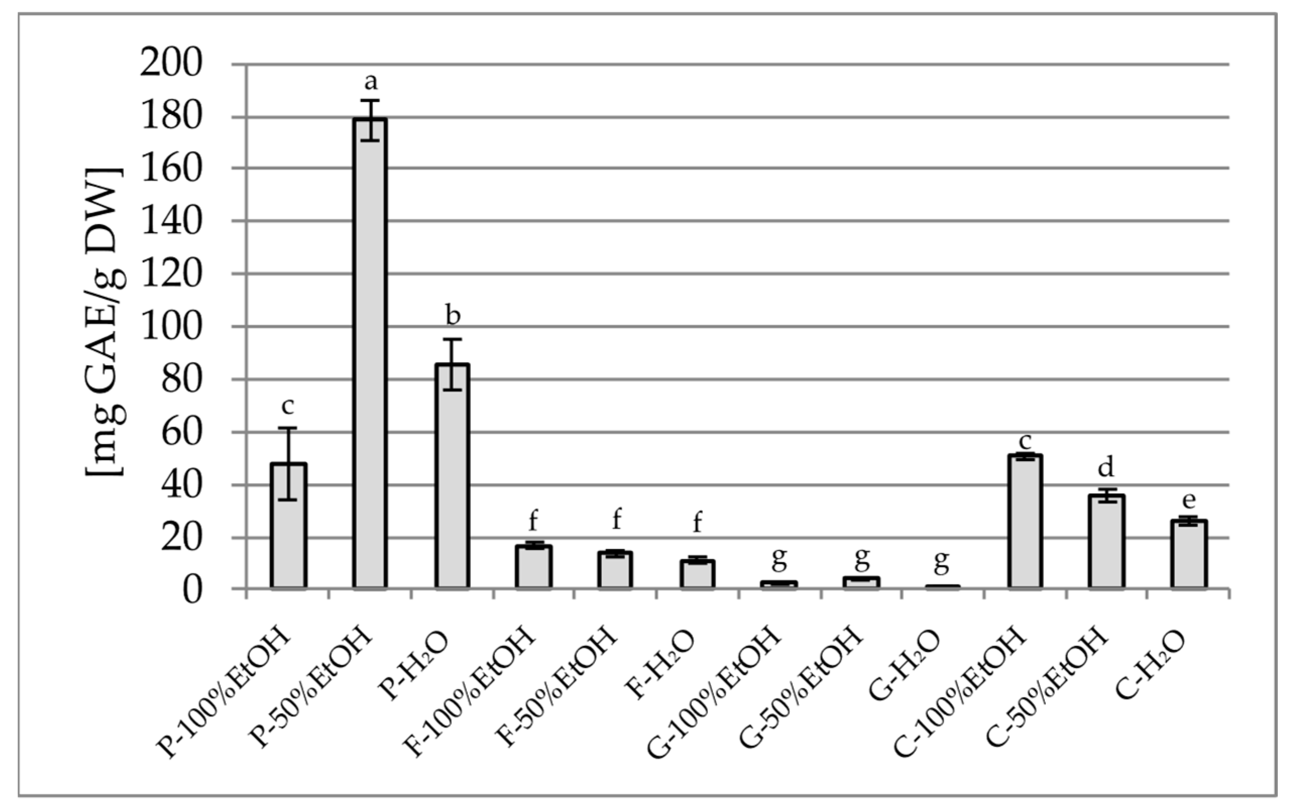
Figure 1. Total polyphenol content in the tested samples, expressed as mg gallic acid equivalent per gram of dry weight [mg GAE/g DW]. P—Paeonia officinalis, F—Forsythia × intermedia, G—Gomphrena globosa, C—Clitoria ternatea, H2O—aqueous extract, EtOH—ethanol extract. Values are presented as mean ± SD. Different letters above the bars indicate significant differences between groups (p < 0.05, one-way ANOVA followed by Newman–Keuls post hoc test).
Plants 14 02603 g001
Figure 2. Total flavonoid content in the tested samples, expressed as mg quercetin equivalent per gram of dry weight [mg QE/g DW]. P—Paeonia officinalis, F—Forsythia × intermedia, G—Gomphrena globosa, C—Clitoria ternatea, H2O—aqueous extract, EtOH—ethanol extract. Values are presented as mean ± SD. Different letters above the bars indicate significant differences between groups (p < 0.05, one-way ANOVA followed by Newman–Keuls post hoc test).
Plants 14 02603 g002
Figure 3. α-Glucosidase inhibitory potential of extracts (25 mg DW/mL). P—Paeonia officinalis, F—Forsythia × intermedia, G—Gomphrena globosa, C—Clitoria ternatea. H2O—aqueous extract, EtOH—ethanol extract. Values are presented as mean ± SD. Different letters above the bars indicate significant differences between groups (p < 0.05, one-way ANOVA followed by Newman–Keuls post hoc test).
Plants 14 02603 g003
Figure 4. α-Amylase inhibitory potential of extracts (25 mg DW/mL). P—Paeonia officinalis, F—Forsythia × intermedia, G—Gomphrena globosa, C—Clitoria ternatea. H2O—aqueous extract, EtOH—ethanol extract. Values are presented as mean ± SD. Different letters above the bars indicate significant differences between groups (p < 0.05, one-way ANOVA followed by Newman–Keuls post hoc test).
Plants 14 02603 g004
Figure 5. Hyaluronidase inhibitory potential of extracts (25 mg DW/mL). P—Paeonia officinalis, F—Forsythia × intermedia, G—Gomphrena globosa, C—Clitoria ternatea. H2O—aqueous extract, EtOH—ethanol extract. Values are presented as mean ± SD. Different letters above the bars indicate significant differences between groups (p < 0.05, one-way ANOVA followed by Newman–Keuls post hoc test).
Plants 14 02603 g005
Figure 6. The effect of flower 50% ethanol extracts on nitric oxide release in LPS-stimulated RAW 264.7 macrophages. P—Paeonia officinalis, F—Forsythia × intermedia, G—Gomphrena globosa, C—Clitoria ternatea. LPS—lipopolysaccharide, UNT—untreated cells, DEX—dexamethasone (reference drug). Different letters above the bars indicate significant differences between groups (p < 0.05, one-way ANOVA followed by Newman–Keuls post hoc test). Asterisks indicate statistically significant differences vs. LPS-treated cells (* p < 0.05; ** 0.01 ≤ p < 0.001; *** p < 0.001, one-way ANOVA followed by Dunnett post hoc test).
Plants 14 02603 g006
Figure 7. AChE inhibitory potential of extracts (25 mg DW/mL). P—Paeonia officinalis, F—Forsythia × intermedia, G—Gomphrena globosa, C—Clitoria ternatea. H2O—aqueous extract, EtOH—ethanol extract. Values are presented as mean ± SD. Different letters above the bars indicate significant differences between groups (p < 0.05, one-way ANOVA followed by Newman–Keuls post hoc test).
Plants 14 02603 g007
Figure 8. BChE inhibitory potential of extracts (25 mg DW/mL). P—Paeonia officinalis, F—Forsythia × intermedia, G—Gomphrena globosa, C—Clitoria ternatea. H2O—aqueous extract, EtOH—ethanol extract. Values are presented as mean ± SD. Different letters above the bars indicate significant differences between groups (p < 0.05, one-way ANOVA followed by Newman–Keuls post hoc test).
Plants 14 02603 g008
Table 1. The selected phenolic acids and flavonoid content in the extracts of four edible flower species (Peonia officinalis, Forsythia × intermedia, Gomphrena globosa, Clitoria ternatea) determined using HPLC.
ExtractCompound Detected Amount [mg/100 g DW]
Gallic AcidChlorogenic Acidp-Coumaric AcidFerulic AcidRutin
P-100% EtOH493.31 ± 18.43 bndndnd239.59 ± 29.66 c
P-50% EtOH390.27 ± 15.48 cndndnd167.65 ± 0.56 d
P-H2O603.67 ± 10.10 andndnd207.56 ± 0.41 c,d
F-100% EtOH1.42 ± 0.07 d319.79 ± 4.21 bnd6.37 ± 0.64 e2828.71 ± 41.11 b
F-50% EtOH19.87 ± 3.36 d796.03 ± 2.4 a1.11 ± 0.09 c,d8.80 ± 1.12 d4657.71 ± 66.37 a
F-H2O4.82 ± 0.54 d0.31 ± 0.28 c1.11 ± 0.38 c,d0.33 ± 0.30 h0.90 ± 0.31 f
G-100% EtOH0.08 ± 0.06 d2.83 ± 0.37 c0.88 ± 0.06 d4.53 ± 1.18 f34.90 ± 0.38 f
G-50% EtOH0.05 ± 0.07 d0.70 ± 0.47 c0.86 ± 0.09 d6.05 ± 1.1038.44 ± 0.37 f
G-H2O0.17 ± 0.10 d2.55 ± 0.19 c5.07 ± 0.10 a12.18 ± 0.23 c23.39 ± 0.27 f
C-100% EtOHnd0.17 ± 0.04 c0.06 ± 0.02 e3.17 ± 0.01 g11.69 ± 0.89 f
C-50% EtOHnd1.93 ± 0.83 c1.32 ± 0.08 c85.95 ± 1.02 a160.35 ± 0.99 d
C-H2Ond1.59 ± 0.16 c1.70 ± 0.35 b57.76 ± 0.69 b91.87 ± 4.47 e
nd—not detected. Values are presented as mean ± SD. Different letters indicate significant differences between groups (p < 0.05, one-way ANOVA followed by Newman–Keuls post hoc test).
Table 2. Compounds identified in the extracts of four edible flower species (Peonia officinalis, Forsythia × intermedia, Gomphrena globosa, Clitoria ternatea) using GC-MS method.
PlantRt (min)Quantity (%)Compound NameFormulaMain Characteristic m/z
Peonia officinalis15.751.53Propanoic acid, 2-(3-acetoxy-4,4,14-trimethylandrost-8-en-17-yl)-C27H42O473, 147, 207, 281, 355, 444
22.428.83Octadecane, 3-ethyl-5-(2-ethylbutyl)-C26H5457, 71, 85, 99, 141, 207
23.2367.62Oleic acid, 3-(octadecyloxy)propyl esterC39H76O357, 207, 281, 407, 462, 519
24.02247.31PentacosaneC25H5257, 71, 85, 99, 127, 141, 207, 225, 281, 322
25.55229.15TetratetracontaneC44H9057, 71, 85, 97, 207, 281, 326, 372., 423, 533
26.1915.56Spirost-8-en-11-one, 3-hydroxy-, (3ß,5a,14ß,20ß,22ß,25R)-C27H40O457, 69, 96, 207, 281, 355, 415
Forsythia × intermedia22.1881.4622-HexadecanolC16H34O55, 69, 97, 207
22.2920.349Oleic AcidC18H34O255, 69, 83, 97, 207, 237, 281
22.4053.931Hexadecane, 2,6,10,14-tetramethyl-C20H4257, 71, 85, 99, 113, 141, 207, 238, 266
23.0691.2099-OctadecenamideC18H35NO55, 59, 72, 83, 154, 207, 281
23.7242.102HeptacosaneC27H5657, 71, 85, 99, 127, 141, 183, 207, 239, 388
23.8241.17717-PentatriaconteneC35H7057, 69, 83, 97, 207, 281, 350, 379, 445
24.0173.915HentriacontaneC31H6457, 71, 85, 99, 127, 155, 183, 207, 239, 265, 295, 323, 352
25.5444.212TetratetracontaneC44H9057, 71, 85, 97, 133, 207, 281, 337, 383, 575, 624
27.070.370Ergosta-5,22-dien-3-ol, acetate, (3ß,22E)-C30H48O257, 67, 91, 133, 145, 207, 281, 341, 393
27.4861.602Octadecane, 3-ethyl-5-(2-ethylbutyl)-C26H5457, 71, 85, 99, 127, 207, 281
30.85879.670Dihydrofuran-2-one, 4-(3,4-dimethoxybenzyl)-3-(4-hydroxy-3-methoxybenzyl)-C21H24O677, 122, 137, 151, 177, 189, 207, 235, 265, 293, 321, 372
Gomphrena globosa21.8387.2083,10B-DihydrofluorantheneC16H12101, 202, 282
23.1143.6585,7,9(11)-Androstatriene, 3-hydroxy-17-oxo-C19H24O259, 77, 115, 133, 195, 207, 284
24.031.075Oleic acid, 3-(octadecyloxy)propyl esterC39H76O357, 133, 207, 281, 309, 403
25.5514.978HeptacosaneC27H5657, 71, 85, 99, 113, 207, 281, 341, 389
27.49241.740TetratetracontaneC44H9057, 71, 85, 97, 127, 207, 281, 377, 429
30.31530.131HexatriacontaneC36H7457, 71, 85, 99, 127, 155, 207, 281, 322, 391, 436
34.60911.210Tetracosane, 11-decyl-C34H7057, 71, 85, 99, 207, 281, 355, 402
Clitoria ternatea11.6120.917SpironolactoneC24H32O4S55, 73, 91, 147, 207, 251, 341, 429
13.8071.117Octadecane, 1,1′-[1,3-propanediylbis(oxy)]bis-C39H80O255, 73, 85, 97, 147, 207, 251, 281, 327, 415, 504
18.1374.95217-Octadecynoic acidC18H32O255, 68, 81, 95, 123, 207, 281
18.2542.6522-HexadecanolC16H34O55, 69, 79, 95, 123, 207, 237
18.3892.459Ethanol, 2-(9-octadecenyloxy)-, (Z)-C20H40O255, 69, 81, 96, 207, 250, 281, 325
18.5853.42413-Heptadecyn-1-olC17H32O55, 67, 81, 95, 123, 207, 278
25.5494.120HeptacosaneC27H5657, 71, 85, 99, 133, 207, 281, 355, 402
27.48914.287TetratetracontaneC44H9057, 71, 85, 97, 127, 207, 281, 306, 347, 391, 439, 481, 535
30.31149.099Octadecane, 3-ethyl-5-(2-ethylbutyl)-C26H5457, 71, 85, 97, 127, 155, 207, 281, 322, 355
34.60216.309HexatriacontaneC36H7457, 71, 85, 95, 207, 281, 341
36.6340.662Benz[e]azulene-3,8-dione, 3a,4,6a 7,9,10,10a,10b-octahydro-3a,10a-dihydroxy-5-(hydroxymethyl)-7-(1-hydroxy-1-methylethyl)-2,10-dimethyl-, [3aR-(3aa,6aa,7a,10ß,10aß,10bß)]-C20H28O653, 69, 73, 69, 133, 179, 207, 281, 314
Table 3. Antioxidant activity assays—DPPH, ABTS, Fe2+ chelation, CUPRAC, and FRAP.
DPPH [%]ABTS [%]Fe2+ chelation [%]CUPRAC [mg TE/g DW]FRAP [mg TE/g DW]
P-100% EtOH97.65 ± 0.60 a100.00 ± 0.99 and175.06 ± 1.04 a144.06 ± 4.56 a
P-50% EtOH90.42 ± 0.89 c99.51 ± 0.41 a73.78 ± 2.58 c151.44 ± 2.18 c140.29 ± 1.10 b
P-H2O91.01 ± 0.41 c99.78 ± 0.18 a83.36 ± 0.44 a162.04 ± 0.01 b137.52 ± 3.09 c
F-100% EtOH95.48 ± 0.27 b65.09 ± 1.02 bnd72.58 ± 0.87 e51.94 ± 0.21 e
F-50% EtOH89.28 ± 0.64 d99.10 ± 0.53 a52.76 ± 1.32 e109.30 ± 2.82 d81.30 ± 0.76 d
F-H2O28.44 ± 0.49 e43.26 ± 0.91 e77.38 ± 0.56 b24.24 ± 0.49 f20.36 ± 0.33 f
G-100% EtOH10.56 ± 0.93 i10.50 ± 0.32 ind6.32 ± 0.05 i5.36 ± 0.02 f
G-50% EtOH6.89 ± 0.84 j15.82 ± 0.90 h52.68 ± 4.27 e7.81 ± 0.31 h,i7.36 ± 0.07 g
G-H2O12.34 ± 0.37 h29.63 ± 0.94 f80.81 ± 0.90 a8.56 ± 0.49 h8.37 ± 0.04 g
C-100% EtOH15.87 ± 1.78 g20.30 ± 0.76 gnd8.90 ± 0.35 h5.83 ± 0.04 g
C-50% EtOH21.83 ± 0.77 f54.36 ± 1.60 c58.62 ± 6.27 d23.41 ± 0.40 f20.30 ± 0.24 f
C-H2O22.06 ± 0.72 f47.43 ± 1.71 d71.28 ± 3.24 c21.70 ± 0.12 g19.46 ± 0.17 f
P—Paeonia officinalis, F—Forsythia × intermedia, G—Gomphrena globosa, C—Clitoria ternatea. Values are presented as mean ± SD. Different letters indicate significant differences between groups (p < 0.05, one-way ANOVA followed by Newman–Keuls post hoc test).
Disclaimer/Publisher’s Note: The statements, opinions and data contained in all publications are solely those of the individual author(s) and contributor(s) and not of MDPI and/or the editor(s). MDPI and/or the editor(s) disclaim responsibility for any injury to people or property resulting from any ideas, methods, instructions or products referred to in the content.