Article

Root-Zone Temperature Drives Coordinated Photosynthesis, Root Architecture, and Metabolism Responses in Schisandra chinensis (Trucz.) Baill

1
College of Chemistry, Chemical Engineering and Resource Utilization, Northeast Forestry University, Harbin 150040, China
2
Key Laboratory of Forest Plant Ecology, Northeast Forestry University, Ministry of Education, Harbin 150040, China
3
School of Life Sciences, Heilongjiang University, Harbin 150040, China
*
Authors to whom correspondence should be addressed.
Plants 2025, 14(16), 2595; https://doi.org/10.3390/plants14162595
Submission received: 19 June 2025 / Revised: 21 July 2025 / Accepted: 18 August 2025 / Published: 20 August 2025
(This article belongs to the Section Plant Response to Abiotic Stress and Climate Change)

Abstract

Soil warming due to climate change has a significant potential impact on crop yield and quality. Schisandra chinensis (Trucz.) Baill, a multipurpose plant disseminated in the highly climate-sensitive region of Northeast Asia, is affected by soil warming, which limits the supply and quality of raw materials. This study investigated the differential responses of biomass accumulation and bioactive ingredient production across various organs to root-zone temperature (RZT) variations, employing both physiological assessments and metabolomic profiling. Elevated root temperatures may increase plant biomass and indirectly increase photosynthetic rates by promoting root growth; however, biomass responses differ among organs. A 20 °C root temperature promoted stem and leaf growth and inhibited root development, whereas a 30 °C root temperature significantly promoted root growth but reduced leaf biomass. Schisanhenol A, a key bioactive lignan serving as a quality marker for S. chinensis, displayed synthesis dependent on temperature. Concurrently, flavonoid biosynthesis is coordinated accumulation at the naringenin nodal point. A 15 °C RZT inhibited lignan production in roots while triggering stress-responsive phenol accumulation in leaves (41.39%). Conversely, at 20 °C and 30 °C RZTs, schisanhenol synthesis was repressed in leaves but accumulated in roots (9.8–25.71%). It is worth noting that the increase in RZT significantly promoted the synthesis and accumulation of schisandrol A in the aboveground part of the plant (43.88%). This research underscores that a suitable elevation in root-zone temperature can augment the medicinal attributes of the aerial components of S. chinensis.

1. Introduction

Schisandra chinensis (Turcz.) Baill. (S. chinensis) is a phytotherapeutic agent of significance in traditional Chinese medicine, as well as in the traditional pharmacopeias of Korea, Russia, and Japan. Phytochemical analyses confirm this species to be a rich source of bioactive compounds, including lignans, triterpenoids, phenolic acids, flavonoids, organic acids, vitamins, and polysaccharides [1]. Among the bioactive constituents, lignans are the characteristic bioactive compounds. Pharmacological investigations have demonstrated its broad-spectrum therapeutic efficacy, including having cardioprotective, hepatoprotective, antioxidant, hypoglycemic, antimicrobial, anti-inflammatory, and antitumor effects, as well as enhancing the ability to sleep, remember, and concentrate [2,3,4]. Phytochemical profiling also revealed lignan presence in both aglycone and glycoside configurations across all organs, including the stems, leaves, and roots of the plant [1]. Increasing demand for effective disease treatments, coupled with growing interest in cosmetics, functional foods, and diversified food sources, has driven greater utilization of the non-fruit plant parts (stems, leaves, and roots) of S. chinensis [5]. Current industrial utilization has expanded to include functional food engineering, particularly in the development of specialty products such as leaf teas, bioactive beverages, nutraceuticals, and phytochemical-enriched condiments [6,7]. This growing demand is further propelled by an expanding range of applications and a heightened focus on the sustainable sourcing of raw materials.
Lignans have been confirmed to be the most bioactive constituents of S. chinensis. Comprehensive investigation has led to the isolation of nearly 200 lignans from S. chinensis plants. Thorough analysis of the current literature identifies schisandrol A, schisandrol B, schisantherin B, schizandrin A, schizandrin B, schizandrin C, and schisantherin A as particularly significant among these lignans. Notably, schisandrol A has emerged as a pivotal marker compound for quality evaluation [8,9]. Lignans are widely distributed across plant tissues, exhibiting concentrations in vegetative organs (roots, stems, leaves) comparable to or exceeding those found in the fruits [10,11]. Given the limitations of fruit-derived lignans in meeting market demand, S. chinensis roots, stems, and leaves are emerging as promising alternative sources. The tissues under consideration are abundant in bioactive compounds, particularly lignans. This characteristic renders them ideal candidates for the development of functional foods and dietary supplements. Lignans are synthesized in a separate branch of the phenylpropanoid pathway [12,13,14]. The biosynthesis pathway of phenylpropane is initiated by phenylalanine metabolism, yielding lignin, flavonoids, and phenolic compounds as its byproducts. It has been determined that this pathway is of significant importance in the synthesis of secondary metabolites and the strength of plant resistance. The chemical composition and content of the compounds are subject to variation due to their growth characteristics and environmental factors, including temperature, light intensity, and precipitation. Therefore, further research into the environmental factors influencing the bioactive compound content is essential. This research is essential for enhancing the quality of locally produced S. chinensis for use as a functional raw material.
Climate change has increased the frequency of extreme high-temperature events, such as heat waves, adversely impacting plant development and agricultural productivity through exacerbated thermal stress [15,16]. S. chinensis inhabits northeastern China, the Russian Far East, and the Korean Peninsula, regions characterized by seasonally frozen soils and cold-adapted ecosystems. In contrast to variable aboveground air temperatures, the root-zone soil temperature displays significant thermal inertia, allowing aberrant states to persist more readily [17]. Roots are highly sensitive to their soil environment, and temperature significantly influences plant growth and development [18]. Root-zone temperature (RZT) is a pivotal soil factor governing root growth and function, significantly influencing plant growth, root physiology, and assimilate distribution [19,20]. Suboptimal RZTs impede shoot and root biomass accumulation, even when aerial tissues experience optimal temperatures [21]. Elevated temperatures can adversely affect key physiological processes, including photosynthesis, respiration, water balance, and membrane stability [22]. In response to root-zone temperature changes, plants enhance physiological performance by modulating secondary metabolism to maintain yield [23,24]. The production of plant secondary metabolites is closely related to the secretion of RZT. However, in contrast to extensive research on the temperature responses of shoots, the soil temperature response of roots is still relatively unexplored [20,24,25]. We conducted experiments partitioning the temperature between the aerial and root tissues to evaluate the effects of soil warming on S. chinensis growth, development, and the synthesis of lignans and flavonoids across different organs, including their metabolic coordination.

2. Results and Analysis

2.1. Effects on Plant Growth of Different RZT Treatments

Following 30-day exposure to varying root-zone temperatures, morphological and growth differences in S. chinensis plants were observed (Figure 1a). Compared to the controls (25 °C), plants exposed to a 15 °C root-zone temperature showed significant reductions in plant height, root length, and stem thickness. At a 20 °C RZT, S. chinensis showed the greatest increase in plant height, although both the basal diameter and the root length remained significantly lower than the control values. Plants subjected a 30 °C RZT (HT) exhibited significantly greater stem thickness and root length than plants in other temperature groups, whereas plant height was significantly lower than that in the 15 °C (LT) treatment. Root biomass under HT conditions was significantly greater than in other treatments (p < 0.05).
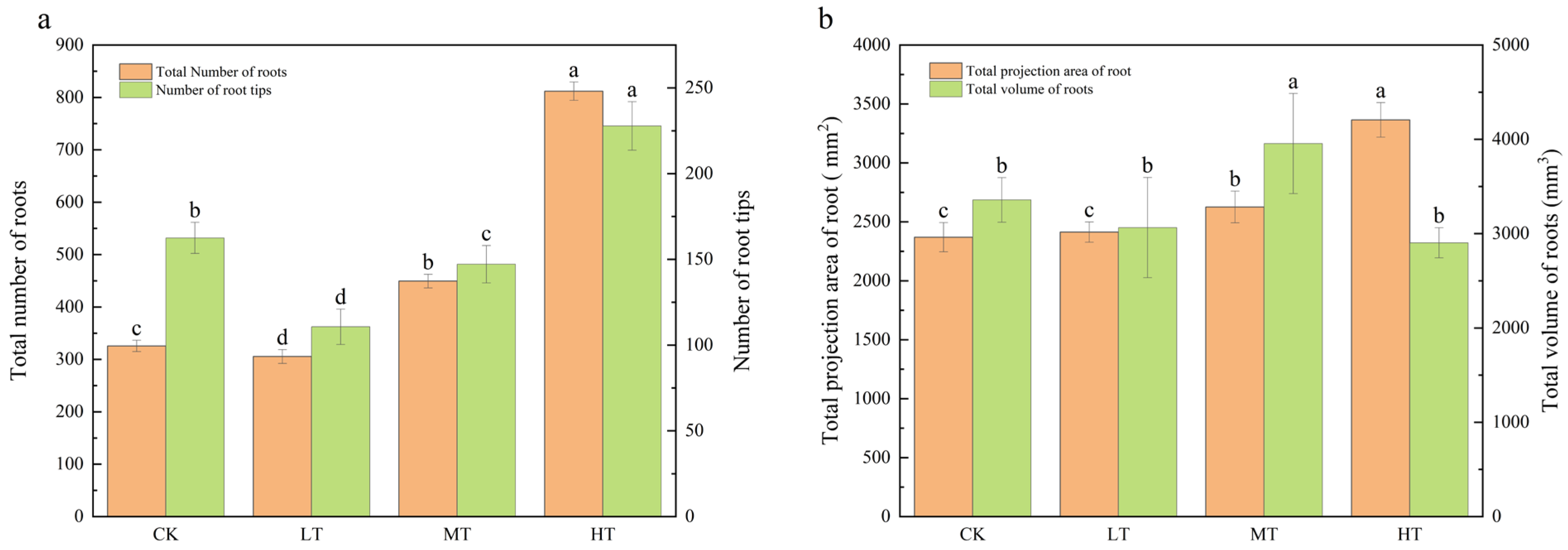
Relative to the control (25 °C), the number of root tips in S. chinensis exhibited temperature-dependent variation: a 32.31% reduction at 15 °C, a 9.45% reduction at 20 °C, and a 39.69% increase at 30 °C (Figure 2a). The root-zone temperature significantly influenced the total root count, showing 38.02% and 149.28% enhancements at 20 °C and 30 °C, respectively, relative to the controls. The 30 °C treatment induced a 42.01% greater root projection area compared to controls, while the 20 °C treatment increased root volume by 17.81% (Figure 2b). It was found that the low-temperature treatment (15 °C) inhibited the root development process, characterized by a significant reduction in total root number, projected area, volume, and root-tip density.

2.2. Response of Gas Exchange Under Different RZTs

The impact of root-zone temperature on the photosynthetic characteristics of S. chinensis seedlings was investigated. On day 30, Pn, Tr, and Gs values were significantly elevated relative to the control; however, Gs showed no significant difference between treatments and remained stable. In contrast, Ci exhibited distinct variation patterns compared to other photosynthetic parameters (Table 1). We also investigated the influence of the root-zone temperature on the levels of photosynthetic pigments (e.g., chlorophyll and carotenoids). Root-zone temperature treatments increased the content of photosynthetic pigments compared to the control. Chlorophyll a, chlorophyll b, and carotenoid contents were consistently higher than those in the control. At the same time, the photosynthetic pigment content gradually increased with increasing root-zone temperature. At 15 °C and 20 °C RZTs, the photosynthetic pigment content showed slight, non-significant elevations.

2.3. Response of Chlorophyll Fluorescence Level Under Different RZT Conditions

The chlorophyll fluorescence characteristics of S. chinensis varied significantly under different root-zone temperature conditions, with some treatments exhibiting significant changes. Low root-zone temperatures resulted in photoinhibition of PSII in S. chinensis leaves. Specifically, the Fv/Fm of PSII in S. chinensis leaves decreased by 16.66% and 19.33% in the 15 °C and 20 °C low-temperature treatments, respectively, both values reaching statistical significance (p < 0.05; Figure 3). The photochemical quenching coefficient (qP) increased by 18% and 55.34% compared to CK at RZTs of 20 °C and 30 °C, respectively, while no significant change occurred at an RZT of 15 °C. Furthermore, compared to CK, the PSII quantum yield [Y(II)] of leaves increased by 28.85% and 78.76% at 20 °C and 30 °C, respectively (p < 0.05). However, a root-zone temperature of 15 °C did not cause significant changes in the leaf PSII quantum yield.

2.4. Response of Lignan Content Under Different RZT Conditions

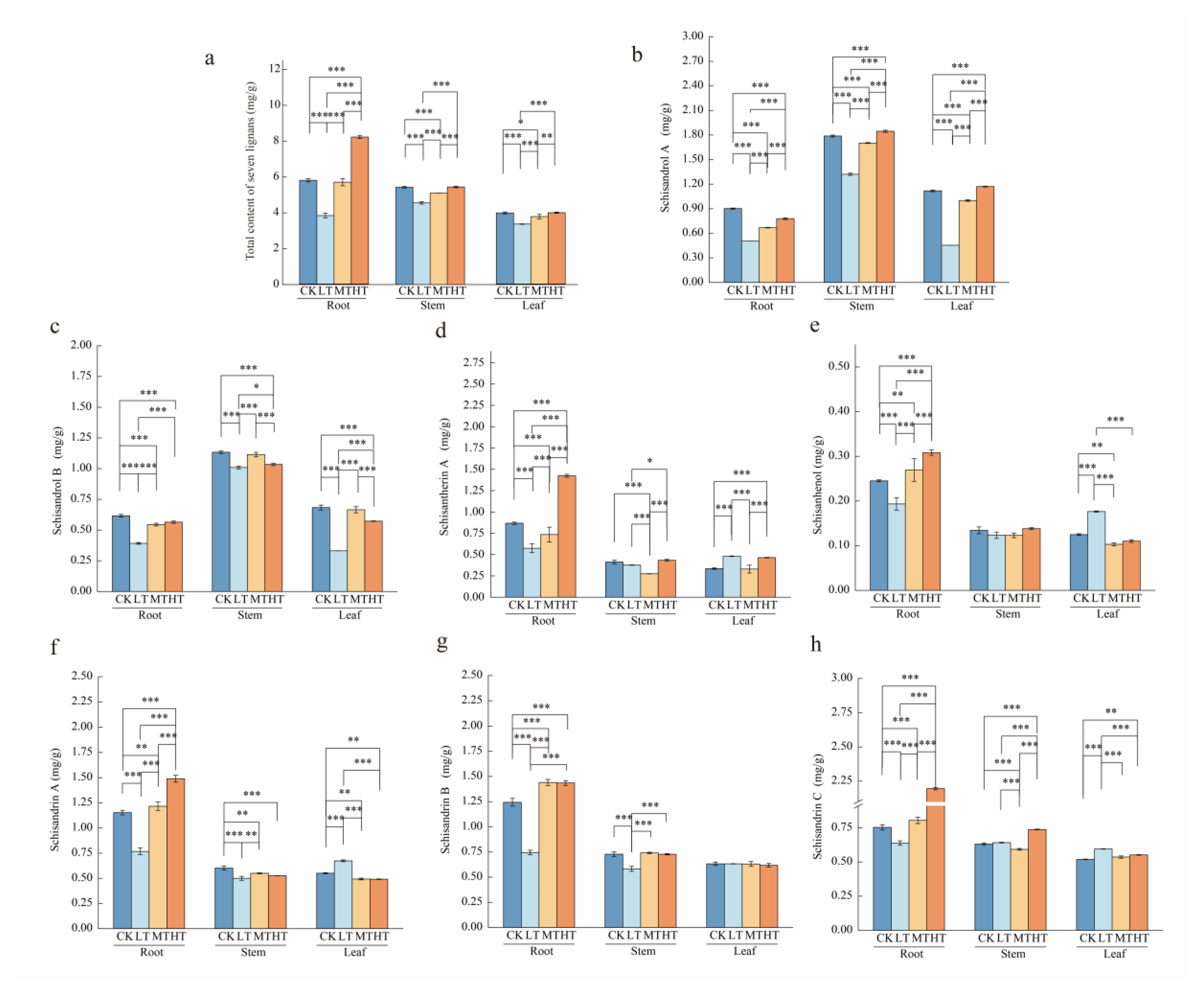
Seven major S. chinensis lignans were quantified by HPLC (Figure 4). Compared to the control (CK), the 15 °C RZT treatment significantly suppressed lignan accumulation in the roots, stems, and leaves, resulting in reduced lignan content across all organs. At a 20 °C RZT, lignan synthesis in the aboveground parts was inhibited, while the roots showed no significant changes. At a 30 °C RZT, the lignan content reached its peak in the roots. Although the lignan content increased in aboveground parts, this increase was not statistically significant compared to CK (Figure 5a). The content of specific lignans varied across plant components (Figure 5b–h). Specifically, compared to control roots, the levels of individual lignans decreased significantly at a 15 °C RZT. At a 20 °C RZT, schisandrol A, schisandrol B, and schisantherin A showed significantly lower levels. Conversely, the root levels of all individual lignans, except schisandrol A and schisandrol B, increased significantly with rising RZT. Compared to the stems in the control group, at a 15 °C RZT, the synthesis of individual lignans was inhibited. At a 30 °C RZT, the schisandrin C content peaked in roots, increasing by 16.94% compared to CK. In leaves, schisantherin A, schisanhenol, schisandrin A, and schisandrin C levels increased by 41.16%, 41.39%, 22.37%, and 14.65%, respectively, compared to the control. In contrast, when the RZT was 15 °C, schisandrol A and schisandrol B decreased by 59.44% and 51.55%.

2.5. PLS-DA Analysis of Secondary Metabolites

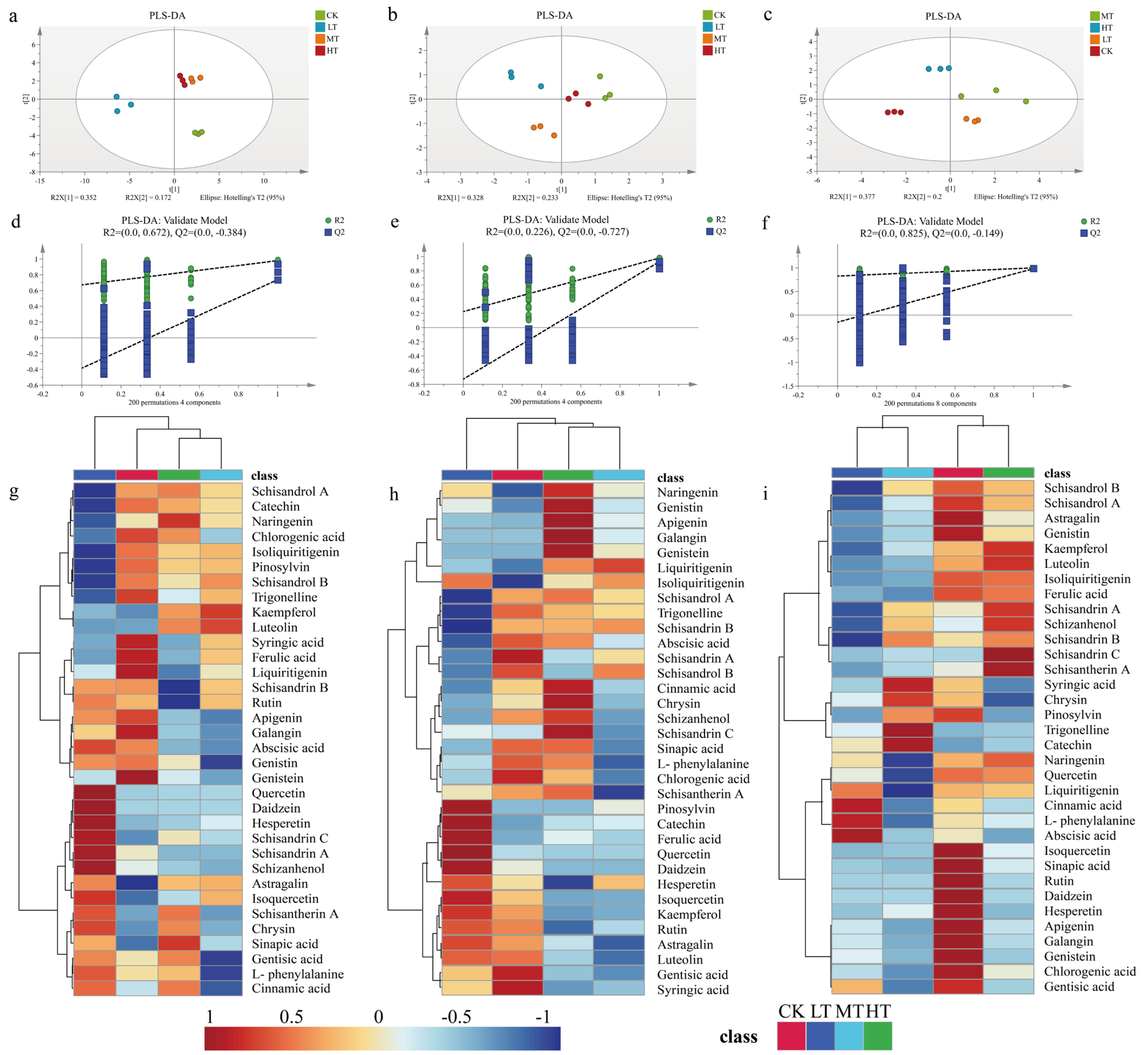
To investigate the impact of RZT variations on secondary metabolite accumulation in S. chinensis, we profiled metabolites using partial least squares discriminant analysis (PLS-DA) and hierarchical clustering heatmaps. The multivariate analysis revealed significant changes in metabolite profiles under different root-zone temperature regimes, with temperature-dependent accumulation patterns observed across experimental groups (Figure 6). Principal component analysis (PCA) of roots showed clear separation among treatments along the first two principal axes, accounting for 37.7% and 20.0% of total variance, respectively (Figure 6a). PLS-DA models for stems and leaves explained 52.4% and 56.1% of the variance, respectively, between the CK and temperature-treated groups (Figure 6b,c). Model validation through 200 permutation tests confirmed statistical robustness, with positive regression line slopes and Q2 intercepts < 0.05, eliminating concerns regarding overfitting or underfitting (Figure 6e–g). These validated models were then used to screen differential metabolites.

2.6. Screening of Phenolic Differentially Expressed Metabolites (DEMs)

Metabolic network structural analysis of phenolic DEMs was performed using fold change (FC) and variable importance in projection (VIP) criteria. DEMs were defined as metabolites meeting |log2FC| ≥ 1 and VIP ≥ 0.9 thresholds, with differential expression patterns visualized through volcano plots (Figure 7). Our analysis revealed temperature-dependent modulation of secondary metabolite biosynthesis pathways. Likewise, we found 15 DEMs (two up- and one down-regulated for leaves, one up- and two down-regulated for stems, and seven down-regulated for roots) for the CK vs. MT treatments (Figure 7b). For the CK vs. HT group, we observed 19 DEMs (2 up-regulated and 4 down-regulated in leaves; 3 up-regulated and 3 down-regulated in stems; and 7 down-regulated in roots) (Figure 7c).

2.7. Structural Analysis of Secondary Metabolism Network

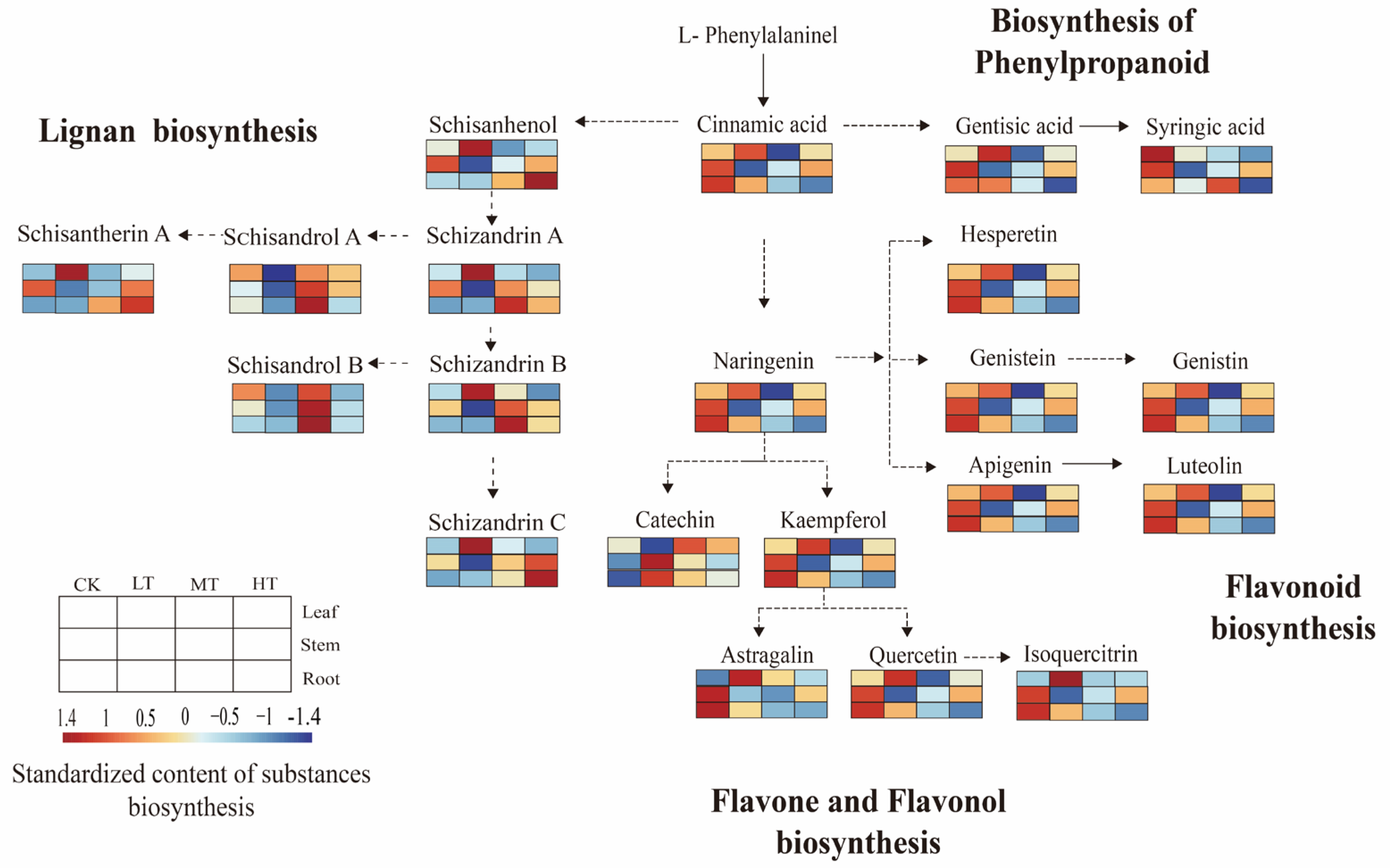
To investigate changes in metabolic processes in S. chinensis tissues under different RZTs, we mapped the identified differentially expressed metabolites (DEMs) and seven lignans using KEGG pathway analysis. The secondary metabolic network focused on four pathways: phenylpropanoid biosynthesis, flavonoid biosynthesis, flavonol biosynthesis, and lignan biosynthesis (Figure 8).
The results demonstrated significant temperature-dependent accumulation patterns. At a 15 °C RZT, cinnamic acid and most flavonoids showed elevated accumulation in leaves compared to CK, with synthesis inhibition observed at 20 °C and 30 °C RZTs. Schisanhenol accumulated preferentially in leaves at a 15 °C RZT, but showed suppressed synthesis in stems and roots. Root-specific lignan synthesis was enhanced at a 20 °C RZT. Schisandrol A, a key quality indicator, accumulated to high levels in the roots at a 30 °C RZT, whereas synthesis was inhibited when the RZT was 15 °C. These differential accumulation patterns may contribute to S. chinensis’ physiological adaptation to RZT variations.

3. Discussion

The root-zone temperature significantly influences plant development and physiological processes through its modulation of root growth and initiation [26,27]. Our results showed that S. chinensis plants exhibited significant growth inhibition when the RZT was 15 °C. Low-RZT conditions suppressed root developmental patterns and morphological architecture by altering the root growth rate, surface area expansion, cell elongation, and lateral root formation, ultimately reducing the overall growth rate and significantly decreasing organ biomass [28,29,30]. Notably, strawberry (Fragaria × ananassa (Weston) Duchesne ex Rozier) plants exposed to a 5 °C RZT displayed impaired growth and compromised fruit quality [31]. At a 20 °C RZT, S. chinensis showed enhanced shoot growth and increased leaf biomass, concomitant with root growth suppression. These findings align with previous reports demonstrating strong interactive effects between the RZT and cucumber (Cucumis sativus L.) growth parameters, including dry weight allocation, root elongation patterns, and leaf area expansion [32,33]. At an RZT of 30 °C, the root growth of S. chinensis was enhanced, while leaf biomass decreased. Xia et al. [34] demonstrated that an RZT of 30 °C significantly enhanced maize (Zea mays L.) growth relative to 24 °C (CK) and 36 °C (HT) conditions, with marked increases in plant height, stem diameter, root length, and dry matter accumulation. Compared to the C3 plant S. chinensis, maize, a C4 species, exhibits markedly divergent physiological responses to RZT. This discrepancy can be attributed to the inherent differences in their photosynthetic mechanisms, resource allocation strategies, and evolutionary adaptability [35]. The aboveground and underground parts of plants may exhibit differential responses to temperature that influence growth and resource allocation [36,37]. Changes in the root-zone temperature affect hormones like auxin and abscisic acid by impacting how the hormones are made and balanced in the root zone, which can lead to growth problems in different plant parts [38,39]. Research has demonstrated the impact of root zone temperatures on the transport of hormones from roots to aboveground parts [40]. The synthesis and upward transport of CTK and GA in the root are generally reduced at low root-zone temperatures, while the synthesis and transport of ABA are increased [41]. This differential response could account for the limited growth of certain vegetative organs of S. chinensis at both the 20 °C and 30 °C RZTs.
In addition to affecting plant growth, RZT also affects the production and enrichment of secondary metabolites in medicinal plants, thereby altering their quality characteristics [42,43]. The biosynthesis of rosmarinic acid and salvianolic acid B in Salvia miltiorrhiza Bunge is stimulated by specific environmental stresses, particularly low temperature [44]. Schisanhenol is one of the initial compounds synthesized in the lignan pathway, and schisandrol A serves as the standard index for the quality evaluation of S. chinensis. It is a representative dibenzocyclooctadiene lignan with significant medicinal value [8,45]. Schisanhenol synthesis was inhibited in the roots and the molecule accumulated in leaves at a 15 °C RZT. At 20 °C and 30 °C RZTs, accumulation occurred in the roots, while leaf synthesis was inhibited. Notably, the marker compound for quality (schisandrol A) accumulated in leaves and stems at a 30 °C RZT and its synthesis was inhibited at 15 °C RZT. High-temperature stress activates the biosynthesis of phenylpropanoids in plants, resulting in the accumulation of secondary metabolites. Compounds such as lignans play an important role in protecting cells from DNA damage [23,46]. Furthermore, elevated temperatures have been shown to facilitate the translocation of secondary metabolites from roots to shoots by means of activating the ABA (abscisic acid) signaling pathway, thereby enhancing antioxidant defense mechanisms [47]. However, the biological synthesis of dibenzocyclooctadiene lignans is still incompletely understood [12,48]. Whether there is reciprocal transformation between individual lignan compounds at different root-zone temperatures requires further research.
As a further product of the phenylpropanoid pathway, flavonoids have been shown to regulate auxin transport, promote pollen germination, and act as antioxidants [12,14]. In this study, the flavonoid content showed tissue specificity. Naringenin, which is associated with the biosynthesis of flavonoids and flavonols, showed higher enrichment in leaves at a 15 °C RZT, and its synthesis was inhibited at 20 °C and 30 °C RZTs. In previous studies, the content of naringenin in Pinellia ternata (Thunb.) Breit. was greatly increased under cold stress, and the accumulation of naringin was decreased under high-temperature stress [49]. Compared to the control, a 15 °C RZT upregulated the biosynthesis of cinnamic acid and lignans in leaves, resulting in their increased accumulation. Concurrently, flavonoid production via the naringenin node was significantly enhanced, leading to an increased flavonoid content. We hypothesize that the co-accumulation of lignans and flavonols substantially contributes to the defense and resistance of plants to stress under different RZTs [7,50,51,52].
The present study used a novel approach of conducting differential temperature control experiments on the aerial and subterranean parts of the plant. In this study, we investigated how the soil temperature affects the development of various vegetative organs of S. chinensis. The results revealed a suitable elevation in root-zone temperature was advantageous for enhancing the effective components of S. chinensis.

4. Materials and Methods

4.1. Plant Material and Root Zone Temperature (RZT) Treatments

S. chinensis seeds were obtained from Benxi, Liaoning Province, China. Following harvest, they were immersed in clean water, massaged to remove the pulp, and allowed to dry in the shade. The seeds were stored at 0–4 °C for 30 days after soaking for 24 h. They were then combined with wet dirt in a volume ratio of 1:3. Germination took place in climate-controlled chambers, 60% relative humidity, and temperatures of 25/15 °C. In the four-leaf stage, plants with consistent growth were divided into four groups and moved to the greenhouse. The aboveground temperature of seedlings was maintained at a constant 25 °C.
The administration of root-zone temperature treatments was conducted using a refrigerated circulating water bath system, maintaining constant temperatures of 15 °C (LT), 20 °C (MT), and 30 °C (HT). Control plants (CK) were maintained under ambient root zone temperatures equivalent to the shoot environment conditions. On day 30 after treatment initiation, plants were destructively sampled for comprehensive growth analysis. Root systems were carefully excised and subjected to two sequential washes in distilled water to remove adhering substrate particles. Cleaned roots were scanned using a high-resolution root imaging system, and architectural parameters were analyzed using LA-S root analysis software (Wan Sheng Hangzhou).

4.2. Determination of Chlorophyll Content and Photosynthetic Parameters

After 30 days of exposure, the third totally enlarged leaf was picked randomly from the top of each group, and the parameters related to photosynthesis in S. chinensis seedlings were determined using a portable photosynthetic assay system (Li-6400, LI-COR Inc., Lincoln, NE, USA). The estimation of the maximum quantum yield (Fv/Fm), the actual quantum yield [Y(II)], and the photosynthetic electron transport rate (ETR) of the leaves was accomplished by means of a portable modulated chlorophyll fluorescence meter (PAM-2500, Walz, Effeltrich, Germany).
In order to extract pigments, three plant leaves (100 mg, WM) were soaked in 5 mL dimethyl sulfoxide at 60 °C for 2 h. The degree of light absorption by the supernatant at 480, 649, and 665 nm was determined using a spectrophotometer (723PC, Shanghai Jinghua Technology Co., Ltd., Shanghai, China). Absorbance was calculated using Wang et al.’s [53] formula.

4.3. Analysis of Secondary Metabolites

4.3.1. Determination of Lignan Content

The lignans of S. chinensis were extracted using the method of Mocan et al. [54], with a slight modification. The 100 mg (dry basis) sample was extracted with 5 mL of high-performance liquid chromatography-grade methanol for 45 minutes, and the supernatant was dried.
Each sample was then redissolved in 1 mL methanol and filtered through a 0.22 µm nylon membrane. The filtrated extracts were then analyzed by HPLC. The analysis was performed using a Thermo Vanquish Core liquid phase system with an Agilent TC-C18 column (4.6 × 250 mm, 5 µm, Agilent Inc., Santa Clara, CA, USA). According to Chang et al. [55], the HPLC analysis used deionized water (A) and acetonitrile (B) as mobile phases. The method for gradient elution was executed as follows: The initial 0–25 min of the process was conducted at a flow rate of 55–75% B. This was followed by a 25–30 min period during which the flow rate was adjusted to 75–75% B. The subsequent 30–35 min involved a transition to 75–55% B, with a constant flow rate of 1 mL/min. The system temperature was maintained at 30 °C, and the injection volume was set to 20 μL. The wavelength of the detectors was set at 254 nm, and the total duration of the procedure was 35 min. Standard solutions with varying concentrations were prepared for the quantification of lignans. We used standard calibration curves of seven lignan standards (≥99%, Chengdu Must Biotechnology Co., Ltd., Chengdu, China) to compute the results.

4.3.2. LC-MS Analysis of Phenolic Compounds

Metabolic products were isolated from samples of roots, stems, and leaves using LC-MS as reported by Liu et al. [56]. Using an electronic balance, weigh the biological sample (DM), add 3 mL of 70% methanol, and sonicate (100 kHz) at 40 °C for 45 min; then, mix and evaporate using a rotary evaporation apparatus twice. For UPLC-MS/MS analysis, all samples were diluted in 1 mL with 70% methanol, sandwiched between a 0.22 µm screen, and placed in an injection bottle.
The separation system consisted of an HPLC equipped with an LC-20 AD pump, temperature control, a column temperature box, an SIL 20 A autosampler (Watts, Shanghai, China), and a BEHC 18 column (1.7 um, 2.1 mm × 50 mm) equipped with the same type of precolumn. The experiment kept the column at 30 °C. We used a mixture of 0.05% acetic acid and water (A) and 0.05% acetic acid and acetonitrile (B) as the solvent system. The flow rate was 0.25 mL/min. After 23 min, there was 5% B and 95% B, and after 25 min, there was 95% B and 5% B. The mass spectrometer operates in positive electron mode in the range 50 to 1000 m/z.

4.4. Statistical Analysis

The data were analyzed using SPSS Statistics (IBM SPSS Statistics, v. 25.0). To analyze the differences in plant growth and phenolic compounds at different root-zone temperatures, one-way analysis of variance (ANOVA) and least significant difference (LSD) tests were used. We used SIMCA 14.1 (Umetrics, Umeö, Sweden) for multivariate statistical analysis, including partial least squares discriminant analysis (PLS-DA), to compare secondary metabolites in the CK, LT, MT, and HT treatments. To screen for differentially expressed metabolites (DEMs), variable importance in prediction (VIP) > 0.9 and |log2 fold change (FC)| ≥ 1 were used. Pathways were constructed from the Kyoto Encyclopedia of Genes and Genomes (KEGG) pathway database.

5. Conclusions

This study investigated the response of different organs of S. chinensis to various root-zone temperatures (RZTs). The results indicated that low-temperature treatment (a 15 °C RZT) significantly inhibited plant growth. The 20 °C RZT regime promoted shoot growth in S. chinensis, whereas the 30 °C RZT regime stimulated root growth. The physiological pathway of photosynthesis was affected by different RZT treatments (15 °C, 20 °C, and 30 °C) due to their regulation of root growth and development, resulting in changes in chlorophyll fluorescence parameters. The regulation of different RZT levels on the lignan content in the roots, stems, and leaves of S. chinensis demonstrated notable organ specificity. A 15 °C RZT inhibited the synthesis of S. chinensis lignans. At a 20 °C RZT, lignan synthesis in the aboveground parts was inhibited, with no significant changes observed in the roots. Conversely, at 30 °C, the lignan content was highest in the roots. Although there was an increase in the aboveground parts, the difference compared to the control group was not significant. Notably, the 30 °C RZT treatment significantly promoted the synthesis of Schisandrol A, increasing its accumulation by 43.88% in aboveground parts. At the metabolic level, the dynamic changes in the content of certain phenolic compounds, such as cinnamic acid and naringenin, indicate that lignans and flavonols co-accumulate under varying root-zone temperatures to protect plants from stress. Studies have demonstrated that a moderate increase in RZT is beneficial for enhancing the medicinal value of the aboveground parts of S. chinensis.

Author Contributions

H.T.: project management, supervision, validation, original drafting, review and editing. X.S. and L.J.: methodology and supervision. W.Z., J.Z., L.Z. and Q.Y.: investigation. X.G. and Y.S.: methodology and supervision. Z.Z.: writing, reviewing, and editing. Z.T. and C.Z.: resource procurement, guidance, and financial support. All authors have read and agreed to the published version of the manuscript.

Funding

This work received financial support from three distinct funding bodies: the Heilongjiang Province Seed Industry Innovation and Development Funding Program (ZQTYB231700002), the Heilongjiang Province Seed Industry Innovation and Development Funding Program (20230202), and the Key R&D Project of Heilongjiang Province (JD22A008).

Data Availability Statement

The original contributions presented in this study are included in the article. Further inquiries can be directed to the corresponding author(s).

Conflicts of Interest

The authors have no known competing financial interests or personal relationships that could have appeared to influence the work reported in this paper.

References

  1. Szopa, A.; Ekiert, R.; Ekiert, H. Current knowledge of Schisandra chinensis (Turcz.) Baill. (Chinese magnolia vine) as a medicinal plant species: A review on the bioactive components, pharmacological properties, analytical and biotechnological studies. Phytochem. Rev. 2016, 16, 195–218. [Google Scholar] [CrossRef] [PubMed]
  2. Yang, S.; Yuan, C. Schisandra chinensis: A comprehensive review on its phytochemicals and biological activities. Arab. J. Chem. 2021, 14, 103310. [Google Scholar] [CrossRef]
  3. Fu, K.; Zhou, H.; Wang, C.; Gong, L.; Ma, C.; Zhang, Y.; Li, Y. A review: Pharmacology and pharmacokinetics of Schisandrin A. Phytother. Res. 2022, 36, 2375–2393. [Google Scholar] [CrossRef]
  4. Yang, K.; Qiu, J.; Huang, Z.; Yu, Z.; Wang, W.; Hu, H.; You, Y. A comprehensive review of ethnopharmacology, phytochemistry, pharmacology, and pharmacokinetics of Schisandra chinensis (Turcz.) Baill. and Schisandra sphenanthera Rehd. et Wils. J. Ethnopharmacol. 2022, 284, 114759. [Google Scholar] [CrossRef]
  5. Liu, G.; Liu, Y.; Sun, Y.; Li, X.; Xu, Z.; Jiang, P.; Rong, X.; Yang, B.; Kuang, H. Lignans and Terpenoids from the Leaves of Schisandra chinensis. Chem. Biodivers. 2020, 17, e2000035. [Google Scholar] [CrossRef]
  6. Kopustinskiene, D.M.; Bernatoniene, J. Antioxidant Effects of Schisandra chinensis Fruits and Their Active Constituents. Antioxidants 2021, 10, 620. [Google Scholar] [CrossRef]
  7. Li, X.; Liang, J.; Zhang, D.-Y.; Kuang, H.-X.; Xia, Y.-G. Low-polymerization compositional fingerprinting for characterization of Schisandra polysaccharides by hydrophilic interaction liquid chromatography-electrospray mass spectrometry. Int. J. Biol. Macromol. 2021, 185, 983–996. [Google Scholar] [CrossRef]
  8. National Pharmacopoeia Committee. Pharmacopoeia of the People ‘s Republic of China: Volume I.; Pharmaceutical Science and Technology Press: Beijing, China, 2020; pp. 13–14. [Google Scholar]
  9. He, D.; Pei, X.; Liu, B.; Li, J.; Dong, J.; Efferth, T.; Ma, P. Lignan contents of Schisandra chinensis (Turcz.) Baill. from different origins—A new model for evaluating the content of prominent components of Chinese herbs. Phytomedicine 2024, 128, 155361. [Google Scholar] [CrossRef]
  10. Wu, Z.; Jia, M.; Zhao, W.; Huang, X.; Yang, X.; Chen, D.; Qiaolongbatu, X.; Li, X.; Wu, J.; Qian, F.; et al. Schisandrol A, the main active ingredient of Schisandrae Chinensis Fructus, inhibits pulmonary fibrosis through suppression of the TGF-β signaling pathway as revealed by UPLC-Q-TOF/MS, network pharmacology and experimental verification. J. Ethnopharmacol. 2022, 289, 115031. [Google Scholar] [CrossRef] [PubMed]
  11. Liu, J.; Mu, X.; Liang, J.; Zhang, J.; Qiang, T.; Li, H.; Li, B.; Liu, H.; Zhang, B. Metabolic profiling on the analysis of different parts of Schisandra chinensis based on UPLC-QTOF-MS with comparative bioactivity assays. Front. Plant Sci. 2022, 13, 970535. [Google Scholar] [CrossRef] [PubMed]
  12. Chen, C.-Y.; Liu, S.-Y.; Yan, Y.; Yin, L.; DI, P.; Liu, H.-M.; Liu, H.-Z. Candidate genes involved in the biosynthesis of lignan in Schisandra chinensis fruit based on transcriptome and metabolomes analysis. Chin. J. Nat. Med. 2020, 18, 684–695. [Google Scholar] [CrossRef]
  13. Ford, J.D.; Huang, K.-S.; Wang, H.-B.; Davin, L.B.; Lewis, N.G. Biosynthetic pathway to the cancer chemopreventive secoisolariciresinol diglucoside−hydroxymethyl glutaryl ester-linked lignan oligomers in flax (Linum usitatissimum) Seed. J. Nat. Prod. 2001, 64, 1388–1397. [Google Scholar] [CrossRef]
  14. Suzuki, S.; Umezawa, T. Biosynthesis of lignans and norlignans. J. Wood Sci. 2007, 53, 273–284. [Google Scholar] [CrossRef]
  15. Gray, S.B.; Brady, S.M. Plant developmental responses to climate change. Dev. Biol. 2016, 419, 64–77. [Google Scholar] [CrossRef] [PubMed]
  16. Miller, S.; Chua, K.; Coggins, J.; Mohtadi, H. Heat Waves, Climate Change, and Economic Output. J. Eur. Econ. Assoc. 2021, 19, 2658–2694. [Google Scholar] [CrossRef]
  17. Su, H.; Li, L.; Ma, H.; Lyu, D.; Sun, J. Calcium Alleviates Temperature Stress by Regulating Nitrogen and Respiratory Metabolism in Malus baccata Roots. Int. J. Agric. Biol. 2016, 18, 286–292. [Google Scholar] [CrossRef]
  18. Guo, E.; Liu, X.; Zhang, J.; Wang, Y.; Wang, C.; Wang, R.; Li, D. Assessing spatiotemporal variation of drought and its impact on maize yield in Northeast China. J. Hydrol. 2017, 553, 231–247. [Google Scholar] [CrossRef]
  19. Ambebe, T.F.; Dang, Q.-L.; Li, J. Low soil temperature inhibits the effect of high nutrient supply on photosynthetic response to elevated carbon dioxide concentration in white birch seedlings. Tree Physiol. 2009, 30, 234–243. [Google Scholar] [CrossRef]
  20. Lee, S.; Wang, W.; Huq, E. Spatial regulation of thermomorphogenesis by HY5 and PIF4 in Arabidopsis. Nat. Commun. 2021, 12, 3656. [Google Scholar] [CrossRef]
  21. Bai, L.; Deng, H.; Zhang, X.; Yu, X.; Li, Y.; Cheng, Z. Gibberellin Is Involved in Inhibition of Cucumber Growth and Nitrogen Uptake at Suboptimal Root-Zone Temperatures. PLoS ONE 2016, 11, e0156188. [Google Scholar] [CrossRef]
  22. Wahid, A.; Close, T.J. Expression of dehydrins under heat stress and their relationship with water relations of sugarcane leaves. Biol. Plant. 2007, 51, 104–109. [Google Scholar] [CrossRef]
  23. Sharma, A.; Shahzad, B.; Rehman, A.; Bhardwaj, R.; Landi, M.; Zheng, B. Response of Phenylpropanoid Pathway and the Role of Polyphenols in Plants under Abiotic Stress. Molecules 2019, 24, 2452. [Google Scholar] [CrossRef]
  24. Gaillochet, C.; Burko, Y.; Platre, M.P.; Zhang, L.; Simura, J.; Willige, B.C.; Kumar, S.V.; Ljung, K.; Chory, J.; Busch, W. HY5 and phytochrome activity modulate shoot-to-root coordination during thermomorphogenesis in Arabidopsis. Development 2020, 147, dev192625. [Google Scholar] [CrossRef]
  25. Hatfield, J.L.; Prueger, J.H. Temperature extremes: Effect on plant growth and development. Weather Clim. Extrem. 2015, 10, 4–10. [Google Scholar] [CrossRef]
  26. Balliu, A.; Sallaku, G. The environment temperature affects post-germination growth and root system architecture of pea (Pisum sativum L.) plants. Sci. Hortic. 2021, 278, 109858. [Google Scholar] [CrossRef]
  27. Zhao, Y.; Zhang, Q.; Li, J.; Yan, X.; He, H.; Gao, X.; Jia, G. High temperature in the root zone repressed flowering in Lilium × formolongi by disturbing the photoperiodic pathway and reconfiguring hormones and primary metabolism. Environ. Exp. Bot. 2021, 192, 104644. [Google Scholar] [CrossRef]
  28. Chen, Y.; Lo, S.; Sun, P.; Lu, C.; Ho, T.D.; Yu, S. A late embryogenesis abundant protein HVA1 regulated by an inducible promoter enhances root growth and abiotic stress tolerance in rice without yield penalty. Plant Biotechnol. J. 2014, 13, 105–116. [Google Scholar] [CrossRef]
  29. Inoue, T.; Akaji, Y.; Noguchi, K. Distinct responses of growth and respiration to growth temperatures in two mangrove species. Ann. Bot. 2021, 129, 15–28. [Google Scholar] [CrossRef]
  30. Fanello, D.D.; Kelly, S.J.; Bartoli, C.G.; Cano, M.G.; Alonso, S.M.; Guiamet, J.J. Plasticity of root growth and respiratory activity: Root responses to above-ground senescence, fruit removal or partial root pruning in soybean. Plant Sci. 2020, 290, 110296. [Google Scholar] [CrossRef]
  31. Gonzalez-Fuentes, J.A.; Shackel, K.; Lieth, J.H.; Albornoz, F.; Benavides-Mendoza, A.; Evans, R.Y. Diurnal root zone temperature variations affect strawberry water relations, growth, and fruit quality. Sci. Hortic. 2016, 203, 169–177. [Google Scholar] [CrossRef]
  32. Yan, Q.; Duan, Z.; Mao, J.; Li, X.; Dong, F. Effects of root-zone temperature and N, P, and K supplies on nutrient uptake of cucumber (Cucumis sativus L.) seedlings in hydroponics. Soil Sci. Plant Nutr. 2012, 58, 707–717. [Google Scholar] [CrossRef]
  33. Yan, Q.-Y.; Duan, Z.-Q.; Mao, J.-D.; Li, X.; Dong, F. Low Root Zone Temperature Limits Nutrient Effects on Cucumber Seedling Growth and Induces Adversity Physiological Response. J. Integr. Agric. 2013, 12, 1450–1460. [Google Scholar] [CrossRef]
  34. Xia, Z.Q.; Si, L.Y.; Jin, Y.; Fu, Y.F.; Wang, Q.; Lu, H.D. Effects of Root Zone Temperature Increase on Physiological Indexes and Photosynthesis of Different Genotype Maize Seedlings. Russ. J. Plant Physiol. 2021, 68, 169–178. [Google Scholar] [CrossRef]
  35. Bai, L.; Han, G.; Jiang, L.; Crowther, T.W.; Zohner, C.M.; Yu, K.; Xu, Z.; Wang, Z.; Wu, Q.; Zhu, Y.; et al. Contrasting Responses of Flowering Phenology in C3 and C4 Plants Shape Grassland Community Structure under Global Change. Ecology 2025, 106, e70139. [Google Scholar] [CrossRef]
  36. Nagel, K.A.; Kastenholz, B.; Jahnke, S.; van Dusschoten, D.; Aach, T.; Mühlich, M.; Truhn, D.; Scharr, H.; Terjung, S.; Walter, A.; et al. Temperature responses of roots: Impact on growth, root system architecture and implications for phenotyping. Funct. Plant Biol. 2009, 36, 947–959. [Google Scholar] [CrossRef]
  37. González-García, M.P.; Conesa, C.M.; Lozano-Enguita, A.; Baca-González, V.; Simancas, B.; Navarro-Neila, S.; Sánchez-Bermúdez, M.; Salas-González, I.; Caro, E.; Castrillo, G.; et al. Temperature changes in the root ecosystem affect plant functionality. Plant Commun. 2022, 4, 100514. [Google Scholar] [CrossRef]
  38. Wang, J.; Jin, D.; Deng, Z.; Zheng, L.; Guo, P.; Ji, Y.; Song, Z.; Zeng, H.Y.; Kinoshita, T.; Liao, Z.; et al. The apoplastic pH is a key determinant in the hypocotyl growth response to auxin dosage and light. Nat. Plants 2025, 11, 279–294. [Google Scholar] [CrossRef]
  39. Zeng, H.Y.; Deng, S.; Jin, C.; Shang, Z.; Chang, L.; Wang, J.; Han, X.; Wang, A.; Jin, D.; Wang, Y.; et al. Origin and evolution of auxin-mediated acid growth. Proc. Natl. Acad. Sci. USA 2024, 121, e2412493121. [Google Scholar] [CrossRef]
  40. Wu, B.; Zuo, J.; Niu, H.; Jia, R.; Li, Q.; Chen, X.; Yang, Z.; Zhang, C.; Sun, F.; Xi, Y. Transcriptome and metabolome analyses reveal the potential mechanism of root zone temperature regulates the growth rhythm of switchgrass (Panicum virgatum L.) seedlings. Ind. Crop. Prod. 2025, 230, 121041. [Google Scholar] [CrossRef]
  41. Luo, Z.R. Relationship between plant hormones and cold resistance. Plant Physiol. Commun. 1989, 3, 1–5. [Google Scholar] [CrossRef]
  42. Kim, N.; Jayakodi, M.; Lee, S.; Choi, B.; Jang, W.; Lee, J.; Kim, H.H.; Waminal, N.E.; Lakshmanan, M.; van Nguyen, B.; et al. Genome and evolution of the shade-requiring medicinal herb Panax ginseng. Plant Biotechnol. J. 2018, 16, 1904–1917. [Google Scholar] [CrossRef]
  43. Yuan, Y.; Tang, X.; Jia, Z.; Li, C.; Ma, J.; Zhang, J. The Effects of Ecological Factors on the Main Medicinal Components of Dendrobium officinale under Different Cultivation Modes. Forests 2020, 11, 94. [Google Scholar] [CrossRef]
  44. Yu, H.; Liao, J.; Jiang, Y.; Zhong, M.; Tao, S.; Chai, S.; Wang, L.; Lin, L.; Yang, R.; Deng, X.; et al. Ecotype-specific phenolic acid accumulation and root softness in Salvia miltiorrhiza are driven by environmental and genetic factors. Plant Biotechnol. J. 2025, 23, 2224–2241. [Google Scholar] [CrossRef]
  45. Zhou, Y.; Men, L.; Sun, Y.; Wei, M.; Fan, X. Pharmacodynamic effects and molecular mechanisms of lignans from Schisandra chinensis Turcz. (Baill.), a current review. Eur. J. Pharmacol. 2021, 892, 173796. [Google Scholar] [CrossRef] [PubMed]
  46. Singh, P.; Singh, A.; Choudhary, K.K. Revisiting the role of phenylpropanoids in plant defense against UV-B stress. Plant Stress 2023, 7, 100143. [Google Scholar] [CrossRef]
  47. Soyk, S.; Lemmon, Z.H.; Sedlazeck, F.J.; Jiménez-Gómez, J.M.; Alonge, M.; Hutton, S.F.; Van Eck, J.; Schatz, M.C.; Lippman, Z.B. Duplication of a domestication locus neutralized a cryptic variant that caused a breeding barrier in tomato. Nat. Plants 2019, 5, 471–479. [Google Scholar] [CrossRef] [PubMed]
  48. Qiang, T.-Y.; Liu, J.-S.; Dong, Y.-Q.; Mu, X.-L.; Chen, Y.; Luo, H.-M.; Zhang, B.-G.; Liu, H.-T. Identification, Molecular Cloning, and Functional Characterization of a Coniferyl Alcohol Acyltransferase Involved in the Biosynthesis of Dibenzocyclooctadiene Lignans in Schisandra chinensis. Front. Plant Sci. 2022, 13, 881342. [Google Scholar] [CrossRef]
  49. Guo, L.; Tan, J.; Deng, X.; Mo, R.; Pan, Y.; Cao, Y.; Chen, D. Integrated analysis of metabolome and transcriptome reveals key candidate genes involved in flavonoid biosynthesis in Pinellia ternata under heat stress. J. Plant Res. 2023, 136, 359–369. [Google Scholar] [CrossRef]
  50. Singh, P.; Arif, Y.; Bajguz, A.; Hayat, S. The role of quercetin in plants. Plant Physiol. Biochem. 2021, 166, 10–19. [Google Scholar] [CrossRef]
  51. Austen, N.; Walker, H.J.; Lake, J.A.; Phoenix, G.K.; Cameron, D.D. The Regulation of Plant Secondary Metabolism in Response to Abiotic Stress: Interactions Between Heat Shock and Elevated CO2. Front. Plant Sci. 2019, 10, 1463. [Google Scholar] [CrossRef]
  52. Ri, I.; Pak, S.; Pak, U.; Yun, C.; Tang, Z. How does UV-B radiation influence the photosynthesis and secondary metabolism of Schisandra chinensis leaves? Ind. Crop. Prod. 2024, 208, 117832. [Google Scholar] [CrossRef]
  53. Wang, W.-J.; He, H.S.; Yu, G.; Wen-Xin, L.I.; Zhong-Hua, Z.; Yuan-Gang, Z.U. Methodological Comparison of Chlorophyll and Carotenoids Contents of Plant Species Measured by DMSO and Acetone-extraction Methods. Bull. Bot. Res. 2009, 29, 224–229. [Google Scholar]
  54. Mocan, A.; Schafberg, M.; Crișan, G.; Rohn, S. Determination of lignans and phenolic components of Schisandra chinensis (Turcz.) Baill. using HPLC-ESI-ToF-MS and HPLC-online TEAC: Contribution of individual components to overall antioxidant activity and comparison with traditional antioxidant assays. J. Funct. Foods 2016, 24, 579–594. [Google Scholar] [CrossRef]
  55. Shi, C.; Gao, C.; Guo, X.R.; Wang, H.Z.; Xu, M.Y.; Liu, Y.; Zhang, J.; Xing, K.X.; Tang, Z.H. Optimization of ultrasonic extraction process of lignans from Schisandra chinensis vine stem by analytic hierarchy process-entropy weight method combined with response surface methodology. J. Food Saf. Qual. 2023, 14, 293–302. [Google Scholar] [CrossRef]
  56. Liu, J.; Liu, Y.; Wang, Y.; Abozeid, A.; Zu, Y.-G.; Tang, Z.-H. The integration of GC–MS and LC–MS to assay the metabolomics profiling in Panax ginseng and Panax quinquefolius reveals a tissue- and species-specific connectivity of primary metabolites and ginsenosides accumulation. J. Pharm. Biomed. Anal. 2017, 135, 176–185. [Google Scholar] [CrossRef]
Figure 1. Responses of morphology (a), biomass (b), plant height (c), stem thickness (d), and root length (e) of S. chinensis to temperatures in the root zone. Values are mean and standard error of six repeated measurements. Small letters indicate significant differences (p < 0.05).
Plants 14 02595 g001
Figure 2. S. chinensis (a) total root number and root tip number and (b) root projection area and total root volume response to different root zone temperatures. Values represent the mean and standard error of six replicates. Different smaller letters indicate that there are significant differences in S. chinensis among the different root-zone temperature treatment groups (p < 0.05).
Plants 14 02595 g002
Figure 3. Chlorophyll fluorescence level changes in S. chinensis under different RZT conditions. Statistical analyses were performed using one-way ANOVA, p < 0.05, followed by the LSD test. Values are expressed as means ± SD (n = 5). Stars (* p < 0.05, ** p < 0.01, *** p < 0.001) above the connecting lines indicate significance levels between different RZT treatments.
Plants 14 02595 g003
Figure 4. Mixed standard (a) and sampled (b) by high-performance liquid chromatography (HPLC). (1) schisandrol A, (2) schisandrol B, (3) schisantherin A, (4) schisanhenol, (5) schisandrin A, (6) schisandrin B, (7) schisandrin C.
Plants 14 02595 g004
Figure 5. Root, stem, and leaf accumulation of seven lignans (bh), and total accumulation (a) under different root-zone temperatures for S. chinensis. The data are expressed as mean ± SD (n = 3). Significant differences between root-zone temperature treatments are indicated by *** (p < 0.001) and ** (p < 0.01), * p < 0.05 above the line.
Plants 14 02595 g005
Figure 6. Changes in the content of secondary metabolites in the roots (a,d,g), stems (b,e,h), and leaves (c,f,i) of S. chinensis after 30 d (n = 3) of treatment at different temperatures in the root zone.
Plants 14 02595 g006
Figure 7. Volcanogram of differential metabolites of S. chinensis after 30 days of exposure to 15°C (a), 20 °C (b) and 30 °C (c) RZT (n = 3).
Plants 14 02595 g007
Figure 8. Metabolic pathway network of secondary metabolites in S. chinensis root zones under temperature stress. Color gradients reflect normalized metabolite concentrations (red: high abundance; blue: low abundance). Dashed and solid lines denote indirect and direct metabolic conversions, respectively, with arrows indicating biochemical transformation directions.
Plants 14 02595 g008
Table 1. Photosynthetic performance and photosynthetic pigmentation of S. chinensis under different root zone temperature treatments.
Pn
(μmol m−2 s−1)
Gs
(μmol m−2 s−1)
Ci
(mmol m−2 s−1)
Tr
(mmol m−2 s−1)
Chla (mg/g)Chlb
(mg/g)
Car
(mg/g)
Chla + chlb
CK2.13 ± 0.21d0.035 ± 0.0041b302.4 ± 20.82a0.87 ± 0.13c0.76 ± 0.14c0.47 ± 0.05b0.24 ± 0.02c1.23 ± 0.09c
LT3.72 ± 0.32c0.037 ± 0.0042b213.8 ± 36.49b1.16 ± 0.13b1.45 ± 0.01ab0.52 ± 0.05b0.39 ± 0.01b1.97 ± 0.05b
MT5.87 ± 0.28b0.050 ± 0.0039a291.6 ± 49.93a1.72 ± 0.13a1.41 ± 0.10b0.53 ± 0.05b0.39 ± 0.03b1.94 ± 0.15b
HT6.71 ± 0.27a0.037 ± 0.0038b218.4 ± 27.54b1.18 ± 0.09b1.60 ± 0.06a0.62 ± 0.03a0.45 ± 0.02a2.22 ± 0.09a
Values are presented as the mean ± standard deviation (n = 6). Means with the same letter (a, b, c, d) are not significantly different (p < 0.05).
Disclaimer/Publisher’s Note: The statements, opinions and data contained in all publications are solely those of the individual author(s) and contributor(s) and not of MDPI and/or the editor(s). MDPI and/or the editor(s) disclaim responsibility for any injury to people or property resulting from any ideas, methods, instructions or products referred to in the content.