Article

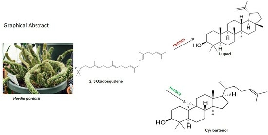
Identification and Functional Characterization of Oxidosqualene Cyclases from Medicinal Plant Hoodia gordonii

1
National Center for Natural Products Research, School of Pharmacy, University of Mississippi, University, MS 38677, USA
2
Natural Products Utilization Research Unit, Agricultural Research Service, United States Department of Agriculture, University, MS 38677, USA
3
Department of Pathology and Laboratory Medicine, Memorial Sloan Kettering Cancer Center, New York, NY 10065, USA
4
Division Pharmacognosy, Department of BioMolecular Sciences, School of Pharmacy, University of Mississippi, University, MS 38677, USA
*
Authors to whom correspondence should be addressed.
Plants 2024, 13(2), 231; https://doi.org/10.3390/plants13020231
Submission received: 19 December 2023 / Revised: 4 January 2024 / Accepted: 8 January 2024 / Published: 14 January 2024
(This article belongs to the Section Phytochemistry)

Abstract

:
Oxidosqualene cyclases (OSCs) are the key enzymes accountable for the cyclization of 2,3-oxidosqualene to varied triterpenoids and phytosterols. Hoodia gordonii (from the family Apocynaceae), a native of the Kalahari deserts of South Africa, Namibia, and Botswana, is being sold as a prevalent herbal supplement for weight loss. The appetite suppressant properties are attributed to P57AS3, an oxypregnane steroidal glycoside. At the molecular level, the enzymes involved in the biosynthesis of triterpenes and phytosterols from H. gordonii have not been previously reported. In the current study, predicted transcripts potentially encoding oxidosqualene cyclases were recognized first by searching publicly available H. gordonii RNA-seq datasets. Two OSC-like sequences were selected for functional analysis. A monofunctional OSC, designated HgOSC1 which encodes lupeol synthase, and HgOSC2, a multifunctional cycloartenol synthase forming cycloartenol and other products, were observed through recombinant enzyme studies. These studies revealed that distinct OSCs exist for triterpene formation in H. gordonii and provided opportunities for the metabolic engineering of specific precursors in producing phytosterols in this plant species.

Graphical Abstract

1. Introduction

Sterols and triterpenoids comprise a vast set of natural products that are abundant in plants and play important roles in various biological functions. In eukaryotes, sterols are the constituents of cell membranes and serve as the precursors for hormones, thus constituting primary metabolites. Different types of skeletal triterpenes exist in plants and are regarded as secondary metabolites because they are species-specific, and their function is unknown in plants. However, some triterpenes, such as betulinic acid, glycyrrhizin, and avenacins, have medicinal properties and hence these are widely used to mitigate various disorders in traditional medicine [1,2]. The common precursor for both sterols and triterpenes is 2,3-oxidosqualene. The cyclization of oxidosqualene into tetracyclic and pentacyclic carbon skeletons of sterols and triterpenes is catalyzed by oxidosqualene cyclases (OSCs). OSCs are further classified into monofunctional and multifunctional based on single or multi-cyclic products formed after cyclization. 2,3-oxidosqualene cyclization involves carbocationic cyclization followed by a cascade of rearrangement steps that are finally stabilized by deprotonation [3,4,5]. Besides these cyclization events, how 2,3-oxidosqualene is folded into the enzyme active site also determines the diversity of end products. The two conformations chair–boat–chair (CBC) and chair–chair–chair (CCC) generate protosteryl and dammarenyl cations, respectively. The dammarenyl cation is converted to different triterpenes such as α-amyrins, β-amyrins, and lupeol, whereas protosteryl cation leads to the formation of cycloartenol which is the precursor of phytosterols and steroid hormones [4]. Apart from OSCs, the chemical diversity of steroids and triterpenes is also attributed to the catalytic activity of different enzyme families, such as cytochromes P450 (CYPs) oxidoreductases and UDP-dependent glucosyltransferases [6]. However, the OSCs at the branch points are responsible for moving the metabolic profile of the plant and, hence, for the production of different secondary metabolites. Therefore, these OSCs could be used to produce specific secondary metabolites through genetic manipulations. However, detailed characteristics of the candidate OSCs covering different branch points and their mechanism of action are required for the desired genetic manipulation. Several OSCs cDNA involved in sterol and triterpene biosynthesis have been cloned and characterized from various plants. For example, β-amyrin synthase and cycloartenol synthase from P. ginseng were characterized by Kushiro et al. [7]. Five OSCs from Kalanchoe daigremontiana were reported by Wang et al. [8]. Kim et al. [9] characterized dammaranediol synthase from Centella asiatica. Several other OSCs were also reported, such as β-amyrin synthase from Aster sedifolius and Arabidopsis thaliana [10] and OSC from Aster tataricus [11] and from tomato [12]. Dhar et al. [13] isolated three branch-point OSCs from Withania somnifera. Sandeep et al. [4] reported four OSCs from the medicinal tree banaba, and Guo et al. [5] reported cycloartenol synthase from Paris polyphylla. All these OSCs have been cloned and functionally characterized in the mutant yeast strain GIL77, which has lanosterol synthase deficiency.
Hoodia gordonii (Masson) Sweet ex Decne., of the family Apocynaceae, is a leafless, spiny succulent plant with medicinal properties. The indigenous people of the Kalahari Desert in South Africa and Namibia used the plant as an appetite suppressant and to treat indigestion and small infections. Due to its appetite-depressant properties, the genus has become a popular dietary supplement for weight loss. The active ingredient P57AS3, a steroidal glycoside responsible for the putative appetite-suppressant effect, was patented by CSIR South Africa in 2002 (U.S. Patent No. 6376657). Chemical fingerprints and LC-MS/UV detection were developed to identify P57AS3. Furthermore, more than 70% of the components in the extracts from the whole plant were identified as steroid glycosides that consisted of a triterpenoid or steroidal skeleton [14]. Despite this progress at the chemical level, very little or no progress has been initiated at the molecular level for producing such specialized metabolites. To our knowledge, none of the genes have yet been cloned and characterized for triterpene or sterol biosynthesis from H. gordonii. The current study was initiated by searching public RNA-seq data repositories to find out the expressed gene members of the Hoodia OSCs gene family. The two newly identified oxidosqualene cyclase genes from H. gordonii were cloned and functionally characterized using recombinant technology.

2. Results

2.1. Isolation and Identification of cDNA Clones for Triterpene Synthases

Published transcriptomic and metabolomic data for H. gordonii on the Medicinal Plant Genome Resources (MPGR) server (http://mpgr.uga.edu/ (accessed on 25 June 2019)) have significantly expanded resources for molecular biology research for this species. To mine the H. gordonii transcriptome data, we downloaded the RNA sequences from MPGR. A total of 41,793 transcripts were annotated, and the unique sequences in the dataset were assigned to GO terms (Figure 1a). Of particular interest, “other metabolic processes” was the largest assignment within the “biological process” category (44.85% of all terms assigned), suggesting a high level of metabolic activity for this plant species. A KEGG analysis of the selected metabolic pathways and biosynthesis of secondary metabolites indicated that 99 transcripts were associated with terpenoid biosynthesis (Figure 1b). We then performed BLAST searches against the transcriptomic dataset using a cycloartenol synthase (CAS) sequence from Arabidopsis thaliana as the query sequence. Nine homologs were identified, and two predicted transcripts that showed considerable similarity to established plant OSCs were prioritized. Attempts to obtain analogous full-length ORFs through the rapid amplification of cDNA ends (RACE) were thus initiated using high-quality total RNAs prepared from stem tissue. The corresponding full-length cDNA clones were then isolated and designated as HgOSC1 and HgOSC2, respectively. The ORF of HgOSC1 was 2.289 kb and that of HgOSC2 was 2.277 kb. BLASTn analysis for the HgOSC1 sequence exhibited 76% similarity to lupeol synthase from Olea europaea and HgOSC2 showed 79% similarity to Centella asiatica cycloartenol synthase.

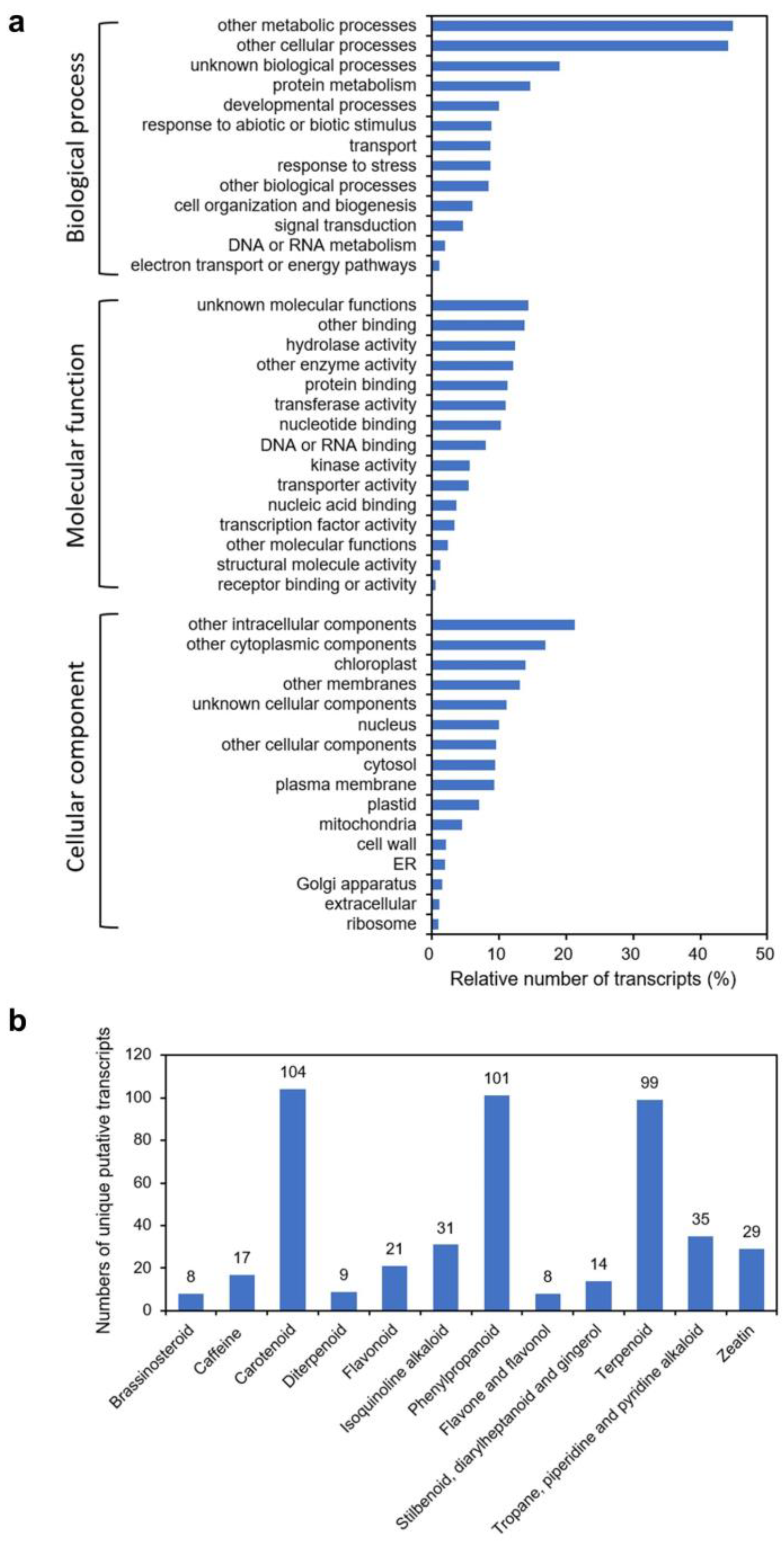
2.2. In Silico Characterization and Phylogenetic Analysis

The ORFs coding for HgOSC1 and HgOSC2 corresponded to proteins of 87 (762 amino acids) and 86 (758 amino acids) kDa, respectively. The amino acid sequence of HgOSC1 showed 79% similarity to lupeol synthase from Vitis vinifera, while 83% sequence identity was obtained for HgOSC2 with cycloartenol synthase from Sesamum indicum and Panax quinquefolius. The NCBI conserved domain search indicated that these Hoodia OSCs possess the conserved superfamily domains SQCY_1 (cd02892) and “ISOPREN_C2_like”. Proteins having these domains belong to class II terpene cyclases including 2,3-oxidosqualene cyclases (Figure S1).
The two OSC proteins were aligned with previously published plant lupeol and cycloartenol synthases and analyzed in MegAlign Pro (DNA Star v15). Both the cloned OSCs possess the amino acid motif DCTAE that is associated with substrate binding and the four characteristic motifs of the OSC superfamily. The prenyl transferase and squalene oxidase repeat Motif A are present in both OSCs. The conserved Motif B (MLCYCR) is present only in HgOSC1 and lupeol synthases, and absent in HgOSC2 and cycloartenol synthases from other plants. The analysis also revealed the presence of conserved catalytic aspartic acid (Motif C) in both HgOSC1 and HgOSC2. The conserved Motif C is responsible for converting squalene into a carbocation, which is necessary for initiating ring cyclization by protonating the first C=C bond. Lastly, the highly conserved Motif D, the QW motif, is present in both lupeol and cycloartenol synthases (Figure S2).
Phylogenetic analyses using the neighbor-joining tree method showed that the HgOSC1 clusters together with the previously characterized lupeol synthases, while the HgOSC2 most closely clusters with the previously described cycloartenol synthases (Figure 2).

2.3. Functional Characterization of OSCs in Yeast

For biochemical characterization of the putative OSCs from H. gordonii, the full-length ORFs of these cDNA clones were heterologously expressed in Saccharomyces cerevisiae. The yeast mutant GIL77 strain (Wang et al., 2011 [12]) was used for this expression as it lacks lanosterol synthase and accumulates a high concentration of 2,3-oxidosqualene which can serve as the substrate for OSC-catalyzed cyclization. However, external ergosterol must be added to the medium to grow the mutant strain. The HgOSC ORF was subcloned into the yeast expression vector pYES2, and the transgenic construct was transformed into the mutant yeast GIL77 strain. An empty vector was also transformed in the mutant yeast strain to serve as a negative control. The induced yeast cultures were pelleted and saponified with 6% methanolic KOH, and the hexane-extractable fractions were analyzed via GC-MS to identify different triterpenoids. All the transformants were found to contain squalene and ergosterol. In addition, the extracts from yeast expressing HgOSC1 cDNA contained one more compound, identified as lupeol (Figure 3 and Figure S3). The extracts were further separated by thin-layer chromatography (TLC). The TLC chromatogram showed the presence of lupeol in the hexane extract (Figure S4). The lupeol structure was confirmed by comparing the mass spectra with the commercial standard in 1H and 13C NMR spectral analyses (Figure S4). On the other hand, our initial attempt at the characterization of HgOSC2 in the mutant yeast was unsuccessful. The expression of HgOSC2 in the mutant yeast did not generate detectable new products in the yeast extracts compared to the control strain harboring the empty vector. However, the gene recoding of HgOSC2 to optimize codon usage (Figure S5) for the expression in yeast in the mutant strain led to activity. The extracts from the strain harboring this recoded HgOSC2 cDNA exhibited many compounds that were not present in the empty vector control, with one being cycloartenol (Figure 4 and Figure S6). Among the other identified compounds were squalene, ergosterol, and cycloartenol acetate. Most of the additional compounds in the yeast extract expressing HgOSC2 are yet to be identified. Nevertheless, this result suggests that HgOSC2 may be a multifunctional triterpene synthase.

2.4. Yeast Spotting Assay

The growth of yeast cells harboring the pYES2-HgOSC2 construct was examined on a selective medium (Figure 5). Growth assessment in the form of yeast spotting assays was conducted after five days. On the culture medium supplied with ergosterol, yeast harboring either pYES2-HgOSC2 or empty vector exhibited similar growth profiles along different ranges of dilutions (1, 0.1, 0.01, and 0.001). However, in the absence of ergosterol, the cells containing pYES2-HgOSC2 grew significantly compared to those with vector pYES2. However, the growth of the yeast transformants with pYES2-HgOSC2 slowed down as indicated by the reduced number of spots at higher dilutions (Figure 5).

2.5. Functional Expression of HgOSC1 and HgOSC2 in Nicotiana benthamiana

To validate the results obtained by the expression of the Hoodia oxidosqualene cyclases in yeast, constructs for plant transformation were prepared to express full-length cDNAs coding for HgOSC1 and HgOSC2 under the control of the constitutive cauliflower mosaic virus (CaMV) 35S promoter and were mobilized into Agrobacterium tumefaciens. Transient expression was performed in N. benthamiana plants using A. tumefaciens harboring the plasmids. Triterpene products from transiently expressed HgOSC1 and HgOSC2 were extracted 6 days after A. tumefaciens infiltration and analyzed via GC-MS. As shown in Figure 6, consistent with yeast expression results, the transient expression of HgOSC1 in N. benthamiana produced a compound not detected in the extracts of the leaves infiltrated with the empty vector control. The triterpene product was identified as lupeol via GC-MS by comparing its retention time and MS characteristics with an authentic standard. However, cycloartenol was not detected in the sample extracted from the leaves transiently expressing HgOSC2.

3. Discussion

Hoodia gordonii is endemic to the Kalahari Desert in South Africa and Namibia. The indigenous people used the Hoodia plants as appetite suppressants and medicinal herb to treat various ailments. H. gordonii is one of the top dietary supplements marketed for weight loss. Most of the studies on this species mainly focused on the active chemical molecule P57, a steroidal glycoside, and other secondary metabolites, such as pregnane glycosides. However, no studies are available on the molecular characterization of the genes involved in triterpene and steroid metabolic pathways. To our knowledge, this is the first investigation on the cloning and molecular characterization of genes responsible for triterpene or sterol biosynthesis from H. gordonii. According to our initial study, lupeol and cycloartenol accumulated in the young tissues of H. gordonii. However, a detailed understanding of biosynthetic pathways for lupeol and cycloartenol is required to improve their production. The common precursor for the sterols and triterpenes is 2,3-oxidosqualene (OS). The cyclization of OS into sterols and triterpenes is catalyzed by oxidosqualene cyclases (OSCs). Numerous 2,3-oxidosqualene cyclase genes encoding oxidosqualene cyclases responsible for the production of triterpenes and sterols have been isolated from a wide range of plant species [4,7,12,13]. We have also successfully cloned and characterized two genes (HgOSC1 and HgOSC2) of the OSC superfamily that belong to dammarenyl and protosteryl cation groups, respectively, from H. gordonii. Both the cloned OSCs were found to be closely related to A. thaliana OSCs and possessed the amino acid motif DCTAE, which is associated with substrate binding and the four characteristics of QW motifs that are features of the OSC superfamily [15]. The phylogenetic analyses using the neighbor-joining tree method showed that both the HgOSCs were clustered with their respective previously characterized lupeol and cycloartenol synthases, thus confirming their homology.
The functional characterization of HgOSC1 in the lanosterol-deficient yeast strain GIL77 yielded lupeol as the major cyclization product. The GC-MS analysis of the HgOSC1 compound, along with the reference standard, confirmed that the expressed product was lupeol. NMR spectral analyses were carried out to further confirm the structure of lupeol by comparing it with 1H and 13C NMR spectral data of the reference standard. Hence, it was concluded that HgOSC1 encodes the lupeol synthase in H. gordonii. The expression pattern of monofunctional Hoodia lupeol synthase is similar to what has been previously reported for other plants such as Eleutherococcus trifoliatus [16], Withania somnifera [13], and Cleome arabica [17]. However, when HgOSC2 was expressed in the GIL77 yeast strain, the cyclization of the 2,3-oxidosqualene resulted in multiple products, with one of the cyclization products being identified as cycloartenol, although it was not a major product (Figure 4). After the cyclization events, how 2,3-oxidosqualene is folded into the enzyme active site also determines the diversity of end products. The two conformations, chair–boat–chair (CBC) and chair–chair–chair (CCC) generate protosteryl and dammarenyl cations, respectively. The protosteryl cation leads to the formation of cycloartenol, which is the precursor of phytosterols and steroid hormones. In the case of Hoodia, cycloartenol synthase was found to be a multifunctional enzyme. In most plants, cycloartenol synthases are monofunctional enzymes, e.g., in pea [18], Fritillaria thunbergii [19], Withania somnifera [13] and Paris polyphylla [5]. On the other hand, some plants like Kalanchoe produce cycloartenol along with some other side products when OSCs are expressed in yeast [8]. Similarly, in the present study, the heterologous expression of Hoodia OSC (HgOSC2) in yeast resulted in the production of cycloartenol along with cycloartenol acetate, squalene, and ergosterol, as well as other side compounds that are yet to be identified (Figure 4). The multifunctionality of such an enzyme may contribute to the wide range and high total steroid glycoside levels (>70% of plant extracts) in Hoodia plants [14]. To further confirm the cycloartenol synthase expression in yeast, the growth of yeast harboring pYES2-HgOSC2 was examined via the spotting assay on a minimal selective medium in the presence or absence of ergosterol. In the presence of ergosterol, yeast containing pYES2 or pYES2-HgOSC2 showed a similar growth profile along a range of dilutions. However, in the absence of ergosterol, only the yeast cells expressing HgOSC2 could grow as compared to the empty vector. The reduced number of spots evidenced the slow growth of the yeast cells. This reinforces the previous studies showing that cycloartenol could support yeast growth to some extent in the absence of lanosterol and supports further that cycloartenol could be metabolized to C-4 desmethyl sterols [2]. The function of HgOSC1 in plants was confirmed by transient expression in N. benthamiana. However, cycloartenol was not detected in the sample extracted from the leaves transiently expressing HgOSC2. Since, cycloartenol was not a major product, as shown in the results of the expression in mutant yeast cells (Figure 4), it can be challenging to identify cycloartenol in the HgOSC2-infiltrated leaves under the current experimental conditions.

4. Materials and Methods

4.1. Plant Materials

Authenticated H. gordonii plant materials were collected from the Maynard W. Quimby Medicinal Plant Garden at the University of Mississippi. The plants were grown in a greenhouse under controlled temperature and humidity. The young and mature shoot tissues were collected during April, flash-frozen and stored at −80 °C until further use.

4.2. Isolation of RNA

RNA isolation was performed using the Qiagen RNA isolation kit (Qiagen, Valencia, CA, USA) as per the manufacturer’s directions. The isolated RNAs were treated with RNase-free DNase to remove possible contaminant DNA using the RNeasy Mini Kit (Qiagen, Valencia, CA, USA). RNA quantity and quality were determined using the NanoDrop ND-1000 Spectrophotometer (Thermo Fisher Scientific, Waltham, MA, USA). RNA sample integrity was also assessed via agarose gel electrophoresis.

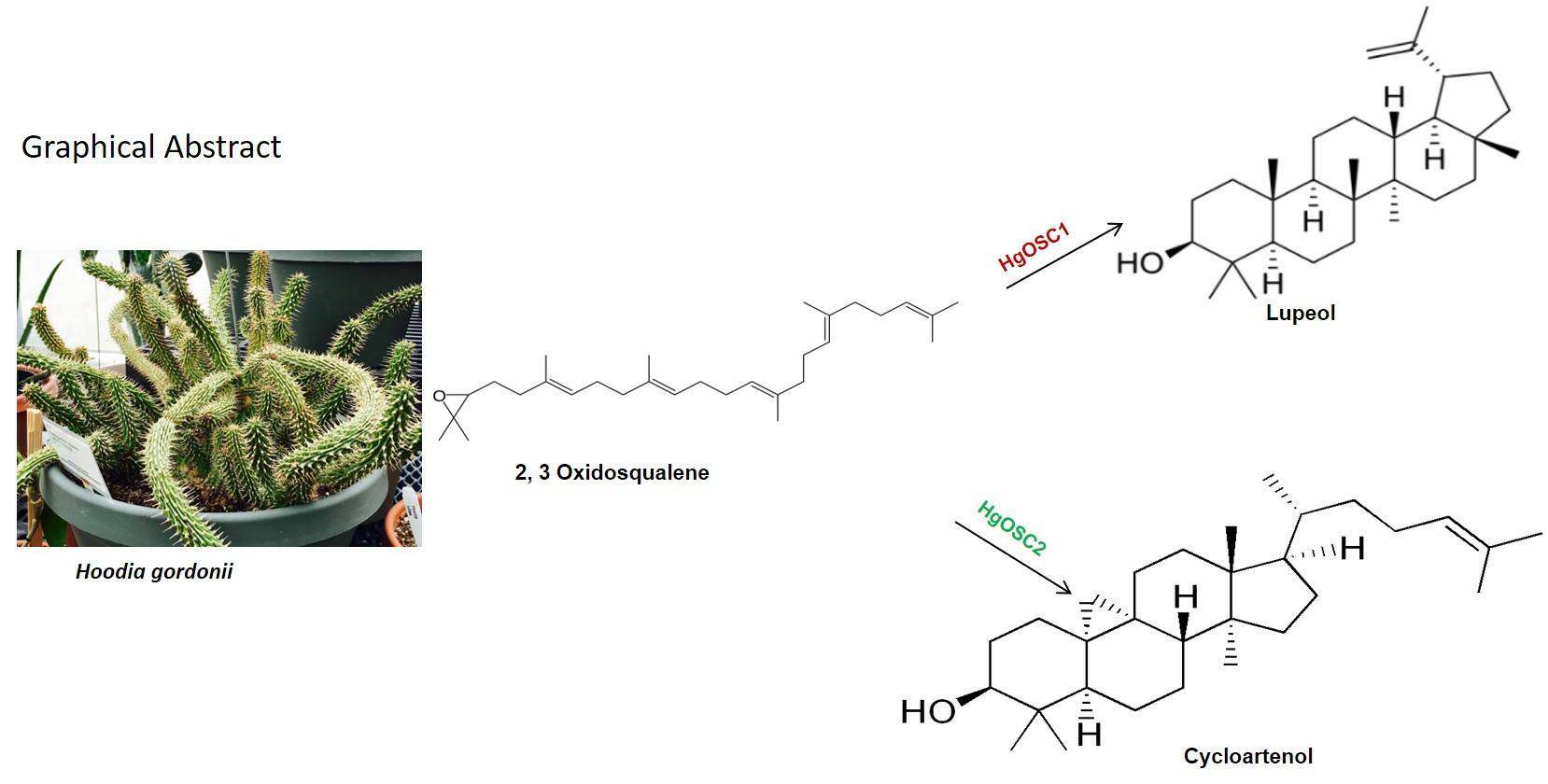
4.3. Functional Annotation of the Transcriptome Dataset and Pathway Analyses

The transcriptome data were downloaded from the Medicinal Plant Metabolomics Resource website (http://mpgr.uga.edu/ (accessed on 25 June 2019)). Functional annotation was performed using the custom pipeline on the Linux computing cluster at the Chinese Academy of Medical Sciences and Peking Union Medical College. The unique putative transcripts were aligned against the NCBI non-redundant nucleotide using BLASTN [20] with an E-value cutoff of 10−5. BlastX was performed to search protein databases including NCBI Non-redundant (Nr) and Swiss-Prot (https://www.uniprot.org/ (accessed on 26 June 2019)). TAIR Gene Ontology (TAIR-GO, http://www.arabidopsis.org (accessed on 26 June 2019)) analyses were performed using a custom script to execute gene categories. Each transcript annotated to TAIR was classified based on TAIR GO terms. The percentage of each functional categorization was computed by the TAIR GO functional categorization method, which represents the percentage of the number of genes annotated to terms in the GO slim category and the total number of transcripts. Metabolic pathways were identified using KEGG (http://www.genome.jp/kegg/pathway.html (accessed on 28 June 2019)) pathway mapper using PERL script. The annotated transcripts were mapped to the specific pathways based on KEGG Ontology (KO) groups and the KEGG pathway maps.

4.4. Isolation of cDNA Clones for Terpene Synthases

To obtain full-length cDNA clones, the rapid amplification of both 5′ and 3′ cDNA ends (RACE) was carried out using the BD SMART™ RACE cDNA Amplification Kit (Clontech, Mountain View, CA, USA) according to the manufacturer’s instructions. Different primer pairs were used for the RACE PCR to amplify the two selected genes (Table S1). The amplified products with single bands were extracted from the gel, and the purified cDNA was cloned into the TOPO-TA cloning vector. The clones were sent for Sanger’s sequencing (GENEWIZ, Burlington, MA, USA) to confirm the amplified product. The full-length cDNAs were then amplified with primer pairs complementary to the 5′ and 3′ ends of the open reading frames (ORFs) identified in RACE experiments using Pfu thermostable DNA polymerase (Stratagene, La Jolla, CA, USA) and first-strand cDNA generated from H. gordonii plants. ProtoScript M-MULV first strand cDNA synthesis kit (New England Biolabs, Ipswich, MA, USA) was used to prepare the cDNAs. The HgOSC1 and HgOSC2 sequences reported in this article have been deposited in the GenBank database (accession nos. HgOSC1, OR133751; HgOSC2, OR133752).

4.5. Phylogenetic Analysis

HgOSC1 and HgOSC2 protein sequences were compared with functionally characterized oxidosqualene cyclases from other plant gene sequences retrieved from GenBank via phylogenetic comparison. Following protein alignments using ClustalW, the neighbor-joining tree was constructed with five hundred bootstrap iterations in MEGAX software version 10 [21].

4.6. Expression of OSCs in Saccharomyces cerevisiae

The cDNA encoded in the ORFs of HgOSC1 and HgOSC2 was ligated to pYES2 expression vector via XbaI and KpnI sites for HgOSC1 and via KpnI and XhoI sites for HgOSC2 to generate pYES2-HgOSC1 and pYES2-HgOSC2, respectively. Lanosterol-deficient strain GIL77 (gal2 hem3-6 erg7 ura3-167), provided by Dr. Reinhard Jetter (The University of British Columbia), was transformed either with an empty vector or with pYES2-HgOSC1 and pYES2-HgOSC2 constructs. The ORF of HgOSC2 was synthesized via GenScript (Piscataway, NJ, USA) for optimized codon usage for yeast and used for the expression experiments due to lack of detectable activity of the original clone. The lithium-acetate method [22] was used to transform these plasmids into the mutant yeast strain GIL77. The transformed yeast cells were selected for the uracil prototype and were grown at 30 °C for three days in the presence of exogenous ergosterol and hemin. The functional characterization of these yeast strains was carried out according to the methods previously described (Wang et al., 2011 [12]). Briefly, the expression of OSCs was induced by galactose (2%) added to the minimal medium along with ergosterol and incubated at 30 °C for 16 hrs. The induced cells were then transferred to a resting medium containing phosphate buffer (pH 7.0) with glucose and hemin. The cells were incubated for one day at 30 °C and then pelleted down with 3500 rpm centrifugation for 5 min. The pelleted cells were frozen at −80 °C for later use in GC-MS analysis.

4.7. GC-MS Analysis

To isolate and analyze the metabolites, the frozen yeast cells (approximately 100 mg) were saponified in 6% potassium hydroxide methanolic solution at 80 °C for 1 h. They were then extracted twice in hexane and dried in a Speedvac. The sample was resuspended in 500 µL methylene chloride prior to GC/Q-ToF analysis. Samples were analyzed on an Agilent 7890B GC (Santa Clara, CA, USA) equipped with an Agilent PAL RSI85 auto-sampler. The mass spectral detector was an Agilent 7250 High-Resolution Accurate Mass Q-TOF MS operated in the full spectral acquisition mode. The Agilent J&W HP-5MS UI capillary columns (30 m × 0.25 mm i.d. × 0.25 µm film thickness) were used for the analysis. The carrier gas was helium with a 1 mL/min flow rate. The GC oven temperature was held at 50 °C for 2 min, programmed at 30 °C/min to 200 °C, then ramped up at 3 °C/min to 300 °C and held for 10 min. One microliter of each sample was injected with a split-less injection mode. The inlet temperature was fixed at 280 °C.
The Q-ToF mass spectrometer was equipped with an electron ionization source, operated with an electron energy of 70 eV and an emission current of 5.0 µA. The source, quadrupole, and transfer line temperatures were set to 280 °C, 150 °C, and 280 °C, respectively. Data were acquired at a rate of 5 Hz from 45 to 550 m/z with a 5 minute solvent delay. Automated ToF mass calibration was performed after every injection using a keyword command in the sequence table. Data were acquired using Agilent MassHunter Acquisition Software (version B.10.0). Data analysis was performed using Agilent MassHunter Qualitative (version B.10.0) and Quantitative (version B.10.0) analysis software. The identification of compounds began with a comparison to the spectral databases (Wiley and NIST) using a probability-based matching algorithm. The identification was confirmed based on standard references purchased from commercial sources.

4.8. TLC and NMR Analysis of HgOSC1 Products

The transformed yeast GIL77 strain with pYES2-HgOSC1 construct and the pYES2 plasmid without any gene insert that served as control were grown on a minimal medium for three days. A few colonies of each were grown in 500 mL of minimal medium lacking uracil and supplemented with ergosterol and hemin. The yeast cultures were grown with shaking at 210 rpm at 30 °C for two days. The cells were then pelleted down via centrifugation (3500 rpm) for 5 min. The pellet was then resuspended in 500 mL induction medium containing 2% galactose supplemented with ergosterol and hemin. The cells were grown in the induction medium for sixteen hours. After induction, the cells were transferred to a resting medium. An equal volume of 6% methanolic KOH was added to the culture to extract the triterpenes. The mixture was then incubated at 80 °C with shaking (200 rpm) for one hour. The unsaponifiable fractions were then extracted with an equal volume of hexane, followed by drying in a rotary evaporator without heating.
To determine the products of the HgOSC1 clone, the hexane extract was then analyzed via thin-layer chromatography (TLC) to detect the presence of lupeol. The TLC was performed on the GF254 silica gel 60 plate (EMD Millipore) with chloroform/methanol (10:0.1, v/v) as the solvent system for development. The reference standard of lupeol (Sigma-Aldrich, St. Louis, MO, USA) was spotted on the plate beside the extract for the purpose of identification. The isolate zones were observed using UV light (254 nm) and then visualized by spraying with 1% vanillin-H2SO4 reagent followed by heating. To further verify the presence and identity of lupeol produced by HgOSC1, nuclear magnetic resonance (NMR) analysis was conducted for the hexane extract. 1H and 13C NMR spectra at 25 °C were obtained with a OneNMR probe on an Agilent DD2-500 NMR spectrometer (Santa Clara, CA, USA). The 1H and 13C NMR spectra of the hexane extract and the lupeol standard’s spectra were stacked for comparison.

4.9. Yeast Spotting Assay

Isolated yeast colonies containing either pYES2 empty vector or pYES2-HgOSC2 were grown overnight at 30 °C in yeast dropout medium (SGal) supplemented with ergosterol and hemin. Yeast cells were pelleted, washed, and resuspended in sterile water to reach an OD600 equal to 1. Serial dilutions were made as 1, 0.1, 0.01, and 0.001 in sterile water, and 5 µL of each was spotted on SGal agar media with or without exogenous ergosterol and hemin. The plates were incubated at 30 °C for five days.

4.10. Transient Expression of Hoodia OSCs in Nicotiana benthamiana

The agrobacterium T-DNA binary vectors for the expression of the Hoodia OSCs were constructed according to the methods previously described [23]. Briefly, the ORFs of HgOSC1 and HgOSC2 were amplified using PfuUltra HS DNA polymerase (Agilent) and subcloned into the shuttle vector between the 2x35S promoter and the octopine synthase terminator (Tosc). The expression cassettes were then subcloned into pLH7000 [24] using the flanking SfiI sites to generate the binary expression vectors pLH-OSC1 and pLH-OSC2, respectively. The constructs were then mobilized into Agrobacterium tumefaciens strain EHA105. Four-week-old N. benthamiana leaves were co-infiltrated with the strain expressing either pLH-OSC1 or pLH-OSC2 along with the strain expressing the suppressor of silencing, p19, in a ratio of 3:1 at an OD600 of 0.6 in the infiltration buffer (10 mM 2-(N-morpholino) ethanesulfone, 10 mM MgCl2, pH 5.7, and 100 μM acetosyringone) by injection into the lower leaf epidermis using a 1 mL syringe. Plants were maintained in a Conviron growth chamber under a 16 h/8 h, light/dark cycle at 25 °C, 150 µmol m−2s−1 light intensity. Agroinfiltrated N. benthamiana leaves were collected 6 days post-infiltration, ground to a fine powder under liquid N2 in a mortar and pestle, and stored at −80 °C until analysis.
In total, 100 mg of powdered frozen N. benthamiana leaf sample was extracted with 600 µL of methanol/chloroform (2:1) twice via centrifugation at 1000× g for 5 min. The resultant supernatants were combined and dried using a centrifugal vacuum evaporator. The dried sample was then saponified in 300 μL of methanol/10% potassium hydroxide (4:1, v/v) for 1 h at room temperature with shaking at 150 rpm. Then, 600 μL water was added to the saponified sample and extracted three times with 300 μL hexane. The hexane fractions were pooled, washed three times with 300 μL water, and then evaporated to dryness. The dried hexane extract was resuspended in 100 μL of 100% methanol for GC-MS analysis. The experiments were independently repeated three times.

5. Conclusions

In conclusion, two different OSCs have been isolated from Hoodia gordonii and characterized via heterologous expression in yeast. The two different triterpenoids, lupeol and cycloartenol, were identified as the products of the enzymes. This is the first report on the identification and biochemical characterization of lupeol and cycloartenol synthase from Hoodia. This study will provide an opportunity to elucidate the metabolic pathways of sterol and triterpenoid production in this particular plant species.

Supplementary Materials

The following supporting information can be downloaded at: https://www.mdpi.com/article/10.3390/plants13020231/s1.

Author Contributions

Z.P., I.P., and I.A.K. conceived and designed the study; I.P. and Z.P. performed all molecular biology experiments; Y.Z. and Z.P. performed RNA-seq data mining and processing; M.W., J.Z., J.L. and A.G.C. performed chemical analysis; I.P. and Z.P. prepared the manuscript. All authors contributed to the writing of this manuscript. All authors have read and agreed to the published version of the manuscript.

Funding

This research is supported in part by “Science Based Authentication of Botanical Ingredients” funded by the Center for Food Safety and Applied Nutrition, US Food and Drug Administration grant number 5U01FD004246 and Cooperative Agreement 58-6060-6-015 grant to the University of Mississippi, USA.

Data Availability Statement

Data are contained within the article and supplementary materials.

Acknowledgments

We thank Reinhard Jetter (The University of British Columbia) for providing the yeast strain GIL77. We also thank Marilyn Ruscoe for providing excellent technical support for this work and Jeff Solomon for reviewing the manuscript.

Conflicts of Interest

The authors have no relevant financial or non-financial interests to disclose.

References

  1. Papadopoulou, K.; Melton, R.E.; Leggett, M.; Daniels, M.J.; Osbourn, A.E. Compromised disease resistance in saponin-deficient plants. Proc. Natl. Acad. Sci. USA 1999, 96, 12923–12928. [Google Scholar] [CrossRef] [PubMed]
  2. Gas-Pascual, E.; Berna, A.; Bach, T.J.; Schaller, H. Plant oxidosqualene metabolism: Cycloartenol synthase-dependent sterol biosynthesis in Nicotiana benthamiana. PLoS ONE 2014, 9, e109156. [Google Scholar] [CrossRef] [PubMed]
  3. Abe, I. The oxidosqualene cyclases: One substrate, diverse products. In Natural Products: Discourse, Diversity and Design; Osbourn, A., Goss, R.J., Carter, G.T., Eds.; Wiley Blackwell: Oxford, UK, 2014; pp. 297–316. [Google Scholar]
  4. Sandeep Misra, R.C.; Chanotiya, C.S.; Mukhopadhyay, P.; Ghosh, S. Oxidosqualene cyclase and CYP716 enzymes contribute to triterpene structural diversity in the medicinal tree banaba. New Phytol. 2019, 222, 408–424. [Google Scholar] [CrossRef] [PubMed]
  5. Guo, S.-Y.; Yin, Y.; Lei, T.; Shi, Y.-H.; Gao, W.; Zhang, X.-N.; Li, J. A cycloartenol synthase from the steroidal saponin biosynthesis pathway of Paris. polyphylla. J. Asian Nat. Prod. Res. 2021, 23, 353–362. [Google Scholar] [CrossRef] [PubMed]
  6. Chen, C.; Pang, Y.; Chen, Q.; Li, C.; Lu, B. Oxidosqualene cyclases in triterpenoids biosynthesis: A review. Chin. J. Biotechnol. 2022, 38, 443–459. [Google Scholar]
  7. Kushiro, T.; Shibuya, M.; Ebizuka, Y. Beta-amyrin synthase—Cloning of oxidosqualene cyclase that catalyzes the formation of the most popular triterpene among higher plants. Eur. J. Biochem. 1998, 256, 238–244. [Google Scholar] [CrossRef]
  8. Wang, Z.; Yeats, T.; Han, H.; Jetter, R. Cloning and characterization of oxidosqualene cyclases from Kalanchoe daigremontiana: Enzymes catalyzing up to 10 rearrangement steps yielding friedelin and other triterpenoids. J. Biol. Chem. 2010, 285, 29703–29712. [Google Scholar] [CrossRef]
  9. Kim, O.T.; Lee, J.W.; Bang, K.H.; Kim, Y.C.; Hyun, D.Y.; Cha, S.W.; Choi, Y.E.; Jin, M.L.; Hwang, B. Characterization of a dammarenediol synthase in Centella asiatica (L.) Urban. Plant Physiol. Biochem. 2009, 47, 998–1002. [Google Scholar] [CrossRef]
  10. Shibuya, M.; Katsube, Y.; Otsuka, M.; Zhang, H.; Tansakul, P.; Xiang, T.; Ebizuka, Y. Identification of a product specific beta-amyrin synthase from Arabidopsis thaliana. Plant Physiol. Biochem. 2009, 47, 26–30. [Google Scholar] [CrossRef]
  11. Sawai, S.; Uchiyama, H.; Mizuno, S.; Aoki, T.; Akashi, T.; Ayabe, S.I.; Takahashi, T. Molecular characterization of an oxidosqualene cyclase that yields shionone, a unique tetracyclic triterpene ketone of Aster tataricus. FEBS Lett. 2011, 585, 1031–1036. [Google Scholar] [CrossRef]
  12. Wang, Z.; Guhling, O.; Yao, R.; Li, F.; Yeats, T.H.; Rose, J.K.; Jetter, R. Two oxidosqualene cyclases responsible for biosynthesis of tomato fruit cuticular triterpenoids. Plant Physiol. 2011, 155, 540–552. [Google Scholar] [CrossRef]
  13. Dhar, N.; Rana, S.; Razdan, S.; Bhat, W.W.; Hussain, A.; Dhar, R.S.; Vaishnavi, S.; Hamid, A.; Vishwakarma, R.; Lattoo, S.K. Cloning and functional characterization of three branch point oxidosqualene cyclases from Withania somnifera (L.) dunal. J. Biol. Chem. 2014, 289, 17249–17267. [Google Scholar] [CrossRef]
  14. Russell, P.J.; Swindells, C. Chemical characterisation of Hoodia gordonii extract. Food Chem. Toxicol. 2012, 50, S6–S13. [Google Scholar] [CrossRef] [PubMed]
  15. Poralla, K.; Hewelt, A.; Prestwich, G.D.; Abe, I.; Reipen, I.; Sprenger, G. A specific amino acid repeat in squalene and oxidosqualene cyclases. Trends Biochem. Sci. 2012, 19, 157–158. [Google Scholar] [CrossRef] [PubMed]
  16. Ma, L.; Wang, S.; Tseng, Y.; Lee, Y.; Chu, F. Cloning and characterization of a 2,3-oxidosqualene cyclase from Eleutherococcus trifoliatus. Holzforschung 2012, 67, 463–471. [Google Scholar] [CrossRef]
  17. Ladhari, A.; Chappell, J. Unravelling triterpene biosynthesis through functional characterization of an oxidosqualene cyclase (OSC) from Cleome arabica L. Plant Physiol. Biochem. 2019, 144, 73–84. [Google Scholar] [CrossRef] [PubMed]
  18. Morita, M.; Shibuya, M.; Lee, M.S.; Sankawa, U.; Ebizuka, Y. Molecular cloning of pea cDNA encoding cycloartenol synthase and its functional expression in yeast. Biol. Pharm. Bull. 1997, 20, 770–775. [Google Scholar] [CrossRef]
  19. Zhu, Z.; Liu, W.; Ge, L.; Yu, Q.; Zhao, Z.; Yang, J.; Wan, H. Molecular cloning and characterization of a cDNA encoding cycloartenol synthase from Fritillaria thunbergii Miq. Afr. J. Biotechnol. 2012, 11, 6896–6903. [Google Scholar]
  20. Altschul, S.F.; Madden, T.L.; Schäffer, A.A.; Zhang, J.; Zhang, Z.; Miller, W.; Lipman, D.J. Gapped BLAST and PSI-BLAST: A new generation of protein database search programs. Nucleic Acids Res. 1997, 25, 3389–3402. [Google Scholar] [CrossRef]
  21. Kumar, S.; Stecher, G.; Li, M.; Knyaz, C.; Tamura, K. MEGA X: Molecular Evolutionary Genetics Analysis across Computing Platforms. Mol. Biol. Evol. 2018, 35, 1547–1549. [Google Scholar] [CrossRef]
  22. Gietz, R.D.; Woods, R.A. Transformation of yeast by lithium acetate/single-stranded carrier DNA/polyethylene glycol method. Methods Enzymol. 2002, 350, 87–96. [Google Scholar] [PubMed]
  23. Pan, Z.; Bajsa-Hirschel, J.; Vaughn, J.N.; Rimando, A.M.; Baerson, S.R.; Duke, S.O. In vivo assembly of the sorgoleone biosynthetic pathway and its impact on agroinfiltrated leaves of Nicotiana benthamiana. New Phytol. 2021, 230, 683–697. [Google Scholar] [CrossRef] [PubMed]
  24. Hausmann, L.; Toepfer, R. Development of plasmid vectors. In Bioengineering of Custom-Tailored Rape Varieties; Brauer, D., Roebbelen, G., Toepfer, R., Eds.; Gesellschaft fuer Pflanzenzuechtung: Goettingen, Germany, 1999; pp. 155–171. [Google Scholar]
Figure 1. Distribution of unique putative transcripts annotated in the Hoodia gordonii transcriptome dataset. (a) Functional annotation of transcripts from H. gordonii based on GO categories. The percentage within each GO category (biological process, molecular function, and cellular component) is indicated on the x-axis. (b) KEGG classification of biosynthetic pathways. The numbers of the unique transcripts related to selected secondary metabolic pathways are shown.
Plants 13 00231 g001
Figure 2. Phylogenetic relationships of selected functionally characterized oxidosqualene cyclases from plants. Amino acid sequences were aligned in ClustalW, and the phylogenetic tree was constructed using the neighbor-joining method with 500 bootstrap iterations in MEGAX. HgOSC1 showed a close relationship with lupeol synthases, whereas HgOSC2 formed a clade with other previously characterized cycloartenol synthases.
Plants 13 00231 g002
Figure 3. Identification of lupeol in transgenic yeast. GC/MS analyses showed peaks of the cell extracts from mutant yeast expressing HgOSC1 (B) and the empty vector pYES2 control (C) vs. lupeol as a reference standard (A). Results are depicted as extracted ion chromatograms (EICs) of the parent mass m/z 426.
Plants 13 00231 g003
Figure 4. Total ion chromatogram (TIC) of GC/MS analysis of yeast cell extracts. TICs of extracts from the yeast strain transformed with pYES2-HgOSC2 (A) and vector pYES2 as a control (B). Compounds identified were: 1, squalene; 2, ergosterol; 3, cycloartenol; and 4, cycloartenol acetate. The compound identification was based on the spectra search with NIST and Wiley libraries, and further confirmed with reference standards.
Plants 13 00231 g004
Figure 5. Yeast spotting assays with lanosterol deficient strain GIL77. Growth of GIL77 yeast strains transformed with vector pYES2 or construct pYES2-HgOSC2 was tested on SGal medium supplemented with (+ERG) or without (−ERG) exogenous ergosterol. After 5 days of incubation, the plates supplemented with ergosterol showed fully grown colonies in both yeast strains, whereas Sgal plates without ergosterol allowed only a limited growth of yeast expressing HgOSC2, indicating that HgOSC2 can partially overcome ergosterol deficiency.
Plants 13 00231 g005
Figure 6. Expression of Hoodia oxidosqualene cyclase (HgOSC1) in Nicotiana benthamiana. HgOSC1 transiently co-expressed with the suppressor of the gene silencing P19 in N. benthamiana leaves by agroinfiltration. Leaf metabolites were extracted (see Materials and Methods) and analyzed via GC-MS (b), depicted as EIC, vs. lupeol reference standard (a). Leaves expressing P19 with an empty vector only served as an experimental control. Mass spectra of products corresponding to the peaks shown in a (c) and b (d). The experiments were repeated three times independently with identical results. ND, not determined.
Plants 13 00231 g006
Disclaimer/Publisher’s Note: The statements, opinions and data contained in all publications are solely those of the individual author(s) and contributor(s) and not of MDPI and/or the editor(s). MDPI and/or the editor(s) disclaim responsibility for any injury to people or property resulting from any ideas, methods, instructions or products referred to in the content.

