Article

Antiquorum and Antibiofilm Activities of Piper bogotense C. DC. against Pseudomonas aeruginosa and Identification of Bioactive Compounds

by
Andrés G. Sierra-Quitian
1,
Lida V. Hernandez-Moreno
1,
Ludy C. Pabon-Baquero
2,
Juliet A. Prieto-Rodriguez
3,* and
Oscar J. Patiño-Ladino
1
1
Departamento de Química, Facultad de Ciencias, Universidad Nacional de Colombia, Sede Bogotá, Bogotá 111321, Colombia
2
Escuela de Ciencias Básicas y Aplicadas, Universidad de La Salle, Bogotá 111711, Colombia
3
Departamento de Química, Facultad de Ciencias, Pontificia Universidad Javeriana, Bogotá 110231, Colombia
*
Author to whom correspondence should be addressed.
Plants 2023, 12(9), 1901; https://doi.org/10.3390/plants12091901
Submission received: 18 April 2023 / Revised: 25 April 2023 / Accepted: 27 April 2023 / Published: 6 May 2023
(This article belongs to the Special Issue Plant-Derived Natural Products and Their Applications)

Abstract

:
The present study describes the anti-biofilm and quorum sensing (QS) inhibitory potential of extracts and chemical constituents from Piper bogotense. Antibiofilm potential was determined through crystal violet assay against Pseudomonas aeruginosa, while QS inhibition efficacy was determined through violacein inhibition assay using Chromobacterium violaceum as a bacterial model. Additionally, this study reports the effects of the chemical constituents isolated in P. bogotense against various virulent factors associated with QS, such as the percentage decrease in pyocyanin, elastase, and protease production. The chemical study led to the isolation and identification of two prenylated benzoic acids (1 and 2) and a prenylated hydroquinone 3, of which compounds 1 and 2 are reported for the first time for P. bogotense. The ethanolic extract and the DCM fraction from P. bogotense stand out for reducing violacein production in C. violaceum, as well as the biofilm formation in P. aeruginosa. Compounds 2 and 3 stand out for having the lowest violacein production (43.8% and 68.3%), as well as the lowest production of virulence factors such as elastase (60.2% and 51.4%) and pyocyanin (39.7% and 33.2%). These results demonstrate the potential of P. bogotense components to be used as an alternative control against multidrug-resistant P. aeruginosa.

1. Introduction

Antimicrobial resistance (AMR) is one of the top 10 global public health threats due to the increased burden of infectious diseases and high costs of medical care; it is estimated that AMR can cause around 10 million deaths a year by 2050 [1,2]. Among the control alternatives to reduce AMR is the inhibition of the quorum sensing system (QS), a cellular communication mechanism that controls virulence factors and the production of biofilms, which favors the survival of bacteria in the host [3,4]. The importance of controlling this system lies in controlling the virulence factors associated with QS, reducing the evolutionary pressure generated in bacteria when treated with antibiotics, and prolonging their lifespan using combination therapies [5,6,7]. Pseudomonas aeruginosa is a Gram-negative pathogen of clinical importance since it is one of the most common agents of infections observed in Intensive Care Units (ICU) and is responsible for high mortality. [8,9]. It is reported as one of the main microorganisms causing nosocomial infections in developed countries, with a prevalence of 3–12%, and in underdeveloped countries, between 5–19% with a predisposition to be multidrug-resistant [10,11,12,13,14]. This bacterium can form biofilms and produce virulence factors that help to impede the action of antibiotics and promote their pathogenicity in the host, which is the reason it has been declared by the World Health Organization (WHO, Geneva, Switzerland) as a critical priority pathogen as it presents multidrug resistance to a wide range of antibiotics [15,16,17,18,19]. QS controls the virulence factors and the formation of biofilms, so the search for anti-QS strategies could be a potential objective to prevent infection by P. aeruginosa.
Plants have been considered a source of various bioactive substances, including some with antibacterial and anti-QS potential. Regulation of biofilm formation, Inhibition of QS, and attenuation of virulence factors in P. aeruginosa, have been reported for a wide variety of extracts, essential oils, and chemical constituents [20,21,22,23,24,25]. Plant-derived secondary metabolites with the potential to inhibit QS include phenols, flavonoids, coumarins, terpenoids, amides, phenylpropanoids, alkaloids, and benzoic acid derivatives, among others [23,24,25]. Although many herbal extracts exhibit anti-QS effects, investigations are needed to determine some bioactive species’ chemical constituents and characterize the molecular mechanisms involved in the control and perturbation of QS signaling.
Among the plant families where some of its species have shown potential for QS control is the Piperaceae family, which is recognized worldwide for its economic importance due to its applications in food, industry, and medicine [26,27,28]. The Piper genus is one of the most representative of the family and comprises about 1457 species distributed worldwide, mainly in tropical and subtropical regions [29]. Many chemical constituents have been found in species of the Piper genus, highlighting the presence of amides, alkylbenzenoids, flavonoids, terpenoids, phenylpropanoids, and prenylated benzoic acid derivatives [30,31]. The antimicrobial potential of species of this genus against different pathogenic bacteria has been described in the literature, including P. aeruginosa [32,33,34,35]. However, there are few studies aimed at determining the effect of products derived from species of Piper genus to inhibit QS and regulate the biofilm formation of P. aeruginosa [36,37,38,39,40,41,42]. The antiQS on C. violaceum and antibiofilm on P. aeruginosa potential of extracts from P. betle, P. nigrum and P. longum have been reported in the literature [37,38,40,41]. The ability to inhibit the production of pyocyanin and the swarming motility of P. aeruginosa was reported for the extract from P. betle [37,41]. The EOs from P. bogotense, P. brachypodom and P. bredemeyeri, showed inhibiting QS on C. violaceum CV026 with IC50 of 514, 93 and 45 μg/mL, respectively [43]. The essential oil of P. nigrum at 50 μL/mL caused a 26% inhibition in the formation of P. aeruginosa biofilms and a 16% inhibition in the production of violacein [41,44]. Investigations on two amides (piperine and trichostaquine) present in the extract of P. nigrum showed that they have the potential to inhibit the production of violacein from C. violaceum, while they cannot inhibit the biofilm formation of P. aeruginosa [40]. Flavonoids from P. delineatum exhibited the ability to disrupt QS-mediated bioluminescence downstream LuxO and showed a strong dose-dependent inhibition of biofilm formation in the bacterial model Vibrio harveyi [39].
Piper bogotense C. DC. (Synonyms: P. barbatum C. DC, P. duriraneum C. DC, P. fistulosum C. DC), popularly known as matico, cordoncillo, bordoncillo, or pipilongo, is a native species distributed in tropical countries such as Ecuador, Colombia and Panama [28,45]. This species is of great agroforestry interest in Colombia due to its medicinal applications, uses as fodder and biofertilizer, and because it contributes to the protection and recovery of soils [46,47]. Additionally, the ripe fruits of P. bogotense are a source of food for bats and native and migratory birds [47]. In traditional medicine, this species is used as a healing, anti-inflammatory, analgesic and for the treatment of hepatic pain [48,49]. The presence of prenylated hydroquinones and benzoquinones from fruits, as well as flavonoids, alkaloids, lactams and sterols from stems and leaves, have been reported in P. bogotense [43,50,51,52,53]. The essential oil from its leaves mainly contains trans-sabinene hydrate, α-phellandrene, α-pinene and limonene [43,53,54]. In addition to the previously mentioned antiQS potential for this essential oil, its antifungal and antiparasitic activity has been determined [53,55]. Cytotoxic activity against various cell lines has been reported for extracts from P. bogotense [56,57].
The present investigation describes the anti-QS activity on C. violaceum, and antibiofilm and antivirulence effects on P. aeruginosa of extracts and chemical compounds from P. bogotense.

2. Results and Discussion

2.1. Phytochemical Study and Anti-QS and Anti-Biofilm Activity

The phytochemical study of the aerial part of P. bogotense led to the obtaining of five fractions of different polarity (DCM, EtOAc, IPA, EtOH:H2O). The fractions and the extract did not show significant effects (p < 0.05) on the growth of P. aeruginosa at the concentrations evaluated, while positive control gentamicin affected total growth inhibition at 2 µg/mL (Table S1 in Supplementary Material). Previous reports of Piper species that have been evaluated against P. aeruginosa show that some do not affect bacterial growth, as reported for P. betle at concentrations of 150–50 ppm and P. nigrum at 3.2 and 1 mg/mL [33,37]. However, other Piper species have been shown to affect P. aeruginosa growth at higher concentrations than those evaluated in this study. For example, the ethanolic extract of P. tuberculatum a MIC > 1024 µg/mL is reported [58], while P. longum and P. nigrum are reported inhibition halos higher than 18 mm at concentrations between 5–10 mg/mL in the disk diffusion assay [34]. In the case of P. umbelatum, DCM extract has been reported to have the potential to inhibit the growth of P. aeruginosa ATCC 27853 at 1000 µg/mL [35]. The results of this assay allowed us to determine that the concentrations evaluated did not affect the growth of P. aeruginosa to avoid selective pressure and prevent resistance; therefore, these were selected for subsequent assays.
Subsequently, P. bogotense extract and fractions were evaluated on the attenuation of C. violaceum QS in the violacein production assay. The results showed that the extract and DCM, EtOAc and EtOH:H2O fractions reduced violacein production directly proportional to the concentration, contrary to the IPA fraction, where an inverse relationship was observed. It was found that the extract and IPA and EtOH:H2O fractions had a violacein production lower than 5% at the highest concentration, being even lower than the inhibition control thymol (Figure 1A). Regarding studies of the anti-QS activity of the ethanolic extract of P. bogotense this is the first report; however, previously, it has been reported that the essential oil of this species, whose major components correspond to trans-sabinene, α-pinene, and α-phellandrene, has violacein inhibitory effects on C. violaceum strain CV026 with IC50 of 513.8 μg/mL [43]. Previous studies found that the extracts of P. betle and P. nigrum showed anti-QS activity in the quantification assay of violacein produced by C. violaceum using concentrations between 1 and 3 mg/mL [37,42].
Next, the ability to reduce the formation of P. aeruginosa biofilms by exposure to the extract and fractions of P. bogotense is evaluated. Figure 1B shows the results, finding that the extract and the DCM fraction were the only ones that managed to reduce biofilm formation significantly. For the ethanolic extract, an inversely proportional effect to the concentration was found. It was evidenced that the extract significantly reduced biofilm formation with a percentage of formation of 19% at 62.5 μg/mL, followed by the DCM fraction, which had a percentage of formation of 18.9% at 250 μg/mL being these results close to the effect of the quercetin inhibition control which produced 13% at 3.9 μg/mL. For the EtOAc fraction, a slight reduction in biofilm formation was observed, with a percentage of formation of approximately 70% at the lowest concentrations. For this species, the effect on P. aeruginosa biofilm formation has not been reported; however, for other species of the genus, it has. For example, for P. betle it is found that the ethanolic extract of leaves inhibits P. aeruginosa PAO1 biofilm by 75% at 200 μg/mL [37,41], and for P. nigrum, it is reported for the ethanolic extract of seeds that it does not affect P. aeruginosa biofilm at 500 μg/mL [40]. Considering the results obtained, the DCM fraction was chosen for the purification and identification of the bioactive compounds because it inhibited the production of violacein from C. violaceum and because it was the fraction that had the best effect on the reduction of the P. aeruginosa biofilm.
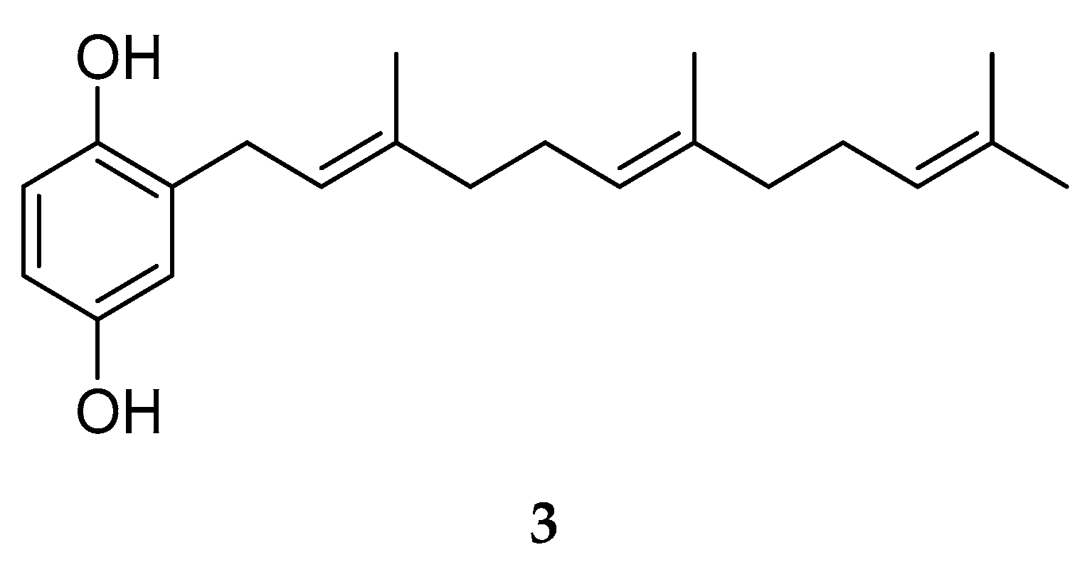
The phytochemical study carried out on the DCM fraction from the aerial part of P. bogotense led to the isolation of three compounds, which were identified by spectroscopic methods and by comparison with data reported in the literature. In this way, two derivatives of benzoic acid (1 and 2) and hydroquinone (3) were isolated and identified (Figure 2). This is the first report of 1 and 2 for the species, while 3 has been previously described in fruits from P. bogotense [52]. All compounds have been identified in other species of the genus Piper and their structural characteristics agree with the chemotaxonomy of the genus. In some of its species, it is common to observe the presence of benzenoids with carboxyl, oxygenated and/or prenylated substituents [31].
NMR analysis for compound 1 (see Figures S1 and S2 in Supplementary Material ) indicated the presence of a 1,3,4-trisubstituted aromatic ring by the signals in 1H-NMR at δH 7.99 (d, J = 8.6 Hz, 2.2 Hz, 1H, H-2), 7.91 (s, J = 2.2 Hz, 1H, H-6) and 6.88 (d, J = 8.6 Hz, 1H, H-3) and signals in APT with δC 161.7 (C), 131.3 (CH), 109.4 (CH), 130.1 (CH), 121.2 (C), and 130.0 (C). Additionally, NMR analysis determined the presence of a carboxyl group, a methoxy group, and a farnesyl-type aliphatic chain located on the aromatic ring, which by comparison with literature data, allowed the identification of 1 as 3-farnesyl-4-methoxybenzoic acid [59]. This compound has been previously described in P. marginatum [31,59,60], however, its biological properties are unknown.
Compound 2 presented an NMR profile like 1. The difference between their spectra was because for 2 the characteristic signal of the methoxy group was not observed in 1H-NMR. However, in APT at δC 158.0, a characteristic signal of oxygenated aromatic carbon was observed, which suggested the presence of a hydroxyl group in position 4 of the aromatic ring. From the analysis carried out and by comparison with the data reported in the literature, 2 was identified as 3-farnesyl-4-hydroxybenzoic acid [61], which has been previously reported in P. auritum, P. marginatum, P. obliquum, P. tricuspe and Dictyopteris divaricate [61,62,63,64]. This compound has been described in the literature for its antiparasitic (Plasmodium falciparum), antibacterial (Escherichia coli and Bacillus subtilis) properties and cell lines (MCF-7, H-460, and SF-268) [57,62], and for its ability to inhibit β-secretase-1, an important enzyme in the development of Alzheimer’s disease [36].
Although compound 3 presents an NMR profile like 1, it does not present characteristic signals for the carboxyl and methoxy groups. Additionally, the spectroscopic analysis led to the determination that 3 has a farnesyl-type substituent and two hydroxyl groups located on the aromatic ring. By comparison with the spectroscopic data reported in the literature, 3 was identified as 2-farnesylhydroquinone [52], which has been previously reported from P. bogotense and P. tricuspe [52,62]. The antiparasitic activity against P. falciparum was described for 3 with an IC50 of 1.37 μM [62].

2.2. Effect of P. bogotense Compounds against C. violaceum Quorum Sensing, Biofilm Formation and Production of Virulence Factors in P. aeruginosa

In the bacterial growth assay, it was observed that the different evaluated concentrations of the compounds (250–62.5 μg/mL) had no significant effect on the growth of P. aeruginosa compared to the control, with a statistical significance level of p < 0.05 (Table S4 in Supplementary Material). These results allowed us to identify that the evaluated concentrations did not affect the growth of bacteria and were chosen for further testing; thus, resistance could be decreased by not causing selective pressure [65]. This is the first report on the effect of compounds 1 to 3 on P. aeruginosa. In previous studies, it has been reported that prenylated derivatives of benzoic acid and prenylated hydroquinones have inhibitory effects on the growth of P. aeruginosa. For example, 3-farnesyl-2-hydroxybenzoic acid has a MIC of 6.25 μg/mL [64,66], and 2-farnesyl-6-methylbenzoquinone has caused mild inhibition of P. aeruginosa with 60 µg in disk inhibition assay [67].
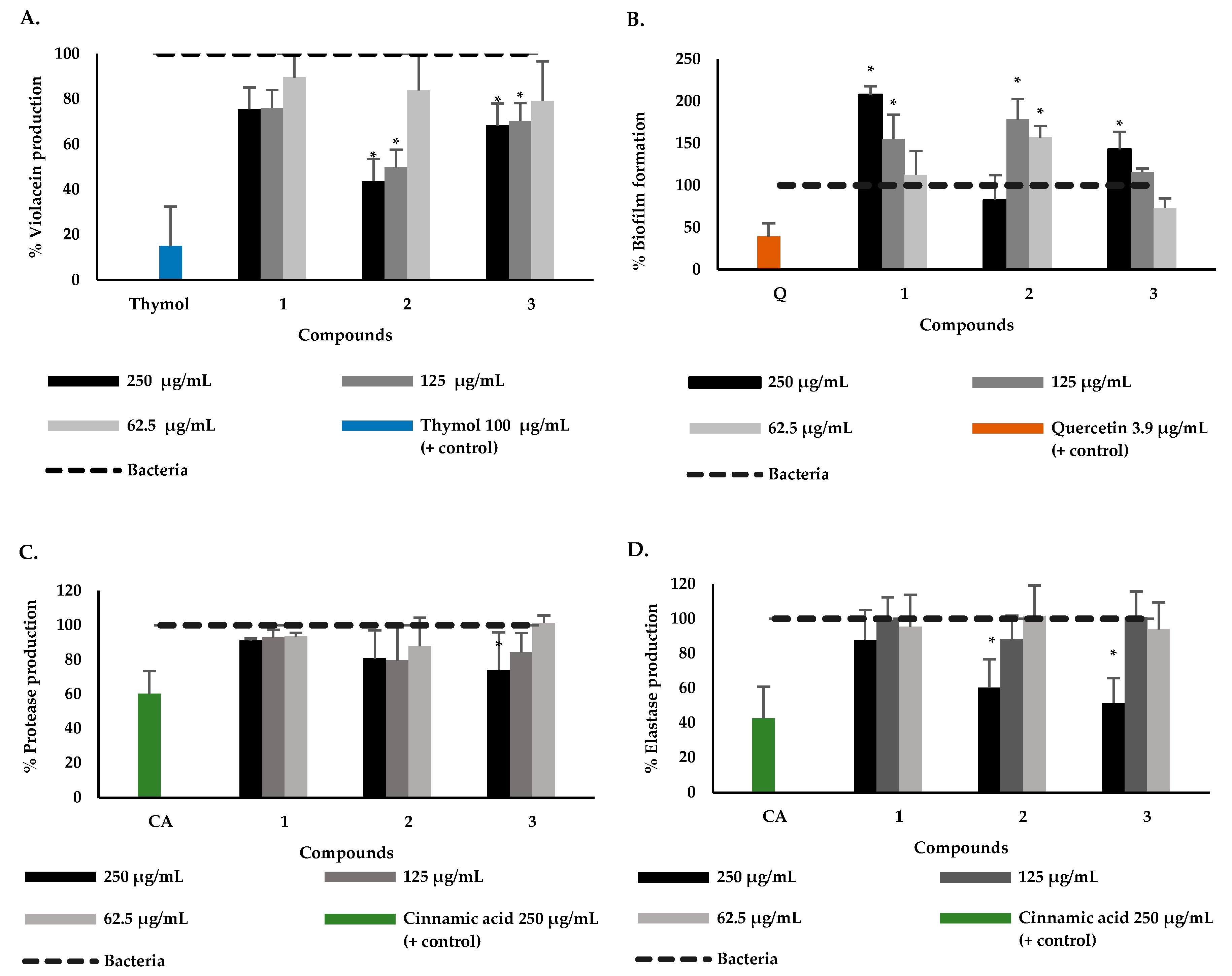
To establish whether compounds 1 to 3 are some of those responsible for the bioactivity observed for the DCM fraction, their effect on violacein production in C. violaceum and biofilm formation in P. aeruginosa was determined and the results are presented in Figure 3A,B. The results showed that the isolated compounds from P. bogotense showed lower activity in inhibiting the production of violacein compared to the extract and DCM fraction. However, it is noteworthy that compounds 2 and 3 significantly reduced the violacein production at the two highest concentrations, with reductions of 43.8% and 49.7% for 2 and 68.3% and 70.2% for 3, respectively (Figure 3A). These results indicate that compounds of P. bogotense can inhibit the QS of the C. violaceum strain since violacein is a product of this system. It is worth noting that the quantification of violacein in C. violaceum has long been utilized as a biosensor assay to search for QS inhibitors, given that this strain can produce and detect acyl-homoserine lactones (AHL) by itself through the expression of this pigment [68,69,70,71,72].
The study demonstrated that compounds 1 to 3 promoted biofilm formation in P. aeruginosa, contrary to what was observed with quercetin (Figure 3B), a compound for which the ability to inhibit P. aeruginosa biofilm has previously been reported [73,74]. These compounds have no previous reports on antibiofilm activity, however, other similar compounds, such as benzoic acid derivatives have shown the potential to reduce the biofilm of P. aeruginosa. For example, gallic acid has been shown to have the ability to reduce the formation by 30.1% (200 µg/mL) [75] and vanillic acid by 46% (4 mmol/L) [76]. It has also been found that hydroquinone-like compounds, such as avarol and sesquiterpene hydroquinone, exhibited inhibitory effects on P. aeruginosa PAO1 biofilm formation at subinhibitory concentrations [77]. It has been reported that phenolic compounds present antimicrobial activity and protect plants from phytopathogenic bacteria. However, in this study, it was found that the evaluated compounds promoted the formation of biofilm in P. aeruginosa, an effect that has previously been reported as a strategy that facilitates the survival and protection of the bacteria against this type of compounds [75]. Among the possible mechanisms that explain the increase in the formation of the film is the effect on the synthesis of AHL, finding that the production of 3-oxo-C12-HSL increases under the action of different phenols at subinhibitory concentrations, leading to an effect in the LasI/LasR complex. Additionally, it has been reported that this type of compound does not directly affect the expression of lasI and rhlI genes, so it is possible that they do not mimic AHL [75].
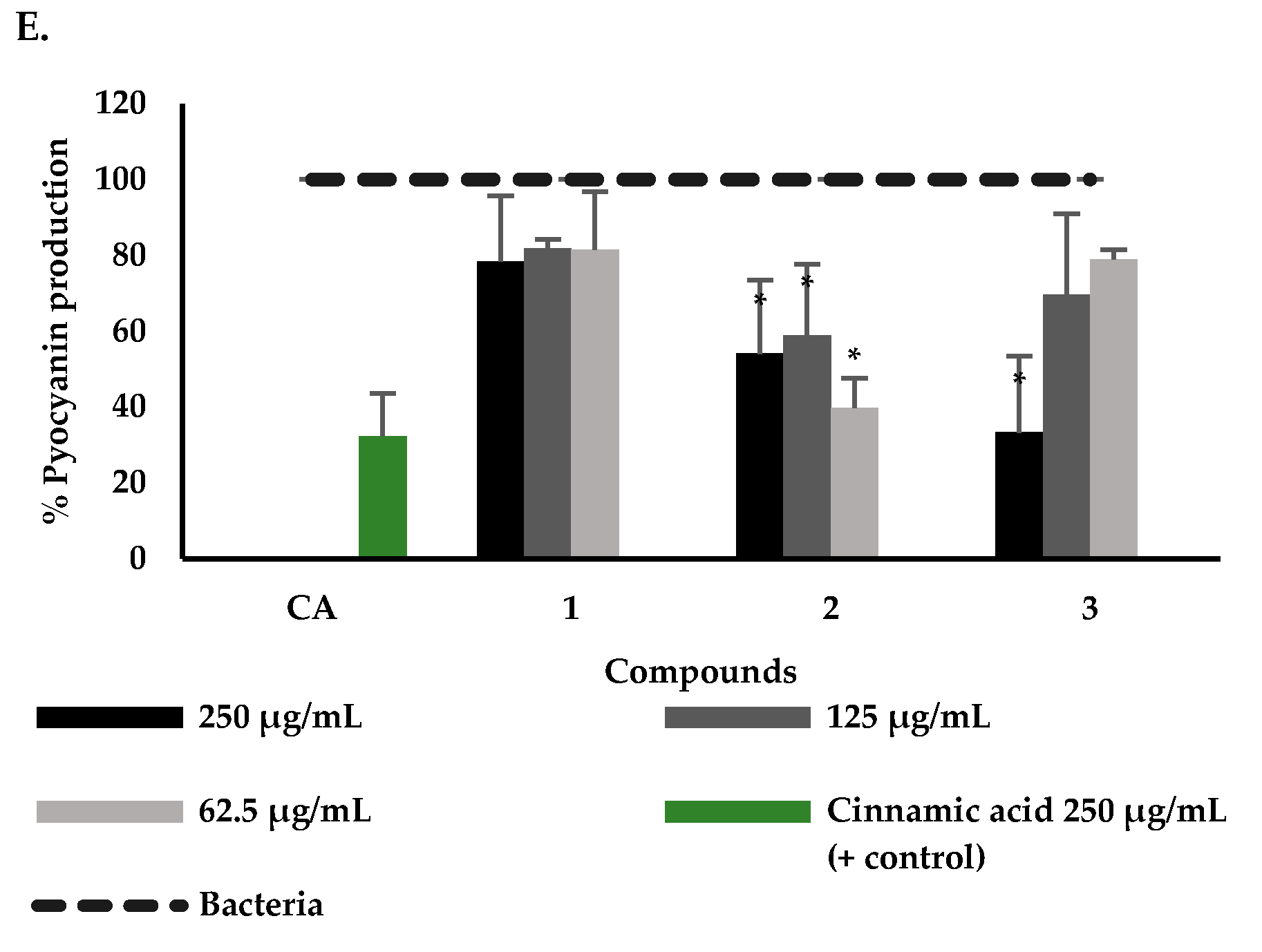
The results obtained indicate that the compounds have the capacity to affect the QS in C. violaceum but not the formation of biofilms in P. areuginosa. Taking into account that biofilm formation is a multifactorial process that involves QS, exopolysaccharide, and c-di-GMP systems [77], it cannot be definitively concluded that P. bogotense compounds do not act on one of the QS pathways of P. aeruginosa. Therefore, to corroborate the effect of these compounds on the QS system of P. aeruginosa, their effect on three virulence factors was evaluated. The effect of compounds 1 to 3 on the production of elastases, proteases, and pyocyanin are shown in Figure 3C–E. It was found that only 3 reduced the production of proteases at the highest concentration (250 µg/mL). In the other two factors’ production, it was found that 2 and 3 exerted an inhibitory effect, with a percentage of elastase production of 60% and 51% at (250 µg/mL), respectively, while pyocyanin production was 39% (62.5 µg/mL) and 33% (250 µg/mL), respectively. In previous studies, cinnamic acid was used as an inhibition control and showed inhibitory activity similar to that found in this study on virulence factors at a concentration of 250 µg/mL [78]. This is the first report of activity on P. aeruginosa virulence factors of P. bogotense compounds. It has been found for similar compounds, such as benzoic acid derivatives, to affect virulence factors; for example, for vanillic acid, its potential to reduce P. aeruginosa pyocyanin production by 16% (4 mmol/L) is reported [76] and in other bacteria such as Serratia marcescens it inhibits protease biosynthesis by 50% [79]. For other hydroquinone-type compounds, such as avarol, it was reported to reduce P. aeruginosa pyocyanin production by 39% [77]. From the analysis of the results obtained with compounds 1 to 3 and the structural characteristics, the following qualitative approximations of structure-activity can be made. When comparing the results obtained with compounds 1 and 2 in the five bioassays, it is observed that compound 1 had no activity in any assay; therefore, it can be indicated that the inactivity of this compound is related to the presence of the methoxyl group in position 4 of the aromatic ring. Comparing now the results found for 2 and 3, it is possible to emphasize that the presence of the carboxyl group in position 1 of the aromatic ring, instead of a hydroxyl group, favors the inhibitory effect on the production of violacein, elastase, pyocyanin, and proteases.
Considering the results, the capacity of the P. bogotense compounds evaluated stands out, since they had an effect on the inhibition of some virulence factors and are considered as an alternative to reduce the pathogenicity of P. aeruginosa without affecting growth. It is important to highlight that compounds capable of altering bacterial QS could potentially be used in combination with conventional antibiotics to increase the efficiency of current antimicrobials by reducing the minimum inhibitory concentration, an approach that may also help to overcome antimicrobial resistance and side effects of drugs [76]. The results of this study do not allow us to know the mechanism of action against the QS system of P. aeruginosa, so it is important to expand the studies to determine it among the possible mechanisms that have been reported for natural molecules as QS inhibitors may be antagonists, inhibitors of AHL biosynthesis and degradation, and AHL receptor mimetics. This could suggest that these molecules would act on the signals produced by the LasR, RhlR, or PQS system, which regulate the virulence factors evaluated [80,81,82].

3. Materials and Methods

3.1. General Experimental Procedures

The solvents were technical grade and distilled before use. Thin-layer chromatography (TLC) was performed on SiliaPlateTM alumina plates pre-coated with silica gel 60 F254 (SiliCycle® Inc., Quebec, QC, Canada). Vacuum Liquid Chromatography (VLC) was performed on SiliaPlateTM silica gel F254 of size 5–20 μm (SiliCycle® Inc., Quebec, QC, Canada). Flash Chromatography (FC) was performed on SiliaFlash® silica gel P60 of size 40–63 μm (SiliCycle® Inc., Quebec, QC, Canada). NMR measurements were performed on Bruker Advance AC-400 spectrometer (Bruker®, Leipzig, Germany) 1H-NMR and APT experiments, operating at 400 MHz for 1H and 100 MHz for APT. The reference strains selected in this study were P. aeruginosa ATCC BAA-47 and C. violaceum ATCC 12472, obtained commercially from the American Type Culture Collection. Azocasein (Sigma-Aldrich®, E.E.U.U., Burlington, MA, USA) was used as substrate for the protease assay, and congo red elastin (Sigma-Aldrich®, E.E.U.U., Burlington, MA, USA) was used for the elastase assay. An orbital shaker with Jeio tech temperature (Jeio tech®, Daejeon, Republic of Korea) was used for the growth of the microorganisms. Optical Density (OD) measurements were performed on a Multiskan sky microplate reader (Multiskan sky Thermo scientific®, E.E.U.U., Waltham, MA, USA).

3.2. Plant Material

The aerial part of the plant was collected in the “Enrique Olaya Herrera” National Park, located in the city of Bogotá D.C. (Colombia). The species was determined by the biologist Ricardo Callejas, and a specimen of P. bogotense C. DC. was deposited in the Universidad de Antioquia Herbarium.

3.3. Extraction and Isolation of Compounds

The dried and ground aerial part of P. bogotense (1000 g) was subjected to extraction with 96% ethanol using the maceration method at room temperature. The resulting solution was concentrated by distillation under reduced pressure to obtain 458 g of ethanolic extract. A part of the extract (150 g) was fractionated by vacuum liquid chromatography (VLC) using solvents of different polarity: dichloromethane (DCM), ethyl acetate (EtOAc), Isopropanol (IPA) and ethanol–water mixture (EtOH:H2O) (80:20). After evaporation of the solvents under reduced pressure, the fractions DCM (40.4 g), EtOAc (20.5 g), IPA (14.0 g) and EtOH:H2O (15.0 g) were obtained. The DCM fraction (40.4 g) was subjected to flash chromatography (FC) eluting with a hexane:EtOAc mixture in increasing polarity (95:5 to 50:50), obtaining 75 fractions that were combined into 4 final fractions according to the TLC profile. Fraction 2 (15.6 g) was subjected to purification by repeated FC eluting with a mixture of hexane:EtOAc (97:3), hexane:acetone (80:20), and hexane:EtOAc (70:30), obtaining a crystalline white solid corresponding to 3-farnesyl-4-methoxybenzoic acid 1 (500.2 mg, m.p. 57–59 °C). Fraction 3 (3.6 g) was subjected to successive FC with a mixture of hexane: EtOAc (95:5) and DCM:EtOAc (80:20), obtaining a green solid corresponding to 3-farnesyl-4-hydroxybenzoic acid 2 (70.5 mg, m.p. 52–54 °C). Fraction 4 (1.8 g) was repeatedly fractionated by successive FC with a mixture of DCM:EtOAc:acetone (60:30:10), Hexane:MEK (80:20), hexane:CHCl3 (60:40), and hexane:EtOAc (70:30), obtaining a white solid corresponding to 2-farnesyl-1,4-hydroquinone 3 (18.0 mg, m.p. 52–54 °C).
3-farnesyl-4-methoxybenzoic acid (1): White solid, melting point (m.p.): 57–59 °C. 1H NMR (400 MHz, CDCl3): δH 7.99 (d, J = 8.6 Hz, 2.2 Hz, 1H, H-2), 7.91 (s, J = 2.2 Hz, 1H, H-6), 6.88 (d, J = 8.6 Hz, 1H, H-3), 5.33 (t, J = 7.2 Hz, 1H, H-2′), 5.12 (dt, J = 17.4, 6.3 Hz, 2H, H-6′, H-10′), 3.91 (s, 3H, H-7), 3.36 (d, J = 7.2 Hz, 2H, H-1′), 2.21—1.84 (m, J = 16.7, 8.2 Hz 8H, H-4′, H-5′, H-8′, H-9′), 1.73 (s, 3H, H-13′), 1.67 (s, 3H, H-14′), 1.61 (s, 3H, H-15′), 1.60 (s, 3H, H-12′). APT (100 MHz, CDCl3): δC 171.8 (C=O; C-8), 161.7 (C-1), 136.6 (C-3′), 134.9 (C-7′), 131.3 (C-6), 131.0 (C-11′), 130.1 (C-2), 130.0 (C-5), 124.3 (C-6′), 124.0 (C-10′), 121.4 (C-2′), 121.2 (C-4), 109.4 (C-3), 55.4 (C-7), 39.6 (C-4′), 39.5 (C-5′), 28.1 (C-1′), 26.6 (C-8′), 26.5 (C-9′), 25.5 (C-13′), 17.5 (C-14′), 15.9 (C-15′), 15.8 (C-12′). The spectroscopic data were consistent with those reported in the literature for 3-farnesyl-4-methoxybenzoic acid [59]. The spectroscopic data can be consulted in Figures S1 and S2 Supplementary Material.
3-farnesyl-4-hydroxybenzoic acid (2): Green solid, melting point (m.p.): 52–54 °C. 1H-NMR (400 MHz, CDCl3): δH 7.91 (d, J = 3.2 Hz, 1H, H-6), 7.90 (s, 1H, = 9.0, 3.2 Hz, H-2), 6.85 (d, J = 9.0 Hz, 1H, H-3), 5.34 (t, J = 6.9 Hz, 1H, H-2′), 5.09 (dt, J = 5.5, 3.8 Hz, 2H, H-6′, H-10′), 3.42 (d, J = 7.1 Hz 2H, H-1′), 2.17–1.95 (m, 8H, J = 5.5, 3.8 Hz, H-4′, H-5′, H-8′, H-9′), 1.79 (s, 3H, H-13′), 1.67 (s, 3H, H-14′), 1.60 (s, 6H, H-15′, H-12′). APT (100 MHz, CDCl3): δC 171.8 (C=O, C-8), 159.6 (C-4), 139.6 (C-3′), 135.7 (C-7′), 132.6 (C-6), 131.3 (C-11′), 130.6 (C-2), 126.8 (C-5), 124.4 (C-10′), 123.5 (C-6′), 121.7 (C-1), 120.3 (C-2′), 115.8 (C-3), 39.7 (C-4′), 39.7 (C-8′), 29.7 (C-1′), 26.7 (C-9′), 26.3 (C-5′), 25.7 (C-12′), 17.9 (C-15′), 16.3 (C-14′), 16.0 (C-13′). The spectroscopic data were consistent with those reported in the literature for 3-farnesyl-4-hydroxybenzoic acid [61,63,64,65]. The spectroscopic data can be consulted in Figures S3 and S4 Supplementary Material.
2-farnesylhydroquinone (3): White solid, melting point (m.p.): 52–54 °C. 1H-NMR (400 MHz, CDCl3): δH 6.67 (d, J = 8.4 Hz, 1H, H-6), 6.61 (d, J = 3.2 Hz, 1H, H-3), 6.59 (dd, J = 8.4, 3.2 Hz, 1H, H-5), 5.30 (t, J = 7.1 Hz, 1H, H-2′), 5.10 (t, J = 6.2 Hz, 2H, H-6′, H-10′), 4.85 (s, 1H, OH), 4.67 (s, 1H, OH), 3.30 (d, J = 7.1 Hz, 2H, H-1′), 2.23–1.89 (m, J = 6.2 Hz, 8H, H-4′, H-5′, H-8′, H-9′), 1.76 (s, 3H, H-13′), 1.68 (s, 3H, H-12′), 1.60 (s, 6H, H-14′, H-15′). APT (100 MHz, CDCl3): δC 149.1 (C-4), 148.0 (C-1), 138.5 (C-3′), 135.4 (C-7′), 131.2 (C-11′), 128.1 (C-2), 124.2 (C-10′), 123.5 (C-6′), 121.1 (C-2′), 116.4 (C-3), 116.4 (C-6), 113.6 (C-5), 39.5 (C-4′)(C-8′), 29.5 (C-1′), 26.5 (C-9′), 26.2 (C-5′), 25.5 (C-12′), 17.5 (C-15′), 16.0 (C-13′), 15.9 (C-14′). The spectroscopic data were consistent with those reported in the literature for 2-farnesylhydroquinone [52]. The spectroscopic data can be consulted in Figures S5 and S6 Supplementary Material.

3.4. Strains and Bacterial Growth Conditions

For the long-term conservation of the morphological, physiological, and genetic characteristics of the strains, the freezing method was used at −70 °C and 10% glycerol in Luria Bertani (LB) broth was used as cryoprotectant. The growth temperature for P. aeruginosa was 37 °C and for C. violaceum, it was 30 °C.

3.5. Evaluation of Bacterial Growth on P. aeruginosa

The determination of the bacterial susceptibility of P. aeruginosa at extract, fractions, and compounds of P. bogotense was performed following the methodology of the Clinical & Laboratory Standards Institute (CLSI) protocol with some modifications [83]. For the extract and fractions, concentrations of 1000, 500 and 250 µg/mL (final in the well), and the isolated compounds 250, 125 and 62.5 µg/mL previously solubilized in dimethyl sulfoxide, DMSO was evaluated. A 96-well plate was used in which 2 µL of each treatment and 2 µL of the inoculum were added to obtain a concentration of 1 × 10⁵ CFU/mL in the final well, which was made up to 200 µL with LB broth. In the assay, five replicates were performed with two independent repetitions. Gentamicin (2 µg/mL), pure DMSO (solvent for the extract), and medium, extract, and growth of P. aeruginosa were used as controls. After 24 h, readings were taken at OD 600 nm, and the concentrations that had no inhibitory effect were identified compared to the control.

3.6. Biofilm Formation and Quantification Assay in P. aeruginosa

Evaluation of P. aeruginosa biofilm formation was performed by the crystal violet staining method [84,85]. A P. aeruginosa culture was prepared and left overnight (24 h) at 37 °C at 180 rpm in an orbital shaker incubator. In a 96-well plate, the extract and fractions were added at the same concentrations evaluated in the previous assay that did not show inhibitory effects on bacterial growth were added, and a static incubation was performed for 24 h. Subsequently, the planktonic cells were discarded, and the plate was washed twice with PBS and allowed to dry completely for half an hour. Next, 250 µL of crystal violet (0.1%) was added for 15 min, discarded, and washed three times with PBS to remove excess dye. Finally, 250 µL of ethanol was added to solubilize the crystal adhering to the biofilm. For all assays, five replicates were performed with two independent repetitions. Quercetin (3.9 µg/mL), pure DMSO (solvent of the extract or compound) and medium, extract or compound, and P. aeruginosa biofilm formation were used as controls. Readings were taken at an OD of 570 nm, and the percentage of biofilm formation was calculated (Equation (1)).
%   B i o f i l m   f o r m a t i o n = A b s   t r e a t m e n t A b s   c o n t r o l × 100

3.7. Quorum Sensing Inhibition Assay: Quantification of Violacein Production from C. violaceum

The evaluation of quorum sensing detection in C. violaceum was performed by the violacein quantification method described previously [86,87]. A bacterial suspension of C. violaceum was prepared in LB broth and adjusted to an OD of 0.4 nm. The extract and fractions were added at the same concentrations evaluated in the biofilm assay. It was left to incubate for 24 h at 30 °C. After this time, the culture was centrifuged at 15,000 rpm for 10 min to separate the cells, and the supernatant was discarded. 500 µL of DMSO was added to solubilize the pigment and centrifuged again at 15,000 rpm for 10 min. A volume of 200 µL of the supernatant was taken, and its absorbance was read at 585 nm in a 96-well plate using a microplate reader. Thymol (100 µg/mL), pure DMSO (solvent of the extract or compound), and medium, compound, and bacteria (C. violaceum) were used as controls. Five replicates with two independent repetitions were performed for all assays, and the results were expressed as percent violacein production (Equation (2)).
%   V i o l a c e i n   p r o d u c t i o n = A b s   t r e a t m e n t A b s   c o n t r o l × 100

3.8. Assays on Virulence Factors in P. aeruginosa

The evaluation of compounds isolated from P. bogotense extract on virulence factors in P. aeruginosa was performed following previously described methodologies [88]. An overnight culture of P. aeruginosa was placed in contact with different concentrations of the compounds (250, 125, and 62.5 µg/mL). After the 24 h incubation, the cultures were centrifuged at 10,000 rpm for 10 min, and the supernatant was used to quantify the production of proteases, elastases, and the pigment pyocyanin. For all assays, cinnamic acid (250 µg/mL), pure DMSO (compound solvent), and medium, compound, and bacteria (P. aeruginosa) were used as controls; and five replicates with two independent repetitions were carried out (Equation (3)).
%   P r o d u c t i o n = A b s   t r e a t m e n t A b s   c o n t r o l × 100

3.8.1. Quantification of Pyocyanin Production

The determination of pyocyanin production was performed using the method described previously [88]. The pigment was extracted from 750 µL of the supernatant with 375 µL of chloroform, the supernatant was removed and the organic layer (blue color) was acidified with 300 µL of 0.2 M HCl. The supernatant took a pink coloration, and from this, 150 µL was taken, which was neutralized with 150 µL of a tris buffer at 200 mM to be able to read absorbance at 390 nm.

3.8.2. Quantification of Elastase Production

Protease production was evaluated following the methodology described previously [88]. Twenty-five µL of the supernatant was taken and 225 µL of 100 mM tris buffer pH 7.5, supplemented with 10 mg/mL elastin red Congo (ERC) was added. This mixture was incubated for 3 h at 37 °C under agitation. After this time, PBS buffer at pH 6.0 was added and allowed to cool for 2 min to stop the reaction. Finally, the mixture was centrifuged at 10,000 rpm for 10 min to separate the insoluble ERC and measured absorbance at 495 nm.

3.8.3. Quantification of Protease Production

Proteolytic activity was performed using protease production and quantification by the method previously described [88]. 37.5 µL of the supernatant was taken and a solution of 100 mM tris buffer pH 8.0 supplemented with 0.3% azocasein was added and incubated for one hour at 37 °C statically. Subsequently, trichloroacetic acid (TCA 10%) was added to stop the reaction and insoluble azocasein was centrifuged at 10,000 rpm for 10 min to measure absorbance at 490 nm.

3.9. Data Analysis

The one-way ANOVA statistical test was performed considering the assumptions of the test (normality, homogeneity of variances, independence, randomness, and outliers) to determine if there were significant differences in the trials. The data that presented significant differences were subjected to additional multiple comparison tests, such as Duncan for normal data, to confirm in which group the differences occurred. These analyses were performed in the R studio statistical program. All the results reported correspond to the mean of five replicates and their respective standard deviation, using a statistical significance of p < 0.05.

4. Conclusions

This study represents the first report on the isolation of two benzoic acid derivatives (3-farnesyl-4-methoxybenzoic acid 1 and 3-farnesyl-4-hydroxybenzoic acid 2) from the aerial part of P. bogotense. Furthermore, it is the first time that the antibacterial and antiQS activity of the ethanolic extract of P. bogotense and its isolated compounds has been tested against P. aeruginosa and C. violaceum. Our findings indicate that chemical components of P. bogotense have the potential to attenuate the QS system of P. aeruginosa, mainly compound 2 (3-farnesyl-4-hydroxybenzoic acid), which exhibits inhibition of violacein, pyocyanin, elastases, protease and biofilm training. Therefore, the phenolic compounds identified from P. bogotense, capable of altering the bacterial QS, could be used in combination with conventional antibiotics to increase the efficiency of current antimicrobials by reducing the minimum inhibitory concentration, reducing resistance to antimicrobials and drug side effects.

Supplementary Materials

The following supporting information can be downloaded at: https://www.mdpi.com/article/10.3390/plants12091901/s1, Scheme S1: Isolation scheme of compounds 1 to 3 from P. bogotense; Tables S1–S3: supplementary data on the effect of the extract and fractions on susceptibility bioassays; biofilm formation; violacein production, Tables S4 and S5: supplementary data on the effect of the compounds of P. bogotense on susceptibility bioassays; biofilm formation; violacein production and virulence factor production; Figures S1–S6: 1H-NMR and APT spectra of compounds 1 to 3.

Author Contributions

Conceptualization, A.G.S.-Q., L.V.H.-M., O.J.P.-L., L.C.P.-B. and J.A.P.-R.; methodology, A.G.S.-Q., L.V.H.-M., O.J.P.-L. and L.C.P.-B.; formal analysis, A.G.S.-Q., L.V.H.-M., O.J.P.-L. and L.C.P.-B.; study investigation, A.G.S.-Q. and L.V.H.-M.; data curation and interpretation, A.G.S.-Q., L.V.H.-M., O.J.P.-L., L.C.P.-B. and J.A.P.-R.; original draft preparation, L.C.P.-B., J.A.P.-R. and O.J.P.-L.; supervision, O.J.P.-L., L.C.P.-B. and J.A.P.-R.; funding acquisition, O.J.P.-L., L.C.P.-B. and J.A.P.-R. All authors contributed to the writing of the manuscript. All authors have read and agreed to the published version of the manuscript.

Funding

This research was funded by Universidad Nacional de Colombia, Universidad de La Salle and Ministerio de Ciencia Tecnología e Innovación (MINCIENCIAS) through contract 835-2017 with code 1101-777-58105, announcement “777-2017 Convocatoria para proyectos de ciencia, tecnología e innovación en salud 2017”and by Pontificia Universidad Javeriana.

Data Availability Statement

Not applicable.

Acknowledgments

The authors extend their gratitude to the research groups (QUIPRONAB and BioMolUN belonging to the Universidad Nacional de Colombia, BIOMIGEN belonging to the Universidad de La Salle, and GIFUJ belonging to the Pontificia Universidad Javeriana) for their collaboration in the development of the research.

Conflicts of Interest

The authors declare no conflict of interest.

References

  1. WHO. Global Antimicrobial Resistance and Use Surveillance System (GLASS) Report. Available online: https://apps.who.int/iris/bitstream/handle/10665/341666/9789240027336-eng.pdf (accessed on 29 March 2023).
  2. World Bank. Drug-Resistant Infections: A Threat to Our Economic Future. Available online: www.worldbank.org (accessed on 29 March 2023).
  3. Haque, S.; Ahmad, F.; Dar, S.A.; Jawed, A.; Mandal, R.K.; Wahid, M.; Lohani, M.; Khan, S.; Singh, V.; Akhter, N. Developments in strategies for quorum sensing virulence factor inhibition to combat bacterial drug resistance. Microb. Pathog. 2018, 121, 293–302. [Google Scholar] [CrossRef] [PubMed]
  4. Lu, L.; Li, M.; Yi, G.; Liao, L.; Cheng, Q.; Zhu, J.; Zhang, B.; Wang, Y.; Chen, Y.; Zeng, M. Screening strategies for quorum sensing inhibitors in combating bacterial infections. J. Pharm. Anal. 2022, 12, 1–14. [Google Scholar] [CrossRef] [PubMed]
  5. Santhakumari, S.; Ravi, A.V. Targeting quorum sensing mechanism: An alternative anti-virulent strategy for the treatment of bacterial infections. S. Afr. J. Bot. 2019, 120, 81–86. [Google Scholar] [CrossRef]
  6. Saeki, E.K.; Kobayashi, R.K.T.; Nakazato, G. Quorum sensing system: Target to control the spread of bacterial infections. Microb. Pathog. 2020, 142, 104068. [Google Scholar] [CrossRef] [PubMed]
  7. Vadakkan, K. Molecular mechanism of bacterial quorum sensing and its inhibition by target specific approaches. ACS Symp. Ser. 2020, 1374, 21–234. [Google Scholar] [CrossRef]
  8. Botelho, J.; Grosso, F.; Peixe, L. Antibiotic resistance in Pseudomonas aeruginosa—mechanisms, epidemiology and evolution. Drug Resist. Updates 2019, 44, 100640. [Google Scholar] [CrossRef]
  9. Elabbadi, A.; Pont, S.; Verdet, C.; Plésiat, P.; Cretin, F.; Voiriot, G.; Fartoukh, M.; Djibré, M. An unusual community-acquired invasive and multi systemic infection due to ExoU-harboring Pseudomonas aeruginosa strain: Clinical disease and microbiological characteristics. J. Microbiol. Immunol. Infect. 2019, 53, 647–651. [Google Scholar] [CrossRef]
  10. Høiby, N.; Bjarnsholt, T.; Givskov, M.; Molin, S.; Ciofu, O. Antibiotic resistance of bacterial biofilms. Int. J. Antimicrob. Agents 2010, 35, 322–332. [Google Scholar] [CrossRef]
  11. Aguilar, F.R.; Aguilar, S.L.; Cubas, D.M.; Coaguila, L.Á.; Fernández, D.A.; Mario, M.M.; Campos, R.; Guevara-Vásquez, G.; Díaz, R.S. Portadores de bacterias multirresistentes de importancia clínica en áreas críticas (UCI-UCIN) de un hospital al norte del Perú. Horiz. Médico 2016, 16, 50–57. [Google Scholar] [CrossRef]
  12. Karakonstantis, S.; Kritsotakis, E.I.; Gikas, A. Pandrug-resistant gram-negative bacteria: A systematic review of current epidemiology, prognosis and treatment options. J. Antimicrob. Chemother. 2020, 75, 271–282. [Google Scholar] [CrossRef]
  13. Luo, J.; Dong, B.; Wang, K.; Cai, S.; Liu, T.; Cheng, X.; Lei, D.; Chen, Y.; Li, Y.; Kong, J.; et al. Baicalin inhibits biofilm formation, attenuates the quorum sensing-controlled virulence and enhances Pseudomonas aeruginosa clearance in a mouse peritoneal implant infection model. PLoS ONE 2017, 12, 0176883. [Google Scholar] [CrossRef]
  14. Silva, A.; Silva, V.; Igrejas, G.; Poeta, P. Chapter 17: Carbapenems and Pseudomonas aeruginosa: Mechanisms and Epidemiology. In Antibiotics and Antimicrobial Resistance Genes in the Environment: Volume 1 in the Advances in Environmental Pollution Research Series; Elsevier: Amsterdam, The Netherlands, 2020; Volume 1, pp. 253–268. ISBN 9780128188828. [Google Scholar]
  15. Yendo, T.M.; de Pante, C.C.; Miyamoto, D. Pseudomonas aeruginosa as an uncommon agent of infectious panniculitis. An. Bras. Dermatol. 2022, 97, 326–331. [Google Scholar] [CrossRef] [PubMed]
  16. Sambrano, H.; Castillo, J.C.; Ramos, C.W.; de Mayorga, B.; Chen, O.; Durán, O.; Ciniglio, C.; Aguilar, C.; Cisterna, O.; de Chial, M. Prevalence of antibiotic resistance and virulent factors in nosocomial clinical isolates of Pseudomonas aeruginosa from Panamá. Braz. J. Infect. Dis. 2021, 25, 101038. [Google Scholar] [CrossRef] [PubMed]
  17. Silby, M.W.; Winstanley, C.; Godfrey, S.A.C.; Levy, S.B.; Jackson, R.W. Pseudomonas genomes: Diverse and adaptable. FEMS Microbiol. Rev. 2011, 35, 652–680. [Google Scholar] [CrossRef] [PubMed]
  18. Pirnay, J.-P.; Bilocq, F.; Pot, B.; Cornelis, P.; Zizi, M.; Van Eldere, J.; Deschaght, P.; Vaneechoutte, M.; Jennes, S.; Pitt, T.; et al. Pseudomonas aeruginosa population structure revisited. PLoS ONE 2009, 4, e7740. [Google Scholar] [CrossRef] [PubMed]
  19. WHO. WHO Publishes List of Bacteria for Which New Antibiotics Are Urgently Needed. Available online: https://www.who.int/news/item/27-02-2017-who-publishes-list-of-bacteria-for-which-new-antibiotics-are-urgently-needed (accessed on 29 September 2022).
  20. Amaning Danquah, C.; Osei-Djarbeng, S.; Appiah, T.; Duah Boakye, Y.; Adu, F. Combating Biofilm and Quorum Sensing: A New Strategy to Fight Infections. In Bacterial Biofilms; IntechOpen: London, UK, 2020. [Google Scholar]
  21. Mohsenipour, Z.; Hassanshahian, M. The effects of Allium sativum extracts on biofilm formation and activities of six pathogenic bacteria. Jundishapur J. Microbiol. 2015, 8, e18971. [Google Scholar] [CrossRef] [PubMed]
  22. Qais, F.A.; Ahmad, I. Anti-quorum sensing and biofilm inhibitory effect of some medicinal plants against gram-negative bacterial pathogens: In vitro and in silico investigations. Heliyon 2022, 8, e11113. [Google Scholar] [CrossRef]
  23. Lu, L.; Hu, W.; Tian, Z.; Yuan, D.; Yi, G.; Zhou, Y.; Cheng, Q.; Zhu, J.; Li, M. Developing natural products as potential anti-biofilm agents. Chin. Med. 2019, 14, 11. [Google Scholar] [CrossRef]
  24. Abdel Bar, F.M.; Alossaimi, M.A.; Elekhnawy, E.; Alzeer, M.A.A.; Abo Kamer, A.; Moglad, E.; ElNaggar, M.H. Anti-quorum sensing and anti-biofilm activity of Pelargonium × hortorum root extract against Pseudomonas aeruginosa: Combinatorial effect of catechin and gallic acid. Molecules 2022, 27, 7841. [Google Scholar] [CrossRef]
  25. Ćirić, A.D.; Petrović, J.D.; Glamočlija, J.M.; Smiljković, M.S.; Nikolić, M.M.; Stojković, D.S.; Soković, M.D. Natural products as biofilm formation antagonists and regulators of quorum sensing functions: A comprehensive review update and future trends. S. Afr. J. Bot. 2019, 120, 65–80. [Google Scholar] [CrossRef]
  26. dos Santos, V.L.P.; Rodrigues, I.C.G.; Berté, R.; Raman, V.; Messias-Reason, I.J.; Budel, J.M. Review of Piper species growing in the Brazilian State of Paraná with emphasize on the vegetative anatomy and biological activities. Bot. Rev. 2021, 87, 23–54. [Google Scholar] [CrossRef]
  27. Ashokkumar, K.; Murugan, M.; Dhanya, M.K.; Pandian, A.; Warkentin, T.D. Phytochemistry and therapeutic potential of black pepper [Piper nigrum (L.)] essential oil and piperine: A review. Clin. Phytoscience 2021, 7, 52. [Google Scholar] [CrossRef]
  28. de Tamokou, J.D.; Mbaveng, A.T.; Kuete, V. Chapter 8: Antimicrobial Activities of African Medicinal Spices and Vegetables. In Medicinal Spices and Vegetables from Africa; Academic Press: Cambridge, MA, USA, 2017; pp. 207–237. ISBN 9780128094419. [Google Scholar]
  29. The Plant List. Available online: http://www.theplantlist.org/ (accessed on 19 October 2022).
  30. Fan, D.; Zhou, C.; Chen, C.; Li, X.; Ma, J.; Hu, Y.; Li, G.; Ruan, J.; Wu, A.; Li, L.; et al. Lignans from the genus Piper L. and their pharmacological activities: An updated review. Fitoterapia 2023, 165, 105403. [Google Scholar] [CrossRef] [PubMed]
  31. Parmar, V.S.; Jain, S.C.; Bisht, K.S.; Jain, R.; Taneja, P.; Jha, A.; Tyagi, O.D.; Prasad, A.K.; Wengel, J.; Olsen, C.E.; et al. Phytochemistry of the genus Piper. Phytochemistry 1997, 46, 597–673. [Google Scholar] [CrossRef]
  32. Chakraborty, D.; Shah, B. Antimicrobial, antioxidative and antihemolytic activity of Piper betel leaf extracts. Int. J. Pharm. Pharm. Sci. 2011, 3, 192–199. [Google Scholar]
  33. Ganesh, P.; Suresh Kumar, R.; Saranraj, P. Phytochemical analysis and antibacterial activity of Pepper (Piper nigrum L.) against some human pathogens. Sch. Res. Libr. Cent. Eur. J. Exp. Biol. 2014, 3, 36–41. [Google Scholar]
  34. Trivedi, M.N.; Khemani, A.; Vachhani, U.D.; Shah, C.P.; Santani, D.D. Pharmacognostic, phytochemical analysis and antimicrobial activity of two Piper species. Pharm. Glob. 2011, 7, 1–4. [Google Scholar]
  35. Vallejo, A.; Feitosa, A.; Gourlart, A.; Pires, L.; Mosquera, O. Tamizaje de acción antimicrobiana de 34 extractos vegetales contra bacilos gramnegativos. Salud. Soc. Uptc. 2014, 1, 34–39. [Google Scholar]
  36. Prabhu, A.; Chembili, V.; Kandal, T.; Punchappady-Devasya, R. Piper nigrum seeds inhibit biofilm formation in Pseudomonas aeruginosa strains. Res. J. Pharm. Technol. 2017, 10, 3894–3898. [Google Scholar] [CrossRef]
  37. Datta, S.; Jana, D.; Maity, T.R.; Samanta, A.; Banerjee, R. Piper betle leaf extract affects the quorum sensing and hence virulence of Pseudomonas aeruginosa PAO1. 3 Biotech 2016, 6, 18. [Google Scholar] [CrossRef]
  38. Zaki, A.A. Assessment of anti-quorum sensing activity for some ornamental and medicinal plants native to Egypt. Sci. Pharm. 2013, 81, 251–258. [Google Scholar] [CrossRef] [PubMed]
  39. Martín-Rodríguez, A.J.; Ticona, J.C.; Jiménez, I.A.; Flores, N.; Fernández, J.J.; Bazzocchi, I.L. Flavonoids from Piper delineatum modulate quorum-sensing-regulated phenotypes in Vibrio harveyi. Phytochemistry 2015, 117, 98–106. [Google Scholar] [CrossRef] [PubMed]
  40. Vázquez-Martínez, J.; Buitemea-Cantúa, G.V.; Gutierrez-Villagomez, J.M.; García-González, J.P.; Ramírez-Chávez, E.; Molina-Torres, J. Bioautography and GC-MS based identification of piperine and trichostachine as the active quorum quenching compounds in black pepper. Heliyon 2020, 6, e03137. [Google Scholar] [CrossRef]
  41. Tan, L.Y.; Yin, W.F.; Chan, K.G. Piper nigrum, Piper betle and Gnetum gnemon-Natural food sources with anti-quorum sensing properties. Sensors 2013, 13, 3975–3985. [Google Scholar] [CrossRef]
  42. Srinivasan, R.; Santhakumari, S.; Ravi, A.V. In vitro antibiofilm efficacy of Piper betle against quorum sensing mediated biofilm formation of luminescent Vibrio harveyi. Microb. Pathog. 2017, 110, 232–239. [Google Scholar] [CrossRef]
  43. Olivero, J.T.V.; Pájaro, N.P.C.; Stashenko, E. Antiquorum sensing activity of essential oils isolated from different species of the genus Piper. Vitae 2011, 18, 77–82. [Google Scholar] [CrossRef]
  44. Amrutha, B.; Sundar, K.; Shetty, P.H. Spice oil nanoemulsions: Potential natural inhibitors against pathogenic E. coli and Salmonella spp. from fresh fruits and vegetables. LWT Food Sci. Technol. 2017, 79, 152–159. [Google Scholar] [CrossRef]
  45. Sen, S.; Rengaian, G. A Review on the Ecology, Evolution and Conservation of Piper (Piperaceae) in India: Future Directions and Opportunities. Bot. Rev. 2021, 88, 333–358. [Google Scholar] [CrossRef]
  46. Muñoz, D.; Calvache, D.; Yela, J. Especies forestales con potencial agroforestal para las zonas altas en el departamento de Nariño. Rev. De Cienc. Agrícolas 2013, 30, 38–53. [Google Scholar]
  47. de Bogota, S.A. de Cordoncillo: Beneficioso Para Los Procesos de Restauración Ecológica y Para La Salud—Historial de Noticias—Secretaría Distrital de Ambiente. Available online: https://ambientebogota.gov.co/es/historial-de-noticias/-/asset_publisher/VqEYxdh9mhVF/content/cordoncillo-beneficioso-para-los-procesos-de-restauracion-ecologica-y-para-la-salud?redirect=https%3A%2F%2Fambientebogota.gov.co%2Fes%2Fhistorial-de-noticias%3Fp (accessed on 10 April 2022).
  48. Secretaria de Ambiente de Bogotá Propiedades de La Planta Cordoncillo Que Habita En Bogotá|Bogota.Gov.Co. Available online: https://bogota.gov.co/mi-ciudad/ambiente/propiedades-de-la-planta-cordoncillo-que-habita-en-bogota (accessed on 29 March 2023).
  49. Tene, V.; Malagón, O.; Finzi, P.V.; Vidari, G.; Armijos, C.; Zaragoza, T. An ethnobotanical survey of medicinal plants used in Loja and Zamora-Chinchipe, Ecuador. J. Ethnopharmacol. 2007, 111, 63–81. [Google Scholar] [CrossRef]
  50. Trelease, W.; Yuncker, G.T. The Piperaceae of Northern South America; University of Illinois Press: Champaign, IL, USA, 1950; Volume 1. [Google Scholar]
  51. López-Ogalla, J.; García-Palomero, E.; Sánchez-Quesada, J.; Rubio, L.; Delgado, E.; García, P.; Medina, M.; Castro, A.; Muñoz, P. Bioactive prenylated phenyl derivatives derived from marine natural products: Novel scaffolds for the design of BACE inhibitors. Medchemcomm 2014, 5, 474–488. [Google Scholar] [CrossRef]
  52. Peña, L.A.; Avella, E.; Puentes de Díaz, A.M. Benzoquinona e hidroquinona preniladas y otros constituyentes aislados de Piper bogotense c. dc. Rev. Colomb. De Química 2000, 29, 25–37. [Google Scholar]
  53. Tangarife-Castaño, V.; Correa-Royero, J.B.; Roa-Linares, V.C.; Pino-Benitez, N.; Betancur-Galvis, L.A.; Durán, D.C.; Stashenko, E.E.; Mesa-Arango, A.C. Anti-dermatophyte, anti-fusarium and cytotoxic activity of essential oils and plant extracts of Piper genus. J. Essent. Oil Res. 2014, 26, 221–227. [Google Scholar] [CrossRef]
  54. Castañeda, M.L.; Martínez Químico, J.R.; Stanshenko, E.E. Estudio de la composición química y la actividad biológica de los aceites esenciales de diez plantas aromáticas colombianas. Sci. Tech. 2007, XIII, 165–166. [Google Scholar] [CrossRef]
  55. Durant-Archibold, A.A.; Santana, A.I.; Gupta, M.P. Ethnomedical Uses and Pharmacological Activities of Most Prevalent Species of Genus Piper in Panama: A Review. J. Ethnopharmacol. 2018, 217, 63–82. [Google Scholar] [CrossRef] [PubMed]
  56. Muñoz, D.R.; Sandoval-Hernandez, A.G.; Delgado, W.A.; Arboleda, G.H.; Cuca, L.E. In vitro anticancer screening of colombian plants from Piper genus (Piperaceae). J. Pharmacogn. Phytother. 2018, 10, 174–181. [Google Scholar] [CrossRef]
  57. Calderón, Á.I.; Vázquez, Y.; Solís, P.N.; Caballero-George, C.; Zacchino, S.; Gimenez, A.; Pinzón, R.; Cáceres, A.; Tamayo, G.; Correa, M. Screening of latin american plants for cytotoxic activity. Pharm. Biol. 2006, 44, 130–140. [Google Scholar] [CrossRef]
  58. Da Costa, J.G.; Campos, A.R.; Brito, S.A.; Pereira, C.K.B.; Souza, E.O.; Rodrigues, F.F.G. Biological screening of araripe basin medicinal plants using Artemia salina Leach and pathogenic bacteria. Pharmacogn. Mag. 2010, 6, 331. [Google Scholar] [CrossRef]
  59. Maxwell, A.; Rampersad, D. Prenylated 4-hydroxybenzoic acid derivatives from Piper marginatum. J. Nat. Prod. 1988, 51, 370–373. [Google Scholar] [CrossRef]
  60. De Oliveira Chaves, M.C.; De Oliveira Santos, B.V. Constituents from Piper marginatum fruits. Fitoterapia 2002, 73, 547–549. [Google Scholar] [CrossRef]
  61. Ampofo, S.A.; Roussis, V.; Wiemer, D.F. New prenylated phenolics from Piper auritum. Phytochemistry 1987, 26, 2367–2370. [Google Scholar] [CrossRef]
  62. Sáez Vega, A.; Rojano, B.; Blair, S.; Segura, C.; Figadere, B.; Seon, B.; Grellier, P.; Sáez, J. Antimalarials and antioxidants compounds from Piper tricuspe (Piperaceae). Pharmacologyonline 2008, 1, 1–8. [Google Scholar]
  63. López, S.N.; Lopes, A.A.; Batista, J.M.; Flausino, O.; da Bolzani, V.S.; Kato, M.J.; Furlan, M. Geranylation of benzoic acid derivatives by enzymatic extracts from Piper crassinervium (Piperaceae). Bioresour. Technol. 2010, 101, 4251–4260. [Google Scholar] [CrossRef] [PubMed]
  64. Malami, I. Prenylated benzoic acid derivatives from Piper species as source of anti-infective agents. Int. J. Pharm. Sci. Res. 2012, 3, 1554–1559. [Google Scholar] [CrossRef]
  65. Lee, B.; Boucher, H.W. Targeting Antimicrobial-Resistant Bacterial Respiratory Tract Pathogens. Curr Opin Pulm Med. 2015, 21, 293–303. [Google Scholar] [CrossRef] [PubMed]
  66. Rüegg, T.; Calderón, A.I.; Queiroz, E.F.; Solís, P.N.; Marston, A.; Rivas, F.; Ortega-Barría, E.; Hostettmann, K.; Gupta, M.P. 3-Farnesyl-2-hydroxybenzoic acid is a new anti-Helicobacter pylori compound from Piper multiplinervium. J. Ethnopharmacol. 2006, 103, 461–467. [Google Scholar] [CrossRef]
  67. Fang, H.Y.; Chokkalingam, U.; Chiou, S.F.; Hwang, T.L.; Chen, S.L.; Wang, W.L.; Sheu, J.H. Bioactive chemical constituents from the brown alga Homoeostrichus formosana. Int. J. Mol. Sci. 2015, 16, 736–746. [Google Scholar] [CrossRef]
  68. Steindler, L.; Venturi, V. Detection of quorum-sensing N -acyl homoserine lactone signal molecules by bacterial biosensors. FEMS Microbiol. Lett. 2007, 266, 1–9. [Google Scholar] [CrossRef]
  69. Papenfort, K.; Bassler, B.L. Quorum sensing signal–response systems in Gram-negative bacteria. Nat. Rev. Microbiol. 2016, 14, 576–588. [Google Scholar] [CrossRef]
  70. Blosser, R.S.; Gray, K.M. Extraction of violacein from Chromobacterium violaceum provides a new quantitative bioassay for N-acyl homoserine lactone autoinducers. J. Microbiol. Methods 2000, 40, 47–55. [Google Scholar] [CrossRef]
  71. Rai, N.; Rai, R.; Venkatesh, K.V. Quorum Sensing Biosensors. In Quorum Sensing vs Quorum Quenching: A Battle with No End in Sight; Springer India: New Delhi, India, 2015; pp. 173–183. [Google Scholar]
  72. Lozano Juzgado, R. Nuevas Estrategias En La Lucha Contra La Resistencia a Los Antibióticos: Inhibición Del Quorum Sensing. Bachelor’s Thesis, Universidad Complutense, Madrid, España, 2017. [Google Scholar]
  73. Ouyang, J.; Sun, F.; Feng, W.; Sun, Y.; Qiu, X.; Xiong, L.; Liu, Y.; Chen, Y. Quercetin is an effective inhibitor of quorum sensing, biofilm formation and virulence factors in Pseudomonas aeruginosa. J. Appl. Microbiol. 2016, 120, 966–974. [Google Scholar] [CrossRef]
  74. Geng, Y.F.; Yang, C.; Zhang, Y.; Tao, S.N.; Mei, J.; Zhang, X.C.; Sun, Y.J.; Zhao, B.T. An innovative role for luteolin as a natural quorum sensing inhibitor in Pseudomonas aeruginosa. Life Sci. 2021, 274, 119325. [Google Scholar] [CrossRef]
  75. Plyuta, V.; Zaitseva, J.; Lobakova, E.; Zagoskina, N.; Kuznetsov, A.; Khmel, I. Effect of plant phenolic compounds on biofilm formation by Pseudomonas aeruginosa. APMIS 2013, 121, 1073–1081. [Google Scholar] [CrossRef] [PubMed]
  76. Ugurlu, A.; Karahasan Yagci, A.; Ulusoy, S.; Aksu, B.; Bosgelmez-Tinaz, G. Phenolic compounds affect production of pyocyanin, swarming motility and biofilm formation of Pseudomonas aeruginosa. Asian Pac. J. Trop. Biomed. 2016, 8, 698–701. [Google Scholar] [CrossRef]
  77. Thi, M.T.T.; Wibowo, D.; Rehm, B.H.A. Pseudomonas Aeruginosa Biofilms. Int J Mol Sci. 2020, 21, 8671. [Google Scholar] [CrossRef] [PubMed]
  78. Rajkumari, J.; Borkotoky, S.; Murali, A.; Suchiang, K.; Mohanty, S.K.; Busi, S. Cinnamic acid attenuates quorum sensing associated virulence factors and biofilm formation in Pseudomonas aeruginosa PAO1. Biotechnol. Lett. 2018, 40, 1087–1100. [Google Scholar] [CrossRef] [PubMed]
  79. Sethupathy, S.; Ananthi, S.; Selvaraj, A.; Shanmuganathan, B.; Vigneshwari, L.; Balamurugan, K.; Mahalingam, S.; Pandian, S.K. Vanillic acid from Actinidia deliciosa impedes virulence in Serratia marcescens by affecting S-layer, flagellin and fatty acid biosynthesis proteins. Sci. Rep. 2017, 7, 16328. [Google Scholar] [CrossRef] [PubMed]
  80. Lee, J.; Zhang, L. The Hierarchy Quorum Sensing Network in Pseudomonas Aeruginosa. Protein Cell. 2015, 6, 26–41. [Google Scholar] [CrossRef]
  81. Asfour, H. Anti-Quorum Sensing Natural Compounds. J Microsc Ultrastruct. 2018, 6, 1. [Google Scholar] [CrossRef]
  82. Bouyahya, A.; Dakka, N.; Et-Touys, A.; Abrini, J.; Bakri, Y. Medicinal Plant Products Targeting Quorum Sensing for Combating Bacterial Infections. Asian Pac J Trop Med. 2017, 10, 729–743. [Google Scholar] [CrossRef]
  83. Clinical and Laboratory Standards Institute (CLSI). Methods for Dilution Antimicrobial Susceptibility Tests for Bacteria That Grow Aerobically; Approved Standard—Tenth Edition. CLSI Document M07-A10. Wayne, PA: Clinical and Laboratory Standards Institute; 2015, 11th ed.; CLSI standard M07, Ed.; Clinical and Laboratory Standards Institute: Wayne, NJ, USA, 2018; ISBN 1-56238-836-3. [Google Scholar]
  84. O’Toole, G.A. Microtiter dish biofilm formation assay. J. Vis. Exp. 2011, 47, e2437. [Google Scholar] [CrossRef]
  85. O’Toole, G.A.; Kolter, R. Initiation of biofilm formation in Pseudomonas fluorescens WCS365 proceeds via multiple, convergent signalling pathways: A genetic analysis. Mol. Microbiol. 1998, 28, 449–461. [Google Scholar] [CrossRef] [PubMed]
  86. Luís, Â.; Duarte, A.; Gominho, J.; Domingues, F.; Duarte, A.P. Chemical composition, antioxidant, antibacterial and anti-quorum sensing activities of Eucalyptus globulus and Eucalyptus radiata essential oils. Ind. Crops Prod. 2016, 79, 274–282. [Google Scholar] [CrossRef]
  87. Aleksic, I.; Ristivojevic, P.; Pavic, A.; Radojević, I.; Čomić, L.R.; Vasiljevic, B.; Opsenica, D.; Milojković-Opsenica, D.; Senerovic, L. Anti-quorum sensing activity, toxicity in zebrafish (Danio rerio) embryos and phytochemical characterization of Trapa natans leaf extracts. J. Ethnopharmacol. 2018, 222, 148–158. [Google Scholar] [CrossRef] [PubMed]
  88. Filloux, A.; Ramos, J. Pseudomonas Methods and Protocols Methods in Molecular Biology 1149; Filloux, A., Ramos, J.-L., Eds.; Springer: New York, NY, USA, 2014; Volume 1149, ISBN 978-1-4939-0472-3. [Google Scholar]
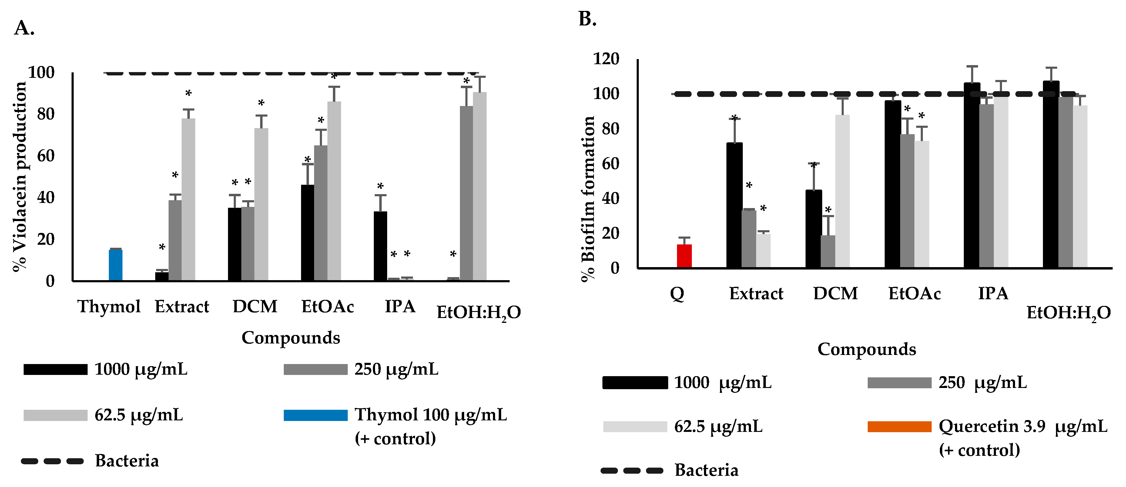
Figure 1. Effect of the extract and fractions of P. bogotense on the production of violacein of C. violaceum (A) and the formation of biofilm in P. aeruginosa (B). Data are represented by the mean ± standard deviation of five independent replicates. * Indicate a significant difference according to Duncan’s test (p < 0.05).
Plants 12 01901 g001
Figure 2. Chemical constituents isolated and identified from P. bogotense.
Plants 12 01901 g002aPlants 12 01901 g002b
Figure 3. Effect of P. bogotense compounds on violacein production in C. violaceum (A), biofilm formation (B), protease (C), elastase (D), and pyocyanin (E) production of P. aeruginosa. Data are represented the mean ± standard deviation of five independent replicates. * Indicate a significant difference according to Duncan’s test (p < 0.05).
Plants 12 01901 g003aPlants 12 01901 g003b
Disclaimer/Publisher’s Note: The statements, opinions and data contained in all publications are solely those of the individual author(s) and contributor(s) and not of MDPI and/or the editor(s). MDPI and/or the editor(s) disclaim responsibility for any injury to people or property resulting from any ideas, methods, instructions or products referred to in the content.