Article

The Biological Properties of the Essential Oil from the Jordan Accession of Phagnalon sinaicum Bornm. & Kneuck.

by
Natale Badalamenti
1,2,†,
Michela Di Napoli
3,†,
Giusy Castagliuolo
3,
Mario Varcamonti
3,
Maurizio Bruno
1,2,4,*,‡ and
Anna Zanfardino
3,‡
1
Department of Biological, Chemical and Pharmaceutical Sciences and Technologies (STEBICEF), University of Palermo, Viale delle Scienze, 90128 Palermo, Italy
2
NBFC, National Biodiversity Future Center, 90133 Palermo, Italy
3
Department of Biology, University of Naples “Federico II”, Via Cinthia 6, 80100 Naples, Italy
4
Centro Interdipartimentale di Ricerca “Riutilizzo Bio-Based Degli Scarti da Matrici Agroalimentari” (RIVIVE), Università di Palermo, 90127 Palermo, Italy
*
Author to whom correspondence should be addressed.
These authors contributed equally to this work.
These authors also contributed equally to this work.
Plants 2023, 12(23), 4007; https://doi.org/10.3390/plants12234007
Submission received: 3 November 2023 / Revised: 24 November 2023 / Accepted: 27 November 2023 / Published: 28 November 2023
(This article belongs to the Special Issue Application and Chemical Characterization of Plant Natural Products)

Abstract

:
The genus Phagnalon Cass. (Asteraceae) is composed of widely distributed species and most of them, due to the medicinal properties they possess, are widely used in folk medicine but also as spices in the culinary field. The polar and non-polar extracts, as well as the complex mixtures of their essential oils, from several Phagnalon species and ssp., have shown antibiotic, antiviral, cytotoxic, and several other biological properties. In this work, the chemical composition and the antimicrobial, cytotoxic, and antioxidant properties of the Jordan accession of Phagnalon sinaicum Bornm. & Kneuck. essential oil (EO), an extremely rare plant that grows in ravines in the Middle East, were investigated. The EO, analyzed by GC-MS, was found to be rich in terpenoid compounds, and, in particular, in oxygenated monoterpenes, with the main compound being artemisia ketone (22.3%), followed by α-thujone (17.7%), and santolin alcohol (14.8%). The EO had good antimicrobial activity, especially against Escherichia coli Gram-negative bacterium (3 mg/mL MIC values) and was also effective in counteracting in vitro biofilm formation. Furthermore, this EO showed low cytotoxicity against immortalized human keratinocytes lines, but had good antioxidant activity on the same eukaryotic cellular models.

1. Introduction

The genus Phagnalon Cass. is a genus including thirty-five species and subspecies accepted by the POWO database [1], and almost all the suffruticose chamaephytes distributed in the main regions of the Mediterranean, and is persistently present in Iran, Turkey, and Syria. Numerous studies have been carried out on this genus of plants, which include ethnobotanical, medicinal, and phytochemical investigations, with the extraction, characterization, and individualization of the biological properties of its secondary metabolites. In fact, many works reported the medicinal use of different Phagnalon sp., highlighting how the use of the plants in the form of an infusion has shown anesthetic, anti-asthma, anti-inflammatory, and pain-relieving properties, and many of these studies reported the widespread use of these plants in the Palestinian region [2]. Furthermore, once dried, the leaves are used to eliminate kidney stones, possible infections [3], depressive disorders, and malfunctions of the anti-inflammatory system in the Jordan region [4,5,6]. In several Spanish areas, for example, P. saxatile (L.), is used in various applications: as an analgesic, anti-hypertensive, or to lower blood cholesterol levels [7,8], and, instead, a P. sordidum (L.) Reichenb infusion, in the Balearic Islands, is used to inhibit and to prevent the formation of kidney stones [9].
A recent review on the Phagnalon genus has highlighted the phytochemical characteristics of the plants investigated to date, revealing how both the extracts of the roots and those of the aerial parts contained several important metabolites such as flavonoids, benzofuran derivatives, hydroquinones, and derivatives of cinnamic acid [10]. Various biological properties have been reported in the extracts of different Phagnalon ssp. The polar extracts, obtained from ethanol, methanol, or hydroalcoholic mixtures of Phagnalon sinaicum Bornm. & Kneuck., collected in the Sinai region (Egypt), were evaluated through in vitro and in vivo tests for their antifungal potential against Phytophthora infestans, one of the major problems that was found in tomato crops. The results obtained showed how the use of this plant extract was able to inhibit and break down this oomycete and could be effective and widely used for the fight against Phytophthora in organic crops through preventing the practice of different chemical methods [11]. Further studies carried out on P. sinaicum, in this case, on the aqueous extract alone, showed promising antifungal activities against Aspergillus flavus, A. niger, Curvularia lunata, Fusarium moniliforme, and Penicillium chrysogenum [12], and moderate actions against Botrytis fabae, Fusarium oxysporum, and P. italicum [13].
Antibiotic resistance is a serious and present-day problem. An alternative to conventional antibiotics could be the use of essential oils (EOs), to gain control over antibiotic-resistant microorganisms. EOs are liquid, volatile, natural, and complex mixtures of low-molecular-weight compounds and are formed by aromatic plants as secondary metabolites, which are naturally synthesized by plants in response to attacks perpetrated by insects, herbivores, and other organisms. The presence of different compounds in the EO makes it possible to use it as an antimicrobial agent that has a low risk of developing microbial resistance [14]. The interest in natural antimicrobial products has increased in recent years. The most important and well-studied compounds come from plants, which exhibit many medicinal and antimicrobial properties, including potential activity against biofilm formation. This phenomenon causes the failure of some antimicrobial agents, and 65–80% of infections may be due to the formation of biofilms. For this reason, the EOs can be used as natural antibiofilm agents [15]. Also, the biofilm-inhibiting effect of plant extracts (extracts and EOs) has been reported against different microorganisms: Escherichia coli, Listeria monocytogenes, Staphylococcus aureus, and Candida albicans [16].
P. sinaicum Bornm. and Kneuck., a species belonging to the Asteraceae family, is a plant with a restricted geographical distribution, is rare, and spreads in Arabia, Palestine, the Egyptian region of Sinai, Yemen, Israel, and Jordan. It occurs essentially spontaneously in desert regions or in dry scrub. It is woody, perennial with a height around 15–25 cm. The leaves and branches, silvery green, are dotted with small, rounded glands at the ends. The leaves, linear-lanceolate, have an amplexicaular base and an acute to acuminate apex. The flower heads, 12–15 mm in diameter, are multi-flowered. This species can be easily distinguished from other Phagnalon species by the presence of glandular branches and stems [17].
There is only one literary publication, from 1994, that carried out phytochemical investigations on P. sinaicum [18]. It reports the presence, in the aerial parts, of metabolites such as thymol, some of its derivatives, γ-dienyl acetate, phytol, and squalene, all compounds structurally confirmed by spectrophotometric and spectroscopic methods. However, there is no scientific publication regarding the analysis of the EO of Jordanian accessions. Only six investigations on the compositions of Phagnalon ssp. EOs are reported, and their diversity will be discussed later. Continuing our studies aimed at investigating the chemotaxonomic diversity of Phagnalon Mediterranean species [19], the chemical and biological findings of a Jordanian accession of P. sinaicum Bornm. and Kneuck. EO (PSEO) are reported here.

2. Results and Discussion

2.1. GC and GC-MS Analysis

From the extraction process, a straw-yellow essential oil (PSEO) was obtained. In total, twenty-two compounds were identified from the GC-MS analysis, representing 97.3% of the total composition, as reported in Table 1. All the compounds are listed according to the calculation of the linear retention indices (LRIs) on the DB-5 MS column and to the comparisons of LRIs present in the literature. The most abundant class was that of the oxygenated monoterpenes, representing 61.7% of the total, with β-thujone (13.8%) and santolina alcohol (21.4%) as the majority of the compounds of the aforementioned class. The second richest class was that of the sesquiterpene hydrocarbons (28.4%), with β-sesquiphellandrene (16.9%) being the main metabolite of this class. Limited and reduced quantities, however, were found for the oxygenated sesquiterpenes (4.0%) and monoterpene hydrocarbons (2.2%), together with the class of other compounds (1.0%) (Table 1).
The PSEO differed greatly from the EO composition of P. rupestre ssp. illyrian, of P. saxatile var. viride, and of the sample obtained from the Sicilian P. graecum species [19]. In fact, all these EOs were found to be totally free of oxygenated monoterpenoids and very rich in monoterpene hydrocarbons. Moderate amounts of oxygenated compounds, especially monoterpenes, were found in the EOs extracted from two different accessions of P. sordidum, one Algerian [20] and the other French (Corsica) [21], but the majority of the PSEO compounds (α-thujone, santolin alcohol, etc.) were not detected. Among the sesquiterpene hydrocarbons, β-sesquiphellandrene has never been detected in any other Phagnalon accession and, in this case, germacrene D, present in high quantities in the Turkish species of P. graecum (21.30%) [22] and P. sordidum (3.12–7.65%) [20,21], was totally absent. The compounds identified, like artemisia ketone and santolina alcohol, are biogenetically irregular derivatives and have been identified in several species of the genus Artemisia and Santolina [23], Achillea [24], and Ericephalus [25]. These two compounds were both identified only in one EO of the Phagnalon species, collected in the state of Israel [26] and were very similar qualitatively to PSEO.

2.2. Antimicrobial Activity of PSEO

The first experimental approach to verify the antimicrobial activity of a new compound is the Kirby and Bauer assay. It is a qualitative method that quickly returns information on the inhibition of the microorganism growth around a compound-tested drop. The antimicrobial activity of the PSEO was tested against a Gram-negative Escherichia coli and a Gram-positive Staphylococcus aureus bacterial model. Figure 1 shows that the resulting inhibition zone on the plate grows as the quantity of EO increases. As can also be seen from the figure, the antimicrobial activity seems to be directed more towards Gram-negative bacteria (orange bars).
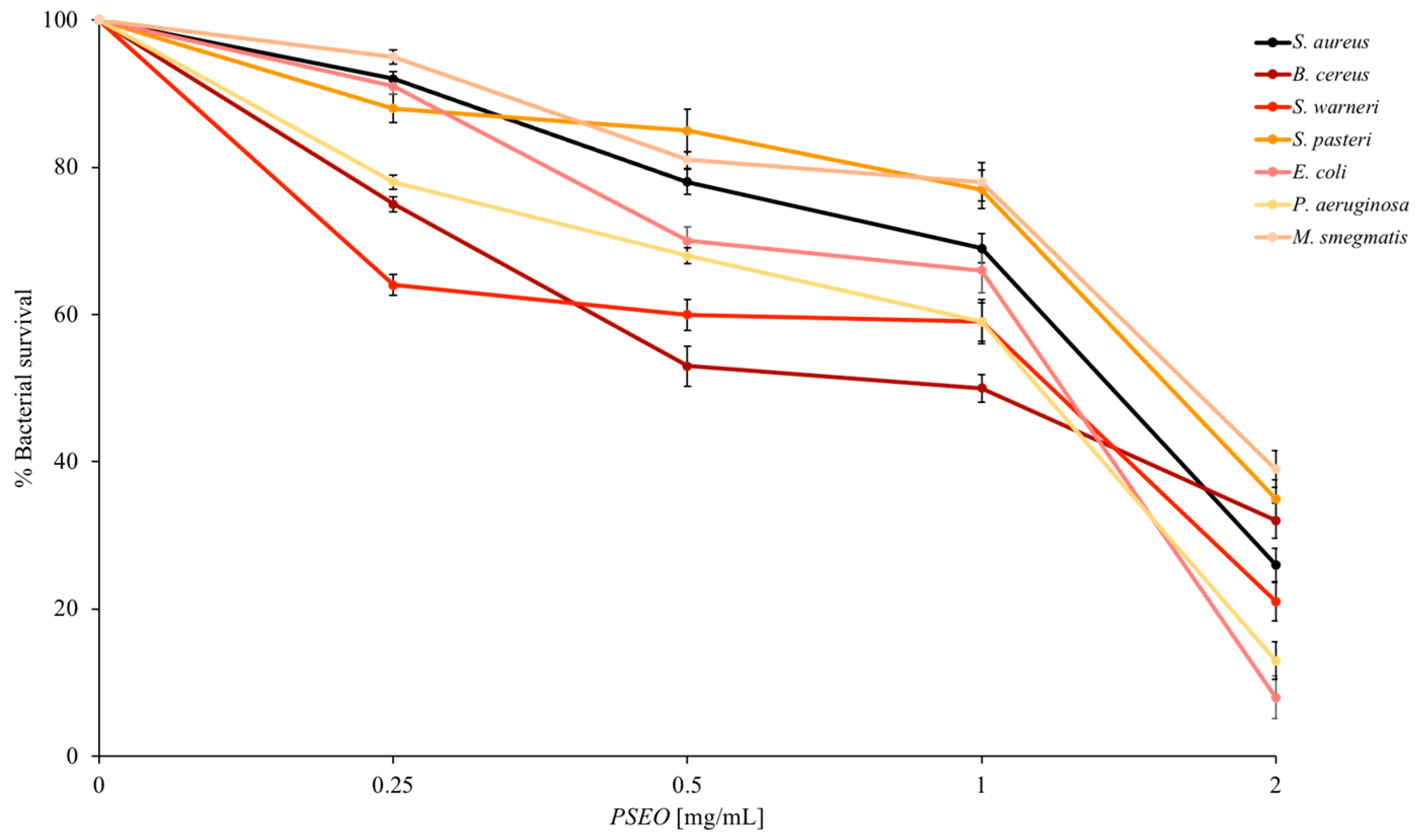
The construction of the dose–response curves (Figure 2) made it possible to validate the data in a more quantitative way. The indicator strains set has been expanded, using different bacteria that establish interactions with human cells. These are both Gram-negative and -positive bacteria, including bacterial strains in oral, gastro-intestinal, and skin infections. The proportionality between the quantity of PSEO administered to the bacteria and the decrease in their cell survival remains stable at relatively low concentrations for a mixture of substances such as EO (2 mg/mL). The PSEO is particularly effective against E. coli or P. aeruginosa, which have a very low survival rate (around 10%). Furthermore, these data are in agreement with the qualitative data, in which PSEO is more active against Gram-negative bacteria.
To complete the antimicrobial activity analysis, a minimum inhibitory concentration (MIC) test for microbial growth was performed against all indicator strains. As shown in Table 2, the lowest MIC values are obtained for the Gram-negative strains, in agreement with the data previously shown.
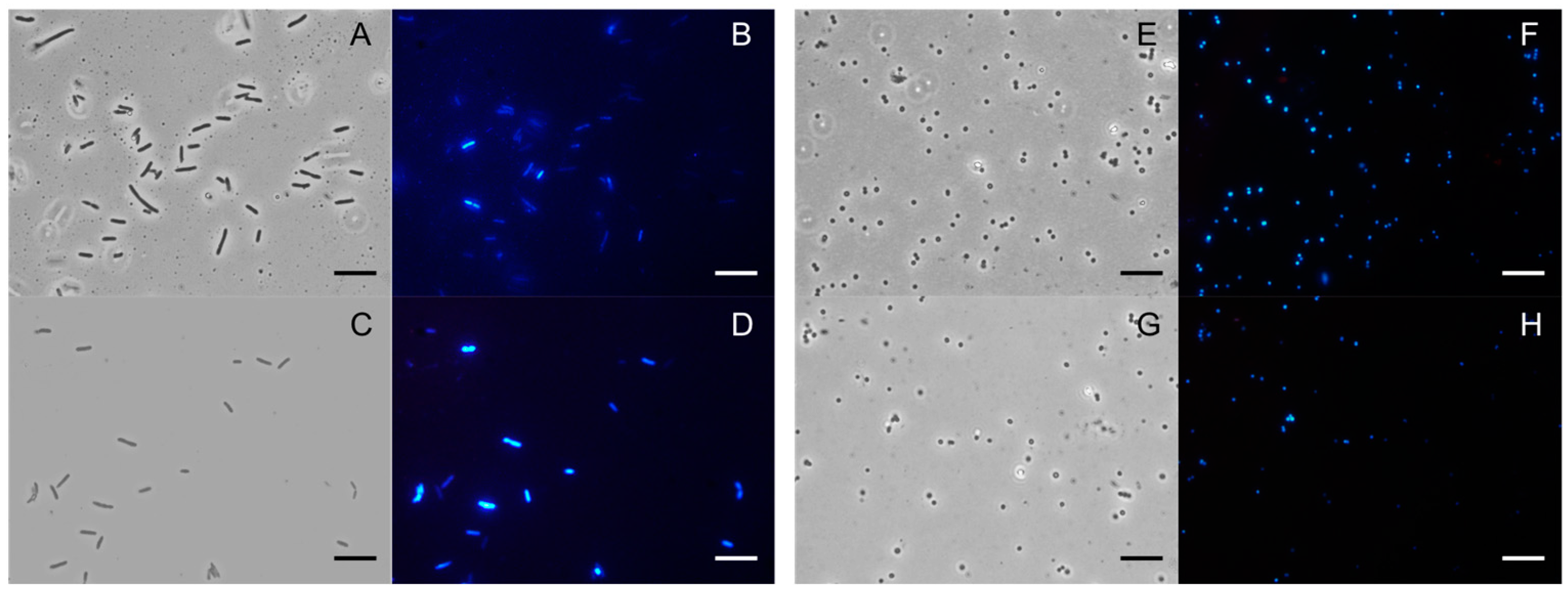
To find information on the possible antimicrobial target, fluorescence microscopy experiments were performed. As shown in Figure 3, no damaged membranes are observed after incubation with the PSEO at a concentration of 2 mg/mL (for negative strain) and 5 mg/mL (for positive strain) for approximately 4 h. Two specific DNA dyes were added to the control and to the sample also containing the EO: DAPI, which gave blue fluorescence and passed through intact membranes, and propidium iodide (PI), which gave red fluorescence and only passed through damaged membranes. Both the Gram-negative (panels A–D) and Gram-positive bacteria (panels E–H) did not show red fluorescence and gave similar results to the controls (B and F). This result highlighted that the EO target did not appear to be bacterial membranes. Our group has been engaged for years in researching new natural antimicrobial substances, compared to other EOs such as thyme, fennel, orchid, and others [27]. In fact, there are many mechanisms of action implemented given the EO’s components [28]. Although many EOs target membranes, some compounds and mixtures of compounds can affect growth regulation, nutritional balances, and energy conversion in bacteria [29].

2.3. Antibiofilm Activity of PSEO

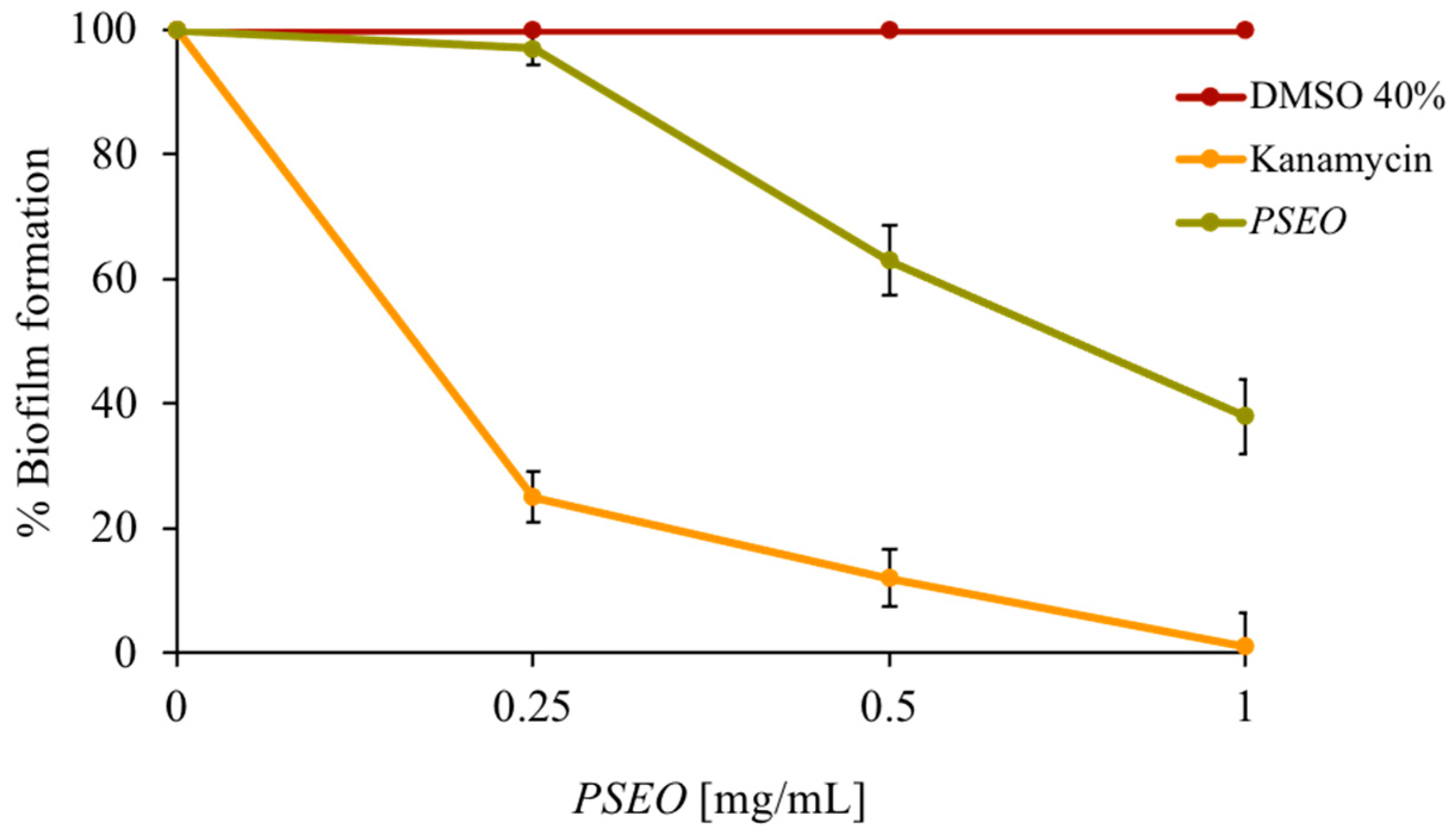
Demonstrating the effectiveness of a new substance only on planktonic cells is a good starting point, but implementing the research at the level of a complicated microbial organization, such as the biofilm, is necessary. The ability to form a biofilm is a significant concern, and a biofilm’s resistance is supported by genetic, physical, and physiological mechanisms [30]. In biofilms, cells are more resistant to disinfectants and antibiotics, and are also more protected and increase their genetic variability [31]. Mycobacterium smegmatis was used as a model organism in the biofilm development study. After analyzing the dose–response curves obtained with this strain by progressively increasing the amount of EO, the concentrations that did not cause the death of the microorganism were chosen for the biofilm inhibition experiments. If the planktonic cells die, it is clear that they cannot produce biofilm, standardizing the experiment for the number of cells added and considering the biofilm formation of a control (no PSEO added). In Figure 4, it is noted that the PSEO inhibits biofilm formation by 60%. DMSO was used as a negative control and the antibiotic kanamycin as a positive control, which gives the total biofilm inhibition at 1 mg/mL. The good antibiofilm activity of the PSEO made it a good candidate in various sectors, ranging from oral hygiene to nutraceutical and cosmetic use. However, in order to pursue this kind of application, in vivo biofilm studies need to be carried out.

2.4. Antioxidant Activity of PSEO

For nutraceutical and cosmetic companies, an important property of a substance is its antioxidant power. In vitro tests were carried out, verifying the ability to screen oxygen radicals, as can be seen in Table 3. The PSEO is very effective at screening hydrogen peroxide: even at very low concentrations, there is a 50% inhibition of ROS (around 0.025 mg/mL). In the same table, we can see how the scavenging activity against ABTS is less marked, with an IC50 of approximately 2 mg/mL.
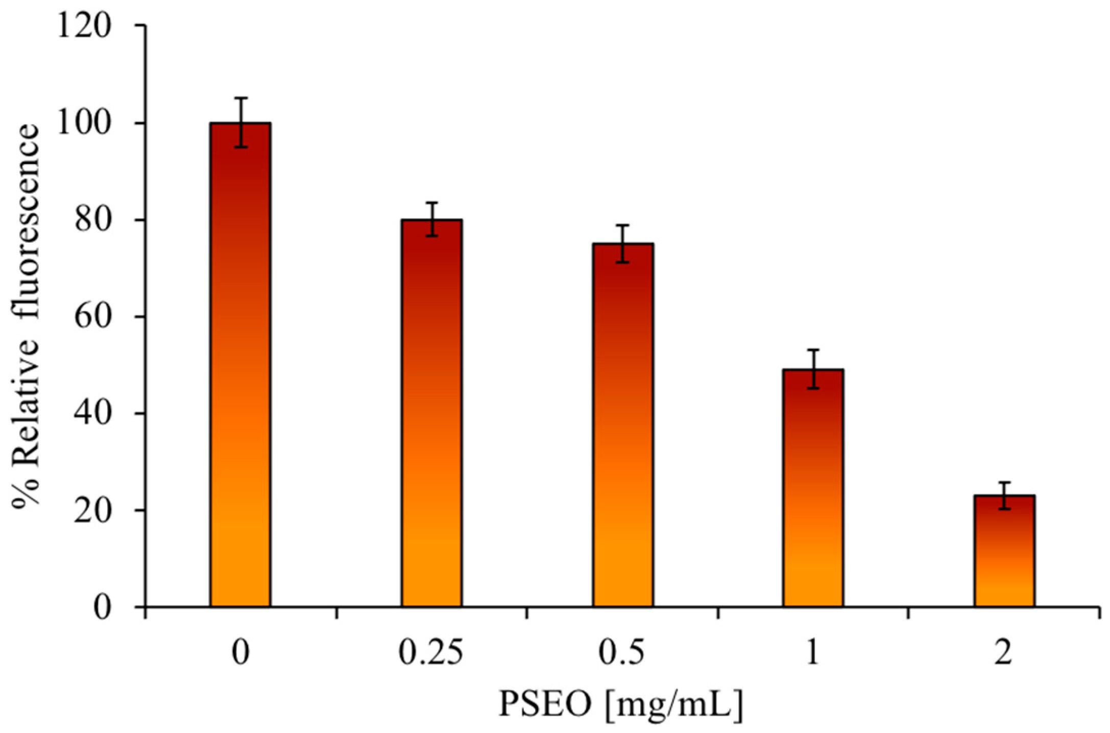
Before testing the antioxidant activity on the HaCat cell line, it was necessary to carry out a cytotoxicity assay using the PSEO at different concentrations up to 6 mg/mL, both at 4 h and 24 h. No cytotoxicity was observed under these experimental conditions (Figure S1). The ROS assay stain was used to confirm the obtained in vitro data about the antioxidant activity of the PSEO. By pre-treating the eukaryotic cells with increasing concentrations of the PSEO, and subsequently giving a stimulus with hydrogen peroxide, the ROS concentration decreases. As shown in Figure 5, there was a massive production of ROS in the control (untreated cells), with an increase in absorbance. In the PSEO samples, the absorbance decreased as the EO concentration increased, confirming that the PSEO pretreatment significantly lowers the concentration of ROS produced by the cells following a H2O2 pulse.

3. Materials and Methods

3.1. Plant Material Collection

The flowering aerial parts of wild P. sinaicum were collected, cleaned, and stored at 0 °C in Al-Jafr, a small city located in the far south of Jordan, about 200 km from the capital Amman (30°17′36″ N 36°12′50″ E), in April 2023. The collected samples were identified by Prof. Vincenzo Ilardi and one exemplary was preserved at the University of Palermo Herbarium (Voucher no. PAL 108769).

3.2. Isolation of Essential Oil

The samples (≈300 g), still fresh, were chopped manually and then through a Waring mixer and finally subjected to hydrodistillation for 3 h, according to the standard procedure described in the European Pharmacopoeia [32]. The extracted EO was dried over Na2SO4 and refrigerated under N2 until the GC analysis. The yield obtained was 0.10% (w/w).

3.3. GC and GC-MS Analysis

GC-MS analysis was performed using a Shimadzu QP 2010 plus gas chromatograph equipped with an AOC-20i autoinjector (Shimadzu, Kyoto, Japan) equipped with a FID, capillary column (DB-5 MS) 30 m × 0.25 mm i.d., 0.25 μm film thickness, and a data processor. The oven program was as follows: temperature increase to 40 °C for 5 min, at a rate of 2 °C/min up to 260 °C, then isothermal for 20 min. Helium was used as carrier gas (1 mL min−1). The injector and detector temperatures were set to 250 °C and 290 °C, respectively. A total of 1 μL of EO solution (3% EO/hexane v/v) was injected in split mode 1.0; MS range 40–600. The settings were as follows: ionization voltage, 70 eV; electron multiplier energy, 2000 V; transfer line temperature, 295 °C; solvent delay, 3.5 min. Linear retention indices (LRIs) were determined using retention times of n-alkanes (C8–C40) and peaks were identified by comparison with mass spectra and comparison of their retention indices with WILEY275 libraries, NIST 17, ADAMS, and FFNSC2.

3.4. Bacterial Strains

The antimicrobial activity was evaluated using Gram-positive and Gram-negative strains: Staphylococcus aureus ATCC6538P, Bacillus cereus ATCC10987, Streptococcus warneri, Streptococcus pasteuri, Escherichia coli DH5α, Pseudomonas aeruginosa PAOI and Mycobacterium smegmatis mc2 155.

3.5. Antimicrobial Activity Assay

The presence of antimicrobial molecules in PSEO was detected using the agar diffusion assay of Kirby–Bauer [33]. Masses of 2.5, 5, 10 mg of PSEO were placed on Luria–Bertani agar plates which were overlaid with 10 mL of soft agar (0.7%) mixed with 10 μL of strains grown for 24 h at 37 °C. The negative control was dimethylsulfoxide (DMSO) 40%, used to resuspend the EO; the positive control was represented by the antibiotic ampicillin. Plates were incubated overnight at 37 °C and the antimicrobial activity was calculated according to the equation cited by Napolitano et al. [34]. For the second experiment, samples with a final volume of 500 μL were prepared; they contained bacterial cells, extract of PSEO at different concentrations (0.25, 0.5, 1, and 2 mg/mL), and 20 mM pH 7.0 of NaP buffer up to final volume. Samples without extract were used as a positive control. The negative control contained bacterial cells with DMSO 40%. After 4 h of incubation at 37 °C, all samples were prepared and then plated on LB agar in Petri dishes that were finally incubated at 37 °C overnight. The following day, the surviving percent of bacterial cells was estimated by counting the number of colonies. Each experiment was performed in triplicate and the reported result was an average of three independent experiments. (p value was <0.05).

3.6. Determination of Minimal Inhibitory Concentration

Minimal inhibitory concentrations (MICs) of PSEO against all strains were determined as described by Di Napoli et al. [35]. Following overnight incubation at 37 °C, MIC values were determined as the lowest extract concentration responsible for no visible bacterial growth.

3.7. DAPI/PI Dual Staining and Fluorescence Microscopy Image Acquisition

For dual staining, 100 µL of E. coli DH5α and S. aureus ATCC6538P (mid-logarithmic phase) was incubated in the dark for 4 h at 37 °C in agitation in the presence or absence of PSEO, at concentrations of 2 mg/mL (for E. coli) and 5 mg/mL (for S. aureus). After 4 h, 10 µL of bacterial culture was mixed with DAPI solution (40, 6-diamidino-2-phenylindole dihydrochloride; Sigma Aldrich, Milan, Italy) and PI (propidium iodide; Sigma Aldrich, Milan, Italy). Samples were observed using an Olympus BX51 fluorescence (Olympus, Tokyo, Japan) using a DAPI filter (excitation/emission: 358/461 nm) [35].

3.8. Antibiofilm Activity Assay

Crystal violet dye was used to evaluate the biofilm formation of M. smegmatis mc2 155. A 24-well plate was prepared in which each well contained a final volume of 1 mL; the negative control was represented by only bacterial cells and medium, the positive control was represented by bacterial cells with antibiotic kanamycin 2 µg/mL, the other samples contained cells and PSEO [0.25, 0.5, 1 mg/mL]. The plate was incubated at 37 °C for 36 h. The OD of the crystal violet present in the dual staining solution was measured at 570 nm.

3.9. Antioxidant Activity

3.9.1. ABTS Scavenging Capacity Assay

This assay was performed according to Re et al. [36], which is based on ABTS radical cation scavenging. A total of 1 mL ABTS solution was added to 100 μL of PSEO (0.05–2 mg/mL concentrations). Finally, the absorbance was measured at 734 nm against a blank, and the percentage inhibition of ABTS radicals was determined from the following equation: ABTS+ radical scavenging activity (%) = (1 * AS/AC) × 100, where AC is the absorbance of the ABTS solution and AS is the absorbance of the sample at 734 nm. The concentration required for 50% inhibition was determined and represented as IC50.

3.9.2. Hydrogen Peroxide Scavenging Assay

The hydrogen peroxide stability was measured as the absorbance at 240 nm of 1 mL of fresh hydrogen peroxide solution (50 mM potassium phosphate buffer, pH 7.0; 0.036% (w/w) H2O2) [37]. Different concentrations of PSEO (0.05–2 mg/mL) were incubated at 20 °C in 1 mL of hydrogen peroxide solution. The percentage of peroxide removed was calculated as follows: peroxide removed (%) = (1 * AS/AC) × 100, where AC is the absorbance of 1 mL of hydrogen peroxide solution and AS is the absorbance of the sample at 240 nm.

3.10. Eukaryotic Cell Culture and Antioxidant Test

HaCat (human keratinocytes) cells are a spontaneously transformed aneuploid immortal keratinocyte cell line from adult human skin, widely used in scientific research. PSEO was added in a complete growth medium for the cytotoxicity assay [38]. HaCaT cells were plated in 12-well plates and then incubated at 37 °C with 5% CO2 for 24 h. Cells were treated with PSEO at different concentrations (0.25, 0.5, 1, and 2 mg/mL). After 60 min of treatment, H2O2 (800 µM) was added to each well for the next 3 h before detecting intracellular ROS. ROS Assay Stain (88-5930, Invitrogen, Waltham, MA, USA) was administered to cells in culture media according to the manufacturer’s protocol. Fluorescence intensity (530 nm) was measured using a Synergy H4 hybrid microplate reader (Agilent, Santa Clara, CA, USA).

4. Conclusions

In this study, for the first time, a chemical analysis of harvested P. sinaicum essential oil, collected in Jordan, was carried out using GC and GC-MS. The essential oil was found to be rich in terpenes with oxygenated monoterpenes as the most abundant class (61.73%), with β-thujone (13.77%) and santolina alcohol (21.43%) as the main compounds. β-Sesquiphellandrene (16.89%) was, instead, the major compound of sesquiterpene hydrocarbons class (28.39%). The P. sinaicum essential oil appeared to have good antimicrobial activity, especially directed against Gram-negative bacteria, in which bacterial membranes are not the primary target. Furthermore, according to some investigations, synergistic volatile compounds such as santolina alcohol and β-sesquiphellandrene contained in essential oils are mainly responsible for antibacterial activity. The ability of this essential oil to inhibit biofilm formation, causing oral diseases and infections on the skin, is very interesting. Furthermore, the P. sinaicum essential oil ROS scavenging activity is an important property for nutraceutical or cosmetic applications.

Supplementary Materials

The following supporting information can be downloaded at https://www.mdpi.com/article/10.3390/plants12234007/s1, Figure S1: Determination of the cytotoxic activity of PSEO. The image shows the percentage of cell survival of the HaCat cell line treated with different concentrations (x-axis) of EO for 4 and 24 h. Cell survival percentage is shown on the y-axis. The tests were performed in three independent experiments. Standard deviations are always less than 5%.

Author Contributions

Conceptualization, A.Z., N.B. and M.B.; methodology, N.B., M.D.N. and G.C.; software, N.B. and G.C.; validation, N.B., M.B. and A.Z.; formal analysis, M.D.N. and N.B.; investigation, M.D.N. and N.B.; resources, N.B. and M.B.; data curation, M.D.N., G.C. and N.B.; writing—original draft preparation, A.Z. and N.B.; writing—review and editing, A.Z., N.B., M.B. and M.V.; visualization, N.B.; supervision, A.Z., M.V., N.B. and M.B. All authors have read and agreed to the published version of the manuscript.

Funding

This research received external funding from the National Biodiversity Future Center S.c.a.r.l., Piazza Marina 61 (c/o Palazzo Steri) Palermo, Italy, C.I. CN00000033—CUP UNIPA B73C22000790001.

Data Availability Statement

All data and materials are available upon request from the corresponding author.

Conflicts of Interest

The authors declare no conflict of interest.

References

  1. POWO. Plants of the World Online. Available online: https://powo.science.kew.org/taxon/urn:lsid:ipni.org:names:445949-1 (accessed on 15 May 2023).
  2. Ali-Shtayeh, M.S.; Yaghmour, R.M.R.; Faidi, Y.R.; Salem, K.; Al-Nuri, M.A. Antimicrobial activity of 20 plants used in folkloric medicine in the Palestinian area. J. Ethnopharmacol. 1998, 60, 265–271. [Google Scholar] [CrossRef] [PubMed]
  3. Abu-Rabia, A. Palestinian plant medicines for treating renal disorders: An inventory and brief history. Altern. Complement. Ther. 2005, 11, 295–300. [Google Scholar] [CrossRef]
  4. Hudaib, M.; Mohammad, M.; Bustanji, Y.; Tayyem, R.; Yousef, M.; Abuirjeie, M.; Aburjai, T. Ethnopharmacological survey of medicinal plants in Jordan, Mujib Nature Reserve and surrounding area. J. Ethnopharmacol. 2008, 120, 63–71. [Google Scholar] [CrossRef] [PubMed]
  5. Friedman, J.; Yaniv, Z.; Dafni, A.; Palewitch, D. A preliminary classification of the healing potential of medicinal plants, based on a rational analysis of an ethnopharmacological field survey among Bedouins in the Negev Desert, Israel. J. Ethnopharmacol. 1986, 16, 275–287. [Google Scholar] [CrossRef] [PubMed]
  6. Alzweiri, M.; Sarhan, A.A.; Mansi, K.; Hudaib, M.; Aburjai, T. Ethnopharmacological survey of medicinal herbs in Jordan, The Northern Badia region. J. Ethnopharmacol. 2011, 137, 27–35. [Google Scholar] [CrossRef]
  7. Pardo De Santayana, M.; Blanco, E.; Morales, R. Plants known as te in Spain: An ethno-pharmaco-botanical review. J. Ethnopharmacol. 2005, 98, 1–19. [Google Scholar] [CrossRef] [PubMed]
  8. Gonzàlez-Tejero, M.R.; Casares-Porcel, M.; Sanchez-Rojas, C.P.; Ramiro-Gutierrez, J.M.; Molero-Mesa, J.; Pieroni, A.; Giusti, M.E.; Censorii, E.; de Pasquale, C.; Della, A. Medicinal plants in the Mediterranean area: Synthesis of the results of the project Rubia. J. Ethnopharmacol. 2008, 116, 341–357. [Google Scholar] [CrossRef]
  9. Epifano, F.; Marcotullio, M.C.; Menghini, L. Constituents of Phagnalon sordidum. Chem. Nat. Comp. 2002, 38, 204–205. [Google Scholar] [CrossRef]
  10. Cicio, A.; Badalamenti, N.; Bruno, M. The ethnobotany, phytochemistry, and biological properties of genus Phagnalon (Asteraceae)—A review. Nat. Prod. Res. 2022, 37, 2083–2097. [Google Scholar] [CrossRef]
  11. Baka, Z.A.M. Antifungal activity of extracts from five Egyptian wild medicinal plants against late blight disease of tomato. Arch. Phytopathol. Plant Prot. 2014, 47, 1988–2002. [Google Scholar] [CrossRef]
  12. Baka, Z.A.M. Plant extract control of the fungi associated with different Egyptian wheat cultivars grains. J. Plant Prot. Res. 2014, 54, 231–237. [Google Scholar] [CrossRef]
  13. Baka, Z.A.M. Efficacy of wild medicinal plant extracts against predominant seed-borne fungi of broad bean cultivars. Acta Phytopathol. Entomol. Hung. 2015, 50, 45–65. [Google Scholar] [CrossRef]
  14. Chávez-gonzález, M.L.; Rodríguez-herrera, R.; Aguilar, C.N. Essential oils: A natural alternative to combat antibiotics resistance. In Antibiotic Resistance, Mechanisms and New Antimicrobial Approaches; Kon, K., Rai, M., Eds.; Elsevier: Amsterdam, The Netherlands, 2016; pp. 227–337. [Google Scholar]
  15. Coenye, T.; Nelis, H.J. In vitro and in vivo model systems to study microbial biofilm formation. J. Microbiol. Methods 2010, 83, 89–105. [Google Scholar] [CrossRef] [PubMed]
  16. Bazargani, M.M.; Rohloff, J. Antibiofilm activity of essential oils and plant extracts against Staphylococcus aureus and Escherichia coli biofilms. Food Control 2016, 61, 156–164. [Google Scholar] [CrossRef]
  17. Qaiser, M.; Lack, H.W. The Genus Phagnalon (Asteraceae, Inuleae) in Arabia. Willdenowia 1985, 15, 3–22. [Google Scholar]
  18. El-Dahmy, S.I.; Abdel Aal, M.; Abd El-Fatah, H.; Fid, F. Thymol derivatives from Phagnalon sinaicum Bornm and Kneuck. Acta Pharm. Hung. 1994, 64, 115–116. [Google Scholar]
  19. Vaglica, A.; Badalamenti, N.; Ilardi, V.; Bruno, M. The chemical composition of the aerial parts essential oil of four Phagnalon species collected in Sicily (Italy) and Greece. Nat. Prod. Res. 2023, in press. [Google Scholar] [CrossRef]
  20. Chikhi, I.; Allali, H.; Costa, J. Chemical composition and biological activities of essential oils of Phagnalon sordidum (L.) Rchb. (Asteraceae) from Algeria. Agric. Conspec. Sci. 2019, 84, 271–281. [Google Scholar]
  21. Brunel, M.; Vitrac, C.; Costa, J.; Mzali, F.; Vitrac, X.; Muselli, A. Essential oil composition of Phagnalon sordidum (L.) from Corsica, chemical variability and antimicrobial activity. Chem. Biodivers. 2016, 13, 299–308. [Google Scholar] [CrossRef] [PubMed]
  22. Erdogan Orhan, I.; Senol, F.S.; Demirci, B.; Ozturk, N.; Baser, K.H.C.; Sener, B. Phytochemical characterization of Phagnalon graecum Boiss. by HPLC and GC-MS with its enzyme inhibitory and antioxidant activity profiling by spectrophotometric methods. Food Anal. Methods 2013, 6, 1–9. [Google Scholar] [CrossRef]
  23. Alves-Silva, J.M.; Piras, A.; Porcedda, S.; Falconieri, D.; Maxia, A.; Gonçalves, M.J.; Cruz, M.T.; Salgueiro, L. Chemical characterization and bioactivity of the essential oil from Santolina insularis, a Sardinian endemism. Nat. Prod. Res. 2023, 36, 445–449. [Google Scholar] [CrossRef] [PubMed]
  24. Marinas, I.C.; Oprea, E.; Gaboreanu, D.M.; Gradisteanu Pircalabioru, G.; Buleandra, M.; Nagoda, E.; Badea, I.A.; Chifiriuc, M.C. Chemical and biological studies of Achillea setacea Herba essential oil-first report on some antimicrobial and antipathogenic features. Antibiotics 2023, 12, 371. [Google Scholar] [CrossRef] [PubMed]
  25. Khalil, N.; Elhady, S.S.; Diri, R.M.; Fekry, M.I.; Bishr, M.; Salama, O.; El-Zalabani, S.M. Salicylic acid spraying affects secondary metabolites and radical scavenging capacity of drought-stressed Eriocephalus africanus L. Agronomy 2022, 12, 2278. [Google Scholar] [CrossRef]
  26. Badalamenti, N.; Porrello, A.; Merra, R.; Ilardi, V.; Bruno, M. The chemical composition of the aerial parts essential oil of Phagnalon sinaicum collected in the Negev Desert (Israel). Nat. Prod. Res. 2023, in press. [Google Scholar] [CrossRef]
  27. Badalamenti, N.; Russi, S.; Bruno, M.; Maresca, V.; Vaglica, A.; Ilardi, V.; Zanfardino, A.; Di Napoli, M.; Varcamonti, M.; Cianciullo, P.; et al. Dihydrophenanthrenes from a Sicilian Accession of Himantoglossum robertianum (Loisel.) P. Delforge showed antioxidant, antimicrobial, and antiproliferative activities. Plants 2021, 10, 2776. [Google Scholar] [CrossRef]
  28. Nazzaro, F.; Fratianni, F.; De Martino, L.; Coppola, R.; De Feo, V. Effect of essential oils on pathogenic bacteria. Pharmaceuticals 2013, 6, 1451–1474. [Google Scholar] [CrossRef] [PubMed]
  29. Caillet, S.; Ursachi, L.; Shareck, F.; Lacroix, M. Effect of gamma radiation and oregano essential oil on murein and ATP concentration of Staphylococcus aureus. J. Food Sci. 2009, 74, M499–M508. [Google Scholar] [CrossRef] [PubMed]
  30. Ciofu, O.; Tolker-Nielsen, T. Tolerance and resistance of Pseudomonas aeruginosa biofilms to antimicrobial agents—How P. aeruginosa can escape antibiotics. Front. Microbiol. 2019, 10, 913. [Google Scholar] [CrossRef]
  31. Drago, L. Bacteria and biofilm in respiratory tract infections. Infez. Med. 2009, 17 (Suppl. 2), 3–9. [Google Scholar]
  32. European Pharmacopoeia 10.3. 2020. Determination of Essential Oils in Herbal Drugs, 2.8.12., 307. Available online: https://www.edqm.eu/ (accessed on 26 November 2023).
  33. Bauer, A.W.; Kirby, W.M.M.; Sherris, J.C.; Turck, M. Antibiotic susceptibility testing by a standardized single disk method. Am. J. Clin. Pathol. 1966, 45, 493–496. [Google Scholar] [CrossRef]
  34. Napolitano, A.; Di Napoli, M.; Castagliuolo, G.; Badalamenti, N.; Cicio, A.; Bruno, M.; Piacente, S.; Maresca, V.; Cianciullo, P.; Capasso, L.; et al. The chemical composition of the aerial parts of Stachys spreitzenhoferi (Lamiaceae) growing in Kythira island (Greece), and their antioxidant, antimicrobial, and antiproliferative properties. Phytochemistry 2022, 203, 113373. [Google Scholar] [CrossRef] [PubMed]
  35. Di Napoli, M.; Maresca, V.; Sorbo, S.; Varcamonti, M.; Basile, A.; Zanfardino, A. Proteins of the fruit pulp of Acca sellowiana have antimicrobial activity directed against the bacterial membranes. Nat. Prod. Res. 2021, 35, 2942–2946. [Google Scholar] [CrossRef] [PubMed]
  36. Re, R.; Pellegrini, N.; Proteggente, A.; Pannala, A.; Yang, M.; Rice-Evans, C. Antioxidant activity applying an improved ABTS radical cation decolorization assay. Free Radic. Biol. Med. 1999, 26, 1231–1237. [Google Scholar] [CrossRef] [PubMed]
  37. Beers, R.F.; Sizer, I.W. A spectrophotometric method for measuring the breakdown of hydrogen peroxide by catalase. J. Biol. Chem. 1952, 195, 133–140. [Google Scholar] [CrossRef] [PubMed]
  38. Napoli, M.D.; Luccia, B.D.; Vitiello, G.; D’Errico, G.; Carpentieri, A.; Pezzella, A.; Pizzo, E.; Notomista, E.; Varcamonti, M.; Zanfardino, A. Characterisation of EFV12 a bio-active small peptide produced by the human intestinal isolate Lactobacillus gasseri SF1109. Benef. Microbes 2020, 11, 815–824. [Google Scholar] [CrossRef] [PubMed]
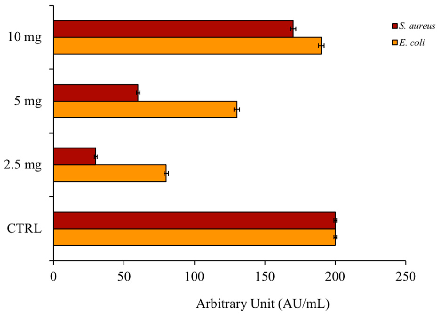
Figure 1. Inhibition of bacterial growth. The image shows the inhibition halos, expressed in AU/mL, of PSEO against E. coli and S. aureus. The positive control was ampicillin and the negative control was DMSO.
Plants 12 04007 g001
Figure 2. Antimicrobial activity against Gram-positive and Gram-negative strains shown with dose–response curves. The tests were performed in three independent experiments. Standard deviations are always less than 5%.
Plants 12 04007 g002
Figure 3. Analysis with fluorescence microscopy. Panels show E. coli bacterial cells (AD) and S. aureus bacterial cells (EH). Panels (A,C,E,G) show the cells observed under the optical microscope, and panels (B,D,F,H) show the cells observed under the fluorescence microscope. Untreated bacterial cells (A,B,E,F); cells treated with PSEO (C,D,G,H). Scale bars: 1 µm (AH).
Plants 12 04007 g003
Figure 4. Antibiofilm activity. The percent inhibition of M. smegmatis biofilm is shown in the figure. Different concentrations of PSEO were tested (x-axis). The tests were performed in three independent experiments. Standard deviations are always less than 5%.
Plants 12 04007 g004
Figure 5. Effects of PSEO on intracellular ROS generation in HaCaT cells upon H2O2 treatment.
Plants 12 04007 g005
Table 1. Chemical composition of Jordan accession of Phagnalon sinaicum (PSEO) essential oil.
No.Compounds aLRI bLRI cArea d (%)
1α-Pinene9319380.4
2β-Phellandrene9719780.6
3β-Pinene9829800.1
4α-Terpinene101810221.1
Monoterpene Hydrocarbons 2.2
5Eucalyptol103410402.1
6Santolina alcohol1038104721.4
7Artemisia ketone1061108013.2
8Artemisia alcohol109211000.2
9α-Thujone110211258.8
10β-Thujone1127113513.8
11Linalool111011761.0
12Terpinen-4-ol118011850.4
13α-Terpineol119911990.8
Oxygenated Monoterpenes 61.7
14α-Copaene137213711.0
15trans-β-Farnesene145514580.6
16Germacrene D148014805.2
17Bicyclogermacrene149414904.7
18β-Sesquiphellandrene1523153116.9
Sesquiterpene Hydrocarbons 28.4
19Spathulenol157115712.1
20α-Cadinol165316521.9
Oxygenated Sesquiterpenes 4.0
21Hexanal8028080.3
222-Hexenal8328620.7
Other Compounds 1.0
Total Composition (%) 97.3
a Compounds are classified in order of LRIs of the apolar column (DB-5 MS); b LRIs on a DB-5 MS apolar column; c LRIs reported for DB-5 MS column reported in the literature; d area is the peak volume percentage of compound in the essential oil (EO) sample.
Table 2. Determination of minimum concentration (MIC) values that inhibit bacterial growth. The MIC is expressed in mg/mL of PSEO against Gram-positive and Gram-negative bacteria. Values were obtained from a minimum of three independent experiments.
StrainsMIC [mg/mL]
S. aureus6
S. warneri10
S. pasteuri10
B. cereus10
E. coli3
P. aeruginosa4
M. smegmatis>10
Table 3. Concentration at 50% scavenging activity. ABTS: 2,20-azino-bis (3-ethyl-benzothiazoline-6-sulfonic acid); H2O2: hydrogen peroxide. The positive control is ascorbic acid for ABTS and resveratrol for H2O2.
SampleIC50 of ABTS (mg/mL)SampleIC50 of H2O2 (mg/mL)
PSEO2PSEO0.025
Ascorbic acid0.00003Resveratrol0.00005
Disclaimer/Publisher’s Note: The statements, opinions and data contained in all publications are solely those of the individual author(s) and contributor(s) and not of MDPI and/or the editor(s). MDPI and/or the editor(s) disclaim responsibility for any injury to people or property resulting from any ideas, methods, instructions or products referred to in the content.