Article

Indonesian Euphorbiaceae: Ethnobotanical Survey, In Vitro Antibacterial, Antitumour Screening and Phytochemical Analysis of Euphorbia atoto

1
Institute of Pharmacognosy, University of Szeged, H-6720 Szeged, Hungary
2
Department of Pharmacy, Faculty of Medicine, University of Mataram, Mataram 83126, Indonesia
3
Department of Medical Microbiology, Albert Szent-Györgyi Health Center and Albert Szent-Györgyi Medical School, University of Szeged, H-6725 Szeged, Hungary
4
Department of Biology Education, Faculty of Teacher Training and Education, University of Mataram, Mataram 83126, Indonesia
5
ELKH-USZ Biologically Active Natural Products Research Group, University of Szeged, H-6720 Szeged, Hungary
*
Author to whom correspondence should be addressed.
Plants 2023, 12(22), 3836; https://doi.org/10.3390/plants12223836
Submission received: 16 October 2023 / Revised: 30 October 2023 / Accepted: 9 November 2023 / Published: 13 November 2023

Abstract

:
Indonesia is among the countries with the most significant biodiversity globally. Jamu, the traditional medicine of Indonesia, predominantly uses herbal materials and is an integral component of the Indonesian healthcare system. The present study reviewed the ethnobotanical data of seven Indonesian Euphorbiaceae species, namely Euphorbia atoto, E. hypericifolia, Homalanthus giganteus, Macaranga tanarius, Mallotus mollissimus, M. rufidulus, and Shirakiopsis indica, based on the RISTOJA database and other literature sources. An antimicrobial screening of the plant extracts was performed in 15 microorganisms using the disk diffusion and broth microdilution methods, and the antiproliferative effects were examined in drug-sensitive Colo 205 and resistant Colo 320 cells by the MTT assay. The antimicrobial testing showed a high potency of M. tanarius, H. giganteus, M. rufidulus, S. indica, and E. atoto extracts (MIC = 12.5–500 µg/mL) against different bacteria. In the antitumour screening, remarkable activities (IC50 0.23–2.60 µg/mL) were demonstrated for the extracts of H. giganteus, M. rufidulus, S. indica, and E. atoto against Colo 205 cells. The n-hexane extract of E. atoto, with an IC50 value of 0.24 ± 0.06 µg/mL (Colo 205), was subjected to multistep chromatographic separation, and 24-methylene-cycloartan-3β-ol, jolkinolide E, tetra-tert-butyl-diphenyl ether, α-tocopherol, and β-sitosterol were isolated.

1. Introduction

Indonesia is considered to have substantial biodiversity based on its high number of indigenous plants. From the 40,000 tropical plant species that are native to Indonesia, 39% are considered endemics. Most Indonesians, especially those in rural regions, employ traditional herbal medicines, collectively known as jamu, to treat diseases because of the abundance of medicinal plants. Besides their medicinal uses, certain plants are used for specific purposes, like tribal ceremonies, whereas others are grown for daily consumption and trade [1,2].
The Health Ministry of the Republic of Indonesia, through the National Institute of Health Research and Development (NIHRD) in collaboration with some leading universities, has conducted comprehensive research on medicinal plants and jamu (“Riset Tanaman Obat dan Jamu”, RISTOJA). RISTOJA is an ethnomedicinal research program that explores the utilization of medicinal plants in Indonesia. The RISTOJA database collected findings for 32,014 medicinal plants in Indonesia during 2012–2017 from 405 ethnic groups in 34 provinces. Since access to the RISTOJA database is not open to the public, data can only be used through proposals submitted to the NIHRD. The Indonesian government, industry, and academic communities agree that further studies are needed to determine the efficacy and safety of different traditional jamu preparations to ensure their safe and effective use.
The analysis of jamu preparations as rational phytotherapies requires a variety of pharmacological and phytochemical studies of extracts and isolated compounds. The results of in vitro and in vivo preclinical studies can support or refute the traditional use of medicinal plants. Scientific investigations of medicinal plants used in indigenous medical systems have been increasingly relevant, and the role of ethnobotanical information in the search for new drugs has been of continuous importance. Knowledge of traditional medicine can serve as the starting point for drug discovery. Therefore, screening of the biological activity of the used plants can provide valuable data for selecting plant extracts for isolating and identifying the active ingredients [3].
Continuing our current research interest in species of the spurge family [4,5], the present study (a) reviewed the data on the traditional therapeutic uses of seven Euphorbiaceae species, namely Euphorbia atoto G.Forst., E. hypericifolia L., Homalanthus giganteus Zoll. & Moritzi, Macaranga tanarius Müll.Arg., Mallotus mollissimus (Geiseler) Airy Shaw, M. rufidulus Müll.Arg., and Shirakiopsis indica (Willd.) Esser; (b) conducted an in vitro antimicrobial and antitumour screening of extracts with different polarities prepared from the plants; and (c) isolated compounds from E. atoto. This study used the metadata from the RISTOJA collected in 2012, 2015, and 2017 together with ethnobotanical data from the literature to evaluate the traditional uses of the seven selected species. The antimicrobial activities of the extracts were investigated in seven Gram-positive, four Gram-negative, and four fungal strains, and their antiproliferative activity was examined in human colon adenocarcinoma cell lines (Colo 205 and Colo 320). The n-hexane extract of E. atoto that exhibited a strong antiproliferative effect was subjected to multistep chromatographic separation to isolate five compounds (15), among which, four were isolated for the first time from this species.

2. Results and Discussion

2.1. Ethnobotanical Data

2.1.1. Illness Categories

Fifty-six ethnobotanical uses were described in the RISTOJA database and in the literature for the selected seven Euphorbiaceae species (E. atoto, E. hypericifolia, H. giganteus, M. tanarius, M. mollissimus, M. rufidulus, and S. indica) and classified into 12 categories (Table 1). The categorization was based on the ICPC-3 (available at https://www.who.int/standards/classifications/other-classifications/international-classification-of-primary-care, accessed on 15 June 2023) that employs the perception of patients or interviewees to classify ethnobotanical information. This framework aligns with the objective of ethnobotanical research for studying the indigenous aspects of medicinal plants [6].
Using the categorization of the ICPC-3, the majority of plants were frequently utilized to treat skin-related problems (17). Other relevant uses were associated with general symptoms (10), digestive system ailments (9), and musculoskeletal issues (6). The plants were less frequently employed to address genital system concerns (3), pregnancy and childbearing issues (3), eye-related conditions (2), neurological system disorders (2), respiratory system conditions (1), blood-related disorders and immune system conditions (1), endocrine, metabolic, and nutritional system disorders (1), and urinary system ailments (1).

2.1.2. Ethnobotanical Uses of the Selected Species

In our study, the most frequently cited plant species were M. tanarius and E. atoto, accounting for 78% of the reported uses (Table 1). M. tanarius is a tree commonly found in wet tropical regions globally, including all islands of Indonesia [7]. It has a long history of traditional medicinal use, particularly in the Pacific region [8,9,10,11,12]. M. tanarius was mentioned 35 times in the RISTOJA database and other literature, and it has been used to treat various ailments. It is most notable for its activity against skin-related problems such as sores, boils, and itching and for its wound-healing effects (Table 2). This plant is frequently applied for the treatment of gastrointestinal disorders, such as a gastric ulcer, stomach ache, and diarrhoea. Infectious diseases, such as malaria, eye diseases, bloody diarrhoea, and sores, were also listed among its traditional applications. The pharmacological activities of the plant have been studied by several groups, and antidiabetic, antiacetylcholinesterase, cytotoxic, and antiherbivore activities were reported [13,14,15,16,17]. These activities can be attributed to the presence of numerous compounds, specifically prenylated flavonoids that have been isolated from this species [17,18,19,20,21,22].
E. atoto is a shrub primarily found in wet, sandy, tropical biomes, particularly in sandy beach areas or dunes near the seashore [23]. E. atoto was mentioned nine times in the RISTOJA database and other literature, and it was mainly used to treat skin-related conditions. The leaf, bark, and whole plant were used in Jambi, Indonesia, the Nicobar Islands of India, and Samoa to treat ulcers, sores, old wounds, abscesses, rheumatism, lumbago, and headaches (Table 2). Despite the potential therapeutic applications of this species, there have been limited pharmacological investigations of its effects. Trachylobane (euphoratones A–B, 3-oxo-ent-trachyloban-17-oic acid), ent-kaurane (ent-kauran-16β-ol-3-one, ent-kaurane-3-oxo-16β,17-diol, ent-kaurane-3β,16 β-diol), ent-sanguinolane diterpenoids, baccatin, campesterol, stigmasterol, β-sitosterol, and (+)-9-aza-1-methylbicyclo [3,3,1]nonan-3-one were identified in E. atoto in previous studies [24,25,26]. Some of the isolated diterpenoids were found to have weak anti-inflammatory activity against mouse RAW264.7 cells [26]. It is important to note that the research conducted by Norhanom and Yadav in 1995 described the tumour-promoting activity of several Malaysian plants belonging to the Euphorbiaceae family, including Euphorbia atoto, which was tested in vitro on a human lymphoblastoid cell line harbouring the Epstein-Barr virus (EBV) genome. Based on this observation, the authors pointed out that regular use of E. atoto could well be an etiological factor for the promotion of tumours among rural Malaysian peoples [27].
Regarding E. hypericifolia, its traditional uses in South Africa and Uganda included the treatment of gonorrhoea, snakebites, and scorpion bites. Previous pharmacological studies demonstrated its anticancer effects against colorectal cancer [28].
S. indica fruits and leaves were utilized to treat constipation and measles, respectively, in the traditional medicine of Thailand and North Sumatra. S. indica has been studied for its anti-Helicobacter pylori, anti-HIV, wound-healing, and anti-inflammatory properties [29,30,31,32].
H. giganteus, M. rufidulus, and M. mollissimus were used in the healing system of Indonesia, and their indications included urinary tract diseases, bone injury, fever, and flatulence (Table 2). No records were found regarding pharmacological investigations of these plants.
Table 2. Ethnobotanical data related to the selected species.
SpeciesPart UsedLocationTraditional UsesReferences
M. tanarius (L.)
Müll.Arg.
LeafAcehHigh cholesterol[8]
West JavaItchy, skin diseases in babies, wounds, gastric ulcers[8]
GorontaloItchy[8]
North SulawesiLeucorrhoea, diarrhoea, sores, wounds, stomach ache[8]
BantenGastric ulcer[33]
Southeast SulawesiInternal organ treatments[11]
West TimorMalaria[8]
North MalukuFertility, eye diseases[8,11]
North SumateraMuscle injury[10]
BarkPapuaEye diseases, skin diseases,
malaria, headache, snake bite treatment, lymphoid problems
[11]
MalukuBloody diarrhoea, postpartum care, mouth ulcer[8,9]
West JavaHaemorrhoids[8]
East Nusa TenggaraBone injury, postpartum
haemorrhage
[11]
FruitsPapuaMalaria[11]
RootNorth MalukuCough[10]
Exudate/sapSoutheast MalukuBoils[11]
North MalukuSkin diseases[10]
QueenslandSores, wound healing[12]
M. mollissimus
(Geiseler) Airy Shaw.
LeafEastern Indonesia *Urinary tract diseases[34]
East Nusa TenggaraFlatulence[11]
BarkEast Nusa TenggaraBone injury[11]
H. giganteus Zoll. & MoritziLeafNorth SumatraItchy, fever[35]
Seeds Fever[36]
M. rufidulus (Miq.)
Müll.Arg.
LeafWest KalimantanBone injury[37]
 
S. indica (Willd.)
Esser.
FruitsThailandConstipation[38]
LeafNorth SumatraMeasles[39]
E. atoto G.Forst.LeafJambiPostpartum care[11]
Nicobar IslandsUlcer, sores, old wound healing[40]
Rheumatism, lumbago[40]
BarkJambiHeadache[11]
Whole plantsSamoaWound healing, abscess[41]
E. hypericifolia L.LeafSouth AfricaGonorrhoea[42]
JuiceUgandaSnake bite treatment, scorpion bite treatment[43]
* Eastern Indonesia includes the islands of Bali, Nusa Tenggara, Sulawesi, Maluku and Papua.

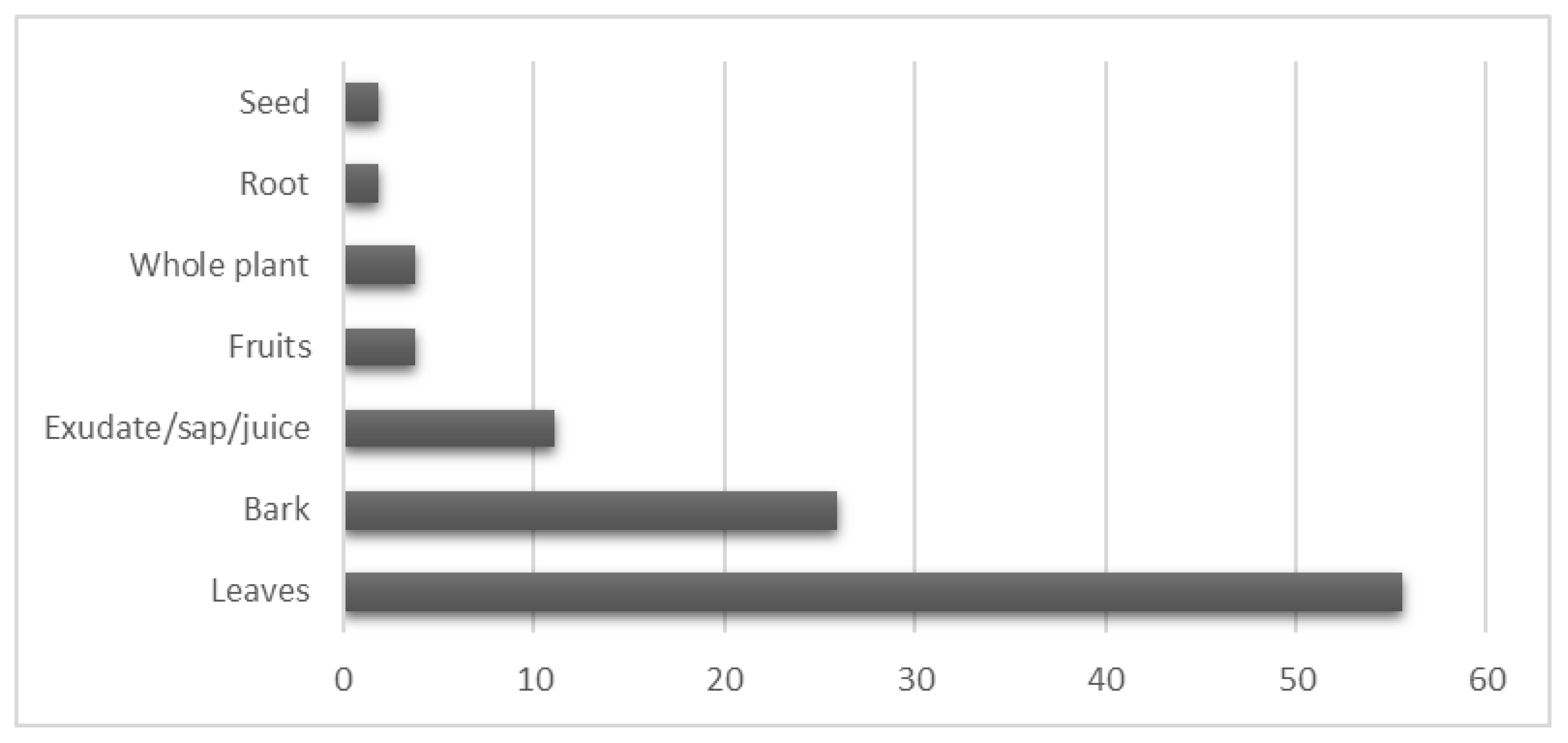
2.1.3. Plant Parts Used

From the collected ethnobotanical data, more than 56% of the recorded symptoms were treated using leaves. Leaves were commonly used in the treatment of skin-related problems (M. tanarius, H. giganteus, S. indica, and E. atoto) as well as digestive system disorders (M. tanarius and M. mollissimus). The second most frequently used plant part was the bark, accounting for more than 25% of the total utilization. Exudates/sap/juices constituted 11% of the mentioned remedies, whereas whole plants and fruits each represented 4% of the applications. The least utilized plant parts were seeds and roots, accounting for 2% each (Figure 1). The predominant use of leaves as the primary plant part in traditional medicine is well documented by numerous studies [44,45,46]. This preference for leaves can be attributed to their abundance and easy accessibility in nature. Leaves offer versatility in their application, as they can be utilized in dried, fresh, or processed forms. Additionally, leaves are known for their convenience in preservation, further contributing to their widespread usage in traditional medicinal practices [36].

2.2. Antimicrobial Activity Screening

The antimicrobial activities of the extracts of E. atoto, E. hypericifolia, H. giganteus, M. tanarius, M. mollissimus, M. rufidulus, and S. indica were tested against 15 microorganisms, including Gram-positive (seven), Gram-negative (four), and fungal (four) strains. The dried plant materials were extracted with MeOH, and after evaporation, the extracts were subjected to a solvent–solvent partition to obtain n-hexane, chloroform, ethyl acetate, and aqueous methanolic extracts. All 28 extracts of different polarities were first screened for antimicrobial activity by the disk diffusion method, and extracts exhibiting high activities were tested by the broth microdilution method. This is the first comprehensive analysis of the antimicrobial effects of extracts of these Euphorbiaceae species.

2.2.1. Screening by the Disk Diffusion Method

The screening results illustrated that the strains most sensitive to the tested extracts were S. aureus ATCC 29213, M. catarrhalis ATCC 25238, and N. glabrata ATCC 2001 (Table S1). In general, better activities were recorded against Gram-positive bacteria than against Gram-negative bacteria, excluding M. catarrhalis ATCC 25238, against which remarkable activities were detected. Among the fungal strains, noteworthy inhibitory activities were registered against C. parapsilosis ATCC 22019 and N. glabrata ATCC 2001. The largest zones of inhibition were measured for N. glabrata ATCC 2001 (up to 33 mm).
The M. tanarius extracts were most effective against Gram-positive strains, and its ethyl acetate extract had zones of inhibition larger than 15 mm against six bacterial and fungal strains. The M. mollissimus and M. rufidulus extracts exhibited modest activities against Gram-positive bacteria, but they were more effective against the N. glabrata ATCC 2001 stain. Among the H. giganteus extracts, the ethyl acetate was most potent against Staphylococcus strains, M. catarrhalis ATCC 25238, C. parapsilosis ATCC 22019, and N. glabrata ATCC 2001. In the case of the S. indica extracts, the antifungal effects predominated in addition to effects against M. catarrhalis ATCC 25238. Extracts of the two Euphorbia species displayed similar antimicrobial effects with moderate potency against bacteria and higher activity against fungal strains (Table S1).

2.2.2. Determination of MICs by the Microdilution Method

Seventeen extracts displaying significant activity (>15 mm diameter) in the antimicrobial screening by the disk diffusion method were further investigated, and their MICs were determined by the broth microdilution method (Table 3). The strongest activities were measured for the M. tanarius, H. giganteus, and E. atoto ethyl acetate extracts against M. catarrhalis ATCC 25238 (MIC = 12.5 µg/mL), followed by the S. indica aqueous methanolic extract (MIC = 25 µg/mL). Outstanding effects of the M. tanarius chloroform extract (MIC = 15.6 µg/mL) and H. giganteus ethyl acetate extract (MIC = 25 µg/mL) were observed against S. epidermidis ATCC 12228. The chloroform extract of M. rufidulus was the only extract in our study with potency against B. subtilis ATCC 6633 (MIC = 25 µg/mL). The M. tanarius ethyl acetate extract was the most effective against N. glabrata ATCC 2001 (MIC = 50 µg/mL). Further moderate activities (MIC = 100–500 µg/mL) were recorded against S. aureus ATCC 29213, MRSA ATCC 43300, C. parapsilosis ATCC 22019, and N. glabrata ATCC 2001. None of the n-hexane extracts was effective as an antimicrobial agent, but it is noteworthy that the ethyl acetate extract exhibited more favourable results in comparison to other solvents. The advantageous use of ethyl acetate as an extraction solvent has been mentioned in previous publications [47,48].

2.3. Antitumour Activity

Extracts with different polarities prepared from E. atoto, E. hypericifolia, H. giganteus, M. tanarius, M. mollissimus, M. rufidulus, and S. indica were tested for antiproliferative activity in doxorubicin-sensitive Colo 205 cells and doxorubicin-resistant Colo 320/MDR-LRP cells using the MTT assay. The aim of this experiment was to map the antitumour potential of the plant extracts because antiproliferative, cytotoxic, and multidrug resistance-reversing activities are characteristic of many Euphorbiaceae species [49].
Strong antiproliferative activity (IC50 < 1 µg/mL) was observed for the n-hexane and chloroform extracts of E. atoto and H. giganteus and the chloroform extracts of M. rufidulus and S. indica (IC50 = 1.04 µg/mL) in Colo 205 cells (Table 4). Colo 320 cells were less sensitive, and the highest activities, as indicated by IC50 values, ranged from 7 to 12 µg/mL. In most cases, the antiproliferative activity of the extracts was stronger against Colo 205 cells than against Colo 320 cells. The chloroform extract of M. tanarius was an exception because it had IC50 values 8.46 ± 0.36 µg/mL and 23.02 ± 0.86 µg/mL for Colo 320 and Colo 205 cells, respectively. This indicates that the M. tanarius chloroform extract has a selective antiproliferative effect in drug-resistant cells, and a relative resistance ratio (RR) of 0.37 was calculated as the ratio of the IC50 between the resistant and sensitive cancer cell lines. RR ≤ 0.5 indicates that an extract contains compounds with drug resistance-modifying activity and possesses collateral sensitivity effects [50]. Extracts of E. hypericifolia were marginally effective as an antiproliferative agent (n-hexane extract: IC50 = 55.94 ± 0.64 µg/mL; chloroform extract: IC50 = 36.54 ± 0.64 µg/mL) against Colo 205 and Colo 320 cells; however, its compounds displayed cytotoxic activities against the colorectal cancer cell line HCT-116 [28].
Interestingly, in this screening, none of the ethyl acetate and aqueous methanolic extracts exhibited antiproliferative activity (Table 4).

2.4. Isolation and Identification of Compounds from E. atoto

Among the studied Euphorbiaceae species, E. atoto was selected for detailed phytochemical studies because its n-hexane extract displayed strong antitumour activity (IC50 = 0.24 ± 0.06 µg/mL), and it was available in an appropriate amount (8.12 g). The n-hexane extract was first separated using OCC on polyamide, and the fractions obtained in this chromatography were then purified using VLC, PTLC, HPLC, and crystallization to yield five compounds (15, Figure 2). The structures of the isolated compounds were determined using 1H NMR, 13C NMR JMOD, 1H–1H COSY, HSQC, HMBC, and NOESY experiments.
Compound 1 was identified as 24-methylene-cycloartan-3β-ol based on a comparison of its spectral data with literature data [51]. This compound was previously isolated from several Euphorbia species [52]. Compound 2 was revealed to be identical to jolkinolide E based on its NMR spectroscopic characteristics. Jolkinolide E (2) is an abietane diterpene previously found in E. jolkini, E. helioscopia, and E. calyptrate [53].
Compound 3 was identified as tetra-tert-butyl-diphenyl ether [1,10-oxybis(2,4-di-tert-butylbenzene)], and it is an unusual structural type regarding the presence of tert-butyl groups in the molecule. Despite its “unnatural” structure, it was previously isolated from nature twice. Specifically, tetra-tert-butyl-diphenyl ether (3) was obtained from the marine mollusc Onchidium struma [54] and Dioscorea collettii rhizomes [55]. Jing et al. supposed that this unique structure is most likely attributed to the environmental stresses of plants (e.g., salinity, water supply). The monomer of 3, 2,4-di-tert-butylphenol (2,4-DTBP), has been detected in different living organisms, such as bacteria, fungi, bryophytes, pteridophytes, and higher plants. It was presumed that endocidal regulation is the primary function of 2,4-DTBP derivatives in the producing organisms [56].
Two widely occurring compounds were also isolated from the n-hexane extract of E. atoto: α-tocopherol (4) [57] and β-sitosterol (5) [58].

2.5. Investigation of the Antitumour Effects of 1

Among the isolated compounds, 24-methylene-cycloartan-3β-ol (1) was available in the amount required for cytotoxic assays. The antiproliferative activity of the compound was tested in colon adenocarcinoma cells (Colo 205), breast cancer cells (HTB-26, MCF-7), and nontumourous, human foetal lung fibroblasts (MRC-5) by the MTT assay. Compound 1 exhibited antiproliferative effects against the HTB-26 and Colo 205 cells with an IC50 of 24.83 µM and 30.68 µM, respectively (Table 5). Compound 1 had similar activity as the positive control, cisplatin (IC50 = 26.64 µM), but it displayed less tumour cell selectivity, as it was more cytotoxic in the MRC-5 cells (IC50 = 15.67 µM).

3. Materials and Methods

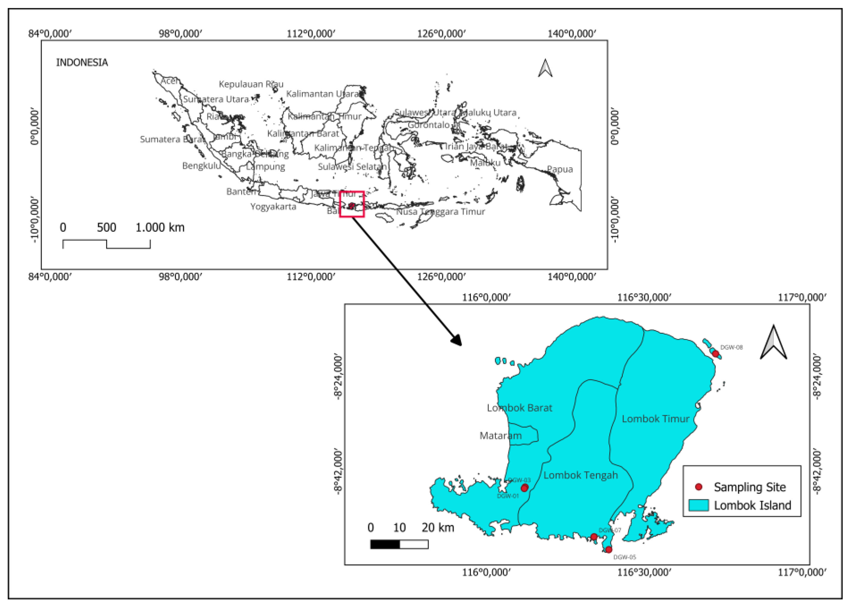
3.1. Plant Materials

Plant materials were collected in August 2021 from Lombok, West Nusa Tenggara, Indonesia (Figure 3). The identification of the plants was performed by I Gde Mertha (Department of Biology Education, Faculty of Teacher Training and Education, University of Mataram, Mataram, Indonesia). Voucher specimens were prepared for each species and deposited in the Laboratory of Silviculture, Department of Forestry, Faculty of Agriculture, University of Mataram (Table 6). The scientific names of the plants cited were verified in accordance with The World Flora Online [59]. The collected plant materials were washed under running tap water, cut into appropriate sizes, and airdried in an oven (Memmert, Universal Oven UN 55, Schwabach, Germany) at 40 °C ± 2 °C.

3.2. Ethnobotanical Data and Analysis

The primary ethnopharmacological data of the plants was extracted from the RISTOJA collected in 2012, 2015, and 2017. This database is not accessible to the public, and permission was requested from the NIHRD, Ministry of Health of the Republic of Indonesia to use the data for seven Euphorbiaceae species in this study. One of the authors (D. G. Wirasisya) participated in the collection of data in the RISTOJA program in 2017. Additional ethnobotanical information was obtained from the following databases: Web of Knowledge, Science Direct, PubMed, SciFinder, and Google Scholar. Initially, the ethnobotanical information obtained from the RISTOJA was documented in the local language. A careful process was undertaken to translate the data into English medical terminology. Then, all of the information was combined and categorized according to the classification framework of the International Classification of Primary Care (ICPC) system developed by the Wonca International Classification Committee and accepted by the World Health Organization.

3.3. General Experiment Procedures

NMR spectra were recorded in CDCl3 or CD3OD on a Bruker Avance DRX 500 spectrometer (Bruker, Billerica, MA, USA) at 500 (1H) or 125 MHz (13C). The signals from the deuterated solvents were taken as references. Two-dimensional NMR measurements were obtained using the standard Bruker software. In the 1H–1H COSY, NOESY, HSQC, and HMBC experiments, gradient-enhanced versions were applied. The separation by open column chromatography (OCC) was performed using polyamide (MP Polyamide, 50–160 μm, MP Biomedicals, Irvine, CA, USA), normal-phase vacuum liquid chromatography (NP-VLC) was conducted using silica gel (15 μm, Merck, Darmstadt, Germany). Thin-layer chromatography (TLC) was performed on silica 60 F254 (Merck) with detection under UV light (254 and 336 nm). Concentrated sulfuric acid was used as the spray reagent, followed by heating at 105 °C for 5 min. High-performance liquid chromatography (HPLC) purification was performed on the Shimadzu LC-10AS, equipped with an SPD-10A UV–VIS detector (Shimadzu Inc., Kyoto, Japan) using the LiChrosphere Si 60 column (5 µm, 250 × 4 mm, Merck).

3.4. Preparation of Plant Extracts and Fractions

The dried plant materials were ground into a powder using a mechanical blender (Retsch, Grindomix GM 200, Hann, Germany) and percolated with MeOH at room temperature. The crude extracts were then concentrated in vacuo. The concentrated extracts were dissolved in 50% aqueous methanol and subjected to a continuous solvent–solvent partition to afford n-hexane, chloroform, ethyl acetate, and water-soluble residual fractions. The yields of the extracts and fractions are presented as percentages of the total mass of the dried plant material (Table 7).

3.5. Isolation of Compounds from E. atoto

The n-hexane fraction (8.12 g) obtained from E. atoto (Table 7) was subjected to open column chromatography (OCC) on polyamide using mixtures of MeOH–H2O (4:6, 6:4, 8:2, and 100% MeOH) as eluents. The fractions yielded from 40% and 60% (0.63 g and 0.32 g, respectively). The MeOH elutions by polyamide OCC were found to be free of chlorophyll. These fractions were combined and further separated using VLC on silica gel. The separation was performed using a gradient system of cyclohexane–ethyl acetate–EtOH (100:0:0, 90:10:0, 80:20:0, 70:30:0, 60:40:1, 50:50:1, 50:50:5, 50:50:10, 0:80:20). TLC monitoring was employed, and fractions with similar composition were combined, resulting in 17 fractions (A–Q). Compound 1 (17 mg) was obtained by crystallization from the MeOH solution of fraction B (48.7 mg). The remaining mother liquor of fraction B was subjected to further separation via normal phase (NP)-HPLC using a mobile phase of cyclohexane–ethyl acetate (94:6) that led to the isolation of compound 2 (0.7 mg).
The fraction obtained by polyamide OCC using an elution with 80% MeOH (4.36 g) was separated using VLC on silica gel utilizing the gradient system of cyclohexane–ethyl acetate–EtOH (100:0:0, 90:10:0, 80:20:0, 70:30:0, 60:40:1, 50:50:1, 50:50:5, 50:50:10, 0:80:20), yielding 15 fractions (A–O). Fraction B (305.9 mg) was further purified by VLC on silica gel by employing a gradient system of n-hexane–chloroform–acetone (80:10:0, 60:40:0, 60:40:0.5, 80:20:0.5, 60:40:10, and 60:40:5), resulting in the isolation of ten subfractions (B1–10). Subfraction B8 (219.6 mg) was subsequently subjected to preparative TLC (PTLC) using cyclohexane–chloroform–acetone (6:4:0.3) as the mobile phase, yielding five subfractions (B8/1–5). PTLC using a mobile phase of cyclohexane–ethyl acetate (9:1) was then applied to separate B8/4 (115.9 mg), resulting in five subfractions (B8/4/1–5). The final purification of subfraction B8/4/3 (41.9 mg) was accomplished using NP-HPLC with cyclohexane–ethyl acetate (94:6) as the mobile phase, affording pure compounds 3 (0.7 mg) and 4 (2.8 mg). Compound 5 (60.4 mg) was obtained using crystallization from the MeOH solution of fraction E (437.5 mg).

3.6. Bacterial and Fungal Strains and Culture Conditions for Antimicrobial Assays

The test microorganisms included the standard Gram-positive strains Staphylococcus aureus (ATCC 29213), methicillin-resistant S. aureus (MRSA) (ATCC 43300), S. epidermidis (ATCC 12228), Streptococcus pyogenes (ATCC 19615), S. agalactiae (ATCC 13813), Bacillus subtilis (ATCC 6633), and Enterococcus faecalis (ATCC 29212). The standard Gram-negative strains were Escherichia coli (ATCC 35218), E. coli (K-12 AG-100), Pseudomonas aeruginosa (ATCC 27853), and Moraxella catarrhalis (ATCC 25238). The fungal strains used were Candida albicans (ATCC 10231), C. parapsilosis (ATCC 22019), C. tropicalis (ATCC 750), and Nakaseomyces glabrata (ATCC 2001). The bacterial cultures were grown on standard Mueller–Hinton (MH) agar, and fungal cultures were grown on RPMI plates (Diagnosticum Zrt., Budapest, Hungary) at 37 °C overnight in an aerobic environment.

3.7. Determination of Antibacterial Activity Using the Disk Diffusion Method

The disk diffusion method was employed to screen extracts for their antibacterial and antifungal activities against standard bacterial and fungal strains to determine their zones of inhibition. Briefly, the samples were dissolved in DMSO at a concentration of 50 mg/mL. Sterile filter paper disks [6 mm in diameter, Whatman antibiotic paper disk (Cytiva, Marlborough, MA, USA)] coated with 10 μL of the sample solutions were placed on top of the bacterial suspension (inoculum: 0.5 McFarland, 1.5 × 10⁸ CFU mL−1). Disks containing antibiotics (ciprofloxacin and ampicillin) and antifungal agents (nystatin) were used as positive controls, and disks containing DMSO served as the negative controls. Under aerobic conditions, the plates were incubated at 37 °C ± 2 °C for 20 h. The diameters of the zones of inhibition caused by the compounds, including the disk, were measured in triplicate. For each of the three repetitions, an average zone of inhibition was calculated [60].

3.8. Determination of the Minimum Inhibitory Concentration (MIC)

Bacteria: The MICs of all tested fractions were determined according to the Clinical and Laboratory Standards Institute (CLSI) guidelines in three independent assays [61]. The starting concentration of tested fractions was 200 µg/mL or 500 µg/mL.
Fungi: Determination of the MIC values was performed according to the European Committee on Antimicrobial Susceptibility Testing (EUCAST) standards in three independent assays [62]. The starting concentration of tested fractions was 200 µg/mL or 500 µg/mL.
The values are given as the mean determined for three replicates from three independent experiments. DMSO was assayed to ensure there was no antibacterial and antifungal effect at the concentration (2 v/v%) applied in the assay.

3.9. Cell Line Cultures

Two human colon adenocarcinoma cell lines, namely Colo 205 (ATCC-CCL-222) doxorubicin-sensitive parent cells and Colo 320/MDR-LRP (ATCC-CCL-220.1) doxorubicin-resistant cells expressing ABCB1, were purchased from LGC Promochem (Teddington, UK). The cells were cultured in RPMI 1640 medium supplemented with 10% heat-inactivated foetal bovine serum (FBS), 2 mM L-glutamine, 1 mM sodium pyruvate, and 100 mM HEPES. HTB-26 breast adenocarcinoma cell line was purchased from LGC Promochem (Teddington, UK). The cell line was cultured in RPMI 1640 medium supplemented with 10% heat-inactivated foetal bovine serum, 2 mM L-glutamine, and 1 mM sodium pyruvate. The breast cancer cell line, MCF-7 (ATCC HTB-22), was purchased from LGC Promochem (Teddington, UK). The MCF-7 cell line was grown in Eagle’s minimal essential medium (EMEM), containing 4.5 g/L glucose supplemented with a non-essential amino acid mixture, a selection of vitamins, and 10% heat-inactivated FBS. The MRC-5 human embryonic lung fibroblast cell line (ATCC CCL-171) was purchased from LGC Promochem (Teddington, UK). The cell line was cultured in EMEM supplemented with a non-essential amino acid mixture, a selection of vitamins, 10% FBS, 2 mM L-glutamine, 1 mM Na-pyruvate, nystatin, and a penicillin–streptomycin mixture in concentrations of 100 U/L and 10 mg/L, respectively. All of the cells were incubated at 37 °C in a 5% CO2 and 95% air atmosphere.

3.10. Antiproliferative Assay

The antiproliferative effects of the extracts were tested in decreasing serial dilutions (2-fold dilutions starting from 100 µg/mL) in human cancer cell lines (Colo 205, Colo 320) in 96-well, flat-bottomed microtiter plates. First, the extracts were diluted in 100 µL of the medium, and then 6 × 10³ cells (Colo 205, Colo 320) in 100 µL of RPMI medium were added to each well, excluding the medium control wells. The culture plates were incubated at 37 °C for 72 h. Following incubation, 20 μL of 3-(4,5-dimethylthiazol-2-yl)-2,5-diphenyl tetrazolium bromide (MTT) solution (from a 5 mg/mL stock solution) was added to each well. After incubation at 37 °C for 4 h, 100 μL of the sodium dodecyl sulphate (SDS) solution (10% SDS in 0.01 M HCl) was added to each well, and the plates were further incubated at 37 °C overnight. Cell growth was determined by measuring the optical density at 540 nm (ref.: 630 nm) using a Multiskan EX ELISA reader (Thermo Labsystems, Cheshire, WA, USA). The concentration that decreased cell viability by 50% was expressed as the IC50 (µg/mL) for each extract. The antiproliferative effect of compound 1 was determined on the Colo 205, HTB-26, MCF-7, and MRC-5 as described for the extracts. Meanwhile, a 5 mM stock solution in DMSO and a starting concentration of 100 µM were applied.

4. Conclusions

Ethnobotanical data were collected for seven Indonesian Euphorbiaceae species from the RISTOJA database and other literature sources and evaluated from different perspectives. It was stated that E. atoto, E. hypericifolia, H. giganteus, M. tanarius, M. mollissimus, M. rufidulus, and S. indica are medicinal plants of the jamu healing system utilized in Indonesia and surrounding areas. E. hypericifolia was the only species with no record in Asian ethnomedicine. However, this plant is used in South Africa and Uganda to treat gonorrhoea and snake and scorpion bite. The ethnobotanical uses of the seven plants included the treatment of skin disorders (17), general symptoms (10), digestive system ailments (9), and musculoskeletal issues (6). Many plant utilizations, such as abscesses, sores, boils, and other skin diseases; fever; urinary tract problems; cough; bloody diarrhoea; leukorrhea; and eye diseases, could be related to bacterial or fungal infections, whereas other symptoms could be reflective of colon cancer, such constipation, flatulence, stomach aches, and diarrhoea. Therefore, the plant extracts were screened for antimicrobial and antitumour activities. Fractions of different polarities (n-hexane, chloroform, ethyl acetate, and aqueous methanolic) were prepared from the plants and tested against 15 microorganisms, including Gram-positive (seven), Gram-negative (four), and fungal (four) strains. Antibacterial effects were observed for the M. tanarius, H. giganteus, and M. rufidulus extracts against the Gram-positive strains, S. aureus ATCC 29213, MRSA ATCC 43300, S. epidermidis ATCC 12228, and B. subtilis ATCC 6633 (MIC = 15.6–500 µg/mL). The Gram-negative bacteria, E. coli ATCC 35218, E. coli K-12 AG-100, and P. aeruginosa ATCC 27853, were not sensitive to most plant extracts, whereas strong antibacterial activity was observed for the M. tanarius, H. giganteus, S. indica, and E. atoto extracts against M. catarrhalis ATCC 25238 (MIC = 12.5–25 µg/mL). The detected antibacterial effects of M. tanarius and H. giganteus can support their application for different skin disorders, fever, cough, and eye diseases. The antifungal screening proved the activity of all plants against C. parapsilosis ATCC 22019 and/or N. glabrata ATCC 2001 with MICs of 50–500 µg/mL. The strongest activity was observed for the M. tanarius (MIC 50 µg/mL) and M. mollissimus (MIC 100 µg/mL) ethyl acetate extracts.
The antitumour activities of 28 plant extracts were screened in drug-sensitive Colo 205 and drug-resistant Colo 320 colon adenocarcinoma cells. Remarkable antiproliferative activities (IC50 = 0.23–2.60 µg/mL) were displayed by the lipophilic extracts of H. giganteus, M. rufidulus, S. indica, and E. atoto, indicating that these extracts can serve as excellent sources of compounds with strong antitumour activity. Another interesting observation was the selective antiproliferative effect of the M. tanarius chloroform extract in Colo 320 cells, suggesting the presence of compounds with drug resistance-modifying activity in this extract.
The E. atoto n-hexane extract, which exhibited strong antiproliferative activity, was subjected to a detailed phytochemical investigation, and five compounds, 24-methylene-cycloartan-3β-ol (1), jolkinolide E (2), tetra-tert-butyl-diphenyl ether (3), α-tocopherol (4), and β-sitosterol (5) were isolated. Compounds 14 were detected in this species for the first time in this study. Antitumour testing of 24-methylene-cycloartan-3β-ol (1) revealed its antiproliferative effect for the first time against HTB-26 human breast (IC50 = 24.83 µM) and Colo 205 human colon adenocarcinoma cells (IC50 30.68 µM) without tumour cell selectivity. It is most probable that the compounds of the E. atoto n-hexane extract responsible for its strong antitumour activity could only partially be identified in the present experiment. Therefore, a bioassay-guided isolation procedure starting from a larger amount of extract will be needed to fully identify the active ingredients.

Supplementary Materials

The following supporting information can be downloaded at: https://www.mdpi.com/article/10.3390/plants12223836/s1, Figures S1–S12, NMR spectra of compounds 14, and Table S1: Antimicrobial activities of the fractions at a concentration of 50 µg/mL by the agar disk diffusion method.

Author Contributions

Conceptualization, D.G.W. and J.H.; methodology, L.V., G.S. and I.G.M.; validation, G.S. and I.G.M.; investigation, D.G.W., A.K., L.V., N.S. and A.B.; data curation, D.G.W., A.K., N.S. and A.B.; writing—original draft preparation, D.G.W.; writing—review and editing, G.S., J.H.; visualization, A.K.; supervision, J.H.; funding acquisition, J.H. All authors have read and agreed to the published version of the manuscript.

Funding

This research was funded by the National Research, Development and Innovation Fund, Hungary (NKFI) under grant numbers K135845 and Ministry of Innovation and Technology of Hungary from the NKFIH Fund, project no. KP2021-EGA-32.

Data Availability Statement

Data are contained within the article and supplementary materials.

Acknowledgments

The authors wish to thank Laboratory of Data Management NIHRD, Ministry of Health of the Republic of Indonesia for providing the RISTOJA subset data.

Conflicts of Interest

The authors declare no conflict of interest.

References

  1. Elfahmi; Woerdenbag, H.J.; Kayser, O. Jamu: Indonesian traditional herbal medicine towards rational phytopharmacological use. J. Herb. Med. 2014, 4, 51–73. [Google Scholar] [CrossRef]
  2. Budiarti, M.; Maruzy, A.; Mujahid, R.; Sari, A.N.; Jokopriyambodo, W.; Widayat, T.; Wahyono, S. The use of antimalarial plants as traditional treatment in Papua Island, Indonesia. Heliyon 2020, 6, e05562. [Google Scholar] [CrossRef]
  3. Heinrich, M. Ethnobotany and its role in drug development. Phytother. Res. 2000, 14, 479–488. [Google Scholar] [CrossRef]
  4. Hammadi, R.; Kúsz, N.; Dávid, C.Z.; Mwangi, P.W.; Berkecz, R.; Szemerédi, N.; Spengler, G.; Hohmann, J.; Vasas, A. Polyoxypregnane ester derivatives and lignans from Euphorbia gossypina var. coccinea pax. Plants 2022, 11, 1299. [Google Scholar] [CrossRef] [PubMed]
  5. Wirasisya, D.G.; Hohmann, J. An Overview of the traditional use, phytochemistry, and biological activity of the genus Homalanthus. Fitoterapia 2023, 166, 105466. [Google Scholar] [CrossRef]
  6. Staub, P.O.; Geck, M.S.; Weckerle, C.S.; Casu, L.; Leonti, M. Classifying diseases and remedies in ethnomedicine and ethnopharmacology. J. Ethnopharmacol. 2015, 174, 514–519. [Google Scholar] [CrossRef] [PubMed]
  7. Plant of the World Online. Macaranga tanarius (L.) Müll.Arg. Available online: http://powo.science.kew.org/taxon/urn:lsid:ipni.org:names:351381-1 (accessed on 19 July 2023).
  8. Mustaqim, W.A. Macaranga tanarius (L.) Müll.Arg. Euphorbiaceae. In Ethnobotany of the Mountain Regions of Southeast Asia; Franco, F.M., Ed.; Ethnobotany of Mountain Regions; Springer International Publishing: Cham, Switzerland, 2021; pp. 651–662. [Google Scholar]
  9. National Institute of Health Research and Development. National Report: Exploration on Local Knowledge of Ethnomedicine and Community-Based Medicinal Plants in Indonesia (Ristoja 2017); NIHRD Publisher Agency: Jakarta, Indonesia, 2017. [Google Scholar]
  10. National Institute of Health Research and Development. National Report: Exploration on Local Knowledge of Ethnomedicine and Community-Based Medicinal Plants in Indonesia (Ristoja 2015); NIHRD Publisher Agency: Jakarta, Indonesia, 2015. [Google Scholar]
  11. National Institute of Health Research and Development. National Report: Exploration on Local Knowledge of Ethnomedicine and Community-Based Medicinal Plants in Indonesia (Ristoja 2012); NIHRD Publisher Agency: Jakarta, Indonesia, 2012. [Google Scholar]
  12. Turpin, G.; Ritmejerytė, E.; Jamie, J.; Crayn, D.; Wangchuk, P. Aboriginal medicinal plants of Queensland: Ethnopharmacological uses, species diversity, and biodiscovery pathways. J. Ethnobiol. Ethnomed. 2022, 18, 54. [Google Scholar] [CrossRef]
  13. Amir Rawa, M.S.; Nurul Azman, N.A.; Mohamad, S.; Nogawa, T.; Wahab, H.A. In vitro and in silico anti-acetylcholinesterase activity from Macaranga tanarius and Syzygium jambos. Molecules 2022, 27, 2648. [Google Scholar] [CrossRef]
  14. Chien, Y.-H.; Yu, Y.-H.; Ye, S.-R.; Chen, Y.-W. Antibacterial and antioxidant activity of the fruit of Macaranga tanarius, the plant origin of Taiwanese green propolis. Antioxidants 2022, 11, 1242. [Google Scholar] [CrossRef]
  15. Heil, M.; Fiala, B.; Maschwitz, U.; Linsenmair, K.E. On benefits of indirect defence: Short- and long-term studies of antiherbivore protection via mutualistic ants. Oecologia 2001, 126, 395–403. [Google Scholar] [CrossRef]
  16. Hsu, Y.-C.; Chang, C.-C.; Hsieh, C.-C.; Shih, Y.-H.; Chang, H.-C.; Lin, C.-L. Therapeutic potential of extracts from Macaranga tanarius (mte) in diabetic nephropathy. Plants 2023, 12, 656. [Google Scholar] [CrossRef]
  17. Péresse, T.; Jézéquel, G.; Allard, P.-M.; Pham, V.-C.; Huong, D.T.M.; Blanchard, F.; Bignon, J.; Lévaique, H.; Wolfender, J.-L.; Litaudon, M.; et al. Cytotoxic prenylated stilbenes isolated from Macaranga tanarius. J. Nat. Prod. 2017, 80, 2684–2691. [Google Scholar] [CrossRef] [PubMed]
  18. Kawakami, S.; Harinantenaina, L.; Matsunami, K.; Otsuka, H.; Shinzato, T.; Takeda, Y. Macaflavanones a−g, prenylated flavanones from the leaves of Macaranga tanarius. J. Nat. Prod. 2008, 71, 1872–1876. [Google Scholar] [CrossRef]
  19. Kumazawa, S.; Murase, M.; Momose, N.; Fukumoto, S. Analysis of antioxidant prenylflavonoids in different parts of Macaranga tanarius, the plant origin of Okinawan propolis. Asian Pac. J. Trop. Med. 2014, 7, 16–20. [Google Scholar] [CrossRef] [PubMed]
  20. Lee, J.-H.; Kim, Y.-G.; Khadke, S.K.; Yamano, A.; Woo, J.-T.; Lee, J. Antimicrobial and antibiofilm activities of prenylated flavanones from Macaranga tanarius. Phytomedicine 2019, 63, 153033. [Google Scholar] [CrossRef]
  21. Phommart, S.; Sutthivaiyakit, P.; Chimnoi, N.; Ruchirawat, S.; Sutthivaiyakit, S. Constituents of the leaves of Macaranga tanarius. J. Nat. Prod. 2005, 68, 927–930. [Google Scholar] [CrossRef]
  22. Tseng, M.-H.; Chou, C.-H.; Chen, Y.-M.; Kuo, Y.-H. Allelopathic prenylflavanones from the fallen leaves of Macaranga tanarius. J. Nat. Prod. 2001, 64, 827–828. [Google Scholar] [CrossRef] [PubMed]
  23. Plant of the World Online. Euphorbia atoto G.Forst. Available online: http://powo.science.kew.org/taxon/urn:lsid:ipni.org:names:345695-1 (accessed on 18 July 2023).
  24. Chen, M.-T.; Kuoh, C.-S.; Lee, K.-K. Studies on the constituents of Euphorbia atoto Forst.f. Taiwan Pharm. J. 1979, 31, 114–117. [Google Scholar]
  25. Hart, N.K.; Johns, S.R.; Lamberton, J.A. (+)-9-aza-1-methylbicyclo[3,3,1]nonan-3-one, a new alkaloid from Euphorbia atoto Forst. Aust. J. Chem. 1967, 20, 561–563. [Google Scholar] [CrossRef]
  26. Zhao, H.; Duan, R.-J.; Kong, C.-H.; Dai, H.-F.; Mei, W.-L.; Xu, F.-Q.; Huang, S.-Z. Two new anti-inflammatory trachylobane diterpenoids from Euphorbia atoto. J Asian Nat. Prod. Res. 2023. [Google Scholar] [CrossRef]
  27. Norhanom, A.W.; Yadav, M. Tumour promoter activity in Malaysian Euphorbiaceae. Br. J. Cancer 1995, 71, 776–779. [Google Scholar] [CrossRef] [PubMed]
  28. Hu, R.; Sang, J.; Li, W.; Tian, Y.; Zou, M.-F.; Tang, G.-H.; Yin, S. Structurally diverse triterpenoids with cytotoxicity from Euphorbia hypericifolia. Fitoterapia 2021, 151, 104888. [Google Scholar] [CrossRef] [PubMed]
  29. Azis, H.A.; Taher, M.; Susanti, D.; Zakariac, Z.A. In vitro wound healing activity of Sapium indicum willd leaf extracts. Afr. J. Tradit. Complement. Altern. Med. 2015, 12, 126–132. [Google Scholar]
  30. Chumkaew, P.; Karalai, C.; Ponglimanont, C.; Chantrapromma, K. Antimycobacterial activity of phorbol esters from the fruits of Sapium indicum. J. Nat. Prod. 2003, 66, 540–543. [Google Scholar] [CrossRef] [PubMed]
  31. Mokmued, K.; Dechayont, B.; Phuaklee, P.; Liplung, C.; Muangpoolsawad, H.; Nuengchamnong, N.; Prommee, N. Evaluation of anti-inflammatory, cytotoxic, anti-h. pylori, antioxidant activities, and phytochemical compositions of Shirakiopsis indica (Willd.) Esser. ScienceAsia 2021, 47, 549. [Google Scholar] [CrossRef]
  32. Tanaka, N.; Takahashi, S.; Yoshino, Y.; Nakatani, M.; Ahmed, F.A.; Hossain, G.M.; Chen, C.-H.; Lee, K.-H.; Kashiwada, Y. Tigliane-type diterpene esters from the fruits of Shirakiopsis indica and their anti-HIV activity. J. Nat. Prod. 2022, 85, 2687–2693. [Google Scholar] [CrossRef]
  33. Khastini, R.O.; Wahyuni, I.; Saraswati, I. Ethnobotanical study of digestive systems disorders in Baduy ethnic, Indonesisa. BIOTROPIA-Southeast Asian J. Trop. Biol. 2021, 28. [Google Scholar]
  34. Nisa, U.; Astana, P.R.W.; Triyono, A.; Ardiyanto, D.; Fitriani, U.; Zulkarnain, Z.; Novianto, F.; Jannah, W.D.M. Ethnobotanical study of medicinal plants used for treating urinary tract problems in eastern Indonesia. IOP Conf. Ser. Earth Environ. Sci. 2021, 905, 012119. [Google Scholar] [CrossRef]
  35. Silalahi, M.; Nisyawati, N.; Walujo, E.B.; Mustaqim, W. Ethnomedicine of medicinal plants by Batak Phakpak subethnic in the Surung Mersada village, Phakpak Bharat district, North Sumatera. J. Ilmu Dasar 2018, 19, 77–92. [Google Scholar] [CrossRef]
  36. Silalahi, M.; Nisyawati; Walujo, E.B.; Supriatna, J.; Mangunwardoyo, W. The local knowledge of medicinal plants trader and diversity of medicinal plants in the Kabanjahe traditional market, North Sumatra, Indonesia. J. Ethnopharmacol. 2015, 175, 432–443. [Google Scholar] [CrossRef]
  37. Mustofa, F.; Rahmawati, N.; Saryanto, S. Ethnomedicine of Medicinal plants used by traditional healers to facilitate bone injury healing in Kalimantan, Indonesia. J. Tumbuh. Obat Indones. 2021, 14, 36–54. [Google Scholar] [CrossRef]
  38. Maneenoon, K.; Khuniad, C.; Teanuan, Y.; Saedan, N.; Prom-in, S.; Rukleng, N.; Kongpool, W.; Pinsook, P.; Wongwiwat, W. Ethnomedicinal plants used by traditional healers in Phatthalung province, Peninsular Thailand. J. Ethnobiol. Ethnomed. 2015, 11, 43. [Google Scholar] [CrossRef]
  39. Susiarti, S.; Sambas, E.N. Local knowledge on medicinal plants of Batak Mandailing and Nias communities in Batang Toru, North Sumatra, Indonesia. IOP Conf. Ser. Earth Environ. Sci. 2018, 197, 012008. [Google Scholar] [CrossRef]
  40. Dagar, H.S. Plant folk medicines among Nicobarese tribals of car Nicobar island, India. Econ. Bot. 1989, 43, 215–224. [Google Scholar] [CrossRef]
  41. Uhe, G. Medicinal Plants of Samoa: A preliminary survey of the use of plants for medicinal purposes in the Samoan islands. Econ. Bot. 1974, 28, 1–30. [Google Scholar] [CrossRef]
  42. Asowata-Ayodele, A.M.; Afolayan, A.J.; Otunola, G.A. Ethnobotanical survey of culinary herbs and spices used in the traditional medicinal system of Nkonkobe municipality, Eastern Cape, South Africa. S. Afr. J. Bot. 2016, 104, 69–75. [Google Scholar] [CrossRef]
  43. Okot, D.F.; Anywar, G.; Namukobe, J.; Byamukama, R. Medicinal plants species used by herbalists in the treatment of snakebite envenomation in Uganda. Trop. Med. Health 2020, 48, 44. [Google Scholar] [CrossRef] [PubMed]
  44. Jima, T.T.; Megersa, M. Ethnobotanical Study of Medicinal plants used to treat human diseases in Berbere District, Bale Zone of Oromia regional state, South East Ethiopia. J. Evid.-Based Complement. Altern. Med. 2018, 2018, 8602945. [Google Scholar] [CrossRef]
  45. Muthu, C.; Ayyanar, M.; Raja, N.; Ignacimuthu, S. Medicinal plants used by traditional healers in Kancheepuram district of Tamil Nadu, India. J. Ethnobiol. Ethnomed. 2006, 2, 43. [Google Scholar] [CrossRef] [PubMed]
  46. Jadid, N.; Kurniawan, E.; Himayani, C.E.S.; Andriyani; Prasetyowati, I.; Purwani, K.I.; Muslihatin, W.; Hidayati, D.; Tjahjaningrum, I.T.D. An ethnobotanical study of medicinal plants used by the tengger tribe in Ngadisari village, Indonesia. PLoS ONE 2020, 15, e0235886. [Google Scholar] [CrossRef]
  47. Su, P.-W.; Yang, C.-H.; Yang, J.-F.; Su, P.-Y.; Chuang, L.-Y. Antibacterial activities and antibacterial mechanism of Polygonum cuspidatum extracts against nosocomial drug-resistant pathogens. Molecules 2015, 20, 11119–11130. [Google Scholar] [CrossRef] [PubMed]
  48. Toh, S.C.; Lihan, S.; Bunya, S.R.; Leong, S.S. In vitro antimicrobial efficacy of Cassia alata (linn.) leaves, stem, and root extracts against cellulitis causative agent Staphylococcus aureus. BMC Complement. Med. Ther. 2023, 23, 85. [Google Scholar]
  49. Vasas, A.; Hohmann, J. Euphorbia diterpenes: Isolation, structure, biological activity, and synthesis (2008–2012). Chem. Rev. 2014, 114, 8579–8612. [Google Scholar] [CrossRef]
  50. Hall, M.D.; Handley, M.D.; Gottesman, M.M. Is resistance useless? Multidrug resistance and collateral sensitivity. Trends Pharmacol. Sci. 2009, 30, 546–556. [Google Scholar] [CrossRef] [PubMed]
  51. Shamsabadipour, S.; Ghanadian, M.; Saeedi, H.; Rahimnejad, M.R.; Mohammadi-Kamalabadi, M.; Ayatollahi, S.M.; Salimzadeh, L. Triterpenes and steroids from Euphorbia denticulata lam. with anti-herpes symplex virus activity. Iran. J. Pharm. Res. 2013, 12, 759–767. [Google Scholar]
  52. Benjamaa, R.; Moujanni, A.; Kaushik, N.; Choi, E.H.; Essamadi, A.K.; Kaushik, N.K. Euphorbia species latex: A comprehensive review on phytochemistry and biological activities. Front. Plant Sci. 2022, 13, 1008881. [Google Scholar] [CrossRef]
  53. Crespi-Perellino, N.; Garofano, L.; Arlandini, E.; Pinciroli, V.; Minghetti, A.; Vincieri, F.F.; Danieli, B. Identification of new diterpenoids from Euphorbia calyptrata cell cultures. J. Nat. Prod. 1996, 59, 773–776. [Google Scholar] [CrossRef]
  54. Sun, B.-N.; Shen, H.-D.; Wu, H.-X.; Cui, F.; Yao, L.-X.; Cheng, Z.-Q.; Diao, Y. Chemical constituents of Onchidium struma from Chongming Island. Shanghai. Nat. Prod. Res. Dev. 2014, 26, 987–989. [Google Scholar]
  55. Jing, S.; Qu, Z.; Zhao, C.; Li, X.; Guo, L.; Liu, Z.; Zheng, Y.; Gao, W. Dihydroisocoumarins and dihydroisoflavones from the rhizomes of Dioscorea collettii with cytotoxic activity and structural revision of 2,2′-oxybis(1,4-di-tert-butylbenzene). Molecules 2021, 26, 5381. [Google Scholar] [CrossRef]
  56. Zhao, F.; Wang, P.; Lucardi, R.D.; Su, Z.; Li, S. Natural sources and bioactivities of 2,4-di-tert-butylphenol and its analogs. Toxins 2020, 12, 35. [Google Scholar] [CrossRef]
  57. Wang, A.; Fraga, R.P.A.; Hörmann, E.; Pfaltz, A. Iridium-catalyzed asymmetric hydrogenation of unfunctionalized, trialkyl-substituted olefins. Chem. Asian J. 2011, 6, 599–606. [Google Scholar] [CrossRef] [PubMed]
  58. Erwin, W.R.P.; Safitri, R.D.; Marliana, E.; Kusuma, I.W. Isolation and characterization of stigmasterol and β-sitosterol from wood bark extract of Baccaurea macrocarpa Miq. Mull. Arg. Rasayan J. Chem. 2020, 13, 2552–2558. [Google Scholar] [CrossRef]
  59. World Flora Online WFO: Published on the Internet. Available online: http://www.worldfloraonline.org/ (accessed on 17 May 2023).
  60. Ghazal, T.S.A.; Schelz, Z.; Vidács, L.; Szemerédi, N.; Veres, K.; Spengler, G.; Hohmann, J. Antimicrobial, multidrug resistance reversal and biofilm formation inhibitory effect of Origanum majorana extracts, essential oil and monoterpenes. Plants 2022, 11, 1432. [Google Scholar] [CrossRef] [PubMed]
  61. Clinical and Laboratory Standard Institute. Performance Standards for Antimicrobial Susceptibility Testing, 30th ed.; CLSI: Wayne, PA, USA, 2020. [Google Scholar]
  62. Rodríguez-Tudela, J.L.; Barchiesi, F.; Bille, J.; Chryssanthou, E.; Cuenca-Estrella, M.; Denning, D.; Donnelly, J.P.; Dupont, B.; Fegeler, W.; Moore, C.; et al. Method for the determination of minimum inhibitory concentration (mic) by broth dilution of fermentative yeasts. Clin. Microbiol. Infect. 2003, 9, i–viii. [Google Scholar] [CrossRef]
Figure 1. Frequencies of the use of medical plants parts (%).
Plants 12 03836 g001
Figure 2. Structures of compounds 1–3 isolated from E. atoto.
Plants 12 03836 g002
Figure 3. Map of Indonesia and the areas from which plants were collected.
Plants 12 03836 g003
Table 1. The use of the selected Euphorbiaceae species by category.
Treatment CategoriesNo. Indications or Symptoms
Skin17
General symptoms10
Digestive system9
Musculoskeletal system6
Genital system3
Pregnancy and childbearing3
Eye2
Neurological system2
Respiratory system1
Blood, blood-forming organs and immune system1
Endocrine, metabolic and nutritional system1
Urinary system1
Total56
Table 3. MICs in µg/mL of the selected fractions of Euphorbiaceae species against bacteria and fungi using the broth microdilution method.
SpeciesFr. aBacteriaFungi
S. aureus ATCC 29213MRSA
ATCC 43300
S. epidermidis ATCC 12228B. subtilis ATCC 6633M. catarrhalis ATCC 25238C. parapsilosis ATCC 22019N. glabrata ATCC 2001
Euphorbia atotoE 12.5 250
MW >500500
Euphorbia
hypericifolia
C >500
E 500250
MW 500
Homalanthus giganteusE25025025 12.5>500200
MW 500
Macaranga tanariusC 15.6 500
E500250100 12.5 50
Mallotus
mollissimus
C 500
E 100
Mallotus
rufidulus
C 25 250
E 250
Shirakiopsis
indica
H >500
C >500
E 500250
MW 25500250
Ciprofloxacin 0.010.210.10.020.02
Ampicillin 0.264.141.040.010.003
Nystatin 0.31250.3125
DMSO (%)>2%>2%>2%>2%>2%>2%>2%>2%
a H = n-hexane, C = chloroform, E = ethyl acetate, MW = aqueous methanolic extract.
Table 4. Antiproliferative activities of the plant extracts in colorectal adenocarcinoma cells.
Plant Name IC50 Value (µg/mL) a
FractionColo 205Colo 320
Euphorbia atoton-Hex0.24 ± 0.0655.02 ± 0.91
CHCl30.23 ± 0.0453.17 ± 0.49
EtOAcNN
Aq-MeOH--
Euphorbia hypericifolian-Hex55.94 ± 0.6472.05 ± 0.82
CHCl3N36.54 ± 0.64
EtOAcNN
Aq-MeOHNN
Homalanthus giganteusn-Hex0.92 ± 0.1531.92 ± 2.69
CHCl314.42 ± 1.3026.18 ± 1.77
EtOAcNN
Aq-MeOHNN
Macaranga tanariusn-Hex56.58 ± 1.1466.57 ± 2.01
CHCl323.02 ± 0.868.46 ± 0.36
EtOAcNN
Aq-MeOHNN
Mallotus mollissimusn-Hex37.29 ± 1.1384.40 ± 1.22
CHCl357.18 ± 0.4162.03 ± 0.42
EtOAcNN
Aq-MeOHNN
Mallotus rufidulusn-Hex23.18 ± 2.2257.35 ± 0.58
CHCl30.56 ± 0.077.10 ± 0.60
EtOAcNN
Aq-MeOHNN
Shirakiopsis indican-Hex2.60 ± 0.3555.53 ± 1.54
CHCl31.04 ± 0.0111.93 ± 0.82
EtOAcNN
Aq-MeOHNN
Doxorubicin b 1.45 ± 0.191.76 ± 0.09
a IC50 values are means of triplicate (n = 3) ± standard deviations. N, no IC50 shown or the IC50 was above 100 µg/mL. IC50 of DMSO, >2% v/v. b Positive control.
Table 5. Antiproliferative effects of 1 (IC50 in µM).
Cell StrainCompound 1DoxorubicinCisplatin
MeanSDMeanSDMeanSD
Colo 20530.680.700.330.00924.091.47
HTB-2624.832.460.370.02126.641.82
MCF-7>100-0.600.04520.611.26
MRC-515.670.590.500.0332.790.17
Table 6. Scientific name, location of plant collection, and plant parts used in the experiments.
Voucher
No
Scientific NameAuthor NamePlace of CollectionPlant Part
DGW-07Euphorbia atotoG.Forst.Gerupuk bay, Central LombokAerial parts
DGW-08Euphorbia hypericifoliaL.Sulat, East LombokAerial parts
DGW-03Homalanthus giganteusZoll. & MoritziMareje, West LombokLeaves
DGW-01Macaranga tanariusMüll.Arg.Mareje, West LombokLeaves
DGW-02Mallotus mollissimus(Geiseler) Airy ShawMareje, West LombokLeaves
DGW-04Mallotus rufidulusMüll.Arg.Mareje, West LombokLeaves
DGW-05Shirakiopsis indica(Willd.) EsserPujut, Central LombokLeaves
Table 7. Yields of plant extracts and fractions.
Plants (Dry Weight)Part MeOH (L)Yield g (%)
MeOH ext.n-Hex fr.CHCl3 fr.EtOAc fr.Aq-MeOH fr.
M. tanarius (60.06 g)Leaves2.415.451.363.602.38 g6.16 g
(25.72)(8.80)(23.30)(15.40)(39.87)
M. mollissimus (69.15 g)Leaves3.017.754.270.720.0711.60
(25.67)(24.06)(4.06)(0.39)(65.35)
H. giganteus (116.4 g)Leaves3.051.396.371.842.2219.7
(44.14)(12.39)(3.58)(4.32)(38.33)
M. rufidulus (54.12 g)Leaves2.821.152.650.460.256.29
(39.08)(12.53)(2.17)(1.18)(29.74)
S. indica (117.61 g)Leaves4.077.9812.700.568.0031.62
(66.30)(16.28)(0.72)(10.26)(40.54)
E. atoto (234.70 g)Aerial parts4.183.558.120.780.0847.80
(35.59)(9.71)(0.93)(0.09)(57.21)
E. hypericifolia (14.42 g)Aerial parts0.83.340.320.201.061.10
(24.20)(9.58)(5.99)(31.73)(32.93)
Disclaimer/Publisher’s Note: The statements, opinions and data contained in all publications are solely those of the individual author(s) and contributor(s) and not of MDPI and/or the editor(s). MDPI and/or the editor(s) disclaim responsibility for any injury to people or property resulting from any ideas, methods, instructions or products referred to in the content.

