Article

Lanthanum Supplementation Alleviates Tomato Root Growth Suppression under Low Light Stress

1
College of Life Sciences, Ritsumeikan University, Kusatsu 525-8577, Japan
2
School of Agriculture, Meiji University, Kawasaki 214-8571, Japan
3
Graduate School of Life Sciences, Tohoku University, Sendai 980-8577, Japan
*
Authors to whom correspondence should be addressed.
Present address: Niigata Research Laboratory, Mitsubishi Gas Chemical Co., Niigata 950-3112, Japan.
Present address: Bioinformation and DDBJ Center, National Institute of Genetics, Mishima 411-8540, Japan.
Plants 2023, 12(14), 2663; https://doi.org/10.3390/plants12142663
Submission received: 22 June 2023 / Revised: 10 July 2023 / Accepted: 13 July 2023 / Published: 16 July 2023

Abstract

:
Supplementation with rare earth elements (REEs) such as lanthanum and cerium has been shown to promote plant elongation and/or increase crop yields. On the other hand, there are reports that REE supplementation of plants has no such effect. The appropriate modes for REE utilization and the underlying mechanism are not fully understood. In this study, we investigated how REE supplementation of plants under low light stress affects plant growth and gene expression. Under low light stress conditions, tomato root elongation was observed to be reduced by about half. This suppression of root elongation was found to be considerably alleviated by 20 mM lanthanum ion supplementation. This effect was plant-species-dependent and nutrient-condition-dependent. Under low light stress, the expression of the genes for phytochrome-interacting factor, which induces auxin synthesis, and several auxin-synthesis-related proteins were markedly upregulated by lanthanum ion supplementation. Thus, we speculate that REE supplementation of plants results in auxin-induced cell elongation and alleviates growth suppression under stress conditions.

1. Introduction

Rare earth elements (REEs) belong to group 3 of the periodic table and are composed of 15 lanthanides, including lanthanum, as well as scandium and yttrium. Although REEs contain the word “rare”, the amount of lanthanum and cerium present in the Earth’s crust is not small and is comparable to the amount of copper and zinc. It is known that REEs have effects on living organisms. The addition of scandium or lanthanum to media for culture of Streptomyces and Bacillus species resulted in upregulated expression of secondary metabolite biosynthesis operons [1,2]. Methylotrophic bacteria, which were isolated from the surface of plants, require lanthanum for survival, and proteins that bind lanthanum ions (La3+), such as lanmodulin and malate dehydrogenase, have been found in these bacteria [3,4,5].
REEs also act on plants and have positive effects on plant growth and crop yield [6,7,8]. It was found more than 100 years ago that supplementation with low concentrations of lanthanum or cerium promoted cell division and increased petiole length in hyacinths [9]. It was also reported several decades ago that REE supplementation increased pea yield [10] and accelerated the growth of oat cotyledon sheaths [11]. Agricultural fertilizers containing REEs have been used in China since around the 1980s [8,12]. Appropriate concentrations of REEs (10−6–10−5 M) in fertilizers promote germination and root elongation [13,14]. However, some reports showed that even at such concentrations, REEs had no elongation-promoting effect [15]. It is not clear in what plants and under what conditions REEs act on plants to have a positive effect [6,7,8]. In addition, when REEs are applied to plants at concentrations around 10−4–10−3 M, elongation inhibition occurs [16,17,18].
The action of REE in plants at the molecular level are partially understood [6,7,8]. It has been shown that low pH or amino acids promote REE uptake into plants [19,20], REEs are incorporated into cell wall polysaccharides [21,22], and REEs increase peroxidase and nitrate reductase activity [23], inhibit redox homeostasis [24,25], increase the content of chlorophyll [24] or specialized metabolites [26,27], and induce cellular uptake by clathrin-dependent endocytosis [22,25,28,29]. However, the molecular mechanism for the action of REEs in plants is not fully understood.
To determine the conditions under which REEs act in plants to bring about positive effects and the underlying molecular mechanisms, we investigated the effects of REEs on plants under stress conditions. This knowledge will be useful in furthering our understanding of the molecular mechanisms of the REE actions in plants and in the development and use of fertilizers containing REEs. Low light is an environmental stress that affects plant growth [30,31]. In this study, we observed how REE supplementation of tomatoes grown under low light stress affected their root elongation and gene expression and attempted to elucidate some of the molecular mechanisms of REE action in plants.

2. Results

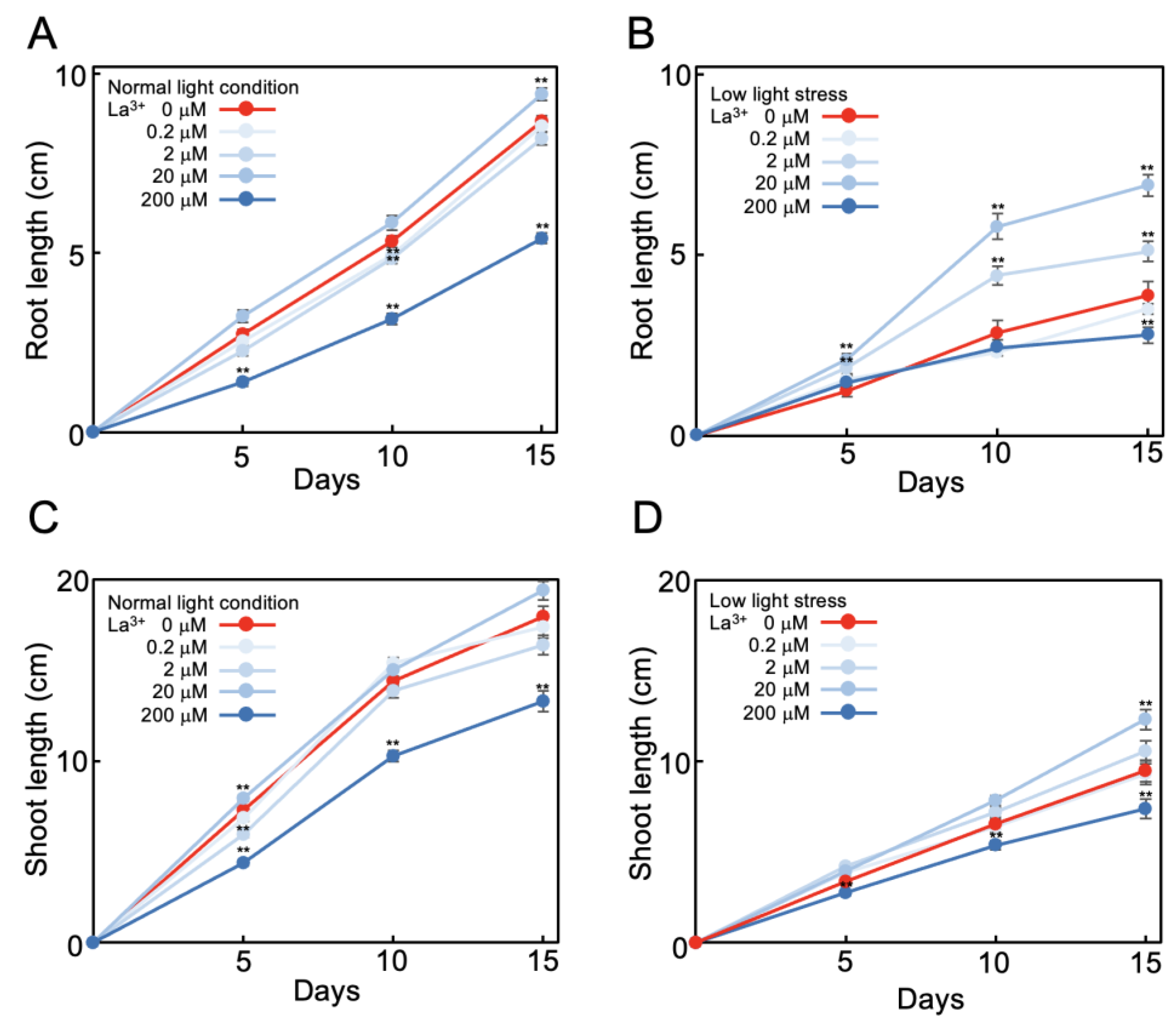
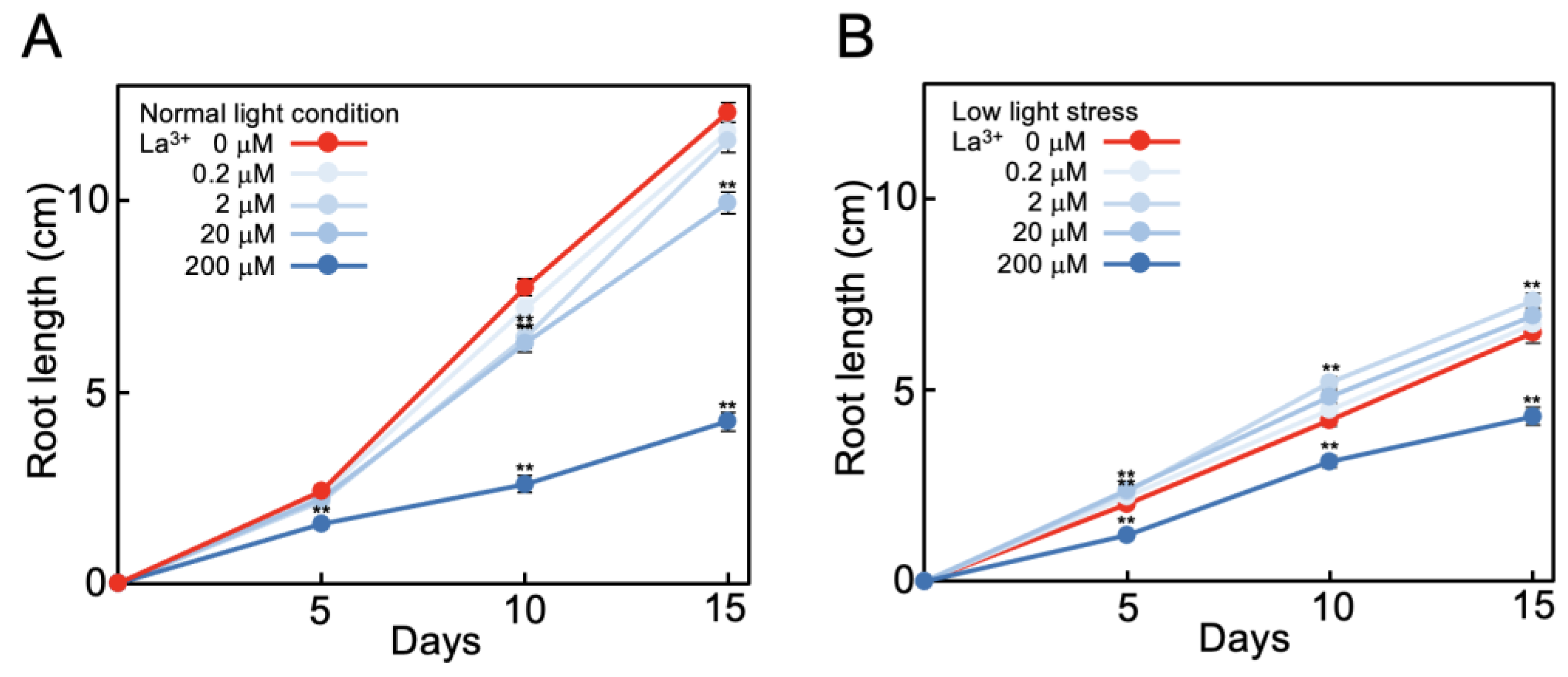
2.1. La3+ Supplementation Alleviates Suppression of Tomato Root Elongation under Low Light

Tomato seeds were sown on agar medium and at, 7 days after sowing, seedlings were transferred to lanthanum ion (La3+)-supplemented (0 to 200 μM) minimal-nutrient hydroponic medium (MGRL medium) [32]. At this time, root length was approximately 5 cm. The length of elongation was measured after transfer to hydroponic solution for 15 days at 25 °C, 16 h light, and 8 h dark conditions. At normal light intensity (photon flux density 132 ± 3 μmol m−2 s−1), during the 15 days of measurements, the mean increases in length of the roots and shoots were 8.7 cm and 17.8 cm, respectively (Figure 1A,C; red lines). At low light intensity (51 ± 4 μmol m−2 s−1), the mean increases in length of the roots and shoots were 3.9 cm (45% of normal light intensity increase) and 9.5 cm (53% of normal light intensity increase), respectively (Figure 1B,D; red lines). Thus, under low light intensity the elongation of tomato roots and shoots during the 15 days was reduced by half. Subsequent experiments were conducted under this low light stress condition.
At normal light intensity, 20 μM La3+ supplementation resulted in 110% elongation of roots (Figure 1A) and 108% elongation of shoots (Figure 1C) compared to no La3+ supplementation during the 15 days. La3+ supplementation at lower concentration (0.2 μM or 2 μM) had no effect on elongation, while 200 μM La3+ supplementation inhibited elongation; that is, elongation of roots was 62% (Figure 1A) and shoots was 74% (Figure 1C) compared to no La3+ supplementation. Thus, at normal light intensity, 20 μM La3+ supplementation promoted elongation by about 10%. Under low light stress with 20 μM La3+ supplementation, root elongation was 177% (Figure 1B) and shoot 129% (Figure 1D) compared to no La3+. That is, root elongation, which was suppressed to 45% under low light stress compared to that under normal light intensity, recovered to 82% with 20 μM La3+ supplementation (Figure 1A,B). Shoot elongation, which was suppressed to 53% under low light stress, recovered to 69% with 20 μM La3+ supplementation (Figure 1C,D). This indicates that La3+ supplementation alleviated the suppression of root and shoot growth caused by low light stress.
This alleviation was La3+-concentration-dependent. Under low light stress, with 2 μM La3+ supplementation, roots elongated 131% compared to no La3+ (Figure 1B). La3+ supplementation at a concentration of 0.2 μM had no effect on growth of roots or shoots, and 200 μM La3+ supplementation inhibited elongation of roots and shoots (72% and 78%, respectively, compared to no La3+) (Figure 1B,D). These results were obtained under the minimal nutritional conditions with MGRL medium [32]. In a replication of the measurements with higher nutrient conditions (1/2 Murashige and Skoog medium [33]) and under low light stress, the root elongation enhancement by 20 μM La3+ supplementation was 144% compared to no La3+ supplementation, which was lower than under the minimal-nutrient condition (177%; Figure 1B). This showed that the effect of REE supplementation was stronger in the lower nutrient conditions.
As the suppression of root elongation due to low light stress was alleviated by supplementation with 20 μM La3+ (Figure 1), we then investigated whether other REEs had the same effect by using 15 commercially available REE chlorides. All REEs tested alleviated elongation suppression by low light stress (Table 1). The optimal concentration of each REE was 0.2–20 μM (Table 1). Of these, La3+ was the most effective and Sc3+ was the next most effective. The REEs were classified into those that significantly reduced root elongation inhibition under low light stress (La3+, Sc3+, Ce3+, Pr3+, Nd3+, Dy3+, Er3+, and Tm3+) and those that reduce it to a lesser extent (Y3+, Sm3+, Eu3+, Gd3+, Tb3+, Yb3+, and Lu3+). A possible relationship between the stress suppression effect of REEs and their chemical properties was investigated [34] (Table 1) but no relationship could not be found. The most effective REE, that is La3+, was used in the following experiments.

2.2. Effect of La3+ Supplementation on Tomato Plant Growth under Low Light Stress

The cell length was measured at 3 cm from the root tip of tomato plants on days 0 and 15 after transfer to hydroponic media. Under normal light intensity, there was no significant difference in cell length between plants in media with and without 20 μM La3+ supplementation during the 15 days (mean cell length increased from 49 μm at day 0 to 67 μm at day 15) (Figure 2). In comparison, the mean cell length under low light stress increased from 46 μm to 52 μm without La3+ supplementation and from 48 μm to 62 μm with 20 μM La3+ supplementation. Thus, alleviation of root elongation suppression by La3+ supplementation under low light stress approximately corresponded to the change in root cell length.
Under low light stress, other characteristics were also altered by La3+ supplementation. Under normal light intensity with 0 and 20 μM La3+ supplementation, the numbers of lateral roots per plant were 18.4 and 23.3, respectively, and, under low light stress, 14.4 and 21.0, respectively (Figure 3A). This showed that La3+ supplementation also alleviated the stress-induced reduction in the number of lateral roots (Figure 3A). The concentrations of chlorophyll in extracts from tomato leaves without and with La3+ supplementation grown under normal light intensity were 22.4 μg/mL and 24.2 μg/mL and, under low light stress, 17.1 μg/mL and 23.6 μg/mL, respectively (Figure 3B). Thus, La3+ supplementation also improved chlorophyll levels that were reduced due to low light stress.

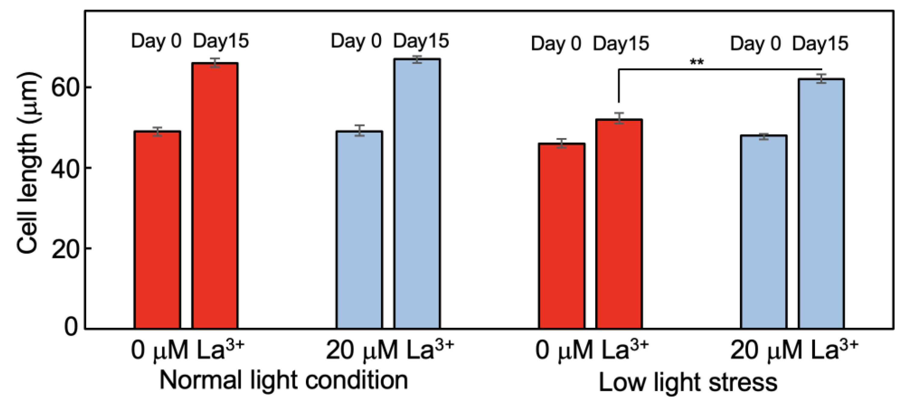
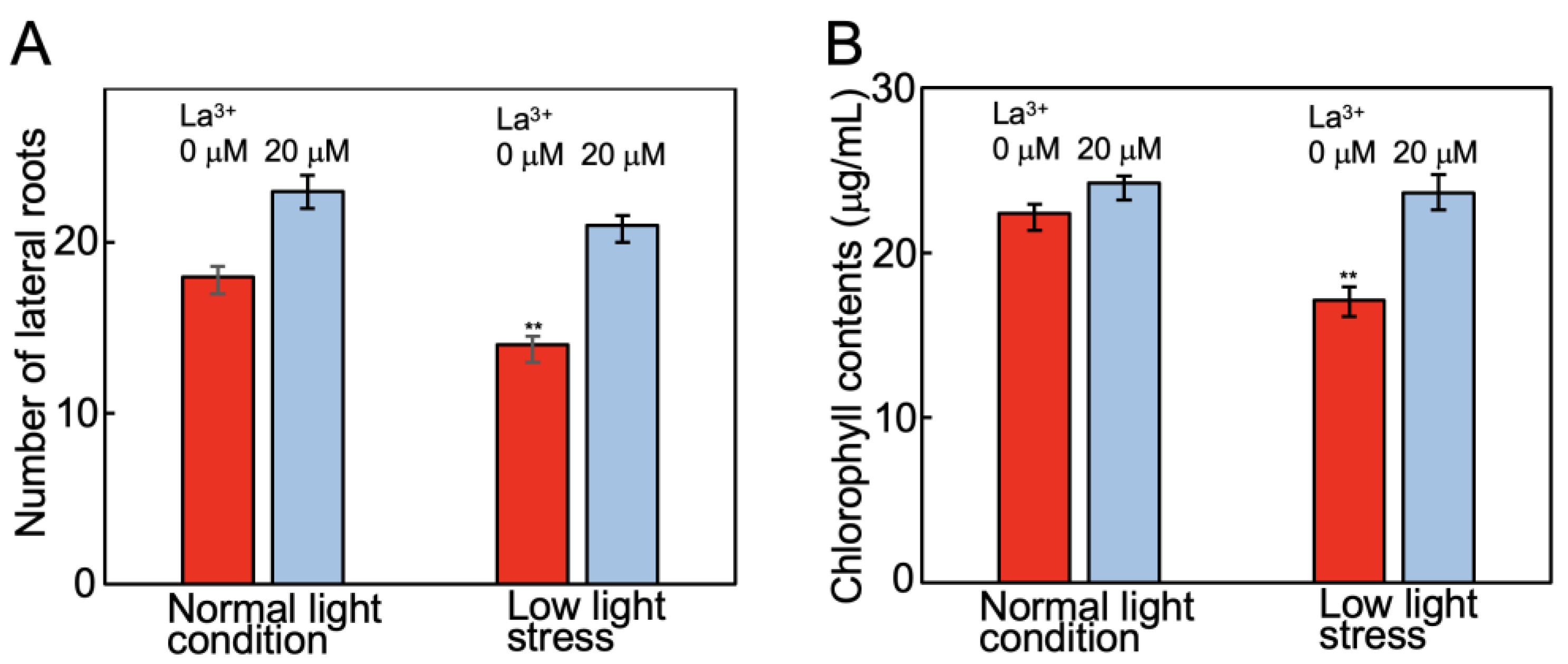
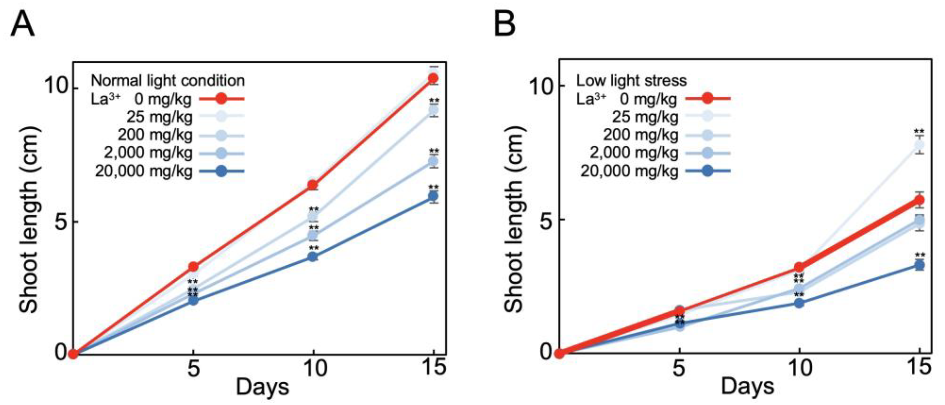
2.3. Analysis of Tomato Plants Grown in La3+-Supplemented Vermiculite

As we found that La3+ supplementation in a minimal-nutrient hydroponic medium alleviated the suppression of tomato root growth due to low light stress, we next examined the effect of La3+ supplementation on tomato growth in an oligotrophic vermiculite supplemented with MGRL solution. The mean increase in length of the shoots after 15 days at normal light intensity was 10.4 cm, and this did not change significantly with La3+ supplementation (Figure 4A). The mean increase in length the shoots grown under low light stress with 25 mg/kg La3+ supplementation was 7.8 cm; thus, the mean increase in length was 136% compared to no La3+ supplementation (Figure 4B). Thus, La3+ supplementation also alleviated low light stress in vermiculite medium. As in the hydroponic medium, this effect of La3+ supplementation in vermiculite was not observed when the eutrophic solution (1/2 Murashige and Skoog medium) was used instead of the minimal-nutrient solution. These results indicate that La3+ supplementation in vermiculite is also effective for plant growth improvement in low light stress and in relatively low-nutrient vermiculites.

2.4. Effect of La3+ Supplementation on Arabidopsis Root Elongation under Low Light Stress

The effect of La3+ supplementation to alleviate low light stress described above was observed in solanaceous tomato plants; we, therefore, investigated whether the same effects could be observed in a different plant family, cruciferous Arabidopsis. In Arabidopsis, normal light intensity was set at a photon flux density of 110 μmol m−2 s−1 and low light intensity at 20 μmol m−2 s−1. Root length elongation during 15 days was 12.3 cm under normal light intensity (Figure 5A) and 6.5 cm (53% of the elongation normal light intensity) under low light stress (Figure 5B). Under normal light intensity, no effect of La3+ supplementation on root elongation was observed (Figure 5A). A slight alleviation of low light stress was observed with 2 mM La3+ fertilization; root length was 7.3 cm with 2 mM La3+, 115% of that without La3+ (Figure 5B). Higher concentration (200 μM) La3+ supplementation inhibited root growth significantly. Thus, the suppression of elongation due to low light stress in cruciferous Arabidopsis was slightly reduced by La3+ supplementation but to a much lesser extent than that observed in solanaceous tomatoes. These results indicate that the alleviation of low light stress by La3+ supplementation varies among plant species.

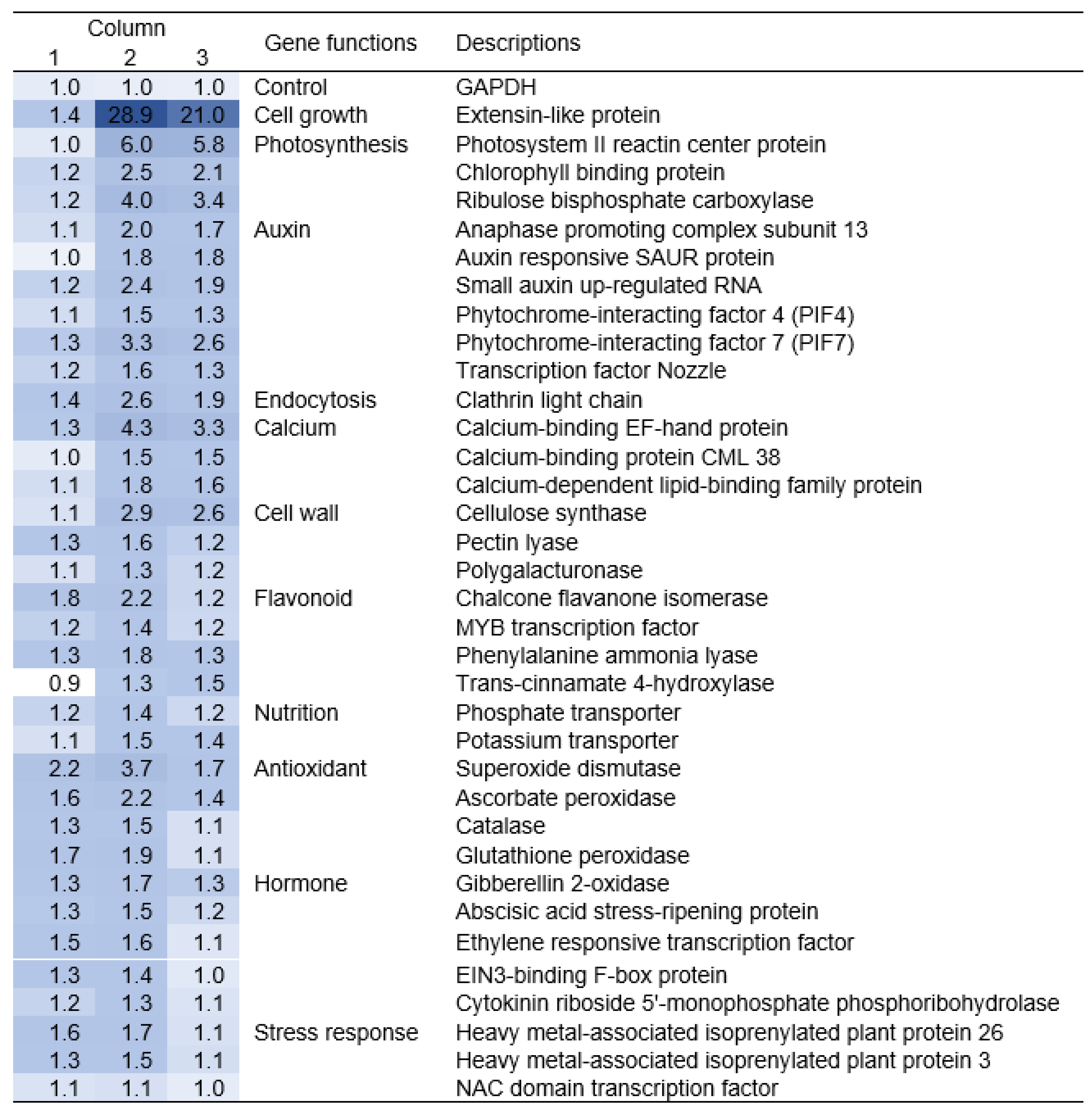
2.5. Gene Expression of Tomato Roots Grown with La3+ Supplementation under Low Light Stress

As we found that La3+ supplementation alleviates the suppression of root elongation caused by low light stress, to explore the molecular mechanism of this effect, we analyzed gene expression in tomato roots grown with 0 and 20 μM La3+ supplementation using qRT-PCR. A total of 37 genes were analyzed (Table S1), including antioxidant, endocytosis, photosystem, stress response, and plant-growth-related genes, whose expression was reported to be altered by La3+ supplementation in previous studies [24,25,29,35]. In Figure 6, values in column 1 are the fold changes in expression of 37 genes with 20 μM compared to 0 μM La3+ supplementation at normal light intensity, and values in column 2 are the corresponding changes in expression at low light stress. Column 3 is the values in column 2 divided by the values in column 1; thus, the values above 1 in column 3 indicate that the gene expression was upregulated with La3+ supplementation under low light stress. These were cell-growth-related extension, photosynthesis-related photosystem II reaction center protein, and auxin-related genes, including phytochrome-interacting factor 7, endocytosis-related clathrin light chain, and calcium-binding EF-hand protein (Figure 6, column 3). La3+ supplementation under low light stress induced changes in the expression levels of these genes, which was associated with alleviation of low light stress.

3. Discussion

We observed that 20 μM La3+ supplementation alleviated tomato root growth suppression caused by low light stress (Figure 1 and Figure 4). The ability of REEs to alleviate low light stress was particularly significant under low nutrient conditions.
The effect of La3+ supplementation on tomato root elongation was not so noticeable at normal light intensity, but its effect was greater under low light stress. The results presented here showing that the action of REEs on plants is more effective under low light intensity are consistent with the previous literature. At normal light intensity (100 to 200 μmol m−2 s−1), root elongation and chlorophyll content has been found to increase 1.1~1.2-fold with REE supplementation [13,29,36]. Moreover, at low light intensity (40 μmol m−2 s−1), chlorophyll content was about 1.5 times higher with REE supplementation [24]. Thus, there seems to be a general trend that the effects of REE supplementation are observed at lower light intensity.
The concentrations of REEs that were effective in alleviating low light-stress-induced elongation suppression ranged from 0.2 to 20 μM (Figure 1 and Table 1), which are similar concentrations to those reported in previous studies observing positive effects of REEs [6,7,8].
The effect of La3+ supplementation was observed in tomatoes of the Solanaceae family but not so much in Arabidopsis of the Cruciferae family (Figure 4). In the solanaceous sweet pepper, La3+ or Ce3+ supplementation was found to double the biomass [37,38]. On the other hand, the effect of La3+ supplementation was not so apparent in cruciferous Arabidopsis and mustard [39,40]. Thus, our results correspond to previous reports, and indicate that the responses to REE supplementation under stress conditions are dependent on the plant species.
The La3+ supplementation effects also depended on the nutrient conditions. The greatest effect of La3+ supplementation was observed when the plants were grown on minimal-nutrient media (Figure 1 and Figure 4). Under eutrophic conditions, the La3+ supplementation effect was low. When barley was grown under irrigated conditions, no La3+ supplementation effect was observed but, under drought conditions (water-deficit stress), La3+ supplementation increased water use efficiency and increased the number of tillers [41,42]. This means that La3+ supplementation alleviated the reduction in barley yield under drought-stress conditions. This effect of La3+ supplementation was observed only under low-nutrient conditions. This is the same as in the present study, where the effect of La3+ was observed under low-nutrient conditions and low light stress. Taken together, it appears that La3+ alleviates the stress response in plants that are under stress conditions in addition to low nutrients.
There have been many reports that REEs promote plant growth [13,14], as well as reports that they do not [15]. Considering the present results, it seems reasonable to assume that REEs have a positive effect on plants in response to stress only under certain conditions. The previously reported plant-growth-promoting effects of REEs may be simply observations that growth-suppressing stresses were alleviated by REEs. Fertilizers containing REEs are used in China, and these REEs may have a role in alleviating stresses such as poor nutrition, low light, and shortage of water.
One of the genes significantly upregulated by La3+ supplementation under low light stress was phytochrome-interacting factors (Figure 6). These are transcription factors that activate the auxin biosynthesis-related genes [43,44]. Consistent with this, auxin-related genes were also upregulated in the presence of La3+ under low light stress (Figure 6). PIF7 is known to interact with the light sensor phytochrome B and respond to shade (low light stress) [44]. This suggests a mechanism for the response to La3+ supplementation; that is, under low light stress, La3+ supplementation triggers a stress adaptation response, activates PIF-mediated auxin synthesis, and promotes auxin-mediated root elongation. It is known that responses to various stresses, such as salt, osmotic, and drought, lead to high expression of auxin-inducible transcription factors, increasing stress tolerance [45,46]. Thus, these results suggest that La3+ supplementation under low light stress leads to auxin biosynthesis, resulting in elongation promotion or the release of growth suppression.
REE supplementation activates clathrin-dependent endocytosis, which increases the Mg2+ and N content necessary for chlorophyll synthesis, resulting in an increased chlorophyll content [25,47]. In this study, the expression of genes for clathrin light chain, photosystem II reaction center protein, and chlorophyll a-b binding protein was particularly elevated with La3+ supplementation under low light stress (Figure 6). In addition, tryptophan, an auxin precursor, is actively synthesized using the increased N content, leading to increased auxin synthesis [48,49].
An La3+-binding protein, lanmodulin, has been identified in methylotrophic bacteria that were isolated from plant surfaces [5]. Lanmodulin has amino acid sequence homology with calmodulin. In this study, seeds were sterilized in each experiment, so it is unlikely that this bacterium and their lanmodulin contributed to the results of this study. Although lanmodulin has not been found in plants, it is known that REEs bind to two of the four metal-binding sites of calmodulin and that calmodulin responds to REE-induced endocytosis [50]. La3+ supplementation under low light stress increased the expression of some calcium-binding protein genes (Figure 6). It has been noted that calmodulin is associated with auxin-mediated cell expansion [51]. It is also possible that La3+ supplementation promotes elongation by such a mechanism.
From the results obtained to date, a putative molecular mechanism for the response to La3+ supplementation under low light stress may be proposed. That is, REE supplementation of plants under stress conditions may result in auxin-induced cell elongation or alleviation of growth suppression.

4. Materials and Methods

4.1. Plant Growing Conditions

The seeds of tomatoes (Solanum lycopersicum cv Momotaro) were purchased from Takii & Co., Ltd (Kyoto, Japan). The Arabidopsis ecotype used was Col-0 of Arabidopsis thaliana. The seeds were placed in 1.5 mL tubes, stirred with 1 mL of 70% ethanol for 1 min, and washed three times with 1 mL of distilled water. The seeds were then agitated with 1 mL of 10-fold diluted sodium hypochlorite for 20 min, washed three times with 1 mL of distilled water, and 2 μL of plant preservative mixture (Nacalai tesque, Kyoto, Japan) was added. The seeds were then placed at 4 °C for 1 day in the dark before sowing.
The seeds were sown and germinated on MGRL agar medium [32], which was composed of 1.5 mM MgSO4, 2.0 mM Ca(NO3)2, 3.0 mM KNO3, 67 μM Na2EDTA, 10.3 μM FeSO4, 6.5 μM MnSO4, 30 μM H3BO3, 1.0 μM ZnSO4, 24 nM (NH4)6Mo7O24, 130 nM CoCl2, 1.0 μM CuSO4, 2.3 mM MES, and 0.6% Gelrite (Fujifilm Wako Chemicals, Osaka, Japan). The pH was adjusted to 5.7 using KOH solution, and 50 mL of the medium was poured into a square petri dish (140 × 100 mm). One-week-old tomato seedlings were transferred to hydroponics or solid medium containing rare earth element chlorides, and their root and shoot elongations were measured for 15 days. For hydroponics, MGRL medium supplemented with each REE chloride (final concentration 0, 0.2, 2.0, 20, and 200 μM) was poured into a 1 L beaker and the pH was adjusted to 5.7 using KOH solution. One-week-old tomato seedings were put into 3 cm long straws (6 mm inner diameter), and these straws were inserted into a floating tube rack on hydroponics. The seedlings were fixed in the straws by inserting a piece of tissue paper. For solid medium, vermiculite containing LaCl3 at a concentration of 25 to 20,000 mg/kg was placed in germination pots (7.5 cm diameter) and MGRL solution was poured into the vermiculite. One-week-old tomato seedings were planted in this solid medium. Tomato seedlings were grown at normal light intensity (photon flux density 132 ± 3 μmol m−2 s−1) or low light stress (51 ± 4 μmol m−2 s−1) at 25 °C, 16 h light, and 8 h dark.
Arabidopsis seedlings were grown at normal light intensity (110 ± 2 μmol m−2 s−1) and low light stress (20 μmol m−2 s−1) at 22 °C, 16 h light, and 8 h dark for 15 days on MGRL agar medium.

4.2. Tomato Root and Shoot Lengths and Chlorophyll Content Measurements

The elongated lengths of tomato roots and shoots were measured for 15 days, starting on day 0, when the plants were transferred from the agar medium to the hydroponic or vermiculite medium. ImageJ was used to measure the length of tomato roots and shoots. Lengths of 30 samples per condition were measured and the mean and standard error were calculated. Chlorophyll was extracted by placing 0.05 g of tomato leaves cut with a cork borer (ø10 mm) in 1 mL of N,N-dimethylformamide overnight at 4 °C in the dark. The absorbance of the solution was measured at 646.8 and 663.8 nm, and the chlorophyll content was determined by the formula (Chla+b = 17.67 × A646.8 + 7.12 × A663.8) [52]. The mean values and standard errors for 30 samples were calculated.

4.3. Observation of Tomato Root Cells

Tomato roots were fixed in FAA (formalin: glacial acetic acid: 95% ethanol: distilled water 10:5:50:35) for 1 h. A longitudinal section of the root was prepared by placing a root between slits in a 1 cm square piece of styrofoam and cutting with a razor blade. Sections were stored in TBS-T (25 mM Tris, 140 mM NaCl, 2.7 mM KCl, 1% Tween 20, pH 7.0) at 4 °C. Cell size was measured using an optical microscope BX53 (Olympus, Tokyo, Japan) and ImageJ. Fifty cells were measured, and the mean values and standard errors of their lengths were calculated.

4.4. Gene Expression Analysis Using qRT-PCR

RNA was extracted from tomato roots pulverized with a mortar and a pestle under liquid nitrogen using the RNeasy Mini Kit (Qiagen, Venlo, Netherland) [53]. qRT-PCR analysis was performed on RNA with RIN values greater than 7.0 measured using a Bioanalyzer 2100 (Agilent Technologies, Santa Clara, California). The cDNA was synthesized using PrimeScript Ⅱ 1st strand cDNA synthesis Kit (Takara Bio, Kusatsu, Japan). qRT-PCR was performed using StepOnePlus (Thermo Fischer Scientific, Waltham, Massachusetts). The mixtures were composed of 100 ng cDNA, 0.4 μM primer, 10 μL of TB Green Premix Ex Taq II, and 0.4 μL of ROX reference dye and were incubated at 95 °C for 30 s, (95 °C for 5 s, 60 °C for 30 s) for 40 cycles. Gene expression levels in tomato roots grown at normal light intensity without La3+ supplementation were set to 1. Relative expression levels were calculated using the ΔΔCt method [54] and compared by correcting the Ct values obtained from the target genes with those of the internal control GAPDH. Experiments were performed in triplicate, and means and standard deviations were calculated. The primers shown in Table S1 were designed to have amplified regions of 70–250 bp, primer lengths of 18–24, Tm values of 55 °C to 65 °C, and GC contents of 40–60%.

Supplementary Materials

The following is available online at https://www.mdpi.com/article/10.3390/plants12142663/s1, Table S1: Primers for qRT-PCR analysis of tomato root expressed genes under low light stress.

Author Contributions

Conceptualization, M.W. and T.I.; methodology, S.I., T.T., E.K., Y.T., M.W. and T.I.; validation, S.I., M.W. and T.I.; investigation, S.I., T.T., E.K., Y.T., S.K., M.W. and T.I.; writing—original draft preparation, S.I. and T.I.; writing—review and editing, S.I., E.K., M.W. and T.I.; visualization, S.I., M.W. and T.I.; supervision, M.W. and T.I.; funding acquisition, M.W. and T.I. All authors have read and agreed to the published version of the manuscript.

Funding

This work was supported by the Japan Society for the Promotion of Science KAKENHI (Grant Nos. JP16K15085, JP19H03252, JP21H02162, JP22H05172, and JP22H05179).

Data Availability Statement

All data were provided within the article.

Acknowledgments

We thank Naoki Nakajima (Ritsumeikan University), Hiromi Masuko-Suzuki, and Kana Ito (Tohoku University) for their technical supports.

Conflicts of Interest

The authors declare no conflict of interest.

References

  1. Tanaka, Y.; Hosaka, T.; Ochi, K. Rare earth elements activate the secondary metabolite-biosynthetic gene clusters in Streptomyces coelicolor A3(2). J. Antibiot. 2010, 63, 477–481. [Google Scholar] [CrossRef] [Green Version]
  2. Inaoka, T.; Ochi, K. Scandium stimulates the production of amylase and bacilysin in Bacillus subtilis. Appl. Environ. Microbiol. 2011, 77, 8181–8183. [Google Scholar] [CrossRef] [Green Version]
  3. Hibi, A.; Asai, K.; Arafuku, H.; Hamajima, M.; Iwama, T.; Kawai, K. Molecular structure of La3+-induced methanol dehydrogenase-like protein in Methylobacterium radiotolerans. J. Biosci. Bioeng. 2011, 111, 547–549. [Google Scholar] [CrossRef]
  4. Pol, A.; Barends, T.R.M.; Dietl, A.; Khadem, A.F.; Eygensteyn, J.; Jetten, M.S.M.; Op den Camp, H.J.M. Rare earth metals are essential for methanotrophic life in volcanic mudpots. Environ. Microbiol. 2013, 16, 255–264. [Google Scholar] [CrossRef]
  5. Cotruvo, J.A., Jr.; Featherston, E.R.; Mattocks, J.A.; Ho, J.V.; Laremore, T.N. Lanmodulin: A highly selective lanthanide-binding protein from a lanthanide-utilizing bacterium. J. Am. Chem. Soc. 2018, 140, 15056–15061. [Google Scholar] [CrossRef]
  6. Kovaříková, M.; Tomášková, I.; Soudek, P. Rare earth elements in plants. Biol. Plant. 2019, 63, 20–32. [Google Scholar] [CrossRef]
  7. Tommasi, F.; Thomas, P.J.; Pagano, G.; Perono, G.A.; Oral, R.; Lyons, D.M.; Toscanesi, M.; Trifuoggi, M. Review of rare earth elements as fertilizers and feed additives: A knowledge gap analysis. Arch. Environ. Contam. Toxicol. 2021, 81, 531–540. [Google Scholar] [CrossRef] [PubMed]
  8. Tao, Y.; Shen, L.; Feng, C.; Yang, R.; Qu, J.; Ju, H.; Zhang, Y. Distribution of rare earth elements (REEs) and their roles in plant growth: A review. Environ. Pollut. 2022, 298, 118540. [Google Scholar] [CrossRef] [PubMed]
  9. Evans, W.H. The influence of the carbonates of the rare earths (cerium, lanthanum, yttrium) on growth and cell-division in hyacinths. Biochem. J. 1913, 7, 349–355. [Google Scholar] [CrossRef] [Green Version]
  10. Drobkov, A.A. Influence of cerium, lanthanum, and samarium on development of peas. Proc. USSR Acad. Sci. 1941, 32, 669–670. [Google Scholar]
  11. Harmet, K.H. Rapid growth responses of Avena coleoptile segments to lanthanum and other cations. Plant Physiol. 1979, 64, 1094–1098. [Google Scholar] [CrossRef] [PubMed]
  12. Pang, X.; Li, D.; Peng, A. Application of rare-earth elements in the agriculture of China and its environmental behavior in soil. J. Soils Sediments 2001, 1, 124–129. [Google Scholar] [CrossRef]
  13. Liu, D.; Wang, X.; Zhang, X.; Gao, Z. Effects of lanthanum on growth and accumulation in roots of rice seedlings. Plant Soil Environ. 2013, 59, 196–200. [Google Scholar] [CrossRef] [Green Version]
  14. Ramírez-Olvera, S.M.; Trejo-Téllez, L.I.; García-Morales, S.; Pérez-Sato, J.A.; Gómez-Merino, F.C. Cerium enhances germination and shoot growth, and alters mineral nutrient concentration in rice. PLoS ONE 2018, 13, e0194691. [Google Scholar] [CrossRef] [Green Version]
  15. Diatloff, E.; Asher, C.J.; Smith, F.W. The effects of rare earth elements on the growth and nutrition of plants. Mater. Sci. Forum 1999, 315, 354–360. [Google Scholar] [CrossRef]
  16. Chen, W.; Tao, Y.; Gu, Y.; Zhao, G. Effect of lanthanide chloride on photosynthesis and dry matter accumulation in tobacco seedlings. Biol. Trace Elem. Res. 2001, 79, 169–176. [Google Scholar] [CrossRef]
  17. Hu, H.; Wang, L.; Li, Y.; Sun, J.; Zhou, Q.; Huang, X. Insight into mechanism of lanthanum (Ⅲ) induced damage to plant photosynthesis. Ecotoxicol. Environ. Saf. 2016, 127, 43–50. [Google Scholar] [CrossRef]
  18. Liu, Y.; Wang, R.; Zhang, P.; Sun, L.; Xu, J. Involvement of reactive oxygen species in lanthanum-induced inhibition of primary root growth. J. Exp. Bot. 2016, 67, 6149–6159. [Google Scholar] [CrossRef] [PubMed] [Green Version]
  19. Cao, X.; Chen, Y.; Wang, X.; Deng, X. Effects of redox potential and pH value on the release of rare earth elements from soil. Chemosphere 2001, 44, 655–661. [Google Scholar] [CrossRef] [PubMed]
  20. Wu, J.; Chen, A.; Peng, S.; Wei, Z.; Liu, G. Identification and application of amino acids as chelators in phytoremediation of rare earth elements lanthanum and yttrium. Plant Soil 2013, 373, 329–338. [Google Scholar] [CrossRef]
  21. Lai, Y.; Wang, Q.; Yang, L.; Huang, B. Subcellular distribution of rare earth elements and characterization of their binding species in a newly discovered hyperaccumulator Pronephrium simplex. Talanta 2006, 70, 26–31. [Google Scholar] [CrossRef]
  22. Wang, L.; Cheng, M.; Yang, Q.; Li, J.; Wang, X.; Zhou, Q.; Nagawa, S.; Xia, B.; Xu, T.; Huang, R.; et al. Arabinogalactan protein-rare earth element complexes activate plant endocytosis. Proc. Natl. Acad. Sci. USA 2019, 116, 14349–14357. [Google Scholar] [CrossRef] [Green Version]
  23. Song, W.; Hong, F.; Wan, Z.; Zhou, Y. Effects of lanthanum and europium on rooting of plantlet Eriobotrya japonica Lindl. in vitro. J. Rare Earths 2002, 20, 658–662. [Google Scholar]
  24. Fan, Z.; Zhang, K.; Wang, F.; Zhao, X.; Bai, R.; Liu, B. Effect of rare earth elements on growth and determination of secondary metabolites under in vitro conditions in Salvia miltiorrhiza. HortScience 2020, 55, 310–316. [Google Scholar] [CrossRef] [Green Version]
  25. Ben, Y.; Cheng, M.; Liu, Y.; Wang, L.; Yang, Q.; Huang, X. The stimulatory effect and mechanism of low-dose lanthanum on soybean leaf cells. J. Hazard. Mater. 2023, 441, 129924. [Google Scholar] [CrossRef]
  26. Yuan, X.; Wang, Q.; Zhao, B.; Wang, Y. Improved cell growth and total flavonoids of Saussurea medusa on solid culture medium supplemented with rare earth elements. Biotechnol. Lett. 2002, 24, 1889–1892. [Google Scholar] [CrossRef]
  27. Ouyang, J.; Wang, X.; Zhao, B.; Yuan, X.; Wang, Y. Effects of rare earth elements on the growth of Cistanche deserticola cells and the production of phenylethanoid glycosides. J. Biotechnol. 2003, 102, 129–134. [Google Scholar] [CrossRef] [PubMed]
  28. Wang, L.; Li, J.; Zhou, Q.; Yang, G.; Ding, X.L.; Li, X.; Cai, C.X.; Zhang, Z.; Wei, H.Y.; Lu, T.H.; et al. Rare earth elements activate endocytosis in plant cells. Proc. Natl. Acad. Sci. USA 2014, 111, 12936–12941. [Google Scholar] [CrossRef]
  29. Cheng, M.; Wang, L.; Zhou, Q.; Chao, D.; Nagawa, S.; He, D.; Zhang, J.; Li, H.; Tan, L.; Gu, Z.; et al. Lanthanum(Ⅲ) triggers AtrbohD- and jasmonic acid-dependent systemic endocytosis in plants. Nat. Commun. 2021, 12, 4327. [Google Scholar] [CrossRef]
  30. Du, Y.; Ji, X.; Zhang, J.; Li, J.; Sun, H.; Zhao, Q. Research progress on the impacts of low light intensity on rice growth and development. Chin. J. Eco-Agric. 2013, 21, 1307–1317. [Google Scholar] [CrossRef]
  31. Lu, T.; Yu, H.; Li, Q.; Chai, L.; Jiang, W. Improving plant growth and alleviating photosynthetic inhibition and oxidative stress from low-light stress with exogenous GR24 in tomato (Solanum lycopersicum L.) seedling. Front. Plant Sci. 2019, 10, 490. [Google Scholar] [CrossRef] [PubMed]
  32. Fujiwara, T.; Hirai, M.; Chino, M.; Komeda, Y.; Naito, S. Effects of sulfur nutrition on expression of the soybean seed storage protein genes in transgenic petunia. Plant Physiol. 1992, 99, 263–268. [Google Scholar] [CrossRef]
  33. Murashige, T.; Skoog, F. A revised medium for rapid growth and bio assays with tobacco tissue cultures. Physiol. Plant. 1962, 15, 473–497. [Google Scholar] [CrossRef]
  34. Voncken, J.H.L. Physical and chemical properties of the rare earths. In The rare earth elements; Springer: Cham, Switzerland, 2016; pp. 53–72. [Google Scholar]
  35. Liu, Y.; Zhang, J. Lanthanum promotes bahiagrass (Paspalum notatum) roots growth by improving root activity, photosynthesis and respiration. Plants 2022, 11, 382. [Google Scholar] [CrossRef]
  36. Song, K.; Gao, J.; Li, S.; Sun, Y.; Sun, H.; An, B.; Hu, T.; He, X. Experimental and theoretical study of the effects of rare earth elements on growth and chlorophyll of alfalfa (Medicago sativa L.) seedling. Front. Plant Sci. 2021, 12, 731838. [Google Scholar] [CrossRef]
  37. García-Jiménez, A.; Gómez-Merino, F.C.; Tejeda-Sartorius, O.; Trejo-Téllez, L.I. Lanthanum affects bell pepper seeding quality depending on the genotype and time of exposure by differentially modifying plant height, stem diameter and concentrations of chlorophylls, sugars, amino acids, and proteins. Front. Plant Sci. 2017, 8, 308. [Google Scholar] [CrossRef] [Green Version]
  38. Abou El-Nour, H.H.; Attia, R.M. Evaluate the effects of rare earth elements on sweet pepper seeds germination process, seedlings growth and plants productivity. GSC Adv. Res. Rev. 2022, 13, 23–38. [Google Scholar] [CrossRef]
  39. He, Y.; Loh, C. Cerium and lanthanum promote floral initiation and reproductive growth of Arabidopsis thaliana. Plant Sci. 2000, 159, 117–124. [Google Scholar] [CrossRef]
  40. Xiong, S.; Xiong, Z.; Chen, Y.; Huang, H. Interactive effects of lanthanum and cadmium on plant growth and mineral element uptake in crisped-leaf mustard under hydroponic conditions. J. Plant Nutr. 2006, 29, 1889–1902. [Google Scholar] [CrossRef]
  41. Buckingham, S.; Maheswaran, J.; Meehan, B.; Peverill, K. The role of applications of rare earth elements in enhancement of crop and pasture production. Mater. Sci. Forum 1999, 315, 339–347. [Google Scholar] [CrossRef]
  42. Reddy, N.; Maheswaran, J.; Meehan, B.; Peverill, K. The application of rare earth elements in enhancement of crop production in Australia, Part 2. In Proceedings of the 4th International Conference on Rare Earth Development and Application; Yu, Z., Yan, C., Xu, G., Niu, J., Chen, Z., Eds.; China Scientific Books: WanChai, Hong Kong, 2001; pp. 251–254. [Google Scholar]
  43. Franklin, K.A.; Lee, S.H.; Patel, D.; Kumar, S.V.; Spartz, A.K.; Gu, C.; Ye, S.; Yu, P.; Breen, G.; Cohen, J.D.; et al. Phytochrome-interacting factor 4 (PIF4) regulates auxin biosynthesis at high temperature. Proc. Natl. Acad. Sci. USA 2011, 108, 20231–20235. [Google Scholar] [CrossRef]
  44. Li, L.; Ljung, K.; Breton, G.; Schmitz, R.J.; Pruneda-Paz, J.; Cowing-Zitron, C.; Cole, B.J.; Ivans, L.J.; Pedmale, U.V.; Jung, H.; et al. Linking photoreceptor excitation to changes in plant architecture. Genes Dev. 2012, 26, 785–790. [Google Scholar] [CrossRef] [Green Version]
  45. Bielach, A.; Hrtyan, M.; Tognetti, V.B. Plants under stress: Involvement of auxin and cytokinin. Int. J. Mol. Sci. 2017, 18, 1427. [Google Scholar] [CrossRef] [PubMed] [Green Version]
  46. Korver, R.A.; Koevoets, I.T.; Testerink, C. Out of shape during stress: A key role for auxin. Trends Plant Sci. 2018, 23, 783–793. [Google Scholar] [CrossRef]
  47. Ben, Y.; Cheng, M.; Wang, L.; Zhou, Q.; Yang, Z.; Huang, X. Low-dose lanthanum activates endocytosis, aggravating accumulation of lanthanum or/and lead and disrupting homeostasis of essential elements in the leaf cells of four edible plants. Ecotoxicol. Environ. Saf. 2021, 221, 112429. [Google Scholar] [CrossRef]
  48. Majda, M.; Robert, S. The role of auxin in cell wall expansion. Int. J. Mol. Sci. 2018, 19, 951. [Google Scholar] [CrossRef] [PubMed] [Green Version]
  49. Salazar-Iribe, A.; De-la-Peña, C. Auxins, the hidden player in chloroplast development. Plant Cell Rep. 2020, 39, 1595–1608. [Google Scholar] [CrossRef] [PubMed]
  50. Wang, L.; Cheng, M.; Chu, Y.; Li, X.; Chen, D.D.Y.; Huang, X.; Zhou, Q. Responses of plant calmodulin to endocytosis induced by rare earth elements. Chemosphere 2016, 154, 408–415. [Google Scholar] [CrossRef]
  51. Du, M.; Spalding, E.P.; Gray, W.M. Rapid auxin-mediated cell expansion. Annu. Rev. Plant Biol. 2020, 71, 379–402. [Google Scholar] [CrossRef]
  52. Porra, R.J.; Thompson, W.A.; Kriedemann, P.E. Determination of accurate extinction coefficients and simultaneous equations for assaying chlorophylls a and b extracted with four different solvents: Verification of the concentration of chlorophyll standards by atomic absorption spectroscopy. Biochim. Biophys. Acta 1989, 975, 384–394. [Google Scholar] [CrossRef]
  53. Takenaka, Y.; Kato, K.; Ogawa-Ohnishi, M.; Tsuruhama, K.; Kajiura, H.; Yagyu, K.; Takeda, A.; Takeda, Y.; Kunieda, T.; Nishimura, I.H.; et al. Pectin RG-I rhamnosyltransferases represent a novel plant-specific glycosyltransferase family. Nat. Plants 2018, 4, 669–676. [Google Scholar] [CrossRef] [PubMed]
  54. Pfaffl, M.W. A new mathematical model for relative quantification in real-time RT-PCR. Nucleic Acids Res. 2001, 29, e45. [Google Scholar] [CrossRef] [PubMed]
Figure 1. Effect of La3+ supplementation on tomato growth under low light stress. Elongated lengths of tomato root (A,B) and shoot (C,D) were measured during growth for 15 days in La3+-fertilized (0 to 200 μM) MGRL hydroponic solutions under normal light intensity (132 μmol m−2 s−1) (A,C) and low light stress (51 μmol m−2 s−1) (B,D). The growth conditions were 25 °C, 16 h light, and 8 h dark. Root and shoot lengths of 30 samples were measured and the means and standard errors were plotted. Statistical significances were determined by Student t-test compared with 0 μM (**, p < 0.01).
Plants 12 02663 g001
Figure 2. Cell lengths of tomato roots grown in La3+-supplemented medium under low light stress. Cell lengths observed in sections at 3 cm from the tip of tomato roots grown with and without La3+ supplementation under normal light intensity or low light stress. Roots were sampled at 0 and 15 days after transfer to the hydroponic medium. The lengths of 50 cells were measured and the mean and standard error were calculated. Statistical significance was determined by Student t-test (**, p < 0.01).
Plants 12 02663 g002
Figure 3. Effect of La3+ supplementation on lateral root number and chlorophyll content in leaves under low light stress. (A) Tomato lateral root number and (B) chlorophyll a+b content in tomato leaves grown with and without La3+ supplementation under normal light intensity or low light stress. In both cases, 30 samples were measured and the mean and standard errors were calculated. Statistical significances were determined by Student t-test compared with 0 μM La3+ under normal light condition (**, p < 0.01).
Plants 12 02663 g003
Figure 4. Growth of tomatoes in La3+-supplemented vermiculite. Elongated lengths of tomato shoots were measured for 15 days in La3+-supplemented vermiculite under normal light condition (A) or low light stress (B). The growth conditions were 25 °C, 16 h light, and 8 h dark. Shoot lengths of 30 samples were measured and the mean and standard errors were plotted. Statistical significances were determined by Student t-test compared with 0 mg/kg (**, p < 0.01).
Plants 12 02663 g004
Figure 5. Effect of La3+ supplementation on Arabidopsis root elongation under low light stress. Elongated Arabidopsis root lengths were observed for 15 days in La3+-fertilized (0 to 200 μM) MGRL solid medium under normal light intensity (110 μmol m−2 s−1) (A) and low light stress (20 μmol m−2 s−1) (B). The growth conditions were 25 °C, 16 h light, and 8 h dark. Root lengths of 30 samples were measured and the means and standard errors were plotted. Statistical significances were determined by Student t-test compared with 0 μM (**, p < 0.01).
Plants 12 02663 g005
Figure 6. Gene expression by qRT-PCR in tomato roots grown with La3+ supplementation medium under low light stress. Gene expression levels in tomato roots grown without La3+ supplementation were set to 1. The fold changes in gene expression with La3+ fertilization were shown for roots under normal light intensity (column 1) and low light stress (column 2). Column 3 shows the fold change under low light stress (column 2) divided by the fold change under normal light (column 1). Dark blue indicates higher fold changes.
Plants 12 02663 g006
Table 1. Effects of rare earth elements on elongation of tomato roots under low light stress and their chemical properties.
REE IonOptimum Concentration
(μM)
Elongation with REE under Low Light Relative to Normal Light, % *Elongation with REE Relative to No REE under Low Light, %Atomic WeightIon Radius
(Å)
Density
(g/cm3)
Number of Unpaired 4f Electrons
None45100
Sc3+207216145.00.752.990
Y3+205111488.90.904.470
La3+2082177138.91.036.150
Ce3+265144140.11.018.161
Pr3+264143140.90.996.772
Nd3+263140144.20.987.013
Pm3+not available145.00.977.264
Sm3+252115150.40.967.525
Eu3+0.248107151.90.955.246
Gd3+0.250112157.30.947.907
Tb3+2050112158.90.928.236
Dy3+0.262138162.50.918.555
Ho3+not available164.90.908.804
Er3+2065145167.30.899.073
Tm3+0.260133168.90.889.322
Yb3+252116173.00.876.971
Lu3+252115175.00.869.840
* With no REE, root elongation was suppressed to 45% under low light stress. Values in this column above 45% indicate root elongation under low light stress was alleviated by REE fertilization.
Disclaimer/Publisher’s Note: The statements, opinions and data contained in all publications are solely those of the individual author(s) and contributor(s) and not of MDPI and/or the editor(s). MDPI and/or the editor(s) disclaim responsibility for any injury to people or property resulting from any ideas, methods, instructions or products referred to in the content.

