Article

Photosynthetic Parameters and Oxidative Stress during Acclimation of Crepe-Myrtle (Lagerstroemia speciosa (L.) Pers.) in a meta-Topolin-Based Micropropagation System and Genetic Fidelity of Regenerated Plants

1
Plant Biotechnology Laboratory, Department of Botany, Aligarh Muslim University, Aligarh 202002, India
2
Department of Botany & Microbiology, College of Science, King Saud University, P.O. Box 2455, Riyadh 11451, Saudi Arabia
3
Plant & Food Research Australia Pty Ltd., Waite Research Precinct, Plant Breeding #46, Waite Road, Urrbrae, SA 5064, Australia
*
Author to whom correspondence should be addressed.
Plants 2022, 11(9), 1163; https://doi.org/10.3390/plants11091163
Submission received: 1 March 2022 / Revised: 12 April 2022 / Accepted: 23 April 2022 / Published: 26 April 2022

Abstract

:
An improved and stable micropropagation system using the cytokinin, meta-Topolin (N6 (3-hydroxybenzylamino purine—mT), with nodal explants in Lagerstroemia speciosa L. was established. Among the different doses of mT, the maximum number of shoots with the highest shoot length was obtained using Murashige and Skoog’s (MS) medium supplemented with 5.0 µM mT. The results were consistent throughout the proliferation period, when recorded at week 4, 8, and 12 of being cultured, with an average of 16.4 shoots per nodal explant, and having a mean length of 4.10 cm at week 8. Shoot proliferation rates could be further improved by a combination of 5.0 µM mT with 0.5 µM α-naphthalene acetic acid in MS medium; nodal explants produced an average of 24.3 shoots with a mean length of 5.74 cm after 8 weeks of being cultured. Among the five different concentrations of three auxins tested for the rooting of microshoots in MS medium, a 1.0 µM indole-3-butyric acid treatment was the best, with an average of 10.3 roots per microshoot at an average length of 3.56 cm in 93% of microshoots within 4 weeks of being transferred to this medium. A significant reduction of both chlorophyll a and b in leaves during the first week of acclimation corresponded with a high accumulation of malondialdehyde (MDH), indicating that lipid peroxidation affected chlorophyll pigments. From the second week of acclimation, photosynthetic pigment content significantly increased and MDH content decreased. The net photosynthetic rate and leaf carotenoid content showed almost linear increases throughout the acclimation period. Activity of antioxidant enzymes, namely, superoxide dismutase, catalase, and peroxidases, consistently increased throughout the acclimation period, corresponding with the accumulation of photosynthetic pigments, thus demonstrating the role of the improved antioxidant enzymatic defense system during acclimation. A comparison of parent plant DNA with that of the greenhouse acclimated plants using random amplified polymorphic DNA and inter-simple sequence repeat markers showed a monomorphic pattern indicating genetic stability and the suitability of the method for micropropagation of L. speciosa.

1. Introduction

Queen Crape Myrtle Lagerstroemia speciosa Pers. (Lythraceae) is a tropical, medium-large sized tree that can grow up to 25 m and is of commercial importance as an ornamental plant and avenue tree. Tracing its origin to “Tropical Southern Asia” [1], its aesthetic beauty, accompanied by spectacular blooming with large bright pink to lavender flowers with various shades, has made it the “Queen” among flowers where it is grown, and it is also known as “Pride of India”. Having moderately hard timber, the tree is used to make decorative furniture, agricultural implements, pulp, paper, ploughs, boats, fence posts, and so on. The species is also used in biofertilizer manufacturing [2] as a fodder component for afforestation and ecological restoration [3]. The demand for plants is high in many countries because their uses are multipurpose, including plantation establishment, urban horticulture, reforestation, desert rehabilitation, use in furniture, paper, pulp, and timber industries, as well as for medicinal purposes [3,4].
Bioactive compounds in the leaves and fruits of L. speciosa, such as ellagitannins (lagerstroemin, reginin A, flosin B, reganin C and D) [5], alanine, methionine, lageracetal, amyl alcohol, and β-sitosterol [6], serve as raw materials in pharmaceutical industries. Additionally, the leaf extracts have anti-oxidant, anti-inflammatory, anti-hypertension, analgesic, diuretic, thrombolytic, anti-cancer [7,8], antifungal, and antiviral properties [9]. The leaves act as a febrifuge and cleansing agent to regulate metabolic processes [10]. Leaf beverages (tea) possess anti-oxidative [11], anti-inflammatory [3,6], anti-hypertensive [8], diuretic, purgative, anti-ulcer, and anti-gout properties [12,13]. Numerous commercial formulations and industrial products (for example, Swanson Banaba extract) are available, including medicinal tablets, capsules, cosmetics, lotions, hair tonics, and hypoglycemic foods. The majority of formulations contain corosolic acid derived from the plant. Corosolic acid is known as “phyto-insulin” or “botanical insulin” due to its insulin-like activities, and its high level in L. speciosa implies that this plant may have anti-diabetic therapeutic potential. [14]. Hypoglycemic effects of L. speciosa have been proven not only in animal and in vitro studies, but also in clinical studies, for example, using GlucosolTM (standardized to 1% corosolic acid) in Type II diabetes patients [15].
Due to its many valuable characteristics, L. speciosa is has been over-exploited in its natural habitat, leading to a scarcity of mature trees [16]. A low density of plants and a scarcity of pollinators means that much of the seed is fertilized through self-pollination, resulting in aborted seed [17]. Viable seeds have a short lifespan (1–2 months), making the establishment of seedlings difficult [18]. Additionally, vegetative propagation of this plant is not widely practiced. To meet these challenges, improved micropropagation methods are required. In vitro propagation of this species is complicated by poor rooting after use of the widely used synthetic cytokinin (CK) and N6-benzylaminopurine (BAP) at the proliferation stage, requiring forced ventilation of vessels [19]. When kinetin (Kn) was used, callus formation was an impediment both at the proliferation and rooting stages, with low numbers of shoots produced [20].
meta-Topolin (mT) (6-(3-hydroxybenzylamino) purine), is an innate aromatic cytokinin (ArCK) originally isolated from Populus X robusta leaves [21,22]. Being the most biologically active ArCK belonging to the group “Topolins”, mT has been found to have distinct advantages in morphogenesis, differentiation, and subsequent rooting and acclimation in the micropropagation industry [21]. It is a hydroxylated form of BAP that produces an O-glucoside with relatively higher activity than other derivatives, resulting in less hyperhydricity in tissue culture, thus facilitating easier rooting and acclimation of rooted plantlets [22,23,24].
Recently, the use of mT to replace more traditional CKs, such as BAP and zeatin, at the proliferation stage of micropropagation has been reported to result in better rooting and acclimation of plantlets in diverse species, including Spathiphyllum floribundum (Araceae) [22], Ananas comosus (Bromeliaceae) [25], Eucalyptus spp. (Myrtaceae) [26], Cannabis sativa (Cannabaceae) [23], Corylus colurna (Betulaceae) [27], Manihot esculenta (Euphorbiaceae) [28], and Pterocarpus marsupium (Fabaceae) [29]. Replacement of BAP with mT reduced hyperhydricity, increased the multiplication rate, and resulted in the spontaneous rooting of Cannabis sativa [23] and Eucalyptus spp. [26]. In comparison with BAP, mT supplementation resulted in more shoots with leaves, which had better morphological and anatomical features, and a higher amount of chlorophyll in Sesamum indicum (Pedaliaceae) [30], Oxystelma esculentum (Asclepiadaceae) [31], and Vanilla planifolia (Orchidaceae) [32]. In a study involving 14 contrasting genotypes of gooseberry (Ribes grossularia—Grossulariaceae), Kucharska, et al. [33] found that mT-supplementation improved the shoot proliferation to 2.8, in comparison with 1.8, which was obtained using the optimal concentration of BAP, which resulted in longer and healthier shoots in subsequent subcultures. Experiments involving multiple CKs, such as Kn and BAP [29], Kn, BAP and thidiazuron [34], and BAP, Kn, and 2-isopentyl adenine [24], have also shown the superiority of mT in its capacity to produce healthy shoots that are more amenable to rooting and acclimation. Furthermore, the problem of recalcitrance of kiwifruit genotypes (Actinidia chinensis—Actinidiaceae) to the rooting of shoots proliferated in BAP or zeatin media, could be solved by switching to mT in culture media for micropropagation [35], and in the management of a large in vitro collection [36].
These advantages of mT over some other CKs used in plant tissue culture can be attributed to the lesser oxidative stress experienced by explants cultured on mT-supplemented media. For example, plants grown in mT-supplemented media had lower H2O2 content than in BAP-supplemented media in Daphne mezereum (Thymelaeaceae) [37]. Corylus colurna (Betulaceae) plantlets cultured in a medium containing BAP showed a significantly higher H2O2 content, enhanced antioxidant enzyme (superoxide dismutase (SOD), ascorbate peroxidase, and catalase (CAT)) activities, and lower chlorophyll than shoots derived from the medium containing mT [27].
However, there are no reports on the use of mT in L. speciosa micropropagation or in any other species in the plant family Lythraceae; therefore, the objective of our work was to optimize the concentration of mT in the proliferation and multiplication stages of L. speciosa micropropagation, and to investigate if further improvements can be achieved when combined with different auxins. Additionally, we used molecular markers to test if the L. speciosa plants regenerated using mT in proliferation media were true-to-type, and if the chlorophyll content and oxidative stress status of plants during the acclimation period could improve our understanding of the ability of tissue cultured plants to recover and re-establish themselves.

2. Results

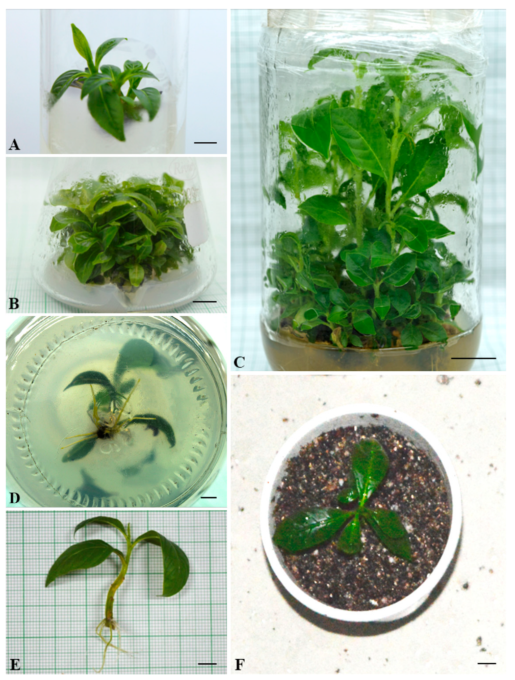
2.1. In Vitro Establishment and Optimizing Shoot Proliferation and Rooting of Shoots

Nodal explants collected from mature L. speciosa plants were successfully established within 2–3 weeks of initiation. All of the nine meta-Topolin (mT) concentrations within the range 0.1–15.0 µM in MS medium resulted in the initiation of multiple shoots from the cultured nodal segments (Table 1). There was no shoot production from nodal explants when the medium was not supplemented with mT. Supplementation of the medium with 5.0 µM mT resulted in a significantly higher number of shoots that were also the longest among the nine concentrations tested (Figure 1A). This result was consistent throughout the period of growth when measurements were taken, from week 4 to 12 (Table 1). With the increase in concentration of mT in the medium beyond 5.0 µM, not only did the number of shoots decrease, but also there was an increase in vitrification, resulting in difficulties with rooting and plant acclimation.
When the optimal mT concentration for shoot proliferation of L. speciosa (5.0 µM mT) from the first experiment was combined with different concentrations of three auxins, α-naphthaleneacetic acid (NAA) produced a better proliferation of shoot buds compared with indole-3-butyric acid (IBA) and indole-3-acetic acid (IAA) (Table 2). After 8 weeks in a culture, 0.5 µM NAA in combination with 5.0 µM mT resulted in a significantly higher number of shoots than any other combination (Table 2; Figure 1B,C), or with mT alone (Table 1), and the highest proportion of explants (95%) producing shoots. Supplementation with IBA or IAA did not have a positive effect on the proliferation of shoot buds, whereas 0.1 and 1.0 µM NAA produced slightly higher numbers of shoots (17.5 and 18.2, respectively) than 5.0 µM mT alone (16.4) in 85% of cultures after 8 weeks (Table 1 and Table 2). The shoots produced using MS medium supplemented with 5.0 µM mT and 0.5 µM NAA were also significantly longer than all other combinations, except the combination with 5.0 µM mT and 1.0 µM NAA (Table 2).
For the rooting experiment, microshoots 3–4 cm in length were harvested from the optimal shoot proliferation medium after 8 weeks of growth and transferred to BM supplemented with one of five concentrations of IAA, NAA, or IBA. The frequency of root initiation varied with auxin concentration (0.5–2.5 µM) (Table 3). BM supplemented with 1 µM IBA produced a significantly higher number of roots per explant and the highest mean length of roots (Table 3; Figure 1D,E). Root development was also the highest in this treatment, with 93% of microshoots producing roots. IBA at 0.5 and 1.5 µM concentrations, and 1.0 µM NAA, also produced high numbers of roots, but the roots in these treatments were much shorter than in the optimum 1 µM NAA concentration. In general, the number of roots produced in IAA-supplemented media was lower than in IBA- and NAA-supplemented media.

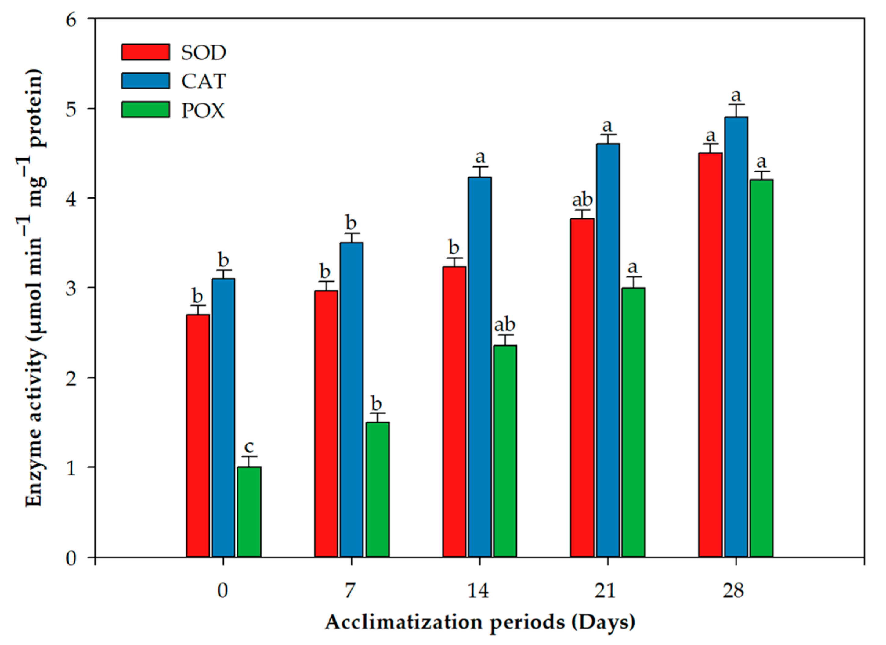
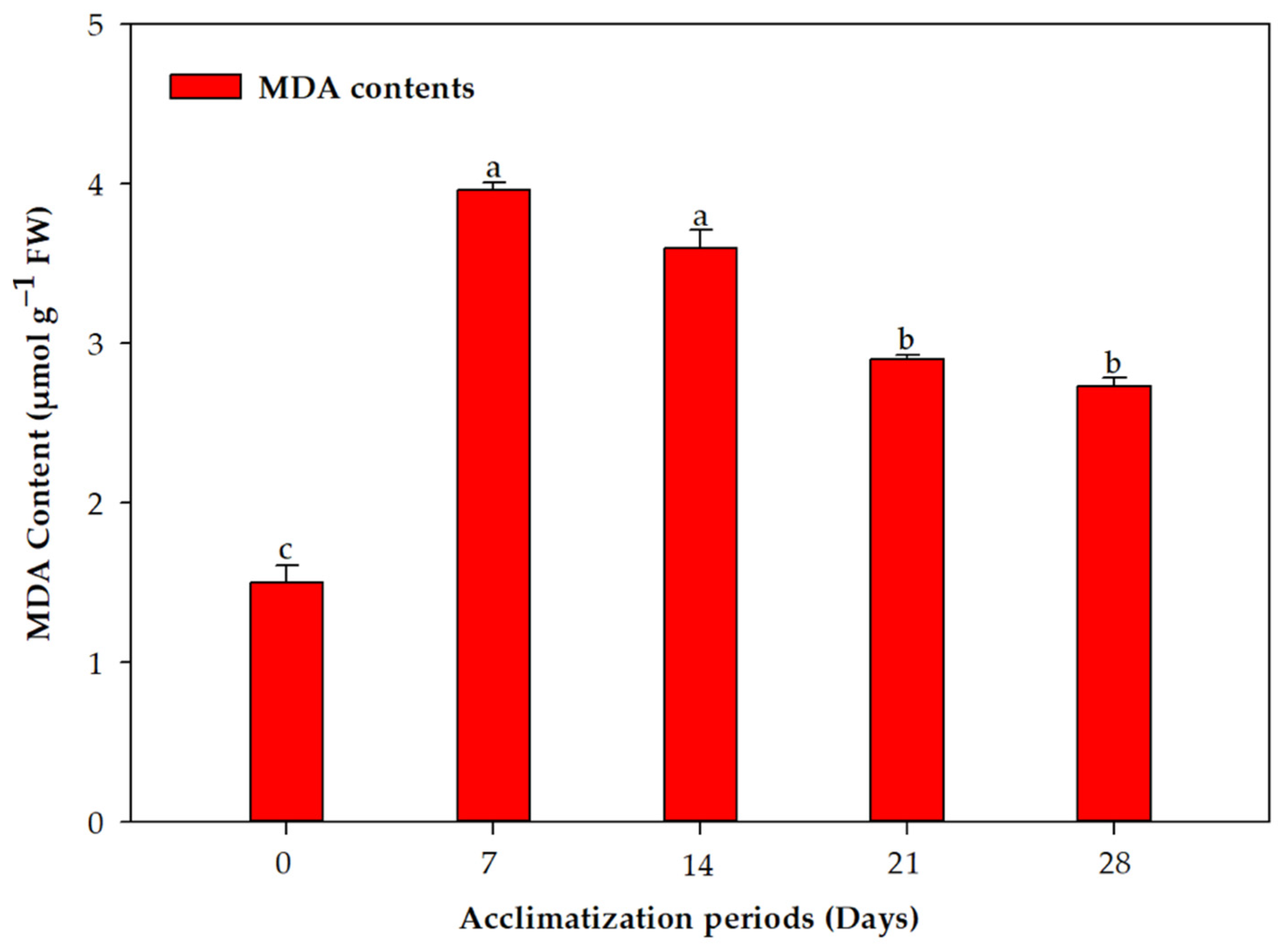
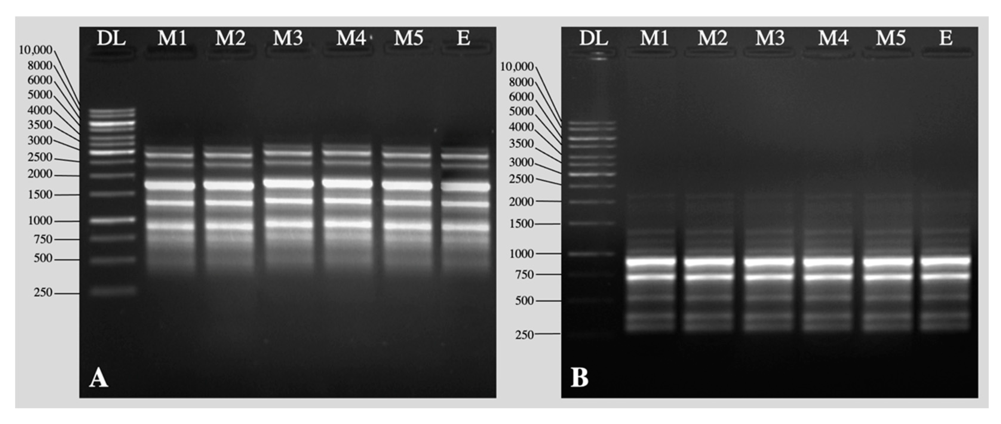
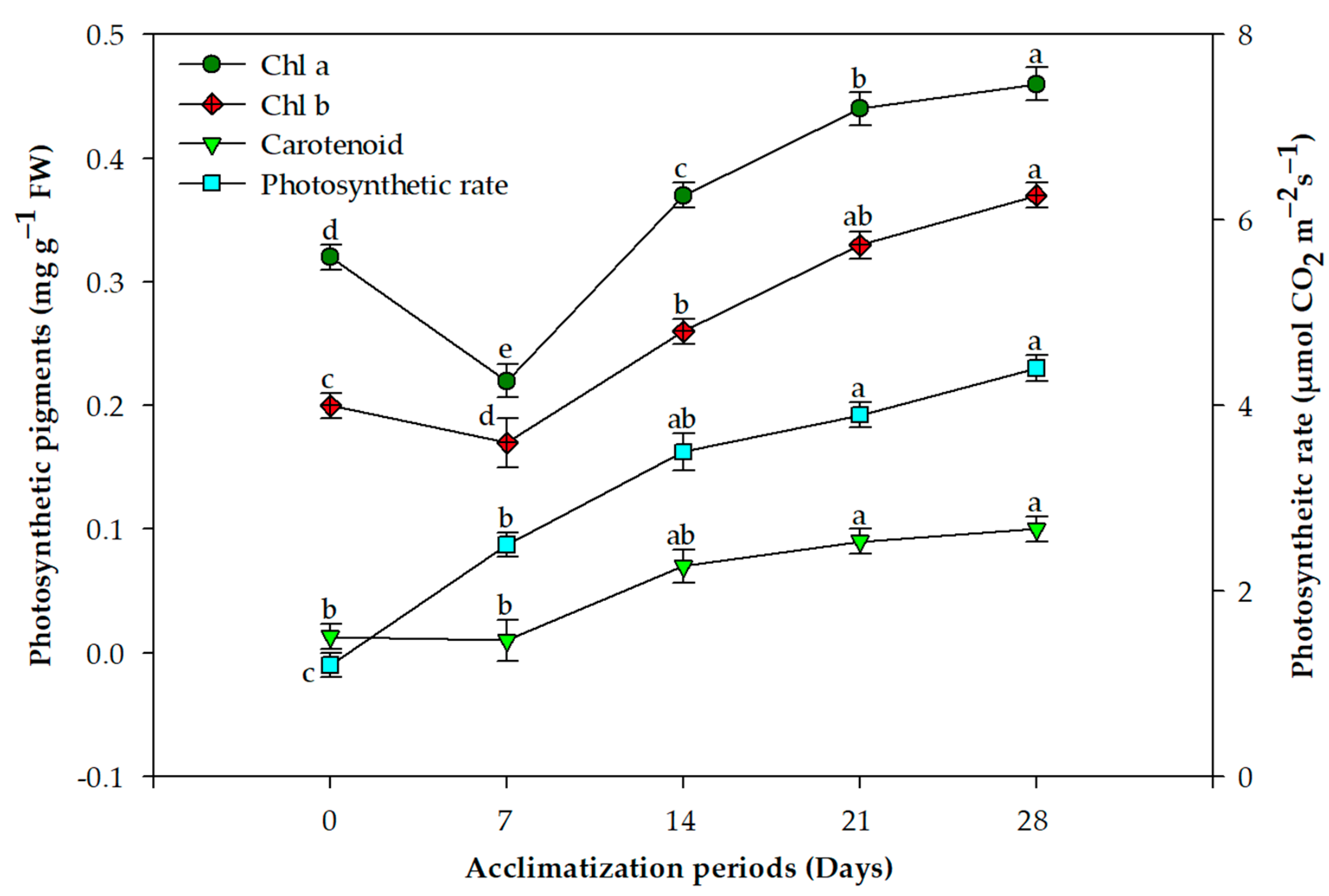
2.2. Acclimation and Assessment of Photosynthetic Pigments, Antioxidative Enzymes and Genetic Fidelity

Overall, 85% plants survived the acclimation process and transfer to garden soil (Figure 1F). The concentration of chlorophyll a and b behaved similarly over the acclimation period; the concentration declined in the first week and quickly recovered by the third and fourth weeks (Figure 2). This difference was statistically significant for both photosynthetic pigments. On the other hand, the photosynthetic rate and carotenoid concentration displayed a more stable increase without any decline after exflasking (Figure 2). The concentrations of all three assayed antioxidant enzymes increased over the period of acclimation, reaching their maximum in the fourth week, when the plants were transferred to the greenhouse (Figure 3). MDA production, an indication of lipid peroxidation, was low in the rooted plantlets at the time of exflasking, but increased almost threefold in the first week of acclimation, after which it decreased gradually until the plants were transferred to the greenhouse (Figure 4).
RAPD and ISSR markers were used for testing the genetic fidelity of micropropagated plants grown in the greenhouse. The mother plant was used as the control. The screening of 18 RAPD primers generated a total of 127 amplified bands with an average of 7.0 bands/primer (Table 4, Figure 5A). Among the 13 ISSR primers screened, 9 primers showed clear, reproducible bands with an average of 7.8 bands/primer (Table 5, Figure 5B). All the bands were monomorphic among the tested plants and the mother plant.

3. Discussion

In this study, we report the development of an efficient micropropagation protocol optimized for shoot proliferation from readily available nodal segments of mature trees of L. speciosa using mT and NAA in a 10:1 proportion, followed by rooting with IBA in MS medium. Previously, when BAP was used at the proliferation stage in L. speciosa, rooting was successful when forced ventilation was used in the containers [19]; however, this is costly and impractical for mass-scale operations. In another experiment with this species, the number of shoots per explant obtained with the optimal BAP concentration after 4 weeks was only two [16], compared with the 12 shoots that were obtained using mT alone (Table 1) in our research. Furthermore, Lim-Ho and Lee [20] reported that Kn in any concentration, or increasing the BAP concentration beyond 1 mg/L, resulted in callus induction and poor plant regeneration in L. speciosa. They managed to increase shoot proliferation using N6-(2-isopentenyl) adenine (2iP) in MS medium; however, CKs with an isoprenoid side chain such as 2iP have the disadvantage of fast oxidation compared with ArCKs [38].
The superiority of mT over BAP in micropropagation has been reported recently in other perennial species with medicinal values such as Oxystelma esculentum [31], Pterocarpus marsupium [29], Scaevola taccada [39], and Bacopa monnieri [40], as well as in perennial species used in horticulture such as Daphne mezereum [37], Ribes grossularia [33], Actinidia chinensis [35], and Franklinia alatamaha [41]. Working on two Prunus rootstocks, Gentile, et al. [42] observed that adventitious regeneration from in vitro leaf explants was possible in leaves from shoots growing in media supplemented with mT but not BAP; therefore, our research on the micropropagation of L. speciosa focused on mT. Our first experiment that used mT alone in MS media was promising, with a higher number of shoots proliferating than other CKs in L. speciosa propagation, based on what has been reported thus far [17,20,21]. When the optimum mT concentration of 5 µM was combined with different auxins, we found that the propagation efficiency and quality of shoots could be further improved in combination with 0.5 µM NAA. The synergistic actions of mT in combination with auxins in achieving significantly improved shoot multiplication rates have been documented in Aloe polyphylla [43], Uniola paniculata [44], and Pterocarpus marsupium [29].
The problems described in most of the research on micropropagation using BAP, zeatin, and Kn in perennial species, include the low regeneration potential, callus induction at the basal end of the explant, vitrification, and difficulty in rooting micropropagated shoots [36,45,46]. The improvement in shoot proliferation without callus induction when BAP or other CKs are replaced by mT has been attributed to its higher activity because of the presence of a hydroxyl group in the N9-position of the purine ring, leading to the formation of the O-glucoside conjugate N6(3-O-β-D-glucopyranosyl) benzyladenine-9-glucoside [22,47]. It has also been shown that substitution at the N9 position of CK’s purine ring significantly enhances acropetal transport of the CK, impeding the accumulation of CK glucoside forms in roots [45,46]. This allows for the gradual release of the active form of CK, enhancing the distribution of endogenous CKs in different plant tissues, and thus improving rooting and overall micropropagation efficiency [38]. In our research, as expected, mT-induced shoots of L. speciosa produced roots with all three auxins tested; however, 1 µM IBA proved to be significantly better in terms of the number of roots, root length, and the percentage of shoots (93%) rooted. This was followed by the successful establishment of a high percentage (95%) of rooted plantlets.
During in vitro culture, plantlets are subjected to diverse stresses that amplify the production of reactive oxygen species (ROS); however, unlike under in vivo conditions, in vitro stress has been studied less in terms of cellular mechanisms to manage stress; namely, constitutive and induced production of radical scavengers, free radical and oxidized-protein enzymatic degradation pathways, and DNA repair mechanisms [48,49]. Oxidative stress in the pathway is associated with the reprogramming of explants to produce fully functional plants results in the peroxidation of lipids, leading to membrane damage, protein degradation, enzyme inactivation, and genetic and epigenetic changes [48,50]. Demonstrating that the elevated amounts of free radicals and their reaction products can lead to morphogenetic recalcitrance in plant tissue culture, Benson [51] emphasized the need for combining studies on oxidative stress with plant development. In our research we focused on the plant acclimation stage, one of the critical stages of micropropagation, particularly because the mT -induced proliferation rates were high and produced shoots that were healthy in the optimized media. Furthermore, only a few studies on the oxidative stress in plants during the acclimation period have been conducted.
We combined studies on photosynthetic rates and pigment production with antioxidant enzyme activity during the period of acclimation. Our results revealed that there is a significant decrease in chlorophyll pigments during the first week of acclimation, and this negatively correlated with MDA content in the leaves, indicating high lipid peroxidation at this stage. Activity of all three oxidative enzymes, CAT, POX and SOD, had similar trends throughout the acclimation period; their activity increased as the plants acclimatized. Similar trends in carotenoid accumulation were also observed. Tissue-culture-raised plantlets are exposed to low PPFD. During acclimation, and in the greenhouse, plants are subjected to elevated PPFD that results in photo-inhibition [52]. Carotenoids play a pivotal role in defending the photosynthetic apparatus from photo-oxidative injuries [53], and their elevation along with high activity of ROS scavenging enzymes in L. speciosa during acclimation was a result of the plants reacting to the stresses of acclimation, resulting in high survival of our regenerated plantlets. Similar to our results, Goncalves, et al. [54] observed that chlorophyll and carotenoid contents in Plantago algarbiensis increased significantly during acclimation compared to the in vitro stage; however, these parameters in P. almogravensis, the other Plantago species they studied, remained high throughout the in vitro, acclimation, and greenhouse stages [54]. This shows a need to study these processes in the latter stages of micropropagation in different species to understand the outcomes. Similarly, Ahmad, et al. [55] found that the chlorophyll and carotenoid contents of Decalepis salicifolia decreased in the first week of acclimation, followed by increases in subsequent weeks; antioxidant enzyme activities also behaved similarly to our results with L. speciosa.
The retention of genetic consistency in regenerated plants is critical for any micropropagation system, as well as for conservation programs; therefore, it is important to appraise the genetic consistency of tissue culture-raised plants prior to commercialization or germplasm conservation. In the present experiment, both ISSR and RAPD primers showed high monomorphic patterns, and a way of authenticating the genetic integrity in the micropropagation system was developed. Our results indicate that axillary bud multiplication using mT and NAA is a safe method for producing true-to-type plants of L. speciosa. The variations observed in the ‘Williams’ banana when topolins or BAP were used, were attributed to excessive subculture cycles [56]. Moreover, the variation was not different between plants regenerated using BAP and mT in the ‘Williams’ banana [57] and sesame [30], whereas the regenerants were stable in Salvia sclarea [34] and Daphne mezereum [37]. There are a number of studies confirming the genetic fidelity of regenerated plants when the cytokinin for axillary bud proliferation is optimized for micropropagation in woody species with medicinal values, such as Withania somnifera [58], Rauvolfia serpentina [59], and Cassia alata [60]. Our results also show that somaclonal variation can be avoided when the growth regulators are used in optimal concentrations and subculturing is performed at regular intervals.

4. Materials and Methods

4.1. Plant Materials and Surface Sterilization

Juvenile healthy shoots of mature L. speciosa plants growing at the Department of Botany of Aligarh Muslim University, Aligarh, Uttar Pradesh, India was collected and washed under running tap water for about 30 min, then treated with the laboratory detergent “Labolene” (Qualigens, Mumbai, India) 5% (v/v) for 5 min, followed by three to four washes with sterile distilled water. The washed shoots were surface sterilized with HgCl2 0.1% (w/v) for 4–5 min followed by repeated washing with autoclaved distilled water under aseptic conditions in a laminar air flow cabinet. Nodal explants (1–1.5 cm) were isolated and cultured on sterile initiation media of different compositions as described below.

4.2. Basal Media and Culture Environment

The basal growth medium (BM) comprised Murashige and Skoog [61] salts (MS), 3% (w/v) sucrose, and 0.8% (w/v) agar (Qualigens Fine Chemicals, Mumbai, India). The medium pH was adjusted to 5.8 prior to adding agar, and the media was then autoclaved at 121 °C for 20 min. The cultures were incubated in a growth room at 25 ± 2 °C with 50 µmol m−2 s−1 photosynthetic photon flux density (PPFD) provided by white 40 W fluorescent lights (Philips India Ltd., Kolkata, India) at culture container level and 16 h photoperiod.

4.3. Shoot Initiation and Proliferation

In the first experiment, nodal explants (axillary buds) were isolated aseptically from surface sterilized shoots and cultured on BM supplemented with different concentrations of mT (0.0, 0.1, 0.5, 1.0, 2.5, 5.0, 7.5, 10.0, 12.5, and 15.0 µM) alone. The number of shoots per explant and the length of shoots were recorded on the 4th, 8th, and 12th week. To test if a combination with auxin would further enhance shoot proliferation, the mT treatment that gave the best proliferation of shoots (5 µM) was combined with α-naphthaleneacetic acid (NAA), indole-3-butyric acid (IBA), or indole-3-acetic acid (IAA) at five different concentrations (0.1, 0.5, 1.0, 1.5, and 2.0 µM) for the second shoot initiation/proliferation experiment. For this experiment, the shoots were initiated as described above and the number of shoots per explant and the length of shoots were recorded on the 8th week. The shoots from the best treatment in this experiment were used for the rooting experiment described below.

4.4. In Vitro Rooting and Plantlet Acclimation

Microshoots (3–4 cm) were dissected aseptically from 8-week-old proliferating axillary bud explants and transferred to root induction media consisting of BM supplemented with one of three auxins viz., IAA, NAA or IBA at 0.5. 1.0, 1.5, 2.0, 2.5 µM concentrations. Root growth was scored after 4 weeks in the culture as rooting percentage, mean number of roots per responding microshoot and mean root length. Microshoots grown on BM with no auxins served as the control.
For acclimation, shoots that had produced roots after 4 weeks of being in the culture under the same conditions as described for shoot organogenesis were removed from the culture tubes, washed to remove agar under tap water, and immediately transferred to polystyrene cups holding sterile SoilriteTM (Keltech Energies Ltd., Bangalore, India). The cups holding plantlets were covered in transparent polythene bags and fed with half-strength MS liquid medium without sucrose at 3-day intervals. The polythene bags were opened gradually over a period of 2 weeks for hardening of the regenerated plantlets. After a further 2 weeks, the acclimatized plants were shifted to pots with regular garden soil and were maintained inside a net house providing 40% filtered sunlight. The number of plants with roots, number of roots per plant, and root length were documented prior to transfer to garden soil.

4.5. Photosynthetic Pigments and Net Photosynthetic Rate Assessment

The method described by Mackinney [62] was used to estimate the content of chlorophyll a and b, and the method of Maclachlan and Zalik [63] for carotenoids in leaf tissue. Approximately 100 mg tissue from the interveinal region of leaves in plants at 0 (at the time of exflasking), 7, 14, 21, and 28 (completely developed leaves) days during acclimation were ground in 5 mL acetone (80%) using a mortar and pestle, and the suspension was filtered through Whatman No. 1 filter paper. The supernatant was ground again, filtered, and the entire filtrate was poured into a test tube and the volume adjusted to 10 mL with 80% acetone. For the chlorophyll content, the optical densities (OD) were recorded at 645 and 663 nm using a spectrophotometer (UV-Pharma Spec 1700, Shimadzu, Japan), whereas, for carotenoids, the OD was recorded at 480 and 510 nm.
The net photosynthetic rate in micropropagated plantlets was quantified at 0 (at the time of exflasking), 7, 14, 21, and 28 days after exflasking using an infra-red gas analyzer (IRGA, LICOR 6400, Lincoln, NE, USA). The photosynthetic rate was quantified using the source of net CO2 exchange among the leaves with the environment, by enfolding leaves in the leaf compartment of the gas analyzer and recording the rate at which the CO2 concentration altered over a 10–20 sec period. The net photosynthetic rate was expressed in µmol CO2 m−2 s−1.

4.6. Extraction and Analysis of Antioxidant Enzymes

For the assessment of leaf antioxidant enzyme concentrations, approximately 0.5 g of newly formed leaf tissue were homogenized in a 2.0 mL extraction buffer containing 1% polyvinylpyrrolidone, 1% Triton x-100, and 0.11 g ethylenediaminetetraacetic acid (EDTA) using a pre-chilled mortar and pestle. Filtration of the homogenate via four layers of cheese cloth was followed by centrifugation at 15,000 g for 20 min. The supernatant was used for the enzyme assays. The extraction process was undertaken in the dark at 4 °C.

4.7. Assessment of Superoxide Dismutase (SOD) Activity

SOD (superoxide:superoxide oxireductase, EC 1.15.1.1) was quantified according to Dhindsa et al. [64] with minor modifications. SOD activity in the supernatant was assessed by its capability to inhibit photochemical reduction of nitroblue tetrazolium (NBT). The reaction mixture consisted of 0.5 M potassium phosphate buffer, 200 mM methionine, 1 M sodium carbonate, 2.5 mM NBT, 3 mM EDTA, 0.1 mL enzyme extract, 60 µM riboflavin, and 1.0 mL doubled distilled water. The assay mixture in uniform transparent tubes was shaken and placed under a 15 W fluorescent lamp (Phillips, Kolkata, India) for 10 min (25–28 °C). The controls consisted of solutions and enzymes placed in the dark (blank A) and the reaction mixture without enzymes was placed in the light (blank B). Absorbance for samples along with blank B was read at 560 nm against the blank A. A 50% reduction in color was considered as one unit of SOD enzyme activity and expressed in mg protein min−1.

4.8. Catalase (CAT) Activity Assay

The method described by Aebi [65] with slight modifications was used for the CAT (H2O2:H2O2 oxidoreductase: EC 1.11.1.6) activity assay in leaves. The reaction mixture consisted of 0.5 M potassium phosphate buffer, 3 mM EDTA, 0.1 mL enzyme extract, and 3 mM H2O2 (pH 7.0), and the reaction was allowed to run for 5 min. CAT action was verified via screening H2O2 loss and quantifying the decline in absorbance at 240 nm. Calculation of CAT activity was carried out by using extinction coefficient (€) 0.036 mM−1 cm−1 and activity was expressed in enzyme units (EU) mg−1 protein min−1. One unit of enzyme was defined as the amount of catalase necessary to decompose 1 µmol of H2O2 per min at pH 7.0 at 25 °C.

4.9. Assessment of Peroxidase (POX) Activity

POX (EC 1.11.1.7) content was measured according to Bergmeyer, et al. [66] Fresh leaves (0.2 mg) were collected and homogenized with 5 mL cold phosphate buffer (50 mM, pH 7.8) using a mortar and pestle, and the homogenate was centrifuged at 10,000 g at 4 °C for 20 min. The supernatant collected was maintained at 4 °C until use in the assay. The assay mixture consisted of 0.1 M phosphate buffer (pH 7.8), 4 mM pyrogallol, 3 mM H2O2, and crude enzyme extract. The assay mixture was transferred to a cuvette and the absorbance was measured at 420 nm using a spectrophotometer (UV-Pharma Spec 1700, Shimadzu, Japan). The enzyme activity was expressed as µmol (H2O2 consumed) mg−1 (protein) s−1.

4.10. Measurement of Lipid Peroxides

A thiobarbituric acid-reactive-substance assay for measuring malondialdehyde (MDA) was used to assess lipid peroxidation. MDA is a secondary end product of polyunsaturated fatty acid oxidation and is considered to be a useful index of general lipid peroxidation [67]. The method described by Cakmak and Horst [68] was used with modifications. Leaf tissue (0.5 g) was homogenized with 5 mL 0.1% trichloroacetic acid (TCA) and centrifuged at 15,000 g for 5 min. Subsequently, approximately 1 mL supernatant was mixed with 4 mL 0.5% (w/v) thiobarbituric acid (TBA) in 20% TCA. The mixture was held in boiling water for 30 min, then instantly cooled on ice to end the reaction. The samples were then centrifuged at 12,000 g for 30 min. The absorbance of the supernatant was read at 532 nm using a UV-VIS spectrophotometer (UV-1700 Pharma Spec), and the reading at 600 nm was deducted to compensate for non-specific absorption. The amount of lipid peroxides was measured in terms of MDA level, with an extinction coefficient of 155 mM−1 cm−1, and expressed as nmol g−1 fresh weight.

4.11. DNA Extraction and Molecular Marker Techniques

Isolation of genomic DNA from leaf tissues of micropropagated plants, as well as donor plants (mother plant), was conducted using a modified cetyltrimethylammonium bromide (CTAB) technique [69]. Quantification and purity of total DNA was assessed by Nanodrop Spectrophotometer (Implen, Munich, Germany). All the samples were diluted to 25 ng/µL in Milli-Q water and stored at 4 °C until further use. Twenty random amplified polymorphic DNA (RAPD) primers (Operon Kit A Technologies Inc., Ebersberg, Germany) and 13 inter-simple sequence repeat (ISSR) primers (UBC, Vancouver, BC, Canada) were used to examine the clonal fidelity among the regenerated plantlets.
PCR amplification reactions were made in 20 µL volumes containing 1X PCR buffer, 25 mM MgCl2 (1.2 µL), 10 mM dNTPs (0.4 µL), 2 µM primers, 3 Unit Taq polymerase (0.2 µL), and 40 ng Template DNA. The amplification of DNA for both RAPD and ISSR analyses was performed in a Thermal Cycler (Biometra, Gottingen, Germany). The PCR amplification program consisted of 45 cycles counting a 94 °C denaturation step of 5 min, an annealing step for 2 min at 35 °C (for RAPD analysis) or 35–58 °C (for ISSR analysis), and a 72 °C elongation of 1 min. A concluding extension was tracked at 72 °C for 10 min. PCR products were analyzed by electrophoresis in 0.8% (w/v) agarose gels with 4 µL ethidium bromide in a TAE buffer (pH 8.0), run at 60 V for 2 h, and visualized on a UV transilluminator (Bio Rad, Hercules, CA, USA). To regulate the uniformity of the experiments, DNA extraction, PCR reactions and electrophoresis were repeated three times. Distinct and reproducible bands were scored. Bands with the identical movement were considered to be homologous fragments, regardless of intensity.

4.12. Statistical Analysis

The two experiments involving mT were established with 20 replicates per treatment in a randomized complete block design with six explants per replicate and the experiments were repeated three times. The rooting experiment with three auxins was established with 10 replicates per treatment in a randomized complete block design with a minimum of six microshoots per replicate. Morphological changes among the cultures were observed and documented at regular intervals. The SPSS version 16 (SPSS Inc. Chicago, IL, USA) was employed for statistical analysis of the data. Duncan’s multiple range test (DMRT) at p ≤ 0.05 was carried out to examine evidence relating to significant variation among mean values, and the results were expressed as the means ± SE of three experiments.
Research manuscripts reporting large datasets that are deposited in a publicly available database should specify where the data have been deposited and provide the relevant accession numbers. If the accession numbers have not yet been obtained at the time of submission, please state that they will be provided during review. They must be provided prior to publication.
Interventionary studies involving animals or humans, and other studies that require ethical approval, must list the authority that provided approval and the corresponding ethical approval code.

5. Conclusions

The present research has successfully established a simple, resourceful, and robust regeneration methodology for micropropagation in L. speciosa that will also be useful for ex situ germplasm conservation. The use of the novel ArCK, mT, provides a broad platform for in vitro propagation of true-to-type plant material suitable for commercialization, conservation strategies, and pharmaceutical needs. L. speciosa shows immense potential for the production of corosolic acid, a “future anti-diabetic drug”, and the plant propagation platform developed in this research could pave the way for commercial production of the plant and its exploitation for bioactives. To the best our knowledge, this mT-based in vitro propagation protocol developed in our research is several times more efficient than existing ones.

Author Contributions

Conceptualization, N.A., N.F. and M.F.; methodology, N.A., N.F., M.F., A.A.A. and R.P.; validation, N.A., M.F. and A.A.A.; formal analysis, N.A., N.F., M.F., A.A.A. and R.P.; investigation, N.A., N.F., M.F., A.A.A. and R.P.; resources, N.A. and M.F.; writing—original draft preparation N.A., N.F. and M.F.; writing—review and editing, N.A., M.F., A.A.A. and R.P.; supervision, M.F. and A.A.A.; project administration, A.A.A.; funding acquisition, M.F. and A.A.A. All authors have read and agreed to the published version of the manuscript.

Funding

This study was supported by the Researchers Supporting Project (RSP-2021/86), King Saud University, Riyadh, Saudi Arabia.

Data Availability Statement

Not applicable.

Acknowledgments

The authors are thankful to the Researchers Supporting Project (RSP-2021/86), King Saud University, Riyadh, Saudi Arabia.

Conflicts of Interest

Authors declare that there are no conflict of interest.

References

  1. Ellis, R.H.; Mai-Hong, T.; Hong, T.D.; Tan, T.T.; Xuan-Chuong, N.D.; Hung, L.Q.; Ngoc-Tam, B.; Le-Tam, V.T. Comparative analysis by protocol and key of seed storage behaviour of sixty Vietnamese tree species. Seed Sci. Technol. 2007, 35, 460–476. [Google Scholar] [CrossRef]
  2. Sannigrahi, A.K. Biodegradation of leaf litter of tree species in presence of cow dung and earthworms. Indian J. Biotechnol. 2009, 8, 335–338. [Google Scholar]
  3. Dhakane, M.U.; Yadav, S.G. Pride of India (Lagerstroemia speciosa (L.) Pers.) forming a silver bullet for 21st century. Int. J. For. Crop Improv. 2015, 6, 79–82. [Google Scholar] [CrossRef]
  4. Deshpande, A.M.; Shirsat, M.K.; Jeyabalan, G. A review of Lagerstroemia speciosa: Pride of India. Int. J. Contemp. Res. Rev. 2018, 9, 20181–20185. [Google Scholar] [CrossRef]
  5. Hayashi, T.; Maruyama, H.; Kasai, R.; Hattori, K.; Takasuga, S.; Hazeki, O.; Yamasaki, K.; Tanaka, T. Ellagitannins from Lagerstroemia speciosa as activators of glucose transport in fat cells. Planta Med. 2002, 68, 173–175. [Google Scholar] [CrossRef]
  6. Kotnala, M.; Chakraborthy, G.; Mazumder, A. Lagerstroemia species: A current review. Int. J. PharmTech Res. 2013, 5, 906–909. [Google Scholar]
  7. Esmail, A.S.A. Medicinal value of Lagerstroemia speciosa: An updated review. Int. J. Curr. Pharm. Res. 2019, 11, 18–26. [Google Scholar] [CrossRef] [Green Version]
  8. Yamaguchi, Y.; Yamada, K.; Yoshikawa, N.; Nakamura, K.; Haginaka, J.; Kunitomo, M. Corosolic acid prevents oxidative stress, inflammation and hypertension in SHR/NDmcr-cp rats, a model of metabolic syndrome. Life Sci. 2006, 79, 2474–2479. [Google Scholar] [CrossRef]
  9. Park, S.W.; Kwon, M.J.; Yoo, J.Y.; Choi, H.-J.; Ahn, Y.-J. Antiviral activity and possible mode of action of ellagic acid identified in Lagerstroemia speciosa leaves toward human rhinoviruses. BMC Complement. Altern. Med. 2014, 14, 171. [Google Scholar] [CrossRef] [Green Version]
  10. Laruan, L.M.V.; Balangod, T.; Balangcod, K.; Patacsil, M.; Apastol, O.; Manuel, J.; Cortez, S.; Vallejn, V. Phytochemical and antibacterial study of Lagerstroemia (L.) Pers and its ethno-medicinal importance to indigenous communities of Benquet provinence, Philippines. Indian J. Od Tradit. Knowl. 2013, 12, 379–383. [Google Scholar]
  11. Pareek, A.; Suthar, M.; Rathore, G.S.; Bansal, V.; Kumawat, T. In vitro antioxidant studies of Lagerstroemia speciosa leaves. Pharmacogn. J. 2010, 2, 357–360. [Google Scholar] [CrossRef]
  12. Unno, T.; Sugimoto, A.; Kakuda, T. Xanthine oxidase inhibitors from the leaves of Lagerstroemia speciosa (L.) Pers. J. Ethnopharmacol. 2004, 93, 391–395. [Google Scholar] [CrossRef] [PubMed]
  13. Chen, L.X.; Schumacher, H.R. Gout: An evidence-based review. J. Clin. Rheumatol. 2008, 14, S55–S62. [Google Scholar] [CrossRef] [PubMed]
  14. Shi, L.; Zhang, W.; Zhou, Y.-Y.; Zhang, Y.-N.; Li, J.-Y.; Hu, L.-H.; Li, J. Corosolic acid stimulates glucose uptake via enhancing insulin receptor phosphorylation. Eur. J. Pharmacol. 2008, 584, 21–29. [Google Scholar] [CrossRef]
  15. Judy, W.V.; Hari, S.P.; Stogsdill, W.W.; Judy, J.S.; Naguib, Y.M.A.; Passwater, R. Antidiabetic activity of a standardized extract (Glucosol™) from Lagerstroemia speciosa leaves in Type II diabetics: A dose-dependence study. J. Ethnopharmacol. 2003, 87, 115–117. [Google Scholar] [CrossRef]
  16. Vijayan, A.; Padmesh Pillai, P.; Hemanthakumar, A.S.; Krishnan, P.N. Improved in vitro propagation, genetic stability and analysis of corosolic acid synthesis in regenerants of Lagerstroemia speciosa (L.) Pers. by HPLC and gene expression profiles. Plant Cell Tissue Organ Cult. 2015, 120, 1209–1214. [Google Scholar] [CrossRef]
  17. Sivadas, D.; Pandurangan, A.G. Non-viable seed set in Lagerstroemia speciosa (Lythraceae) populations in the Western Ghats, India. Trop. Ecol. 2022, 63, 141–144. [Google Scholar] [CrossRef]
  18. Hadiuzzaman, S. In vitro cloning from seedling explants of Lagerstroemia speciosa Pers. and L. thorellii Gagnep. Bangladesh J. Bot. 1992, 21, 59–64. [Google Scholar]
  19. Zobayed, S.M.A. In vitro propagation of Lagerstroemia spp. from nodal explants and gaseous composition in the culture headspace. Environ. Control Biol. 2000, 38, 1–11. [Google Scholar] [CrossRef]
  20. Lim-Ho, C.L.; Lee, S.K. Micropropagation of Lagerstroemia speciosa (L.) Pers. (Lythraceae). Gard. Bull. Singap. 1985, 38, 175–184. [Google Scholar]
  21. Strnad, M.; Hanuš, J.; Vaněk, T.; Kamínek, M.; Ballantine, J.A.; Fussell, B.; Hanke, D.E. Meta-topolin, a highly active aromatic cytokinin from poplar leaves (Populus × canadensis Moench., cv. Robusta). Phytochemistry 1997, 45, 213–218. [Google Scholar] [CrossRef]
  22. Werbrouck, S.P.; Strnad, M.; Van Onckelen, H.A.; Debergh, P.C. Meta-topolin, an alternative to benzyladenine in tissue culture? Physiol. Plant. 1996, 98, 291–297. [Google Scholar] [CrossRef]
  23. Lata, H.; Chandra, S.; Techen, N.; Khan, I.A.; ElSohly, M.A. In vitro mass propagation of Cannabis sativa L.: A protocol refinement using novel aromatic cytokinin meta-topolin and the assessment of eco-physiological, biochemical and genetic fidelity of micropropagated plants. J. Appl. Res. Med. Aromat. Plants 2016, 3, 18–26. [Google Scholar] [CrossRef]
  24. Naaz, A.; Hussain, S.A.; Anis, M.; Alatar, A.A. Meta-topolin improved micropropagation in Syzygium cumini and acclimatization to ex vitro conditions. Biol. Plant. 2019, 63, 174–182. [Google Scholar] [CrossRef]
  25. Teklehaymanot, T.; Wannakrairoj, S.; Pipattanawong, N. Meta-topolin for pineapple shoot multiplication under three in vitro systems. Am.-Eurasian J. Agric. Environ. Sci. 2010, 7, 157–162. [Google Scholar]
  26. Westhuizen, A.V.D. Use of Meta-topolin as an alternative cytokinin in the tissue culture of Eucalyptus species. Acta Hortic. 2014, 1055, 25–28. [Google Scholar] [CrossRef]
  27. Gentile, A.; Frattarelli, A.; Nota, P.; Condello, E.; Caboni, E. The aromatic cytokinin meta-topolin promotes in vitro propagation, shoot quality and micrografting in Corylus colurna L. Plant Cell Tissue Organ Cult. 2017, 128, 693–703. [Google Scholar] [CrossRef]
  28. Chauhan, R.D.; Taylor, N.J. Meta-topolin stimulates de novo shoot organogenesis and plant regeneration in cassava. Plant Cell Tissue Organ Cult. 2018, 132, 219–224. [Google Scholar] [CrossRef]
  29. Ahmad, A.; Anis, M.; Khanam, M.N.; Alatar, A.A. Direct shoot organogenesis from shoot tip explants of a highly medicinal valued tree Pterocarpus marsupium Roxb. Vitr. Cell. Dev. Biol.-Plant 2020, 56, 670–681. [Google Scholar] [CrossRef]
  30. Elayaraja, D.; Subramanyam, K.; Vasudevan, V.; Sathish, S.; Kasthurirengan, S.; Ganapathi, A.; Manickavasagam, M. Meta-Topolin (mT) enhances the in vitro regeneration frequency of Sesamum indicum (L.). Biocatal. Agric. Biotechnol. 2019, 21, 101320. [Google Scholar] [CrossRef]
  31. Jayaprakash, K.; Manokari, M.; Badhepuri, M.K.; Raj, M.C.; Dey, A.; Shekhawat, M.S. Influence of meta-topolin on in vitro propagation and foliar micro-morpho-anatomical developments of Oxystelma esculentum (L.f.)Sm. Plant Cell Tissue Organ Cult. 2021, 147, 325–337. [Google Scholar] [CrossRef]
  32. Manokari, M.; Priyadharshini, S.; Jogam, P.; Dey, A.; Shekhawat, M.S. Meta-topolin and liquid medium mediated enhanced micropropagation via ex vitro rooting in Vanilla planifolia Jacks. ex Andrews. Plant Cell Tissue Organ Cult. 2021, 146, 69–82. [Google Scholar] [CrossRef]
  33. Kucharska, D.; Orlikowska, T.; Maciorowski, R.; Kunka, M.; Wojcik, D.; Pluta, S. Application of meta-Topolin for improving micropropagation of gooseberry (Ribes grossularia). Sci. Hortic. 2020, 272, 109529. [Google Scholar] [CrossRef]
  34. Erisen, S.; Kurt-Gur, G.; Servi, H. In vitro propagation of Salvia sclarea L. by meta-Topolin, and assessment of genetic stability and secondary metabolite profiling of micropropagated plants. Ind. Crops Prod. 2020, 157, 112892. [Google Scholar] [CrossRef]
  35. Saeiahagh, H.; Mousavi, M.; Wiedow, C.; Bassett, H.B.; Pathirana, R. Effect of cytokinins and sucrose concentration on the efficiency of micropropagation of ‘Zes006′ Actinidia chinensis var. chinensis, a red-fleshed kiwifruit cultivar. Plant Cell Tissue Organ Cult. 2019, 138, 1–10. [Google Scholar] [CrossRef]
  36. Debenham, M.; Pathirana, R. Establishment and management of an in vitro repository of kiwifruit (Actinidia spp.) germplasm. In Meta-Topolin: A Growth Regulator for Plant Biotechnology and Agriculture; Ahmad, N., Strnad, M., Eds.; Springer Nature: Cham, Switzerland, 2020; pp. 279–291. [Google Scholar]
  37. Nowakowska, K.; Pacholczak, A. Comparison of the effect of meta-Topolin and benzyladenine during Daphne mezereum L. micropropagation. Agronomy 2020, 10, 1994. [Google Scholar] [CrossRef]
  38. Plíhal, O.; Szüčová, L.; Galuszka, P. N9-substituted aromatic cytokinins with negligible side effects on root development are an emerging tool for in vitro culturing. Plant Signal. Behav. 2013, 8, e24392. [Google Scholar] [CrossRef] [Green Version]
  39. Shekhawat, M.S.; Priyadharshini, S.; Jogam, P.; Kumar, V.; Manokari, M. Meta-topolin and liquid medium enhanced in vitro regeneration in Scaevola taccada (Gaertn.) Roxb. Vitr. Cell. Dev. Biol.-Plant 2021, 57, 296–306. [Google Scholar] [CrossRef]
  40. Pramanik, B.; Sarkar, S.; Bhattacharyya, S.; Gantait, S. meta-Topolin-induced enhanced biomass production via direct and indirect regeneration, synthetic seed production, and genetic fidelity assessment of Bacopa monnieri (L.) Pennell, a memory-booster plant. Acta Physiol. Plant. 2021, 43, 107. [Google Scholar] [CrossRef]
  41. Gladfelter, H.J.; Johnston, J.; Wilde, H.D.; Merkle, S.A. Adventitious shoot-based propagation of Franklinia alatamaha for commercial horticulture and restoration. Vitr. Cell. Dev. Biol.-Plant 2020, 56, 857–864. [Google Scholar] [CrossRef]
  42. Gentile, A.; Gutierrez, M.J.; Martinez, J.; Frattarelli, A.; Nota, P.; Caboni, E. Effect of meta-Topolin on micropropagation and adventitious shoot regeneration in Prunus rootstocks. Plant Cell Tissue Organ Cult. 2014, 118, 373–381. [Google Scholar] [CrossRef]
  43. Bairu, M.W.; Stirk, W.A.; Dolezal, K.; Van Staden, J. Optimizing the micropropagation protocol for the endangered Aloe polyphylla: Can meta-topolin and its derivatives serve as replacement for benzyladenine and zeatin? Plant Cell Tissue Organ Cult. 2007, 90, 15–23. [Google Scholar] [CrossRef]
  44. Valero-Aracama, C.; Kane, M.E.; Wilson, S.B.; Philman, N.L. Substitution of benzyladenine with meta-topolin during shoot multiplication increases acclimatization of difficult- and easy-to-acclimatize sea oats (Uniola paniculata L.) genotypes. Plant Growth Regul. 2009, 60, 43–49. [Google Scholar] [CrossRef]
  45. Podlesakova, K.; Zalabak, D.; Cudejkova, M.; Plihal, O.; Szucova, L.; Dolezal, K.; Spichal, L.; Strnad, M.; Galuszka, P. Novel cytokinin derivatives do not show negative effects on root growth and proliferation in submicromolar range. PLoS ONE 2012, 7, e39293. [Google Scholar] [CrossRef]
  46. Vylíčilová, H.; Bryksová, M.; Matušková, V.; Doležal, K.; Plíhalová, L.; Strnad, M. Naturally Occurring and Artificial N9-Cytokinin Conjugates: From Synthesis to Biological Activity and Back. Biomolecules 2020, 10, 832. [Google Scholar] [CrossRef]
  47. Aremu, A.O.; Bairu, M.W.; Dolezal, K.; Finnie, J.F.; Van Staden, J. Topolins: A panacea to plant tissue culture challenges? Plant Cell Tissue Organ Cult. 2012, 108, 1–16. [Google Scholar] [CrossRef]
  48. Cassells, A.C.; Curry, R.F. Oxidative stress and physiological, epigenetic and genetic variability in plant tissue culture: Implications for micropropagators and genetic engineers. Plant Cell Tissue Organ Cult. 2001, 64, 145–157. [Google Scholar] [CrossRef]
  49. Şen, A. Oxidative stress studies in plant tissue culture. Antioxid. Enzym. 2012, 3, 59–88. [Google Scholar]
  50. Bednarek, P.T.; Orłowska, R. Plant tissue culture environment as a switch-key of (epi)genetic changes. Plant Cell Tissue Organ Cult. 2020, 140, 245–257. [Google Scholar] [CrossRef] [Green Version]
  51. Benson, E.E. Do free radicals have a role in plant tissue culture recalcitrance? Vitr. Cell. Dev. Biol. Plant 2000, 36, 163–170. [Google Scholar] [CrossRef]
  52. Wang, Y.; Zhang, H.; Hou, P.; Su, X.; Zhao, P.; Zhao, H.; Liu, S. Foliar-applied salicylic acid alleviates heat and high light stress induced photoinhibition in wheat (Triticum aestivum) during the grain filling stage by modulating the psbA gene transcription and antioxidant defense. Plant Growth Regul. 2014, 73, 289–297. [Google Scholar] [CrossRef]
  53. Jeon, M.-W.; Ali, M.B.; Hahn, E.-J.; Paek, K.-Y. Effects of photon flux density on the morphology, photosynthesis and growth of a CAM orchid, Doritaenopsis during post-micropropagation acclimatization. Plant Growth Regul. 2005, 45, 139–147. [Google Scholar] [CrossRef]
  54. Goncalves, S.; Martins, N.; Romano, A. Physiological traits and oxidative stress markers during acclimatization of micropropagated plants from two endangered Plantago species: P. algarbiensis Samp. and P. almogravensis Franco. Vitr. Cell. Dev. Biol.-Plant 2017, 53, 249–255. [Google Scholar] [CrossRef]
  55. Ahmad, Z.; Shahzad, A.; Sharma, S.; Parveen, S. Ex vitro rescue, physiochemical evaluation, secondary metabolite production and assessment of genetic stability using DNA based molecular markers in regenerated plants of Decalepis salicifolia (Bedd. ex Hook.f.) Venter. Plant Cell Tissue Organ Cult. 2018, 132, 497–510. [Google Scholar] [CrossRef]
  56. Aremu, A.O.; Bairu, M.W.; Szucova, L.; Dolezal, K.; Finnie, J.F.; Van Staden, J. Genetic fidelity in tissue-cultured ‘Williams’ bananas-The effect of high concentration of topolins and benzyladenine. Sci. Hortic. 2013, 161, 324–327. [Google Scholar] [CrossRef]
  57. Bairu, M.W.; Stirk, W.A.; Doležal, K.; van Staden, J. The role of topolins in micropropagation and somaclonal variation of banana cultivars ‘Williams’ and ‘Grand Naine’ (Musa spp. AAA). Plant Cell Tissue Organ Cult. 2008, 95, 373–379. [Google Scholar] [CrossRef]
  58. Fatima, N.; Ahmad, N.; Anis, M.; Ahmad, I. An improved in vitro encapsulation protocol, biochemical analysis and genetic integrity using DNA based molecular markers in regenerated plants of Withania somnifera L. Ind. Crops Prod. 2013, 50, 468–477. [Google Scholar] [CrossRef]
  59. Faisal, M.; Alatar, A.A.; El-Sheikh, M.A.; Abdel-Salam, E.M.; Qahtan, A.A. Thidiazuron induced in vitro morphogenesis for sustainable supply of genetically true quality plantlets of Brahmi. Ind. Crops Prod. 2018, 118, 173–179. [Google Scholar] [CrossRef]
  60. Ahmed, M.R.; Anis, M.; Alatar, A.A.; Faisal, M. In vitro clonal propagation and evaluation of genetic fidelity using RAPD and ISSR marker in micropropagated plants of Cassia alata L.: A potential medicinal plant. Agrofor. Syst. 2017, 91, 637–647. [Google Scholar] [CrossRef]
  61. Murashige, T.; Skoog, F. A Revised Medium for Rapid Growth and Bio Assays with Tobacco Tissue Cultures. Physiol. Plant. 1962, 15, 473–497. [Google Scholar] [CrossRef]
  62. Mackinney, G.R. Absorption of light by chlorophyll solutions. J. Biol. Chem. 1941, 140, 315–322. [Google Scholar] [CrossRef]
  63. Maclachlan, S.; Zalik, S. Plastid structure, chlorophyll concentration, and free amino acid composition of a chlorophyll mutant of barley. Can. J. Bot. 1963, 41, 1053–1062. [Google Scholar] [CrossRef]
  64. Dhindsa, R.S.; Plumb-Dhindsa, P.; Thorpe, T.A. Leaf senescence: Correlated with increased levels of membrane permeability and lipid peroxidation, and decreased levels of superoxide dismutase and catalase. J. Exp. Bot. 1981, 32, 93–101. [Google Scholar] [CrossRef]
  65. Aebi, H. Catalase in vitro. Methods Enzymol. 1984, 105, 121–126. [Google Scholar] [PubMed]
  66. Bergmeyer, H.U.; Gawehn, K.; Grassl, M. Methods of Enzymatic Analysis; Bergmeyer, H.U., Ed.; Verlag Chemie: Wienheim, Germany, 1974; Volume 1, pp. 481–482. [Google Scholar]
  67. Hodges, D.M.; DeLong, J.M.; Forney, C.F.; Prange, R.K. Improving the thiobarbituric acid-reactive-substances assay for estimating lipid peroxidation in plant tissues containing anthocyanin and other interfering compounds. Planta 1999, 207, 604–611. [Google Scholar] [CrossRef]
  68. Cakmak, I.; Horst, W.J. Effect of aluminium on lipid peroxidation, superoxide dismutase, catalase, and peroxidase activities in root tips of soybean (Glycine max). Physiol. Plant. 1991, 83, 463–468. [Google Scholar] [CrossRef]
  69. Doyle, J. DNA Protocols for Plants. In Molecular Techniques in Taxonomy; Hewitt, G.M., Ed.; NATO AS1 Series; Springer: Berlin/Heidelberg, Germany, 1991; pp. 283–293. [Google Scholar]
Figure 1. Micropropagation of Lagerstroemia speciosa L. (A) Multiple shoot induction from nodal segments cultured for 4 weeks using MS medium supplemented with 5.0 µM meta-Topolin (mT) (Bar = 0.86 cm); (B) mass multiplication of shoots after 8 weeks of being cultured using MS medium supplemented with 5.0 µM mT (Bar = 0.89 cm); (C) multiplication and elongation of shoots after 12 weeks using the optimal concentration of mT (5.0 µM) + NAA (0.5 µM) (Bar = 1.70 cm); (D) rooting of in vitro shoots after 4 weeks using MS medium supplemented with 1 µM indole-3-butyric acid (Bar = 0.60 cm); (E) rooted microshoot ready for transplantation (Bar 0.71 cm); and (F) a 4-week-old acclimatized plant (Bar = 0.71).
Plants 11 01163 g001
Figure 2. Changes in chlorophyll a, chlorophyll b, carotenoid content, and net photosynthetic rate during the period of acclimation for Lagerstroemia speciosa L. plantlets. Line graphs represent mean ± SE of three repeated experiments, each with 20 replications. Means within a category with different letters are significantly different according to Duncan’s multiple range test (p ≤ 0.05).
Plants 11 01163 g002
Figure 3. Changes in catalase (CAT), peroxidase (POX), and superoxide dismutase (SOD) activity in leaves of Lagerstroemia speciosa L. during the acclimation period. Bars represent mean ± SE of three repeated experiments, each with 20 replications. Means of an enzyme activity with different letters are significantly different according to Duncan’s multiple range test (p ≤ 0.05).
Plants 11 01163 g003
Figure 4. Changes in malondialdehyde (MDA) concentration in Lagerstroemia speciosa L. leaves during the acclimation period. Bars represent the mean ± SE of three repeated experiments each with 20 replications. Means in a column with different letters are significantly different according to Duncan’s multiple range test (p ≤ 0.05).
Plants 11 01163 g004
Figure 5. Random amplified polymorphic DNA (RAPD) and inter-simple sequence repeat (ISSR) profiles of Lagerstroemia speciosa L. (A) Amplified profile from RAPD primer OPA-01; (B) amplified profile from ISSR primer UBC-880. DL—DNA ladder; lanes 1–5 randomly selected in vitro plants; lane E—Donor plant.
Plants 11 01163 g005
Table 1. Effect of different concentrations of meta-Topolin in MS basal medium on in vitro shoot regeneration from nodal explants of Lagerstroemia speciosa L.
meta-Topolin Concentration (µM)4 Weeks8 Weeks12 Weeks
Number of Shoots Shoot Length (cm)Number of ShootsShoot Length (cm)Number of ShootsShoot Length (cm)
0.00.00 ± 0.00 j0.00 ± 0.00 g0.00 ± 0.00 i0.00 ± 0.00 f0.00 ± 0.00 j0.00 ± 0.00 f
0.12.04 ± 0.04 i0.88 ± 0.02 f5.20 ± 0.00 h0.92 ± 0.16 e6.85 ± 0.03 i1.60 ± 0.10 e
0.53.20 ± 0.20 h1.32± 0.08 e7.62 ± 0.17 e1.62 ± 0.18 d10.2 ± 0.18 h3.36 ± 0.16 d
1.04.36 ± 0.36 g2.50 ± 0.00 c12.8 ±0.04 c2.70 ± 0.33 bc14.1 ± 0.10 f3.86 ± 0.02 e
2.58.10 ± 0.10 d3.20 ± 0.20 b14.4 ± 0.18 b3.08 ± 0.50 b18.5 ± 0.15 c4.60 ± 0.00 b
5.012.0 ± 0.02 a3.76 ± 0.06 a16.4± 0.12 a4.10 ± 0.10 a22.4 ± 0.02 a5.22 ± 0.00 a
7.510.0 ± 0.00 b2.52 ± 0.11 c11.5 ± 0.15 d3.12 ± 0.09 b20.2 ± 0.20 b4.84 ± 0.08 b
10.09.16 ± 0.04 c2.00 ± 0.00 d6.50 ± 0.00 f2.30 ± 0.08 cd15.6 ± 0.10 d4.40 ± 0.04 b
12.57.10 ± 0.10 e1.20 ± 0.00 e5.60 ± 0.17 g1.50 ± 0.00 de14.7 ± 0.06 e3.80 ± 0.40 c
15.05.00 ± 0.00 f0.80 ± 0.00 f5.00 ± 0.00 h1.20 ± 0.20 de11.4 ± 0.04 g2.96 ± 0.06 d
Values represent mean ± SE of three repeated experiments, each with ten replications. Means in a column with different letters (superscript) are significantly different according to Duncan’s multiple range test (p ≤ 0.05).
Table 2. Effect of different concentrations of auxins in combination with 5 µM meta-Topolin in MS basal medium on shoot initiation and proliferation from nodal segments of Lagerstroemia speciosa L. after 8 weeks of being cultured.
Plant Growth Regulators (µM)% RegenerationNumber of Shoots/ExplantMean Shoot Length (cm)
IAAIBANAA
0.1 7012.5 ± 0.19 gh4.40 ± 0.40 ef
0.5 7514.4 ± 0.40 e5.00 ± 0.00 cd
1.0 608.70 ± 0.12 j4.92 ± 0.02 cd
1.5 708.00 ± 0.00 k4.20 ± 0.00 efg
2.0 657.48 ± 0.12 l3.92 ± 0.02 g
0.1 8013.6 ± 0.08 f4.68 ± 0.08 de
0.5 8516.0 ± 0.00 d5.22 ± 0.02 bc
1.0 7514.6 ± 0.16 e4.34 ± 0.04 ef
1.5 6012.9 ± 0.31 g3.84 ± 0.05 g
2.0 6010.0 ± 0.00 i2.60 ± 0.10 h
0.18517.5 ± 0.36 c5.16 ± 0.05 bc
0.59524.3 ± 0.19 a5.74 ± 0.06 a
1.08518.2 ± 0.20 b5.50 ± 0.00 ab
1.58015.8 ± 0.30 d5.20 ± 0.20 bc
2.07512.0 ± 0.00 h5.04 ± 0.24 cd
Values represent mean ± SE of three repeated experiments each with ten replications. Means in a column with different letters (superscript) are significantly different according to Duncan’s multiple range test (p ≤ 0.05). Plant growth regulators: IAA—Indole-3-acetic acid, IBA—Indole-3-butyric acid and NAA—α-naphthaleneacetic acid.
Table 3. In vitro rooting response of Lagerstroemia speciosa L. microshoots in basal medium supplemented with indole-3-acetic acid (IAA), indole-3-butyric acid (IBA), and α-naphthaleneacetic acid (NAA) after 4 weeks in the culture.
Plant Growth Regulators (µM)% Shoots with RootsNumber of Roots Per MicroshootRoot Length (cm)
IAAIBANAA
0.0 000.00 ± 0.00 k0.00 ± 0.00 h
0.5 853.44 ± 0.14 g2.46 ± 0.06 b
1.0 854.36 ± 0.14 f2.51 ± 0.22 b
1.5 703.84 ± 0.04 gh2.02 ± 0.12 d
2.0 653.24 ± 0.06 h1.98 ± 0.12 cd
2.5 602.60 ± 0.10 ij1.60 ± 0.10 ef
0.5 657.36 ± 0.16 c2.20 ± 0.20 bc
1.0 9310.3 ± 0.12 a3.56 ± 0.05 a
1.5 708.20 ± 0.20 b1.76 ± 0.06 de
2.0 606.52 ± 0.21 d1.54 ± 0.04 ef
2.5 503.20 ± 0.20 h1.36 ± 0.16 f
0.5705.69 ± 0.22 e1.78 ± 0.12 de
1.0757.24 ± 0.26 c2.24 ± 0.07 bc
1.5656.10 ± 0.04 de2.08 ± 0.20 cd
2.0652.72 ± 0.13 i1.54 ± 0.05 ef
2.5552.20 ± 0.00 j0.84 ± 0.04 g
Values represent mean ± SE of three repeated experiments, each with 20 replications. Means in a column with different letters (superscript) are significantly different according to Duncan’s multiple range test (p ≤ 0.05). Plant growth regulators: IAA—Indole-3-acetic acid, IBA—Indole-3-butyric acid and NAA—α-naphthaleneacetic acid.
Table 4. Randomly amplified polymorphic DNA primers (RAPD) used to screen ten micropropagated plantlets of Lagerstroemia speciosa L.
S. No.Kit A
PrimersPrimers Sequences (5′-3′)No. of Bands
1OPA 01GTTTCGCTCG9
2OPA 02TGATCCCTGG11
3OPA 03CATCCCCCTG8
4OPA 04GGACTGGAGT10
5OPA 05TGCGCCCTTC6
6OPA 06TGCTCTGCCC3
7OPA 07GGTGACGCAG10
8OPA 08GTCCACACGG11
9OPA 09TGGGGGACTC9
10OPA 10CTGCTGGGAC9
11OPA 11GTAGACCCGT8
12OPA 12CCTTGACGCA5
13OPA 13TTCCCCCGCT3
14OPA 14TCCGCTCTGGNil
15OPA 15GGAGGGTGTT3
16OPA 16TTTGCCCGGA3
17OPA 17AGGGAACGAG9
18OPA 18CCACAGCAGT7
19OPA 19ACCCCCGAAG3
20OPA 20GGACCCTTACNil
Table 5. Inter-simple sequence repeats (ISSR) primers used to validate the genetic fidelity of micropropagated plantlets of Lagerstroemia speciosa L.
S. No.PrimersPrimers Sequences (5′-3′)Annealing Temperature (°C)No. of Bands
1UBC-801(AT)8T35.03
2UBC-811(GA)8C49.05
3UBC-825(AC)8T45.7Nil
4UBC-827(AC)8G49.0Nil
5UBC-834(AG)8YT49.09
6UBC-841(GA)8YC49.0Nil
7UBC-855(AC)8YT49.06
8UBC-866(CTC)655.4Nil
9UBC-868(GAA)645.72
10UBC-880(GGGGT)3G49.012
11UBC-889DBDA(CA)6C45.713
12UBC-891HVHT (GT)6G45.710
13UBC-900ACTTCCCCACAGGTTAACAC58.011
Publisher’s Note: MDPI stays neutral with regard to jurisdictional claims in published maps and institutional affiliations.

