Communication

Structural Characterization of Withanolide Glycosides from the Roots of Withania somnifera and Their Potential Biological Activities

1
School of Pharmacy, Sungkyunkwan University, Suwon 16419, Korea
2
New Material Development Team, COSMAX BIO Ltd., 255 Pangyo-ro, Bungdang-gu, Seongnam 13486, Korea
3
College of Pharmacy and Research Institute of Pharmaceutical Sciences, Gyeongsang National University, Jinju 52828, Korea
4
School of Chemical Engineering, Sungkyunkwan University, Suwon 16419, Korea
*
Authors to whom correspondence should be addressed.
Plants 2022, 11(6), 767; https://doi.org/10.3390/plants11060767
Submission received: 27 February 2022 / Revised: 10 March 2022 / Accepted: 11 March 2022 / Published: 13 March 2022

Abstract

:
Withania somnifera (Solanaceae), commonly known as “ashwagandha”, is an ayurvedic medicinal plant that has been used for promoting good health and longevity. As part of our ongoing natural product research for the discovery of bioactive phytochemicals with novel structures, we conducted a phytochemical analysis of W. somnifera root, commonly used as an herbal medicine part. The phytochemical investigation aided by liquid chromatography-mass spectrometry (LC/MS)-based analysis led to the isolation of four withanolide glycosides (14), including one new compound, withanoside XII (1), from the methanol (MeOH) extract of W. somnifera root. The structure of the new compound was determined by nuclear magnetic resonance (NMR) spectroscopic data, high-resolution (HR) electrospray ionization (ESI) mass spectroscopy (MS), and electronic circular dichroism (ECD) data as well as enzymatic hydrolysis followed by LC/MS analysis. In addition, enzymatic hydrolysis of 1 afforded an aglycone (1a) of 1, which was identified as a new compound, withanoside XIIa (1a), by the interpretation of NMR spectroscopic data, HR-ESIMS, and ECD data. To the best of our knowledge, the structure of compound 2 (withagenin A diglucoside) was previously proposed by HRMS and MS/MS spectral data, without NMR experiment, and the physical and spectroscopic data of withagenin A diglucoside (2) are reported in this study for the first time. All the isolated compounds were evaluated for their anti-Helicobacter pylori, anti-oxidant, and anti-inflammatory activities. In the anti-Helicobacter pylori activity assay, compound 2 showed weak anti-H. pylori activity with 7.8% inhibition. All the isolated compounds showed significant ABTS radical scavenging activity. However, all isolates failed to show inhibitory activity against nitric oxide (NO) production in lipopolysaccharide-stimulated RAW 264.7 macrophage cells. This study demonstrated the experimental support that the W. somnifera root is rich in withanolides, and it can be a valuable natural resource for bioactive withanolides.

1. Introduction

Withania somnifera (L.) Dunal, also well-known as “Indian ginseng” or “ashwagandha” belongs to the plant family Solanaceae [1,2]. Its roots and leaves have been largely used for purposes to promote longevity and good health in the ayurvedic system of traditional Indian medicine for over 3000 years [2]. Until present, the roots of W. somnifera have been widely consumed as a functional food due to the fact of therapeutic properties, consisting of invigoration, enhancement of cognitive ability, and stress release activities. The root extracts are also commonly consumed as a powder, liquid, tablet, and capsule due to the fact of its prominent health benefits.
Withanolides are the primary constituents for the medicinal effects of this plant. It is defined as a class of steroidal lactones based on an ergostane skeleton, where C-22 and C-26 are oxidized to make a six-membered lactone ring [3]. Previous studies have reported withanolides as a high-priority topic for pharmacological research. Numerous reports showed a vast array of biological properties such as anticancer, neuroprotective, anti-inflammatory, immunomodulatory, and antioxidant activities of W. somnifera extracts and withanolides [4,5,6,7,8]. To date, more than 40 withanolides, including novel structural variants, such as withanone and withaferin A, have been isolated from W. somnifera [9]. Moreover, some reports have also found W. somnifera containing withanolide glycosides or glycowithanolides with a β-d-glucopyranose with glycosidic linkage at C-3 or C-27 [10]. Previous research has shown promising therapeutic potential for these withanolide glycosides, similar to the withanolide scaffold alone [11,12,13]. Novel glycosidic derivatives, such as withanosides I–XI, have been isolated from W. somnifera with reports of anti-Alzheimer’s, anti-stress, and neuroprotective activity [11]. Withanolide glycosides, possessing antiviral activity, have also proven to serve as potential therapeutic agents against COVID-19 [12,13]. Thus, withanolide glycosides have exhibited promising biological properties that can contribute to the bioactive constituents of natural medicines.
As part of a continuing natural product research to discover bioactive phytochemicals with novel structures [14,15,16,17], we explored bioactive phytochemicals from a methanolic extract of W. somnifera roots [18,19,20], commonly used as an herbal medicine part. In our recent studies of W. somnifera roots, we reported bioactive withanolides, including new compounds, namely, withasilolides A–F [18] and withasomniferol D [20], some of which showed cytotoxicity against some human cancer cells and anti-adipogenic activity. Moreover, new phenylpropanoid esters, namely, withaninsams A and B, along with known phenolic compounds and alkaloids showing anti-inflammatory effects were identified in our recent study [19]. As an ongoing study for the discovery of new metabolites from W. somnifera roots, we focused on polar fraction to isolate withanolide glycosides in the present study, since withanolide glycosides have been relatively un-investigated from W. somnifera. The intensive chemical analysis of the MeOH extracts of W. somnifera roots aided by liquid chromatography-mass spectrometry (LC/MS)-based analysis led to the isolation of four withanolide glycosides (14), including one new compound, withanoside XII (1), from the n-BuOH-soluble fraction. Enzymatic hydrolysis of 1 afforded a new compound, withanoside XIIa (1a), an aglycone of 1. Herein, we describe the separation and structural elucidation of compounds 14 and evaluation of their anti-Helicobacter pylori, anti-oxidant, and anti-inflammatory activities.

2. Results and Discussion
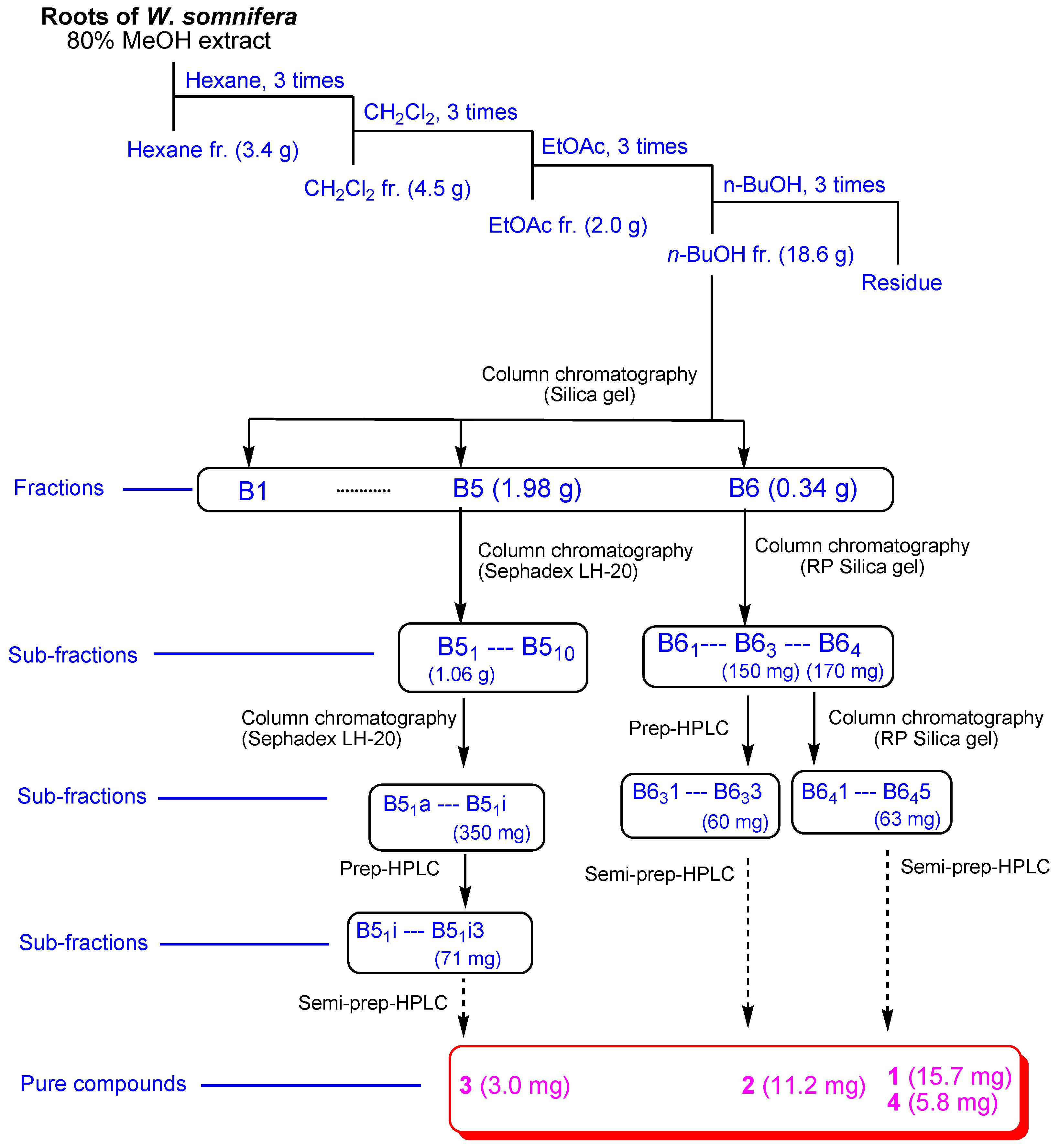
2.1. Isolation of Compounds 14

The roots of W. somnifera were extracted using 80% MeOH under reflux to provide the crude MeOH extract followed by the rotary evaporation. The methanolic extract was sequentially applied to the solvent partition process by four solvents, namely, hexane, dichloromethane, ethyl acetate, and n-butanol, yielding four main solvent fractions (Figure 1). The LC/MS analysis of each fraction using reference to an in-house-built UV library database revealed that the n-butanol-soluble fraction contained withanolide glycosides. The phytochemical examination of the n-butanol fraction by application of repeated column chromatography and semi-preparative high-performance liquid chromatography (HPLC) (Figure 1) with the guidance of LC/MS analysis led to the separation of four withanolide glycosides (14) (Figure 2).

2.2. Structural Elucidation of the Isolated Compounds 14

Compound 1, obtained as a white amorphous powder, possessed the molecular formula of C40H62O16 deduced from positive high-resolution electrospray ionization mass spectroscopy (HR-ESIMS), which revealed a [M + Na]+ ion peak at m/z 821.3920 (calcd. for C40H62 NaO16, 821.3936). The infrared (IR) spectrum of 1 showed distinctive absorptions for the hydroxy (3439 cm−1) and α,β-unsaturated ketone (1705 cm−1) functional groups. The 1H NMR data (Table 1) of compound 1, assigned by the aid of heteronuclear single quantum correlation (HSQC) experiment, showed the presence of signals for five methyls (δH 0.89 (3H, s), 1.02 (3H, s), 1.29 (3H, s), 1.87 (3H, s), and 2.01 (3H, s)), four oxygenated methines (δH 3.75 (1H, t, J = 4.0 Hz), 3.86 (1H, br s), 4.09 (1H, m), and 4.27 (1H, dd, J = 13.5, 3.5 Hz)), and one olefinic proton (δH 5.77 (1H, d, J = 4.0 Hz)) as well as two indicative anomeric protons (δH 4.40 (1H, d, J = 8.0 Hz), 4.41 (1H, d, J = 8.0 Hz) for sugar moieties. The 13C NMR data (Table 1) of 1, combined with heteronuclear multiple bond correlation (HMBC) experiment, revealed 40 carbon resonances, including 28 carbons for aglycone and 12 carbons for sugar units, where the carbon resonances assignable to the sugar units were typical of glucose [21]. Comprehensive inspection of the NMR data revealed that the NMR data of 1 was similar to those of withanoside VII, previously isolated from W. somnifera roots [22], but the apparent differences between the structures of 1 and withanoside VII were detected in the side chain due to the discrepancy of NMR data corresponding to C-20 and C-21.
The different partial structure of 1 was characterized by the analysis of the key HMBC correlations from H3-21 (δH 1.29)/C-17 (δC 54.3), C-20 (δC 74.9), and C-22 (δC 81.2) (Figure 3). Importantly, the linkage positions of the two glucoses were confirmed by the key COSY correlations from H-1 to H2-4, and key HMBC correlations of H-1′/C-3, and H-1″/C-6′ (Figure 3). Finally, the complete planar structure of 1 was elucidated by analysis of COSY and HMBC experiments (Figure 3).
The stereochemistry of 1 was established by the correlations obtained from the rotating frame Overhauser effect spectroscopy (ROESY) experiment, vicinal proton coupling constants observed in the 1H NMR specrtum, and electronic circular dichroism (ECD) data. The α-position of the hydroxy groups at C-1 and C-7 was determined by the ROESY correlations of H3-19/H-1 and H-4β, and H-8/H-7, H3-18, and H3-19 (Figure 4). The ECD spectrum of 1 showed a positive Cotton effect at 255 nm oriented from the n → π* transition of the α,β-unsaturated δ-lactone [18,23], which unambiguously confirmed a 22R-configuration. The configuration of C-22 was also supported by the typical coupling constants (J = 13.5 and 3.5 Hz) of H-22 showing a doublet of doublets [18,23]. In addition, almost the same 13C NMR chemical shifts observed for C-20 and the carbons of D ring and lactone ring of 1 to the related known withanolides, including withasilolide A [18], 20β-hydroxy-1-oxo-(22R)-witha-2,5,24-trienolide [24], withacoagin, [25], dunawithanine B [26], withacoagulin E [27], and 1α,3β,20αF-trihydroxy-20R,22R-witha-5,24-dienolide [28]. Suggested that the configuration of C-20 of 1 is the same as that of the related compounds. Finally, to identify the absolute configuration of sugar units, enzymatic hydrolysis of 1 using glucosidase was carried out, which afforded aglycone 1a and the sugar moieties from 1. The absolute configuration of two glucoses of 1 was concluded as D-configuration by LC/MS-based analysis after their thiocarbamoyl–thiazolidine derivatization [29]. The typical coupling constant (J = 8.0 Hz) of the anomeric protons was characteristic of β-form in glucopyranose [30], which demonstrated that both the sugar units of 1 were β-d-glucopyranoses. Therefore, the chemical structure of 1 was elucidated as shown in Figure 1, and compound 1 was named withanoside XII.
Compound 1a, obtained as an aglycone of 1 by enzymatic hydrolysis, possessed the molecular formula of C28H42O6 confirmed by HRESIMS, which showed molecular ion peak at m/z 473.2904 [M − H] (calcd. for C28H41O6, 473.2903) in the negative-ion mode. The NMR data of 1a showed clearly similar signals to those of compound 1, without signals responsible for the sugar moieties. The up-field shifted signals (δH 3.91/δC 65.1) for C-3 of 1a indicated the absence of the sugar units at C-3. Likewise, the planar structure of 1a was further confirmed by the interpretation of COSY and HMBC experiments (Figure 5). The observed ROESY correlations and a positive Cotton effect at 251 nm in the ECD spectrum of 1a confirmed the same stereochemistry of 1a to that of 1. Accordingly, the structure of 1a was also assigned as a new compound, as illustrated in Figure 5 and compound 1a, was named withanoside XIIa.
The known compounds were identified as withagenin A diglucoside (2) [31], withanoside II (3) [22], and withanoside IV (4) [22] by comparing their physical features and NMR spectroscopic data with those previously reported, and the data from LC/MS analysis. To the best of our knowledge, the compound 2 (withagenin A diglucoside) has not been isolated as a natural product, and the structure of withagenin A diglucoside (2) was previously proposed by HRMS and MS/MS spectral data without NMR experiment [31]. Compound 2 was isolated as a white amorphous powder, and the molecular formula of 2 was deduced as C40H62O17 from the (−)–HRESIMS data showing a [M − H] ion peak at m/z 813.3929 (calcd. for C40H61O17, 813.3909). Detailed 1H NMR data analysis of 2 revealed that the 1H NMR data of 2 was almost identical to those of withanoside II (3) [22], isolated in this study, with an apparent difference of one hydroxylated methylene group [δH 4.34 (1H, d, J = 12.0 Hz, 27-Ha) and 4.40 (1H, d, J = 12.0 Hz, 27-Hb)] of C-27 in 1. Based on the NMR data, and the molecular formula (C40H62O17) determined by (−)–HRESIMS data, the compound 2 was unambiguously confirmed as withagenin A diglucoside, which was also supported by the comparable UV and ECD data of 2 to withanoside II (3) [22]. Here, we reported the physical and spectroscopic data of withagenin A diglucoside (2) for the first time.

2.3. Evaluation of Biological Activity of the Isolated Compounds

Helicobacter pylori is a major health problem worldwide, affecting approximately 50% of the global population [32,33]. In our anti-H. pylori activity assay, we found that the crude MeOH extract of W. somnifera roots showed weak antibacterial activity with 22.4% inhibition against H. pylori strain 51. Several previous studies have reported anti-bacterial activity of W. somnifera, and withaferin A, a major withanolide from this plant is known to inhibit H. pylori-induced inflammation [34,35,36,37]. Thus, the isolated withanolide glycosides (14) were evaluated for anti-H. pylori activity. Among the isolates, compound 2 exhibited weak anti-H. pylori activity with 7.8% growth inhibition against H. pylori strain 51, and the other compounds failed to show inhibitory activity. An epoxide group at C-6/C-7 and the hydroxyl groups at C-5 and C-27 may play a role in anti-H. pylori activity of compound 2, compared with the structures of inactive compounds.
Anti-oxidant activity has been known to be related to various human diseases including cancer and inflammation, and the methanolic extract of W. somnifera has been reported to show anti-oxidant activity [36]. In this study, the crude MeOH extract of W. somnifera roots showed significant ABTS radical scavenging activity with 24.3 ± 4.9 μmol Trolox equivalent (TE)/g·DW (dry weight). Thus, the isolated compounds 14 were evaluated for anti-oxidant activity. Although the activity of 14 was lower than the extract, all the compounds also displayed significant anti-oxidant activity, and the TE values of 14 were 10.5 ± 3.2, 13.1 ± 2.1, 9.4 ± 1.8, and 11.6 ± 3.9 μmol TE/g·DW, respectively. Significant differences in the activity were not found among the isolated compounds.
In addition, the anti-inflammatory activity of the isolates was evaluated because ethanolic extract of W. somnifera root, withaferin A, and phenolic compounds from this plant have been reported to have anti-inflammatory activity [19,38]. However, all the isolates (14) failed to show inhibitory activity against lipopolysaccharide-induced nitric oxide (NO) production in RAW 264.7 murine macrophage cells.

3. Materials and Methods

3.1. General Experimental Procedure and Plant Material

Detailed information on the general experimental procedure and identification of plant material are provided in the Supplementary Materials.

3.2. Extraction and Separation of the Compounds

Dried roots of W. somnifera (1.28 kg) were extracted using 80% aqueous MeOH (each 3.0 L for 3 days) under reflux three times and filtered at room temperature. After the filtrate was concentrated through a rotavapor, the methanolic extract (189.6 g) was suspended in water (700 mL) and then partitioned with each 700 mL of hexane, dichloromethane (CH2Cl2), ethyl acetate (EtOAc), and n-butanol (n-BuOH). Four fractions were provided in corresponding order: hexane (3.4 g), CH2Cl2 (4.5 g), EtOAc (2.0 g), and n-BuOH soluble (18.6 g) fractions. Four fractions from the solvent partitioning were subjected to LC/MS analysis combined with the reference of an in-house-built UV spectra library, which indicated the presence of withanolide glycosides in the n-BuOH fraction since some peaks in the n-BuOH fraction exhibited the UV pattern (λmax 200–230 nm) related to that reported for withanolides [18] and the molecular ion peaks ranging m/z 780–820.
The n-BuOH soluble fraction (18.6 g) was applied to silica gel column chromatography (150 g, eluted with CH2Cl2/MeOH (30:1 → 1:1) to give six fractions (B1–B6)). Fraction B5 (1.98 g) was subjected to Sephadex LH-20 column chromatography (100% MeOH) to yield ten subfractions (B51–B510). Subfraction B51 (1.06 g) was separated by Sephadex LH-20 column chromatography again to yield nine subfractions (B51a–B51i). Subfraction B51i (350 mg) was further separated through preparative HPLC (65% MeOH → 80% MeOH, gradient solvent system) to give three subfractions (B51i1–B51i3). Subfraction B51i3 (71 mg) was purified using semi-preparative HPLC (66% MeOH) to yield compound 3 (tR 35.3 min, 3.0 mg). Fraction B6 (340 mg) was subjected to reverse-phase (RP) silica gel column chromatography (40% MeOH → 100% MeOH, gradient solvent system) twice, yielding 4 subfractions (B61–B64), and subfraction B63 (150 mg) was applied to preparative HPLC (65% MeOH → 80% MeOH, gradient solvent system) to give three subfractions (B631–B633). Subfraction B633 (60 mg) was separated using semi-preparative HPLC (49% MeOH) to give compound 2 (tR 60.0, 11.2 mg). Subfraction B64 (170 mg) was fractionated by RP silica gel column chromatography (40% MeOH → 100% MeOH, gradient solvent system) to yield five subfractions (B641–B645). Finally, subfraction B645 (63 mg) was purified by semi-preparative HPLC (53% MeOH) to give compounds 4 (tR 30.0, 5.8 mg) and 1 (tR 34.8, 15.7 mg).

3.2.1. Withanoside XII (1)

White amorphous powder; ( α ) D 25   −24.9 (c 0.78, MeOH); UV (MeOH) λmax (log ε) 194 (2.8) nm; IR (KBr) νmax 3439, 2921, 1705, 1643, 1411, and 1034 cm−1; ECD (MeOH) λmaxε) 221 (+0.37), 255 (+16.5) nm; 1H (850 MHz) and 13C NMR (212.5 MHz), see Table 1; (+)–ESIMS m/z 821 [M + Na]+; (+)–HRESIMS m/z 821.3920 [M + Na]+ (calcd. for C40H62 NaO16, 821.3936).

3.2.2. Withagenin A Diglucoside (2)

White amorphous powder; ( α ) D 25   −18.4 (c 0.55, MeOH); UV (MeOH) λmax (log ε) 228 (3.8) nm; IR (KBr) νmax 3435, 2931, 1698, 1645, 1387, and 1044 cm−1; ECD (MeOH) λmax (Δε) 250 (+5.5) nm; 1H NMR (850 MHz CD3OD) δ: 0.85, 0.90, 2.13 (3H each, all s, 18, 19, 28-H3), 1.06 (3H, d, J = 7.0 Hz, 21-H3), 2.95 (1H, d, J = 4.0 Hz, 6-H), 3.25 (1H, dd-like, 7-H), 3.72 (1H, t, J = 3.0 Hz, 1-H), 4.34 (1H, d, J = 12.0 Hz, 27-Ha), 4.40 (1H, d, J = 12.0 Hz, 27-Hb), 4.43 (1H, d, J = 8.0 Hz, 1″-H), 4.45 (1H, d, J = 8.0 Hz, 1-H), 4.47 (1H, overlap, 3-H), and 4.50 (1H, m, 22-H); (−)–ESIMS m/z 813 [M − H].

3.3. Enzymatic Hydrolysis and Absolute Configuration Determination of the Sugar Moieties of Compound 1

The absolute configurations of the sugar moieties were determined using the method previously described [24] with minor modifications. Briefly, compound 1 (1.0 mg) was hydrolyzed with crude glucosidase (10 mg, from almonds, Sigma-Aldrich, St. Louis, MO, USA) for 72 h at 37 °C, and CH2Cl2 was used for the extraction of aglycone. The CH2Cl2 fraction was confirmed to contain the aglycone 1a by LC/MS analysis. Detailed description for absolute configuration determination of the sugar moieties in the aqueous layer is provided in the Supplementary Materials.

Withanoside XIIa (1a)

White amorphous powder; ( α ) D 25   −46.5 (c 0.03, MeOH); UV (MeOH) λmax (log ε) 200 (2.6) nm; IR (KBr) νmax 3715, 2940, 2834, 1698, 1555, 1112, and 1025 cm−1; ECD (MeOH) λmax (Δε) 219 (+8.5), 251 (+17.3) nm; 1H (850 MHz) and 13C NMR (212.5 MHz), see the Table 1; (−)–ESIMS m/z 519 [M + HCOOH]; (−)–HRESIMS m/z 473.2904 [M − H] (calcd. for C28H41O6, 473.2903).

3.4. Anti-Helicobacter pylori Activity

The anti-H. pylori activity was evaluated using a previously described method [39]. The detailed information on the experimental procedure is included in the Supplementary Materials.

3.5. Antioxidant Activity Test with ABTS Radical

The ABTS radical was generated using a previously reported method [40]. Each sample (20 μL) was reacted with 180 μL of the ABTS•+ solution at room temperature, and the absorbance was measured at 734 nm after 10 min. The antioxidant activity of each sample was expressed as Trolox (Sigma, St. Louis, MO, USA) equivalents per gram (μmol TE/g).

3.6. Anti-Inflammatory Activity

The anti-inflammatory activity was evaluated using our previously reported method [19]. The detailed information on the experimental procedure is included in the Supplementary Materials.

3.7. Statistical Analysis

All of the contents and the antioxidant activities are expressed as the means ± standard deviations (SDs) of triplicate determinations. The differences among samples were statistically evaluated via one-way analysis of variance (ANOVA). The values were evaluated at the 5% significance level using two-sided tests. Pearson’s correlation coefficients were obtained using IBM SPSS Statistics 24.0 software (Armonk, NY, USA).

4. Conclusions

In conclusion, we isolated and characterized four withanolide glycosides (14), including one new compound, withanoside XII (1), in the n-BuOH fraction of the MeOH extracts of W. somnifera root via the LC/MS-based analysis [41]. The structure of withanoside XII was established by 1D and 2D NMR spectroscopic methods, HR-ESIMS, and ECD data as well as enzymatic hydrolysis followed by LC/MS analysis. We also characterized the new compound, withanoside XIIa (1a), as an aglycone of 1, obtained by enzymatic hydrolysis of 1. The physical and spectroscopic data of withagenin A diglucoside (2) were reported in this study for the first time. In the anti-H. pylori activity assay, compound 2 showed weak anti-H. pylori activity with 7.8% inhibition. In addition, all the isolated withanolide glycosides showed significant anti-oxidant activity in the ABTS radical scavenging assay. The present study provided the experimental support that W. somnifera root is rich in withanolides, and it can be a valuable natural resource for bioactive withanolides.

Supplementary Materials

The following supporting information can be downloaded at: https://www.mdpi.com/article/10.3390/plants11060767/s1, Figure S1: The HR-ESIMS data of 1; Figure S2: The 1H NMR spectrum of 1 (CD3OD, 850 MHz); Figure S3: The 1H-1H COSY spectrum of 1 (CD3OD); Figure S4: The HSQC spectrum of 1 (CD3OD); Figure S5: The HMBC spectrum of 1 (CD3OD); Figure S6: The ROESY spectrum of 1 (CD3OD); Figure S7: The ECD spectrum of 1; Figure S8: The HR-ESIMS data of 1a; Figure S9: The 1H NMR spectrum of 1a (CD3OD, 850 MHz); Figure S10: The 1H-1H COSY spectrum of 1a (CD3OD); Figure S11: The HSQC spectrum of 1a (CD3OD); Figure S12: The HMBC spectrum of 1a (CD3OD); Figure S13: The ROESY spectrum of 1a (CD3OD); Figure S14: The ECD spectrum of 1a; Figure S15: The HR-ESIMS data of 2; Figure S16: The 1H NMR spectrum of 2 (CD3OD, 850 MHz); General experimental procedure.

Author Contributions

Conceptualization, J.K.K. and K.H.K.; formal analysis, J.W.H., J.S.Y., B.S.L., D.-M.K. and M.-J.A.; investigation, J.W.H. and D.-M.K.; writing—original draft preparation, J.W.H. and K.H.K.; writing—review and editing, K.H.K.; visualization, J.W.H., J.S.Y. and B.S.L.; supervision, J.K.K. and K.H.K.; project administration, K.H.K.; funding acquisition, K.H.K. All authors have read and agreed to the published version of the manuscript.

Funding

This work was supported by a National Research Foundation of Korea (NRF) grant funded by the Korean government (MSIT; grant numbers: 2019R1A5A2027340 and 2021R1A2C2007937).

Institutional Review Board Statement

Not applicable.

Informed Consent Statement

Not applicable.

Data Availability Statement

Not applicable.

Conflicts of Interest

The authors declare no conflict of interest.

References

  1. Kulkarni, S.; Dhir, A. Withania somnifera: An Indian ginseng. Prog. Neuropsychopharmacol. Biol. Psychiat. 2008, 32, 1093–1105. [Google Scholar] [CrossRef]
  2. Pandey, V.; Ansari, W.A. Withania somnifera: Advances and implementation of molecular and tissue culture techniques to enhance its application. Front. Plant Sci. 2017, 8, 1390. [Google Scholar] [CrossRef] [PubMed] [Green Version]
  3. Glotter, E. Withanolides and related ergostane-type steroids. Nat. Prod. Rep. 1991, 8, 415–440. [Google Scholar] [CrossRef] [PubMed]
  4. Chakraborti, S.; De Barun, K.; Bandyopadhyay, T. Variations in the antitumor constituents of Withania somnifera Dunal. Experientia 1974, 30, 852–853. [Google Scholar] [CrossRef] [PubMed]
  5. Minguzzi, S.; Barata, E.S.L. Cytotoxic withanolides from Achnistus arborescens. Phytochemistry 2002, 59, 635–641. [Google Scholar] [CrossRef]
  6. Zhao, J.; Nakamura, N. Withanolides derivatives from the roots of Withania somnifera and their neurite outgrowth activities. Chem. Pharm. Bull. 2002, 50, 760–765. [Google Scholar] [CrossRef] [Green Version]
  7. Zhang, H.; Cao, C.-M.; Gallagher, R.J.; Timmermann, B.N. Antiproliferative withanolides from several solanaceous species. Nat. Prod. Res. 2014, 28, 1941–1951. [Google Scholar] [CrossRef] [PubMed] [Green Version]
  8. Ma, T.; Zhang, W.-N.; Yang, L.; Zhang, C.; Lin, R.; Shan, S.-M.; Zhu, M.-D.; Luo, J.-G.; Kong, L.-Y. Cytotoxic withanolides from Physalis angulate var. villosa and the apoptosis-inducing effect via ROS generation and the activation of MAPK in human osteosarcoma cells. RSC Adv. 2016, 6, 53089–53100. [Google Scholar] [CrossRef]
  9. Tomar, V.; Beuerle, T. A validated HPTLC method for the simultaneous quantifications of three phenolic acids and three withanolides from Withania somnifera plants and its herbal products. J. Chromatogr. B 2019, 1124, 154–160. [Google Scholar] [CrossRef] [PubMed]
  10. Girme, A.; Verma, M.K. Investigating 11 Withanosides and Withanolides by UHPLC-PDA and Mass Fragmentation Studies from Ashwagandha (Withania somnifera). ACS Omega 2020, 5, 27933–27943. [Google Scholar] [CrossRef]
  11. Saroya, A.S.; Singh, J. Neuropharmacology of Nardostachys jatamansi DC. In Pharmacotherapeutic Potential of Natural Products in Neurological Disorders; Springer: Singapore, 2018; pp. 167–174. [Google Scholar]
  12. Chikhale, R.V.; Gurav, S.S.; Patil, R.B.; Sinha, S.K.; Prasad, S.K.; Shakya, A.; Shrivastava, S.K.; Gurav, N.S.; Prasad, R.S. SARS-CoV-2 host entry and replication inhibitors from Indian ginseng: An in-silico approach. J. Biomol. Struct. Dyn. 2021, 39, 4510–4521. [Google Scholar] [CrossRef]
  13. Tripathi, M.K.; Singh, P.; Sharma, S.; Singh, T.P.; Ethayathulla, A.S.; Kaur, P. Identification of bioactive molecule from Withania somnifera (Ashwagandha) as SARS-CoV-2 main protease inhibitor. J. Biomol. Struct. Dyn. 2021, 39, 5668–5681. [Google Scholar] [CrossRef]
  14. Lee, S.R.; Kang, H.S.; Yoo, M.J.; Yi, S.A.; Beemelmanns, C.; Lee, J.C.; Kim, K.H. Anti-adipogenic Pregnane Steroid from a Hydractinia-associated Fungus, Cladosporium sphaerospermum SW67. Nat. Prod. Sci. 2020, 26, 230–235. [Google Scholar]
  15. Lee, S.; Ryoo, R.; Choi, J.H.; Kim, J.H.; Kim, S.H.; Kim, K.H. Trichothecene and tremulane sesquiterpenes from a hallucinogenic mushroom Gymnopilus junonius and their cytotoxicity. Arch. Pharm. Res. 2020, 43, 214–223. [Google Scholar] [CrossRef]
  16. Ha, J.W.; Kim, J.; Kim, H.; Jang, W.; Kim, K.H. Mushrooms: An Important Source of Natural Bioactive Compounds. Nat. Prod. Sci. 2020, 26, 118–131. [Google Scholar]
  17. Yu, J.S.; Park, M.; Pang, C.; Rashan, L.; Jung, W.H.; Kim, K.H. Antifungal phenols from Woodfordia uniflora Collected in Oman. J. Nat. Prod. 2020, 83, 2261–2268. [Google Scholar] [CrossRef]
  18. Kim, S.; Yu, J.S.; Lee, J.Y.; Choi, S.U.; Lee, J.; Kim, K.H. Cytotoxic withanolides from the roots of Indian ginseng (Withania somnifera). J. Nat. Prod. 2019, 82, 765–773. [Google Scholar] [CrossRef]
  19. Baek, S.C.; Lee, S.; Kim, S.; Jo, M.S.; Yu, J.S.; Ko, Y.J.; Cho, Y.C.; Kim, K.H. Withaninsams A and B: Phenylpropanoid esters from the roots of Indian ginseng (Withania somnifera). Plants 2019, 8, 527. [Google Scholar] [CrossRef] [Green Version]
  20. Lee, B.S.; Yoo, M.J.; Kang, H.; Lee, S.R.; Kim, S.; Yu, J.S.; Kim, J.C.; Jang, T.S.; Pang, C.; Kim, K.H. Withasomniferol D, a New Anti-adipogenic Withanolide from the Roots of Ashwagandha (Withania somnifera). Pharmaceuticals 2021, 14, 1017. [Google Scholar] [CrossRef]
  21. Harinantenaina, L.R.R.; Kasai, R.; Yamasaki, K. Ent-kaurane Diterpenoid Glycosides from the Leaves of Cussonia racemosa, a Malagasy Endemic Plant. Chem. Pharm. Bull. 2002, 50, 268–271. [Google Scholar] [CrossRef] [Green Version]
  22. Matsuda, H.; Murakami, T.; Kishi, A.; Yoshikawa, M. Structures of withanosides I, II, III, IV, V, VI, and VII, new withanolide glycosides, from the roots of Indian Withania somnifera Dunal. and inhibitory activity for tachyphylaxis to clonidine in isolated guinea-pig ileum. Bioorg. Med. Chem. 2001, 9, 1499–1507. [Google Scholar] [CrossRef]
  23. Xu, Y.M.; Wijeratne, E.M.K.; Brooks, A.D.; Tewary, P.; Xuan, L.J.; Wang, W.Q.; Sayers, T.J.; Gunatilaka, A.A.L. Cytotoxic and other withanolides from aeroponically grown Physalis philadelphica. Phytochemistry 2018, 152, 174–181. [Google Scholar] [CrossRef]
  24. Atta-ur-Rahman; Dur-e-Shahwar; Naz, A.; Choudhary, M.I. Withanolides from Withania coagulans. Phytochemistry 2003, 63, 387–390. [Google Scholar]
  25. Neogi, P.; Kawai, M.; Butsugan, Y.; Mori, Y.; Suzuki, M. Withacoagin, a new withanolide from Withania coagulans roots. Bull. Chem. Soc. Jpn. 1988, 61, 4479–4481. [Google Scholar] [CrossRef] [Green Version]
  26. Adam, G.; Chien, N.Q.; Nguyen, H.K. Dunawithanine A and B, first plant withanolide glycosides from Dunalia australis. Naturwissenschaften 1981, 68, 425–426. [Google Scholar] [CrossRef]
  27. Huang, C.-F.; Ma, L.; Sun, L.-J.; Ali, M.; Arfan, M.; Liu, J.-W.; Hu, L.-H. Immunosuppressive withanolides from Withania coagulans. Chem. Biodivers. 2009, 6, 1415–1426. [Google Scholar] [CrossRef]
  28. Velde, V.V.; Lavie, D. New withanolides of biogenetic interest from Withania somnifera. Phytochemistry 1981, 20, 1359–1364. [Google Scholar] [CrossRef]
  29. Tanaka, T.; Nakashima, T.; Ueda, T.; Tomii, K.; Kouno, I. Facile Discrimination of Aldose Enantiomers by Reversed-Phase HPLC. Chem. Pharm. Bull. 2007, 55, 899–901. [Google Scholar] [CrossRef] [Green Version]
  30. Coxon, B. Two-Dimensional J-Resolved Proton Nuclear Magnetic Resonance Spectrometry of Hydroxyl-Coupled α- and β-d-Glucose. Anal. Chem. 1983, 55, 2361–2366. [Google Scholar] [CrossRef]
  31. Bolleddula, J. Identification of metabolites in Withania sominfera fruits by liquid chromatography and high-resolution mass spectrometry. Rapid Commun. Mass Spectrom. 2012, 26, 1277–1290. [Google Scholar] [CrossRef]
  32. McGee, D.J.; George, A.E.; Trainor, E.A.; Horton, K.E.; Hildebrandt, E.; Testerman, T.L. Cholesterol enhances Helicobacter pylori resistance to antibiotics and LL-37. Antimicrob. Agents Chemother. 2011, 55, 2897–2904. [Google Scholar] [CrossRef] [Green Version]
  33. Kang, H.; Lim, J.W.; Kim, H. Inhibitory effect of Korean Red Ginseng extract on DNA damage response and apoptosis in Helicobacter pylori-infected gastric epithelial cells. J. Ginseng Res. 2020, 44, 79–85. [Google Scholar] [CrossRef]
  34. Bisht, P.; Rawat, V. Antibacterial activity of Withania somnifera against Gram-positive isolates from pus samples. AYU 2014, 35, 330–332. [Google Scholar]
  35. Owais, M.; Sharad, K.S.; Shehbaz, A.; Saleemuddin, M. Antibacterial efficacy of Withania somnifera (ashwagandha) an indigenous medicinal plant against experimental murin salmonellosis. Phytomedicine 2005, 12, 229–235. [Google Scholar] [CrossRef]
  36. Alam, N.; Hossain, M.; Mottalib, M.A.; Sulaiman, S.A.; Gan, S.H.; Khalil, M.I. Methanolic extracts of Withania somnifera leaves, fruits and roots possess antioxidant properties and antibacterial activities. BMC Complement. Altern. Med. 2012, 12, 175. [Google Scholar] [CrossRef] [Green Version]
  37. Kim, G.; Kim, T.-H.; Kang, M.-J.; Choi, J.-A.; Pack, D.-Y.; Lee, I.-R.; Kim, M.-G.; Han, S.-S.; Kim, B.-Y.; Oh, S.-M.; et al. Inhibitory effect of witaferin A on Helicobacter pylori-induced IL-8 production and NF-κB activation in gastric epithelial cells. Mol. Med. Rep. 2016, 13, 967–972. [Google Scholar] [CrossRef]
  38. Singh, D.; Aggarwal, A.; Maurya, R.; Naik, S. Withania somnifera inhibits NF-kappaB and AP-1 transcription factors in human peripheral blood and synovial fluid mononuclear cells. Phytother. Res. 2007, 21, 905–913. [Google Scholar] [CrossRef]
  39. Khalil, A.A.K.; Park, W.S.; Lee, J.; Kim, H.J.; Akter, K.M.; Goo, Y.M. A new anti-Helicobacter pylori juglone from Reynoutria japonica. Arch. Pharmacal Res. 2019, 42, 505–511. [Google Scholar] [CrossRef]
  40. Park, W.S.; Kim, H.-J.; Li, M.; Lim, D.H.; Kim, J.; Kwak, S.-S.; Kang, C.-M.; Ferruzzi, M.G.; Ahn, M.-J. Two Classes of Pigments, Carotenoids and C-Phycocyanin, in Spirulina Powder and Their Antioxidant Activities. Molecules 2018, 23, 2065. [Google Scholar] [CrossRef] [Green Version]
  41. Lee, K.H.; Kim, J.K.; Yu, J.S.; Jeong, S.Y.; Choi, J.H.; Kim, J.C.; Ko, Y.J.; Kim, S.H.; Kim, K.H. Ginkwanghols A and B, osteogenic coumaric acid-aliphatic alcohol hybrids from the leaves of Ginkgo biloba. Arch. Pharmacal Res. 2021, 44, 514–524. [Google Scholar] [CrossRef]
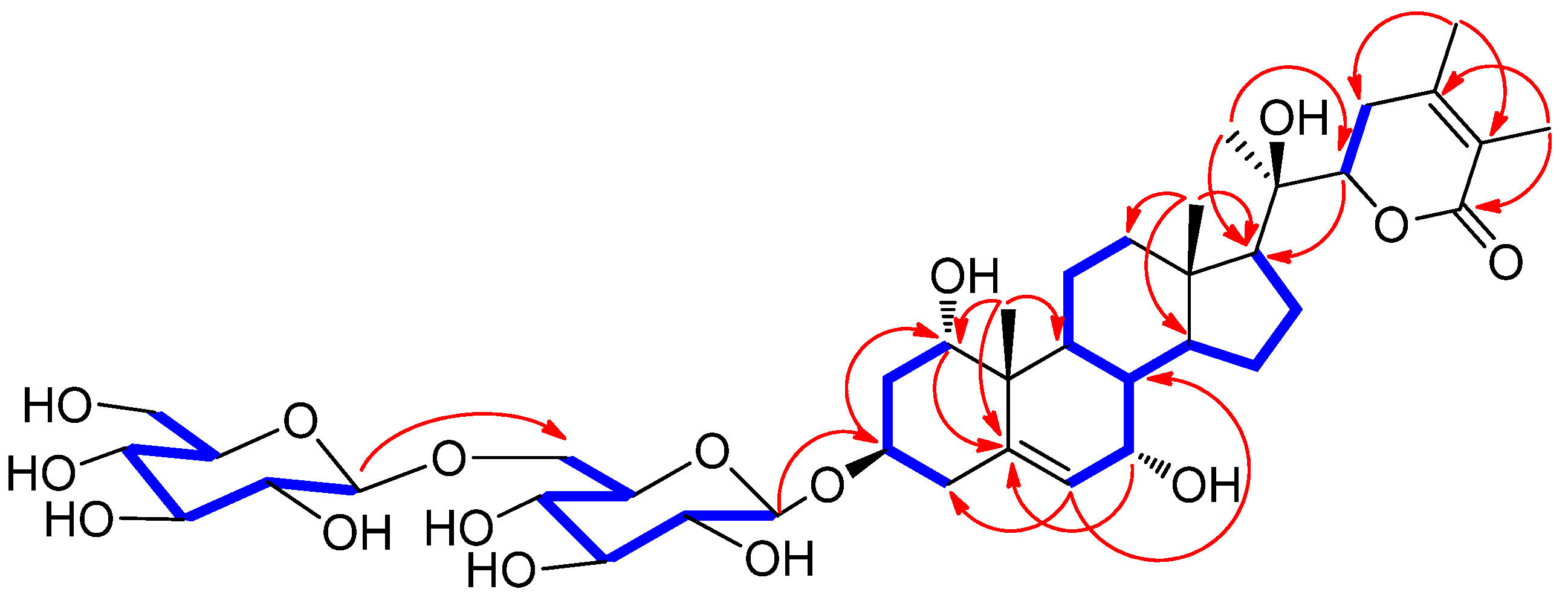
Figure 1. The separation scheme of compounds 14.
Plants 11 00767 g001
Figure 2. Chemical structures of compounds 14.
Plants 11 00767 g002
Figure 3. Key 1H-1H COSY ( Plants 11 00767 i001) and HMBC ( Plants 11 00767 i002) correlations for 1.
Plants 11 00767 g003
Figure 4. Key ROESY correlations for 1.
Plants 11 00767 g004
Figure 5. Key 1H-1H COSY ( Plants 11 00767 i001) and HMBC ( Plants 11 00767 i002) correlations for 1a.
Plants 11 00767 g005
Table 1. 1H (850 MHz) and 13C NMR (212.5 MHz) data of compounds 1 and 1a in CD3OD (δ in ppm) a.
Position11a
δH (J in Hz)δCδH (J in Hz)δC
13.86 dd (1.5, 1.5)72.2 d3.82 dd (1.0, 1.0)72.0 d
2α2.55 ddd (13.0, 5.5, 1.5)37.7 t2.01 ddd (13.0, 5.5, 1.0)37.6 t
2β2.40 ddd (13.0, 12.5, 1.5) 1.73 ddd (13.0, 12.5, 1.0)
34.09 dddd (12.5, 12.5, 5.5, 5.5)73.8 d3.91 dddd (12.5, 12.5, 5.5, 5.5)65.1 d
4α2.23 dd (13.0, 5.5)36.1 t2.36 dd (13.0, 5.5)40.9 t
4β1.85 dd (13.0, 12.5) 2.32 dd (13.0, 12.5)
5 142.7 s 142.9 s
65.77 d (4.0)126.2 d5.72 d (4.0)126.0 d
73.75 t (4.0)64.4 d3.72 t (4.0)64.5 d
81.44 m37.0 d1.46 m37.0 d
91.92 m33.4 d1.92 m33.8 d
10 41.8 s 41.6 s
11α1.57 m19.5 t1.51 m19.3 t
11β1.52 m 1.56 m
12α1.37 m39.2 t2.00 m39.3 t
12β2.01 m 1.36 m
13 42.3 s 42.1 s
141.57 m49.6 d1.57 m49.8 d
15α1.86 m23.1 t1.19 m22.3 t
15β1.22 m 1.84 m
16α1.70 m21.5 t1.69 m21.3 t
16β1.91 m 1.91 m
171.82 m54.3 d1.82 m54.1 d
180.89 s12.6 q0.89 s12.6 q
191.02 s17.5 q1.02 s17.3 q
20 74.9 s 75.0 s
211.29 s19.5 q1.28 s19.3 q
224.27 dd (13.5, 3.5)81.2 d4.25 dd (13.5, 3.5)81.0 d
23α2.32 m30.8 t2.32 m30.8 t
23β2.56 m 2.54 t (15.0)
24 151.5 s 151.2 s
25 120.6 s 120.6 s
26 167.8 s 167.6 s
271.87 s11.1 q1.87 s10.8 q
282.01 s19.2 q2.00 s19.1 q
1′4.40 d (8.0)101.8 d
2′3.18 dd (9.0, 8.0)73.8 d
3′3.38 overlap76.4 d
4′3.28 overlap70.3 d
5′3.49 overlap75.6 d
6′4.14 dd (12.0, 2.0);
3.80 dd (12.0, 6.0)
68.5 t
1″4.41 d (8.0)103.5 d
2″3.23 (9.0, 8.0)73.8 d
3″3.36 overlap76.4 d
4″3.30 overlap70.0 d
5″3.28 overlap76.5 d
6″3.88 dd (12.0, 2.0);
3.69 dd (12.0, 5.5)
61.3 t
a J values are in parentheses (shown in Hz); 13C NMR assignments were based on HSQC and HMBC experiments.
Publisher’s Note: MDPI stays neutral with regard to jurisdictional claims in published maps and institutional affiliations.

