Article

Silver Nanoparticle Production Mediated by Vitis vinifera Cane Extract: Characterization and Antibacterial Activity Evaluation

Department of Biotechnology, Faculty of Food and Biochemical Technology, UCT Prague, Technická 5 Dejvice, Praha 6, 166 28 Prague, Czech Republic
*
Author to whom correspondence should be addressed.
Plants 2022, 11(3), 443; https://doi.org/10.3390/plants11030443
Submission received: 30 December 2021 / Revised: 31 January 2022 / Accepted: 2 February 2022 / Published: 5 February 2022
(This article belongs to the Special Issue Biological Activities of Plant Extracts)

Abstract

:
The ever-growing range of possible applications of nanoparticles requires their mass production. However, there are problems resulting from the prevalent methods of nanoparticle production; physico-chemical routes of nanoparticle synthesis are not very environmentally friendly nor cost-effective. Due to this, the scientific community started exploring new methods of nanoparticle assembly with the aid of biological agents. In this study, ethanolic Vitis vinifera cane extract combined with silver nitrate was used to produce silver nanoparticles. These were subsequently characterized using UV-visible (UV-Vis) spectrometry, transmission electron microscopy, and dynamic light-scattering analysis. The antimicrobial activity of produced nanoparticles was tested against the planktonic cells of five strains of Gram-negative bacterium Pseudomonas aeruginosa (PAO1, ATCC 10145, ATCC 15442, DBM 3081, and DBM 3777). After that, bactericidal activity was assessed using solid medium cultivation. In the end, nanoparticles’ inhibitory effect on adhering cells was analyzed by measuring changes in metabolic activity (3-(4,5-dimethylthiazol-2-yl)-2,5-diphenyltetrazolium bromide assay-MTT). Our results confirmed that ethanolic Vitis vinifera cane extract is capable of mediating silver nanoparticle production; synthesis was conducted using 10% of extract and 1 mM of silver nitrate. The silver nanoparticles’ Z-average was 68.2 d nm, and their zeta potential was –30.4 mV. These silver nanoparticles effectively inhibited planktonic cells of all P. aeruginosa strains in concentrations less than 5% v/v and inhibited biofilm formation in concentrations less than 6% v/v. Moreover, minimum bactericidal concentration was observed to be in the range of 10–16% v/v. According to the results in this study, the use of wine agriculture waste is an ecological and economical method for the production of silver nanoparticles exhibiting significant antimicrobial properties.

Graphical Abstract

1. Introduction

Nanoparticles rose to the forefront of scientific interest decades ago; however, their significance and potential is still being extended by new discoveries. Their unique physical and chemical properties, mainly caused by their high specific surface area, are already being used in a wide range of science and industry, from electronics or material design to food processing [1]. In recent years, though, another characteristic of nanoparticles started to become more and more important. With the rise of resistant pathogenic microorganisms, which cause infections not responding to traditional antibiotic therapy, there appeared a great need to find new ways of combating these once easily curable illnesses. Metal nanoparticles, in particular, have since been extensively studied for their antimicrobial properties against the dreaded resistant pathogenic microorganisms. For example, silver, gold, copper, platinum, and palladium nanoparticles have already proven their inhibiting effect on pathogenic bacteria such as Pseudomonas aeruginosa, Escherichia coli, Staphylococcus aureus (including methicillin resistant Staphylococcus aureus), Bacillus subtilis, and Streptococcus bovis [2].
Still, the ever-growing potential of metal nanoparticle applications foreshadows future problems with their mass production. Physico-chemical methods of nanoparticle manufacture are not cost-effective and can have dire environmental consequences due to the use of toxic solvents. The production of nanoparticles using biological agents, however, promises comparable yields and stability without wasting energy and damaging the environment [3]. Biomolecules such as proteins, amino acids, vitamins, reducing sugars, terpenoids, polyphenols, and organic acids act as reducing and stabilizing agents in this type of nanoparticle production. One of the proposed mechanisms in plant extract mediated synthesis involves polyphenols such as flavonoids or quercetin. The hydroxyl groups present in polyphenols reduce metal ions, themselves oxidizing to carbonyl groups. The resulting carbonyl groups subsequently electrostatically stabilize the nanoparticles [4]. In a study by Shameli et al. [5], silver nanoparticles (AgNPs) were synthesized using stem bark extract of Callicarpa maingayi. It was identified that aldehydes contained in the extract reduced silver ions to metallic AgNPs, forming complexes with amide and polypeptide groups, preventing the NPs from aggregation. The presence of biologically formed capping agents is not considered a hindrance, especially in the context of the antimicrobial effect; on the contrary, the capping molecules could have a synergistic effect with the action of nanoparticles. Moreover, the capping agents of biological origin could potentially lower the toxicity of nanoparticles for human cells [6].
In this regard, multiple different approaches were already employed and reported in the literature; live microorganisms, microbial lysates, plant tissue, and even plant extracts were successfully used as a source of reducing and stabilizing molecules for nanoparticle production [7,8,9,10,11]. When it comes to the microbial production of nanoparticles, Bacillus cereus, Escherichia coli, and Pseudomonas proteolytica have proven their ability to form silver nanoparticles, whereas Shewanella alga and Rhodopseudomonas capsulate were able to produce gold nanoparticles [12,13,14]. In regard to the use of plant extracts, Acalypha indica, Boswellia serrata, Cinnamon zeylanicum, and Withania somnifera were used for the production of silver nanoparticles [15,16,17,18]. In a study by Gnanajobitha et al. [19], Vitis vinifera fruit extract was used for the synthesis of AgNPs. In another study by Saratale et al. [20], tannin extracted from grape pomace was used for the mediation of silver nanoparticle production. A variety of different parts of plants are used in the literature: leaves, flowers, seeds, roots, fruits, stems, and peels [21]. The most industrially feasible solution, however, seems to be the use of extracts made from dry plant material, which can be easily stored. Moreover, this material should preferably not have any other major purpose (e.g., agricultural waste).
In this study, we observed the antimicrobial activity of AgNPs, produced using V. vinifera extract made from agricultural waste and silver nitrate solution, against the planktonic cells of Pseudomonas aeruginosa.

2. Results

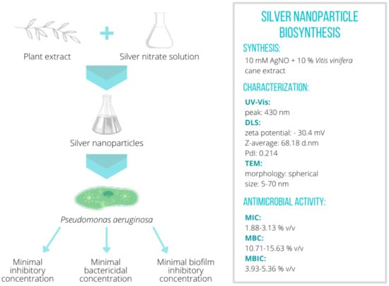
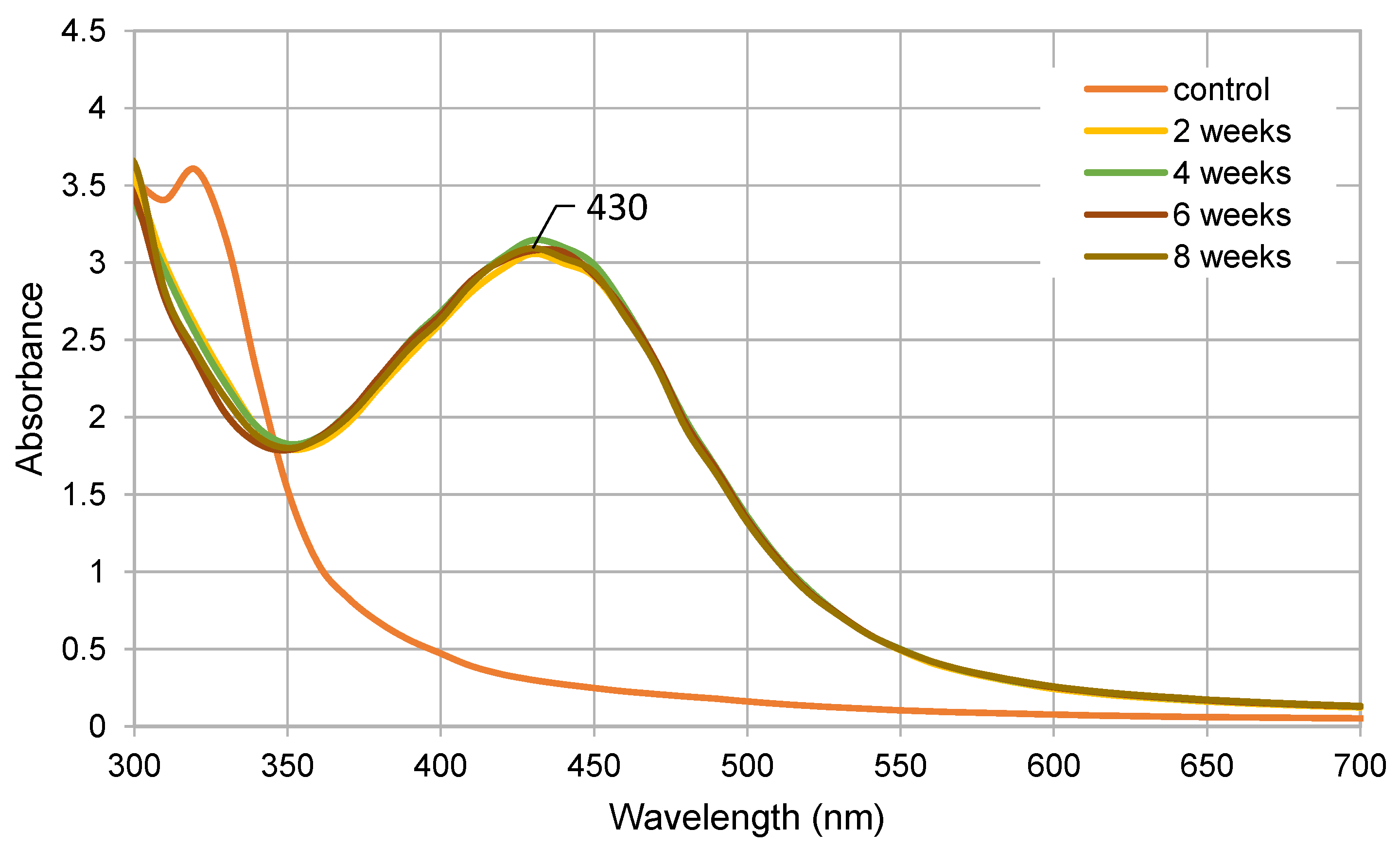
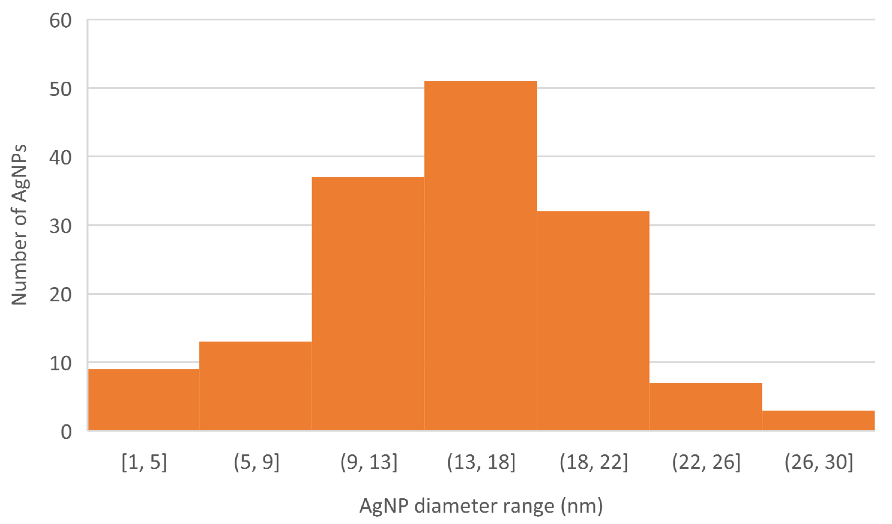
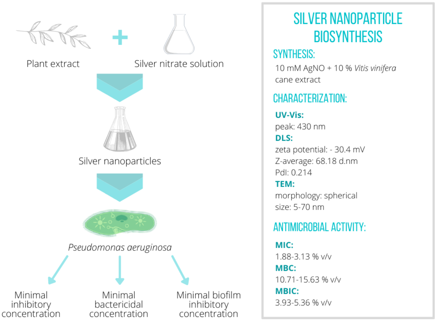
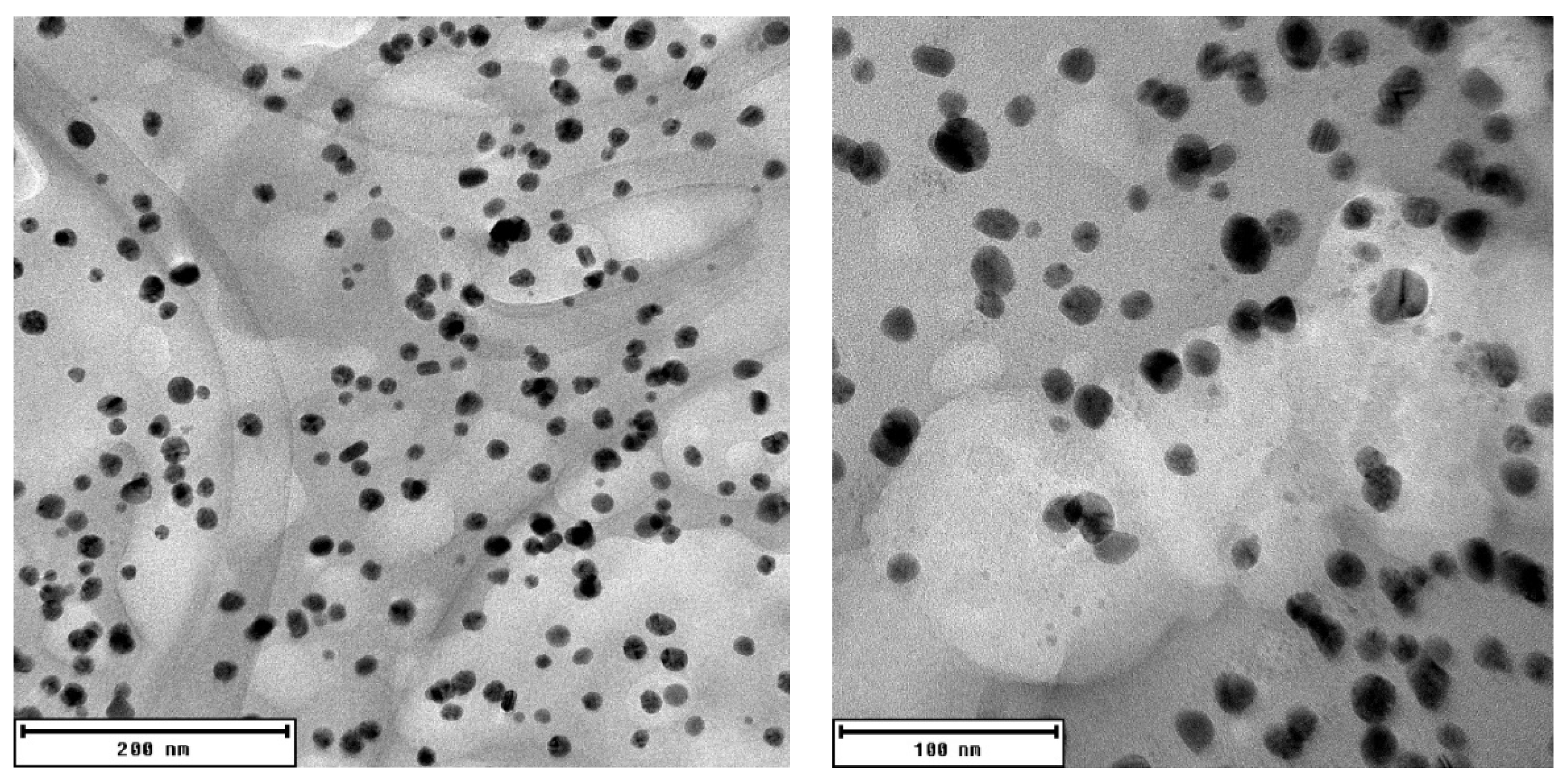
2.1. Nanoparticle Biosynthesis and Characterization

After the nanoparticle synthesis was completed, UV-visible (UV-Vis) spectrophotometric analysis was carried out, and it revealed a distinct peak at 430 nm (see Figure 1). This peak was apparent with all reagent ratios used (10% v/v of V. vinifera extract with 0.1, 0.25, 0.5, 1, or 2 mM of silver nitrate; 1 mM of silver nitrate with 0.5, 1, 2.5, 5, 10, 15, or 20% v/v of V. vinifera extract)—data not shown. The results from this screening experiment were used to choose a reagent ratio producing the highest and narrowest peak (1 mM of silver nitrate and 10% of V. vinifera extract); nanoparticles for subsequent antimicrobial experiments were produced accordingly. After that, the stability of produced AgNPs stored in the dark at room temperature was assessed over a period of 2 months (see Figure 1).
After that, AgNPs were subjected to dynamic light-scattering (DLS) analysis (see Table 1). The zeta potential of AgNP suspension was determined as –30.4 mV, where zeta potential values exceeding –25 or 25 mV signify a stable nanoparticle suspension, which will likely not aggregate due to van der Waals interactions [22]. The size of produced AgNPs was characterized using the DLS analysis; the Z-average was calculated as 68.18 d nm. The polydispersity index (PdI) of AgNP suspension was 0.214.
The results of the DLS size analysis were later confirmed by photographs from transmission electron microscopy (TEM) of the AgNPs (see Figure 2). TEM analysis revealed that the produced AgNPs are predominantly spherical in shape, and the particle size distribution of the suspension is relatively high, with most nanoparticles having a diameter of around 13–18 nm (see Figure 3).
The yield of nanoparticle synthesis was assessed using atomic absorption spectrometry (AAS). It was discovered that 65% of Ag ions present in the initial mixture were transformed into nanoparticles, making the final concentration of AgNPs 70.1 mg/L, with 37.8 mg/L of remaining silver ions.

2.2. Minimal Inhibition Concentration of AgNPs against Planktonic Cells of P. aeruginosa

The antimicrobial activity of the AgNP suspension was able to inhibit the growth of planktonic cells of all the P. aeruginosa strains tested (see Table 2). MIC80 was observed at 3.13% v/v of AgNPs for P. aeruginosa DBM 3081 and DBM 3777. The planktonic cells of P. aeruginosa ATCC 15442 were inhibited by more than 90% in the presence of 2.50% v/v of AgNPs. Regarding the last two strains, P. aeruginosa ATCC 10145 and PAO1, 1.88% v/v of AgNPs was needed for total inhibition.

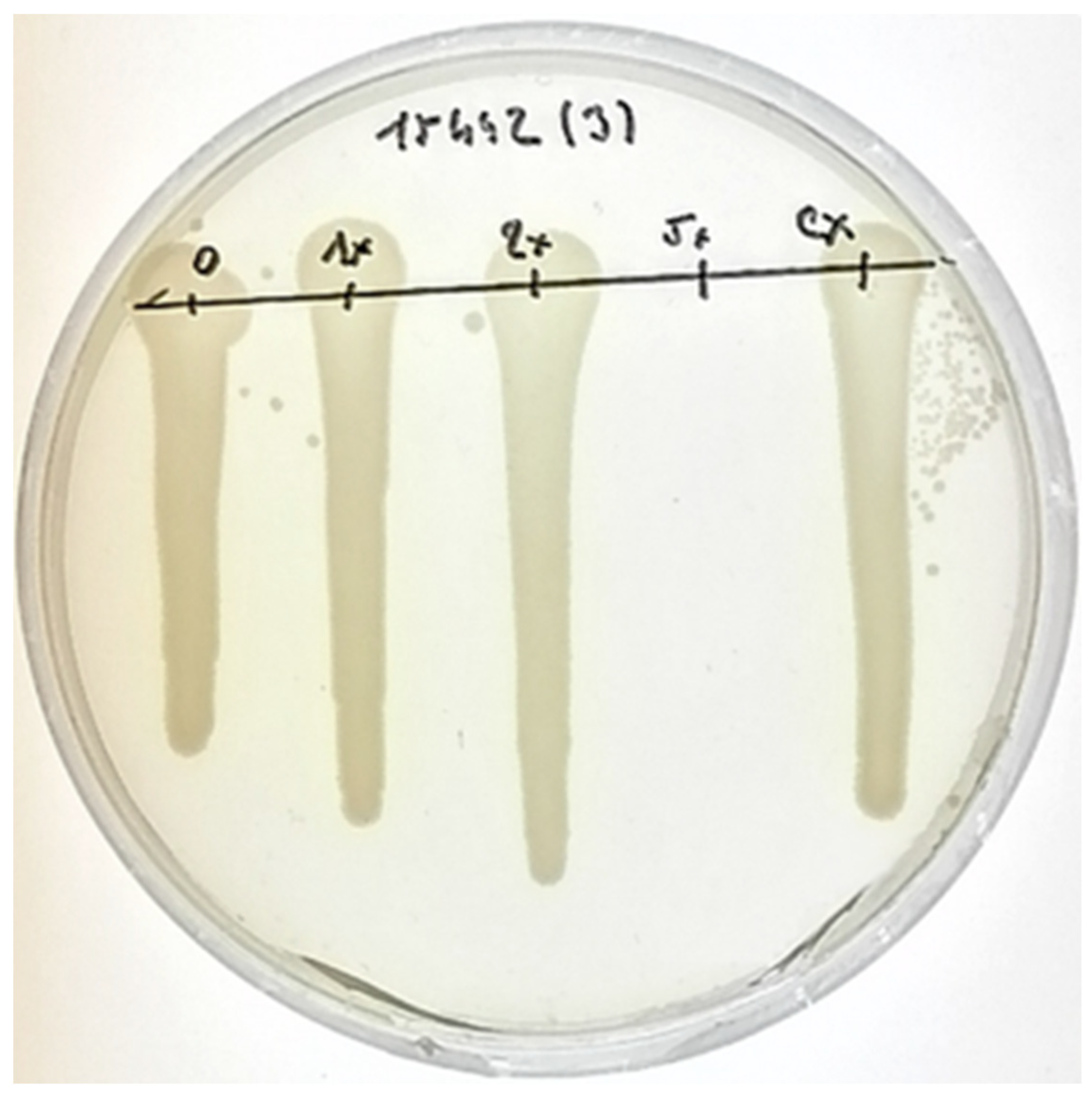
2.3. Bactericidal Activity of AgNPs against P. aeruginosa Cells

The bactericidal activity experiment was carried out to find the MBC of biosynthesized AgNPs (see Table 3). The MBC was observed for all the strains tested apart from P. aeruginosa ATCC 10145 in the concentration range used. For both P. aeruginosa DBM 3081 and P. aeruginosa DBM 3777, the MBC was determined as 15.63% v/v of AgNPs. Other strains exhibited even lower tolerance for the biosynthesized AgNPs. The MBC against P. aeruginosa ATCC 15442 was determined to be 12.50% v/v (see Figure 4), and the MBC against P. aeruginosa PAO1 was determined as 10.71% v/v.

2.4. Biological Activity of AgNPs against Adhering Cells of P. aeruginosa

After the effect of biosynthesized AgNPs on planktonic cells was evaluated, the experiments exploring the ability of AgNPs to inhibit cell adhesion were carried out (Table 4). MBIC50 was found in the range of 3.21–5.36% v/v for all the strains studied apart from P. aeruginosa DBM 3777. MBIC90 was 3.93, 5.36, and 5.36% v/v when assessing the adhesion inhibition against the cells of PAO1, P. aeruginosa ATCC 15442, and P. aeruginosa DBM 3081, respectively. MBIC90 was not found for either P. aeruginosa ATCC 10145 and P. aeruginosa DBM 3777 in the concentration range tested.

3. Discussion

In the age of antibiotic resistance, there is growing pressure to inhibit the growth of pathogenic microorganisms using alternatives to traditional antibiotic agents. Metal nanoparticles have thus become a subject of research in this regard [23]. However, their wider application is being partially hindered by inconveniences regarding the physico-chemical methods of their production. Therefore, “green” approaches to nanoparticle production have been studied intensively in recent decades [24]. In this study, we used agricultural waste, in adherence to circular economy principles, for the mediation of silver nanoparticle production.
AgNPs in this study exhibited a distinct peak at 430 nm when subjected to UV-Vis spectrophotometry. According to the literature, a peak between 410 and 450 nm is characteristic of silver nanoparticle suspension. In a study by Packialakshmi and Naziya [25], a peak at 420 nm was observed when silver nanoparticles were synthesized with the aid of Garcinia mangostana stem extract. A similar study by Bharathi et al. [26] was carried out, observing nanoparticle formation after the addition of Diospyros montana bark extract to silver nitrate solution, after which the UV-Vis spectra analysis revealed a peak at 432 nm. In a study using tannin extracted from grape pomace, silver nanoparticle synthesis was confirmed by a distinct peak at 420 nm [20]. In another study, using V. vinifera fruit extract, it was found that a peak at 450 pointed to a successful synthesis of silver nanoparticles [19]. Using multipole scattering theory, it was found that the position of a UV-Vis spectrophotometric peak can be used to calculate the size of nanoparticles produced, with higher wavelengths implying larger nanoparticles and vice versa [27].
Through the DLS analysis of AgNPs, in this study, we determined the average diameter as 68.18 nm and the PdI to be 0.214. In a study by Nayak et al. [28], the DLS analysis revealed that nanoparticles synthesized using Ficus benghalensis and Azadirachta indica bark extracts had average diameters of 85.95 and 90.13 nm, respectively, and a PdI of 0.247 and 0.314, respectively. In another study by Rao and Tang [29], it was demonstrated that reaction temperature can have a direct impact on average size and PdI. Their analysis showed that AgNPs synthesized with the aid of Eriobotrya japonica leaf extract had an average diameter of 76.10 nm with a PdI of 0.571 at 25 °C and an average diameter of 54.47 nm with a PdI of 0.281 at 80 °C. AgNPs produced in this study exhibited a relatively low PdI; this implies a more uniform nanoparticle suspension, which can be an advantage in the case of medicinal applications.
According to the DLS analysis, AgNPs in this study exhibited a zeta potential of −30.4 mV, which insinuates their long-term stability. In a study by Ravichandran et al. [30], the characterization of AgNPs produced using Atrocarpus altilis leaf extract revealed a zeta potential of −12.7 mV. A study by Anandalakshmi et al. [31] demonstrated silver nanoparticles synthesized using Pedalium murex leaf extract with a zeta potential of only −7.66 mV. In another study by Singh et al. [32], the zeta potential of silver nanoparticles synthesized with the aid of Cannabis sativa fiber extract was −29.2 mV. This value implied the stability of the AgNPs produced, which was subsequently confirmed by repeated UV-Vis spectra assessment (see Figure 1).
TEM analysis showed the morphology of AgNPs produced in this study to be spherical. Moreover, TEM analysis showed the size of AgNPs as being between 1 and 30 nm. In a study by Sadeghi and Gholamhoseinpoor [33], it was discovered by TEM analysis that AgNPs formed using Ziziphora tenuior extract were spherical in shape and that their size ranged from 8 to 40 nm. Another study by Ahmed et al. [34] also reported spherical AgNPs with a diameter of 34 nm when Azadirachta indica extract was used as the reducing and stabilizing agent. Irregular spherical shape and size between 20 and 100 nm was described in a study by Vijayakumar [35], where Justicia gendarussa extract was used for the phytofabrication of AgNPs.
Although AgNPs have been proved to exhibit powerful antimicrobial action against both Gram-negative and Gram-positive bacteria (Staphylococcus aureus, Bacillus cereus), viruses (HIV 1), and fungi (Candida, Aspergillus), we will focus on exploring their activity against P. aeruginosa [36]. When it comes to planktonic cell inhibition, the dispersion in this study was effective in the range of 1.32–2.21 mg/L of AgNPs with a remaining 0.71–1.18 mg/L of silver ions against all the strains tested. To compare the results with a traditional antibiotic, MIC80 of polymyxin B was observed between 2.5 and 3.5 mg/L against P. aeruginosa cells [37]. Regarding other studies using AgNPs, in a study by Pompilio et al. [38], the planktonic cells of P. aeruginosa were inhibited by AgNPs in the range of concentrations 1.06–4.25 mg/L. In another study by Singh et al. [39], AgNP MIC against multidrug-resistant strains of P. aeruginosa was observed to be between 6.25–12.5 mg/L. Finally, in a study by Radzig et al. [40], it was described that MIC of silver ions against P. aeruginosa PAO1 is 0.3 mg/L, and MIC of AgNPs is 8.0 mg/L. According to these results, combining silver ions with AgNPs could lower the concentration of silver necessary for the successful inhibition of the planktonic cells of P. aeruginosa compared to using silver ions or nanoparticles alone. The AgNP suspensions produced in this study combined the actions of nanoparticles and silver ions and proved more effective than the AgNPs in other cited studies.
In the case of biofilm formation inhibition, MBIC was achieved when 2.27–3.79 mg/L of AgNPs and 1.21–2.02 mg/L of silver ions were used. In a study by Palanisamy et al. [41], a 67% inhibition of biofilm formation of P. aeruginosa was achieved when 20 mg/L of AgNPs was used. In a different study by Markowska et al. [42], it was observed that AgNP MBIC against P. aeruginosa is 4 mg/L. In comparison, MBIC80 of polymyxin B against P. aeruginosa was found to be between 5 and 15 mg/L [37]. Regarding the last antimicrobial experiment, total bactericidal effect was observed when 7.57–11.05 mg/L of AgNPs with 4.05–5.91 mg/L of silver ions was used. In a study by Lara et al. [43], the MBC was found to be in the range of 8.88–10.7 mg/L. In another study by Mohan et al. [44], the bactericidal activity against P. aeruginosa was achieved when 12.5 mg/L of AgNPs was used. The combined effect of nanoparticles and silver ions in AgNP suspension produced in this study provided at least two-times stronger antibiofilm and bactericidal activity than the nanoparticle suspensions in other studies.
According to the results of experiments in this study, AgNP suspension manufactured using agricultural waste from wine production can be a cheap and effective alternative to traditionally produced nanoparticle suspensions when it comes to bactericidal activity or the inhibition of both the planktonic cells and the formation of biofilm.

4. Materials and Methods

4.1. Materials for Biosynthesis

V. vinifera canes, from both white and red varieties that were obtained during the dormancy period in January 2017 from vineyards in the Czech Republic, silver nitrate (Sigma Aldrich, St. Louis, MO, USA), ethanol (40%), and distilled water.

4.2. Extraction Method

The extraction method was modified according to Rollová et al. [45]. Briefly, V. vinifera canes were homogenized using a blade blender. Then, 150 g of the homogenized canes were mixed with 600 mL of 40% ethanol solution. This mixture was macerated for 24 h; then, it was filtered through an 8 μm filter paper and then again through a 0.2 μm filter paper. The liquid ethanolic extract was stored at 7 °C for further experimentation. The extract was characterized using polyphenolic content determination, high performance liquid chromatography, and ultra-high performance liquid chromatography coupled with high-resolution mass spectrometry in an earlier study [45].

4.3. Green Synthesis of Silver Nanoparticles

For the synthesis of AgNPs, a green synthesis method was employed using ethanolic extract of V. vinifera and silver nitrate solution. The synthesis was carried out using a number of different reagent ratios. First, 10 mL of V. vinifera ethanolic extract (see above) was added to 90 mL of silver nitrate dissolved in water to obtain various final concentrations (0.1, 0.25, 0.5, 1, 2, and 10 mM of silver nitrate). In the second layout, different amounts of V. vinifera ethanolic extract (0.5, 1, 2.5, 5, 10, and 15 mL) were added to silver nitrate dissolved in water at a constant final concentration of 1 mM to make a final volume of 100 mL. The mixture was left to react for 48 h, and the resulting AgNP suspension was then characterized using UV-Vis spectrophotometry. All experiments were performed in eight parallels. The final reagent ratios for antimicrobial testing stock dispersion were picked using these pilot experiments—10% v/v of Vitis vinifera cane extract and 1 mM of silver nitrate solutions were used (see Figure 1). Physical characteristics (apart from AAS) and antimicrobial properties of AgNPs were observed without separation from V. vinifera extract.

4.4. Characterization of Nanoparticles

UV-Vis spectrometry was carried out using a Reader Infinite M900Pro (TECAN MTP, Männedorf, Switzerland). Spectral analysis (250–700 nm) results were plotted and evaluated to confirm the presence of nanoparticles. UV-Vis spectrometry was also used for stability assessments when the nanoparticle dispersion spectra were measured 2, 4, 6, and 8 weeks after synthesis, while in the meantime, being stored in the dark at room temperature. After that, the nanoparticles were analyzed by transmission electron microscopy using EFTEM Jeol 2200 FS (Jeol Ltd., Tokyo, Japan). Dynamic light-scattering analysis was also carried out using Malvern Zetasizer Nano ZS (ATA Scientific Pty Ltd., Caringbah, Australia).
The yield of synthesis was assessed using a nanoparticle dispersion filtrate by AAS using Agilent 280FS AA. The nanoparticles were extracted from the dispersion by centrifugation. After that, the concentration of silver in the resulting supernatant was analyzed by AAS. The yield of synthesis was assessed as we compared the concentration of silver in the AgNP dispersion supernatant and the initial concentration of silver ions at the start of the synthesis.

4.5. Microbial Strains and Growth Media

These bacterial strains of P. aeruginosa were used: PAO1, ATCC 10145, ATCC 15442, DBM 3081, and DBM 3777. Glycerol cryopreserves of the microorganisms were stored at −70 °C.
All P. aeruginosa strains were precultivated before each experiment in a Luria-Bertani (LB) liquid medium at 37 °C for 24 h to achieve the exponential phase of growth (100 mL in Erlenmeyer flasks, 150 rpm).
Negative controls were conducted for the antimicrobial activity of the corresponding concentrations of both ethanol and extract to rule out the possibility of interference.

4.6. Evaluation of Minimal Inhibitory Concentration (MIC)

The antimicrobial effects of biosynthesized AgNPs against planktonic cells was determined using a microcultivation device Bioscreen C (Growth Curves Ltd., Turku, Finland) by a microdilution method, according to Sharma et al. [46]. The cells of tested P. aeruginosa strains were cultivated for 24 h in a microtiter plate in the presence of different% v/v of AgNPs. Each experiment was done in 10 parallels. For each well, there was 160 µL of LB medium and 30 µL of inoculum (OD600 = 0.1), and the remaining 130 µL were filled with varying ratios of phosphate buffer saline solution and AgNPs (0.31, 0.63, 1.25, 1.88, 2.50, and 3.13% v/v). Control parallels were filled with 160 µL of LB medium, 30 µL of inoculum (OD600 = 0.1), and 130 µL of phosphate buffer saline solution. After that, the minimal inhibitory concentration (MIC80) was determined by evaluating growth curves constructed from the data provided by the microcultivation device as the lowest concentration of antimicrobial agent causing 80% inhibition of growth compared to control after overnight cultivation [47].

4.7. Evaluation of Minimal Bactericidal Concentration (MBC)

The bactericidal effect of biosynthesized nanoparticles was determined through a subsequent solid medium (LB agar plates) cultivation of planktonic cells taken from a cultivation in the presence of nanoparticles. The cells of tested P. aeruginosa strains were cultivated for 24 h in a microtiter plate in the presence of different% v/v of AgNPs derived from their respective MICs (1× MIC, 2× MIC, 5× MIC); each experiment was done in three parallels. Control parallels were filled with 160 µL of LB medium, 30 µL of inoculum (OD600 = 0.1), and 130 µL of phosphate buffer saline solution. Subsequently, 10 µL of cell suspension representing each concentration of nanoparticles and the control were inoculated onto a LB agar plate and cultivated for an additional 24 h. After that, the minimal bactericidal concentration (MBC) was determined as a concentration yielding the growth of fewer than five colonies (>99% killing) by evaluating the growth on solid medium, according to Rahal and Simberkoff [48].

4.8. Evaluation of Minimal Biofilm Inhibitory Concentration (MBIC)

4.8.1. Cultivation Conditions

The cultivation was carried out in polystyrene 96-well microtiter plates (TPP, Trasadingen, Switzerland), according to Shin and Eom [49]. The cells of P. aeruginosa strains were cultivated for 24 h in the presence of different% v/v of AgNPs, which were derived from their respective MICs (from 0% v/v to 3× MIC). For each well, there was 210 µL of inocculum (OD600 = 0.8) with 70 µL of differing ratios of phosphate buffer saline solution and AgNPs. Control parallels were filled with 160 µL of LB medium, 30 µL of inoculum (OD600 = 0.8), and 130 µL of phosphate buffer saline solution. Eight parallels were carried out for each concentration of AgNPs, including the control. The microtiter plate was covered with a lid and incubated at 37 °C and 150 rpm for 24 h.

4.8.2. MTT Assay

The metabolic activity of the biofilm was measured using a 3-(4,5-dimethylthiazol-2-yl)-2,5-diphenyltetrazolium bromide (MTT) reduction assay, according to Sabaeifard et al. [50]. The wells were washed three times by saline. Afterwards, 60 µL of glucose solution (57.4 mg/mL), and 50 µL of MTT solution (1 mg/mL) were added into each well. The microtiter plate was then covered with a lid and incubated in the dark at 37 °C and 150 rpm for 1 h. After incubation, 100 µL of wash solution was added to every well, and the plate was put on a shaker at room temperature for 30 min, where the wash solution was a mixture of dimethylformamide, phosphate buffer saline solution, acetic acid, and sodium dodecyl sulfate. After 30 min, color intensity was measured using a Reader Infinite M900Pro (TECAN MTP, Männedorf, Switzerland)at 570 nm. Each experiment was performed in eight parallels. Hence, the minimal biofilm inhibitory concentration (MBIC50 and MBIC90) was determined as the lowest concentration of nanoparticle suspension which inhibited biofilm formation by 50 and 90%, respectively, compared to control [49].

4.9. Statistical Analysis

For the evaluation of microbial growth or bioflm assays data, we used Dixon’s Q test to exclude outlying values. Arithmetic means and standard deviations (SD) were calculated for each concentration tested by mentioned assays in relative percentages (control samples were 100%).

5. Conclusions

This study developed a successful method of silver nanoparticle manufacture using a plant extract from agricultural waste. The process of synthesis used in this study is cost-effective and produces stable silver nanoparticles ranging in size between 1 and 30 nm, which can be stored at room temperature for up to two months. Moreover, these nanoparticles exhibit significant inhibiting, bactericidal, and antibiofilm activity against P. aeruginosa in concentrations between 2.8 and 15.7% v/v. With this simple method, silver nanoparticles for antimicrobial purposes can be synthesized while using agricultural waste, thus adhering to circular economy principles.

Author Contributions

Conceptualization, J.M. (Jana Michailidu); methodology, J.M. (Jana Michailidu) and O.M.; validation, J.M. (Jana Michailidu) and O.M.; resources, J.M. (Jana Michailidu); writing—original draft preparation, J.M. (Jana Michailidu); writing—review and editing, O.M. and J.M. (Jana Michailidu); supervision, J.M. (Jan Masák) and A.Č.; funding acquisition, I.K. All authors have read and agreed to the published version of the manuscript.

Funding

This research received no external funding.

Institutional Review Board Statement

Not applicable.

Informed Consent Statement

Not applicable.

Data Availability Statement

Not applicable.

Acknowledgments

This work was supported from the grant of Specific university research–grant No A2_FPBT_2021_044.

Conflicts of Interest

The authors declare no conflict of interest.

References

  1. Thakkar, K.N.; Mhatre, S.S.; Parikh, R.Y. Biological synthesis of metallic nanoparticles. Nanomed. Nanotechnol. Biol. Med. 2010, 6, 257–262. [Google Scholar] [CrossRef] [PubMed]
  2. Baptista, P.V.; McCusker, M.P.; Carvalho, A.; Ferreira, D.A.; Mohan, N.M.; Martins, M.; Fernandes, A.R. Nano-Strategies to Fight Multidrug Resistant Bacteria—“A Battle of the Titans”. Front. Microbiol. 2018, 9, 1441. [Google Scholar] [CrossRef] [Green Version]
  3. Kulkarni, N.; Muddapur, U. Biosynthesis of Metal Nanoparticles: A Review. J. Nanotechnol. 2014, 354, 1–8. [Google Scholar] [CrossRef] [Green Version]
  4. El-Seedi, H.R.; El-Shabasy, R.M.; Khalifa, S.A.M.; Saeed, A.; Shah, A.; Shah, R.; Iftikhar, F.J.; Abdel-Daim, M.M.; Omri, A.; Hajrahand, N.H.; et al. Metal nanoparticles fabricated by green chemistry using natural extracts: Biosynthesis, mechanisms, and applications. RSC Adv. 2019, 9, 24539–24559. [Google Scholar] [CrossRef] [Green Version]
  5. Shameli, K.; Bin Ahmad, M.; Jaffar Al-Mulla, E.A.; Ibrahim, N.A.; Shabanzadeh, P.; Rustaiyan, A.; Abdollahi, Y.; Bagheri, S.; Abdolmohammadi, S.; Usman, M.S.; et al. Green biosynthesis of silver nanoparticles using Callicarpa maingayi stem bark extraction. Molecules 2012, 17, 8506–8517. [Google Scholar] [CrossRef] [PubMed]
  6. Amini, S.M. Preparation of antimicrobial metallic nanoparticles with bioactive compounds. Mater. Sci. Eng. C 2019, 103, 109809. [Google Scholar] [CrossRef] [PubMed]
  7. Anshup, A.; Venkataraman, J.S.; Subramaniam, C.; Kumar, R.R.; Priya, S.; Kumar, T.R.S.; Omkumar, R.V.; John, A.; Pradeep, T. Growth of gold nanoparticles in human cells. Langmuir 2005, 21, 11562–11567. [Google Scholar] [CrossRef] [PubMed]
  8. Kowshik, M.; Deshmukh, N.; Vogel, W.; Urban, J.; Kulkarni, S.K.; Paknikar, K.M. Microbial synthesis of semiconductor CdS nanoparticles, their characterization, and their use in the fabrication of an ideal diode. Biotechnol. Bioeng. 2002, 78, 583–588. [Google Scholar] [CrossRef] [PubMed]
  9. Lengke, M.F.; Fleet, M.E.; Southam, G. Biosynthesis of silver nanoparticles by filamentous cyanobacteria from a silver(I) nitrate complex. Langmuir 2007, 23, 2694–2699. [Google Scholar] [CrossRef]
  10. Parveen, K.; Banse, V.; Ledwani, L. Green synthesis of nanoparticles: Their advantages and disadvantages. In AIP Conference Proceedings; AIP Publishing LLC.: Melville, NY, USA, 2016; p. 020048. [Google Scholar]
  11. Rautaray, D.; Ahmad, A.; Sastry, M. Biosynthesis of CaCO3 Crystals of Complex Morphology Using a Fungus and an Actinomycete. J. Am. Chem. Soc. 2003, 125, 14656–14657. [Google Scholar] [CrossRef]
  12. Shahverdi, A.R.; Minaeian, S.; Shahverdi, H.R.; Jamalifar, H.; Nohi, A.-A. Rapid synthesis of silver nanoparticles using culture supernatants of Enterobacteria: A novel biological approach. Processes Biochem. 2007, 42, 919–923. [Google Scholar] [CrossRef]
  13. Raheman, F.; Deshmukh, S.; Ingle, A.; Gade, A.; Rai, M. Silver Nanoparticles: Novel Antimicrobial Agent Synthesized from an Endophytic Fungus Pestalotia sp. Isolated from leaves of Syzygium cumini (L). Nano Biomed. Eng. 2011, 3, 174–178. [Google Scholar] [CrossRef]
  14. Sunkar, S.; Nachiyar, C.V. Biogenesis of antibacterial silver nanoparticles using the endophytic bacterium Bacillus cereus isolated from Garcinia xanthochymus. Asian Pac. J. Trop. Biomed. 2012, 2, 953–959. [Google Scholar] [CrossRef] [Green Version]
  15. Kora, A.J.; Sashidhar, R.B.; Arunachalam, J. Aqueous extract of gum olibanum (Boswellia serrata): A reductant and stabilizer for the biosynthesis of antibacterial silver nanoparticles. Processes Biochem. 2012, 47, 1516–1520. [Google Scholar] [CrossRef]
  16. Chandran, K.; Song, S.; Yun, S.-I. Effect of size and shape controlled biogenic synthesis of gold nanoparticles and their mode of interactions against food borne bacterial pathogens. Arab. J. Chem. 2014, 12, 1994–2006. [Google Scholar] [CrossRef] [Green Version]
  17. Raut, R.W.; Mendhulkar, V.D.; Kashid, S.B. Photosensitized synthesis of silver nanoparticles using Withania somnifera leaf powder and silver nitrate. J. Photochem. Photobiol. B Biol. 2014, 132, 45–55. [Google Scholar] [CrossRef] [PubMed]
  18. Sathishkumar, M.; Sneha, K.; Won, S.W.; Cho, C.W.; Kim, S.; Yun, Y.S. Cinnamon zeylanicum bark extract and powder mediated green synthesis of nano-crystalline silver particles and its bactericidal activity. Colloids Surf. B Biointerfaces 2009, 73, 332–338. [Google Scholar] [CrossRef]
  19. Gnanajobitha, G.; Paulkumar, K.; Vanaja, M.; Rajeshkumar, S.; Malarkodi, C.; Annadurai, G.; Kannan, C. Fruit-mediated synthesis of silver nanoparticles using Vitis vinifera and evaluation of their antimicrobial efficacy. J. Nanostructure Chem. 2013, 3, 67. [Google Scholar] [CrossRef] [Green Version]
  20. Saratale, R.G.; Saratale, G.D.; Ahn, S.; Shin, H.-S. Grape Pomace Extracted Tannin for Green Synthesis of Silver Nanoparticles: Assessment of Their Antidiabetic, Antioxidant Potential and Antimicrobial Activity. Polymers 2021, 13, 4355. [Google Scholar] [CrossRef]
  21. Kuppusamy, P.; Yusoff, M.M.; Maniam, G.P.; Govindan, N. Biosynthesis of metallic nanoparticles using plant derivatives and their new avenues in pharmacological applications—An updated report. Saudi Pharm. J. 2016, 24, 473–484. [Google Scholar] [CrossRef]
  22. Jacobs, C.; Kayser, O.; Müller, R.H. Nanosuspensions as a new approach for the formulation for the poorly soluble drug tarazepide. Int. J. Pharm. 2000, 196, 161–164. [Google Scholar] [CrossRef]
  23. Aruguete, D.M.; Kim, B.; Hochella, M.F.; Ma, Y.; Cheng, Y.; Hoegh, A.; Liu, J.; Pruden, A. Antimicrobial nanotechnology: Its potential for the effective management of microbial drug resistance and implications for research needs in microbial nanotoxicology. Environ. Sci. Process. Impacts 2012, 15, 93–102. [Google Scholar] [CrossRef] [PubMed]
  24. Makarov, V.V.; Love, A.J.; Sinitsyna, O.V.; Makarova, S.S.; Yaminsky, I.V.; Taliansky, M.E.; Kalinina, N.O. “Green” nanotechnologies: Synthesis of metal nanoparticles using plants. Acta Nat. 2014, 6, 95. [Google Scholar] [CrossRef] [Green Version]
  25. Packialakshmi, N.; Naziya, S. Green Synthesis of Silver Nanoparticles from Stem Extracts of Caralluma Fimbriyata and Its Antibacterial Activity. Int. J. Appl. Sci. Biotechnol. 2014, 2, 305–310. [Google Scholar] [CrossRef] [Green Version]
  26. Bharathi, D.; Diviya Josebin, M.; Vasantharaj, S.; Bhuvaneshwari, V. Biosynthesis of silver nanoparticles using stem bark extracts of Diospyros montana and their antioxidant and antibacterial activities. J. Nanostruct. Chem. 2018, 8, 83–92. [Google Scholar] [CrossRef] [Green Version]
  27. Haiss, W.; Thanh, N.T.K.; Aveyard, J.; Fernig, D.G. Determination of Size and Concentration of Gold Nanoparticles from UV−Vis Spectra. Anal. Chem. 2007, 79, 4215–4221. [Google Scholar] [CrossRef]
  28. Nayak, D.; Ashe, S.; Rauta, P.R.; Kumari, M.; Nayak, B. Bark extract mediated green synthesis of silver nanoparticles: Evaluation of antimicrobial activity and antiproliferative response against osteosarcoma. Mater. Sci. Eng. C 2016, 58, 44–52. [Google Scholar] [CrossRef]
  29. Rao, B.; Tang, R.-C. Green synthesis of silver nanoparticles with antibacterial activities using aqueous Eriobotrya japonica leaf extract. Adv. Nat. Sci. Nanosci. Nanotechnol. 2017, 8, 015014. [Google Scholar] [CrossRef] [Green Version]
  30. Ravichandran, V.; Vasanthi, S.; Shalini, S.; Ali Shah, S.A.; Harish, R. Green synthesis of silver nanoparticles using Atrocarpus altilis leaf extract and the study of their antimicrobial and antioxidant activity. Mater. Lett. 2016, 180, 264–267. [Google Scholar] [CrossRef]
  31. Anandalakshmi, K.; Venugobal, J.; Ramasamy, V. Characterization of silver nanoparticles by green synthesis method using Pedalium murex leaf extract and their antibacterial activity. Appl. Nanosci. 2016, 6, 399–408. [Google Scholar] [CrossRef] [Green Version]
  32. Singh, P.; Pandit, S.; Garnæs, J.; Tunjic, S.; Mokkapati, V.R.S.S.; Sultan, A.; Thygesen, A.; Mackevica, A.; Mateiu, R.V.; Daugaard, A.E.; et al. Green synthesis of gold and silver nanoparticles from Cannabis sativa (industrial hemp) and their capacity for biofilm inhibition. Int. J. Nanomed. 2018, 13, 3571–3591. [Google Scholar] [CrossRef] [PubMed]
  33. Sadeghi, B.; Gholamhoseinpoor, F. A study on the stability and green synthesis of silver nanoparticles using Ziziphora tenuior (Zt) extract at room temperature. Spectrochim. Acta Part A Mol. Biomol. Spectrosc. 2015, 134, 310–315. [Google Scholar] [CrossRef] [PubMed]
  34. Ahmed, S.; Saifullah; Ahmad, M.; Swami, B.L.; Ikram, S. Green synthesis of silver nanoparticles using Azadirachta indica aqueous leaf extract. J. Radiat. Res. Appl. Sci. 2016, 9, 1–7. [Google Scholar] [CrossRef] [Green Version]
  35. Vijayakumar, A.S. Pharmacological activity of silver nanoparticles, ethanolic extract from justicia gendarussa (burm) f plant leaves. Res. J. Life Sci. Bioinform. Pharm. Chem. Sci. 2019, 5, 462–475. [Google Scholar] [CrossRef]
  36. Deshmukh, S.P.; Patil, S.M.; Mullani, S.B.; Delekar, S.D. Silver nanoparticles as an effective disinfectant: A review. Mater. Sci. Eng. C 2019, 97, 954–965. [Google Scholar] [CrossRef]
  37. Vaňková, E.; Paldrychová, M.; Kašparová, P.; Lokočová, K.; Kodeš, Z.; Maťátková, O.; Kolouchová, I.; Masák, J. Natural antioxidant pterostilbene as an effective antibiofilm agent, particularly for gram-positive cocci. World J. Microbiol. Biotechnol. 2020, 36, 101. [Google Scholar] [CrossRef]
  38. Pompilio, A.; Geminiani, C.; Bosco, D.; Rana, R.; Aceto, A.; Bucciarelli, T.; Scotti, L.; Di Bonaventura, G. Electrochemically Synthesized Silver Nanoparticles Are Active Against Planktonic and Biofilm Cells of Pseudomonas aeruginosa and Other Cystic Fibrosis-Associated Bacterial Pathogens. Front. Microbiol. 2018, 9, 1349. [Google Scholar] [CrossRef] [Green Version]
  39. Singh, K.; Panghal, M.; Kadyan, S.; Chaudhary, U.; Yadav, J.P. Green silver nanoparticles of Phyllanthus amarus: As an antibacterial agent against multi drug resistant clinical isolates of Pseudomonas aeruginosa. J. Nanobiotechnol. 2014, 12, 40. [Google Scholar] [CrossRef] [Green Version]
  40. Radzig, M.; Koksharova, O.; Khmel, I.A. Antibacterial effects of silver ions: Effect on gram-negative bacteria growth and biofilm formation. Mol. Gen. Mikrobiol. Virusol 2009, 24, 194–199. [Google Scholar] [CrossRef]
  41. Palanisamy, N.K.; Ferina, N.; Amirulhusni, A.N.; Mohd-Zain, Z.; Hussaini, J.; Ping, L.J.; Durairaj, R. Antibiofilm properties of chemically synthesized silver nanoparticles found against Pseudomonas aeruginosa. J. Nanobiotechnol. 2014, 12, 2. [Google Scholar] [CrossRef] [Green Version]
  42. Markowska, K.; Grudniak, A.M.; Krawczyk, K.; Wróbel, I.; Wolska, K.I. Modulation of antibiotic resistance and induction of a stress response in Pseudomonas aeruginosa by silver nanoparticles. J. Med. Microbiol. 2014, 63, 849–854. [Google Scholar] [CrossRef] [PubMed] [Green Version]
  43. Lara, H.; Ayala-Nunez, V.; Ixtepan Turrent, L.; Rodríguez-Padilla, C. Bactericidal effect of AgNPs against multidrug-resistant bacteria. World J. Microbiol. Biotechnol. 2009, 26, 615–621. [Google Scholar] [CrossRef]
  44. Mohan, S.; Oluwafemi, O.S.; George, S.C.; Jayachandran, V.P.; Lewu, F.B.; Songca, S.P.; Kalarikkal, N.; Thomas, S. Completely green synthesis of dextrose reduced silver nanoparticles, its antimicrobial and sensing properties. Carbohydr. Polym. 2014, 106, 469–474. [Google Scholar] [CrossRef] [PubMed]
  45. Rollová, M.; Gharwalova, L.; Krmela, A.; Schulzová, V.; Hajšlová, J.; Jaroš, P.; Kolouchová, I.; Maťátková, O. Grapevine extracts and their effect on selected gut-associated microbiota: In vitro study. Czech. J. Food Sci. 2020, 38, 137–143. [Google Scholar] [CrossRef]
  46. Sharma, M.; Manoharlal, R.; Negi, A.S.; Prasad, R. Synergistic anticandidal activity of pure polyphenol curcumin I in combination with azoles and polyenes generates reactive oxygen species leading to apoptosis. FEMS Yeast Res. 2010, 10, 570–578. [Google Scholar] [CrossRef]
  47. Serra, E.; Hidalgo-Bastida, L.A.; Verran, J.; Williams, D.; Malic, S. Antifungal Activity of Commercial Essential Oils and Biocides against Candida albicans. Pathogens 2018, 7, 15. [Google Scholar] [CrossRef] [Green Version]
  48. Rahal, J.J.; Simberkoff, M.S. Bactericidal and Bacteriostatic Action of Chloramphenicol Against Meningeal Pathogens. Antimicrob. Agents Chemother. 1979, 16, 13–18. [Google Scholar] [CrossRef] [Green Version]
  49. Shin, D.-S.; Eom, Y.-B. Zerumbone inhibits Candida albicans biofilm formation and hyphal growth. Can. J. Microbiol. 2019, 65, 713–721. [Google Scholar] [CrossRef]
  50. Sabaeifard, P.; Abdi-Ali, A.; Soudi, M.R.; Dinarvand, R. Optimization of tetrazolium salt assay for Pseudomonas aeruginosa biofilm using microtiter plate method. J. Microbiol. Methods 2014, 105, 134–140. [Google Scholar] [CrossRef]
Figure 1. Silver nanoparticle formation using 10% of V. vinifera extract and 1 mM of AgNO3 and their stability 2, 4, 6, and 8 weeks after synthesis while stored in the dark at room temperature; UV-Vis spectra.
Plants 11 00443 g001
Figure 2. TEM images of AgNPs produced using V. vinifera extract.
Plants 11 00443 g002
Figure 3. A histogram of TEM image of the AgNPs produced.
Plants 11 00443 g003
Figure 4. A photograph of a Petri dish with an MBC experiment showing cultivation of P. aeruginosa ATCC 15442 without any biological agent (negative control), with different concentrations of AgNPs (1×, 2×, 5× MIC), and a corresponding concentration of V. vinifera extract (negative control) (from the left).
Plants 11 00443 g004
Table 1. DLS analysis results of nanoparticles produced using V. vinifera extract.
Zeta Potential (mV)Z-Average (d nm)Polydispersity Index
–30.4 ± 3.8868.18 ± 2.650.214
Table 2. Pseudomonas aeruginosa planktonic cell inhibition by AgNPs in V. vinifera cane extract; MIC80; 100% v/v is equivalent to 70.1 mg/L AgNPs with 37.8 mg/L of remaining silver ions.
Pseudomonas aeruginosa StrainPAO1ATCC 10145ATCC 15442DBM 3081DBM 3777
MIC80 (% v/v)1.881.882.503.133.13
Table 3. Bactericidal activity of AgNPs in V. vinifera cane extract against P. aeruginosa strains; 100% v/v is equivalent to 70.1 mg/L AgNPs with 37.8 mg/L of remaining silver ions.
Pseudomonas aeruginosa StrainPAO1ATCC 10145ATCC 15442DBM 3081DBM 3777
MBC (% v/v)10.71-12.5015.6315.63
Table 4. Pseudomonas aeruginosa adhesion inhibition (50 and 80%) by AgNPs in V. vinifera cane extract; MBIC50 and MBIC90; 100% v/v is equivalent to 70.1 mg/L AgNPs with 37.8 mg/L of remaining silver ions.
Pseudomonas aeruginosa StrainPAO1ATCC 10145ATCC 15442DBM 3081DBM 3777
MBIC50 (% v/v)<3.935.00<5.363.21-
MBIC90 (% v/v)3.93-5.365.36-
Publisher’s Note: MDPI stays neutral with regard to jurisdictional claims in published maps and institutional affiliations.

