Article

Biopotential of Underutilized Rosaceae Inflorescences: LC-DAD-MS Phytochemical Profiles Associated with Antioxidant, Antidiabetic, Anti-Inflammatory and Antiproliferative Activity In Vitro

1
Department of Biology, Faculty of Science, University of Zagreb, Horvatovac 102a, 10000 Zagreb, Croatia
2
Department of Agriculture and Nutrition, Institute of Agriculture and Tourism, Karla Huguesa 8, 52440 Poreč, Croatia
3
Department of Agronomy, Biotechnical Faculty, University of Ljubljana, Jamnikarjeva 101, 1000 Ljubljana, Slovenia
4
Division of Molecular Medicine, Ruđer Bošković Institute, 10000 Zagreb, Croatia
5
METRIS Research Centre, Istrian University of Applied Sciences, Riva 6, 52100 Pula, Croatia
*
Author to whom correspondence should be addressed.
These authors contributed equally to this work.
Plants 2022, 11(3), 271; https://doi.org/10.3390/plants11030271
Submission received: 9 December 2021 / Revised: 2 January 2022 / Accepted: 17 January 2022 / Published: 20 January 2022

Abstract

:
The aim of this work was to assess the biopotential of the young inflorescence tissues of Prunus, Malus and Chaenomeles in order to evaluate the possibility of their application in the food industry, and to provide a polyphenolic fingerprint for their quality control. The contents of different bioactive compounds and their antioxidant capacities were spectrophotometrically measured, the main phenolic compounds were identified and quantified using LC-DAD-MS, the antidiabetic potential was determined using α-amylase and α-glucosidase inhibition assays, the anti-inflammatory potential was determined using a 5-lipoxygenase inhibition assay, and the cytotoxicity was determined by MTT assay. Using one-way ANOVA, principal component analysis, hierarchical clustering and Pearson’s correlation coefficient, the relations between the samples, and between the samples and the measured parameters, were revealed. In total, 77 compounds were identified. The concentration of sugars was low in M. purpurea, at 1.56 ± 0.08 mg/g DW. The most effective sample in the inhibition of antidiabetic enzymes and anti-inflammatory 5-lipoxygenase was C. japonica. The inhibition of α-glucosidase was strongly positively correlated with the total and condensed tannins, procyanidin dimers and procyanidin tetramer, and was very strongly correlated with chlorogenic acid. In α-amylase inhibition, C. japonica and P. serrulata ‘Kiku Shidare Zakura’ were equally efficient to the standard inhibitor, maltose. The most effective in the growth and proliferation inhibition of HepG2, HCT116 and HaCaT cells was P. avium. The results suggest Prunus, Malus and Chaenomeles inflorescences as functional food ingredients.

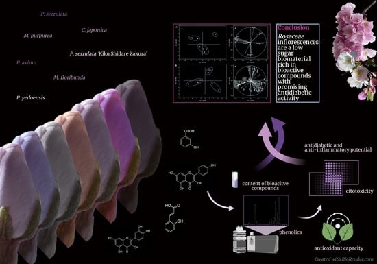
Graphical Abstract

1. Introduction

Inflorescence, as a precursor of a fruit, is composed of several types of metabolically very active tissues. In recent years, the biopotential of the bioactive compounds of this plant structure has been recognized. Namely, the significant bioactivities of inflorescences of industrial hemp [1], Musa species [2], Sorbus aucuparia [3], Cistus salviifolius [4], Lonicera japonica [5], and Caryota urens [6] have been revealed. Moreover, the flower buds of C. salviifolius have been shown to have a higher concentration of phenolics, better antioxidant and anti-superoxide dismutase activity, and stronger cytotoxic activity against human breast cancer and ovarian cancer cells than leaves [4]. Recently, the intrinsic antiradical activity of industrial hemp’s inflorescences’ water extracts and mechanisms related to their anti-inflammatory, antiproliferative and antimycotic activity have been revealed [1]. The inflorescences of Musa species are one of the most widely consumed vegetables in the Southeast Asian region, and their biological activities are well known.
The use of inflorescences of Rosaceae varieties, except those of Sorbus spp. [3], is not that common. Sorbus spp. inflorescences have a higher concentration of total phenolics than the commonly consumed fruits [3]. Furthermore, the inflorescences of different Sorbus species have higher antioxidant activity than their leaves or fruits [7]. Moreover, the extremely high content of phenolics in S. aucuparia inflorescences suggests their great potential as a source for natural health products [3,7]. Different organs of Prunus, Malus, and Chaenomeles taxa have already been investigated for their phytochemical, nutraceutical and bioactivity potential [8,9,10,11,12,13]. However, the inflorescences have been neglected so far.
Therefore, the aim of this work was to assess the biopotential of the young inflorescence tissues of selected Prunus, Malus, and Chaenomeles using a combination of spectrometric, chromatographic, cell culture and chemometric analyses, as well as to evaluate the possibility of their application in the food industry. For that purpose, we (a) spectrophotometrically measured the content of different types of bioactive compounds (total phenolics, flavonoids, non-flavonoids, total and condensed tannins, and soluble sugars), and their antioxidant capacity by the three methods (ABTS, FRAP and DPPH); (b) identified and quantified the main phenolic compounds using the LC-DAD-MS method; (c) determined the antidiabetic potential of the inflorescences using α-amylase and α-glucosidase inhibition assays; (d) measured the anti-inflammatory potential using a 5-lipoxygenase inhibition assay; (e) assessed the cytotoxicity toward HepG2, HCT 116 and HaCaT cells by MTT assay; and (f) statistically—using one-way ANOVA, principal component analysis, hierarchical clustering and Pearson’s correlation coefficient—revealed the relations between the samples, as well as between the samples and the measured parameters. This study is the first to present a comprehensive LC-DAD-MS polyphenolic profile of the inflorescences of Prunus avium (L.) L. (Pa), Prunus serrulata Lindl. (Ps), Prunus serrulata ‘Kiku Shidare Zakura’ (PsKss), Malus x purpurea (E.Barbier) Ehder (Mp), Malus floribunda Siebold ex Van Houtte (Mf) and Chaenomeles japonica (Thunb.) Lindl. ex Spach (Cj), providing a fingerprint for the future quality control of these biomaterials, and the first report on their bioactivity.

2. Results and Discussion

The food industry mainly focuses on Rosaceae fruits, and somewhat less on the flowers, stems and leaves [8], which are mostly used in their dried form for tea. Because inflorescences are composed of several types of metabolically very active tissues, it can be assumed that these plant reproductive structures contain a significant concentration of bioactive phytochemicals, and that they present thus-far-underutilized plant potential. Therefore, in this study, for the first time, we offer data on phytochemical profile and bioactivity of inflorescences of Pa, Ps, PsKss, Py, Mp, Mf and Cj.

2.1. Spectrophotometric Analysis of the Phytochemical Content and Antioxidant Capacity of the Inflorescences

All of the the tested samples had a similar or higher amount of total phenolics than the commonly used flower buds of Capparis spinosa [14]. The highest amount of total phenolics (53.12 ± 0.79 mg GAE/g DW), flavonoids (38.89 ± 4.04 mg CE/g DW) and nonflavonoids (31.32 ± 0.71 mg GAE/g DW) were recorded in PsKss (Table 1). Because only about 10% of the medicinal plant material has total phenolics in a concentration higher than 5% DW of GAE [7,15], PsKss inflorescence is among the plant materials with the highest concentration of these compounds. Compared to the concentration of the total flavonoids in many other commonly used medicinal plants [15], the tested inflorescences had a similar or higher concentration, suggesting their potential for the food industry. Moreover, very recently, it was shown that inflorescences of Pa have far more total phenolics and flavonoids than stems and kernels [16].
In order to reduce the global rise of overweightness and obesity, the content of sugars, as a source of calories, in food should be diminished. As expected, the level of sugars in the inflorescences was lower than that in the more often-used fruits. The interval of the concentrations in the inflorescences was between 1.56 ± 0.08 mg/g DW in Mp and 8.61 ± 0.12 mg/g DW in Py (Table 1), which is relatively low compared to the concentrations recorded in M. domestica fruits [17] and Pa fruits [10]. Namely, the ripe flesh and skin of cultivated stone and pome fruits usually contain soluble sugars in the amount of 70–90% of the DW [18]. This is one of the main reasons why Rosaceae inflorescences have a high potential in the food industry, and should be considered a potent biomaterial.
The antioxidant potential was assessed using three standard methods (ABTS, FRAP and DPPH), and each of them revealed PsKss as the most potent sample (Table 1). Because this taxon had significantly more total phenolics, flavonoids, nonflavonoids (Table 1), and total identified hydroxycinnamic acids than the other samples (Table 2), with the predominant ones being caffeic acid hexoside 1,3-caffeoylquinic acid, 4-caffeoylquinic acid, 5-caffeoylquinic acid 1 (chlorogenic acid), and p-coumaric acid hexoside 1 and 2, we assume that these compounds significantly contributed to its antioxidant potential. The same relations between the content of phenolic compounds and antioxidant activity between different parts of Pa have already been revealed as well [16,19]. The antioxidant activity of Cj inflorescences was similar to that of its fruits [20]. However, inflorescences of Pa showed higher antioxidant potential than the stems and kernels [16]. Mf had a higher antioxidant capacity than Mp (Table 1), and this is probably related to the higher amount of identified hydroxycinnamic acids, especially chlorogenic acid, flavanols, flavanones, flavonols and procyanidins (Table 2). Both Mf and Mp dry inflorescences showed higher antioxidant activity than the fresh flowers of different Malus cultivars [21], highlighting their potential for use in the food industry.

2.2. LC-DAD-MS Analysis of the Individual Phenolics in the Inflorescences

Detailed studies on the phytochemical profile of Rosaceae plant parts other than the fruits (roots, stems, leaves, and flowers) are very scarce. The identification and quantification of chemical compounds in the inflorescences of Rosaceae contributes to the understanding of the basis of their bioactivity. Just recently, the first results of the phytochemical analyses of the stems, leaves and flowers of Pa were published by Jesus et al. [8]; however, inflorescences were not included. Our study is the first to present a comprehensive LC-DAD-MS polyphenolic profile of selected Rosaceae inflorescences, and it provides a fingerprint for the future quality control of the selected inflorescences.
We identified, in total, 77 phenolic compounds (Table 2, Supplementary Table S1). The representative chromatograms, at different wavelengths, of each of the species are presented in Supplementary Figure S1; the highest number of individual compounds (46), as well as the highest concentration of total identified compounds (82.77 ± 0.26 mg/g DW), were recorded in Mf (Table 2; Supplementary Figure S2). This species also contained the highest concentration of procyanidins, with 35.73 ± 0.66 mg/g DW (Table 1). On the other hand, Pa had the lowest concentration of total identified compounds, 18.05 ± 2.34 mg/g DW. In the inflorescences of this species, we identified 36 compounds, which is significantly more than were identified in its stems, leaves and fruits [8,9,19,22,23], emphasising the wealth of different compounds in inflorescences. However, this was the only sample in which we did not identify individual procyanidins (Table 2). The most individual compounds represented with the highest concentration among the samples were detected in Py (20), representing 49% of all of the identified compounds in this species (Table 2, Supplementary Figure S2).
The individual phenolic with the highest concentration in Pa was di-caffeoylquinic acid (3.12 ± 0.08 mg/g DW), in Ps it was quercetin-acetyl hexoside (4.00 ± 0.40 mg/g DW), in PsKss it was caffeic acid hexoside (15.48 ± 2.23 mg/g DW), in Py and Mf it was procyanidin trimer (11.05 ± 0.30 mg/g DW and 8.66 ± 1.08 mg/g DW, respectively), in Mp it was phloridzin (5.14 ± 0.74 mg/g DW), and in Cj the most represented phenolic was procyanidin dimer (7.90 ± 1.48 mg/g DW) (Table 2). In each of the Prunus inflorescence samples, the concentration of quercetin-3-rutinoside was present with a higher concentration than that of quercetin-3-galactoside, which is opposite to the inflorescence of P. serotina [24], and P. avium inflorescence had a similar concentration of chlorogenic acid to the flowers of P. padus [25].
In Pa, PsKss and Py, the predominant identified compounds were hydroxycinnamic acids in Ps and Mp flavonols, and in Mf and Cj they were procyanidins (Table 1). In Pa, epicatechin was the main flavanol, while catechin was present in a smaller concentration, and the same ratio had already been recorded for fruits as well [23]. Among hydroxybenzoic acids, we identified gallic acid in the concentration of 0.28 ± 0.06 mg/g DW. This was previously found in Pa stems and fruits as well; however, it was not found in leaves and flowers [8].
Mf had a significantly higher concentration of total identified flavonols and procyanidins than other samples, at 21.60 ± 0.16 mg/g DW and 35.73 ± 0.66 mg/g DW, respectively (Table 1). It had far more chlorogenic acid than its whole fruit, flesh or peel [26]. Chalcones were identified in Mf and Mp only, in total concentrations of 6.80 ± 0.24 mg/g DW and 5.61 ± 0.28 mg/g DW, respectively (Table 1). Among them, phloridzin predominated, with 23 ± 0.52 mg/g DW and 5.14 ± 0.74 mg/g DW, respectively. Phloretin xylosylglucoside and trilobatin were represented in inverse proportions in Mf and Mp; in Mf trilobatin predominated with a five-times-higher concentration than in Mp, while in Mp phloretin xylosylglucoside predominated with a two-times-higher concentration than in Mf (Table 1). This suggests that the ratio of phloretin xylosylglucoside and trilobatin could be used as a phytochemical differentiator between these two species.
In Cj, we identified 43 polyphenolic compounds (Table 2), compared to the 20 compounds identified in the fruits [27]. This emphasises the value of inflorescences in the richness and variety of polyphenolic compounds. The representation of flavanols, flavonols and flavanones in Cj inflorescences was similar to that in Chaenomeles maulei fruit juices [28]. The most important polyphenol group was procyanidins (Table 1 and Table 2), which was similar to its leaves [12] and fruits [27].

2.3. Antidiabetic Activity of the Inflorescences

The antidiabetic activity of the samples was assessed via the inhibition of α-amylase and α-glucosidase, which are enzymes required for carbohydrate digestion. These enzymes are targets not only when attempting to alleviate diabetes but also hyperlipidemia, obesity and caries [29]. The potential of Pa flower, stem and leaf extracts in the inhibition of α-glucosidase has been recognised [8]. Stem extract inhibited α-glucosidase significantly more than extracts of P. avium Saco and Hedelfinger fruits [22], which shows the biopotential of Rosaceae plant parts other than the commonly consumed fruits.
This work is the first report on α-amylase and α-glucosidase inhibition by Rosaceae inflorescences. Each of the tested samples more efficiently inhibited α-glucosidase than α-amylase (Figure 1A,B).
The same tendency was recorded with extracts of Chaenomeles fruits as well [20]. However, the fact that the inhibitions of α-amylase, Cj and PsKss were equally efficient to the standard maltose at the same concentration is very interesting and promising. Moreover, both samples even showed a tendency to be more effective than maltose. This emphasizes the high antidiabetic potential of the mentioned inflorescences, and we suggest further in vivo investigations of the antidiabetic activity of these biomaterials and the mechanisms behind this activity. One of the possible intermediates in this α-amylase inhibitory activity could be phenolic compounds. Indeed, phenolic acids and flavonoids bind covalently to α-amylase, forming quinones or lactones that react with nucleophilic groups of the enzyme, thus altering the enzyme’s activity. Cj had the highest concentration of total condensed tannins, 51.68 ± 0.38 mg CE/g DW, while PsKss had the highest concentration of total phenolics, flavonoids and nonflavonoid compounds, at 53.12 ± 0.79 mg GAE/g DW, 38.89 ± 4.04 mg CE/g DW and 31.32 ± 0.71 mg GAE/g DW, respectively (Table 1). Therefore, we assume that, among those measured, these compounds mostly contributed to the inhibition of α-amylase. Moreover, α-amylase showed the highest positive Pearson’s correlation coefficient in its total condensed tannins and total flavonoids, with r = 0.585 and r = 0.458, respectively (Table 3).
As in the case of the total condensed tannins, as determined by the spectrophotometric method, which were most represented in the Cj (Table 1), the LC-DAD-MS method also revealed the highest concentrations of individual procyanidins in this species (Table 2). Compared to the other samples, Cj had significantly more procyanidin dimer 1, 2, 4, 5, 6 and procyanidin tetramer, and also 5-p-coumaroylquinic acid 2, naringenin hexoside, isorhamnetin acetyl hexoside 1 and 2, myricetin rutinoside and syringetin acetyl hexoside 2 (Table 2), so we hypothesyze that among these compounds one might potentially find new strong inhibitor/s of α-amylase. Here, we would especially emphasise procyanidins, as they were present in higher amounts—procyanidin dimer 1 even being present at 7.90 ± 1.48 mg/g DW—than the other compounds, and probably mainly contributed to the inhibition of this enzyme. As a support, the significance of tannins for α-amylase inhibition was recognised in work with sorghum as well [30]. An extract of pinhão coat (Araucaria angustifolia) rich in condensed tannins also effectively inhibited α-amylase [29]. On the other hand, in PsKss extract, the predominant compound was caffeic acid hexoside 1 with 15.48 ± 2.23 mg/g DW, which is more than 37% of all of the identified compounds in this species, and this might be one of the key contributors to the inhibition of α-amylase activity. Moreover, recently, fruits from the genus Prunus have been suggested for the preparation of extracts with antidiabetic activities [31].
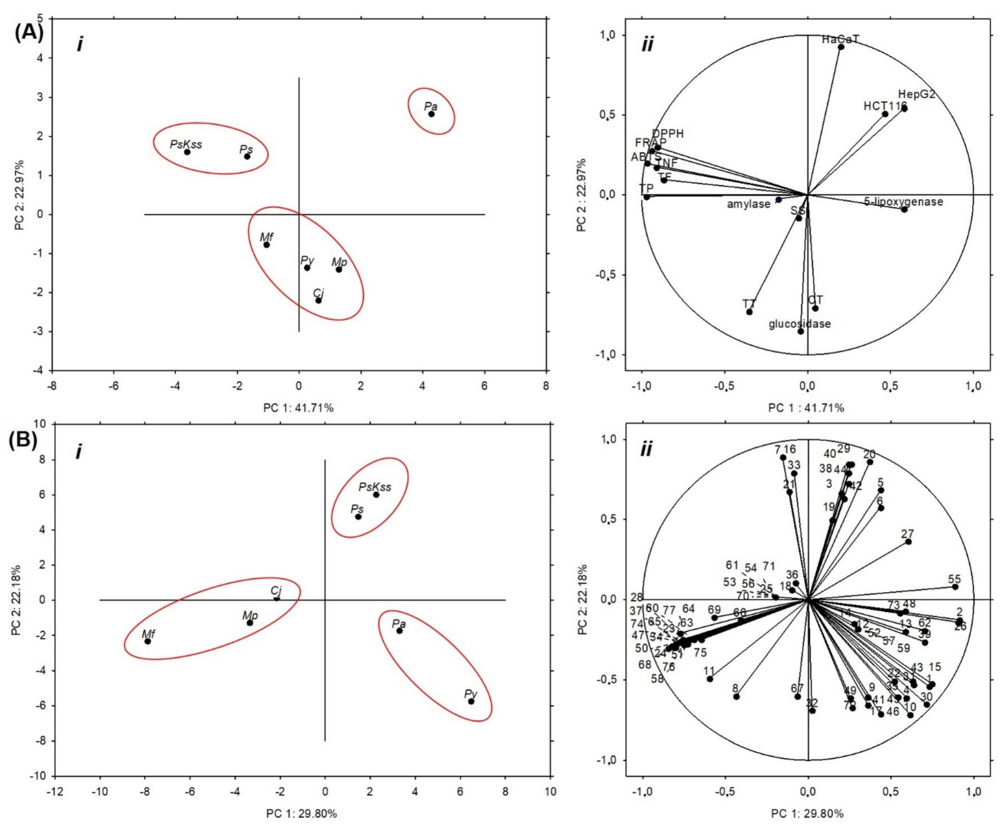
The potential of Pa fruits to inhibit α-glucosidase is known [22]; however, there are no data on their or other Rosaceae inflorescences’ antidiabetic activity. In our study, Mf and Cj showed a significantly higher rate of α-glucosidase inhibition than the other samples (Figure 1B). Moreover, the inhibition percentages (86.29 ± 3.78% and 83.91 ± 0.48%, respectively) were very close to the value of the standard acarbose at the same concentration, 92.70 ± 1.21% (Figure 1). Compared to the inhibition percentages of common vegetables [32], Rosaceae inflorescences can justifiably be considered relevant natural α-glucosidase inhibitors. Acarbose, a pseudotetrasaccharide, is otherwise a highly effective inhibitor of intestinal α-glucosidases; however, it is not effectively absorbed into the bloodstream, but rather retained in the intestine, and may cause gastrointestinal side effects [29]. Therefore, the plant-based α-glucosidase inhibitors with lower side effects are very welcome. This indicates the high potential of Mf and Cj inflorescences to attenuate hyperglycemia, and should definitely be investigated further in in vivo models. For example, just recently, Kumar et al. [33] revealed that the extract of P. amygdalus seed coat applied to diabetic rats significantly reduced the level of blood glucose, and down-regulated hyperglycemic stress, oxidative stress and hyperlipidaemia. Moreover, they found out that the in vivo antidiabetic activity of the extract was accomplished via the inhibition of dipeptidyl peptidase IV (DPP-IV) protein. This is a hydrolase distributed in various tissues and the circulation, which quickly metabolizes glucagon-like peptide-1 (GLP-1). GLP-1, otherwise, maintains the blood glucose level, supporting insulin secretion and β-cell masses, reducing glucagon secretion, and changing the rate of gastric emptying [34,35]. By inhibiting the DPP-IV, the level of GLP-1 can be maintained; in this way the blood glucose level can be maintained as well. We hypothesize that Mf and Cj inflorescences’ extracts might also affect DPP-IV activity in vivo, and this would be good to test in future. Considering the content of phytochemicals, Mf had the highest concentration of total tannins among the samples, 107.85 ± 1.09 mg CE/g DW, and Cj—as mentioned earlier—had the highest concentration of total condensed tannins and individual identified procyanidins (Table 1, Table 2). Therefore, these groups of compounds were probably responsible for the strong α-glucosidase inhibition. Furthermore, PCA revealed that the total and condensed tannins mostly contributed to the inhibition of α-glucosidase (Figure 2Aii).
The significance of condensed tannins in α-glucosidase inhibition has also been detected in the analysis of P. persica pulp [13]. The individual identified compounds predominating in Cj we have already emphasized, and in Mf the predominant compounds were chlorogenic acid (8.41 ± 0.69 mg/g DW), di-caffeoylquinic acid 2, eriodictyol hexoside 1 and 2, quercetin-3-rhamnoside, quercetin-arabinofuranoside, kaempferol rhamnoside, isorhamnetin hexoside, syringetin hexoside 1, phloridzin, trilobatin, procyanidin dimers, procyanidin trimers, and procyanidin tetramer (Table 2). At the same time, Mf had the highest concentration of total identified compounds, total identified procyanidins, chalcones, flavonols and flavanones (Table 1). As with α-amylase, we would also like to draw attention to the identified procyanidins, especially procyanidin trimer 3, which was present in Mf with more than 10% of the total identified compounds in that species, and probably significantly affected the activity of α-glucosidase. In both of these species, in addition to the identified procyanidins, the predominant compound among the individual compounds was chlorogenic acid, at 8.41 ± 0.69 mg/g DW in Mf and 7.04 ± 0.23 mg/g DW in Cj (Table 2); as such, we assume that these compounds may be responsible for potent α-glucosidase inhibition. Another thing that we detected is that Mf and Cj had high concentrations of procyanidins similar to hawthorn (Crataegus spp.) fruits, which are—due to the preventive activity of these compounds toward oxidative stress after ischemia repercussion injury and myocardial infarction—included in the European pharmacopeia as a complementary treatment for chronic heart failure [36]. Therefore, we think, in the future, that it would be wise to test the potential of Mf and Cj inflorescences’ extracts for the prevention or alleviation of cardiovascular diseases.

2.4. Anti-Inflammatory Activity of Inflorescences

5-Lipoxygenase catalyses the first step in the biosynthesis of leukotrienes, which are pivotal lipid mediators of inflammation and allergy. The inhibition of this enzyme is one of the strategies to reduce inflammation, and the search for natural inhibitors of 5-lipoxygenase is very active. It is known that a fraction of Py bark suppressed inflammatory chemokines in human HaCaT keratinocytes, and that it might have anti-atopic dermatitis activity [11]. However, the possibility of inflammation suppression with Rosaceae inflorescences has not been tested so far. Therefore, this is the first report on 5-lipoxygenase inhibition by Rosaceae inflorescences.
In our work, Cj was the most efficient species in the inhibition of 5-lipoxygenase, but it was less efficient than the standard nordihydroguaiaretic acid (Figure 1C). Very recently, Turkiewicz et al. [20] tested the inhibition potential of Chaenomeles species and cultivars’ fruits toward 15-lipoxygenase; compared to their results, Cj inflorescences are much more efficient against 5-lipoxygenase, and thus present a promising biomaterial for further analysis. Our analyses showed that only Cj had naringenin-hexoside, isorhamnetin acetyl hexoside 1 and 2, myricetin rutinoside, syringetin acetyl hexoside 2, and procyanidin dimer 5 and 6 (Table 2), and that it had the highest content of total condensed tannins (Table 1), 5-p-coumaroyl-quinic acid 2, and procyanidin dimer 1, 2 and 4 (Table 2). Thus, we assume that these compounds, or at least some of them, were crucial for the anti-inflammatory activity of this sample.

2.5. Cytotoxic Activity of the Inflorescences

In this study, we evaluated the in vitro anti-proliferative effect of Rosaceae inflorescences on human hepatocellular (HepG2) and colorectal (HCT 116) carcinoma cells, and non-tumorigenic skin keratinocytes (HaCaT). There is evidently a variability in the cell’s metabolic response to the extracts (Table 4).
The most potent cytotoxic activity shown by Pa toward HCT 116 cell line IC50 was 261.97 ± 13.12 μg/mL, toward HepG2 IC50 was 300.89 ± 0.21 μg/mL, and toward HaCaT cells IC50 was 323.84 ± 46.61 μg/mL (Table 4). Jesus et al. [8] speculated that the high concentration of chlorogenic acid in the leaves of Pa might be the reason of their anticancer activity. However, among the tested samples in our work, Pa was not the one with the highest concentration of this acid (Table 2), and still showed the most potent anticancer activity (Table 4). Therefore, we presume that chlorogenic acid, in itself, may not be crucial for the antiproliferative activity of Pa inflorescences, but in combination with other compounds could act synergistically and enhance the cytotoxicity. Interestingly, Pa stem extracts up to the concentration of 400 μg/mL did not show cytotoxicity toward different cancer cell types, including HepG2, while extracts of the fruits revealed selectivity against colon carcinoma HCT-15 [19]. The cytotoxic effects of isolated catechins are known from both in vitro and in vivo investigations; however, in combinations with the other compounds in the extract, their effects might be different [37]. What surprised us was that Pa had the lowest concentrations of catechins and their oligo- and polymers (Supplementary Figure S3). The cytotoxic activity of these phenolics is well known; however, in the extracts of Pa, Ps, PsKss and Py, some chemical interactions occur between compounds which lead to unexpected effects—the highest cytotoxicity was found in samples with the lowest concentration of catechins, and this was especially emphasized with HaCaT cells, suggesting the cell-based specificity of the extracts as well. Moreover, we detected a strong negative linear correlation between the viability of HaCaT cells, and the content of total tannins (R2 = 0.780) and condensed tannins (R2 = 0.977), and the total identified flavan-3-ols (R2 = 0.952) and procyanidins (R2 = 0.990) among the Prunus samples (Supplementary Figure S3). We hypothesize that these compounds in some way interfere with the cytotoxic effect of Prunus inflorescences’ extracts toward HaCaT. When we looked at the individual components, we detected that epicatechin, procyanidin dimer 2 and procyanidin trimer 1 had strong negative correlations (r = −0.622, r = −0.810, and r = −0.686, respectively) with the HaCaT viability (Supplementary Table S2), so we assume that these compounds might attenuate the cytotoxicity of the Prunus extracts. One possible explanation could be that flavan-3-ols (catechins), and their oligomers and polymers, bind the cytotoxic components of the extracts and act as their antagonists. The higher the concentration of the total and condensed tannins, and the total identified procyanidins and flavanols in Prunus inflorescences, the higher the number of viable HaCaT cells. This is useful information for the possible application of the tested Prunus extracts in dermal wound healing.
Ps and PsKss showed similar cytotoxic potentials to Pa toward HaCaT cells. Cell-specific cytotoxicity levels were ascertained for Ps (HaCaT was most susceptible), Mp (HepG2 was most sensitive), and Mf (HCT 116 was most sensitive) (Table 4).
So far, the anticancer properties of the fruits and stems of Pa have been investigated against five human cancer cell lines, and only the extract of the fruits showed activity against colon carcinoma HCT-15 [19]. Because fruits contain anthocyanins, and stems do not, the presumption is that they might be the key to the anticancer properties of Pa fruits. The inflorescences have not been tested so far.
A very strong positive correlation was found between cytotoxic activity against HepG2 and di-caffeoylquinic acid 3, isorhamnetin dihexoside, laricitrin glucuronide and syringetin hexoside 2 (Supplementary Table S1), all of which are predominantly present in Pa. As such, these compounds might be responsible for the inhibition of cell proliferation, and their cytotoxic potential should be investigated further. The cytotoxic activity toward HCT116 was very strongly correlated with di-caffeoylquinic acid 3,5-feruloylquinic acid, isorhamnetin dihexoside, laricitrin glucuronide and syringetin hexoside 2, which were again predominant in Pa. On the other hand, caffeic acid dihexoside was the only compound that was very strongly correlated with an antiproliferative effect on HaCaT cells (Supplementary Table S2).

2.6. Statistical Analysis

The principal component analysis (PCA) based on the measured groups of metabolites and the antioxidant capacity (Table 1), cytotoxicity (Table 4), and hypoglycemic and anti-inflammatory potential (Figure 1) explained 64.68% of the total variation among the samples, where PC1 accounted for 41.71% of the variance and PC2 accounted for 22.97% (Figure 2Ai). The samples were separated into three groups: Ps and PsKss formed one group; Mp, Cj, Py and Mf formed the other; and Pa was separated alone as the most specific sample (Figure 2Ai). The variables that mostly contributed to the group of Ps and PsKss were antioxidant capacity (ABTS, FRAP and DPPH), total phenolics, flavonoids, and nonflavonoid compounds (Figure 2Aii). The separation of Pa was due to the cytotoxic activity toward all of the three tested cell types. The group of Mp, Cj, Py and Mf mostly contributed tannins, soluble sugars, and the inhibition of α-glucosidase.
Based on the individual identified phenolics, the samples were similarly grouped: the only difference was in the grouping of Py, which was closer to Pa, and they grouped together (Figure 2Bi). This suggests that the representation of the individual identified phenolics and the bioactivity of the tested Rosaceae inflorescences are analogously distributed between all of the samples, except for Py. We also noticed that Cj, based on the measured parameters, was closer to the Malus than to the Prunus samples.
HC analysis, an algorithm that creates a dendrogram showing the hierarchical relationships between different datasets, showed the degree of similarities/dissimilarities between the samples. Based on their groups of metabolites, antioxidant capacity, cytotoxicity, and hypoglycemic and anti-inflammatory potential, Ps and PsKss were the most similar samples to each other. Other samples were close to them, while Pa was the most distant from all of the samples (Supplementary Figure S4A). These results indicate that greater genetic similarity does not imply a greater similarity in biological effects (Supplementary Figure S4A).
Based on their individual identified phenolic compounds, the samples were, as expected, more similar (closer) to each other (Supplementary Figure S4B); the most similar were Pa and Ps, while the most distant from them was Py.
Pearson’s correlation coefficient (r) between the groups of metabolites, antioxidant capacity, cytotoxicity, and hypoglycemic and anti-inflammatory potential of the samples revealed a strong positive correlation between antioxidant capacity (ABTS, FRAP and DPPH) and the total phenolics, flavonoids and nonflavonoids (Table 3). The inhibition of α-glucosidase was strongly positively correlated with cytotoxicity toward HaCaT cells (Table 3). Cytotoxicity toward HepG2 was positively correlated with cytotoxicity toward both HCT 116 and HaCaT cells. Cytotoxic activities toward HCT 116 and HaCaT were also strongly correlated. The total tannins exhibited a strong negative correlation with cytotoxicity toward HCT 116 and HaCaT cells. This suggests that a removal of tannins from the extracts might increase the cytotoxic effect/s of the samples toward HCT 116 and HaCaT, along with their inhibition of α-glucosidase, which is worthy of further investigations. The inhibition of α-glucosidase was strongly positively correlated with the total and condensed tannins, which indicates that these compounds might be responsible for the inhibition of this enzyme. These results are in accordance with previous observations regarding the tannin effect on glucosidase activity [38].
As far as individual compounds are concerned, according to Pearson’s correlation coefficient, the only compound that was very strongly correlated with the inhibition of α-glucosidase was chlorogenic acid (Supplementary Table S2); as such, we suggest the further investigation of the antidiabetic potential of this phenolic compound. The results from all three antioxidant methods strongly correlated with caffeic acid hexoside 1, 3-caffeoylquinic acid, 4-caffeoylquinic acid, 3-p-coumaroylquinic acid, p-coumaric acid hexoside 2, catechin, quercetin-hexoside pentoside, quercetin-3-glucoside, kaempferol dihexoside and kaempferol trihexoside. Therefore, we assume that these compounds mostly contributed to the antioxidant activity of the inflorescences. The cytotoxic activity toward HepG2, HCT 116 and HaCaT strongly correlated with di-caffeoylquinic acid 3,3-feruloylquinic acid, 5-feruloylquinic acid, isorhamnetin dihexoside, laricitrin glucuronide and syringetin hexoside 2. The individual compounds that strongly correlated with the inhibition of both antidiabetic enzymes, α-amylase and α-glucosidase, were procyanidin dimer 1 and 4. With the inhibition of α-amylase, the only strong correlations were naringenin hexoside, isorhamnetin acetyl hexoside 1 and 2, myricetin rutinoside, syringetin acetyl hexoside 2, and procyanidin dimer 1, 4, 5 and 6. The inhibition of α-glucosidase strongly correlated with chlorogenic acid; procyanidin dimer 1, 2, 4; and procyanidin tetramer.
Based on the results, we propose the consideration of Prunus, Malus and Chaenomeles inflorescences as low-sugar plant foods rich in polyphenolic bioactive compounds, or at least as functional additives to regular food that could improve human health. In particular, we propose Cj inflorescences for further in vivo studies of their antidiabetic and anti-inflammatory activity, and for possible use as a functional food. Prunus inflorescences would be excellent candidates for further analyses of the influence of monomers, oligomers and polymers of flavanols on HaCaT cells’ proliferation. Finally, PsKss is a material with a respectable amount of total phenolics and flavonoids that shows strong antioxidant activity and α-amylase inhibition; therefore, it is worthy of further in vitro and in vivo investigations.

3. Materials and Methods

3.1. Chemicals and Materials

All of the chemicals and reagents were supplied by Sigma Aldrich GmbH (Taufkirchen, Germany). A Gemini 3 µm C18 110A New Column 150 × 4.6 mm and a Gemini C18 4 × 3.0 mm guard column were purchased from Phenomenex (Torrance, CA, USA). The HaCaT, HCT 116 and HepG2 were obtained from the American Type Culture Collection (ATCC, Manassas, VA, USA).
The inflorescence samples of each of the variety—Pa, Ps, PsKss, Mp, Mf and Cj—were collected separately three times during April 2018 in the Botanical Garden of the Faculty of Science, University of Zagreb, Croatia. The plant material was freeze dried using an Alpha 1–2 Christ freeze-dryer, pulverised using a pestle and mortar, and then used to prepare the extracts.

3.2. Extraction of the Phenolic Compounds

The phenolic compounds were extracted in an ice-cold ultrasonic bath for 60 min with 70% EtOH using an equivalent of 30 mg/mL dry weight. The homogenates were centrifuged for 8 min at 9500 rpm. The supernatant was filtered through a Chromafil AO-20/25 polyamide ester filter produced by Macherey-Nagel (Düren, Germany).

3.3. Spectrophotometric Determination of the Phytochemicals and Antioxidant Capacity

The total phenols, flavonoids, non-flavonoids, tannins, condensed tannins and soluble sugars were determined as in Poljuha et al. [39] and Šola et al. [40,41]. The antioxidant activity of the non-hydrolyzed extracts was measured using three assays (ABTS, DPPH and FRAP), as described in Poljuha et al. [39]. Three repetitions in three independent experiments were performed for the same sample, and all of the absorbance measurements were performed with a Fluostar Optima microplate reader (BMG Labtech GmbH, Offenburg, Germany) at different wavelengths (734 nm for ABTS, 517 nm for DPPH, and 593 nm for FRAP).

3.4. LC-DAD-MS Analysis

The phenolic compounds were analysed on a Dionex HPLC system with a diode array detector (Thermo Scientific, San Jose, CA, USA) using Chromeleon workstation software. A Column Gemini heated at 25 °C was used. The extracts were eluted with aqueous 0.1% formic acid and 3% acetonitrile (A), and 0.1% formic acid and 3% water in absolute acetonitrile (B). All of the phenolic compounds were identified using a mass spectrometer (LTQ XL Linear Ion Trap Mass Spectrometer, Thermo Fisher Scientific, USA) with an electrospray interface (ESI) operating in negative ion mode, under the same conditions as those reported by Mikulic-Petkovsek et al. [42]. The contents of the phenolic compounds were calculated from the peak areas of the sample and the corresponding standards, and were expressed in mg/g dry weight. All of the analyses were performed in triplicate.

3.5. Effect of the Extracts on Antidiabetic (α-Amylase and α-Glucosidase) and Anti-Inflammatory (5-Lipoxygenase) Activity

The α-amylase inhibitory activity was tested as reported in Šola et al. [41], and the absorbance was measured at 544 nm using a microplate reader. The percentage of α-amylase inhibition at a sample concentration of 0.8 mg/mL was calculated using the following equation:
% inhibition = [100 − (At − Atb)/(Ac − Acb)] × 100,
where At was the absorbance of the test (with amylase), Atb was the absorbance of the test blank (without amylase), Ac was the absorbance of the control (with amylase), and Acb was the absorbance of the control blank (without amylase). Maltose was used as a positive control.
The inhibition of α-glucosidase was measured using the pre-incubation method, as described by Salahuddin et al. [43], with slight modifications. In brief, 20 μL extract was mixed with 100 μL p-nitrophenyl-α-D-glucopyranoside (1 mM in 100 Mm phosphate buffer) and pre-incubated for 10 min at 37 °C. A volume of 100 μL α-glucosidase (56.6 mU/mL in phosphate buffer) was added and re-incubated for 20 min at 37 °C. The enzyme reaction activity was terminated by the addition of 500 μL Na2CO3 (1 M). The absorbance was measured at 405 nm using a microplate reader. The enzyme inhibitory activity at a sample concentration of 0.55 mg/mL was calculated similarly to that for the α-amylase inhibition. Acarbose was used as a positive control.
The inhibition of the 5-lipoxygenase activity was assessed according to El Euch et al. [4]. In brief, in 350 µL phosphate buffer (pH 7.4) containing Na2HPO4, KH2PO4 and NaCl, 40 µL lipoxygenase enzyme (500 U) from Glycine max was mixed with 120 µL 3.5 mM linoleic acid and 40 µL 20 mg/mL inflorescence extracts. The mixture was homogenized using a vortex mixer and incubated for 10 min at 25 °C. The absorbance was measured in triplicate at 234 nm using a NanodropTM 2000c (Thermo Scientific). The enzyme inhibitory activity at a sample concentration of 1.45 mg/mL was calculated from the following equation:
% inhibition = [(Acon − Abackground) − (Asample − Abackground) × 100%/(Acon − Abackground)]
where Acon = the absorbance of the control, Abackground = the absorbance of the background (blank), and Asample = the absorbance of the sample. Nordihydroguaiaretic acid was used as a positive control.

3.6. In Vitro Antiproliferative Activity

The in vitro antiproliferative activity (IC50 expressed in μg/mL) was determined for HepG2 (HB-8065), HCT 116 (CCL-247) and HaCaT (CVCL-0038) cells using an MTT assay, which is a quantitative colorimetric assay for the measurement of cell survival and proliferation, as described in Šola et al. [41]. In brief, 4 × 10³ cells were seeded in 96-well plates, and then treated 24 h later with different concentrations of the extracts diluted in growth medium. As a control, the same dilution of 70% ethanol in growth medium was prepared and incubated with the cells. After 72 h, the treatment was removed, 1 × MTT was added, and the cells were incubated for 4 h in the growth conditions, followed by the addition of dimethyl sulfoxide and 20 min incubation with gentle mixing. The absorbance was measured at λ = 570 nm with an ELISA microplate reader (LabSystem Multiskan MS, Artisan Technology group, Champaign, IL, USA).

3.7. Statistical Analysis

The data obtained were statistically processed in the Statistica 13.1 program (Stat Soft Inc., Palo Alto, CA, USA). All of the experiments were performed in triplicate. The comparison of the samples’ means was carried out using one-way variance analysis (ANOVA) and Duncan’s New Multiple Range Test (DNMRT). The statistically significant values were those that differed at the p ≤ 0.05 level. Principal component analysis (PCA) and hierarchical clustering (HC) were performed to evaluate how close the samples were according to the given parameters. The Pearson’s correlation coefficients between the phytochemical content and bioactivities of the inflorescences were calculated.

4. Conclusions

This is the first study that characterised the phytochemical profile, in vitro antioxidant activity, and antidiabetic, anti-inflammatory and antiproliferative potential of inflorescences of Pa, Ps, PsKss, Mp, Mf and Cj. Moreover, for the first time, Rosaceae inflorescences were screened for their antidiabetic and anti-inflammatory activity. The results showed that these inflorescences are a valuable source of phenolics with significant biological activities. The highest amount of total phenolics, flavonoids and nonflavonoids, and the highest antioxidant capacity measured by all three methods was recorded in PsKss. Besides Mp, this species also contained the lowest concentration of soluble sugars. In addition, the sugar content in the inflorescences was lower than those in the more commonly used fruits. Among the tested Malus species, Mf inflorescences had significantly higher total phenolics, flavonoids and tannins, as well as identified polyphenolic compounds, and showed higher antioxidant activity and α-glucosidase inhibition than Mp. The sample that showed the most effective inhibition of both antidiabetic enzymes and anti-inflammatory 5-lipoxygenase was Cj. Therefore, we suggest further in vitro and in vivo analyses of the antidiabetic and anti-inflammatory potential of Cj inflorescences. Beside Cj, PsKss showed strong α-amylase inhibition, and the strong inhibition of α-glucosidase Mf. The cytotoxic activity was both species- and cell-type-specific. The most effective in the inhibition of the growth and proliferation of HepG2, HCT 116 and HaCaT cells was Pa. The antiproliferative effect of extracts from Prunus inflorescences was negatively correlated with the concentration of the total and condensed tannins, identified procyanidins (especially procyanidin dimer 2 and procyanidin trimer 1) and flavanols (especially epicatechin). The inhibition of α-glucosidase showed a strong negative correlation with the total tannins. The results in this work encourage further in vivo studies of these matrices, and suggest innovations for future-oriented diets and food production, such as the incorporation of Prunus, Malus and Chaenomeles inflorescences’ extracts as dietary supplements and functional ingredients.

Supplementary Materials

The following are available online at https://www.mdpi.com/article/10.3390/plants11030271/s1, Figure S1: The representative chromatograms, at 280 and 350 nm, of (A) Prunus avium, (B) P. serrulata, (C) P. serrulata ‘Kiku Shidare Zakura’, (D) P. yedoensis, (E) Malus purpurea, (F) M. floribunda and (G) Chaenomeles japonica. The numbers above the peaks correspond to the numbers given in the Table 2. Figure S2: (A) the number of identified phenolic compounds, (B) the number of compounds with a predominant concentration, and (C) the proportion of predominantly represented compounds (%) in Rosaceae inflorescences. Pa = P. avium, Ps = P. serrulata, PsKss = P. serrulata ‘Kiku Shidare Zakura’, Py = P. yedoensis, Mp = M. purpurea, Mf = M. floribunda, Cj = Chaenomeles japonica. Figure S3: Linear correlation between the HaCaT cells’ viability and the amount of (A) total tannins, (B) total condensed tannins, (C) total identified procyanidins and (D) total identified flavanols in different Prunus varieties’ inflorescences. CE = catechin equivalent. Figure S4: Hierarchical clustering, expressed as Euclidean distance, of Rosaceae inflorescences (Pa, Pss, PsKss, Py, Mp, Mf, Cj) based on their (A) groups of metabolites, antioxidant capacity, cytotoxicity, antidiabetic and anti-inflamatory potential; and (B) identified individual phytochemicals. Pa = P. avium, Ps = P. serrulata, PsKss = P. serrulata ‘Kiku Shidare Zakura’, Py = P. yedoensis, Mp = M. purpurea, Mf = M. floribunda, Cj = Chaenomeles japonica. a, a1-a1-1-1-1-1 = clusters. Table S1: Identification of phenolic compounds in Rosaceae inflorescences in negative ionization with LC-DAD-MS and MS2/MS3. Table S2: Pearson’s correlation coefficient (r) between the individual identified compounds, antioxidant capacity, cytotoxicity, and hypoglycemic and anti-inflammatory potential of Rosaceae inflorescences.

Author Contributions

I.Š.: Conceptualization, writing—draft, writing—review and editing, formal analysis, methodology; D.P.: formal analysis, visualization; M.M.-P.: data curation, investigation; D.D.: data curation, investigation; M.P.: data curation, investigation; J.B.: investigation; R.V.: funding acquisition; M.H.: funding acquisition; G.R.: antioxidant assays and funding acquisition. All authors have read and agreed to the published version of the manuscript.

Funding

This research was financially funded by the University of Zagreb (grant number 20284112) and the programme P4-0013-0481, which was funded by the Slovenian Research Agency (ARRS).

Institutional Review Board Statement

Not applicable.

Informed Consent Statement

Not applicable.

Data Availability Statement

Not applicable.

Conflicts of Interest

The authors declare no conflict of interest. The funders had no role in the design of the study; in the collection, analyses, or interpretation of data; in the writing of the manuscript, or in the decision to publish the results.

References

  1. Orlando, G.; Recinella, L.; Chiavaroli, A.; Brunetti, L.; Leone, S.; Carradori, S.; Di Simone, S.; Ciferri, M.C.; Zengin, G.; Ak, G.; et al. Water extract from inflorescences of industrial hemp futura 75 variety as a source of anti-inflammatory, anti-proliferative and antimycotic agents: Results from in silico, in vitro and ex vivo studies. Antioxidants 2020, 9, 437. [Google Scholar] [CrossRef]
  2. Lau, B.F.; Kong, K.W.; Leong, K.H.; Sun, J.; He, X.; Wang, Z.; Mustafa, M.R.; Ling, T.C.; Ismail, A. Banana inflorescence: Its bio-prospects as an ingredient for functional foods. Trends Food Sci. Technol. 2020, 97, 14–28. [Google Scholar] [CrossRef]
  3. Sarv, V.; Venskutonis, P.R.; Bhat, R. The Sorbus spp.—Underutilised plants for foods and nutraceuticals: Review on polyphenolic phytochemicals and antioxidant potential. Antioxidants 2020, 9, 813. [Google Scholar] [CrossRef]
  4. El Euch, S.K.; Bouajila, J.; Bouzouita, N. Chemical composition, biological and cytotoxic activities of Cistus salviifolius flower buds and leaves extracts. Ind. Crops Prod. 2015, 76, 1100–1105. [Google Scholar] [CrossRef]
  5. Lee, E.J.; Kim, J.S.; Kim, H.P.; Lee, L.-H.; Kang, S.S. Phenolic constituents from the flower buds of Lonicera japonica and their 5-lipoxygenase inhibitory activities. Food Chem. 2010, 120, 134–139. [Google Scholar] [CrossRef]
  6. Ferreres, F.; Andrade, C.; Gomes, N.G.; Andrade, P.B.; Gil-Izquierdo, A.; Pereira, D.M.; Suksungworn, R.; Duangsrisai, S.; Videira, R.A.; Valentão, P. Valorisation of kitul, an overlooked food plant: Phenolic profiling of fruits and inflorescences and assessment of their effects on diabetes-related targets. Food Chem. 2021, 342, 128323. [Google Scholar] [CrossRef]
  7. Olszewska, M.A.; Michel, P. Antioxidant activity of inflorescences, leaves and fruits of three Sorbus species in relation to their polyphenolic composition. Nat. Prod. Res. 2009, 23, 1507–1521. [Google Scholar] [CrossRef] [PubMed]
  8. Jesus, F.; Gonçalves, A.C.; Alves, G.; Silva, L.R. Health benefits of Prunus avium plant parts: An unexplored source rich in phenolic compounds. Food Rev. Int. 2020. [Google Scholar] [CrossRef]
  9. Acero, N.; Gradillas, A.; Beltran, M.; García, A.; Muñoz Mingarro, D. Comparison of phenolic compounds profile and antioxidant properties of different sweet cherry (Prunus avium L.) varieties. Food Chem. 2019, 279, 260–271. [Google Scholar] [CrossRef] [PubMed]
  10. Usenik, V.; Fabčič, J.; Štampar, F. Sugars, organic acids, phenolic composition and antioxidant activity of sweet cherry (Prunus avium L.). Food Chem. 2008, 107, 185–192. [Google Scholar] [CrossRef]
  11. Kang, G.-J.; Lee, H.-J.; Yoon, W.-J.; Yang, E.-J.; Park, S.-S.; Kang, H.-K.; Park, M.-H.; Yoo, E.-S. Prunus yedoensis inhibits the inflammatory chemokines, MDC and TARC, by regulating the STAT1-signaling pathway in IFN-γ-stimulated HaCaT human keratinocytes. Biomol. Ther. 2008, 16, 394–402. [Google Scholar] [CrossRef] [Green Version]
  12. Teleszko, M.; Wojdyło, A. Comparison of phenolic compounds and antioxidant potential between selected edible fruits and their leaves. J. Funct. Foods 2015, 14, 736–746. [Google Scholar] [CrossRef]
  13. Zhang, X.; Su, M.; Du, J.; Zhou, H.; Li, X.; Li, X.; Ye, Z. Comparison of phytochemical differences of the pulp of different peach (Prunus persica (L.) Batsch) cultivars with alpha-glucosidase inhibitory activity variations in China using UPLC-Q-TOF/MS. Molecules 2019, 24, 1968. [Google Scholar] [CrossRef] [Green Version]
  14. Wojdyło, A.; Nowicka, P.; Grimalt, M.; Legua, P.; Almansa, M.S.; Amorós, A.; Carbonell-Barrachina, A.; Hernández, F. Polyphenol compounds and biological activity of caper (Capparis spinosa L.) flowers buds. Plants 2019, 8, 539. [Google Scholar] [CrossRef] [Green Version]
  15. Borneo, R.; León, A.E.; Aguirre, A.; Ribotta, P.; Cantero, J.J. Antioxidant capacity of medicinal plants from the Province of Córdoba (Argentina) and their in vitro testing in a model food system. Food Chem. 2009, 112, 664–670. [Google Scholar] [CrossRef]
  16. Afonso, S.; Oliveira, I.V.; Meyer, A.S.; Aires, A.; Saavedra, M.J.; Gonçalves, B. Phenolic profile and bioactive potential of stems and seed kernels of sweet cherry fruit. Antioxidants 2020, 9, 1295. [Google Scholar] [CrossRef]
  17. Yang, S.; Meng, Z.; Li, Y.; Chen, R.; Yang, Y.; Zhao, Z. Evaluation of physiological characteristics, soluble sugars, organic acids and volatile compounds in ‘Orin’ apples (Malus domestica) at different ripening stages. Molecules 2021, 26, 807. [Google Scholar] [CrossRef]
  18. Walker, R.P.; Battistelli, A.; Bonghi, C.; Drincovich, M.F.; Falchi, R.; Lara, M.V.; Moscatello, S.; Vizzotto, G.; Famiani, F. Non-structural carbohydrate metabolism in the flesh of stone fruits of the genus Prunus (Rosaceae)—A review. Front. Plant Sci. 2020, 11, 549921. [Google Scholar] [CrossRef]
  19. Bastos, C.; Barros, L.; Dueñas, M.; Calhelha, R.C.; Queiroz, M.J.R.; Santos-Buelga, C.; Ferreira, I.C. Chemical characterisation and bioactive properties of Prunus avium L.: The widely studied fruits and the unexplored stems. Food Chem. 2015, 173, 1045–1053. [Google Scholar] [CrossRef] [Green Version]
  20. Turkiewicz, I.P.; Wojdyło, A.; Tkacz, K.; Nowicka, P.; Golis, T.; Bąbelewski, P. ABTS On-line antioxidant, α-amylase, α-glucosidase, pancreatic lipase, acetyl-and butyrylcholinesterase inhibition activity of Chaenomeles fruits determined by polyphenols and other chemical compounds. Antioxidants 2020, 9, 60. [Google Scholar] [CrossRef] [PubMed] [Green Version]
  21. Fang, L.; Meng, W.; Min, W. Phenolic compounds and antioxidant activities of flowers, leaves and fruits of five crabapple cultivars (Malus Mill. species). Sci. Hortic. 2018, 235, 460–467. [Google Scholar] [CrossRef]
  22. Gonçalves, A.C.; Bento, C.; Silva, B.M.; Silva, L.R. Sweet cherries from Fundão possess antidiabetic potential and protect human erythrocytes against oxidative damage. Food Res. Int. 2017, 95, 91–100. [Google Scholar] [CrossRef] [Green Version]
  23. Gonçalves, B.; Landbo, A.K.; Knudsen, D.; Silva, A.P.; Moutinho-Pereira, J.; Rosa, E.; Meyer, A.S. Effect of ripeness and postharvest storage on the phenolic profiles of cherries (Prunus avium L.). J. Agric. Food Chem. 2004, 52, 523–530. [Google Scholar] [CrossRef]
  24. Olszewska, M. Quantitative HPLC analysis of flavonoids and chlorogenic acid in the leaves and inflorescences of Prunus serotina Ehrh. Acta Chromatogr. 2007, 19, 253–269. [Google Scholar]
  25. Olszewska, M.A.; Kwapisz, A. Metabolite profiling and antioxidant activity of Prunus padus L. flowers and leaves. Nat. Prod. Res. 2011, 25, 1115–1131. [Google Scholar] [CrossRef]
  26. Coklar, H.; Akbulut, M.; Alhassan, I.; Kirpitci, Ş.; Korkmaz, E. Organic acids, sugars, phenolic compounds and antioxidant activity of Malus floribunda coccinella fruit, peel and flesh. Acta Sci. Pol. Hortorum Cultus 2018, 17, 47–59. [Google Scholar] [CrossRef]
  27. Du, H.; Wu, J.; Li, H.; Zhong, P.-X.; Xu, Y.-J.; Li, C.-H.; Ji, K.-X.; Wang, L.-S. Polyphenols and triterpenes from Chaenomeles fruits: Chemical analysis and antioxidant activities assessment. Food Chem. 2013, 141, 4260–4268. [Google Scholar] [CrossRef] [PubMed]
  28. Valcheva-Kuzmanova, S.V.; Denev, P.N.; Ognyanov, M.H. Chemical composition and antioxidant activity of Chaenomeles maulei fruit juice. J. Biomed. Clin. Res. 2018, 11, 41–48. [Google Scholar] [CrossRef] [Green Version]
  29. Da Silva, S.M.; Koehnlein, E.A.; Bracht, A.; Castoldi, R.; de Morais, G.R.; Baesso, M.L.; Peralta, R.; de Souza, C.G.M.; de Sá-Nakanishi, A.B.; Peralta, R.M. Inhibition of salivary and pancreatic α-amylases by a pinhão coat (Araucaria angustifolia) extract rich in condensed tannin. Food Res. Int. 2014, 56, 1–8. [Google Scholar] [CrossRef] [Green Version]
  30. Mkandawire, N.L.; Kaufman, R.C.; Bean, S.R.; Weller, C.L.; Jackson, D.S.; Rose, D.J. Effects of sorghum (Sorghum bicolor (L.) Moench) tannins on alpha-amylase activity and in vitro digestibility of starch in raw and processed flours. J. Agric. Food Chem. 2013, 61, 4448–4454. [Google Scholar] [CrossRef] [PubMed]
  31. Papoutsis, K.; Zhang, J.; Bowyer, M.C.; Brunton, N.; Gibney, E.R.; Lyng, J. Fruit, vegetables, and mushrooms for the preparation of extracts with α-amylase and α-glucosidase inhibition properties: A review. Food Chem. 2021, 338, 128119. [Google Scholar] [CrossRef]
  32. Assefa, S.T.; Yang, E.Y.; Chae, S.Y.; Song, M.; Lee, J.; Cho, M.C.; Jang, S. Alpha glucosidase inhibitory activities of plants with focus on common vegetables. Plants 2020, 9, 2. [Google Scholar] [CrossRef] [PubMed] [Green Version]
  33. Kumar, V.; Sachan, R.; Rahman, M.; Sharma, K.; Al-Abassi, F.A.; Anwar, F. Prunus amygdalus extract exert antidiabetic effect via inhibition of DPP-IV: In-silico and in-vivo approaches. J. Biomol. Struct. Dyn. 2021, 39, 4160–4174. [Google Scholar] [CrossRef]
  34. Meier, J.J. GLP-1 receptor agonists for individualized treatment of type 2 diabetes mellitus. Nat. Rev. Endocrinol. 2012, 8, 728–742. [Google Scholar] [CrossRef]
  35. Seino, Y.; Fukushima, M.; Yabe, D. GIP and GLP-1, the two incretin hormones: Similarities and differences. J. Diabetes Investig. 2010, 1, 8–23. [Google Scholar] [CrossRef] [Green Version]
  36. Wang, T.K.; Xu, S.; Li, S.; Zhang, Y. Proanthocyanidins should be a candidate in the treatment of cancer, cardiovascular diseases and lipid metabolic disorder. Molecules 2020, 25, 5971. [Google Scholar] [CrossRef] [PubMed]
  37. Xu, Y.Q.; Gao, Y.; Granato, D. Effects of epigallocatechin gallate, epigallocatechin and epicatechin gallate on the chemical and cell-based antioxidant activity, sensory properties, and cytotoxicity of a catechin-free model beverage. Food Chem. 2021, 339, 128060. [Google Scholar] [CrossRef]
  38. Muccilli, V.; Cardullo, N.; Spatafora, C.; Cunsolo, V.; Tringali, C. α-Glucosidase inhibition and antioxidant activity of an oenological commercial tannin. Extraction, fractionation and analysis by HPLC/ESI-MS/MS and 1H NMR. Food Chem. 2017, 215, 50–60. [Google Scholar] [CrossRef] [PubMed]
  39. Poljuha, D.; Šola, I.; Bilić, J.; Dudaš, S.; Bilušić, T.; Markić, J.; Rusak, G. Phenolic composition, antioxidant capacity, energy content and gastrointestinal stability of Croatian wild edible plants. Eur. Food Res. Technol. 2015, 241, 573–585. [Google Scholar] [CrossRef]
  40. Šola, I.; Vujčić Bok, V.; Dujmović, M.; Rusak, G. Developmentally-related changes in phenolic and L-ascorbic acid content, and antioxidant capacity of Chinese cabbage sprouts. J. Food Sci. Technol. 2020, 57, 702–712. [Google Scholar] [CrossRef]
  41. Šola, I.; Vujčić Bok, V.; Pinterić, M.; Auer, S.; Ludwig-Müller, J.; Rusak, G. Improving the phytochemical profile and bioactivity of Chinese cabbage sprouts by interspecific transfer of metabolites. Food Res. Int. 137, 109726. [CrossRef] [PubMed]
  42. Mikulic-Petkovsek, M.; Samoticha, J.; Eler, K.; Stampar, F.; Veberic, R. Traditional elderflower beverages: A rich source of phenolic compounds with high antioxidant activity. J. Agric. Food Chem. 2015, 63, 1477–1487. [Google Scholar] [CrossRef] [PubMed]
  43. Salahuddin, M.A.H.; Ismail, A.; Kassim, N.K.; Hamid, M.; Ali, M.S.M. Phenolic profiling and evaluation of in vitro antioxidant, α-glucosidase and α-amylase inhibitory activities of Lepisanthes fruticosa (Roxb) Leenh fruit extracts. Food Chem. 2020, 331, 127240. [Google Scholar] [CrossRef] [PubMed]
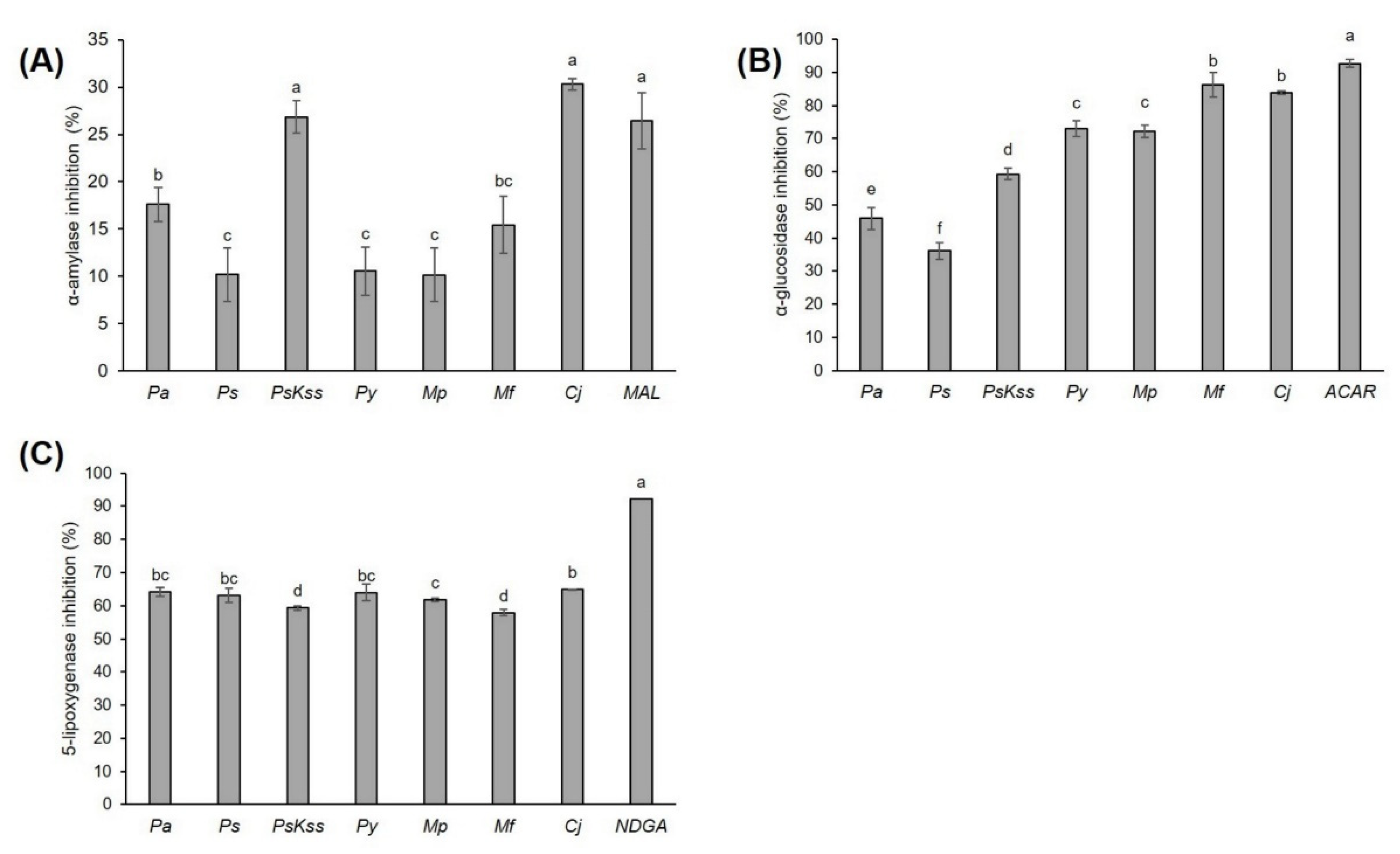
Figure 1. Inhibition of (A) α-amylase, (B) α-glucosidase, and (C) 5-lipoxygenase activity by Rosaceae inflorescences’ extracts (0.80 mg/mL, 0.55 mg/mL and 1.45 mg/mL, respectively). The values represent the mean ± standard deviation of three replicates. Different letters indicate a significant difference among the values (ANOVA, Duncan test, p ≤ 0.05). Pa = P. avium, Ps = P. serrulata, PsKss = P. serrulata ‘Kiku Shidare Zakura’, Py = P. yedoensis, Mp = M. purpurea, Mf = M. floribunda, Cj = Chaenomeles japonica, MAL = maltose 0.80 mg/mL, ACAR = acarbose 0.55 mg/mL, NDGA = nordihydroguaiaretic acid 0.15 mg/mL.
Plants 11 00271 g001
Figure 2. Principal component analysis of (A) the groups of metabolites, antioxidant capacity, cytotoxicity, antidiabetic and anti-inflammatory potential of Rosaceae inflorescences: (i) score plot separating the inflorescence samples based on the measured groups of metabolites, antioxidant capacity, cytotoxicity, and antidiabetic and anti-inflammatory potential, and (ii) loading plot of the measured variables; (B) the individual identified phenolic compounds in Rosaceae inflorescences: (i) score plot separating the inflorescence samples based on the individual identified phenolic compounds they contain, and (ii) the loading plot of the individual phenolics as variables. Pa = P. avium, Ps = P. serrulata, PsKss = P. serrulata ‘Kiku Shidare Zakura’, Py = P. yedoensis, Mp = M. purpurea, Mf = M. floribunda, Cj = Chaenomeles japonica, TP = total phenolics, TF = total flavonoids, TNF = total nonflavonoids, TT = total tannins, CT = condensed tannins, SS = soluble sugars, ABTS = 2,2′-azino-bis(3-ethylbenzothiazoline-6-sulfonic acid) diammonium salt, FRAP = ferric reducing antioxidant power, DPPH = 2,2-diphenyl-1-picrylhydrazyl, 1–77 = numbers related to the individual identified phenolics, as depicted in the Table 2.
Plants 11 00271 g002
Table 1. Concentration (in mg/g dry weight) of the total phenolics (TP), total flavonoids (TF), total nonflavonoids (TNF), total tannins (TT), condensed tannins (CT) and soluble sugars (SS), and the antioxidant capacity (ABTS, FRAP and DPPH) of Rosaceae inflorescences.
Prunus aviumPrunus serrulataPrunus serrulata ‘Kiku Shidare Zakura’Prunus yedoensisMalus purpureaMalus floribundaChaenomeles japonica
TP (mg GAE/g DW)27.83 ± 0.69 f46.84 ± 0.75 b53.12 ± 0.79 a37.31 ± 0.48 e40.29 ± 0.64 c46.74 ± 0.93 b39.42 ± 0.97 d
TF (mg CE/g DW)13.57 ± 0.84 f32.35 ± 2.13 b38.89 ± 4.04 a25.78 ± 1.03 d13.43 ± 0.82 f23.83 ± 0.88 e29.45 ± 0.65 c
TNF (mg GAE/g DW)16.40 ± 0.91 g29.35 ± 0.83 b31.32 ± 0.71 a22.21 ± 0.86 e23.52 ± 0.56 d28.85 ± 0.58 c18.73 ± 0.48 f
TT (mg CE/g DW)27.26 ± 0.22 g59.44 ± 1.32 f71.59 ± 0.33 d83.55 ± 0.55 b64.32 ± 0.45 e107.85 ± 1.09 a80.27 ± 0.33 c
CT (mg CE/g DW)4.25 ± 0.33 e7.74 ± 1.02 d6.99 ± 0.17 d16.52 ± 0.01 b15.45 ± 0.10 b10.98 ± 0.24 c51.68 ± 0.38 a
SS (mg SE/g DW)3.37 ± 0.06 c3.04 ± 0.05 d2.41 ± 0.10 e8.61 ± 0.12 a1.56 ± 0.08 f3.58 ± 0.06 b3.27 ± 0.07 c
ABTS (mg TE/g DW)22.86 ± 4.61 e49.41 ± 7.23 b61.32 ± 5.84 a36.63 ± 4.32 c28.78 ± 2.52 d47.78 ± 6.26 b35.05 ± 4.25 c
FRAP (mg TE/g DW)27.89 ± 0.60 g51.68 ± 0.12 b58.06 ± 0.78 a40.28 ± 1.18 d29.12 ± 0.83 f44.36 ± 0.85 c36.36 ± 1.50 e
DPPH (mg TE/g DW)25.47 ± 2.57 d52.95 ± 4.22 b69.42 ± 3.27 a39.21 ± 4.86 c25.10 ± 3.25 d40.38 ± 3.4 c39.61 ± 3.95 c
Values represent the mean ± standard deviation of three replicates. Different letters indicate a significant difference among the values in a row (ANOVA, Duncan test, p ≤ 0.05). GAE = gallic acid equivalent, CE = catechin equivalent, SE = sucrose equivalent, TE = trolox equivalent.
Table 2. Concentration (mg/g DW ± SD) of the individual phenolic compounds in Rosaceae inflorescences.
Prunus aviumPrunus serrulataPrunus serrulata ‘Kiku Shidare Zakura’Prunus yedoensisMalus purpureaMalus floribundaChaenomeles japonica
1Gallic acid0.28 ± 0.06 b0.15 ± 0.04 c0.06 ± 0.01 d0.38 ± 0.03 a0.17 ± 0.04 c0.03 ± 0.01 dnd
Total identified
hydroxybenzoic acids
0.28 ± 0.06 b0.15 ± 0.04 c0.06 ± 0.01 d0.38 ± 0.03 a0.17 ± 0.04 c0.03 ± 0.01 dnd
2Caffeic acid1.95 ± 0.27 c1.25 ± 0.14 c2.50 ± 0.08 b3.90 ± 0.10 andnd0.53 ± 0.08 d
3Caffeic acid hexoside 10.14 ± 0.03 b0.39 ± 0.06 b15.48 ± 2.23 a0.10 ± 0.01 b0.05 ± 0.01 b0.23 ± 0.02 b0.75 ± 0.04 b
4Caffeic acid hexoside 2ndndnd5.62 ± 0.25 andnd0.02 ± 0.00 b
5Caffeic acid dihexoside0.29 ± 0.01 b0.19 ± 0.04 c0.41 ± 0.02 andndndnd
63-caffeoylquinic acidnd0.36 ± 0.04 b1.70 ± 0.25 a0.54 ± 0.03 bndnd0.02 ± 0.00 c
74-caffeoylquinic acidnd0.27 ± 0.08 a0.35 ± 0.02 and0.13 ± 0.06 b0.15 ± 0.02 b0.07 ± 0.01 b
85-caffeoylquinic acid 11.59 ± 0.18 d0.59 ± 0.17 e0.47 ± 0.02 e5.75 ± 0.15 c1.41 ± 0.30 d8.41 ± 0.69 a7.04 ± 0.23 b
95-caffeoylquinic acid 20.43 ± 0.36 andnd0.27 ± 0.01 a0.31 ± 0.13 and0.14 ± 0.02 a
10di-caffeoylquinic acid 13.12 ± 0.08 b0.15 ± 0.03 c0.31 ± 0.02 c7.06 ± 0.82 a0.65 ± 0.14 c0.50 ± 0.03 c2.83 ± 0.30 b
11di-caffeoylquinic acid 20.13 ± 0.01 bndnd0.29 ± 0.01 b0.16 ± 0.06 b1.02 ± 0.15 and
12di-caffeoylquinic acid 30.17 ± 0.03 andndndndndnd
133-feruloylquinic acid0.24 ± 0.04 a0.03 ± 0.00 c0.05 ± 0.00 bc0.08 ± 0.00 bndnd0.004 ± 0.001 c
145-feruloylquinic acid0.26 ± 0.03 a0.06 ± 0.01 d0.11 ± 0.00 bc0.09 ± 0.01 cd0.01 ± 0.00 e0.13 ± 0.04 b0.01 ± 0.00 e
153-p-coumaroylquinic acid0.52 ± 0.08 b0.14 ± 0.02 d0.21 ± 0.01 c0.73 ± 0.03 a0.03 ± 0.01 e0.15 ± 0.01 cd0.001 ± 0.000 e
164-p-coumaroylquinic acidnd0.27 ± 0.08 a0.35 ± 0.02 and0.13 ± 0.06 b0.15 ± 0.02 b0.07 ± 0.02 b
175-p-coumaroylquinic acid 10.39 ± 0.02 a0.09 ± 0.01 d0.07 ± 0.00 d0.33 ± 0.03 b0.10 ± 0.01 d0.19 ± 0.03 c0.12 ± 0.03 d
185-p-coumaroylquinic acid 20.03 ± 0.00 c0.09 ± 0.01 b0.08 ± 0.01 bc0.10 ± 0.01 b0.07 ± 0.06 bc0.06 ± 0.02 bc0.25 ± 0.04 a
19p-coumaric acid hexoside 10.23 ± 0.06 b0.21 ± 0.04 b0.31 ± 0.01 a0.09 ± 0.00 c0.04 ± 0.01 cd0.22 ± 0.04 b0.001 ± 0.000 d
20p-coumaric acid hexoside 20.37 ± 0.06 b1.42 ± 0.15 a1.34 ± 0.05 a0.21 ± 0.01 c0.02 ± 0.00 d0.11 ± 0.01 cd0.13 ± 0.00 cd
Total identified
hydroxycinnamic acids
9.87 ± 0.08 b5.51 ± 0.06 c23.70 ± 0.18 a25.16 ± 0.10 a3.12 ± 0.06 d11.31 ± 0.09 b11.93 ± 0.05 b
21Catechin0.23 ± 0.04 e2.61 ± 0.27 a2.46 ± 0.09 a0.91 ± 0.02 dnd1.99 ± 0.16 b1.67 ± 0.06 c
22Epicatechin1.10 ± 0.15 d0.91 ± 0.10 d1.81 ± 0.06 b6.92 ± 0.18 a1.18 ± 0.18 d1.38 ± 0.22 c0.40 ± 0.06 e
Total identified flavanols1.33 ± 0.09 e3.52 ± 0.18 cd4.27 ± 0.07 b7.83 ± 0.10 a1.18 ± 0.18 e3.38 ± 0.19 bc2.08 ± 0.06 de
23Eriodictyol hexoside 10.03 ± 0.00 bndndnd0.63 ± 0.04 b2.92 ± 0.86 and
24Eriodictyol hexoside 2ndndndnd0.35 ± 0.09 b1.02 ± 0.15 and
25Naringenin hexosidendndndndndnd0.62 ± 0.04 a
Total identified flavanones0.03 ± 0.00 bndndnd0.98 ± 0.06 b3.93 ± 0.50 a0.62 ± 0.04 b
26Quercetin-glycoside0.18 ± 0.01 c0.13 ± 0.01 d0.21 ± 0.02 b0.36 ± 0.02 andndnd
27Quercetin-3-rutinoside2.78 ± 0.28 b0.58 ± 0.10 d4.79 ± 0.13 a1.93 ± 0.03 c0.08 ± 0.03 e0.21 ± 0.03 e0.72 ± 0.04 d
28Quercetin-3-rhamnoside hexosidendndndnd0.58 ± 0.21 a0.41 ± 0.06 and
29Quercetin-hexoside pentosidend0.25 ± 0.08 b0.59 ± 0.01 andndndnd
30Quercetin-rhamnoside dihexoside 10.15 ± 0.01 bndnd0.20 ± 0.00 andndnd
31Quercetin-rhamnoside dihexoside 2nd0.02 ± 0.01 c0.05 ± 0.00 b0.47 ± 0.01 andndnd
32Quercetin-3-galactoside0.26 ± 0.02 cndnd0.98 ± 0.01 b1.21 ± 0.27 a0.309 ± 0.070 c0.17 ± 0.01 c
33Quercetin-3-glucoside0.03 ± 0.00 d0.19 ± 0.02 a0.12 ± 0.01 bnd0.02 ± 0.00 d0.089 ± 0.004 c0.02 ± 0.00 d
34Quercetin-3-rhamnosidendndndnd2.26 ± 0.38 b4.31 ± 0.51 and
35Quercetin-3-xyloside0.01 ± 0.00 e0.02 ± 0.00 de0.06 ± 0.00 c0.56 ± 0.04 a0.09 ± 0.01 b0.05 ± 0.01 cd0.02 ± 0.00 de
36Quercetin-arabinofuranoside0.13 ± 0.04 c0.15 ± 0.02 c0.25 ± 0.03 b0.17 ± 0.00 c0.03 ± 0.01 d0.31 ± 0.04 a0.004 ± 0.000 d
37Quercetin-arabinopyranoside0.01 ± 0.00 cndndnd2.07 ± 0.20 a1.61 ± 0.05 bnd
38Quercetin-acetyl hexoside 1nd4.00 ± 0.40 a1.53 ± 0.07 b0.22 ± 0.00 cndndnd
39Quercetin-acetyl hexoside 2nd0.14 ± 0.02 b0.08 ± 0.00 c0.35 ± 0.02 andndnd
40Kaempferol trihexosidend1.27 ± 0.11 a0.81 ± 0.08 bndndndnd
41Kaempferol-3-rutinoside0.92 ± 0.02 a0.06 ± 0.00 f0.05 ± 0.01 f0.54 ± 0.01 b0.37 ± 0.08 c0.24 ± 0.03 d0.15 ± 0.01 e
42Kaempferol acetyl hexoside 1nd1.50 ± 0.15 a0.32 ± 0.01 b0.09 ± 0.01 andndnd
43Kaempferol acetyl hexoside 2nd0.07 ± 0.01 and0.33 ± 0.04 andndnd
44Kaempferol dihexosidend0.12 ± 0.01 b0.44 ± 0.03 andndndnd
45Kaempferol pentoside 1ndndnd1.34 ± 0.09 andndnd
46Kaempferol pentoside 2ndndnd0.12 ± 0.01 andndnd
47Kaempferol rhamnoside0.06 ± 0.01 bndndnd0.32 ± 0.08 b4.43 ± 0.63 and
48Kaempferol hexoside 10.01 ± 0.00 e0.20 ± 0.02 b0.08 ± 0.00 d0.27 ± 0.02 a0.03 ± 0.01 e0.02 ± 0.00 e0.14 ± 0.01 c
49Kaempferol hexoside 20.69 ± 0.23 andnd0.38 ± 0.01 b0.01 ± 0.00 c0.34 ± 0.04 bnd
50Kaempferol rhamnosyl hexosidendndndnd0.02 ± 0.01 b0.22 ± 0.03 and
51Isorhamnetin hexosidend0.01 ± 0.00 c0.04 ± 0.00 c0.04 ± 0.00 c0.28 ± 0.04 b3.12 ± 0.21 a0.22 ± 0.00 b
52Isorhamnetin dihexoside0.31 ± 0.02 andndndndndnd
53Isorhamnetin acetyl hexoside 1ndndndndndnd0.98 ± 0.12 a
54Isorhamnetin acetyl hexoside 2ndndndndndnd0.04 ± 0.00 a
55Isorhamnetin-3-rutinoside0.02 ± 0.00 a0.01 ± 0.00 c0.02 ± 0.00 b0.02 ± 0.00 bndndnd
56Myricetin rutinosidendndndndndnd0.004 ± 0.000 a
57Laricitrin glucuronide0.07 ± 0.01 andndndndndnd
58Syringetin hexoside 10.02 ± 0.00 bndndnd0.30 ± 0.03 b5.18 ± 0.62 and
59Syringetin hexoside 20.29 ± 0.10 andndndndndnd
60Syringetin acetyl hexoside 1ndndndnd1.61 ± 0.41 a0.78 ± 0.14 b0.30 ± 0.02 b
61Syringetin acetyl hexoside 2ndndndndndnd0.07 ± 0.00 a
Total identified flavonols5.93 ± 0.05 b8.71 ± 0.06 b9.42 ± 0.03 b8.37 ± 0.02 b9.29 ± 0.11 b21.60 ± 0.16 a2.84 ± 0.02 c
62Apigenin hexosidend0.02 ± 0.00 b0.01 ± 0.00 b0.04 ± 0.00 andndnd
Total identified flavonesnd0.02 ± 0.00 b0.01 ± 0.00 b0.04 ± 0.00 andndnd
63Phloretin xylosylglucosidendndndnd0.18 ± 0.03 a0.09 ± 0.02 bnd
64Phloridzinndndndnd5.14 ± 0.74 a5.23 ± 0.52 and
65Trilobatinndndndnd0.30 ± 0.08 b1.47 ± 0.20 and
Total identified chalconesndndndnd5.61 ± 0.28 a6.80 ± 0.24 and
66Procyanidin dimer 1nd0.33 ± 0.03 c0.52 ± 0.03 c1.24 ± 0.04 c1.45 ± 0.42 bc2.52 ± 0.36 b7.90 ± 1.48 a
67Procyanidin dimer 2ndndnd5.07 ± 0.19 a0.75 ± 0.31 c2.93 ± 0.47 b5.30 ± 0.27 a
68Procyanidin dimer 3ndndndnd0.02 ± 0.01 b3.43 ± 0.40 a0.36 ± 0.02 b
69Procyanidin dimer 4ndndndnd0.03 ± 0.01 c3.43 ± 0.39 b6.31 ± 0.25 a
70Procyanidin dimer 5ndndndndndnd1.21 ± 0.08 a
71Procyanidin dimer 6ndndndndndnd0.50 ± 0.23 a
72Procyanidin trimer 1nd1.18 ± 0.09 d0.86 ± 0.05 d11.05 ± 0.30 a2.15 ± 0.31 c4.76 ± 0.39 b0.02 ± 0.00 e
73Procyanidin trimer 2nd2.63 ± 0.18 c2.68 ± 0.16 c5.97 ± 0.55 a0.12 ± 0.05 d0.07 ± 0.01 d4.52 ± 0.16 b
74Procyanidin trimer 3ndndndnd0.42 ± 0.04 b8.66 ± 1.08 a0.02 ± 0.00 b
75Procyanidin trimer 4ndndndnd2.33 ± 0.58 a1.72 ± 0.28 and
76Procyanidin trimer 5ndndndndnd4.10 ± 1.60 and
77Procyanidin tetramerndndndndnd4.11 ± 1.60 a2.89 ± 0.18 a
Total identified condensed tanninsnd4.14 ± 0.10 c4.06 ± 0.08 c23.33 ± 0.27 b7.27 ± 0.22 c35.73 ± 0.66 a29.03 ± 0.27 b
Total identified compounds16.44 ± 0.06 d22.05 ± 0.07 d41.54 ± 0.06 c65.10 ± 0.09 b27.62 ± 0.14 d82.77 ± 0.26 a46.49 ± 0.09 c
Values represent the mean ± standard deviation of three replicates. Different letters indicate a significant difference among the values in a row (ANOVA, Duncan test, p ≤ 0.05); nd = not detected.
Table 3. Pearson’s correlation coefficient (r) between the groups of metabolites, antioxidant capacity, cytotoxicity, and hypoglycemic and anti-inflammatory potential of Rosaceae inflorescences.
TPTFTNFTTCTSSABTSFRAPDPPHHepG2HCT116HaCaTα-Amylα-Glucos5-Lipoxy
TP1.000
TF0.7611.000
TNF0.9380.6291.000
TT0.4710.0140.3331.000
CT−0.0650.178−0.3670.3141.000
SS0.194−0.2100.2510.2620.0451.000
ABTS0.9370.8750.9080.160−0.1400.1151.000
FRAP0.8730.9130.8620.016−0.176−0.0220.9791.000
DPPH0.8300.9520.762−0.015−0.094−0.1700.9450.9691.000
HepG2−0.459−0.567−0.396−0.336−0.3530.059−0.451−0.489−0.3931.000
HCT116−0.416−0.353−0.327−0.602−0.1710.454−0.250−0.274−0.2650.6891.000
HaCaT−0.148−0.067−0.029−0.670−0.512−0.059−0.0040.0500.1040.7290.6231.000
α-amyl0.1690.458−0.116−0.0380.585−0.0640.2370.1910.3770.1460.2340.1171.000
α-glucos0.077−0.083−0.1010.6450.6270.429−0.065−0.205−0.213−0.312−0.121−0.7860.2631.000
5-lipoxy−0.3450.020−0.371−0.5300.4310.318−0.172−0.114−0.152−0.0770.5340.1540.2320.0111.000
Values in bold represent strong (0.60–0.79) and very strong (0.80–1.00) correlations.
Table 4. In vitro antiproliferative activity (IC50 expressed in μg/mL) of Rosaceae inflorescence exstracts tested on hepatocellular carcinoma (HepG2), colorectal cancer (HCT 116) and keratinocyte (HaCaT) cell lines.
Cell Type (IC50 μg/mL)
HepG2HCT 116HaCaT
Prunus avium300.89 ± 0.21 c A261.97 ± 13.12 c A323.84 ± 46.61 c A
Prunus serrulata473.59 ± 35.69 ab A517.42 ± 37.10 a A377.66 ± 34.85 bc B
Prunus serrulata 2018Kiku Shidare Zakura’409.71 ± 103.52 b A464.01 ± 57.31 a A385.20 ± 7.27 bc A
Prunus yedoensis508.09 ± 26.28 a A537.92 ± 43.0 a A521.64 ± 67.29 a A
Malus purpurea386.2 ± 19.92 b B539.66 ± 45.19 a A461.39 ± 71.56 ab AB
Malus floribunda445.78 ± 27.42 ab A361.83 ± 31.19 b B459.28 ± 43.69 ab A
Chaenomeles japonica452.48 ± 15.18 ab A470.66 ± 48.16 a A473.27 ± 92.54 ab A
Values represent the mean ± standard deviation of three replicates. Different small letters indicate a significant difference among the values in a column, and different capital letters indicate a significant difference among the values in a row (ANOVA, Duncan test, p ≤ 0.05).
Publisher’s Note: MDPI stays neutral with regard to jurisdictional claims in published maps and institutional affiliations.