Article

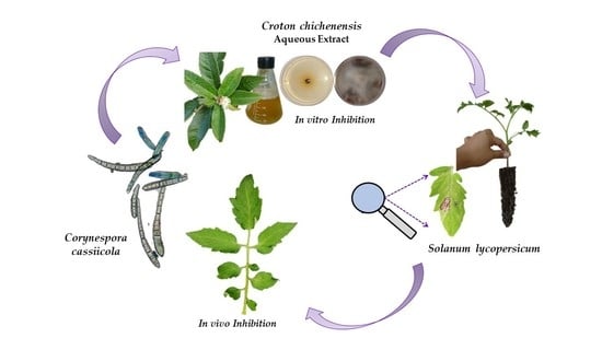
Activity of Aqueous Extracts from Native Plants of the Yucatan Peninsula against Fungal Pathogens of Tomato In Vitro and from Croton chichenensis against Corynespora cassiicola on Tomato

by
Felicia Amalia Moo-Koh
1,2,
Jairo Cristóbal-Alejo
2,*,
José María Tun-Suárez
2,
Irma Leticia Medina-Baizabal
1,
Alejandra Anahi Arjona-Cruz
2 and
Marcela Gamboa-Angulo
1,*
1
Centro de Investigación Científica de Yucatán, Calle 43 No. 130, Col. Chuburná de Hidalgo, Mérida 97205, Yucatán, Mexico
2
Tecnológico Nacional de México, Campus Conkal, Avenida Tecnológico s/n, Conkal 97345, Yucatán, Mexico
*
Authors to whom correspondence should be addressed.
Plants 2022, 11(21), 2821; https://doi.org/10.3390/plants11212821
Submission received: 12 September 2022 / Revised: 14 October 2022 / Accepted: 17 October 2022 / Published: 24 October 2022
(This article belongs to the Section Plant Protection and Biotic Interactions)

Abstract

:
Plant extracts are a valuable alternative to control pathogens of horticultural crops. In the present study, four species of pathogenic fungi were isolated from leaf spots on Solanum lycopersicum and identified by traditional and molecular techniques as Alternaria alternata ITC24, Corynespora cassiicola ITC23, Curvularia lunata ITC22, and Fusarium equiseti ITC32. When 11 aqueous extracts from eight native plants of the Yucatan Peninsula were tested against the four fungi in vitro, the extract from Croton chichenensis roots was most active, inhibiting mycelial growth (79–100%), sporulation (100%), and conidial germination (71–100%) at 3% (w/v). A logarithmic–diagrammatic scale of the pathosystem C. cassiicola–S. lycopersicum was established and used to assess disease severity on inoculated tomato plants in a greenhouse after treatment with the aqueous extract from C. chichenensis roots at 12% (w/v). After 21 days, the disease severity was 57% lower than on the control without extract applied. This dose of the extract was not phytotoxic to tomato leaves and was compatible with the beneficial organisms Bacillus subtilis CBCK47 and Trichodema asperellum Ta13-17. The antifungal efficacy of C. chichenensis is highly promising for incorporation into integrated disease management of tomato crops.

Graphical Abstract

1. Introduction

Phytopathogens are an ongoing threat to plant production because they often persist and evade plant defenses, causing disease and global annual losses of billions of US dollars. To control fungal plant diseases, farmers have used synthetic fungicides that suppress the fungi; however, misuse of these products results in the development of resistance and adverse environmental effects [1,2] on beneficial soil organisms and pollinating insects, other organisms, and humans. The use of natural plant products is thus receiving increased interest from farmers who are focusing on more sustainable production and helping reduce the risk of resistance development in a pathogen [3,4,5].
Plants produce secondary metabolites such as alkaloids, flavonoids, terpenes, phenols, glycosides, tannins, and fatty acids that inhibit the growth of bacteria and fungi. These metabolites can be located in two groups, phytoanticipins and phytoalexins. Phytoanticipins are constitutively present in the plant, and phytoalexins are biosynthesized in response to pathogen attacks [5]. Thus, plant extracts frequently contain a mixture of compounds with different modes and mechanisms of action. In addition, some compounds may act synergistically to inhibit the growth of phytopathogens [4,6]. This effect of a mixture of compounds is also known as compatibility between natural products produced by the same or different organism species [1,5]. However, plant extracts can also affect seed germination and plant growth and be toxic to beneficial organisms. For example, an aqueous extract of Seriphidium kurramense had a toxic effect on Lemna minor at different concentrations (10, 100, and 1000 µg/mL) [7]. An extract of Cleome arabica L. had a toxic effect on seedlings of Lactuca sativa, Raphanus sativus, Peganum harmala, and Silybum marianum [8]. Therefore, the toxicity of the extracts must be evaluated before they can be applied to a crop.
With 2330 native vascular plant species, the Yucatán Peninsula represents 10% of Mexico’s biodiversity [9,10]. In previous studies and following chemotaxonomic, medicinal, or serendipity criteria, plant species from this tropical region were collected and tested against plant pathogenic fungi [11,12,13,14,15]. In vitro tests of extracts from Acacia pennatula, Acalypha gaumeri, Bonellia flammea, Calea jamaicensis, Croton chichenensis, Licaria sp., Mosannona depressa, Parathesis cubana, and Piper neesianum showed antifungal activity [11,12,13,14,15]. Among these extracts, however, only those from A. gaumeri and B. flammea were effective in vivo against Alternaria chrysanthemi, the causal agent of early blight on Chrysanthemum morifolium [16].
Corynespora cassiicola is a phytopathogenic fungus that induces irregular spots on the leaves of more than 530 plants, including tomato (Solanum lycopersicum) [17] among 380 genera in the tropics and subtropics, including numerous economically important crops [18]. The disease incidence in fields can reach 60%. Other phytopathogenic fungi on tomatoes are Alternaria solani, A. tenuis, Cladosporium fulvum, Colletotrichum phomoides, Phytophthora infestans, P. nicotianae var. parasitica, Phoma destructiva, Curvularia spp., and Fusarium spp. [19,20,21,22]. Identifying the presence of C. cassiicola is difficult due to the existence of fungal complexes and secondary parasites that take advantage of the lesions to establish themselves [23]. Tomato leaf spots can affect from 35–58% up to 80% of the plants in greenhouses, so tools to monitor and identify this phytopathogen are essential [24,25]. One such tool that has been developed to monitor phytopathogens in the field and greenhouse is the logarithmic–diagrammatic scale that allows the monitoring of the early symptoms and estimates the intensity of the disease of C. cassiicola in tomato [26,27].
In the present study, we isolated four fungi from tomato leaf spots and confirmed that they caused the leaf spots. We then selected eight native plants previously reported with antifungal effects on plant pathogens, and their aqueous extract assessed in vitro activity against the isolated fungi. These aqueous extracts (11) were from different parts of A. gaumeri, B. flammea, C. chichenensis, C. jamaicensis, Licaria sp., M. depressa, P. cubana, and P. neesianum. We then developed a logarithmic–diagrammatic scale for the early detection of the symptoms induced by C. cassiicola, tested the most effective aqueous extract against the fungus on tomato plants, and evaluated its phytotoxicity and compatibility with beneficial organisms.

2. Results

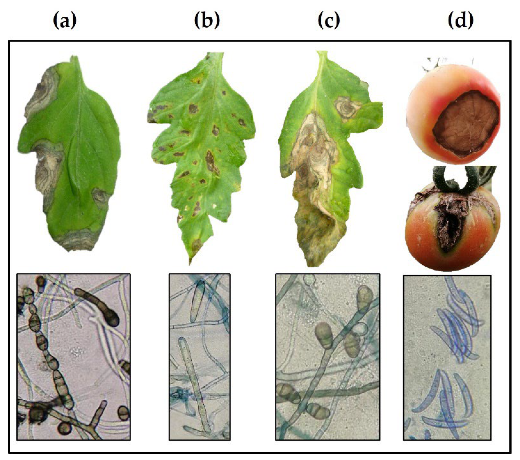
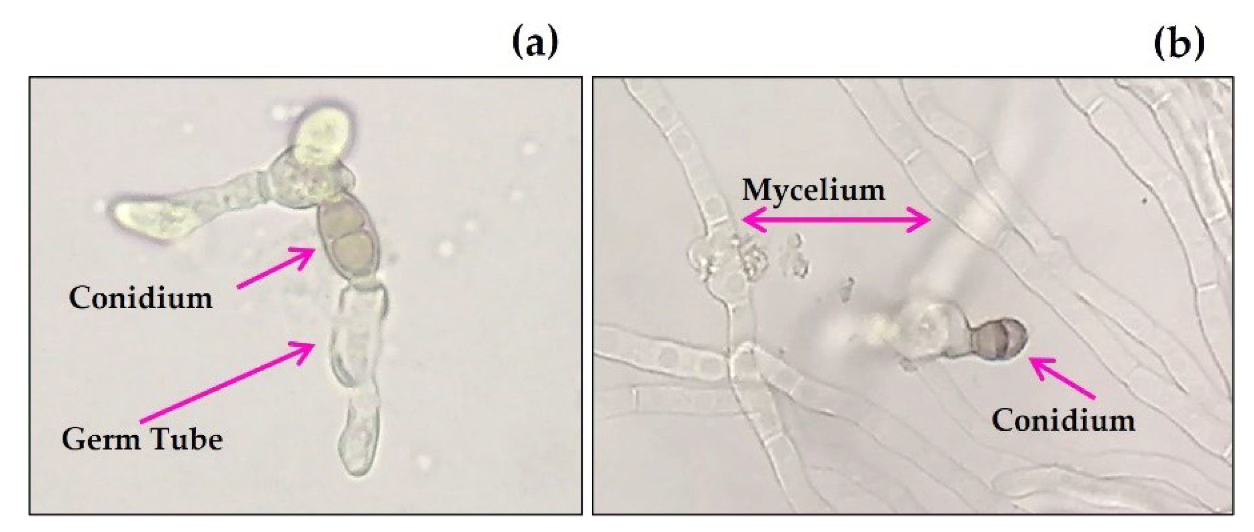
2.1. Identification of Fungal Phytopathogens

Three fungal strains belonging to the genera Alternaria (strain ITC24), Corynespora (strain ITC23), and Curvularia (strain ITC22) were isolated from the adult leaves with dry, circular to oval, chlorotic, and necrotic spots, the typical symptoms of leaf spots on tomato. In addition, Fusarium (strain ITC32) was isolated from tomato fruit with a brown rot at the base, which expanded over about 50% of the fruit surface. According to Koch’s postulates, pathogenicity tests verified that the isolated fungi were responsible for the disease in the S. lycopersicum cultivars (Figure 1).
The four phytopathogenic fungal isolates were morphologically and molecularly identified as Alternaria alternata (Fr.) Keissl strain ITC24, C. cassiicola Berk. & M.A. Curtis strain ITC23, Curvularia lunata (Wakker) strain ITC22 and F. equiseti (Corda) Sacc. strain ITC32 (Table 1). The DNA extracted from each fungal strain was amplified by PCR using universal primers ITS1 and ITS4. The sequencing of the amplified PCR products showed between 400 and 600 bp in length. Comparison of the edited sequences on the Bioedit software for each fungal strain with NCBI sequences showed 99–100% similarity, which were deposited in GenBank (Table 2).

2.2. Antifungal In Vitro Activity of Aqueous Extracts

After 7 days at 3% (w/v) concentration, the evaluated aqueous extracts significantly inhibited at least one of the fungal variables assessed for the four pathogens in relation to the control (p ≤ 0.001). The most active aqueous extract was from C. chichenensis root and caused 99–100% inhibition of mycelial growth (IMG) of C. cassiicola ITC23, C. lunata ITC22, and F. equiseti ITC32. The next most active was the aqueous extract from the stem bark of B. flammea, with 93.7% IMG of C. lunata (Table 2, Figure 2). The aqueous extracts of M. depressa stem bark were less active (19.5–48.9% IMG) against the four pathogens. The remaining eight extracts had no significant (0–28.7% IMG) effect against the pathogens assayed (Table 3).
The total inhibition of sporulation (IS, 100%) of all four pathogenic fungi was provided by the aqueous extract from C. chichenensis roots, from B. flammea bark stem A. alternata ITC24 and C. lunata ITC22 (83.4 and 93.3% against the other two), from A. gaumeri roots and Licaria sp. stem bark against A. alternata ITC24 (100%), C. lunata ITC22 (100, 96.8%, respectively), and F. equiseti ITC32 (89.9 and 100%, respectively) at 3% (w/v) after 7 days. Except for the aqueous extract from P. cubana, the six remaining extracts inhibited the sporulation of two phytopathogenic fungi to varying extents (IS = 10.5–100%) (Table 4).
For the inhibition of conidial germination (ICG), the most active aqueous extract was from C. chichenensis roots at 3% (w/v), and the germination was inhibited in a range of 70.5 to 100% for the four phytopathogenic fungi after 5 h of exposure. The aqueous extract from M. depressa root bark caused less but significant ICG against F. equiseti ITC32 (61.3%), C. cassiicola ITC23 (80.1%), and C. lunata ITC22 (100%). The conidia of C. cassiicola ITC23 were most sensitive to the aqueous extracts evaluated, and germ tubes on conidia that germinated were often deformed (Table 5, Figure 3).
The aqueous extract of the roots from C. chichenensis had the greatest activity against the four phytopathogenic fungi in the sporulation and germination and completely inhibited the mycelial growth of C. cassiicola ITC23, C. lunata ITC22, and F. equiseti ITC32.

2.3. Corynespora cassiicola–Solanum lycopersicum Pathosystem

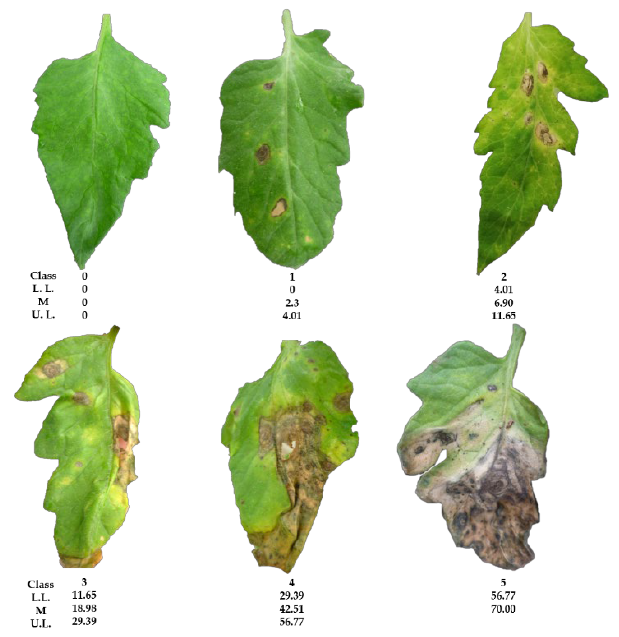
2.3.1. Logarithmic–Diagrammatic Scale Development

The diseased leaves (N = 42) from tomato plants were photographed to calculate the leaf-spot area on each leaf and develop a logarithmic–diagrammatic scale to assess the disease severity. The largest calculated area (56.1 pixels) was equivalent to an estimated 70.0% of disease severity or dead surface. Six classes (0–5) were established, with class zero corresponding to leaves without symptoms (apparently healthy) and class 5 to leaves with severe (midpoint = 70.0%) major damage. Classes 1–4 values correspond to the midpoints with lower disease severity of 2.3, 6.9, 18.0, and 42.5%, respectively, were obtained using the software 2LOG. Finally, a representative image from each class was selected as part of constructing the scale of the pathosystem C. cassiicola–S. lycopersicum (Figure 4).

2.3.2. Validation of Logarithmic–Diagrammatic Scale

The first validation of the logarithmic–diagrammatic scale based on the six severity classes for the pathosystem C. cassiicolaS. lycopersicum showed a precision of 0.79 (r2) and an accuracy of 0.81 (b1); for the second validation, these values were 0.83 and 0.85, respectively. With these results, the scale was used to estimate leaf-spot severity and the efficacy of disease control using the plant extracts.

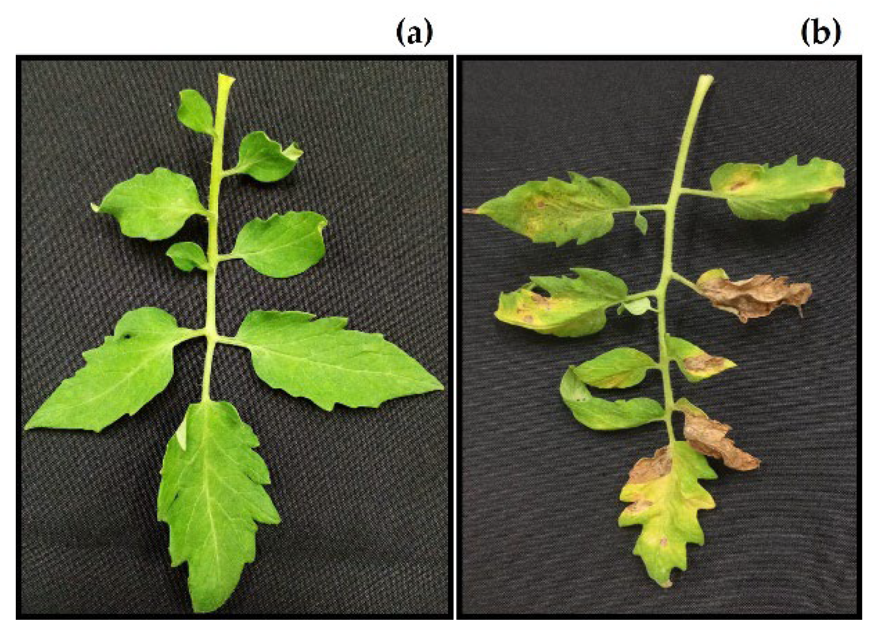
2.4. In Vivo Effect of Extract from Croton chichenensis on Tomato Leaf Spot

The severity scale for leaf spots on the tomato plants showed that the plants treated with the aqueous extract of the C. chichenensis root significantly (p ≤ 0.001) reduced leaf spot area compared with the untreated controls. The area under the disease progress curve (AUDPC) showed a lower percentage of lesion area (64.56% for day) in the treatment with the 12% (w/v) aqueous extract from C. chichenensis roots and the lowest final severity (Yfinal = 10.43%). The progress of the apparent infection rate obtained from the reciprocal of b parameter of the Weibull model with the aqueous extract showed that leaf spot severity was very low (0.01901%/day) compared with the untreated control, which had the highest intensity (0.03012%/day) of the disease (Table 6).
The leaves of the control plants had significant concentric spots and chlorosis by 7 days after inoculation with C. cassiicola, whereas the leaves on the plants treated with the aqueous extract from C. chichenensis roots were green and had no symptoms. At 21 days after inoculation, most of the leaves on the control plants had great (Class 5) disease severity. In contrast, the treated plants still had no symptoms (Class 0) (Figure 5).

2.5. Phytotoxicity of Croton chichenensis Roots

After 7 days on agar containing 12% (w/v) aqueous extract from C. chichenensis roots, the leaves of S. lycopersicum had no necrosis or visible damage to the upper surface, veins, and leaf margin and no hypersensitivity (Figure 6).

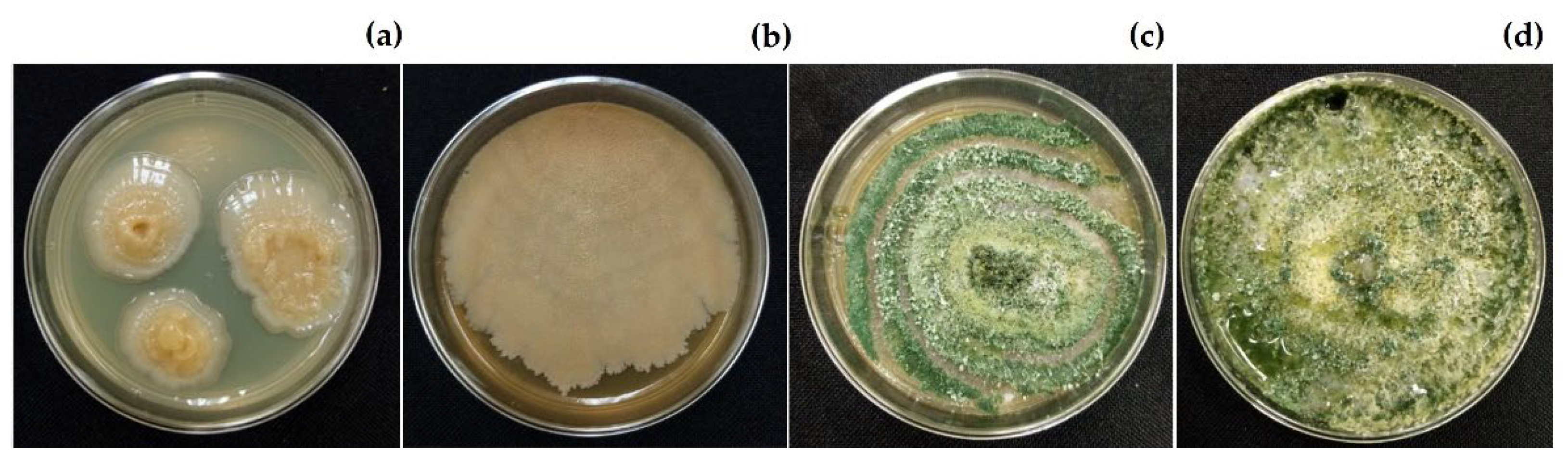
2.6. Compatibility of Croton chichenensis Extract and Beneficial Organisms

The aqueous extract of C. chichenensis roots 12% (w/v) had no statistically significant differences (p ≤ 0.001) with the control on the compatibility assay with beneficial organisms. After 2 days of exposure to the aqueous extract, three colonies of Bacillus subtilis CBCK47 were detected on bacteriological agar medium and grew to a diameter of 2–3 cm in 5 days. The plant growth-promoting organisms Trichoderma asperellum Ta13-17 grew 8 cm in diameter, and conidial was detected on potato dextrose agar (PDA) after 7 days of exposure to the aqueous extract (Figure 7). This extract of C. chichenensis in the medium slightly modified the growth but did not inhibit both organisms, then had 100% compatibility.

3. Discussion

Here, we identified four fungi causing leaf spot diseases in tomato and tested the efficacy of aqueous extracts from native species of the tropical Yucatan Peninsula against these fungi as part of our goal to conserve and take advantage of the regional biodiversity. The morphological identification of the four phytopathogens showed similarity with the morph length and width of the conidial previously reported in the literature [28,29,30,31,32,33,34] (Table 1), which is supported by molecular identification with an admisible percentage of similarity [34]. Genetic studies of species have become increasingly valuable as they complement each other by helping to overcome the difficulties of using traditional morphological identification alone [23,35,36,37].
Tomato leaf spot has been reported in other countries since late 1986 [38,39]. Tomato leaf spot has been attributed to the presence of the fungi Cladosporium cladosporioides and C. fulvum [20,40], Stemphylium lycopersici [41], and C. cassiicola [24]. Among these pathogens, C. cassiicola has the most hosts [18]. In Mexico, C. cassiicola has been isolated from the leaves of Capsicum annuum L., C. chinense Jacq. [13,42], Hibiscus sabdariffa L. [43,44], Gossypium hirsutum L. [45] and Vicia faba L. [46]. In the present study, C. cassiicola ITC23 (Corynesporascaceae) was found together with A. alternata ITC24 and C. lunata (Pleosporaeae), affecting tomato at the foliar level [28]. These three microorganisms form fungal complexes, making their identification difficult [19,47,48]. All three have dark-coloured colonies and conidia and free-form conidiophores [13,49]. A. alternata is known to cause tomato leaf blight [50], and C. lunata causes leaf spots on pepper (Capsicum annuum) [51], but we found no reports of C. lunata infecting tomato.
In the present study, F. equiseti ITC32 formed sporodochia as asexual fruiting structures containing conidia on free conidiophores on tomato fruits [52]. The fungus is cosmopolitan; it has a wide host range and easily adapts to different climatic environments [53,54]. It is also associated with tomato wilt; infection begins in the roots and impacts all plant parts [55]. However, wilt and rot of tomato fruits are associated with F. oxysporum [22,56], but we found no reports of F. equiseti causing a disease of tomato fruits. Fulfilling Koch's postulates confirmed that the mycelium and spores of the fungus and symptoms were caused by F. equiseti in tomato fruits after infection.
Aqueous extracts from plants with bioactive properties against plant pathogens have been described and used worldwide, and more than 2500 species from 235 plant families have been identified as effective against pests [57,58]. As expected, 91% of the aqueous extracts evaluated, except that from P. cubana, had an inhibitory effect greater than 50% against the four pathogens for at least one of the variables measured. The most active aqueous extract that inhibited mycelial growth, sporulation, and conidial germination (70.5% to 100%) was from the roots of C. chichenensis against C. cassiicola ITC23, C. lunata ITC22, and F. equiseti ITC32; and completely inhibited the sporulation of A. alternata ITC24 (100%). On the other hand, the aqueous extract of B. flammea stem bark greatly inhibited sporulation in the four organisms, but it only affected the inhibition of conidial germination of C. lunata ITC22 and F. equiseti ITC32. In a previous report, an aqueous extract from B. flammea (3%, w/v) showed the inhibition of mycelial growth of 50%, conidial germination of 98%, and no effect on the sporulation of C. cassiicola ITC7, fungus isolated from Citrus chinense; and a good effect on the three variables (58–89%) on C. lunata ITC4, fungus isolated from Thrinax radiata [13]. Total or partial inhibition of the mycelial growth of pathogenic fungi with plant extracts is important, but sporulation and germination inhibition tests are variables considered fundamental before further developing a natural fungicide. Aqueous extracts of plants can control common fungi that infest plants and produce conidia [59]; inhibiting conidial germination, which would prevent new infections, is highly desirable for an aqueous extract [60,61]. In our study, the aqueous extract from C. chichenensis root provided the highest inhibition of conidial germination.
The species C. chichenensis is an endemic shrub to the Yucatan Peninsula and belongs to the Euphorbiaceae family with a great diversity of species [9]. Species in the genus produce numerous compounds with potential pharmacological use, including 339 diterpenes, seven sesquiterpenes, one sesterterpene, one triterpene, 21 glycosides, eight alkaloids, three benzoate derivatives, three pyran-2-one derivatives, two cyclopeptides, two propane derivatives, and two limonoids, among others. The activity of ethanolic extracts compared to the aqueous extracts is related to the presence of more polar metabolites such as saponins and other glycosides alkaloids, flavonoids, and tannins, among others [62].
In previous studies, the ethanolic extract from C. chichenensis roots inhibited the mycelial growth of A. tagetica ATTC53771, C. gloeosporioides CICY002, F. oxysporum CICY003, and Rhizopus sp. CICY004 at 2 mg/mL in a dilution agar assay [10]. Compounds from other Croton species also have antifungal activity. For example., the ethanolic extract from C. leptostachyus stem and roots inhibited the growth of F. oxysporum (IC50 of 9726 and 1133 mg/L, respectively) in a dilution agar assay [63]; an ursane-type triterpenoide from C. bonplandianum roots inhibited A. alternata, Colletotrichum camellie, C. gloeosporioides, Curvularia eragrostidies, and F. equiseti (IC50 of 10–15 µg/mL) [64]; and an ethanolic extract from C. heliotropiifolius stem bark inhibited Candida albicans at 2.5 mg/mL in a bioautographic assay [65]. Thus, our report is the first antifungal effects of aqueous extracts from C. chichenensis roots against A. alternata, C. cassiicola, C. lunata, and F. equiseti.
To evaluate the antifungal effect of the aqueous extract from C. chichenensis on plants in the greenhouse, we first developed a logarithmic–diagrammatic scale for the pathosystem C. cassiicola-S. lycopersicum to estimate disease severity. The precise values that we obtained and those of the reported scales demonstrate that the scales are good tools for estimating the severity of the pathogens. For example, the diagrammatic scale for C. annuumCercospora sp. had a range of 0.79 to 0.95 [66],0.61 to 0.95 for Annona cherimolaC. gloeosporioides [26], and 0.81 to 0.92 for the Tar Stain Complex in Maize [27].
In controlling leaf spots in tomato, the aqueous extract of C. chichenensis roots reduces the speed of symptom development over time, resulting in a large difference in severity. In other greenhouse studies, an aqueous extract of Ocimum basilicum at 10% (w/v) reduced by 43% the calculated rate of disease caused by C. cassiicola [67]; an aqueous extract of cinnamon at 0.25% (w/v) reduced the severity of grapevine downy mildew caused by Plasmopara viticola by 67% [68]. Volatile metabolites from the leaves of Solanum habrochaites (LA1777) reduced the severity of the late blight of tomato caused by Phytophthora infestans EG_7 by 97% and lowered the AUDPC [69]. In a field test using the pathosystem Chrysanthemum morifoliumA. chrysanthemi, an aqueous extract from A. gaumeri roots and B. flammea stem bark at 3% (w/v), reduced disease severity by 67% and 50%, respectively, and lowered AUDPC [16].
The toxicity tests of promising candidates are mandatory to evaluate their effects on aerial parts of plants, seeds, beneficial organisms, and human cells. These studies also provide valuable information for future evaluations of the application of plant extracts in the field [7,70,71]. In this study, B. subtilis and T. asperellum were selected for their history of promoting plant growth, other benefits, and their biological plasticity [72,73]. No symptoms of toxicity were apparent on tomato leaves or the beneficial organisms evaluated after treatment with the 12% (w/v) aqueous extract of C. chichenensis roots, which is compatible for its possible combination and application with the biocontrol agents Trichoderma and Bacillus [74,75]. By contrast, in vitro assays showed that the aqueous extracts from Citrus chinense fruits at 50% (w/v) [76] inhibited the population growth of E. coli and Bacillus sp., and the aqueous extracts from Phyllantus sp., Azadirachta indica, and Tagetes patula inhibited the growth of Trichoderma and Metarhizium at 15% (w/v) [77].
The activity of the aqueous extract of C. chichenensis at 12% is promising against tomato pathogens, but its activity needs to be tested on other tomato accessions and pathogens and in the field in different conditions. This promising extract has the potential to be combined with beneficial organisms such as growth promoters and biocontrol agents in an integrated management program of tomato diseases. The active compounds in C. chichenensis roots also need to be isolated and identified.

4. Materials and Methods

4.1. Isolation and Pathogenicity Tests of Phytopathogenic Fungi

The four phytopathogenic fungi used in the present study were isolated from S. lycopersicum plants grown at the Tecnológico Nacional de México/Campus Conkal (Table 1 and Table 2). The leaf and fruit samples with apparent fungal-induced lesions were cut into small fragments (0.2–0.5 cm2) and surface-sterilized with NaClO at 2%, then washed twice with sterile distilled water and blotted dry with sterile absorbent paper. The pieces were placed on potato dextrose agar (PDA) in Petri dishes and kept at 28 ± 2 °C with 12 h light/12 h dark for 7 days until fungal colonies were observed [13]. The colonies had different colours and were purified by transferring the mycelium to PDA.
The pathogenicity of the purified fungi as the cause of the plant symptoms was confirmed using Koch's postulates by spray inoculating healthy leaves and fruits of tomato with a conidial suspension (1 × 10⁶ conidia/mL) of fungi [49]. Each fungal species produced the original symptoms on the host and was re-isolated and purified as described above.

4.2. Identification of the Fungi

The four purified pathogenic fungi were morphologically identified to the genus level using the dichotomous keys of Barnett and Hunter [78] and Manamgoda et al. [79]. The DNA was extracted from the mycelium of the pure fungal strains grown on PDA after three days and using the methods of Moo-Koh et al. [13] and the ZR Fungal/Bacterial DNA MiniPrep Kit (Zymo Research, Irvine, CA, USA). ITS1 and ITS2 between ribosomal genes (rDNA) 18S–5.8S and 28S were amplified by PCR using primer pair ITS1 (5′ CCGTAGGTGAACCTGCGG 3′) and ITS4 (5′ TCCTCCGCTTATTGATATGC 3′) (Integraded DNA Technologies–IDT, Coralville, IA, USA) [80]. The amplified products were sequenced (Macrogen, Inc., Seoul, South Korean), edited with the Bioedit software (https://bioedit.software.informer.com/7.1/: accessed on 21 June 2022), and compared with the data available in the BLAST (https://www.ncbi.nlm.nih.gov/genbank/: accessed on 22 June 2022). The sequences for each fungal species were deposited at the GenBank in National Center for Biotechnological Information (NCBI) (Table 2).

4.3. Plant Material

The plant material of the eight selected species for aqueous extraction was collected from the Yucatan Peninsula by the working group of the Biotechnology Unit of the Centro de Investigación Científica de Yucatán (Table 7) [13,14,15].

4.4. Preparation of Aqueous Extracts

Dried, ground plant material (30 g/L) was transferred to a flask with 500 mL of boiling distilled water and stirred for 20 min. The mixture was cooled, then filtered through Whatman No. 1 filter paper, then through a 22 µm membrane (Millipore Merck, Burlington, MA, USA), and had a final concentration of 6% (w/v). Sterile filtrates were kept for 24 h at 4 °C until used in bioassays [13,15].

4.5. In Vitro Antifungal Bioassay of Aqueous Extracts

4.5.1. Inhibition of Mycelial Growth

The filtered, sterilized aqueous extracts were evaluated in a PDA dilution test (BD, Bioxon, Estado de México, México). The PDA (3.9 g in 50 mL, 2:1 w/v) was sterilized and held at 50 °C for mixing with the aqueous extract to a final concentration of 3% (w/v), 10 mL was added to each plate and held at room temperature for 24 h. A mycelial disc (5 mm diameter) from a 6-to-7-day-old culture of the test fungus was placed in the middle of the dish. A Petri dish of medium without extract was used as a control. Five replicate plates were tested for each extract–fungus combination and incubated at 28 ± 2 °C with 12 h light /12 h dark [13]. The diameter of each colony was measured with a Vernier digital calliper every 24 h until the colony in control covered the agar surface. The efficacy of the extract at inhibiting mycelial growth was calculated using the Abbott formula [81], E (%) = (Control − Treatment)/Control)(100), where E = efficacy (%), Control = mycelial diameter of the control (cm), Treatment = mycelial diameter of the treatment (cm).

4.5.2. Inhibition of Sporulation

Sterile water (9 mL) was added to treatment and control plates in which the fungus grew horizontally and vertically, and the mycelial surface was gently scraped to remove the conidia. The suspension was filtered through two layers of sterile gauze, then a 1:9 (v/v) dilution of suspension to water was homogenized, and 9 µL was transferred to a storage Neubauer counting chamber. Conidia in four fields of view were counted with a light microscope (Leica model DM500, Wetzlar, Germany), and the total number of conidia/mL was calculated as NE = (X/0.1) (1000)(9), where X is the mean number of conidia in 4 fields, 0.1 is the depth of the chamber; 1000: volume (mL); 9: volume of water (mL) added to Petri dish. The values were used in the Abbott formula described above to calculate the inhibition of sporulation [81].

4.5.3. Inhibition of Spore Germination

A 9 µL drop of the undiluted conidial suspension was dropped on each of the quadrants of a Petri dish containing PDA plus the aqueous extracts (3%, w/v). PDA alone was used as the control, and each quadrant was considered a replication. Conidia were considered germinated when a germ tube was observed. Conidia were checked for germination, and the germinated conidia were counted in each quadrant every 60 min for five h, to ensure the germination of the conidia in control, using a light microscope (Leica model DM500, Wetzlar, Germany) with a magnification of 400× (Figure 3).

4.5.4. Statistical Analyses

The experimental design was completely randomized. The percentage data for the inhibition of mycelial growth, sporulation, and conidial germination by not complying with the assumptions of normality of these data were arcsine square-root-transformed before calculating an ANOVA. Tukey’s test (p ≤ 0.05) was used, and separation of means was carried out with the SAS ver. 9.4. for Windows (SAS Institute, Cary, NC, USA) for all analyses.

4.6. Pathosystem Corynespora cassiicola—Solanum lycopersicum

4.6.1. Development of the Logarithmic—Diagrammatic Scale

The severity of C. cassiicola on the tomato crops was measured with a logarithmic–diagrammatic scale developed for this study using 42 photographs of tomato leaves with leaf spots caused by C. cassiicola. For each leaf, the total area with lesions was measured using the software ImageJ (National Institutes of Health, Bethesda, MD, USA) [82]. The total area was then used to calculate the percentage of the severity of the disease per leaf with the formula severity (%) = (diseased leaf area/ total leaf area)(100) [26].
The software 2LOG [83] was used to design a logarithmic–diagrammatic scale using the principle of Horsfall–Barrat [84,85]. The data on the percentage of the severity of the disease obtained previously were analyzed in the 2LOG computer program to obtain the different estimated classes of the scale, with their confidence limits. The number of classes was determined with respect to the evaluator's measurement ability. In other cases, six classes were recommended, and finally, a representative photograph was associated with each class [26] (Figure 4).

4.6.2. Validation of the Logarithmic—Diagrammatic Scale

The logarithmic–diagrammatic scale of the pathosystem C. cassiicolaS. lycopersicum was validated using two evaluations by six participants who estimated the severity of the same 42 leaves and compared them with the classes of the previously elaborated scale. We then compared their estimates with the printed generated scale and the calculated information. Simple linear regressions were used to correlate the values of severity obtained with ImageJ with those estimated by the evaluators. Thus, the precision r2 and accuracy b1 of each evaluator was obtained, and with it, the reproducibility of the scale. The r2 and b1 values of 1.0 indicated the maximum. SAS ver. 9.4. for Windows was used for all analyses [26].

4.7. Evaluation of Croton chichenensis Extract on Tomato Leaf Spot

4.7.1. Tomato Crops and Infection with Corynespora cassiicola

The S. lycopersicum (DR8558) seeds were grown in 200-well polystyrene trays containing Cosmopeat substrate (Cosmocel, Fredericton, NB, Canada) mixed with agrolite (1:1 v/v). Fifteen days after planting, the seedlings were transplanted into nine-inch pots containing Luvisol rhodic-type soil and irrigated and fertilized as recommended for tomato. To prevent red spider mite (Tetranychus urticae) and whitefly (Bemisia tabaci) infestation, we used the botanical product BioDi®e S.A. (argemonine 3.5% + berberine 2.2% + ricinine 2.8% + α-ter-thienil 3.50%) (Ultraquimia, Benito Juárez, Ciudad de México, México).
The aqueous extract of C. chichenensis roots at 12% (w/v) was sprayed on the tomato foliage 30 days after transplanting. After 24 h, the plants were inoculated by being sprayed with a conidial suspension of C. cassiicola (1 × 10⁶ conidia/mL).
When the first disease symptoms were observed, 10 leaves per plant were selected from each treatment. Each leaf was compared with the classes of the diagrammatic logarithmic scale previously elaborated and validated. These records were continued at 7-day intervals for three more weeks with the same selected leaves.

4.7.2. Epidemiological Parameters

The disease progress curves were constructed using the severity percentages [16,26] to estimate the AUDPC using the trapezoidal integration method, the apparent infection rate with the model description of Weibull and by its inverse parameter of b (1/b), and the percentage of disease severity at the end of the evaluations with the scale (Yfinal).

4.8. Phytotoxicity Bioassay on Solanum lycopersicum Leaf

The toxicity of the aqueous extract of the roots of C. chichenensis at doses of 3, 6, and 12% (w/v) was evaluated on the leaves of S. lycopersicum. Ten leaves of 30-day-old S. lycopersicum plants were randomly collected and cut into 2 cm2 pieces surface, sterilized as described above, and placed in Petri dishes containing agar–agar (BD Becton Dickinson, 23 g L−1). The aqueous extract of the C. chichenensis roots (50 µL) was deposited on the cut edge of each leaf piece. Sterile distilled water (50 µL) was used instead for the controls [15]. All of the dose treatments were in triplicate and kept at 28 ± 2 °C, 12 h light/12 h dark. Every 24 h for 15 days, samples were examined for any necrosis or darkened areas at the treatment site.

4.9. In Vitro Compatibility Bioassay with Beneficial Organisms

Bacillus subtilis CBCK47 and Trichoderma asperellum Ta13-17 were provided by the Microbiology and Phytopathology Laboratories of the Tecnológico Nacional de México/ Campus Conkal [62,63].
The bacteriological agar medium (BD Becton Dickinson, Estado de México, México, 23 g/L), sterile at 50 °C, was mixed with the aqueous extract of the roots of C. chichenensis (24%, w/v) for a final concentration of 12% (w/v). The agar medium alone was used as the control. Four plates were streaked with B. subtilis CBCK47 and incubated at 28 ± 2 °C, with 12 h light/12 h dark. After 48 h, the bacterial colonies were counted [86].
For the fungus T. asperellum Ta13-17, the aqueous extract from C. chichenensis roots was prepared in PDA in plates, as described above, at a final concentration of 12% (w/v). PDA alone was used as the negative control. The culture medium was inoculated with a mycelial disk (5 mm in diameter) from a 5-day-old colony of T. asperellum Ta13-17. The diameter of the colony was then measured every 24 h, as described earlier. The compatibility of the extract with the fungus was estimated using the Abbott formula described above.

5. Conclusions

This research is the first contribution aimed at isolating and identifying the phytopathogens associated with tomato leaf spots in the Yucatan Peninsula. Our knowledge of the inhibitory activity of extracts from native species of Yucatan was enriched, in particular, the antifungal spectrum of action of C chichenensis. Moreover, we developed a logarithmic–diagrammatic scale for the pathosystem C. cassiicola–S. lycopersicum to assess the severity of tomato leaf spots. The aqueous extract of C. chichenensis was shown to be a viable alternative to fungicides to reduce the severity of the tomato leaf spots induced by C. cassiicola in the greenhouse. The plant extract had no phytotoxicity and was compatible with microbial biocontrol agents, opening an avenue to reduce the use of synthetic fungicides and ensure sustainable control of plant diseases.

Author Contributions

Direction and supervision of the study, J.C.-A. and M.G.-A.; methodology- preparation of fungal extracts and cultures, F.A.M.-K. and I.L.M.-B.; antifungal bioassays and in vivo evaluations F.A.M.-K. and A.A.A.-C.; financial support of the project M.G.-A., J.C.-A. and J.M.T.-S.; writing, revising and editing the manuscript, all authors. All authors have read and agreed to the published version of the manuscript.

Funding

Consejo Nacional de Ciencia y Tecnología (CONACYT)- International Development Research Centre (IDRC) financed project No. CEAR2019-01; and Tecnológico Nacional de México (TecNM) financed project 10759.21P.

Institutional Review Board Statement

Not applicable.

Informed Consent Statement

Not aplicable.

Data Availability Statement

Not aplicable.

Acknowledgments

The authors thank Consejo Nacional de Ciencia y Tecnología (CONACYT) - International Development Research Centre (IDRC)-Centro de Investigaciones y Estudios Superiores en Antropología Social (CIESAS) project No. CEAR2019-01, Tecnológico Nacional de México, Campus Conkal financed the project TECNM 10759.21P. F.A.M.-K. thanks to CONACyT for postdoctoral scholarship 2019–2022 (CVU 368804) from Estancias Posdoctorales para Mujeres Indigenas (EPMI).

Conflicts of Interest

The authors declare no conflict of interest.

References

  1. Jiménez, M.F.; Carrasco, H.; Olea, A.F.; Silva, E. Natural compounds: A sustainable alternative to the phytopathogens control. J. Chil. Chem. Soc. 2019, 64, 4459–4465. [Google Scholar] [CrossRef]
  2. Romani, A.; Simone, G.; Campo, M.; Moncini, L.; Bernini, R. Sweet chestnut standardized fractions from sustainable circular process and green tea extract: In vitro inhibitory activity against phytopathogenic fungi for innovative applications in green agriculture. PLoS ONE 2021, 16, e0247298. [Google Scholar] [CrossRef]
  3. Fungicide Resistance Action Committee, FRAC. Available online: https://fmcagro.es/blog/ver-entrada/clasificaci%C3%B3ndelosmodosdeacci%C3%B3ndeFRACeIRAC (accessed on 18 July 2022).
  4. Shuping, D.S.S.; Eloff, J.N. The use of plants to protect plants and food against fungal pathogens: A review. Afr. J. Tradit. Complementary Altern. Med. 2017, 14, 120–127. [Google Scholar] [CrossRef] [PubMed] [Green Version]
  5. Ribera, A.E.; Zuñiga, G. Induced plant secondary metabolites for phytopatogenic fungi control: A review. J. Soil. Sci. Plant Nutr. 2012, 12, 893–911. [Google Scholar] [CrossRef]
  6. Masoko, P.; Eloff, J.N. The diversity of antifungal compounds of six South African Terminalia species (Combretaceae) determined by bioautography. Afr. J. Biotechnol. 2005, 4, 1425–1431. Available online: https://www.ajol.info/index.php/ajb/article/view/71476 (accessed on 17 July 2022).
  7. Ahmad, K.; Ali, A.; Afridi, W.A.; Somayya, R.; Ullah, M.J. Antimicrobial, hemagglutination and phytotoxic activity of crude ethanolic and aqueous extracts of Seriphidium kurramense. J. Tradit. Chin. Med. 2018, 38, 433–438. Available online: https://pubmed.ncbi.nlm.nih.gov/32185977/ (accessed on 5 July 2022). [CrossRef]
  8. Ladhari, A.; Omezzine, F.; DellaGreca, M.; Zarrelli, A.; Zuppolini, S.; Haouala, R. Phytotoxic activity of Cleome arabica L. and its principal discovered active compounds. South Afr. J. Bot. 2013, 88, 341–351. [Google Scholar] [CrossRef] [Green Version]
  9. Villaseñor, J.L. Checklist of the native vascular plants of México. Rev. Mex. De Biodivers. 2016, 87, 559–902. [Google Scholar] [CrossRef] [Green Version]
  10. Ramírez-Morillo, I.M. La flora de la península de Yucatán: ¿Diversa? ¿Bien conocida? ¿Protegida? No, no y ¿No? Desde Herb. CICY 2019, 11, 130–137. [Google Scholar]
  11. Peraza-Sánchez, S.R.; Chan-Che, E.O.; Ruiz-Sánchez, E. Screening of Yucatecan plant extracts to control Colletotrichum gloeosporioides and isolation of a new pimarene from Acacia pennatula. J. Agric. Food Chem. 2005, 53, 2429–2432. [Google Scholar] [CrossRef]
  12. Gamboa, M.; Cristóbal, J.; Medina, I.L.; Chí, F.; Méndez, R.; Simá, P.; May, F. Antifungal properties of selected plants from the Yucatan península, México. World J. Microbiol. Biotechnol. 2008, 24, 1955–1959. [Google Scholar] [CrossRef]
  13. Moo, F.A.; Cristóbal, J.; Reyes, A.; Tun, J.M.; Sandoval, R.; Ramírez, J.A. Actividad in vitro del extracto acuoso de Bonellia flammea contra hongos fitopatógenos. Agrociencia 2014, 48, 833–845. Available online: https://agrociencia-colpos.org/index.php/agrociencia/article/view/1123/1123 (accessed on 27 May 2022).
  14. Vargas, A.A.; Gamboa, M.M.; Medina, I.L.; Pérez, D.; Cristóbal, J.; Ruiz, E. Evaluación de extractos de plantas nativas yucatecas contra Alternaria chrysanthemi y espectro de actividad antifúngica de Acalypha gaumeri. Rev. Mex. Fitopatol. 2014, 32, 1–11. Available online: https://www.researchgate.net/publication/317441366_Evaluacion_de_Extractos_de_Plantas_Nativas_Yucatecas_Contra_Alternaria_chrysanthemi_y_Espectro_de_Actividad_Antifungica_de_Acalypha_gaumeri?enrichId=rgreq-cb1c696aa8831adfcf83e29575e415bc-XXX&enrichSource=Y292ZXJQYWdlOzMxNzQ0MTM2NjtBUzo1MTcwOTQzMDU2NjUwMjlAMTUwMDI5NjI5NzYyMQ%3D%3D&el=1_x_3&_esc=publicationCoverPdf (accessed on 22 June 2022).
  15. Cruz, P.; Cristóbal, J.; Ruiz, V.; Carnevali, G.; Vera, M.; Martín, J.; Reyes, F.; Gamboa-Angulo, M. Extracts from six native plants of the Yucatán peninsula hinder mycelial growth of Fusarium equiseti and F. oxysporum, Pathogens of Capsicum chinense. Pathogens 2020, 9, 827. [Google Scholar] [CrossRef]
  16. Vargas, A.A.; Cristóbal, J.; Canto, B.; Gamboa, M.M. Aqueous extracts from Acalypha gaumeri and Bonellia flammea for leaf blight control in chrysanthemum (Chrysanthemum morifolium). Rev. Mex. Fitopatol. 2022, 1, 40–58. [Google Scholar] [CrossRef]
  17. MacKenzie, K.J.; Sumabat, L.G.; Xavier, K.V.; Vallad, G.E. A review of Corynespora cassiicola and its increasing relevance to tomato in Florida. PHP 2018, 19, 303–309. [Google Scholar] [CrossRef]
  18. Ma, D.; Zhu, J.; Jiang, J.; Zhao, Y.; Li, B.; Mu, W.; Liu, F. Evaluation of bioactivity and control efficacy of tetramycin against Corynespora cassiicola. Pestic. Biochem. Physiol. 2018, 152, 106–113. [Google Scholar] [CrossRef]
  19. Wani, A.H. An overview of the fungal rot of tomato. Mycopath 2011, 9, 33–38. Available online: http://pu.edu.pk/images/journal/impp/PDF-FILES/8-FINALReview%20AH%20WANI%2017-5-2012_vol%209(1)2011.pdf (accessed on 30 June 2022).
  20. Griffiths, S.A.; Cox, R.J.; Overdijk, E.J.R.; Mesarich, C.H.; Saccomanno, B.; Lazarus, C.M.; De Wit, P.J.G.M.; Collemare, J. Assignment of a dubious gene cluster to melanin biosynthesis in the tomato fungal pathogen Cladosporium fulvum. PLoS ONE 2018, 13, e0209600. [Google Scholar] [CrossRef] [Green Version]
  21. Ni, L.; Punja, Z.K. Management of fungal diseases on cucumber (Cucumis sativus L.) and tomato (Solanum lycopersicum L.) crops in greenhouses using Bacillus subtilis. In Bacilli and Agrobiotechnology: Phytostimulation and Biocontrol; Islam, T., Rahman, M., Pandey, P., Boehme, M.H., Haesaert, G., Eds.; Springer: Berlin/Heidelberg, Germany, 2019; Volume 2, pp. 1–28. [Google Scholar]
  22. Srinivas, C.; Nirmala, D.D.; Narasimha, M.K.; Dhananjaya, M.C.; Lakshmeesha, T.R.; Singh, B.; Kumar, K.N.; Niranjana, S.R.; Hashem, A.; Alqarawi, A.A.; et al. Fusarium oxysporum f. sp. Lycopersici causal agent of vascular wilt disease of tomato: Biology to diversity—A review. Saudi J. Biol. Sci. 2019, 26, 1315–1324. [Google Scholar] [CrossRef]
  23. Naranjo-Ortiz, M.A.; Gabaldón, T. Fungal evolution: Cellular, genomic and metabolic complexity. Biol. Rev. 2020, 95, 1198–1232. [Google Scholar] [CrossRef] [PubMed] [Green Version]
  24. Kamei, A.; Dutta, S.; Sarker, K.; Das, S.; Datta, G.; Goldar, S. Target leaf spot of tomato incited by Corynespora cassiicola, an emerging disease in tomato production under Gangetic alluvial region of West Bengal, India. Arch. Phytopathol. Plant Prot. 2019, 51, 1039–1048. [Google Scholar] [CrossRef]
  25. Mamode Ally, N.; Neetoo, H.; Ranghoo-Sanmukhiya, M.; Hardowar, S.; Vally, V.; Gungoosingh-Bunwaree, A.; Coutinho, T.A.; Vojvodić, M.; Bulajić, A. First Report of Target Spot on Tomato Caused by Corynespora cassiicola in Mauritius. Plant Dis. 2021, 105, 226. [Google Scholar] [CrossRef]
  26. Tovar, A.; Hernández, M.; Cristóbal, J.; Romero, R.; Mora, G. Escala logarítmica diagramática de severidad de la mancha negra (Colletotrichum gloeosporioides Penz.) en chirimoyo (Annona cherimola Mill.). Rev. Mex. Fitopatol. 2002, 20, 103–109. Available online: https://www.researchgate.net/publication/237037415_Escala_logaritmica_Diagramatica_de_Severidad_de_la_Mancha_Negra_Colletotrichum_gloeosporioides_Penz_en_Chirimoyo_Annona_cherimola_Mill?enrichId=rgreq-91b6601b3ffea507bca95fe69b294c64-XXX&enrichSource=Y292ZXJQYWdlOzIzNzAzNzQxNTtBUzoxMDQ0MTE1Nzc3MTY3NDJAMTQwMTkwNTA2ODYwNQ%3D%3D&el=1_x_2&_esc=publicationCoverPdf (accessed on 30 June 2022).
  27. Hernández, L.; Sandoval, J.S. Escala diagramática de severidad para el complejo mancha de asfalto del maíz. Rev. Mex. Fitopatol. 2015, 33, 95–103. Available online: https://www.scielo.org.mx/scielo.php?script=sci_arttext&pid=S0185-33092015000100095 (accessed on 17 July 2022).
  28. Kustrzeba-Wójcicka, I.; Siwak, E.; Terlecki, G.; Wolańczyk-Mędrala, A.; Mędrala, W. Alternaria alternata and Its Allergens: A Comprehensive Review. Clin. Rev. Allergy Immunol. 2014, 47, 354–365. [Google Scholar] [CrossRef] [PubMed]
  29. Christias, C.H.; Hatzipapas, P.; Dara, A.; Kaliafas, A.; Chrysanthis, G. Alternaria alternata, a new pathotype pathogenic to aphids. BioControl 2001, 46, 105–124. [Google Scholar] [CrossRef]
  30. Qi, Y.X.; Zhang, X.; Pu, J.J.; Liu, X.M.; Lu, Y.; Zhang, H.; Xie, Y.X. Morphological and molecular analysis of genetic variability within isolates of Corynespora cassiicola from different hosts. Eur. J. Plant Pathol. 2011, 130, 83–95. [Google Scholar] [CrossRef]
  31. Voglmayr, H.; Jaklitsch, W.M. Corynespora, Exosporium and Helminthosporium revisited-new species and generic reclassification. Stud. Mycol. 2017, 87, 43–76. [Google Scholar] [CrossRef]
  32. Sivanesan, A. Graminicolous Species of Bipolaris, Curvularia, Drechslera, Exserohilum and Their Teleomorphs; CAB International: Wallingford, UK, 1987; pp. 141–183. [Google Scholar]
  33. Wang, M.M.; Chen, Q.; Diao, Y.Z.; Duan, W.J.; Cai, L. Fusarium incarnatum-equiseti complex from China. Pers. Mol. Phylogeny Evol. Fungi 2019, 43, 70–89. [Google Scholar] [CrossRef] [Green Version]
  34. Ezrari, S.; Lahlali, R.; Radouane, N.; Tahiri, A.; Lazraq, A. First report of Fusarium equiseti causing pre- and postharvest fruit rot on zucchini in Morocco. J. Plant Pathol. 2020, 102, 251. [Google Scholar] [CrossRef] [Green Version]
  35. Tekpinar, A.D.; Kalmer, A. Utility of various molecular markers in fungal identification and phylogeny. Nova Hedwig. 2019, 109, 187–224. [Google Scholar] [CrossRef]
  36. Luchi, N.; Ioos, R.; Santini, A. Fast and reliable molecular methods to detect fungal pathogens in woody plants. Appl. Microbiol. Biotechnol. 2020, 104, 2453–2468. [Google Scholar] [CrossRef] [Green Version]
  37. Crous, P.W.; Rossman, A.Y.; Aime, M.C.; Allen, W.C.; Burgess, T.; Groenewald, J.Z.; Castlebury, L.A. Names of phytopathogenic fungi: A practical guide. Phytopathology 2021, 111, 1500–1508. [Google Scholar] [CrossRef]
  38. Kingsland, G.C. Pathogenicity and epidemiology of Corynespora cassiicola in the Republic of Seychelles. Int. J. Pest Manag. 1986, 32, 283–287. [Google Scholar] [CrossRef]
  39. Kingsland, G.C.; Sitterly, W.R. Studies on fungicides for control of Corynespora cassiicola leaf spot of tomatoes in the Republic of Seychelles. Int. J. Pest Manag. 1986, 32, 31–34. [Google Scholar] [CrossRef]
  40. Robles-Yerena, L.; Ayala-Escobar, V.; Leyva-Mir, S.G.; Bernardi-Lima, N.; Camacho-Tapia, M.; Tovar-Pedraza, J.M. First report of Cladosporium cladosporioides causing leaf spot on tomato in Mexico. J. Plant Pathol. 2019, 101, 759. [Google Scholar] [CrossRef]
  41. Medina, R.; Franco, M.E.; da Cruz Cabral, L.; Bahima, J.V.; Patriarca, A.; Balatti, P.A.; Saparrat, M.C. The secondary metabolites profile of Stemphylium lycopersici, the causal agent of tomato grey leaf spot, is complex and includes host and non-host specific toxins. Australas. Plant Pathol. 2021, 50, 105–115. [Google Scholar] [CrossRef]
  42. Tun-Suárez, J.M.; Castillo Peraza, M.E.; Cristóbal Alejo, J.; Latournerie Moreno, L. Etiología de la mancha foliar del chile dulce (Capsicum annuum L.) y su control in vitro en Yucatán, México. Fitosanidad 2011, 15, 5–10. Available online: http://scielo.sld.cu/scielo.php?script=sci_arttext&pid=S1562-30092011000100001 (accessed on 22 June 2022).
  43. Hernández-Morales, J.; Ochoa-Martínez, D.L.; Ortega-Acosta, S.Á.; Vega-Muñoz, R. Survey on alternative hosts of Corynespora cassiicola, the cause of the leaf and calyx spot, in the surroundings of roselle fields in Mexico. Trop. Plant Pathol. 2018, 43, 263–270. [Google Scholar] [CrossRef]
  44. Ortega, S.A.; Ochoa, D.L.; Hernández, J.; Palemón, F. Morphological and genetic characterization of Corynespora cassiicola isolates obtained from roselle and associated weeds. Rev. Mex. De Fitopatol. 2020, 38, 1–17. [Google Scholar] [CrossRef] [Green Version]
  45. Rondon, M.N.; Lawrence, K. The fungal pathogen Corynespora cassiicola: A review and insights for target spot management on cotton and Soya bean. J. Phytopathol. 2021, 169, 329–338. [Google Scholar] [CrossRef]
  46. Haddoudi, I.; Cabrefiga, J.; Mora, I.; Mhadhbi, H.; Montesinos, E.; Mrabet, M. Biological control of Fusarium wilt caused by Fusarium equiseti in Vicia faba with broad spectrum antifungal plant-associated Bacillus spp. Biol. Control 2021, 160, 104671. [Google Scholar] [CrossRef]
  47. Khalmuminova, G.K.; Kamilov, S.G.; Verushkina, O.A.; Khujanazarova, M.K. Alternaria diseases of agricultural crops in Uzbekistan. GSC Biol. Pharm. Sci. 2020, 13, 062–067. [Google Scholar] [CrossRef]
  48. Ferdinandez, H.S.; Manamgoda, D.S.; Udayanga, D. Molecular phylogeny and morphology reveal three novel species of Curvularia (Pleosporales, Pleosporaceae) associated with cereal crops and weedy grass hosts. Mycol. Prog. 2021, 20, 431–451. [Google Scholar] [CrossRef]
  49. Agrios, G.N. Plant Pathology, 5th ed.; Elsevier Academic Press: Amsterdam, The Netherlands, 2005; pp. 1–838. [Google Scholar]
  50. Meena, R.; Godika, S.; Yadav, P.D.; Chand, K.; Gayithri, M.; Jajorira, A. Management of early blight of tomato Solanum lycopersicum Mill. Incited by Alternaria solani through salicylic acid in vitro and in vivo. J. Pharm. Innov. 2022, 11, 1–6. Available online: https://www.researchgate.net/publication/360238895_Management_of_early_blight_of_tomato_Solanum_lycopersicum_Mill_Incited_by_Alternaria_solani_through_salicylic_acid_in_vitro_and_in_vivo (accessed on 22 June 2022).
  51. Pei, Y.; Sun, Y.; Feng, T.; Chen, Y.; Long, H. Pathogen Identification for the New Leaf Spot Disease of Capsicum annuum L. and Its Biological Characteristics. Chin. J. Tropic. Crops CJTC 2021, 42, 2659–2665. [Google Scholar] [CrossRef]
  52. Thines, M.; Aoki, T.; Crous, P.W.; Hyde, K.D.; Lücking, R.; Malosso, E.; May, T.W.; Miller, A.N.; Redhead, S.A.; Yurkov, A.M.; et al. Setting scientific names at all taxonomic ranks in italics facilitates their quick recognition in scientific papers. IMA Fungus 2020, 11, 25. [Google Scholar] [CrossRef]
  53. Akbar, A.; Hussain, S.; Ullah, K.; Fahim, M.; Ali, G.S. Detection, virulence and genetic diversity of Fusarium species infecting tomato in Northern Pakistan. PLoS ONE 2018, 13, e0203613. [Google Scholar] [CrossRef]
  54. Ngegba, P.M.; Kanneh, S.M.; Bayon, M.S.; Ndoko, E.J.; Musa, P.D. Fungicidal effect of three plants extracts in control of four phytopathogenic fungi of tomato (Lycopersicum esculentum L.) fruit rot. Int. J. Environ. Agric. Biotechnol. (IJEAB) 2018, 3, 112–117. [Google Scholar] [CrossRef]
  55. Manghwar, H.; Hussain, A.; Ali, Q.; Saleem, M.H.; Abualreesh, M.H.; Alatawi, A.; Ali, S.; Munis, M.F.H. Disease severity, resistance analysis, and expression profiling of pathogenesis-related protein genes after the inoculation of Fusarium equiseti in wheat. Agronomy 2021, 11, 2124. [Google Scholar] [CrossRef]
  56. Safari, Z.S.; Ding, P.; Nakasha, J.J.; Yusoff, S.F. Controlling Fusarium oxysporum tomato fruit rot under tropical condition using both chitosan and vanillin. Coatings 2021, 11, 367. [Google Scholar] [CrossRef]
  57. Roy, S.; Handique, G.; Muraleedharan, N.; Dashora, K.; Roy, S.M.; Mukhopadhyay, A.; Babu, A. Use of plant extracts for tea pest management in India. App. Microbiol. Biotechnol. 2016, 100, 4831–4844. [Google Scholar] [CrossRef] [PubMed]
  58. Ngegba, P.M.; Cui, G.; Khalid, M.Z.; Zhong, G. Use of Botanical Pesticides in Agriculture as an Alternative to Synthetic Pesticides. Agriculture 2022, 12, 600. [Google Scholar] [CrossRef]
  59. Alotibi, F.O.; Ashour, E.H.; Al-Basher, G. Evaluation of the antifungal activity of Rumex vesicarius L. and Ziziphus spina-christi (L) Desf. Aqueous extracts and assessment of the morphological changes induced to certain myco-phytopathogens. Saudi J. Biol. Sci. 2020, 27, 2818–2828. [Google Scholar] [CrossRef]
  60. Setlow, P. Germination of spores of Bacillus species: What we know and do not know. J. Bacteriol. 2014, 196, 1297–1305. [Google Scholar] [CrossRef] [Green Version]
  61. Tata, S.; Donli, P.O.; Mohammed, F.K.; Peter, A.; Ahmed, A. Comparative studies on the effect of aqueous and methanolic extracts of some botanicals on growth and sporulation of Colletotrichum graminicola. Int. J. Innov. Res. Adv. Stud. IJIRAS 2018, 5, 1–7. Available online: https://www.ijiras.com/2018/Vol_5-Issue_10/paper_27.pdf (accessed on 5 May 2022).
  62. Xu, W.H.; Liu, W.Y.; Liang, Q. Chemical constituents from Croton species and their biological activities. Molecules 2018, 23, 2333. [Google Scholar] [CrossRef] [Green Version]
  63. Pardo-Rodriguez, D.A.; Ortíz-Romero, L.T.; Tacha, A.L.; Murillo-Perea, E.; Mendez-Arteaga, J.J.; Murillo-Arango, W. Estudio químico y etnobotánico de Croton leptostachyus. La Rev. De La Acad. Colomb. De Cienc. Exactas Físicas Y Nat. 2014, 38, 356–363. [Google Scholar]
  64. Ghosh, P.; Mandal, A.; Rasul, M.G. A new bioactive ursane-type triterpenoid from Croton bonplandianun. Bail. J. Chem. Sci. 2013, 125, 359–364. [Google Scholar] [CrossRef] [Green Version]
  65. Queiroz, M.M.F.; Queiroz, E.F.; Zeraik, M.L.; Marti, G.; Favre-Godal, Q.; Simões-Pires, C.; Marcourt, L.; Carrupt, M.; Cuendet, M.Q.; Paulo, M.Q.; et al. Antifungals and acetylcholinesterase inhibitors from the stem bark of Croton heliotropiifolius. Phytochem. Lett. 2014, 10, lxxxviii. [Google Scholar] [CrossRef]
  66. Michereff, S.J.; Noronha, M.D.A.; Andrade, D.E.G.T.D.; Oliveira, E.P.D.; Xavier, F.M.S.; Moreira, P.A.A. Elaboração e validação de escala diagramática para a cercosporiose do pimentão. Summa Phytopathol. 2006, 32, 260–266. [Google Scholar] [CrossRef]
  67. Ogbebor, N.; Adekunle, A.T. Inhibition of conidial germination and mycelial growth of Corynespora cassiicola (Berk and Curt) of rubber (Hevea brasiliensis muell. Arg.) using extracts of some plants. Afr. J. Biotechnol. 2005, 4, 996–1000. [Google Scholar]
  68. Garcia, C.; Fedrigo, K.; Gabriel, A.; Botelho, R.V.; Rodrigues, J.D.; Ono, E.O. Control of mildew in vines with cinnamon extract and catalase activity in organic production. Res. Soc. Develop. 2021, 10, e214101018885. [Google Scholar] [CrossRef]
  69. Arafa, R.A.; Kamel, S.M.; Taher, D.I.; Solberg, S.Ø.; Rakha, M.T. Leaf extracts from resistant wild tomato can be used to control late blight (Phytophthora infestans) in the cultivate tomato. Plants 2022, 11, 1824. [Google Scholar] [CrossRef]
  70. Kapanen, A.; Itävaara, M. Ecotoxicity tests for compost applications. Ecotoxicol. Environ. Saf. 2001, 49, 1–16. [Google Scholar] [CrossRef]
  71. Oliveros, A.D.J.; Rodríguez, D.C.; Calcagno, M.P. Estandarización de un bioensayo para la búsqueda de compuestos fitotóxicos en extractos vegetales. Ciencia 2011, 19, 187–202. [Google Scholar]
  72. Ruiz-Sánchez, E.; Mejía-Bautista, M.Á.; Cristóbal-Alejo, J.; Valencia-Botín, A.; Reyes-Ramírez, A. Actividad antagónica de filtrados de Bacillus subtilis contra Colletotrichum gloeosporioides (Penz.). Rev. Mex. De Cienc. Agrícolas 2014, 5, 1325–1332. [Google Scholar] [CrossRef] [Green Version]
  73. Celis-Perera, S.E.; Moo-Koh, F.A.; Reyes-Ramirez, A.; Suárez, J.M.T.; Cristóbal-Alejo, J. Antagonismo in vitro de Trichoderma asperellum Samuels, Lieckf. & Nirenberg (Ta13-17) contra hongos patógenos de Solanum lycopersicum L. Rev. Prot. Veg. 2021, 36, 1–7. [Google Scholar]
  74. Elshahawy, I.E.; Haggag, K.H.; Abd-El-Khair, H. Compatibility of Trichoderma spp. with seven chemical fungicides used in the control of soil borne plant pathogens. Res. J. Pharm. Biol. Chem. Sci. 2016, 7, 1772–1785. [Google Scholar]
  75. Manjunath, M.; Singh, A.; Tripathi, A.N.; Prasanna, R.; Rai, A.B.; Singh, B. Bioprospecting the fungicides compatible Trichoderma asperellum isolate effective against multiple plant pathogens in vitro. J. Environ. Biol. 2017, 38, 553–560. [Google Scholar] [CrossRef]
  76. Colivet, J.; Belloso, G.; Hurtado, E. Comparación del efecto inhibidor de extractos de ají dulce (Capsicum chinense) sobre el crecimiento de Escherichia coli y Bacillus sp. SABER 2006, 18, 168–173. [Google Scholar]
  77. Gómez, L.E.; Aguirre, M.C.; Segura, J.D. Evaluación del efecto tóxico de los extractos vegetales acuosos Barbasco (Phyllantus sp.) Neem (Azadirachta indica) y Marigol (Tagetes patula) sobre los microorganismos Trichoderma sp. y Metarhizium sp. Rev. Nataima 2003, 7, 35–40. [Google Scholar]
  78. Hunter, B.B.; Barnett, H.L. Deuteromycetes (fungi imperfecti). In CRC Handbook of Microbiology; CRC Press: Boca Raton, FL, USA, 2019; pp. 448–476. [Google Scholar]
  79. Manamgoda, D.S.; Cai, L.; McKenzie, E.H.C.; Crous, P.W.; Madrid, H.; Chukeatirote, E.; Shivas, R.G.; Tan, Y.P.; Hyde, K.D. A phylogenetic and taxonomic re-evaluation of the Bipolaris-Cochliobolus-Curvularia Complex. Fungal Divers. 2012, 56, 131–144. [Google Scholar] [CrossRef]
  80. White, T.J.; Bruns, T.; Lee, S.; Taylor, J. Amplification and direct sequencing of fungal ribosomal RNA genes for phylogenetics. In PCR Protocols: A Guide to Methods and Applications; Innis, M.A., Gelfand, D.H., Sninsky, J.J., White, T.J., Eds.; Academic Press: San Diego, CA, USA, 1990; p. 315321. [Google Scholar]
  81. Abbott, W.S. A method of computing the effectiveness of an insecticide. J. Econ. Entomol. 1925, 18, 265–267. [Google Scholar] [CrossRef]
  82. Schneider, C.A.; Rasband, W.S.; Eliceiri, K.W. NIH Image to ImageJ: 25 years of image analysis. Nat. Methods 2012, 9, 671–675. [Google Scholar] [CrossRef]
  83. Osada-Velázquez, H.K.; Mora-Aguilera, G. 2LOG Programa para desarrollar escalas de severidad por el método de Horsfall y Barratt. Manual del Usuario. Colegio de Postgraduados, Montecillo, Estado de Mexico, 1997. (Unpublished work). Available online: https://www.colpos.mx/posgrado/assets/pdf/montecillo/fitosanidad/CVU_Fitopatologia/CVU_Gustavo_Mora_Aguilera.pdf (accessed on 11 September 2022).
  84. Horsfall, J.G.; Barrat, R.W. An improved grading system for measuring plant diseases. Phytopathology 1945, 35, 655. Available online: https://ci.nii.ac.jp/naid/10018788083 (accessed on 12 May 2022).
  85. Campbell, C.L.; Madden, L.V. Introduction to Plant Disease Epidemiology; John Wiley and Sons: New York, NY, USA, 1990; p. 532. [Google Scholar]
  86. Basurto-Cadena, M.G.L.; Vázquez-Arista, M.; García-Jiménez, J.; Salcedo-Hernández, R.; Bideshi, D.K.; Barboza-Corona, J.E. Isolation of a new Mexican strain of Bacillus subtilis with antifungal and antibacterial activities. Sci. World J. 2012, 2012, 384978. [Google Scholar] [CrossRef]
Figure 1. Fungal phytopathogens and symptoms induced on Solanum lycopersicum. (a) Alternaria alternata ITC24, (b) Corynespora cassiicola ITC23, (c) Curvularia lunata ITC22, and (d) Fusarium equiseti ITC32.
Plants 11 02821 g001
Figure 2. Effect of aqueous extract from Croton chichenensis roots at 3% (w/v) on mycelial growth of (a) Alternaria alternata ITC24, (b) Corynespora cassiicola ITC23, (c) Curvularia lunata ITC22 and (d) Fusarium equiseti ITC32 in dilution agar assay, left control, right extract.
Plants 11 02821 g002
Figure 3. Effect of (a) aqueous extract from roots of Croton chichenensis at 3% (w/v) after 5 h on conidial germination of Curvularia lunata ITC22, (b) control (water).
Plants 11 02821 g003
Figure 4. Logarithmic–diagrammatic scale of severity of the leaf spot disease in the pathosystem Solanum lycopersicum–Corynespora cassiicola using the software 2LOG. Classes: 0–5, L.L. = Lower Limit (%), M = Midpoint (%) and U.L. = Upper Limit (%).
Plants 11 02821 g004
Figure 5. Efficacy of the (a) aqueous extract of Croton chichenensis (12%, w/v) and (b) control (water) in the biocontrol of leaf spot on Solanum lycopersicum 21 days after inoculation with Corynespora cassiicola ITC23.
Plants 11 02821 g005
Figure 6. Solanum lycopersicum fragment leaves from phytotoxicity test after 7 days on agar with exposition to (a) aqueous extract from Croton chichenensis roots at 12% (w/v) and (b) control (water).
Plants 11 02821 g006
Figure 7. The aqueous extract from Croton chichenensis roots (12%, w/v) had compatibility with (a) Bacillus subtillis CBCK4, (b) control (bacteriological agar medium), (c) Trichodema asperellum Ta13-17, and (d) control (potato dextrose agar medium).
Plants 11 02821 g007
Table 1. Morphological identification of four phytopathogenic fungi from Solanum lycopersicum.
Strain IDLength
(µm)
Width
(µm)
Species
ITC2417–367–12 Alternaria alternata
ITC2310–281–17Corynespora cassiicola
ITC2218–328–16Curvularia lunata
ITC3220–83 3–6Fusarium equiseti
Table 2. Phytopathogenic fungal strains isolated from Solanum lycopersicum with leaf spots.
Strain IDSourceMolecular Identification
SpeciesSimilarity
(%) a
GenBank
Accession
ITC24LeavesAlternaria alternata100ON815355
ITC23LeavesCorynespora cassiicola100ON815356
ITC22LeavesCurvularia lunata100ON804206
ITC32Fruit baseFusarium equiseti99ON815354
a = Percentage identity with GenBank Blast sequences.
Table 3. Inhibition (%) of mycelial growth of four pathogens of Solanum lycopersicum by aqueous extracts from native species of the Yucatan Peninsula at 3% (w/v) in dilution agar assay.
Plant Species Plant PartAlternaria
alternata
(ITC24)
Corynespora cassiicola
(ITC23)
Curvularia lunata
(ITC22)
Fusarium equiseti
(ITC32)
Acalypha gaumeriroot0 d7.1 h1.2 f0 e
Bonellia flammeastem bark0 d27.3 d93.7 b8.1 c
Croton chichenensisroot0 d100 a99.2 a100 a
Calea jamaicensiswhole plant0 d5.7 h0 g0 e
Licaria sp.leaves0 d2.4 i22.1 cd2.3 d
root bark0 d6.5 h4.9 e0 e
stem bark0 d15.5 ef4.8 e0 e
Mosannona depressaroot bark2.4 c12.4 g24.7 cd7.9 c
stem bark35.9 a48.9 b19.5 d44.6 b
Parathesis cubanastem bark0 d37.9 c7.8 e2.2 d
Piper neesianumleaves10.2 b13.8 fg28.7 c0 e
Control (water) 0 d0 j0 g0 e
Note: Values with different letters within a column differed significantly in a Tukey test (p ≤ 0.05).
Table 4. Inhibition (%) of sporulation of fungal pathogens of Solanum lycopersicum by aqueous extracts from native species of the Yucatan Peninsula at 3% (w/v).
Species Plant PartAlternaria
alternata
(ITC24)
Corynespora cassiicola
(ITC23)
Curvularia lunata
(ITC22)
Fusarium equiseti
(ITC32)
Acalypha gaumeriroot100 a0 e100 a89.9 c
Bonellia flammeastem bark100 a83.4 b100 a93.3 b
Croton chichenensisroot100 a100 a100 a100 a
Calea jamaicensiswhole plants100 a10.5 d0 d0 f
Licaria sp. leaves100 a99.5 a0 d49.5 e
root bark89.1 b0 e90.1 c0 f
stem bark100 a20.7 c96.8 b100 a
Mosannona depressaroot bark27.7 c21.7 c100 a69.9 d
stem bark100 a0 e100 a0 f
Parathesis cubanastem bark0 d0 e0 d0 f
Piper neesianumleaves27.7 c21.7 c100 a69.9 d
Control (water) 0 d0 e0 d0 f
Note: Values with different letters within a column differed significantly in a Tukey test (p ≤ 0.05).
Table 5. Inhibition (%) of conidial germination of phytopathogenic fungi with 3% (w/v) aqueous extracts of plant species.
Species Plant PartAlternaria
alternata
(ITC24)
Corynespora
cassiicola
(ITC23)
Curvularia lunata
(ITC22)
Fusarium equiseti
(ITC32)
Acalypha gaumeriroot0.0 b0.0 f100 a100 a
Bonellia flammeastem bark0.0 b20.3 d98.9 a100 a
Croton chichenensisroot80.9 a100 a100 a70.5 b
Calea jamaicensiswhole plants0.0 b11.3 e0.0 c0.0 d
Licaria sp. leaves0.0 b100 a0.0 c0.0 d
root bark0.0 b0.0 f85.2 b0.0 d
stem bark0.0 b19.9 d100 a100 a
Mosannona depressaroot bark0.0 b80.1 c100 a61.3 c
stem bark0.0 b83.7 b100 a0.0 d
Parathesis cubanastem bark0.0 b0.0 f0.0 c0.0 d
Piper neesianumleaves0.0 b80.1 c100 a61.3 c
Control (water) 0.0 b0 f0.0 c0.0 d
Note: Values with different letters within a column differed significantly in a Tukey test (p ≤ 0.05).
Table 6. Effect of aqueous extract from Croton chichenensis roots (12%, w/v) on severity of tomato leaf spot 21 days after inoculation with Corynespora cassiicola ITC23.
TreatmentAUDPC
(%/Day)
Yfinal
(%)
Weibull
Tasa 1/b
(%/Day)
r2
Coefficient of Determination
Extract 64.56 b10.43 b0.01901 b0.98
Control-water1057.01 a67.59 a0.03012 a0.96
Note: Values with different letters within a column differed significantly in a Tukey test (p ≤ 0.05).
Table 7. Plants from the Yucatan Peninsula evaluated against pathogenic fungi of tomato.
SpeciesFamilySiteVoucherPart
Acalypha gaumeri Pax & K. HoffmEuphorbiaceaeYaxcabáPS-2584root
Bonellia flammea (Millsp. ex Mez) B. Ståhl & KällersjöPrimulaceaeYaxcabáPS-2782stem bark
Croton chichenensis Lundell EuphorbiaceaeBacaPS-2571root
Calea jamaicensis (L.) L.AsteraceaeJahuactalGC-8084whole plant
Licaria sp.LauraceaeJahuactalGC-8037leaves
root bark
stem bark
Mosannona depressa (Ball.) ChatrouAnnonaceaeJahuactalGC-8085root bark
stem bark
Parathesis cubana (A. DC.) Molinet & M. GómezPrimulaceaeJahuactalJLT-1133stem bark
Piper neesianum C .DC.PiperaceaeJahuactalGC-8080leaves
Publisher’s Note: MDPI stays neutral with regard to jurisdictional claims in published maps and institutional affiliations.

