Article

Chemical Composition of Reichardia tingitana Methanolic Extract and Its Potential Antioxidant, Antimicrobial, Cytotoxic and Larvicidal Activity

by
Salama A. Salama
1,2,
Zarraq E. Al-Faifi
3 and
Yasser A. El-Amier
4,*
1
Biology Department, Faculty of Science, Jazan University, P.O. Box 114, Jazan 45142, Saudi Arabia
2
Zoology Department, Faculty of Science, Damanhur University, Damanhour 22511, Egypt
3
Center for Environmental Research and Studies, Jazan University, P.O. Box 2097, Jazan 42145, Saudi Arabia
4
Department of Botany, Faculty of Science, Mansoura University, Mansoura 35516, Egypt
*
Author to whom correspondence should be addressed.
Plants 2022, 11(15), 2028; https://doi.org/10.3390/plants11152028
Submission received: 13 July 2022 / Revised: 24 July 2022 / Accepted: 29 July 2022 / Published: 4 August 2022

Abstract

:
The biggest challenges are locating effective, reasonably priced, and eco-friendly compounds to treat diseases caused by insects and microbes. The aim of this study was to employ GC-MS to assess the biological potency and chemical composition of the aerial parts of Reichardia tingitana (L.) Roth. Using this technique, 17 components were interpreted from the extracted plant, accounting for around 100% of total volatile compounds. Commonly, 6,10,14-trimethylpentadecan-2-one (21.98%) and methyl oleate (27.26%) were positioned as the major components, which were ascertained after 19.25, and 23.34 min, respectively. The major components were classified as hydrocarbons (23.82%), fatty acids, esters of fatty acids (57.46%), steroids (17.26%), and terpenes (1.48%). The DPPH antioxidant activity of the R. tingitana extracted components revealed that the shoot extract is the most powerful, with an IC50 value of 30.77 mg L−1 and a radical scavenging activity percentage of 71.91%. According to the current result, methanolic extract of R. tingitana had the maximum zone of inhibition against Salmonella typhimurium and Bacillus cereus (25.71 ± 1.63 and 24.42 ± 0.81 mm, respectively), while Clostridium tetani and Staphylococcus xylosus were the main resistant species. In addition, the 50% methanol crude shoot extract of R. tingitana showed greater potential anticancer activity with high cytotoxicity for two tumor cells HepG-2 and PC3 cells (IC50 = 29.977 and 40.479 µg mL−1, respectively) and noncytotoxic activity for WI-38 normal cells (IC50 = >100 µg mL−1). The MeOH extract of plant sample was more effective against Aedes aegypti larvae with LC50 of extract being 46.85, 35.75, and 29.38 mg L−1, whereas the LC90 is 82.66, 63.82, and 53.30 mg L−1 for the various time periods of 24, 48, and 72 h, respectively. R. tingitana is a possible biologically active plant. Future study will include pure chemical isolation and individual component bioactivity evaluation.

1. Introduction

Wild plants are part of nature’s biodiversity. They are rich in various bioactive molecules and nutrients, and are a great source of food for humans and agroindustry [1]. Since far before recorded history, wild plants have played a significant role in the suppression of dietary and pathogen-related ailments of native people. In most civilizations, particularly in developing countries, wild plants provide enormous direct potential benefits by serving as a major source of sold commodities. In European nations, around 1300 medicinal plants are utilized, with 90% of them being wild [2]. These discreet pharmaceuticals obtained from natural sources have less negative side effects than those obtained from synthetic sources [3].
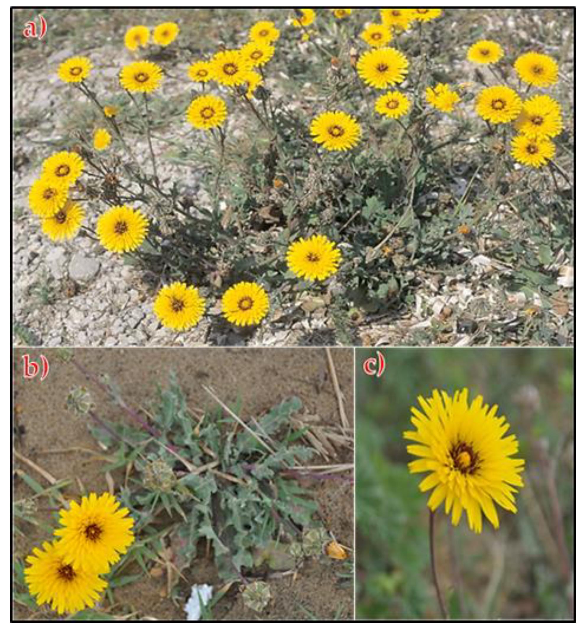
Around 2080–2094 species of seed plants and vascular cryptogams are found in Egypt’s flora [4,5]. Asteraceae family is one of the biggest flowering plant families, with a global distribution. Although the family includes a substantial number of shrubs, vines, and trees, the majority of the members are herbaceous. The Asteraceae family has 98 genera and 228 species in Egypt’s flora [4,6]. Many biological actions have been identified for members of the Asteraceae family, including antibacterial, antifungal, antidiabetic, antihelminthic, immunostimulatory, and anticancer properties [7,8,9]. Only two Reichardia species, R. picroides (L.) Roth and R. tingitana (L.) Roth, were recorded in Egypt’s flora that grows abundantly in Egypt’s coastal and inland deserts. R. tingitana (False sowthistle) is a glabrous erect annual herb that is native to the Mediterranean region and western Asia. The stem branches from the base, the plant grows up to 40 cm tall with the taproot, and it blooms from March to May, with characteristic yellow capitula terminal, solitary or paired on swollen stalks [6]. This genus has eight species that are found across the Mediterranean region, with some species being found in portions of India and Africa [10].
Recent studies have focused on the identification of active chemical components, as well as the biological applications of extracted plants [11,12]. Tumors, hepatic illnesses, poor digestion, constipation, calculi, heart issues, pains, spleen disorders, and hiccough are just a few of the afflictions that the whole plant can help with [13,14]. Several viral diseases, such as dengue fever, chikungunya, Zika fever, Mayaro, and yellow fever viruses, as well as other disease agents, are all conveyed by the mosquito A. aegypti. Continuous use of synthetic pesticides leads to vector resistance, biological multiplication of hazardous compounds via the food chain, and negative consequences for environmental quality and nontarget creatures, including human health [15,16].
The chemistry of R. tingitana extract was reported and it showed antimicrobial, bioherbicide, and cytotoxic activities [9,17]. Constituents, such as phenolics, tannins, flavonoids, coumarins, phytosterols, lactones, and essential oils, have been isolated from aboveground biomass of R. tingitana [9,17,18,19]. Moreover, little is known about the bioactive composition of R. tingitana, and allelochemicals as well as the anti-dengue vector activity of R. tingitana extract was not previously studied. Despite its significance, little study has been conducted on the plant. In order to examine the biochemical components responsible for the biological effects, the current study used GC-MS spectroscopy to assess the chemical constituents of the Egyptian ecospecies of R. tingitana methanol extract. The study looked at the antioxidant activity of the extracted plant using the DPPH free radical scavenging assay, as well as the in vitro antibacterial activities against a range of pathogenic bacteria. The larvicidal activity of the extract against A. aegypti was also investigated.

2. Results and Discussion

2.1. Total Phenolic, Flavonoid, and Tannin Contents

The medicinal benefits of herbal medicines are based on the chemistry of plants. Knowing a plant’s chemical composition helps one comprehend its possible medicinal benefits. Numerous bioactive chemical substances known as secondary plant metabolites are created by the plant cell via the main metabolic pathways [20]. Among them, phenolic compounds were shown to have significant antioxidant [21], anti-inflammatory [22], antihyperglycemic [23], immunomodulatory, and anticancer [24] action. The present study demonstrated that R. tingitana shoot extract is filled with phenolics (143.68 mg gallic acid equivalent/g dry extract), flavonoids (32.41 mg catechin equivalent/g dry extract), and tannins (96.57 mg gallic acid equivalent/g dry extract). Recent studies include our own findings on the antioxidant properties and activities of phenolics and flavonoids. They both have the potential to substantially promote and inhibit oxidative processes in this regard. Additionally, it has been shown that they have the capacity to split up chains of free radicals, which has a strong antioxidant effect.. In addition to phenolics and flavonoids, tannins are a prevalent and significant plant phytochemical. Tannins have strong antibacterial [25] and antioxidant [21] properties. Tannins can have an antinutritional impact in addition to these outcomes [26]. Tannins are phenolic chemicals that exist naturally. They include a lot of phenolic groups and have a high molecular weight (Mr > 500), which leads to the precipitation of proteins [27].
Abdel-Mogib et al. [19] and Abd-ElGawad et al. [9] reported that aboveground biomass has undergone phytochemical analysis, which reveals the presence of phenolics, tannins, flavonoids, coumarins, volatile oils, glycosides, flavonoids, lactones, esters, significant levels of sterols, and/or triterpenes. Numerous intrinsic and extrinsic variables, specialized metabolic processes, and endogenous physiological changes in the plants all contribute to the variation in biochemical content in the various solvent extracts and within the plant parts [28].

2.2. Gas-Chromatography Mass Spectroscopy “GC-MS”

The chemical structures and constitutes of the R. tingitana extract were characterized by GC-MS analysis. Figure 1 demonstrates the relation between the relative abundance of the different components characterized by the extracted plant against the retention time at which a definite component was detected. The results in Table 1 confirmed that 17 components were interpreted from the extracted plant. Commonly, 6,10,14-trimethylpentadecan-2-one (21.98%) and methyl oleate (27.26%) are positioned as the major components, which were ascertained after 19.25, and 23.34 min, respectively. Subsequently, other constitutes were categorized with elevated percent of composition, for instance, palmitic acid (14.85%), (E)-octadec-9-enoic acid (2.16%), methyl palmitate (3.29%), (Z)-octadec-11-enoic acid (3.56%), 3,7,12-trihydroxycholan-24-oic acid (6.63%), and Stigmast-5-en-3-ol, (3α)- (7.57%) (Figure 2). The components of hydrocarbons were recorded at 13.56 “very low relative abundance” and 19.25 min, while the components of fatty acids and esters were recorded with retention times ranging from 16.62 to 33.50 min. The most abundant components of steroids were identified after retention time at 34.45–35.70 min.
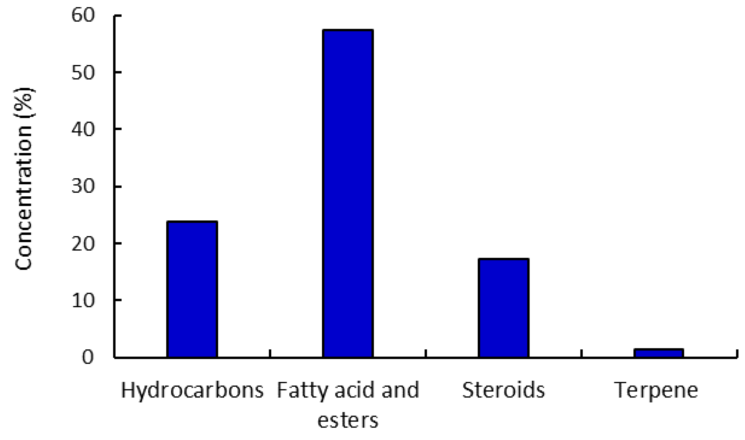
The characterized components were classified under numerous naturally occurring categories, such as hydrocarbons (23.82%), fatty acids, esters of fatty acids (57.46%), steroids (17.26%), and terpenes (1.48%) (Figure 3). The class of fatty acids and/or their ester derivatives retained the majority of these components. Thus, two components were identified for hydrocarbons, ten components were characterized as fatty acid and esters, four components as steroids, and one component as a terpene. Accordingly, 6,10,14-trimethylpentadecan-2-one is the major constituent of the hydrocarbon category, with 21.98% of the total composition of the extracted components. Moreover, methyl oleate is the major constituent of the fatty acid esters class, with 27.26%, while Stigmast-5-en-3-ol, (3α)- as a steroidal sub-class is the major component under this category (7.57%).

2.3. Biological Characteristics of the Plant Extracts

2.3.1. Antioxidant Activity—DPPH Assay

The capacity of R. tingitana methanolic extract to scavenge DPPH free radicals in comparison to ascorbic acid was used to determine its antioxidant activity. Half maximum inhibitory concentration (IC50) values were used to express the scavenging effects of plant extracts and the standard on the DPPH radical; the findings are shown in Table 2. A lower IC50 value indicates a greater capacity to scavenge DPPH radicals. Subsequently, the results, as presented in Table 2, confirmed that the extract from the shoot has the most antioxidant scavenging activity (IC50 = 30.77 mg L−1) compared to the ascorbic acid (IC50 = 12.02 mg L−1). The main element regulating the mechanism of the reactions engaged in the assessment of the antioxidant ability of the examined extract is the predominance of fatty acids and their derivatives (57.46%) and oxygenated hydrocarbons (23.82%) of total separated components. The present results of Rumex vesicarius were in agreement with the results of Abd-ElGawad et al. [9], Cornara et al. [29], and Leonti [30].
On the other hand, fatty acids and lipids, which were extracted from Sisymbrium irio, Aesculus indica, and Abies pindrow, showed strong antioxidant capabilities for scavenging the free radicals in the solution [31]. Palmitic acid and methyl oleate are the two main constituents, with 14.85 and 27.26% acting as significant antioxidant agents, respectively. The ability of reactive oxygen species, such as phenolics, fatty acids, terpenes, oxygenated hydrocarbons, or carbohydrates, to scavenge or stabilize free radicals generally determines the antioxidant capacity of bioactive chemicals [32,33,34].
In this investigation, the antioxidant activity of R. tingitana shoot extract was superior to that of other wild plant extracts in many countries [17]. According to Abd El-Gawad et al. [35], R. tingitana aerial parts have more antioxidant activity in their extract than other common Egyptian plants (Cakile maritime, Centaurea glomerata, Juncus bufonius, and Lactuca serriola). Different studies have demonstrated that the quantity of bioactive chemicals, particularly phenolic compounds, such as flavonoid, phenolic acids, ascorbic acid, and carotenoids, directly affects the antioxidant capabilities of plants [36]. From our study, this plant contains nonvolatile compounds, such as tannins, flavonoids and phenolics.

2.3.2. Antibacterial Activity

The antimicrobial properties of methanolic extract of R. tingitana at a concentration of 10 mg L−1 against four Gram-negative and five Gram-positive bacterial strains have been assessed in this study using agar disc diffusion assay [21]. The results revealed that the methanolic extract of a certain plant effectively inhibits the growth of pathogenic bacteria with varying potency (Table 3). According to the current result, methanolic extract of R. tingitana had the maximum zone of inhibition against S. typhimurium and B. cereus (25.71 ± 1.63 and 24.42 ± 0.81 mm, respectively), while C. tetani and S. xylosus were the main resistant species (Table 3). The tested pathogenic organisms can be arranged in the following order based on their sensitivity: S. typhimurium < B. cereus < Escherichia coli < Staphylococcus aureus < Klebsella pneumonius < Streptococcus pyogenes < Enterobacter aerogenes < S. xylosus < C. tetani. The common antibiotics, including ampicillin, azithromycin, cephradine, and tetracycline, had varying degrees of activity. S. typhimurium was totally resistant to ampicillin and cephradin, but C. tetani was completely resistant to cephradin and azithromycin and E. aerogenes was entirely resistant to tetracycline, ampicillin, and cephradin (Table 3). Additionally, plant extract demonstrated strong antibacterial activity against B. cereus, E. coli, and S. typhimurium (24.42 0.81, 22.85 0.87, and 25.71 1.63 mm, respectively) but less potency against C. tetani and S. xylosus when compared to antibiotics.
In the current investigation, the MeOH extract of R. tingitana had approximately twofold more activity against S. aureus, S. xylosus, and E. coil than did the chloroform extract in Yemen [37]. Additionally, the MeOH extract used in this investigation had stronger antibacterial action against S. aureus than that shown with MeOH derived from Tunisian ecospecies but was ineffective against E. coil [38]. Several previous experiments on the essential oil and n.hexane extract of the aerial parts of R. tingitana confirm that they were potent antibacterials against both Gram positive and negative bacteria [17]. These variations in the antibacterial potential may be explained by variances in the extracts’ chemical composition caused by environmental factors, such as geographical variability, soil type, and climate [39,40].
Generally, the Egyptian ecospecies showed more antimicrobial activity compared to the Yemen and Tunisian ecospecies. This difference could be ascribed to the variation in the chemical composition of the extract. The bioactivities of the plant extract are directly correlated with their chemical compound compositions either individually or synergistically [41]. Furthermore, it has been noted that MeOH extracts of wild plants contain a variety of different chemical compounds, including phenolic compounds and its derivative compounds, the esters of weak acids, fatty acids, and terpenes, and others. As a result, these chemical components can affect multiple target sites against the bacterial cells, with minor variations [40,42,43]. In order to determine the bacterial species’ efficacy against the extracts that were considered, its composition should be studied. Terpenes, steroids, oxygenated hydrocarbons, and carbohydrates showed stronger antibacterial potentials, according to our analysis of the main chemical constituents of the isolated R. tingitana [44,45,46]. Plant-derived phenolics, such as phenolic acids and flavonoids, two nonvolatile compounds found in this plant, have been shown to be effective antibacterial agents against bacteria with various resistances. Regardless of whether the bacteria are Gram-negative or Gram-positive species, the methanol extract of R. tingitana seemed to have stronger antibacterial power against a specific type of bacterial species.

2.3.3. Anticancer Activity

The usage of therapeutic plants and the herbal extracts they are derived from, which are rich in polyphenolic chemicals, may help explain the decreased incidence of cancer. Numerous clinical trials have shown that different herbal medications can exhibit a range of anticancer effects. Additionally, the use of herbal extracts with distinctive therapeutic qualities is advantageous in the field of pharmaceutical chemistry due to their improved effectiveness and biological potency [47,48]. In this study, the cytotoxic activities of the prepared plant sample extract were evaluated using an MTT assay. The sample was tested in vitro against two tumor cells, i.e., HepG-2, and PC3 cell lines (Table 4). Doxorubicin was selected as a reference drug, comparing the results of the tested sample against the different cancer cells. The method was applied for determining the cell metabolic activity based on the aptitude of nicotinamide adenine dinucleotide phosphate (NADPH)-dependent cellular oxidoreductase enzymes to reduce the MTT tetrazolium dye to its formazan “insoluble” that has a purple color. The number of viable cells should increase with growth, decrease with cytotoxic treatments, and remain the same (or plateau) with cytostatic treatments. The control sample is a benefit for calculating the cell viability percent, as it produces 100% viability of healthy cells.
The experiments were run using five concentrations of each plant extract (1.56, 3.125, 6.25, 12.50, 25.00, 50.00, and 100 µg mL−1) prepared in a serial dilution. According to the results, the methanolic extracts of plant exhibited cytotoxic activity in a dose-dependent manner, which was comparable with doxorubicin as a reference standard. At 100 µg mL−1, the extract of R. tingitana showed inhibition activities of 93.14%, 91.41%, and 9.55% for HepG-2 and PC3 human tumor cells and normal cell (WI-38), respectively. However, the lowest concentration (1.56 µg mL−1) shows the lowest cytotoxic activity in all samples (Table 4).
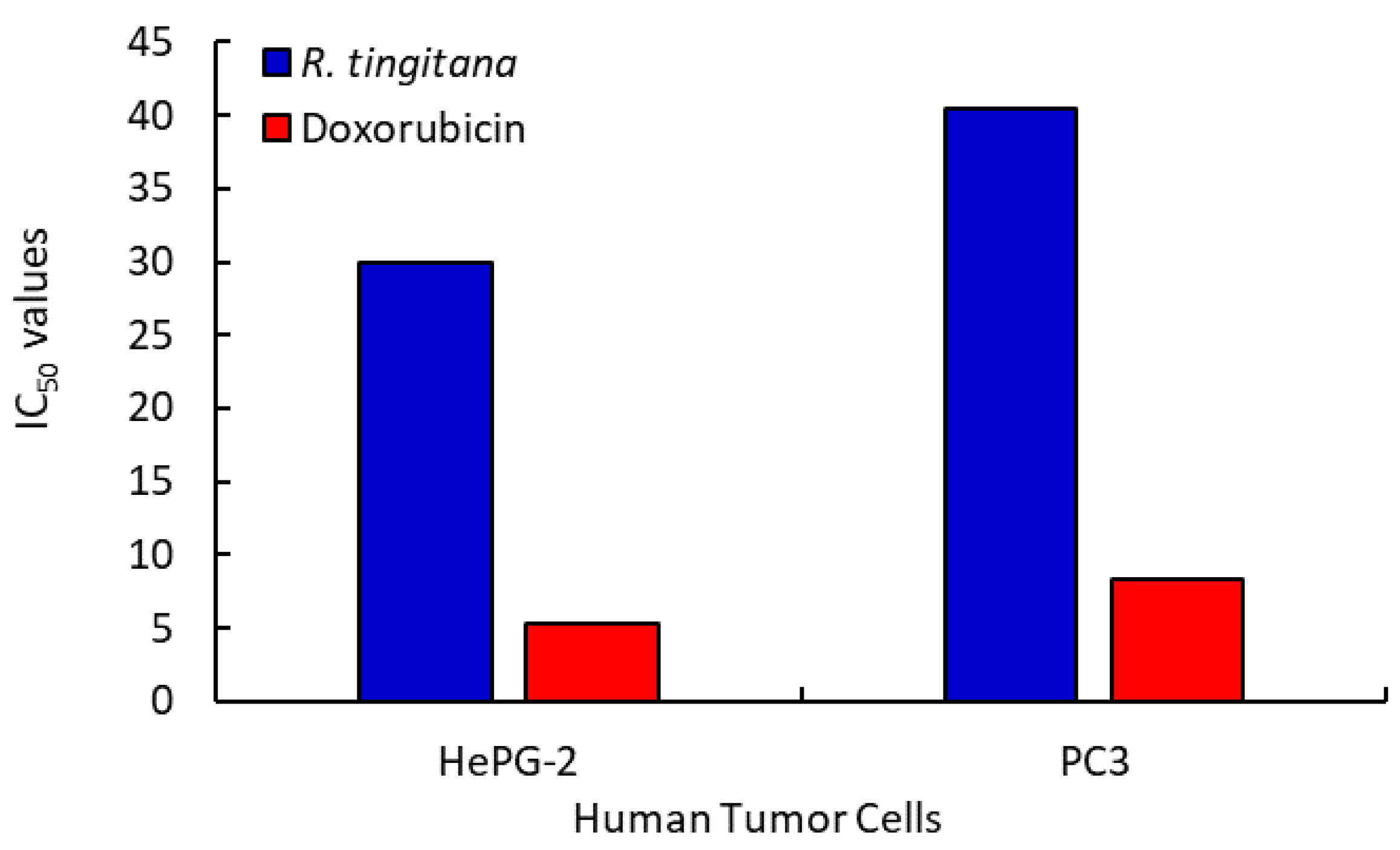
The curves generated by plotting the percentages of cell survival vs. drug concentration (µM) were used to calculate the IC50 values, which expressed the concentration that represented 50% of the inhibition of cell growth. As a result, as the extract concentration and IC50 values decline, the potency of cytotoxicity will increase. For HepG-2 and PC3 cells, as well as WI-38 normal cells, the IC50 values of the MeOH extract from the R. tingitana sample were 29.977, 40.479, and >100 µg mL−1, respectively. To compare the outcomes of the tested compounds against the various cancer cells, which achieved IC50 values of 5.274, 8.303, and >100 µg mL−1 for HepG-2, PC3, and WI-38, respectively, doxorubicin was used as a reference drug (Table 4 and Figure 4). According to the IC50 data, the extracted shoot of this plant shows noncytotoxic activity for normal cells (WI-38) and moderate cytotoxic activity for two human tumor cells (HepG-2 and PC3). This result is in agreement with the result obtained by Csupor-Löffler et al. [49], who studied the ant proliferative activities of aqueous and organic extracts prepared from 26 Hungarian species of the family Asteraceae, which were tested in vitro against HeLa (cervix epithelial adenocarcinoma), A431 (skin epidermoid carcinoma), and MCF7 (breast epithelial adenocarcinoma) cells by using the MTT assay.
It is important to note that the structural nature of each extract’s components and the nature of the cancer cell line are frequently correlated with the cytotoxicity of extracted samples as cytotoxic agents on HepG2 and PC3 tumor cell lines. Furthermore, the characteristics of the extracted plant particles, such as their size, aggregation, and surface shape, may influence their cytotoxicity. The chemical composition of the plant extract, the kind of cancer cell, and the concentration of the extracted plant employed in this assessment were among the parameters that affected how effective the plant extract was as an anticancer agent for tumor cell proliferation [50]. Many polyphenols, such as isoflavones, are phytoestrogens that may bind to estrogen receptors and have an estrogenic impact on the tissues or organs they are intended to affect. Additionally, newly identified substances, such as phenolic acids and flavonoids, could be to blame for the anti-inflammatory and cytotoxic effects of plant extracts [40,51]. R. tingitana contains nonvolatile substances, such flavonoids and phenolics in the current investigation.

2.3.4. Larvicidal Bioassay

Serious human diseases are spread by mosquitoes, which result in millions of fatalities each year. Due to the development of synthetic pesticide resistance, vector control is at risk. In the future, insecticides with a botanical origin could be a good substitute for biocontrol methods [52,53]. Data from Table 5 showed that R. tingitana methanolic extract had larvicidal action against A. aegypti larvae in their third instar at 24 and 48 h after treatment. Results from this investigation showed a substantial increase in larval mortality at all concentration of R. tingitana methanolic extract. The highest larval mortality was recorded at concentrations of 300 mg L−1 for 24, 48, and 72 h (32.54%, 44.28%, and 50.81%, respectively), while the lowest mortality was 6.35%, 10.13%, and 15.74% at 24, 48, and 72 h, when the concentration was found to be 100 mg L−1, compared to 1.05% for control group.
The LC50 (the concentration of extract that causes 50% death) of extract is 46.85, 35.75, and 29.38 mg L−1 for the various time periods of 24, 48, and 72 h, whereas the LC90 (the concentration of extract that causes 90% mortality) is 82.66, 63.82, and 53.30 mg L−1, respectively (Table 5). The different bioactive chemicals, such as phenolics, terpenoids, flavonoids, and alkaloids, either as single or combined compounds, determine the toxicity of the plants [40,54]. Additionally, desert plants, such as R. tingitana, are a rich source of bioactive chemicals that can be used as larvicides, pupicides, or mosquito repellents [55,56].
These outcomes also line up with those attained by Salama et al. [40], who utilized Rumex vesicarius methanolic extract against A. aegypti larvae and found that the LC50 and LC90 values were 19.99 and 36.12 mg L−1 after 24 h. Shehata [57] reported that petroleum ether extract from leaves of Prunus domestica and Rhamnus cathartica was more effective against Culex pipiens (LC50: 33.3 and 63.4 mg L−1, respectively) than chloroform (LC50: 70.8 and 192.1 mg L−1) and methanolic extracts (LC50: 132.7 and 273.5 mg L−1, respectively). Ullah et al. [56] recorded LC50 and LC90 values of Cassia fistula and Nicotiana tabacum extracts of 50.27, 203.99, and 17.77 and 206.49 mg L−1 against larvae of Culex quinquefasciatus. According to Dey et al. [58], Piper longum aqueous extract had the highest 24-h larval death rates when used to treat A. aegypti, A. stephensi, and C. quinquefasciatus.

3. Materials and Methods

3.1. Plant Material and Extraction Process

In April 2021, during the blossoming season, multiple populations of Reichardia tingitana (L.) Roth were gathered from in Al-Hashr Mountain, Jazan region, Saudi Arabia (17°27′21.31″ N 43°2′18.13″ E) (Figure 5). The plant was identified in line with Tackholm [4] and Boulos [6]. At Mansoura University in Egypt’s Faculty of Science, a voucher specimen (Mans. 0011820006) was made and added to the herbarium.
The samples were cleaned and allowed to dry naturally. A conical flask with a capacity of 250 mL and 150 mL of methanol was filled with 10 g of dried plant material. After that, the mixture was put into a water bath shaker (Memmert WB14: Schwabach, Germany), where it was shaken continuously for two hours at room temperature. The mixture was filtered using Whatman filter papers (no. 1, 125 mm, Cat. No. 1001 125, Germany). The methanol extract was dried using a rotary evaporator and the residue was collected in glass vials and stored in the refrigerator at 4 °C until further analyses [59].

3.2. Phytochemical Analysis

Similar to how hot infusions are used in conventional therapy, the active components were extracted using this technique. Twenty grams of the dried plant stems were mixed with 200 mL of deionized water and agitated for 30 min in a water bath system with a temperature of 70 °C. Filtering the extracted product, the filtrate was kept at 4 °C for subsequent use. The total quantities of phenolic, flavonoid, and tannin in the aqueous extract of R. tingitana aerial parts were quantified.

3.2.1. Total Phenolic Contents

By using the Folin–Ciocalteu approach developed by Wolfe et al. [60] and Issa et al. [61], which used gallic acid as a standard, the total phenolic content was evaluated. Using a standard curve (y = 0.00621x, r2 = 0.976), the measured amounts of total phenolic content in the aqueous extract of R. tingitana were converted to milligrams of gallic acid/grams of dry plant extract.

3.2.2. Total Flavonoid Contents

According to Zhishen et al. [62], who used catechin as a reference, a colorimetric analysis using aluminum chloride was used to determine the total flavonoids present. The amounts of total flavonoid constituents were calculated using a standard curve (y = 0.0027x, r2 = 0.979) as equivalents of catechin in milligram per dried plant extract in gram.

3.2.3. Total Tannin Contents

The amount of total tannins was evaluated using the vanillin–hydrochloride test, and the values of the predicted samples were represented as the equivalents of tannic acid in grams/dried plant per 100 g, according to Burlingame [63] and Aberoumand [64].

3.3. Gas Chromatography-Mass Spectrometry Analysis (GC-MS)

The chemical composition of the extracted R. tingitana plant was described by employing the plant extract on a Trace GC-TSQ mass spectrometer (Thermo Scientific: Austin, TX, USA) with a direct capillary column TG-5MS (30 m × 0.25 mm × 0.25 m film thickness) [65]. The temperature of the column oven was first maintained at 50 °C, then raised by 5 °C per minute to reach 250 °C and maintained for 2 min, and then increased by 30 °C per minute to reach the final temperature of 300 °C and maintained for 2 min. The MS transfer line and injector were kept at temperatures of 260 and 270 °C, respectively. As a carrier inert gas, helium (He) was employed at a constant flow rate of 1 mL/min. After 4 min, the solvent was removed, and 1 µL diluted samples were immediately fed into the GC in split mode using an Auto sampler AS1300. In packed scan mode, EI mass spectroscopy data over the m/z range of 50–500 were gathered at an ionization voltage of 70 EV. The ion source’s temperature was set at 200 °C. By comparing the mass spectral data of the various extracted plant materials to those of the mass spectrometry databases WILEY 09 and NIST 14, it was possible to understand the chemical composition of each of the distinct plant materials. Five potential components were suggested by the GC-MS analysis for each identified peak. The probability factors and the primary structure’s fragmentation patterns governed the chosen structure in the case of the different suggested components.

3.4. Antioxidant DPPH Assay

The desired concentrations of R. tingitana were obtained by diluting a stock solution of the extracted plant in methanol (5, 10, 20, 30, 40, and 50 mg L−1). To each concentration of the sample solution that had been made, DPPH solution (1 mL, 0.135 Mm) was added. The tested samples’ concentrations of catechol were utilized as the standard. The samples were left at room temperature in the dark for 30 min, and a UV/Vis spectrophotometer was used to measure the absorbance at a wavelength of 517 nm (Spekol 11 spectrophotometer, analytic Jena AG: Jena, Germany). The following equation was used to compute the percentages of antioxidant scavenging activities, using a DPPH solution in methanol as a reference:
Inhibition   % = A   control     A   sample A   control × 100
The approach was applied while making only minimal alterations to the preceding investigations [66,67]. The inhibitive concentrations were calculated using the exhibited exponential curve [68], which depicted the connection between the sample concentration and the amount of residual DPPH radical (IC50, mg L−1).

3.5. Antimicrobial Activity Procedure

The agar well diffusion experiment was used to determine the antibacterial potential of the examined R. tingitana extract [69,70]. Standard antibiotics were cephradin (CPD), tetracycline (TAC), azithromycin (ATM), and ampicillin (AMC). The microbial species were purchased from the Microbiological Resources Centre (MIRCEN), Faculty of Agriculture, Ain Shams University, and were of animal origin. The tested bacteria were Escherichia coli (NR_112558.1), Enterobacter aerogenes (KX878983.1), Salmonella typhimurium (NR_074910.1), and Klebsella pneumonius (NR_117683.1). Gram-positive bacteria: Bacillus cereus (EMCC_1080), Clostridium tetani (KHO40477.1), Staphylococcus aureus (NR_115606.1), Streptococcus pyogenes (MH916557.1), and Staphylococcus xylosus (NR_113350.1). The inhibitory zone diameters (mm) were determined after the plates had been incubated at 37 °C for 18 to 24 h.

3.6. Anticancer Activity Procedure

Hepatocellular carcinoma (HePG-2), mammary gland carcinoma (MCF-7), and human prostate cancer (PC3) are three particular human tumor cell lines that were purchased from the ATCC holding business for biological products and vaccines (VACSERA: Cairo, Egypt). A common chemotherapeutic anticancer medication was doxorubicin. The chemical reagents used were RPMI-1640 medium, MTT, DMSO (Sigma Co.: St. Louis, MO, USA), and fetal bovine serum (FBS; Gibco Life Technologies: Paisley, UK).
A typical colorimetric MTT assay was performed to monitor cell growth and assess the cytotoxicity of the extracted R. tingitana in accordance with the guidelines given by Bondock et al. [71]. Simply put, the transformation of MTT (2-(4,5-dimethylthiazol2-yl)-3,5-diphenyl-2H-tetrazolium bromide) from yellow to purple was carried out by mitochondrial succinate dehydrogenases of living cells. To create the cell strains, RPMI-1640 media was supplemented with 10% fetal bovine serum. In an incubator with 5% CO2, penicillin (100 units mL−1) and streptomycin (100 g mL−1) antibiotics were added. Cell lines were seeded on a 96-well plate at a density of 1.0 × 10⁴ cells/well suspended in 100 μL of complete medium and incubated at 37 °C for 48 h with 5 % CO2. Cells were cultured for the first 24 h, and then treated with various doses of the test samples. MTT solution (5 mg mL−1, 20 L) was added following a 24-h drug treatment period, and the mixture was once more incubated for 4 h. To dissolve the created violet formazan, 100 L of DMSO was then applied to each well. The colorimetric analysis was conducted, and a plate reader was used to measure the absorbance values at 570 nm (EXL 800, New York, NY, USA). The Origin 8.0® program (OriginLab Corporation, Available online: https://www.originlab.com/; Accessed 27 March 2021) was used to generate the IC50 values using nonlinear regression (sigmoid type). The following equation, in which optical density (OD) expresses the absorbance read of the control and the tested sample, was used to compute the percentage of inhibition in cell growth:
%   Inhibition = OD   control   OD   sample OD   control × 100

3.7. Mosquitocidal Assay

3.7.1. Rearing of Aedes aegypti

The Center for Disease Vector in Jizan provided the A. aegypti larvae, which were raised for six generations in the Center for Environmental Research at the Faculty of Science in Jazan University under carefully controlled conditions of temperature (28 ± 2 °C), relative humidity (70–80%), and 12h/12h light–dark regime. Three days after emergence, adult mosquitoes were housed in (30 × 30 × 30 cm) wooden cages and given cotton pieces every day that had been soaked in a 10.0% sucrose solution. Following this time, females were permitted to have a blood meal from a pigeon host, which is required for egg laying (anautogeny). In the cage for egg laying, a plastic cup oviposition (15 × 15 cm) with dechlorinated tap water was inserted. The finished egg rafts were removed from the plastic dish and placed in plastic pans (25 × 30 × 15 cm) with three liters of tap water that had been left for 24 h. A slice of bread was given to the developing larvae each day as food. This diet was discovered to be the best for female fertility and larval growth [72].

3.7.2. Larvicidal Activity Procedure

The tested material for larvicidal activity was dissolved in 0.1 mL of methanol. To find deaths, several concentration ranges of each relevant extract were generated. In 200 mL plastic cups containing 100 mL of dechlorinated tap water, the tested sample was run. Then, 10 larvae in the third instar were immediately placed into plastic containers that contained various extract concentrations. Typically, three replicates were employed for each concentration under examination. All of the plastic cups were incubated in a mosquito colony under controlled conditions and, thereafter, mortality was noted. A total of 0.1 mL of methanol or 2 drops of Tween 80 were given to control larvae in 100 mL of water. Up to adult emergence, mortality was tracked every day and dead larvae and pupae were eliminated [72]. Failure to react to mechanical stimulation was used to detect larval death. The following equation [73] was utilized to calculate the % larval mortality:
Larval   mortality   % = Number   of   dead   larvae Number   of   treated   larvae × 100

3.8. Statistical Analysis

Using the Costat software (CoHort Software: Monterey, CA, USA), the antioxidant, antibacterial, and larvicidal activity experiments were conducted three times with three replications. The outcomes were then subjected to a one-way ANOVA to assess the significance of the differences between samples.

4. Conclusions

In conclusion, by using GC-MS analysis, 17 components were interpreted from the MeOH extract of R. tingitana aerial parts. The two primary components are commonly reported as 6,10,14-trimethylpentadecan-2-one (21.98%) and methyl oleate (27.26%). Hydrocarbons (23.82%), fatty acids, esters of fatty acids (57.46%), steroids (17.26%), and terpenes (1.48%) were identified as the major components. The R. tingitana shoots demonstrated advantageous biological characteristics, including larvicidal effectiveness against A. aegypti, the dengue virus vector, as well as antioxidant, antibacterial, and cytotoxic agents. Shoot extract exhibited the highest antioxidant activity, showing a better capacity to trap free radicals in the DPPH solution, with an IC50 value of 30.77 mg L−1. The current findings indicate that the R. tingitana extracted shoot exhibited the most potent antibacterial activity against Gram-negative bacterial species, exhibiting broad-spectrum activity against S. typhimurium and B. cereus, while C. tetani and S. xylosus were the main resistant species. Additionally, compared to control, the methanol extract of R. tingitana showed increased potential anticancer activity, with significant cytotoxicity for the two tumor cells: HepG-2 and PC3 cells. Furthermore, A. aegypti larvae were more easily killed by the MeOH extract of the plant sample. The significant biological findings and the impressive percentage of phenolic, flavonoid, and tannin contents found in the R. tingitana extract, supported the possibility of further study on this plant for the creation of drugs from natural sources.

Author Contributions

Conceptualization, Y.A.E.-A. and S.A.S.; validation, Y.A.E.-A. and S.A.S.; formal analysis, Y.A.E.-A. and Z.E.A.-F.; investigation, Y.A.E.-A., Z.E.A.-F. and S.A.S.; data curation, Y.A.E.-A.; writing—original draft preparation, Y.A.E.-A.; writing—review and editing, Y.A.E.-A., Z.E.A.-F. and S.A.S. All authors have read and agreed to the published version of the manuscript.

Funding

The authors extend their appreciation to The Researchers Supporting Project number (ISP20-1), Jazan University, Saudi Arabia.

Institutional Review Board Statement

Not applicable.

Informed Consent Statement

Not applicable.

Data Availability Statement

The data presented in this study are available on request from the corresponding author.

Acknowledgments

The authors extend their appreciation to the deputyship for Research, Innovation, and Ministry of Education in Saudi Arabia for funding this research work through the Project with number: ISP20-1.

Conflicts of Interest

The authors declare no conflict of interest.

References

  1. Memariani, Z.; Farzaei, M.H.; Ali, A.; Momtaz, S. Nutritional and Bioactive Characterization of Unexplored Food Rich in Phytonutrients. In Phytonutrients in Food; Woodhead Publishing, Elsevier: Amsterdam, The Netherlands, 2020; pp. 157–175. [Google Scholar]
  2. Gusain, P.; Uniyal, D.; Joga, R. Conservation and Sustainable Use of Medicinal Plants. In Preparation of Phytopharmaceuticals for the Management of Disorders; Apple Academic Press, Elsevier: Amsterdam, The Netherlands, 2021; pp. 409–427. [Google Scholar]
  3. David, B.; Wolfender, J.-L.; Dias, D.A. The pharmaceutical industry and natural products: Historical status and new trends. Phytochem. Rev. 2015, 14, 299–315. [Google Scholar] [CrossRef]
  4. Tackholm, V. Students’ Flora of Egypt; Cairo University Publishing: Cairo, Egypt, 1974. [Google Scholar]
  5. Boulos, L. Flora of Egypt. Checklist; Al-Hadara Publishing: Cairo, Egypt, 1995. [Google Scholar]
  6. Boulos, L. Flora of Egypt; Al-Hadara Publishing: Cairo, Egypt, 2002; Volume 3. [Google Scholar]
  7. Sharma, P.; Prasad, G.; Archana, S. A comprehensive review: Medicinal plants with potential antidiabetic activity. Res. Rev. J. Herb. Sci. 2018, 4, 29–57. [Google Scholar]
  8. Cazella, L.N.; Glamoclija, J.; Soković, M.; Gonçalves, J.E.; Linde, G.A.; Colauto, N.B.; Gazim, Z.C. Antimicrobial activity of essential oil of Baccharis dracunculifolia DC (Asteraceae) aerial parts at flowering period. Front. Plant Sci. 2019, 10, 27. [Google Scholar] [CrossRef] [PubMed] [Green Version]
  9. Abd-El Gawad, A.M.; El-Amier, Y.A.; Assaeed, A.M.; Al-Rowaily, S.L. Interspecific variations in the habitats of Reichardia tingitana (L.) Roth leading to changes in its bioactive constituents and allelopathic activity. Saudi J. Biol. Sci. 2020, 27, 489–499. [Google Scholar] [CrossRef]
  10. Recio, M.C.; Giner, R.M.; Hermenegildo, M.; Peris, J.B.; Mañez, S.; Rios, J.-L. Phenolics of Reichardia and their taxonomic implications. Biochem. Syst. Ecol. 1992, 20, 449–452. [Google Scholar] [CrossRef]
  11. Khan, I.H.; Javaid, A. Anticancer, antimicrobial and antioxidant compounds of quinoa inflorescence. Adv. Life Sci. 2020, 8, 68–72. [Google Scholar]
  12. Javed, S.; Mahmood, Z.; Khan, K.M.; Sarker, S.D.; Javaid, A.; Khan, I.H.; Shoaib, A. Lupeol acetate as a potent antifungal compound against opportunistic human and phytopathogenic mold Macrophomina phaseolina. Sci. Rep. 2021, 11, 11. [Google Scholar]
  13. Gopal, R.; Vijayakumaran, M.; Venkatesan, R.; Kathiroli, S. Marine organisms in Indian medicine and their future prospects. Indian J. Nat. Prod. Resourc. 2008, 7, 139–145. [Google Scholar]
  14. Mostafa, H.; Elbakry, A.; Eman, A.A. Evaluation of antibacterial and antioxidant activities of different plant parts of Rumex vesicarius L. (Polygonaceae). Int. J. Pharm. Pharm. Sci. 2011, 3, 109–118. [Google Scholar]
  15. Weeratunga, P.; Rodrigo, C.; Fernando, S.D.; Rajapakse, S. Control methods for Aedes albopictus and Aedes aegypti. Cochrane Database Syst. Rev. 2017, 8, CD012759. [Google Scholar]
  16. Carvajal-Lago, L.; Ruiz-López, M.J.; Figuerola, J.; Martínez-de la Puente, J. Implications of diet on mosquito life history traits and pathogen transmission. Environ. Res. 2021, 195, 110893. [Google Scholar] [CrossRef] [PubMed]
  17. El Alfy, T.; El Tantawy, M.E.; Motaal, A.A.; Gamal, F.E.Z. Pharmacological, biological study and GC/MS analysis of the essential oil of the aerial parts and the alcohol soluble fraction of the n. Hexane extract of the flowers of Reichardia tingitana L. Can. J. Pure Appl. Sci. 2015, 9, 3167–3175. [Google Scholar]
  18. Daniewski, W.M.; Skibicki, P.; Gumułka, M.; Drożdż, B.; Grabarczyk, H.; Błoszczyk, E. Sesquiterpene lactones. XXXV. Constituents of Reichardia tingitana L. Roth. and their antifeedant activity. Acta Soc. Bot. Pol. 1988, 57, 539–545. [Google Scholar] [CrossRef] [Green Version]
  19. Abdel-Mogib, M.; Abou-Elzahab, M.; Dawidar, A.; Ayyad, S. A sesquiterpene glucoside from Reichardia tingitana. Phytochemistry 1993, 34, 1434–1435. [Google Scholar] [CrossRef]
  20. Thomford, N.E.; Senthebane, D.A.; Rowe, A.; Munro, D.; Seele, P.; Maroyi, A.; Dzobo, K. Natural products for drug discovery in the 21st century: Innovations for novel drug discovery. Int. J. Mol. Sci. 2018, 19, 1578. [Google Scholar] [CrossRef] [Green Version]
  21. El-Amier, Y.A.; Soufan, W.; Almutairi, K.F.; Zaghloul, N.S.; Abd-ElGawad, A.M. Proximate composition, bioactive compounds, and antioxidant potential of wild halophytes grown in coastal salt marsh habitats. Molecules 2021, 27, 28. [Google Scholar] [CrossRef] [PubMed]
  22. Fu, R.; Zhang, Y.-T.; Guo, Y.-R.; Huang, Q.-L.; Peng, T.; Xu, Y.; Tang, L.; Chen, F. Antioxidant and anti-inflammatory activities of the phenolic extracts of Sapium sebiferum (L.) Roxb. leaves. J. Ethnopharmacol. 2013, 147, 517–524. [Google Scholar] [CrossRef] [PubMed]
  23. Manickam, M.; Ramanathan, M.; Farboodniay Jahromi, M.; Chansouria, J.; Ray, A. Antihyperglycemic activity of phenolics from Pterocarpus marsupium. J. Nat. Prod. 1997, 60, 609–610. [Google Scholar] [CrossRef]
  24. Liu, X.; Zhao, M.; Wu, K.; Chai, X.; Yu, H.; Tao, Z.; Wang, J. Immunomodulatory and anticancer activities of phenolics from emblica fruit (Phyllanthus emblica L.). Food Chem. 2012, 131, 685–690. [Google Scholar] [CrossRef]
  25. Scalbert, A. Antimicrobial properties of tannins. Phytochemistry 1991, 30, 3875–3883. [Google Scholar] [CrossRef]
  26. Serrano, J.; Puupponen-Pimiä, R.; Dauer, A.; Aura, A.M.; Saura-Calixto, F. Tannins: Current knowledge of food sources, intake, bioavailability and biological effects. Mol. Nutr. Food Res. 2009, 53, S310–S329. [Google Scholar] [CrossRef] [Green Version]
  27. Hagerman, A.E.; Riedl, K.M.; Jones, G.A.; Sovik, K.N.; Ritchard, N.T.; Hartzfeld, P.W.; Riechel, T.L. High molecular weight plant polyphenolics (tannins) as biological antioxidants. J. Agric. Food Chem. 1998, 46, 1887–1892. [Google Scholar] [CrossRef]
  28. Arif, Y.; Singh, P.; Siddiqui, H.; Bajguz, A.; Hayat, S. Salinity induced physiological and biochemical changes in plants: An omic approach towards salt stress tolerance. Plant Physiol. Biochem. 2020, 156, 64–77. [Google Scholar] [CrossRef] [PubMed]
  29. Cornara, L.; la Rocca, A.; Marsili, S.; Mariotti, M. Traditional uses of plants in the Eastern Riviera (Liguria, Italy). J. Ethnopharmacol. 2009, 125, 16–30. [Google Scholar] [CrossRef] [PubMed]
  30. Leonti, M. Local Mediterranean food as a source of novel nutraceuticals. In Pharmaceutical Soc Great Britain; Pharmaceutical Press: London, UK, 2006. [Google Scholar]
  31. Nengroo, Z.R.; Rauf, A. Fatty acid composition and antioxidant activities of five medicinal plants from Kashmir. Ind. Crops Prod. 2019, 140, 111596. [Google Scholar] [CrossRef]
  32. Okwu, D.E. Citrus fruits: A rich source of phytochemicals and their roles in human health. Int. J. Chem. Sci. 2008, 6, 451–471. [Google Scholar]
  33. Al-Snafi, A.E. The medical benefit of Gnaphalium luteoalbum—A review. IOSR J. Pharm. 2019, 9, 40–44. [Google Scholar]
  34. Lu-Martínez, A.A.; Báez-González, J.G.; Castillo-Hernández, S.; Amaya-Guerra, C.; Rodríguez-Rodríguez, J.; García-Márquez, E. Studied of Prunus serotine oil extracted by cold pressing and antioxidant effect of P. longiflora essential oil. J. Food Sci. Technol. 2021, 58, 1420–1429. [Google Scholar] [CrossRef]
  35. Abd El-Gawad, A.; Mashaly, I.A.; Al-Nafie, R.I. Antioxidant activity and allelopathic potential of five wild plants on germination and growth Bidens pilosa L. Int. J. Curr. Res. 2015, 7, 21019–21024. [Google Scholar]
  36. Sytařová, I.; Orsavová, J.; Snopek, L.; Mlček, J.; Byczyński, Ł.; Mišurcová, L. Impact of phenolic compounds and vitamins C and E on antioxidant activity of sea buckthorn (Hippophaë rhamnoides L.) berries and leaves of diverse ripening times. Food Chem. 2020, 310, 125784. [Google Scholar] [CrossRef] [PubMed]
  37. Alasbahi, R.H. In vitro antibacterial activity of some Yemeni medicinal plants. Univ. Aden J. Nat. Appl. Sci. 2008, 12, 641–647. [Google Scholar]
  38. Sassi, A.B.; Harzallah-Skhiri, F.; Aouni, M. Investigation of some medicinal plants from Tunisia for antimicrobial activities. Pharm. Biol. 2007, 45, 421–428. [Google Scholar] [CrossRef]
  39. Fayed, E.M.; Abd-EIGawad, A.M.; Elshamy, A.I.; El-Halawany, E.S.F.; EI-Amier, Y.A. Essential oil of Deverra tortuosa aerial parts: Detailed chemical profile, allelopathic, antimicrobial, and antioxidant activities. Chem. Biodivers. 2021, 18, e2000914. [Google Scholar] [CrossRef]
  40. Salama, S.A.; Al-Faifi, Z.E.; Masood, M.F.; El-Amier, Y.A. Investigation and Biological Assessment of Rumex vesicarius L. Extract: Characterization of the Chemical Components and Antioxidant, Antimicrobial, Cytotoxic, and Anti-Dengue Vector Activity. Molecules 2022, 27, 3177. [Google Scholar] [CrossRef] [PubMed]
  41. Vamanu, E.; Gatea, F. Correlations between microbiota bioactivity and bioavailability of functional compounds: A mini-review. Biomedicines 2020, 8, 39. [Google Scholar] [CrossRef] [Green Version]
  42. Ullah, M.F.; Khan, M.W. Food as medicine: Potential therapeutic tendencies of plant derived polyphenolic compounds. Asian Pac. J. Cancer Prev. 2008, 9, 187–196. [Google Scholar] [PubMed]
  43. Shin, J.; Prabhakaran, V.-S.; Kim, K.-S. The multi-faceted potential of plant-derived metabolites as antimicrobial agents against multidrug-resistant pathogens. Microb. Pathog. 2018, 116, 209–214. [Google Scholar] [CrossRef] [PubMed]
  44. El-Amier, Y.A.; Abdelghany, A.M.; Abed Zaid, A. Green synthesis and antimicrobial activity of Senecio glaucus-Mediated silver nanoparticles. Res. J. Pharm. Biol. Chem. 2014, 5, 631–642. [Google Scholar]
  45. Maccelli, A.; Vitanza, L.; Imbriano, A.; Fraschetti, C.; Filippi, A.; Goldoni, P.; Maurizi, L.; Ammendolia, M.G.; Crestoni, M.E.; Fornarini, S. Satureja montana L. Essential oils: Chemical profiles/phytochemical screening, antimicrobial activity and o/w nanoemulsion formulations. Pharmaceutics 2019, 12, 7. [Google Scholar] [CrossRef] [PubMed] [Green Version]
  46. Salehi, B.; Krochmal-Marczak, B.; Skiba, D.; Patra, J.K.; Das, S.K.; Das, G.; Popović-Djordjević, J.B.; Kostić, A.Ž.; Anil Kumar, N.V.; Tripathi, A. Convolvulus plant-A comprehensive review from phytochemical composition to pharmacy. Phytother. Res. 2020, 34, 315–328. [Google Scholar] [CrossRef] [PubMed]
  47. Syed, D.N.; Khan, N.; Afaq, F.; Mukhtar, H. Chemoprevention of prostate cancer through dietary agents: Progress and promise. Cancer Epidemiol. Biomark. Prev. 2007, 16, 2193–2203. [Google Scholar] [CrossRef] [PubMed] [Green Version]
  48. Zaki, A.A.; Ali, Z.; Wang, Y.-H.; El-Amier, Y.A.; Khan, S.I.; Khan, I.A. Cytotoxic steroidal saponins from Panicum turgidum Forssk. Steroids 2017, 125, 14–19. [Google Scholar] [CrossRef] [PubMed]
  49. Csupor-Löffler, B.; Hajdú, Z.; Réthy, B.; Zupkó, I.; Máthé, I.; Rédei, T.; Falkay, G.; Hohmann, J. Antiproliferative activity of Hungarian Asteraceae species against human cancer cell lines. Part II. Phytother. Res. Int. J. Devoted Pharmacol. Toxicol. Eval. Nat. Prod. Deriv. 2009, 23, 1109–1115. [Google Scholar] [CrossRef]
  50. Khacha-Ananda, S.; Tragoolpua, K.; Chantawannakul, P.; Tragoolpua, Y. Antioxidant and anti-cancer cell proliferation activity of propolis extracts from two extraction methods. Asian Pac. J. Cancer Prev. 2013, 14, 6991–6995. [Google Scholar] [CrossRef] [Green Version]
  51. Bazou, D.; Coakley, W.T.; Hayes, A.J.; Jackson, S.K. Long-term viability and proliferation of alginate-encapsulated 3-D HepG2 aggregates formed in an ultrasound trap. Toxicol. Vitr. 2008, 22, 1321–1331. [Google Scholar] [CrossRef] [PubMed]
  52. Peng, Z.; Yang, J.; Wang, H.; Simons, F.E.R. Production and characterization of monoclonal antibodies to two new mosquito Aedes aegypti salivary proteins. Insect Biochem. Mol. Biol. 1999, 29, 909–914. [Google Scholar] [CrossRef]
  53. Mohankumar, T.K.; Shivanna, K.S.; Achuttan, V.V. Screening of methanolic plant extracts against larvae of Aedes aegypti and Anopheles stephensi in Mysore. J. Arthropod-Borne Dis. 2016, 10, 303. [Google Scholar] [PubMed]
  54. Nasir, S.; Nasir, I.; Asrar, M.; Debboun, M. Larvicidal and pupicidal action of medicinal plant extracts against dengue mosquito Aedes albopictus (Skuse)(Diptera: Culicidae). Indian J. Anim. Res. 2017, 51, 155–158. [Google Scholar] [CrossRef] [Green Version]
  55. Elango, G.; Rahuman, A.A.; Bagavan, A.; Kamaraj, C.; Zahir, A.A.; Venkatesan, C. Laboratory study on larvicidal activity of indigenous plant extracts against Anopheles subpictus and Culex tritaeniorhynchus. Parasitol. Res. 2009, 104, 1381–1388. [Google Scholar] [CrossRef]
  56. Ullah, Z.; Ijaz, A.; Mughal, T.K.; Zia, K. Larvicidal activity of medicinal plant extracts against Culex quinquefasciatus Say. (Culicidae, Diptera). Int. J. Mosq. Res 2018, 5, 47–51. [Google Scholar]
  57. Shehata, A.Z. Biological activity of Prunus domestica (Rosaceae) and Rhamnus cathartica (Rhamnaceae) leaves extracts against the mosquito vector, Culex pipiens L.(Diptera: Culicidae). Egypt. Acad. J. Biol. Sci. F Toxicol. Pest Control. 2019, 11, 65–73. [Google Scholar] [CrossRef]
  58. Dey, P.; Goyary, D.; Chattopadhyay, P.; Kishor, S.; Karmakar, S.; Verma, A. Evaluation of larvicidal activity of Piper longum leaf against the dengue vector, Aedes aegypti, malarial vector, Anopheles stephensi and filariasis vector, Culex quinquefasciatus. S. Afr. J. Bot. 2020, 132, 482–490. [Google Scholar] [CrossRef]
  59. Souza, M.M.; Silva, B.D.; Costa, C.S.; Badiale-Furlong, E. Free phenolic compounds extraction from Brazilian halophytes, soybean and rice bran by ultrasound-assisted and orbital shaker methods. An. Acad. Bras. Ciências 2018, 90, 3363–3372. [Google Scholar] [CrossRef] [PubMed]
  60. Wolfe, K.; Wu, X.; Liu, R.H. Antioxidant activity of apple peels. J. Agric. Food Chem. 2003, 51, 609–614. [Google Scholar] [CrossRef]
  61. Issa, N.K.; Abdul Jabar, R.; Hammo, Y.; Kamal, I. Antioxidant activity of apple peels bioactive molecules extractives. Sci. Technol. 2016, 6, 76–88. [Google Scholar]
  62. Zhishen, J.; Mengcheng, T.; Jianming, W. The determination of flavonoid contents in mulberry and their scavenging effects on superoxide radicals. Food Chem. 1999, 64, 555–559. [Google Scholar] [CrossRef]
  63. Burlingame, B. Wild nutrition. J. Food Compos. Anal. 2000, 13, 99–100. [Google Scholar] [CrossRef]
  64. Aberoumand, A. Nutritional evaluation of edible Portulaca oleracia as plant food. Food Anal. Methods 2009, 2, 204–207. [Google Scholar] [CrossRef] [Green Version]
  65. De Dobbeleer, I.; Gummersbach, J.; Huebschmann, H.-J.; Mayer, A.; Silcock, P. Analyzing PBDEs in House Dust Samples with the Thermo Scientific TSQ Quantum XLS Ultra GC-MS/MS in EI-SRM Mode; Thermo Fisher Scientific: Dreieich, Germany, 2012; pp. 1–6. [Google Scholar]
  66. Miguel, M.G. Antioxidant activity of medicinal and aromatic plants. A review. Flavour Fragr. J. 2010, 25, 291–312. [Google Scholar] [CrossRef]
  67. Abd-ElGawad, A.M.; Elshamy, A.I.; El-Amier, Y.A.; El Gendy, A.E.-N.G.; Al-Barati, S.A.; Dar, B.A.; Al-Rowaily, S.L.; Assaeed, A.M. Chemical composition variations, allelopathic, and antioxidant activities of Symphyotrichum squamatum (Spreng.) Nesom essential oils growing in heterogeneous habitats. Arab. J. Chem. 2020, 13, 4237–4245. [Google Scholar] [CrossRef]
  68. Parejo, I.; Codina, C.; Petrakis, C.; Kefalas, P. Evaluation of scavenging activity assessed by Co (II)/EDTA-induced luminol chemiluminescence and DPPH·(2, 2-diphenyl-1-picrylhydrazyl) free radical assay. J. Pharmacol. Toxicol. Methods 2000, 44, 507–512. [Google Scholar] [CrossRef]
  69. Magaldi, S.; Mata-Essayag, S.; de Capriles, C.H.; Pérez, C.; Colella, M.; Olaizola, C.; Ontiveros, Y. Well diffusion for antifungal susceptibility testing. Int. J. Infect. Dis. 2004, 8, 39–45. [Google Scholar] [CrossRef] [PubMed] [Green Version]
  70. Valgas, C.; Souza, S.M.D.; Smânia, E.F.; Smânia, A., Jr. Screening methods to determine antibacterial activity of natural products. Braz. J. Microbiol. 2007, 38, 369–380. [Google Scholar] [CrossRef] [Green Version]
  71. Bondock, S.; Adel, S.; Etman, H.A.; Badria, F.A. Synthesis and antitumor evaluation of some new 1, 3, 4-oxadiazole-based heterocycles. Eur. J. Med. Chem. 2012, 48, 192–199. [Google Scholar] [CrossRef] [PubMed]
  72. El-Sheikh, T.M.; Hassan, M.I.; Moselhy, W.A.; Amer, M.S.; Shehata, A.Z. Evaluation of the biological activity of some Cupressus semprevirens (Cupressaceae) extracts against the mosquito vector Culex pipiens L.(Diptera: Culicidae). Egypt. Acad. J. Biol. Sci. A Entomol. 2011, 4, 33–48. [Google Scholar] [CrossRef] [Green Version]
  73. Briggs, J.D. Reduction of adult house-fly emergence by the effects of Bacillus spp. on the development of immature forms. J. Insect Pathol. 1960, 2, 418–432. [Google Scholar]
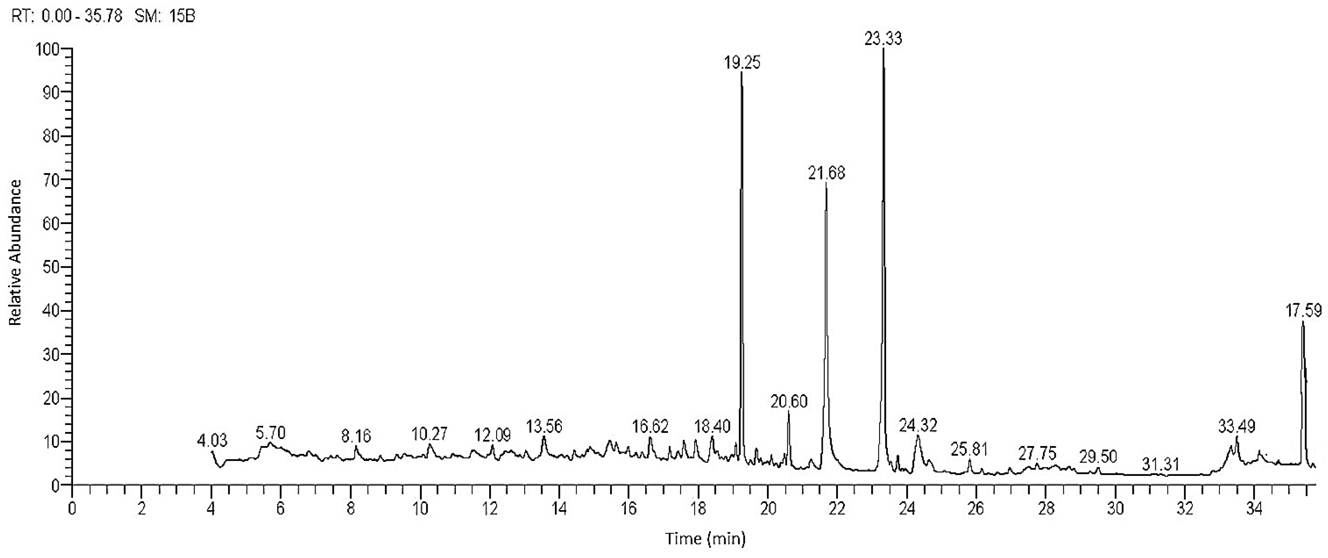
Figure 1. Chromatogram and structures of main components of the methanol extract of R. tingitana shoot by GC-MS.
Plants 11 02028 g001
Figure 2. Chemical structure of the major identified compounds in the MeOH extract of R. tingitana.
Plants 11 02028 g002
Figure 3. The categorized chemical constitutes identified from the extracted R. tingitana by GC-MS analysis.
Plants 11 02028 g003
Figure 4. IC50 values of the tested plant sample and doxorubicin as standard against human cancer cells.
Plants 11 02028 g004
Figure 5. Reichardia tingitana (L.) Roth plant. (a) Overview of the growing herb, (b,c) close view of vegetative stage and flowering branch.
Plants 11 02028 g005
Table 1. The characterized chemical components were isolated from the extracted shoots of R. tingitana.
No.RT aConc. % bCompoundMolecular WeightMolecular Formula
Hydrocarbons
113.561.84 ± 0.045,5,8a-trimethylhexahydro-2H-chromen-4a(5H)-yl acetate240.34C14H24O3
219.2521.98 ± 0.226,10,14-trimethylpentadecan-2-one268.49C18H36O
Fatty Acid and Esters
316.622.16 ± 0.05(E)-octadec-9-enoic acid282.47C18H34O2
417.921.44 ± 0.03Ethyl (9Z,12Z)-octadeca-9,12-dienoate308.51C20H36O2
519.081.13 ± 0.02Oleic acid282.47C18H34O2
619.671.16 ± 0.02Isobutyl octadecyl phthalate474.73C30H50O4
720.63.29 ± 0.07Methyl palmitate270.46C17H34O2
821.6814.85 ± 0.18Palmitic acid256.43C16H32O2
923.3427.26 ± 0.24Methyl oleate296.5C19H36O2
1023.741.06 ± 0.02Methyl stearate298.51C19H38O2
1124.313.56 ± 0.06(Z)-octadec-11-enoic acid282.47C18H34O2
1233.51.55 ± 0.032-hydroxypropane-1,3-diyl (9E,9′E)-bis(octadec-9-enoate)621C39H72O5
Steroids
1318.41.75 ± 0.04Estra-1,3,5(10)-trien-17α-ol256.39C18H24O
1434.451.31 ± 0.02Ethyl 3,7,12-trihydroxycholan-24-oate436.63C26H44O5
1535.396.63 ± 0.053,7,12-trihydroxycholan-24-oic acid408.58C24H40O5
1635.77.57 ± 0.08Stigmast-5-en-3-ol, (3α)-414.72C29H50O
Terpene
1717.591.48 ± 0.02Corymbolone (4a-hydroxy-4,8a-dimethyl-6-(prop-1-en-2-yl) octahydronaphthalen-1(2H)-one)236.36C15H24O2
Total100.0
a retention time, b average concentration of three replications ± standard deviation.
Table 2. Radical scavenging activity percent (%), and IC50 values (mg L−1) at various concentrations of the methanol extracted of R. tingitana and the standard ascorbic acid by DPPH assay.
TreatmentConc. (mg L−1)Radical Scavenging Activity (%)IC50 (mg L−1)
R. tingitana58.32 ± 0.42 F30.77
1023.84 ± 1.64 E
2042.27 ± 2.37 D
3051.61 ± 2.88 C
4062.33 ± 3.20 B
5071.91 ± 3.72 A
LSD0.051.62 ***
Ascorbic acid12.81 ± 0.01 F12.02
2.511.38 ± 0.03 E
538.57 ± 0.19 D
1047.92 ± 0.51 C
1561.34 ± 1.42 B
2072.61 ± 1.55 A
LSD0.051.40 ***
Values are average (n = 3) ± standard deviation. LSD0.05 expressed the calculated least of the smallest significance between two means, as each test was run on those two means (calculated by Factorial ANOVA). Different superscript letters within each treatment (column) express significant variation at a probability level of 0.05 (Duncan’s test). ***: significant at p ≤ 0.001.
Table 3. Antibacterial activity of the methanol extract from the aerial parts of R. tingitana and some selected reference antibiotics at a concentration of 10 mg mL−1.
MicrobesR. tingitana (10 mg mL−1)Standard Antibiotic (10 mg L−1)
CephradinTetracyclineAzithromycinAmpicillin
Gram-Negative Bacteria
Escherichia coli22.85 ± 0.87 B17.81 ± 0.82 D20.34 ± 0.71 BC20.45 ± 0.51 B20.51 ± 0.73 C
Enterobacter aerogenes11.36 ± 0.44 E0.00 F0.00 F14.56 ± 0.63 C0.00 F
Salmonella typhimurium25.71 ± 1.63 A0.00 F12.82 ± 0.54 D9.35 ± 0.07 D0.00 F
Klebsella pneumonius15.13 ± 0.51 D12.08 ± 0.42 E20.17 ± 0.68 C13.75 ± 0.49 C8.03 ± 0.09 E
Gram-Positive bacteria
Bacillus cereus24.42 ± 0.81 AB20.33 ± 0.55 BC12.09 ± 0.50 D19.44 ± 0.42 B10.62 ± 0.42 D
Clostridium tetani9.25 ± 0.04 F0.00 F8.54 ± 0.05 E0.00 D10.51 ± 0.27 D
Staphylococcus aureus18.62 ± 0.64 C21.82 ± 0.57 B22.77 ± 0.61 AB20.08 ± 0.91 B30.67 ± 1.92 A
Streptococcus pyogenes13.66 ± 0.32 D25.60 ± 1.31 A23.63 ± 1.58 A23.74 ± 0.87 A22.07 ± 1.37 C
Staphylococcus xylosus10.54 ± 0.51 E19.73 ± 0.98 C21.15 ± 1.65 ABC22.81 ± 0.69 A25.32 ± 1.51 B
LSD0.050.0001 ***0.0001 ***0.0001 ***0.0001 ***0.0001 ***
Value is the diameter of the inhibition zone (mm) as an average of three replications ± standard error. Different superscript letters within each treatment (column) express significant variation at a probability level of 0.05 (Duncan’s test). LSD: least significant difference. *** p < 0.001.
Table 4. Cytotoxic activity and the IC50 values of the R. tingitana MeOH extract against the tumor and normal cells at different concentrations, and doxorubicin as standard. Hepatocellular carcinoma (HePG-2), human prostate cancer (PC3), and normal cell (WI-38).
SamplesConc. (µg mL−1)In Vitro Cytotoxicity (%)
HePG-2PC3WI-38
R. tingitana10093.1491.419.55
5088.3481.628.33
2585.0775.315.58
12.568.5258.223.67
6.2550.5941.721.48
3.12538.9925.381.32
1.5625.6222.930.95
IC5029.97740.479>100
Doxorubicin10063.1570.0712.23
5054.0346.839.52
2549.5837.616.31
12.541.3932.424.98
6.2534.5624.273.32
3.12523.1414.652.45
1.566.473.871.13
IC505.2748.303>100
IC50: inhibitory concentration (µg): 1–10 (very strong), 11–20 (strong), 21–50 (moderate), 51–100 (weak), and above 100 (noncytotoxic).
Table 5. Larvicidal effect of the methanol extract from the aerial parts of R. tingitana on 3rd larval instar of A. aegypti.
Conc. (mg L−1)R. tingitana
24 h Post-Treatment48 h Post-Treatment72 h Post-Treatment
30032.54 ± 1.41 A41.28 ± 2.03 A50.81 ± 2.37 A
25028.33 ± 1.23 A31.51 ± 1.39 B42.17 ± 1.83 AB
20018.16 ± 0.79 B29.37 ± 1.25 B34.62 ± 1.44 BC
15011.32 ± 0.42 C19.09 ± 0.83 C28.06 ± 1.07 C
1006.35 ± 0.31 CD10.13 ± 0.44 CD15.74 ± 0.52 D
Control1.05 ± 0.05 D1.05 ± 0.05 D1.05 ± 0.05 E
F-value166.13 ***110.75 ***142.10 ***
p-value<0.0001 ***<0.0001 ***<0.0001 ***
LC5046.8535.7529.38
LC9082.6663.8253.30
Mortality was expressed as mean ± SE (standard error) of 3 replicates. LC50: median lethal concentration; LC90: lethal concentration. Different superscript letters within each treatment (column) express significant variation; *** all F-values are significant at p ≤ 0.001 (Duncan’s test).
Publisher’s Note: MDPI stays neutral with regard to jurisdictional claims in published maps and institutional affiliations.