Article

Proteomic Analysis Reveals Salicylic Acid as a Pivotal Signal Molecule in Rice Response to Blast Disease Infection

1
Jiangsu Key Laboratory for Eco-Agricultural Biotechnology around Hongze Lake, Jiangsu Collaborative Innovation Center of Regional Modern Agriculture and Environmental Protection, Huaiyin Normal University, Huai’an 223300, China
2
College of Biology and the Environment, Nanjing Forestry University, Nanjing 210037, China
3
College of Plant Protection, Nanjing Agricultural University, Nanjing 210095, China
4
Jiangsu Grain and Oil Quality Monitoring Center, Nanjing 210031, China
*
Authors to whom correspondence should be addressed.
Plants 2022, 11(13), 1702; https://doi.org/10.3390/plants11131702
Submission received: 1 June 2022 / Revised: 21 June 2022 / Accepted: 21 June 2022 / Published: 27 June 2022

Abstract

:
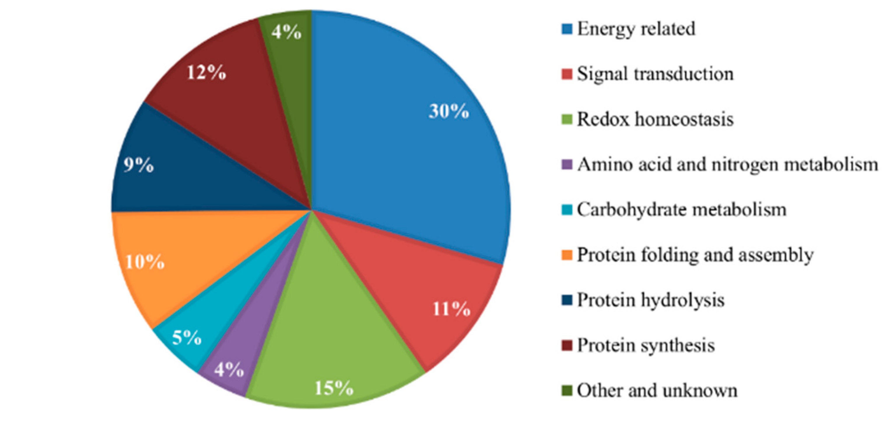
Rice blast disease caused by a fungus, Magnaporthe grisea, is one of the most destructive diseases in rice production worldwide, and salicylic acid (SA) can efficiently decrease the damage of M. grisea. Here, we combined the 2-Dimensional-Liquid Chromatography and the Matrix-assisted laser desorption/ionization time-of-flight mass spectrometer (2D-LC-MALDI-TOF-TOF MS) techniques to compare and identify differentially expressed labelled proteins by the isobaric tags for relative and absolute quantitation (iTRAQ) between the blast-resistant cultivar Minghui and the susceptible rice cultivar Nipponbare in response to blast fungus infection. The group samples were treated with salicylic acid and compared to control samples. A total of 139 DEPs from the two cultivars showed either more than a two-fold change or alternating regulation patterns. Protein functionality analysis also exhibited that these proteins are involved in a wide range of molecular functions including: energy-related activity (30%), signal transduction (11%), redox homeostasis (15%), amino acid and nitrogen metabolism (4%), carbohydrate metabolism (5%), protein folding and assembly (10%), protein hydrolysis (9%), protein synthesis (12%), and other unknown functions (4%). Specifically, we demonstrated that exogenous treatment with salicylic acid promoted recovery in both rice cultivars from Magnaporthe grisea infection by enhancing: the regulation of signal transduction, increasing energy conversion and production through the regulation of the glycolytic pathway, and other various biochemical processes. These findings may facilitate future studies of the molecular mechanisms of rice blast resistance.

1. Introduction

Rice is a staple crop of economic importance in many countries, as well as being a model monocot species for plant research [1]. With an estimated production capacity of at least 600 million tons per annum, its production is affected by both biotic and abiotic factors. Infectious diseases are among the sternest threats to rice crop production, reducing rice productivity by 25% worldwide [2]. The Magnaporthe grisea (Hebert) Barr (anamorph, Pyricularia grisea Sacc.), an ascomycete filamentous fungus, one of the most infamous causative agents of blast disease, continuously threatens rice production globally [3], affecting the Poaceae family including rice [4]. Visible disease symptoms are characterized by white to gray-green lesions or spots with darker borders. However, with the enhancement of agronomic practices, several cultural strategies such as the use of genetically resistant varieties and the application of fungicides have been implemented to control blast disease in rice [5,6]. Recently, Zhang et al. [7] reported on a tripartite interaction among rice plants, insects, and pathogens in the control of rice blast disease. They have demonstrated that pre-infestation by herbivores on rice crops reduces the disease incidence of rice blast by increasing the accumulation of salicylic acid (SA) [7], emphasizing a known fact that phytohormones accumulation including SA improves basal crop resistance to blast disease in rice through reducing plant biochemical processes such as: plant photosynthesis rate and regulating the antioxidant defense activities [8,9].
Measuring rice proteomic changes in response to salicylic acid (SA) has provided new insights into the molecular mechanisms of SA-induced rice defense to blast disease. It has been shown that SA treatment after rice blast infection elevates rice blast disease resistance. Sun et al. [10] identified 33 protein spots that were differentially expressed after SA treatment, evidencing that SA improves basal resistance to rice blast through decreasing plant photosynthesis, regulating the antioxidant defense activities. They concluded that enhanced blast disease resistance in rice plants is coordinated by SA-induced complex cellular activities. Although, full comprehension of the mechanisms of regulation is still yet to be attained. Cell-level research has also demonstrated that SA signaling is mediated by a series of downstream factors such as OsNPR1 and WRK45 acting in parallel, and OsAAA-ATPase1 [11,12,13].
The integration of various powerful fields such as proteomics has also enabled the investigation of rice blast disease at the translational level, disclosing several key genes and proteins associated with rice blast resistance [14]. Hence, utilizing proteomic techniques such as two-dimensional liquid chromatography mass combined with the matrix-assisted laser ionization time-of-flight mass spectrometry (MALDI-TOF/TOF MS) for both qualitative and quantitative characterizations of protein mixtures and post-translational modifications has extended the breadth and depth of proteome analysis [15,16,17]. The 2D-LC proteome analysis has been widely used in the identification of protein expression patterns, to address the mechanisms by which Magnaporthe oryzae regulates G-proteins (GTP-binding protein) and signaling proteins (RGS) that orchestrate blast disease incidence [18]. Likewise, several researchers have also used the MALDI-TOF/TOF MS in the identification and quantification of protein expression patterns in SA-added rice plants resisting the blast disease.
First developed in 2004, the Isobaric Tags for Relative and Absolute Quantification (iTRAQ) has also been shown to pose unique advantages over other conventional proteomics techniques [19]. It enables the identification and quantification of many proteins from specific biological environments using labeled peptides as internal standards identifiable by sensitive mass spectrometers. Furthermore, it utilizes bioinformatics tools and statistical packages to visualize obtained data. The incidence of the iTRAQ approach has enabled many researchers to identify proteins involved in pathogen resistance in several plant species. Ma et al. [20] implemented the iTRAQ to compare and detect differentially expressed proteins (DEPs) from a blast-resistant variety and a susceptible variety. They detected 46 common DEPs among the two varieties involved in plant-pathogen interaction, plant hormone signal transduction, fatty-acid metabolism, and peroxisome biosynthesis [20]. Furthermore, iTRAQ has been implemented in several kinds of research that may include the quantification of rice grains proteins [19], assessing the effect of high night temperature stress on rice [21], assessing the impact of ion pollution and detoxification in rice [22], and assessing the expression patterns of rice proteins under several abiotic stresses [23,24].
Although the iTRAQ approach has been utilized in the past to assess proteins related to rice blast resistance, few proteins have been successfully identified. In this present study, we compared the posttranslational responses of rice cultivars (blast-resistant Minghui and blast-susceptible Nipponbare cultivars) sprayed with salicylic acid against the controls to Magnaporthe grisea fungus infection using the iTRAQ approach to label the peptides and the combined 2D-LC/MALDI-TOF-TOF MS technique for protein separation and identification, with an intent to comprehend the molecular mechanisms underlying blast resistance in rice. This study may facilitate and provide valuable molecular data for rice blast-resistant breeding purposes.

2. Results

2.1. Identification and Relative Quantification of Proteins

In present study, differentially expressed proteins (DEPs) expressed in response to Magnaporthe grisea (M) infection were identified using the isobaric Tags for Relative and Absolute Quantitation (iTRAQ) technique coupled with the 2-D Liquid Chromatography-Matrix-assisted laser desorption/ionization time-of-flight mass spectrometer (2D-LC-MALDI-TOF-TOF MS) analysis. The proteomic differences were contrasted and analyzed from the two rice cultivars, the blast-resistant Minghui (M) and blast susceptible Nipponbare (R) varieties, samples were grouped in four sub-pairs as M-M vs. M-CK, M-SA vs. M-CK, R-M vs. R-CK, and R-SA vs. R-CK. The schematic workflow of the experimental design is shown in Figure 1. A total of 1230 proteins were obtained, which were further separated using the Liquid Chromatography (LC), and 1163 proteins were successfully identified using the MASCOT program. Expression analysis of the observed proteins exhibited that 139 proteins had more than 2.0-fold change and/or less than 0.5-fold difference with p < 0.05 between samples, and these were considered to be significantly altered, as shown in Table 1.
These DEPs were shown to be involved in a wide range of molecular functions, 30%, which was the majority, was constituted in energy-related activities, 11% in signal transduction, 15% in redox homeostasis, 4% in amino acid and nitrogen metabolism, 5% in carbohydrate metabolism, 10% in protein folding and assembly, 9% in protein hydrolysis, 12% in protein synthesis, and the remaining 4% in other unknown functions (Figure 2). This suggests that biological processes with high protein constituency such as energy-related activities are likely to enhance blast disease resistance in rice varieties. A deeper analysis in the energy-related activity showed that 65 of the total DEGs were upregulated, to which 36 and 29 were enriched in Minghui and Nipponbare, respectively. Specifically, the 23 kDa polypeptide of photosystem II, chloroplastic glutamine synthetase, and enoyl-CoA hydratase/isomerase proteins were highly upregulated among the significantly expressed protein in the R-SA group and downregulated in the R-M group in the Nipponbare variety.

2.2. Bioinformatics Analysis of Differentially Expressed Proteins

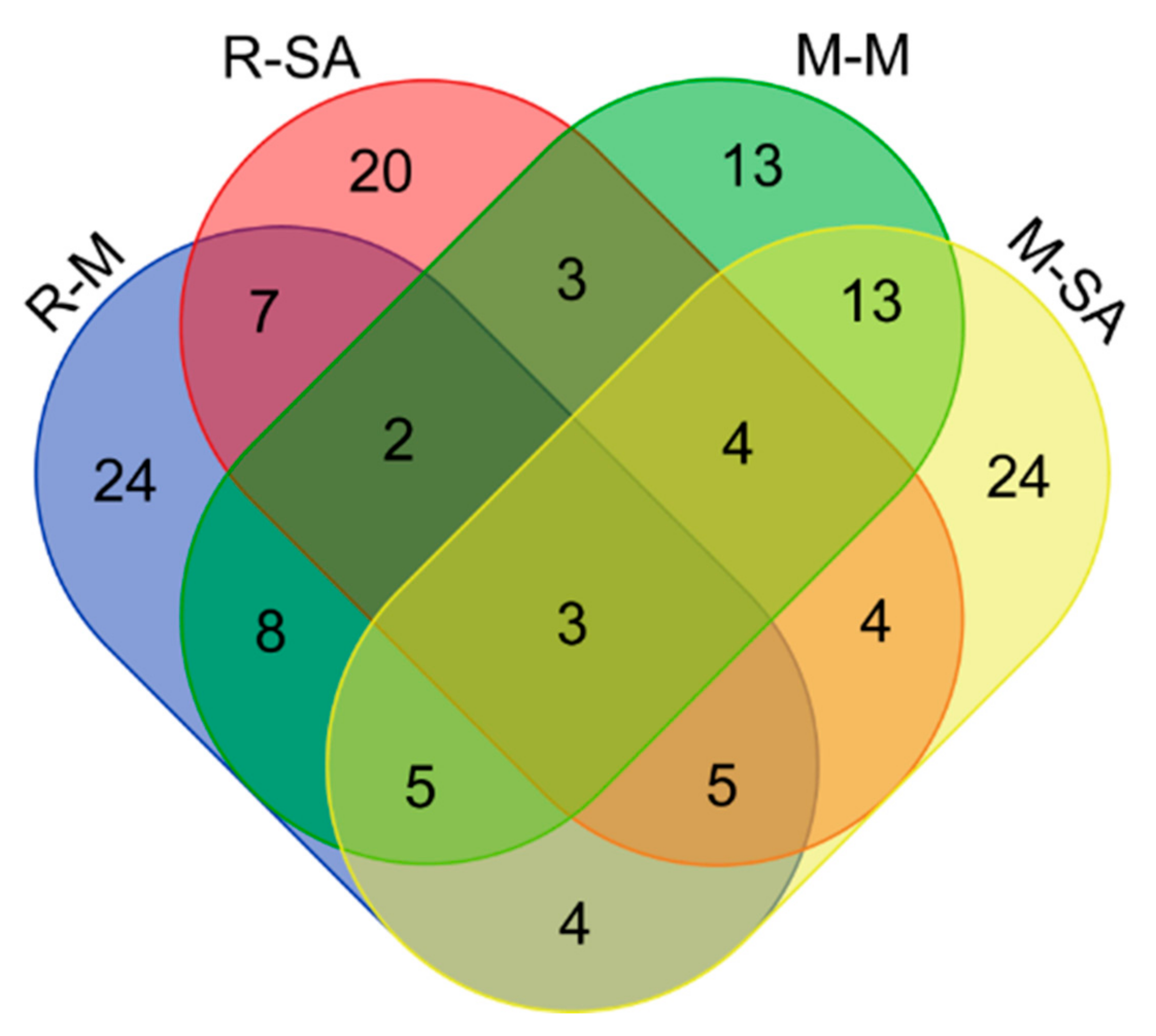
To define the key proteins associated with rice blast resistance and SA-dependent recovery, we did bioinformatics analyses of the four pair samples, M-M vs. M-CK, M-SA vs. M-CK, R-M vs. R-CK, and R-SA vs. R-CK. The differentially expressed analysis of most significant protein changes between the two varieties showed that three common DEPs were expressed in all the groups (Figure 3). This implies that these DEPs may play an important role in all rice cultivars of study and also that there is less in common in the regulation of blast disease infection between the two varieties of study. As expected, the SA-added groups, M-SA and R-SA, made up the largest part of the differentially expressed proteins, 24 and 20, respectively, compared to the control groups. This shows that salicylic acid had an important effect on fungal infection rates. On the other hand, the R-M group also constituted a larger group of DEPs (24) as compared to the M-M group, suggesting that the R- group (Nipponbare variety) was negatively affected by blast disease. Comparisons between two cultivars showed that more DEPs were expressed in the susceptible Nipponbare cultivar as compared to the blast-resistant Minghui cultivar, evidencing the fact SA exhibited positive regulatory roles against the M. grisea infection. Hence, these results can be summarized as that salicylic acid upregulated the expression of DEPs in both varieties.

2.3. Comprehensive Inventory of the Differentially Expressed Proteins

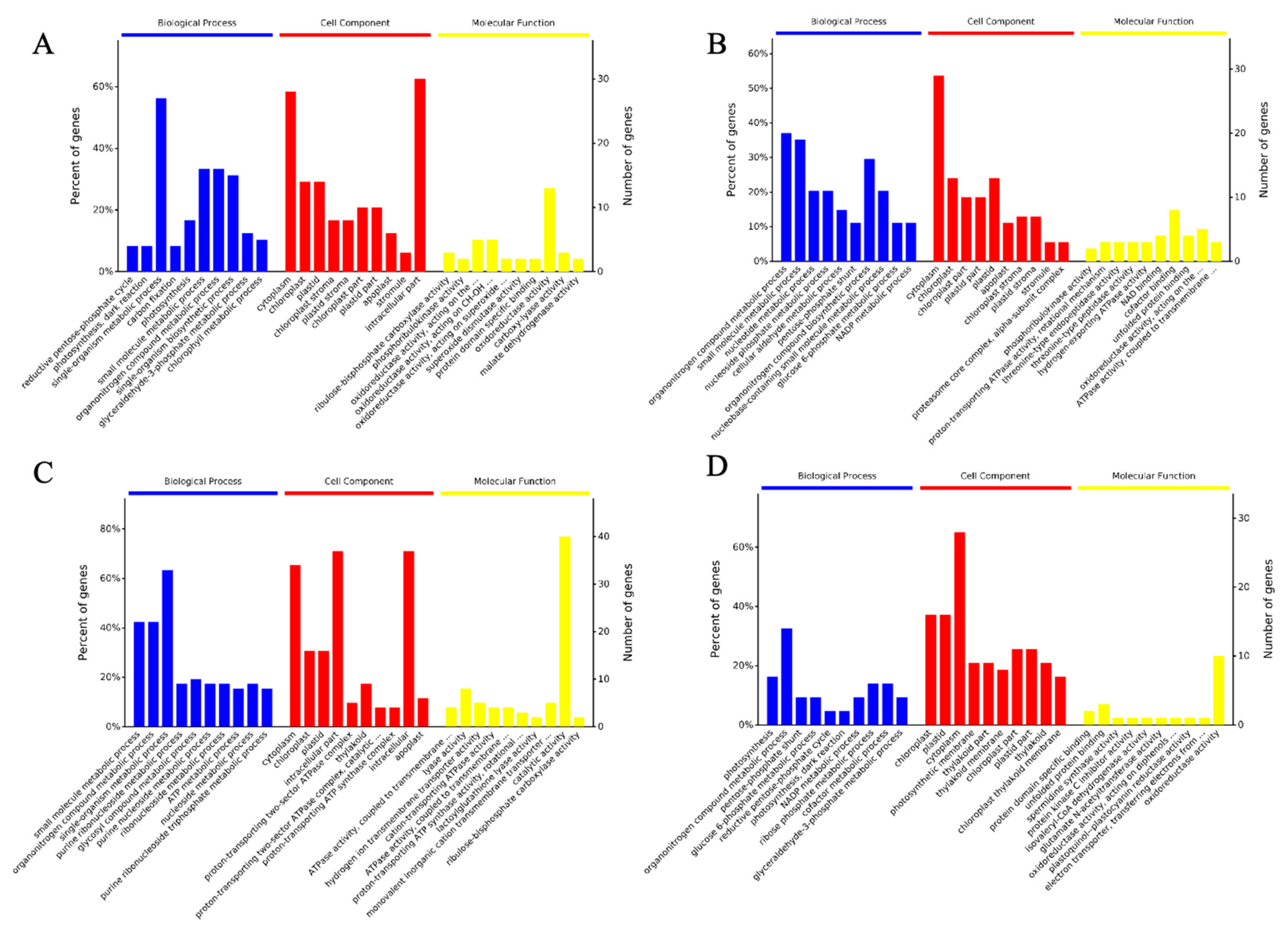
To classify the DEPs in Minghui and Nipponbare varieties, putative proteins induced by M. grisea were separated by the 2Dimensional- Liquid Chromatography (2D-LC) (Figure 4). All the identified DEPs were classified into three GO terms; biological process (BP), molecular function (MF), and cell component (CC). The majority of the identified proteins in the M-M group were involved in single-organism metabolic processes (BP analysis), cytoplasm (CC analysis), and nucleoside phosphate binding (MF analysis). The identified proteins in the M-SA group were concentrated in the organonitrogen compound metabolic process (BP analysis), cytoplasm (CC analysis), oxidoreductase activity, and catalytic activity (MF analysis). In the R-M group, the majority of identified proteins were involved in the small molecule metabolic process (BP analysis), cytoplasm (CC analysis), and catalytic activity (MF analysis). Lastly, the identified proteins in the R-SA group were concentrated in the organonitrogen compound metabolic process (BP analysis), chloroplast and cytoplasm (CC analysis), oxidoreductase activity, and nucleoside phosphate binding (MF analysis). Wholly, these results show that DEPs from different rice cultivars and exposed to different treatments exhibited different functional characteristics, and that more genes were assigned in the susceptible Nipponbare cultivar as compared to the blast-resistant Minghui cultivar.

2.4. Specific Metabolic Processes in Minghui and Nipponbare Are Involved in the Response against M. grisea

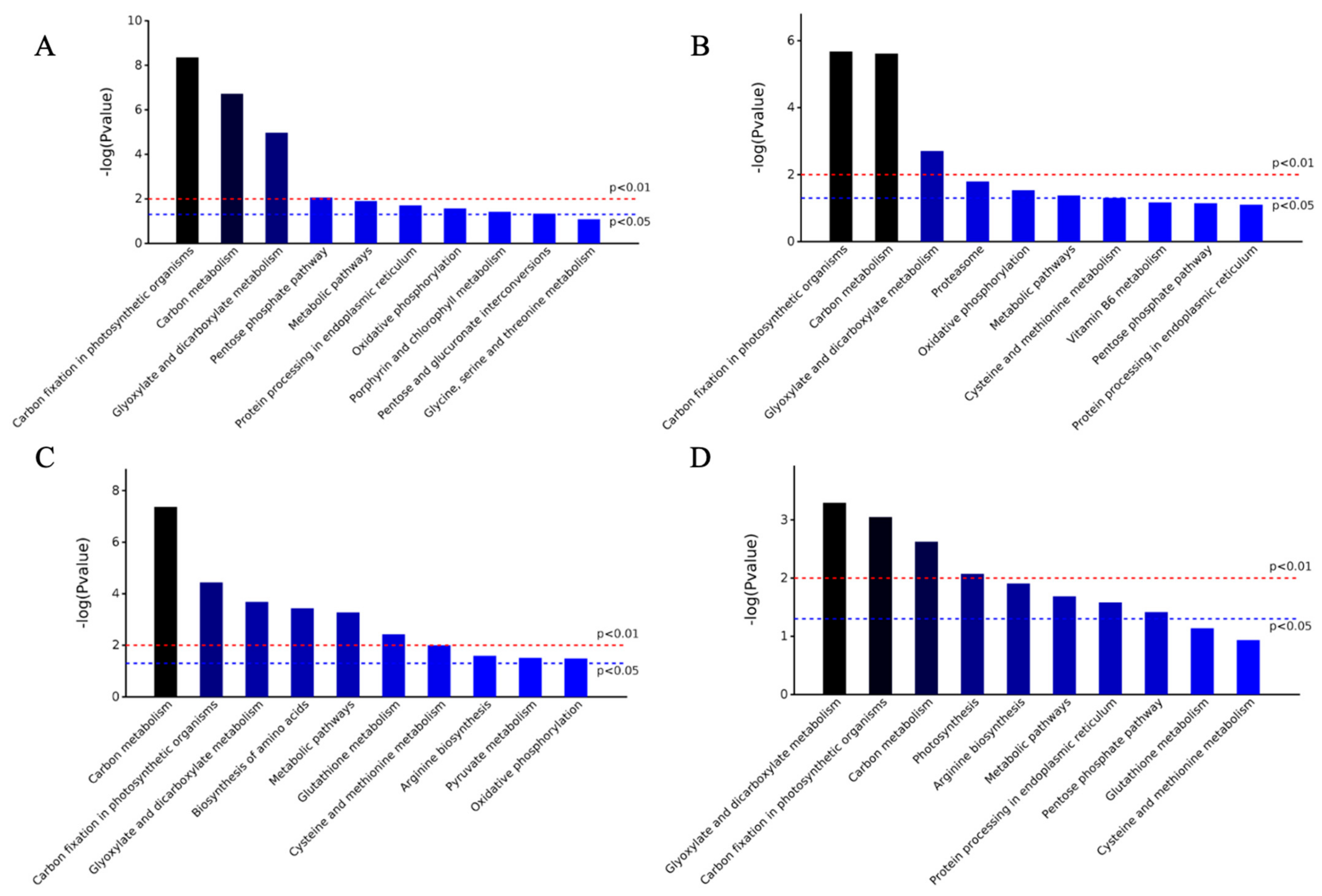
To determine specific metabolic processes in Minghui and Nipponbare following M. grisea inoculation and salicylic acid spray, we used the KEGG database (Figure 5). We noted that only three metabolic pathways were significant at both p-values (p < 0.01 and p < 0.05), including carbon fixation and metabolism, carbon metabolism, glyoxylate and dicarboxylate metabolism. Carbon fixation and metabolism exhibited the highest −log(Pvalue) change in both groups of the Minghui cultivar, while the carbon metabolism, glyoxylate and dicarboxylate metabolism were highly upregulated in the R-M and R-SA groups, respectively. However, significant expression differences were observed when comparing two distinct varieties with the same treatment. In SA-treated cultivars we noted that 50% of the biochemical processes in the M-SA group were not significant at p-value < 0.05 as compared to the R-SA group, to which only two of the investigated processes were insignificant when p-value < 0.05. In the M. grisea-treated groups, similar findings were observed, although all the processes investigated in the R-M group had significant differences when p-value < 0.05, except for the Glycine, serine and threonine metabolism in the M-M group. Based on the results of the KEGG analysis, the salicylic acid added groups from both varieties had higher upregulation expression as compared to the control groups.

2.5. Protein-Protein Interactions

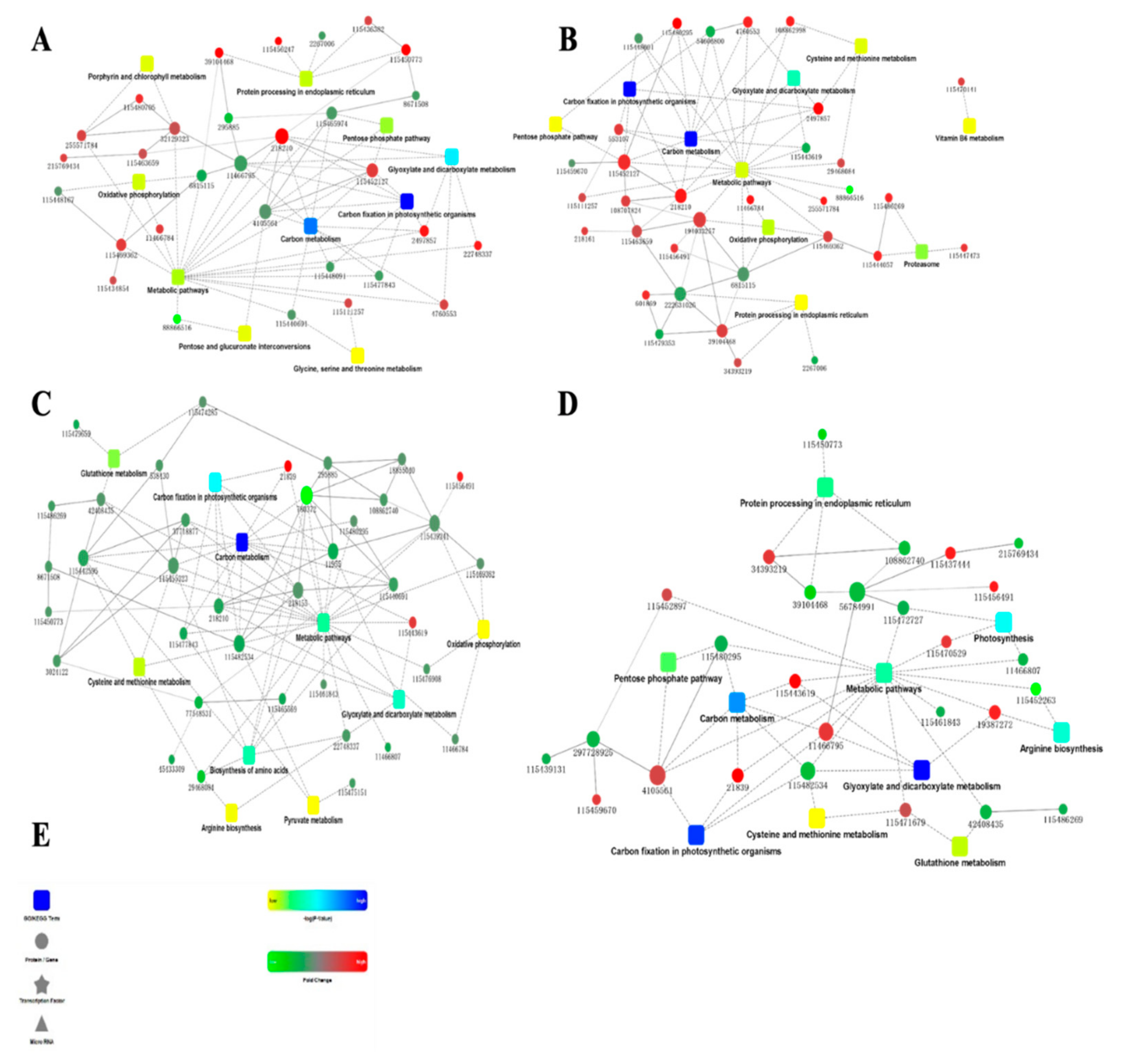
To comprehend the protein network interaction, we constructed protein to protein interactions (PPI) using the STRING database and viewed using Cytoscape software (Figure 6). Generally, the results exhibited that the identified proteins were densely connected and regulated several pathways. In the M-M (Figure 6A) group, the majority of the investigated proteins with high fold change were involved in metabolic pathways, moderately upregulating its enrichment. The carbon fixation in photosynthetic organisms and carbon metabolism pathways were highly enriched with fewer moderately upregulated proteins. The glyoxylate and dicarboxylate metabolism was lowly enriched by several upregulated proteins. Likewise, the glycine, serine, and the threonine metabolism were upregulated and downregulated by two proteins, 2,3-diphosphoglycerate-independent and betaine aldehyde dehydrogenase, respectively. Similar enrichment patterns were observed in the M-SA group (Figure 6B), with fewer proteins involved in the metabolic pathways, and highly upregulated as compared to the M-M group. Specifically, the metabolic pathways were densely connected with several upregulated proteins. However, some of the pathways were absent in the M-M group and were regulated by several proteins. For instance, the pentose phosphate pathway was present in M-SA but absent in R-SA and was lowly enriched by two upregulated proteins. Intriguingly, vitamin B6 metabolism was not linked to any of the pathways within the network, and only one protein was highly upregulating its enrichment.
The R-M group (Figure 6C) was also densely connected with moderately expressed proteins. Carbon metabolism was highly enriched among the observed pathways, with fewer proteins involved as compared to the metabolic pathways. Nonetheless, the metabolic pathways were moderately enriched with two highly expressed proteins involved. The remaining pathways in the R-M group, oxidative phosphorylation, pyruvate metabolism, and arginine biosynthesis, were lowly enriched and had two, three, and four downregulated proteins, respectively, linking their enrichment. The R-SA group was sparsely connected (Figure 6D), with some proteins highly expressed while some were lowly expressed. The majority of the highly expressed proteins with a high fold change moderately enriched the regulation of metabolic pathways, with a few downregulating their enrichment. Nonetheless, the carbon fixation in photosynthetic organisms and glyoxylate and dicarboxylate metabolisms were highly enriched with two and three proteins, respectively, linked.

3. Discussion

Developments in proteomics research over the past decades have enabled the comprehension of plant disease resistance at the protein level. In this study, the proteomics approach was employed to comparatively identify the expressed proteins from the blast-susceptible Nipponbare and the blast-resistant Minghui rice cultivars. To reveal the mechanism of resistance to Magnaporthe grisea infection, the two rice cultivars were treated with the biotic stress hormone, salicylic acid, after infection. A total of 139 DEPs were identified among all samples and were further shown to be involved in energy metabolism, signal transduction, carbohydrate metabolism, nitrogen metabolism, protein hydrolysis, and the synthesis process.

3.1. Differentially Expressed Proteins Related to Energy

Energy deficits are a general symptom of photosynthetic plants under stress due to reductions in photosynthesis and/or respiration, ultimately resulting in cell death and growth arrest. However, plants counter energy shortages by enhancing inherent pathways of carbohydrate metabolism, induction of alternative pathways of glycolysis, and maintaining energy supply and carbon skeletons for key metabolic processes. Sucrose is the primary translocated carbohydrate in the majority of plants; thus, its metabolism is vital in the regulation of plant growth in response to stresses [25,26,27]. We identified 41 energy-related proteins (spots 1–41) involved in the response to blast fungus infection in rice. These proteins mainly included 23 kDa polypeptide of photosystem II, chloroplastic glutamine synthetase, NADP-dependent malic enzyme, oxalate oxidase-like protein, uroporphyrinogen decarboxylase, 6-phosphogluconolactonase, 2,3-bisphosphoglycerate-independent phosphoglycerate mutase, and ATP synthase (Table 1). In plants, these proteins are involved in the glycolysis pathway, tricarboxylic acid cycle, chlorophyll synthesis, pentose phosphate pathway, photosynthesis process energy, and so on.
ATP synthase is widely distributed in the mitochondrial membrane and chloroplasts thylakoid [28], participating in oxidative phosphorylation and photophosphorylation, under the impetus of the transmembrane proton power potential, resulting in ATP hydrolysis and the release of energy, which is the most direct energy source in plants [29,30]. We observed that the levels of ATP synthase proteins significantly declined under the blast fungus infection in Nipponbare (susceptible cultivar) and that treatment with salicylic acid failed to promote significant recovery of ATP synthase protein levels in Nipponbare. Nonetheless, in Minghui (resistant variety) the three levels of ATP synthase proteins spots (13, 14, and 16) were slightly downregulated under blast fungus infection, and treatment with salicylic acid significantly elevated the levels of these proteins. These results demonstrated that Magnaporthe grisea infection significantly reduces energy production in Nipponbare compared to Minghui and that salicylic acid exogenous treatment promotes more energy recovery in Minghui than in Nipponbare after infection.
Thirteen differentially expressed protein spots (1, 5, 24, 28, 30–36, 39, 40) were involved in photosynthesis metabolism, including chlorophyll synthesis; in Minghui under blast fungus infection these proteins were upregulated but not in Nipponbare. Treatment with salicylic acid afterwards further improved the expression of these proteins in Minghui. We identified five proteins (spots 3, 20, 22, 25, 26) involved in the glycolytic pathway. Glycolysis is the process of degrading glucose to produce ATP, providing partial energy for cell activities. In this study, the expression of proteins involved in the glycolytic pathway was downregulated in Nipponbare infected by Magnaporthe grisea. However, in Minghui two of the five proteins (spots 22 and 26) were upregulated significantly. These results showed that the glycolytic pathway was inhibited by blast infection in Nipponbare.

3.2. Proteins Associated with Signal Transduction

Stress signal perception and transmission are important for plant environmental stress response [31]. We identified 15 signal transduction-related proteins, including brain-specific protein, calreticulin precursor, and GTP-binding protein (Table 1). In Nipponbare, most of the expressed proteins were downregulated, while in Minghui the expressed proteins were upregulated. This implies that active regulation of signal transduction is associated with blast infection resistance in Minghui.
Integrins belong to a family of cell surface glycoproteins, which connect the extracellular matrix to the actin cytoskeleton and serve in cell signaling across the plasma membrane to confer cell survival. Overexpressed 14-3-3 beta protein interacts with cytosol B1 integrin promoting cell migration and signal transmission [32,33]. We analyzed the protein level of 14-3-3 beta (spot 50) and noted that its stability was constant in Minghui, while it was downregulated in Nipponbare under blast fungus infection. Spot 59 is a spermidine synthase important in plant stress responses. It is involved in signal transduction associated with plant stress resistance [34]. The expression of the protein was significantly upregulated only in Nipponbare after treatment with salicylic acid. Wholly, this result showed that SA enhances the regulation of signal transduction by up- and downregulating proteins involved. However, the correlation between proteomic changes due to rice blast resistance may deserve further investigation.

3.3. Differentially Expressed Proteins with Redox Homeostasis

There is active redox homeostasis in a plant cell in response to stress stimuli. Oxidants mainly include reactive oxygen and nitrogen species, and reductants (antioxidant molecules). It has been shown that many enzymes are involved in redox homeostasis. Normal cell functions require the balance of oxidation and reduction processes [35,36]. We identified 21 proteins (spots 57–77) associated with redox homeostasis under blast fungus infection, which mainly included glutathione S-transferase, putative glyoxalase I, manganese superoxide dismutase, lactoylglutathione lyase, and superoxide dismutase (Table 1). In general, most of these proteins in Nipponbare were downregulated under the blast fungus infection. In Minghui, 5 out of the 21 proteins were upregulated and 3 downregulated under the blast infection. Treatment with salicylic acid did not change their expression patterns, suggesting the presence of an SA-mediated defense in the resistant cultivar subjected to blast infection.
Glutathione S-transferase is the key enzyme of glutathione binding reaction, and catalyzes the initial steps of the glutathione-binding reaction. It can catalyze the binding reaction of nucleophilic glutathione with various electrophilic exogenous chemicals. Many exogenous chemicals are easily separated from some bioactive intermediates in the biotransformation of the first phase reaction; they can covalently combine with the important components of cell biological macromolecules and cause cell damage. Glutathione can prevent the occurrence of such covalent binding for detoxification [37,38]. In this study, the expression of these proteins in both varieties decreased, but the Minghui cultivars showed a recovery after treatment with salicylic acid (spot 43). This suggests that exogenous treatment with salicylic acid is necessary to induce the glutathione-binding reaction process in Minghui after infection.
Manganese superoxide dismutase (spot 60) is an important antioxidation enzyme [39], alongside Betaine aldehyde dehydrogenase (spot 66) involved in the synthesis of betaine, which is the substance in methylation reactions and detoxification of homocysteine [40]. PDI-like protein (Spot 74) is also crucial; it catalyzes the formation of disulfides and also regulates specific biochemical processes [41]. The expression of these three proteins was significantly upregulated in Minghui infected with Magnaporthe grisea. By contrast, in Nipponbare, expressions of the proteins were significantly downregulated under blast fungus infection.

3.4. Proteins Associated with Amino Acid and Nitrogen Metabolism

Previously, rice blast fungus infection was shown to alter expression patterns of proteins involved in amino acid metabolism, nitrogen assimilation, nucleotide anabolism, and lipid oxidation [42]. In this study, we observed that cysteine synthase, glutamate ammonia ligase, ketol-acid reductoisomerase, and ornithine acetyltransferase expressions were altered in response to blast fungus infection (Table 1). The activities of cysteine synthase determine the level of cysteines, an important amino acid in protein synthesis. The enzyme plays a very important role in antioxidation and stress resistance in plants [43,44]. In Nipponbare, Cysteine synthase was downregulated after blast fungus infection, and recovery was observed after treatment with salicylic acid. In Minghui, the expression of cysteine synthase remained stable under fungus infection.
Glutamate ammonia ligase (spot 81) plays a role in ammonia and glutamate detoxification, acid-base homeostasis, cell signaling, and cell proliferation. Ketol-acid reductoisomerase (spot 82) is one of the key enzymes in the biosynthesis of amino acids with branched chains in plants; however, interruption of the synthesis can cause plant death [45]. Ornithine acetyltransferase (spot 83) regulates the abundance of L-arginine, which plays an important role in maintaining cell functions [46]. Generally, the expressions of these three proteins were inhibited in Nipponbare under the Magnaporthe grisea infection; additional treatment with SA improved the expression of these proteins in blast-infected Nipponbare, while in Minghui their levels remained stable under infection.

3.5. Proteins Associated with Carbohydrate Metabolism

Glucose, a monosaccharide metabolized in plants, is one of the most important carbohydrates in plant cell metabolism [47]. We observed that the expression patterns of seven proteins involved in carbohydrate metabolism were downregulated in response to blast fungus infection in Nipponbare. Additional treatment with SA elevated the expression of these proteins, evidencing the vital role of SA during blast fungus infection. Furthermore, the cytoplasmic malate dehydrogenase (spot 86) plays an important role in the shuttle system of the cytoplasm and organelles, nucleic acid selective channels, gluconeogenesis, and malic acid metabolism [48]. In Nipponbare, this enzyme was significantly downregulated after Magnaporthe grisea infection, suggesting inhibition of corresponding biological processes. In comparison, enzyme levels remained unchanged in Minghui.
S-adenosylmethionine synthetase 2 (spot 90) plays an important role in cell metabolism in all organisms. We observed that the level of S-adenosylmethionine synthetase was downregulated only in Nipponbare infected by Magnaporthe grisea, and later recovered with salicylic acid treatment. Takusagawa et al. [49] have shown the abundance of spot 90 in more than one hundred different kinds of methyltransferase catalytic reaction of a donor in the synthesis of glutathione (GSH) precursor molecules of the sulfur transfer process and polyamine synthesis to aminopropyl process, and also related activity to a variety of enzymes [49].

3.6. Differentially Expressed Proteins Related to Protein Metabolism

Thirty-three proteins (spots 91–133) were shown to be mainly involved in the processes of protein metabolism including protein synthesis, protein folding, and assembly. Proteins involved in protein folding and assembly mainly included heat shock proteins (HSPs), HSP 70 kDa and 90 kDa. Heat shock proteins (HSPs) are synthesized under various stresses [50]. These proteins mainly serve as molecular chaperones to stabilize target proteins subjected to stresses, thereby playing a protective role [51]. In Nipponbare, these proteins were significantly downregulated after infection with Magnaporthe grisea. However, in Minghui the expression of these proteins was upregulated. Proteins involved in protein hydrolysis mainly include aminopeptidase, proteasome, hydrolase, and so on. These enzymes mainly function to degrade target proteins. In Nipponbare, these proteins showed a downward regulation trend in general, whereas the opposite was true in Minghui: of the two species, only a few were exceptions. Similar to the proteins involved in protein folding and assembly discussed above, the levels of the proteins may also indicate the differential response of the two rice cultivars under infection.
Proteins involved in protein synthesis mainly include ribosomal protein (RP), binding protein, assembly protein, and so on. Ribosomal protein is ubiquitous in every cell and forms ribosomes with RNA to function in protein synthesis [52]. In Nipponbare, the proteins were downregulated in response to blast fungus infection, but an afterward treatment with salicylic acid significantly improved the levels of the proteins. By contrast, in Minghui, the proteins were upregulated after infection.

4. Materials and Methods

4.1. Plant Materials

Two rice cultivars, blast-resistant Minghui (M) and blast-susceptible Nipponbare (R), were comparatively assessed for differential expression of proteins (DEPs) in this study. The 20 seeds disinfected with 70% alcohol and rinsed with distilled water were planted into a 10 cm-diameter plant pot in a greenhouse under optimum conditions and supplemented with the nutrient solution. The experiment was designed in two blocks (2 cultivars), 3 treatments (including the control-CK) and 3 replicates. Nine two-week-old rice plants from Minghui were randomly selected and divided into three replicate groups designated as the Minghui set as a control (M-CK), Minghui infected with Magnaporthe grisea fungus (M-M), and Minghui sprayed with salicylic acid (M-SA). Likewise, 3 Nipponbare replicate groups were randomly selected and designated as the Nipponbare set as a control (R-CK), Nipponbare infected with Magnaporthe grisea fungus (R-M), and Nipponbare sprayed with salicylic acid (R-SA). The groups, R-M, R-SA, M-M, and M-SA, were sprayed with a fungus strain of M. grisea, Guy11, at an intensity of 5 × 10⁴/mL. The groups R-CK and M-CK were sprayed with water and served as controls. To investigate the effect of salicylic acid, the groups R-SA and M-SA were sprayed with salicylic acid with a concentration of 8 mmol/L. Then, the rice plants were left for another week to grow. One-week-old treated leaves were harvested and stored at −80 °C before further experiments.

4.2. Protein Extraction

The trichloroacetic acid (TCA)/acetone precipitation method was used to extract proteins [18]. Leaf tissues were ground into a fine powder in liquid nitrogen. The resulting leaf powder was suspended in 10% (w/v) TCA/acetone supplemented with 0.1% DTT, and stored at −20 °C for 2 h for promoting perception. The suspension was centrifuged at 15,000 rpm for 20 min. The resulting pellet was re-suspended in 10% (w/v) TCA/acetone supplemented with 1 mM PMSF and 0.07% (w/v) β-mercaptoethanol, and was stored at −20 °C for 1 h. The suspension was further centrifuged at 15,000 rpm for 20 min to precipitate proteins, which were then subjected to vacuum freeze-drying processes. The resulting protein powder was dissolved in 800 μL lysis solution containing 7 M urea, 2 M thiourea, 4% (w/v) CHAPS, 65 mM DTT, 1 mM PMSF, and 0.5% (v/v) violates. The protein concentration of the sample was quantified according to the Bradford method [53].

4.3. Trypsin Digestion, iTRAQ™ Labels, Off-Line Strong Cation Exchange Chromatography, and Online nano-LC-MALDI-TOF-TOF MS Analysis

A total of 100 μg of proteins from each sample were subjected to reduction, alkylation, and subsequent tryptic digestion at 37 °C overnight. Afterward, the samples M-CK, M-M, M-SA, R-CK, R-M, and R-SA were labeled with iTRAQ reagents (AB Sciex, Framingham, MA, USA), named 113, 114, 115, 116, 117, and 118, respectively. The 6 samples were then desalted and dried (Figure 1) [54,55]. The iTRAQ-labeled peptides were separated by conducting strong cation exchange (SCX) chromatography on a polysulfoethyl column (Poly-SEA 5μ 300 Å 2.0 × 150 mm, Michrom, Auburn, CA, USA). A total of 10 SCX components were collected and concentrated [56,57]. The samples were subsequently loaded onto the Eksigent nanoLC-Ultra™ 2D chromatographic (Sciex, USA) and Proteineer fc II (Bruker Daltonics Inc., Billerica, MA, USA) systems. The resulting tryptic peptides were dripped onto the MTP AnchorChipTM 384 BC target plate and mixed with 5 mg/mL CHCA supplemented with 0.1% TFA and 50% acetonitrile. Matrix-assisted laser desorption/ionization time-of-flight mass spectrometer (MALDI-TOF-TOF MS) analyses were conducted using an ultrafleXtreme MALDI-TOF-TOF instrument (Bruker Daltonics Inc., USA). The mass range 700–3500 Da reflects positive PMF, and 40–1015 Da for lift [58].

4.4. Database Search and iTRAQ Quantification

The spectra of proteins were used for the NCBInr database search with the online MASCOT program (http://www.matrixscience.com, accessed on 2 January 2019). The searching parameters included 0.15 Da mass tolerance for peptides and 0.25 Da mass tolerance for TOF-TOF fragments, one trypsin miscleavage allowed, carbamidomethyl of Cys as fixed modification, and oxidation of Met, pyro-Glu formation of N-terminal Gln and Glu as variable modification. Only significant hits, as defined by the MASCOT probability analysis (p < 0.05), were accepted [59]. Quantification of peptides and proteins was based on signature peak areas (m/z: 113, 114, 115, 116, 117, and 118). Relative quantification ratios of the identified proteins were calculated, averaged, and corrected for any systematic errors caused by iTRAQ™ labeling of peptides. The accuracy of each protein ratio was given by a calculated error factor in the analysis software. The standard of a 95% uncertainty range was adopted [60]. Finally, a more than 2.0-fold or less than 0.5-fold difference (p < 0.05) in expression values was considered as a significant difference in expression.

5. Conclusions

The M. grisea (Hebert) Barr continuously threatens rice production globally, and exogenous application with salicylic acid enhances rice resistance to blast disease infection. In this study, the iTRAQ approach was utilized to investigate the alteration of global protein expression abundance affected blast infection and SA application in rice varieties with different resistance to blast. A total of 139 DEPs were identified and were mainly involved in a wide range of biological processes, including energy-related activity, signal transduction, redox homeostasis, amino acid and nitrogen metabolism, carbohydrate metabolism, protein folding and assembly, protein hydrolysis, protein synthesis, and others. Comparing their expression patterns in two rice cultivars, susceptible Nipponbare and resistant Minghui, treated with SA and untreated with SA, it was found that expression patterns recovered from disease effects after SA exogenous application, which evidenced the potential role of SA to increase rice blast disease resistance. Nonetheless, there are still gaps within the comprehension of the SA mechanism in increasing blast resistance in rice. By contrast, SA-medicated pathways appeared to be constitutively expressed in the resistant Minghui cultivar, which enhances resistance to blast fungus infections.

Author Contributions

Conceptualization, Y.L. and H.M.; methodology, H.Z. and Y.Z.; software, X.M. and Y.Z.; formal analysis, H.Z. and Y.Z.; writing—original draft preparation, H.Z., D.H. and Y.Z.; writing—review and editing, D.H. and H.M.; project administration, H.M. and Y.L.; funding acquisition, H.M. and Y.L. All authors have read and agreed to the published version of the manuscript.

Funding

This research was funded by the Natural Science Foundation of Jiangsu Provincial Department of Education (17KJA180002 and 19KJB180011), and the Open Project of Jiangsu Key Laboratory for Eco-Agricultural Biotechnology around Hongze Lake (HZHLAB2101).

Institutional Review Board Statement

Not applicable.

Informed Consent Statement

Not applicable.

Data Availability Statement

Not applicable.

Acknowledgments

We thank Liming Yang (Nanjing Forestry University) for his critical comments.

Conflicts of Interest

The authors declare no conflict of interest.

References

  1. Pingali, P. Agricultural policy and nutrition outcomes—Getting beyond the preoccupation with staple grains. Food Secur. 2015, 7, 583–591. [Google Scholar] [CrossRef] [Green Version]
  2. Oerke, E.C.; Dehne, H.W. Safeguarding production—Losses in major crops and the role of crop protection. Crop Prot. 2004, 23, 275–285. [Google Scholar] [CrossRef]
  3. Chen, D.; Chen, X.W.; Lei, C.L.; Bing-Tian, M.A. Rice Blast Resistance of Transgenic Rice Plants with Pi-d2 Gene. Rice Sci. 2010, 17, 179–184. [Google Scholar] [CrossRef]
  4. Veneault-Fourrey, C.; Talbot, N.J. Moving toward a systems biology approach to the study of fungal pathogenesis in the rice blast fungus Magnaporthe grisea. Adv. Appl. Microbiol. 2005, 57, 177–215. [Google Scholar]
  5. Liu, X.; Li, W.; Hu, B.; Wang, M.; Wang, J. Identification of isobavachalcone as a potential drug for rice blast disease caused by the fungus Magnaporthe grisea. J. Biomol. Struct. Dyn. 2019, 37, 3399–3409. [Google Scholar] [CrossRef]
  6. Zhang, H.; Zheng, X.; Zhang, Z. The Magnaporthe grisea species complex and plant pathogenesis. Mol. Plant. Pathol. 2016, 17, 796–804. [Google Scholar] [CrossRef] [Green Version]
  7. Zhang, K.L.; Liu, Q.S.; Kang, H.X.; Liu, X.M. Herbivore-induced rice resistance against rice blast mediated by salicylic acid. Insect Sci. 2020, 27, 49–57. [Google Scholar] [CrossRef]
  8. Takatsuji, H. Development of disease-resistant rice using regulatory components of induced disease resistance. Front. Plant Sci. 2014, 5, 630. [Google Scholar] [CrossRef] [Green Version]
  9. Rivas-San Vicente, M.; Plasencia, J. Salicylic acid beyond defence: Its role in plant growth and development. J. Exp. Bot. 2011, 62, 3321–3338. [Google Scholar] [CrossRef] [Green Version]
  10. Sun, R.; Qin, S.; Zhang, T.; Wang, Z. Comparative phosphoproteomic analysis of blast resistant and susceptible rice cultivars in response to salicylic acid. BMC Plant Biol. 2019, 19, 454. [Google Scholar] [CrossRef]
  11. Shimono, M.; Sugano, S.; Nakayama, A.; Jiang, C. Rice WRKY45 Plays a Crucial Role in Benzothiadiazole-Inducible Blast Resistance. Plant Cell 2007, 19, 2064–2076. [Google Scholar] [CrossRef] [PubMed] [Green Version]
  12. Sugano, S.; Jiang, C.J.; Miyazawa, S.I.; Masumoto, C. Role of OsNPR1 in rice defense program as revealed by genome-wide expression analysis. Plant Mol. Biol. 2010, 74, 549–562. [Google Scholar] [CrossRef] [PubMed]
  13. Liu, X.; Inoue, H.; Tang, X.; Tan, Y.; Xu, X. Rice OsAAA-ATPase1 is Induced during Blast Infection in a Salicylic Acid-Dependent Manner, and Promotes Blast Fungus Resistance. Int. J. Mol. Sci. 2020, 21, 1443. [Google Scholar] [CrossRef] [Green Version]
  14. Xue, W.; Mao, X.; Wei, Y.; Ling, L. Proteomic analysis of blast-resistant near-isogenic lines derived from japonica rice, var. Yunyin, infected with Magnaporthe oryzae. Chin. Sci. Bull. 2014, 59, 4312–4322. [Google Scholar] [CrossRef]
  15. Meleady, P. Two-Dimensional Gel Electrophoresis and 2D-DIGE. Methods Mol. Biol. 2018, 1664, 3–14. [Google Scholar] [PubMed]
  16. Kachroo, P.; Lee, K.H.; Schwerdel, C.; Bailey, J.E.; Chattoo, B.B. Analysis of host-induced response in the rice blast fungus Magnaporthe grisea using two-dimensional polyacrylamide gel electrophoresis. Electrophoresis 1997, 18, 163–169. [Google Scholar] [CrossRef] [PubMed]
  17. Kim, S.T.; Kim, S.G.; Hwang, D.H.; Kang, S.Y. Proteomic analysis of pathogen-responsive proteins from rice leaves induced by rice blast fungus, Magnaporthe grisea. Proteomics 2004, 4, 3569–3578. [Google Scholar] [CrossRef] [PubMed]
  18. Zhang, H.; Ma, H.; Xie, X.; Ji, J.; Dong, Y.; Du, Y. Comparative proteomic analyses reveal that the regulators of G-protein signaling proteins regulate amino acid metabolism of the rice blast fungus Magnaporthe oryzae. Proteomics 2014, 14, 2508–2522. [Google Scholar] [CrossRef]
  19. Evans, C.; Noirel, J.; Ow, S.Y.; Salim, M.; Pereira-Medrano, A.G. An insight into iTRAQ: Where do we stand now? Anal. Bioanal. Chem. 2012, 404, 1011–1027. [Google Scholar] [CrossRef]
  20. Ma, Z.; Wang, L.; Zhao, M.; Gu, S.; Wang, C. iTRAQ proteomics reveals the regulatory response to Magnaporthe oryzae in durable resistant vs. susceptible rice genotypes. PLoS ONE 2020, 15, e0227470. [Google Scholar] [CrossRef]
  21. Zhang, H.Y.; Lei, G.; Zhou, H.W.; He, C.; Liao, J.L. Quantitative iTRAQ-based proteomic analysis of rice grains to assess high night temperature stress. Proteomics 2017, 17, 1600365. [Google Scholar] [CrossRef] [PubMed] [Green Version]
  22. Zhang, H.; Zhang, W.; Huang, S.; Xu, P.; Cao, Z. The potential role of plasma membrane proteins in response to Zn stress in rice roots based on iTRAQ and PRM under low Cd condition. J. Hazard Mater. 2022, 429, 128324. [Google Scholar] [CrossRef] [PubMed]
  23. Jia, Y.; Liu, H.; Qu, Z.; Wang, J.; Wang, X. Transcriptome Sequencing and iTRAQ of Different Rice Cultivars Provide Insight into Molecular Mechanisms of Cold-Tolerance Response in Japonica Rice. Rice 2020, 13, 43. [Google Scholar] [CrossRef] [PubMed]
  24. Hussain, S.; Zhu, C.; Bai, Z.; Huang, J.; Zhu, L. iTRAQ-Based Protein Profiling and Biochemical Analysis of Two Contrasting Rice Genotypes Revealed Their Differential Responses to Salt Stress. Int. J. Mol. Sci. 2019, 20, 547. [Google Scholar] [CrossRef] [PubMed] [Green Version]
  25. Baena-González, E.; Rolland, F.; Thevelein, J.M.; Sheen, J. A central integrator of transcription networks in plant stress and energy signalling. Nature 2007, 448, 938–942. [Google Scholar] [CrossRef]
  26. Coleman, H.D.; Yan, J.; Mansfield, S.D. Sucrose synthase affects carbon partitioning to increase cellulose production and altered cell wall ultrastructure. Proc. Natl. Acad. Sci. USA 2009, 106, 13118–13123. [Google Scholar] [CrossRef] [Green Version]
  27. An, X.; Zhang, J.; Dai, L.; Deng, G. Isobaric Tags for Relative and Absolute Quantitation (iTRAQ)-Based Comparative Proteome Analysis of the Response of Ramie under Drought Stress. Int. J. Mol. Sci. 2016, 17, 1607. [Google Scholar] [CrossRef]
  28. Junge, W. Protons, proteins and ATP. Photosynth Res. 2004, 80, 197–221. [Google Scholar] [CrossRef]
  29. Connelly, J.P.; Connelly, J.P.; Müller, M.G.; Bassi, R.; Croce, R.; Holzwarth, A.R. Femtosecond transient absorption study of carotenoid to chlorophyll energy transfer in the light-harvesting complex II of photosystem II. Biochemistry 1997, 36, 281–287. [Google Scholar] [CrossRef]
  30. Abe, R.; Sayama, K.; Sugihara, H. Development of new photocatalytic water splitting into H2 and O2 using two different semiconductor photocatalysts and a shuttle redox mediator IO3-/I. J. Phys. Chem. B 2005, 109, 16052–16061. [Google Scholar] [CrossRef]
  31. Jiang, H.X.; Yang, L.T.; Qi, Y.P.; Lu, Y.B. Root iTRAQ protein profile analysis of two Citrus species differing in aluminum-tolerance in response to long-term aluminum-toxicity. BMC Genom. 2015, 16, 949. [Google Scholar] [CrossRef] [PubMed] [Green Version]
  32. Przybyło, M.; Pocheć, E.; Link-Lenczowski, P.; Lityńska, A. Beta1-6 branching of cell surface glycoproteins may contribute to uveal melanoma progression by up-regulating cell motility. Mol. Vis. 2008, 14, 625–636. [Google Scholar] [PubMed]
  33. Krähling, H.; Mally, S.; Eble, J.A.; Noël, J.; Schwab, A.; Stock, C. The glycocalyx maintains a cell surface pH nanoenvironment crucial for integrin-mediated migration of human melanoma cells. Pflugers Arch. 2009, 458, 1069–1083. [Google Scholar] [CrossRef]
  34. Wu, H.; Min, J.; Ikeguchi, Y.; Zeng, H. Structure and mechanism of spermidine synthases. Biochemistry 2007, 46, 8331–8339. [Google Scholar] [CrossRef] [PubMed]
  35. Kelly, K.A.; Havrilla, C.M.; Brady, T.C.; Abramo, K.H.; Levin, E.D. Oxidative stress in toxicology: Established mammalian and emerging piscine model systems. Env. Health Perspect. 1998, 106, 375–384. [Google Scholar] [CrossRef]
  36. Lamb, G.D.; Westerblad, H. Acute effects of reactive oxygen and nitrogen species on the contractile function of skeletal muscle. J. Physiol. 2011, 589, 2119–2127. [Google Scholar] [CrossRef] [PubMed]
  37. Zhang, K.; Zang, T.Z.; Yang, W.; Sun, Y.; Mu, Y.; Liu, J.Q. Single chain antibody displays glutathione S-transferase activity. J. Biol. Chem. 2006, 281, 12516–12520. [Google Scholar] [CrossRef] [PubMed] [Green Version]
  38. Board, P.G.; Anders, M.W. Glutathione transferase omega 1 catalyzes the reduction of S-(phenacyl)glutathiones to acetophenones. Chem. Res. Toxicol. 2007, 20, 149–154. [Google Scholar] [CrossRef] [PubMed] [Green Version]
  39. De Leo, M.E.; Landriscina, M.; Palazzotti, B.; Borrello, S.; Galeotti, T. Iron modulation of LPS-induced manganese superoxide dismutase gene expression in rat tissues. FEBS Lett. 1997, 403, 131–135. [Google Scholar] [CrossRef] [Green Version]
  40. Marchitti, S.A.; Chen, Y.; Thompson, D.C.; Vasiliou, V. Ultraviolet radiation: Cellular antioxidant response and the role of ocular aldehyde dehydrogenase enzymes. Eye Contact Lens 2011, 37, 206–213. [Google Scholar] [CrossRef] [Green Version]
  41. Wang, S.; Park, S.; Kodali, V.K.; Han, J.; Yip, T. Identification of protein disulfide isomerase 1 as a key isomerase for disulfide bond formation in apolipoprotein B100. Mol. Biol. Cell. 2015, 26, 594–604. [Google Scholar] [CrossRef] [PubMed]
  42. Ask, M.; Bettiga, M.; Duraiswamy, V.R.; Olsson, L. Pulsed addition of HMF and furfural to batch-grown xylose-utilizing Saccharomyces cerevisiae results in different physiological responses in glucose and xylose consumption phase. Biotechnol. Biofuels 2013, 6, 181. [Google Scholar] [CrossRef] [PubMed] [Green Version]
  43. Choi, Y.E.; Kwon, K.W.; Lee, J.C.; Woo, S.Y. Expression of the rice cytoplasmic cysteine synthase gene in tobacco reduces ozone-induced damage. Plant Biotech. Rep. 2007, 1, 93–100. [Google Scholar] [CrossRef]
  44. Zagorchev, L.; Seal, C.E.; Kranner, I.; Odjakova, M. A central role for thiols in plant tolerance to abiotic stress. Int. J. Mol. Sci. 2013, 14, 7405–7432. [Google Scholar] [CrossRef] [PubMed] [Green Version]
  45. Verdel-Aranda, K.; López-Cortina, S.T.; Hodgson, D.A.; Barona-Gómez, F. Molecular annotation of ketol-acid reductoisomerases from Streptomyces reveals a novel amino acid biosynthesis interlock mediated by enzyme promiscuity. Microb. Biotechnol. 2015, 8, 239–252. [Google Scholar] [CrossRef] [PubMed]
  46. Gunina, A.; Kuzyakov, Y. Sugars in soil and sweets for microorganisms: Review of origin, content, composition and fate. Soil Biol. Biochem. 2015, 90, 87–100. [Google Scholar] [CrossRef]
  47. Hung, C.-C.; Guo, L.; Santschi, P.H.; Alvarado-Quiroz, N.; Haye, J.M. Distributions of carbohydrate species in the Gulf of Mexico. Mar. Chem. 2003, 81, 119–135. [Google Scholar] [CrossRef]
  48. Lu, M.C.; Ji, J.-A.; Jiang, Z.-Y.; You, Q.-D. The Keap1-Nrf2-ARE Pathway As a Potential Preventive and Therapeutic Target: An Update. Med. Res. Rev. 2016, 36, 924–963. [Google Scholar] [CrossRef]
  49. Takusagawa, F.; Kamitori, S.; Markham, G.D. Structure and function of S-adenosylmethionine synthetase: Crystal structures of S-adenosylmethionine synthetase with ADP, BrADP, and PPi at 28 angstroms resolution. Biochemistry 1996, 35, 2586–2596. [Google Scholar] [CrossRef]
  50. Silva, D.; Rayol, C.D.; Moura, P.M.M.F.; Andrade, P.P. Partial purification, immunogenicity and putative new localization of a native Leishmania heat shock protein 70. Parasitol. A Latinoam. 2008, 63, 4–11. [Google Scholar] [CrossRef] [Green Version]
  51. Ivanina, A.V.; Taylor, C.; Sokolova, I.M. Effects of elevated temperature and cadmium exposure on stress protein response in eastern oysters Crassostrea virginica (Gmelin). Aquat. Toxicol. 2009, 91, 245–254. [Google Scholar] [CrossRef] [PubMed]
  52. Warner, J.R.; McIntosh, K.B. How common are extraribosomal functions of ribosomal proteins? Mol. Cell 2009, 34, 3–11. [Google Scholar] [CrossRef] [PubMed] [Green Version]
  53. Bradford, M.M. A rapid and sensitive method for the quantitation of microgram quantities of protein utilizing the principle of protein-dye binding. Anal. Biochem. 1976, 72, 248–254. [Google Scholar] [CrossRef]
  54. Jin, G.Z.; Cong, Y.L.; Yu, W.M.H.; Dong, H.; Shu, H. iTRAQ-2DLC-ESI-MS/MS based identification of a new set of immunohistochemical biomarkers for classification of dysplastic nodules and small hepatocellular carcinoma. J. Proteome. Res. 2011, 10, 3418–3428. [Google Scholar] [CrossRef] [PubMed]
  55. Wang, Y.S.; Wang, Y.S.; Cao, R.; Jin, H.; Huang, Y.; Zhang, X.Y. Altered protein expression in serum from endometrial hyperplasia and carcinoma patients. J. Hematol. Oncol. 2011, 4, 15. [Google Scholar] [CrossRef] [PubMed] [Green Version]
  56. Xu, D.; Li, Y.; Li, X.; Wei, L.L.; Pan, Z. Serum protein S100A9, SOD3, and MMP9 as new diagnostic biomarkers for pulmonary tuberculosis by iTRAQ-coupled two-dimensional LC-MS/MS. Proteomics 2015, 15, 58–67. [Google Scholar] [CrossRef] [PubMed]
  57. Chen, G.; Zhang, Y.; Jin, X.; Zhang, L.; Zhou, Y. Urinary proteomics analysis for renal injury in hypertensive disorders of pregnancy with iTRAQ labeling and LC-MS/MS. Proteom. Clin. Appl. 2011, 5, 300–310. [Google Scholar] [CrossRef]
  58. Jing, M.; Ma, H.; Li, H.; Guo, B.; Zhang, X. Differential regulation of defense-related proteins in soybean during compatible and incompatible interactions between Phytophthora sojae and soybean by comparative proteomic analysis. Plant Cell. Rep. 2015, 34, 1263–1280. [Google Scholar] [CrossRef]
  59. Ma, H.; Song, L.; Shu, Y.; Wang, S.; Niu, J. Comparative proteomic analysis of seedling leaves of different salt tolerant soybean genotypes. J. Proteom. 2012, 75, 1529–1546. [Google Scholar] [CrossRef]
  60. Zhang, Z.; Zhang, L.; Hua, Y.; Jia, X.; Li, J.; Hu, S. Comparative proteomic analysis of plasma membrane proteins between human osteosarcoma and normal osteoblastic cell lines. BMC Cancer 2010, 10, 206. [Google Scholar] [CrossRef] [Green Version]
Figure 1. The flowchart depicts the main steps of protein expression analysis in the rice blast-resistant Minghui and the blast susceptible Nipponbare cultivars by conducting six-plex isobaric tagging and the nano Liquid Chromatography-Matrix-assisted laser desorption/ionization time-of-flight mass spectrometer (nanoLC-MALDI-TOF-TOF MS). The treatment groups in the flow chart were abbreviated as M-CK, M-M, and M-SA for the Minghui cultivar sub-groups set as a control, infected with Magnaporthe grisea, and sprayed with salicylic acid, respectively. Treatment samples of the susceptible Nipponbare cultivar were also abbreviated as R-CK, R-M, R-SA for samples set as control, infected with Magnaporthe grisea, and sprayed with salicylic acid, respectively.
Plants 11 01702 g001
Figure 2. Shows the classification of the 139 proteins expressed differentially in two rice cultivars, blast-resistant Minghui and blast-susceptible Nipponbare.
Plants 11 01702 g002
Figure 3. Venn diagram of differentially expressed proteins identified in four experimental groups. Different color schemes denote different group sets, that is, Minghui added with SA (M-SA), Minghui inoculated with M. grisea (M-M), Nipponbare added with SA (R-SA), and Nipponbare inoculated with M. grisea (R-M).
Plants 11 01702 g003
Figure 4. GO annotation of four groups of proteins in three categories: biological process (BP), cellular component (CC), and molecular function (MF). The figures show total percentage of genes assigned to three categories of enrichment analysis of the top ten entries according to the significant degree: biological processes (blue), cell component (red), and molecular function (yellow). From left to right is under the −log (p-value) from a large to a small array, which means that the closer to the left side, the more significant. The vertical axis represents the number and proportion of proteins or genes in each entry. (A) M-M, (B) M-SA, (C) R-M, (D) R-SA.
Plants 11 01702 g004
Figure 5. KEGG analysis of four groups of proteins. The figure shows the top ten entries according to a significant degree. From left to right was the following −log(p-value) from a large to a small array, which meant that the closer to the left side, the more significant. The vertical axis represents −log (p-value). The red line indicates that p = 0.01, while the blue line indicates that p = 0.05. Only when the column is higher than the red line or blue line, does its corresponding metabolic pathway have significant meaning. (A) M-M, (B) M-SA, (C) R-M, (D) R-SA.
Plants 11 01702 g005
Figure 6. The protein-protein interaction of differentially expressed proteins. The figures show the top ten entries according to the same significant degree as the KEGG analysis (Figure 5). (A) Represents the M-M group. (B) The M-SA group. (C) The R-M group. (D) The R-SA group. (E) Marginal data of protein-protein interaction results. The dots represent proteins; red means upregulated; green means expression. The rounded rectangles represent the enriched pathways, color from yellow to blue gradient indicates significance from low to high, respectively. A solid line indicates the protein interaction that had been reported, and dotted line that had no experimental results. The ID or names of dots and rounded rectangles are displayed below.
Plants 11 01702 g006
Table 1. The mascot search results and relative volume (RV) of 139 proteins expressed differentially in two rice cultivars, blast-resistant Minghui and blast susceptible Nipponbare.
a Spot No.b Accessionc Protein named Me SCf MPg TpIh TmwNipponbareMinghui
i R-CKi R-Mi R-SAi M-CKi M-Mi M-SA
Energy related
1gi|11547052923 kDa polypeptide of photosystem II, rice23719%48.6627.11.00.962.82 *1.01.421.84
2gi|19387272Chloroplastic glutamine synthetase, rice40514%66.1849.81.01.023.68 *1.01.191.28
3gi|54606800NADP dependent malic enzyme, rice2749%55.7965.81.00.521.261.00.970.19 *
4gi|115475413Oxalate oxidase-like protein, rice22713%25.4824.71.00.880.48 *1.00.832.13 *
5gi|115452897Uroporphyrinogen decarboxylase, rice965%36.1543.01.00.732.19 *1.01.360.96
6gi|1154406912,3-bisphosphoglycerate-independent phosphoglycerate mutase, rice64517%85.4261.01.00.34 *1.341.01.661.35
7888%35.4261.01.00.851.141.00.44 *1.48
8gi|1154802956-phosphogluconolactonase, rice26220%55.4629.11.00.5 *0.48 *1.01.295.44 *
9gi|46391135ATP synthase beta chain, rice27717%55.6643.11.00.33 *0.561.00.790.69
10gi|56784991ATP synthase beta subunit, rice24119%65.3345.91.00.870.38 *1.00.980.75
11gi|6815115ATP synthase beta subunit, rice70021%85.3854.01.00.901.121.00.25 *0.43 *
12gi|11466784ATP synthase CF1 alpha subunit, rice854%25.9555.71.00.46 *0.701.00.630.45 *
13gi|11466784ATP synthase CF1 alpha subunit, rice65523%105.9555.71.01.251.431.02.08 *1.05
14gi|11466784ATP synthase CF1 alpha subunit, rice51221%85.9555.71.00.16 *0.911.02.74 *3.82 *
15gi|115476908ATP synthase D chain, mitochondrial, rice7721%35.1919.71.00.40 *0.611.00.801.06
16gi|194033257ATP synthase F0 subunit 1, rice33016%65.8555.71.00.881.841.01.932.38 *
17gi|110289207Chaperonin CPN60-1, rice1585%46.9567.61.00.851.191.01.303.72 *
18gi|1154659746-phosphogluconate dehydrogenase, rice37613%65.8552.91.00.861.741.00.41 *1.25
19gi|2267006Endosperm lumenal binding protein, rice1023%25.3073.71.01.011.231.00.37 *0.22 *
20gi|780372Enolase, rice32112%75.4248.31.00.00 *0.651.00.720.76
21gi|115468758Enoyl-CoA hydratase/isomerase, rice1528%56.4847.31.01.074.21 *1.00.951.08
22gi|115452127Fructose-1,6-bisphosphatase, rice2168%45.0044.11.00.550.981.02.77 *2.96 *
23gi|115448167Inorganic pyrophosphatase-like protein, rice20223%45.5624.41.00.981.121.00.48 *0.67
24gi|34393836Ribose-5-phosphate isomerase, rice24922%44.9127.11.00.991.141.01.372.17 *
25gi|27261025Thiamine biosynthesis protein, rice28420%75.4437.21.01.130.01 *1.01.321.68
26gi|553107Triosephosphate isomerase, rice23115%46.6027.81.01.071.441.01.402.48 *
27gi|115469362Vacuolar H+-ATPase subunit A, rice4109%65.2068.71.00.501.391.02.81 *2.30 *
28gi|62733870Chlorophyll a/b-binding protein CP26, rice24328%55.9524.31.00.42 *0.851.01.121.03
29gi|255571784Glutamate-1-semialdehyde 2,1-aminomutase, rice778%35.9950.81.00.620.931.02.51 *3.69 *
30gi|32129323Magnesium chelatase subunit chlD, rice1708%65.3881.01.00.680.991.02.05 *1.23
31gi|115448091Phosphoribulokinase, chloroplast, rice2148%45.6845.21.00.560.711.00.37 *0.40 *
32gi|21839Phosphoribulokinase, rice452%25.8445.41.03.73 *6.29 *1.02.15 *6.65 *
33gi|34394725Photosystem I reaction center subunit IV, rice5722%29.6415.51.00.29 *0.30 *1.00.992.07 *
34gi|11466807Photosystem II protein V, rice9326%24.649.41.00.24 *0.44 *1.01.101.45
35gi|11955Ribulose-1,5-bisphosphate carboxylase, rice 1036%36.1353.41.00.23 *0.771.00.43 *1.43
36gi|11466795Ribulose-1,5-bisphosphate carboxylase, rice12210%46.2253.41.00.652.82 *1.00.351.15
37gi|115439241Similar to ATP synthase beta chain, rice37617%76.1059.61.00.45 *1.051.00.791.19
38gi|218210Ribulose-1,5-bisphosphate carboxylase, rice15620%39.1119.81.00.29 *1.591.06.43 *3.78 *
39gi|115456265NAD-dependent epimerase/dehydratase, rice12411%26.3427.91.01.332.18 *1.01.391.11
40gi|4760553Nad-dependent formate dehydrogenase, rice20714%46.8741.41.00.971.011.02.43 *3.23 *
41gi|115443619NADH-hydroxypyruvate reductase, rice23714%66.5642.31.02.03 *5.27 *1.01.140.20 *
Signal transduction
42gi|11545880614-3-3-like protein GF14-6, rice37627%74.7629.91.00.690.42 *1.00.831.64
43gi|108712139Ankyrin repeat domain protein 2, rice29120%54.6437.31.00.861.681.01.222.21 *
44gi|303859Brain specific protein, rice7711%24.7729.21.00.691.051.00.817.12 *
45gi|303859Brain specific protein, rice43832%64.7729.21.00.48 *0.35 *1.02.01 *2.63 *
46gi|303859Brain specific protein, rice20417%54.7729.21.00.35 *0.40 *1.01.333.09 *
47gi|34393219Calreticulin precursor, rice2217%44.4949.01.00.822.76 *1.01.482.04 *
48gi|115445587GTP-binding protein typA, rice1867%47.0874.01.00.922.65 *1.00.680.76
49gi|12957710GTP-binding protein, rice46018%87.0346.91.01.142.66 *1.00.901.17
50gi|6682927Importin alpha 1b, rice19410%45.1859.11.00.671.171.01.024.29 *
51gi|115471679Spermidine synthase, rice908%25.2335.61.00.752.01 *1.01.001.39
52gi|115476928TaWIN2, rice29021%44.8529.11.00.891.921.03.00 *1.29
53gi|115456491TolB, C-terminal domain protein, rice1915%35.4573.91.02.60 *1.931.01.322.60 *
54gi|115452585Glutamyl endopeptidase, rice1625%45.66104.41.00.990.28 *1.02.32 *3.55 *
55gi|115456491TolB, C-terminal domain protein, rice1217%45.4573.91.01.274.11 *1.01.101.47
56gi|115458498UVB-resistance protein UVR8, rice795%25.3748.71.00.971.821.00.870.50 *
Redox homeostasis
57gi|115479659Glutathione S-transferase GST 23, rice11115%45.5025.31.00.24 *0.801.00.23 *1.30
58gi|11177845Glutathione S-transferase OsGSTF3, rice22815%45.8125.11.00.860.651.01.372.21 *
59gi|115477793Glutathione S-transferase, rice1019%36.8437.51.00.32 *0.45 *1.00.770.76
60gi|601869Manganese superoxide dismutase, rice46935%76.5024.91.00.950.821.03.28 *2.96 *
61gi|50252391Putative glyoxalase I, rice1038%35.8232.41.00.37 *1.291.01.021.39
62gi|538430superoxide dismutase, rice5819%25.7115.31.00.42 *0.731.02.41 *1.94
63gi|115475151Lactoylglutathione lyase, rice26620%55.5132.91.00.38 *0.631.00.591.05
64gi|115439131Peroxiredoxin, rice8716%25.5817.31.00.770.48 *1.01.441.75
65gi|115476190Quinone oxidoreductase-like protein, rice1638%37.6339.71.00.43 *1.041.00.36 *0.38 *
66gi|115111257Betaine aldehyde dehydrogenase, rice1477%35.2955.41.00.750.951.02.37 *2.06 *
67gi|53370754C1-like domain containing protein, rice2417%55.3785.61.00.710.811.00.945.31 *
68gi|1154568282-hydroxyacid dehydrogenase, rice847%26.0034.71.00.964.20 *1.00.30 *0.47 *
69gi|41052915Ferredoxin-NADP(H) oxidoreductase, rice22219%67.9841.11.00.690.881.01.190.10 *
70gi|41052915Ferredoxin-NADP(H) oxidoreductase, rice20621%67.9841.11.00.34 *0.921.01.981.13
71gi|115461843Isovaleryl-CoA dehydrogenase, rice15210%56.5245.11.00.46 *0.48 *1.01.821.89
72gi|115474285L-ascorbate peroxidase, rice50035%65.2127.21.00.50 *0.611.00.840.62
73gi|37718877Methylenetetrahydrofolate reductase, rice20114%46.1042.21.00.37 *0.741.00.951.25
74gi|115453457PDI-like protein, rice27012%54.9564.01.00.13 *0.20 *1.05.09 *13.92 *
75gi|115472727Rieske iron-sulfur protein, rice18011%38.5424.21.00.530.47 *1.01.501.26
76gi|115436382Thioredoxin domain 2 containing protein, rice907%26.4341.11.01.230.841.02.45 *1.52
77gi|297728925Thioredoxin, rice29423%48.1618.91.00.950.43 *1.01.350.96
Amino acid and nitrogen metabolism
78gi|115455323Cysteine synthase, rice15512%35.3534.41.00.42 *0.921.01.241.36
79gi|108862998Cysteine synthase, rice594%18.7643.81.01.080.971.00.923.38 *
80gi|115442595Cysteine synthase, rice53122%76.2842.11.00.30 *0.511.01.231.09
81gi|22748337Glutamate ammonia ligase, rice1396%25.7338.81.00.41 *0.951.04.37 *1.36
82gi|115465569Ketol-acid reductoisomerase, chloroplast, rice2259%46.0162.71.00.26 *1.481.01.891.47
83gi|115452263Ornithine acetyltransferase, rice1186%26.4548.31.00.710.12 *1.01.381.10
Carbohydrate metabolism
84gi|218155Chloroplastic aldolase, rice26711%57.6042.41.00.46 *0.641.01.061.40
85gi|218155Chloroplastic aldolase, rice45614%67.6042.41.00.09 *0.621.01.571.39
86gi|115482534Cytoplasmic malate dehydrogenase, rice17118%45.7535.91.00.26 *0.37 *1.01.661.97
87gi|2497857Malate dehydrogenase, mitochondrial, rice32012%48.8135.91.01.340.981.04.66 *4.70 *
88gi|115477843Malate dehydrogenase, NADP-dependent, rice1886%26.9647.51.00.27 *1.001.00.36 *0.76
89gi|4105561Ribulose-5-phosphate-3-epimerase, rice17311%28.9329.21.00.752.30 *1.00.48 *0.77
90gi|3024122S-adenosylmethionine synthase 2, rice37818%55.6843.31.00.41 *1.021.01.590.77
Protein folding and assembly
91gi|11547935320 kDa chaperonin, chloroplast, rice17336%55.9725.51.00.810.961.01.240.23 *
92gi|222631026Chloroplast, Hsp70, rice3367%45.1273.71.01.001.591.01.950.30 *
93gi|218161Elongation factor 1 beta, rice32923%54.8623.81.01.281.001.01.992.36 *
94gi|115489714Glycine-rich RNA-binding protein 1, rice50645%66.3216.11.00.640.49 *1.00.750.61
95gi|27476086Hsp 70, mitochondrial precursor, rice2888%65.4670.71.00.36 *0.581.01.141.35
96gi|115456247Heat shock 70 kDa protein, rice52314%95.1071.71.00.981.161.05.64 *1.18
97gi|115448989Heat shock 70 kDa protein, rice40613%85.4973.11.00.971.441.03.07 *4.74 *
98gi|18855040Heat shock protein 90, rice43915%94.8993.01.00.48 *1.211.01.331.27
99gi|39104468Heat shock protein 90, rice57919%104.9880.41.00.750.941.01.830.45 *
100gi|39104468Heat shock protein 90, rice43412%84.9880.41.00.700.26 *1.06.29 *2.32 *
101gi|108862740Hsp 90 protein, rice1225%35.0279.71.00.41 *0.37 *1.01.481.40
102gi|115459670Lambda integrase-like, rice33326%65.5725.81.00.962.64 *1.00.760.49 *
103gi|77557101Methyl-CpG binding domain protein, rice15617%44.7431.51.00.911.551.02.07 *1.26
104gi|115470141PDX1-like protein 4, rice18512%56.4133.91.00.821.021.00.722.31 *
Protein hydrolysis
105gi|42408435Aminopeptidase N, rice29611%75.4298.61.00.750.42 *1.01.021.23
106gi|42408435Aminopeptidase N, rice1524%35.4298.61.00.47 *1.981.01.161.86
107gi|42408435Aminopeptidase N, rice654%25.4298.61.00.35 *0.37 *1.01.511.30
108gi|215694277Aminopeptidase, rice964%35.38100.11.02.46 *1.141.01.301.35
109gi|29468084Aspartate aminotransferase, rice39118%65.9046.01.00.08 *0.861.01.592.07 *
110gi|115434854Similar to Ubiquitin-specific protease 14, rice941%15.0889.21.00.621.051.02.20 *1.77
111gi|115449043Subtilisin-like protease, rice1927%45.9581.41.00.19 *0.47 *1.02.45 *1.21
112gi|115482934Glycine cleavage system H protein, rice888%14.9217.51.01.302.39 *1.00.690.79
113gi|8671508Beta 4 subunit of 20S proteasome, rice16016%35.4223.61.00.41 *1.071.00.37 *1.22
114gi|115444057Proteasome subunit alpha type 1, rice21516%35.3729.91.00.710.991.01.083.16 *
115gi|115447473Proteasome subunit alpha type 2, rice19615%45.3925.81.00.760.871.01.052.60 *
116gi|115486269Proteasome subunit alpha type 5, rice25031%54.7026.11.00.37 *0.48 *1.01.583.14 *
117gi|14091862Putative hydrolase, rice2339%39.1741.41.00.930.641.01.862.37 *
Protein synthesis
118gi|11546977060S acidic ribosomal protein P3, rice8622%24.4011.91.00.690.37 *1.00.782.14 *
119gi|6525065Translational elongation factor Tu, rice844%26.0550.51.00.41 *0.651.04.07 *7.31 *
120gi|88866516UDP-glucose pyrophosphorylase, rice1034%25.4351.81.01.151.781.02.211.56
121gi|11544957729 kDa ribonucleoprotein, chloroplast, rice23512%45.1734.91.00.780.721.00.40 *0.63
122gi|10870782430S ribosomal protein S1, chloroplast, rice38018%84.7043.61.01.081.151.01.392.33 *
123gi|11545652530S ribosomal protein S6, chloroplast, rice8212%27.7923.41.00.48 *0.801.02.10 *1.06
124gi|11545652530S ribosomal protein S6, chloroplast, rice12116%37.7923.41.01.872.76 *1.02.11 *2.11 *
125gi|11546365950S ribosomal protein L1, rice26814%36.8638.91.00.710.801.02.08 *2.06 *
126gi|7754853160S acidic ribosomal protein P0, rice1279%25.3834.51.00.15 *0.821.01.601.79
127gi|115442127NAC-alpha-like protein 3, rice8816%24.3922.11.00.510.921.00.48 *1.06
128gi|115480705Nucleic acid-binding protein precursor, rice25421%64.4135.41.00.771.091.03.36 *1.97
129gi|45433309Nucleosome assembly protein 1, rice12510%34.3442.71.00.25 *0.45 *1.00.751.13
130gi|115477393Peptidylprolyl isomerase, rice2239%45.1564.41.01.041.011.01.390.34 *
131gi|115437444RNA helicase, rice787%37.0356.51.00.814.01 *1.01.970.83
132gi|215769434RNA-binding protein 8A, rice19630%55.1822.11.01.020.34 *1.02.54 *1.47
133gi|125541223Zn-dependent peptidases, rice1404%45.41121.91.00.741.401.00.19 *0.14 *
Others and unknown
134gi|115477851CaLB domain containing protein, rice15029%94.7730.41.00.761.021.00.912.70 *
135gi|115450773Cell division control protein 48 homolog A, rice3147%65.1290.41.00.43 *0.24 *1.06.91 *1.55
136gi|295885Actin, rice14013%45.2942.21.00.44 *0.861.00.10 *1.12
137gi|115434036Isoflavone reductase, rice20515%45.6933.51.00.45 *0.861.00.670.78
138gi|115457122O-methyltransferase, rice37617%65.3340.91.00.23 *0.771.00.660.32 *
139gi|4158221Reversibly glycosylated polypeptide, rice1236%25.8241.91.00.16 *0.671.01.670.97
a Numbering of proteins expressed differentially in two rice cultivars. b Accession number from the database. c Names and species of the proteins obtained via the MASCOT software from the NCBInr database. d MOWSE score probability for the entire protein. e The sequence coverage of identified proteins. f The total number of identified peptide. g EpI is experimental isoelectric point. h Emw is experimental molecular mass. i The protein abundance ratio (Treatment/Control) at each particular time point. * Indicates significant (more than 2.0-fold or less than 0.5-fold) difference between control and treatment at 0.05 level.
Publisher’s Note: MDPI stays neutral with regard to jurisdictional claims in published maps and institutional affiliations.