Article

Polyoxypregnane Ester Derivatives and Lignans from Euphorbia gossypina var. coccinea Pax.

1
Department of Pharmacognosy, University of Szeged, Eötvös u. 6, 6720 Szeged, Hungary
2
School of Biological Sciences, University of Nairobi, P.O. Box 30197, Nairobi 00100, Kenya
3
Institute of Pharmaceutical Analysis, University of Szeged, 6720 Szeged, Hungary
4
Department of Medical Microbiology, Albert Szent-Györgyi Health Center, Albert Szent-Györgyi Medical School, University of Szeged, 6725 Szeged, Hungary
5
Interdisciplinary Centre of Natural Products, University of Szeged, Eötvös u. 6, 6720 Szeged, Hungary
*
Author to whom correspondence should be addressed.
Plants 2022, 11(10), 1299; https://doi.org/10.3390/plants11101299
Submission received: 21 April 2022 / Revised: 5 May 2022 / Accepted: 10 May 2022 / Published: 13 May 2022
(This article belongs to the Special Issue Phytochemicals for Plant and Human Health)

Abstract

:
From the aerial parts of Euphorbia gossypina var. coccinea Pax., eight new pregnane glycosides (euphogossypins A–H, 18) of the cynanforidine and deacetylmetaplexigenin aglycons, two new lignans (gossypilignans A and B, 9 and 10), and four known compounds, namely, the pregnane 12-O-benzoyldeaxcylmetaplexigenin (11), the lignan 9α-hydroxypinoresinol (12), and the flavonoids naringenin (13) and quercitrin (14) were isolated. The structure elucidation of the new compounds was carried out by a spectroscopic analysis, including HRMS, 1D (1H, 13C JMOD), and 2D NMR (HSQC, 1H–1H COSY, HMBC, and NOESY) experiments. The obtained pregnane glycosides were substituted with acetyl and benzoyl ester moieties, and sugar chains containing thevetose, cymarose, digitoxose, and glucose monosaccharides. All of the compounds are described for the first time from E. gossypina var. coccinea. The isolated pregnanes and lignans were tested for their antiproliferative activity on HeLa cells using the MTT assay; the compounds exerted no significant effect against the tumor cells.

1. Introduction

Plants belonging to the genus Euphorbia are known to possess considerable chemical, medicinal, and economic importance [1]. Terpenes, including diterpenes and triterpenes, steroids, cerebrosides, glycerols, and phenolic compounds are characteristic constituents of Euphorbia species [2]. In the course of our work focusing on the isolation of special metabolites from various Euphorbia species, the chemical composition of Euphorbia gossypina var. coccinea Pax. (Euphorbiaceae) was thoroughly investigated [3]. The aim of our work was the identification of the special metabolites of the plant. This plant is a perennial, much-branched, succulent, herbaceous, evergreen shrub native to Kenya and Tanzania. Preparations of E. gossypina have long been used in traditional medicine for the treatment of swollen legs and general body pain. Moreover, it is applied as eye drops in the treatment of conjunctivitis and warts [4]. The diluted latex of small twigs is taken to treat laryngitis [5]. In Somalia, the latex is applied to treat mange in livestock [6]. There is no literature data on the phytochemistry and pharmacology of E. gossypina var. coccinea.
Herein, we report the isolation and structure elucidation of fourteen compounds, among them being eight new polyoxypregnane ester derivatives (euphogossypins A–H, 18), two new lignans (gossypilignans A and B, 9 and 10), and four known compounds, including the pregnane 12-O-benzoyldeacylmetaplexigenin (11), the lignan 9α-hydroxypinoresinol (12), and the flavonoids naringenin (13) and querctirin (14) from E. gossypina var. coccinea.
Pregnane glycosides are C21 steroidal natural compounds, in which the pregnane part is attached to different sugars. These compounds demonstrate a fair degree of diversity in their aglycone part with different numbers and types of sugar units being attached to the aglycone at position C-3 [7]. The sugar part of these compounds can be a mono- or disaccharide, or an oligosaccharide chain arranged mainly linearly to the pregnane skeleton through an acetal linkage. The characteristic monosaccharides of pregnane glycosides are d-glucose, l-rhamnose, d-cymarose, d-oleandrose, d-allose, and d-digitoxose. The occurrence of pregnane glyosides is characteristic of the Asclepiadaceae, Apocynaceae, Malpighiaceae, Ranunculaceae, and Zygophyllaceae families [8]. Pregnane glycosides are reported to possess noteworthy pharmacological properties, such as immunosuppressant, cytotoxic, antidepressant, anti-inflammatory, antioxidant, antibacterial, antifungal, and antiproliferative activities [8]. Therefore, the isolated compounds were tested for their antiproliferative activity against the HeLa cell line using the MTT assay.

2. Results and Discussion

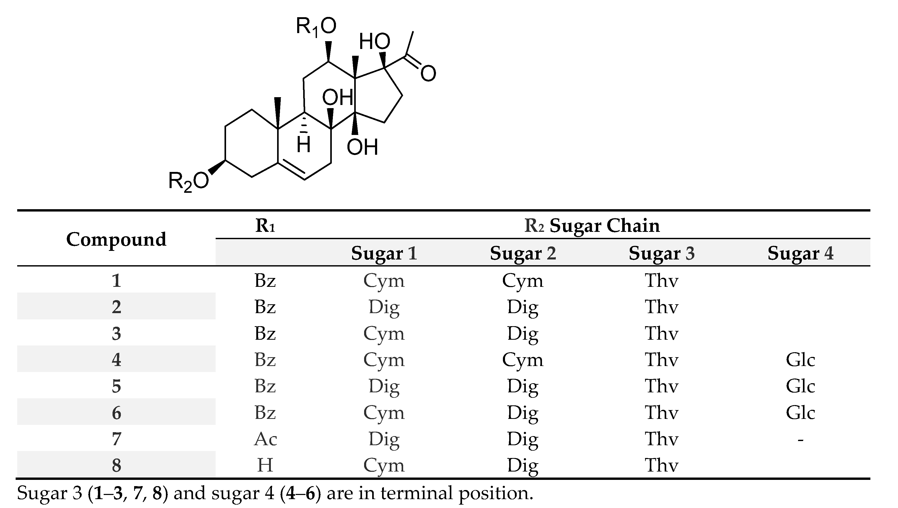
2.1. Isolation of Compounds

The dried and ground aerial parts of the plant were extracted with methanol. After evaporation of the methanol, the extract was dissolved in 50% aqueous methanol, and a solvent–solvent partition was performed with n-hexane, CHCl3, and EtOAc. The CHCl3 extract was separated by normal (NP) and reversed-phase (RP) vacuum liquid chromatography (VLC), and then it was purified by preparative thin layer chromatography (prep. TLC) and high-performance liquid chromatography (HPLC) to yield eight new pregnane glycosides (18), two new lignans (9, 10), one known pregnane and one lignan, and two known flavonoids (Figure 1).

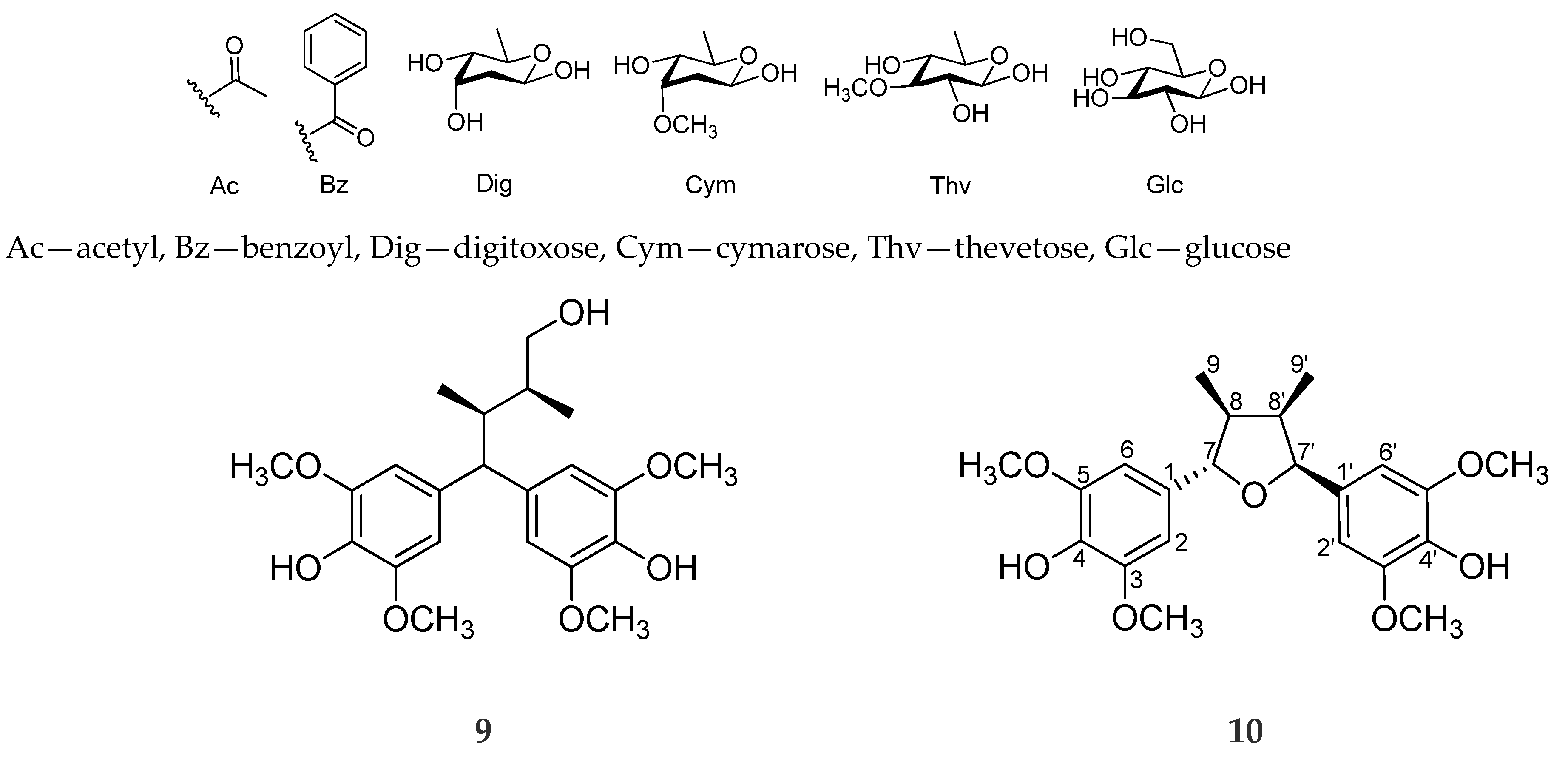
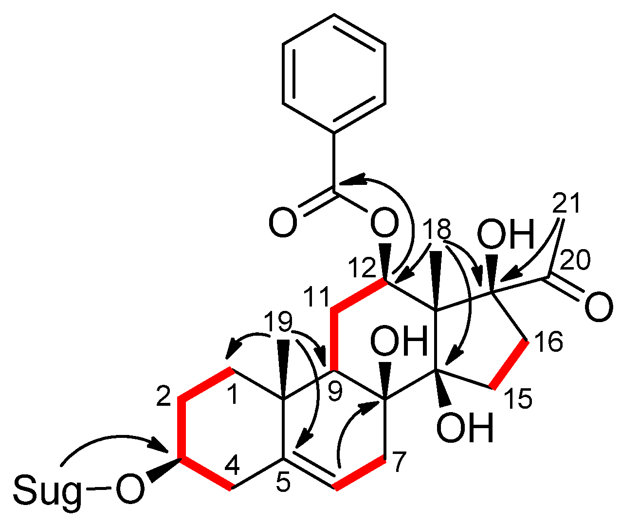
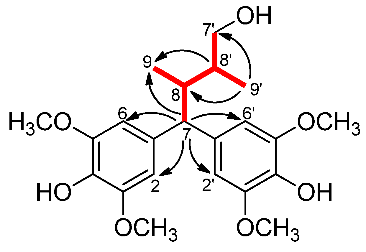
2.2. Structure Elucidation of the Compounds

The structure determination of the isolated compounds was carried out by an extensive spectroscopic analysis, including one- and two-dimensional NMR and HRMS measurements.
Compound 1 was obtained as a white amorphous powder. Its molecular formula was determined as C49H72O17 by the HRESIMS ion at m/z 955.4660 [M + Na]+ (calcd for C49H72O17Na, 955.4662). The 1H NMR spectrum of 1 showed the resonances of three anomeric protons at δH 4.85 (dd, J = 2.0 and 9.0 Hz), 4.76 (dd, J = 1.8 and 9.5 Hz), and 4.30 (d, J = 7.8 Hz), three methoxy groups at δH 3.42, 3.44, and 3.65 (each 3H, s), and three secondary methyl groups at δH 1.22 (d, J = 6.3 Hz), 1.27 (d, J = 6.2 Hz), and 1.31 (d, J = 6.2 Hz), suggesting the presence of a trisaccharide unit in 1 composed of deoxymethyl sugars. Moreover, the 1H NMR spectrum contained signals ascribable to three methyl groups displayed at δH 1.12 (3H, s), 1.54 (3H, s), and 2.06 (3H, s), and to an olefinic proton at δH 5.38 (1H, br s). The proton signals at δH 7.93 (2H, dd), 7.43 (2H, t), and 7.55 (1H, t) showed the presence of a benzoyl group in the molecule. The JMOD spectrum indicated that compound 1 contained two carbonyls, seven non-protonated carbons (of which three were oxygenated), twenty-two methines (of which eleven were oxygenated), nine methylenes, and nine methyl carbons (of which three were methoxy). The 1H and 13C JMOD NMR data dictated that 1 was a pregnane glycoside. Of these, 21 carbons were assigned to a pregnane skeleton, 7 to a benzoyl function, and 19 to a trisaccharide moiety. The HMBC correlations between H-19 (δH 1.12) and C-1 (δC 38.9), C-5 (δC 140.8), C-9 (δC 43.8), and C-10 (δC 37.3) suggested the position of a double bond at C-5/C-6. The HMBC correlations between H-18 (δH 1.54) and C-12 (δC 73.3), C-14 (δC 88.1), and C-17 (δC 91.6), between H-21 (δH 2.06) and C-17 (δC 91.6), and H-6 (δH 5.38) and C-8 (δC 74.4) demonstrated the positions of hydroxy groups at C-8, C-14, and C-17 (Figure 2). The aglycone moiety of 1 was, therefore, determined to be 12β-benzoyloxy-3β,8β,14β,17β-tetrahydroxypregn-5-ene (cynanforidine), a C/D-cis-polyoxypregnane ester [9]. The relative configuration of the molecule was determined by the analysis of NOESY correlations and literature data of similar structures reported previously.
The aglycone of 1 was supposed to have the same configuration as those of pregnanes isolated from Gymnema sylvestre [10]. The multiplicity of H-12 (δH 4.83 (dd, J = 4.2, 11.9 Hz)) implied that the configuration of H-12 was axial (α-configuration, Figure 2). The NOESY correlations between H-3 (δH 3.57), Hα-1 (δH 1.89), and Hα-4 (δH 2.40), and between H-12 (δH 4.83) and H-9 (δH 1.59) determined the configurations of the oxygenated groups at C-3 and C-12 to be β. NOESY correlations detected between Hβ-1 (δH 1.13) and Hβ-4 (δH 2.29) showed the α-position of these protons. The C-12 benzoyl group was confirmed by HMBC correlation from H-12 (δH 4.83) to the benzoyl carbonyl C-1′ (δC 165.5). The large coupling constants between the H-1 and H-2 of monosaccharide moieties, and the HMBC correlations between Thv H-1 (δH 4.30) and Cym II C-4 (δC 82.8), Cym II H-1 (δH 4.76) and Cym I C-4 (δC 82.7), and between Cym I H-1 (δH 4.85) and aglycone C-3 (δC 78.0) indicated the sugar linkages as β-d-thevetopyranosyl-(1→4)-β-d-cymaropyranosyl-(1→4)-β-d-cymaropyranoside and at C-3 of aglycone. Based on the above evidence, the structure of 1 was elucidated as cynanforidine 3-O-β-d-thevetopyranosyl-(1→4)-β-d-cymaropyranosyl-(1→4)-β-d-cymaropyranoside, a new compound named euphogossypin A.
Compound 2 was isolated as a white amorphous powder. Its 1H and JMOD spectra exhibited the characteristic signals of a pregnane aglycone, one benzoyl unit, and three sugar moieties (Table 1). In addition, NMR data of 2 were similar to those of euphogossypin A (1), except for the difference in sugar units at C-3. A careful analysis of NMR data led to the conclusion that two cymarose units were replaced by two digitoxose monosaccharides. The HMBC correlations between Thv H-1 (δH 4.82) and Dig II C-4 (δC 84.1), Dig II H- 1 (δH 5.41) and Dig I C-4 (δC 83.9), and between Dig I H-1 (δH 5.48) and aglycone C-3 (δC 78.1) confirmed the sugar linkages to be 3-O-β-d-thevetopyranosyl-(1→4)-β-d-digitoxopyranosyl-(1→4)-β-d-digitoxopyranoside. Consequently, compound 2 was identified as a new compound, cynanforidine 3-O-β-d-thevetopyranosyl-(1→4)-β-d-digitoxopyranosyl-(1→4)-β-d-digitoxopyranoside, named euphogossypin B.
The molecular formula of 3 was determined as C48H70O17 by the HRESIMS molecular ion at m/z 941.4508 [M + Na]+ (calcd for C48H70O17Na 941.4510). The 1H and JMOD data (Table 1) suggested that the structure of 3 was similar to those of 1 and 2, except for the difference in monosaccharide units in the C-3 sugar chain. The sugar moieties were found to be d-cymarose, d-digitoxose, and d-thevetose. The sugar linkages (β-d-thevetopyranosyl-(1→4)-β-d-digitoxopyranosyl-(1→4)-β-d-cymaropyranoside) to each other and to the pregnane skeleton were confirmed by the HMBC correlations from Thv H-1 (δH 4.35) to Dig C-4 (δC 83.8), from Dig H-1 (δH 4.89) to Cym C-4 (δC 83.9), and from Cym H-4 (δH 3.24) to C-3 (δC 79.3). Similarly to those of 1 and 2, the aglycone was found to be 12β-benzoyloxy-3β,8β,14β,17β-tetrahydroxypregn-5-ene (cynanforidine). Consequently, the structure of 3 was determined as cynanforidine 3-O-d-thevetopyranosyl-(1→4)-β-d-digitoxopyranosyl-(1→4)-β-d-cymaropyranoside and named euphogossypin C.
HRESIMS and NMR data of compounds 46 suggested that they were tetrasaccharide derivatives due to the presence of four anomeric carbon and proton signals, one more than observed for compounds 13. The additional sugar unit was identified as d-glucopyranose (Table 2). The polyoxypregnane ester aglycone was the same as in compounds 13. The large coupling constant between H-1 and H-2 of the glucose unit, and the HMBC correlations between Glc H-1 (δH 4.43) and Thv C-4 (δC 82.8) in 46, indicated the sugar linkages as β-d-glucopyranosyl-(1→4)-β-d-thevetopyranosyl-(1→4)-β-d-cymaropyranosyl-(1→4)-β-d-cymaropyranoside in 4, β-d-glucopyranosyl-(1→4)-β-d-thevetopyranosyl-(1→4)-β-d-digitoxopyranosyl-(1→4)-β-d-digitoxopyranoside in 5, and β-d-glucopyranosyl-(1→4)-β-d-thevetopyranosyl-(1→4)-β-d-digitoxopyranosyl-(1→4)-β-d-cymaropyranoside in 6. Based on the above evidence, the structure of 4 was deduced to be cynanforidine β-d-glucopyranosyl-(1→4)-β-d-thevetopyranosyl-(1→4)-β-d-cymaropyranosyl-(1→4)-β-d-cymaropyranoside, and was named euphogossypin D, 5 was determined as cynanforidine β-d-glucopyranosyl-(1→4)-β-d-thevetopyranosyl-(1→4)-β-d-digitoxopyranosyl-(1→4)-β-d-digitoxopyranoside and named euphogossypin E, and 6 was characterized as cynanforidine β-d-glucopyranosyl-(1→4)-β-d-thevetopyranosyl-(1→4)-β-d-digitoxopyranosyl-(1→4)-β-d-cymaropyranoside and named euphogossypin F.
The NMR data of compound 7 were very similar to that of 2, with the only difference between them being the replacement of the benzoyl moiety into an acetyl substituent at C-12. Therefore, the aglycone of 7 was identified as metaplexigenin (Table 3) [11]. The connecting sugar chain was determined as β-d-thevetopyranosyl-(1→4)-β-d-digitoxopyranosyl-(1→4)-β-d-digitoxopyranoside based on the 1D and 2D NMR spectral data. Compound 7 was, thus, characterized as metaplexigenin β-d-thevetopyranosyl-(1→4)-β-d-digitoxopyranosyl-(1→4)-β-d-digitoxopyranoside and named euphogossypin G.
Compound 8 wore the same C-3 trisaccharide sugar chain, β-d-thevetopyranosyl-(1→4)-β-d-digitoxopyranosyl-(1→4)-β-d-cymaropyranoside, as 3 (Table 3). By the NMR data, it was determined to be a deacetylmetaplexigenin derivative and named euphogossypin H.
Gossypilignan A (9) was obtained as a pale yellow, amorphous solid. The molecular formula of 9 was determined as C22H30O7 from its HRESIMS ion observed at m/z 429.1889 [M + Na]+ (calcd for C22H30O7Na, 429.1884). The 1H and 13C JMOD NMR data indicated the presence of four methoxy (δH 2 × 3.82, s, and 2 × 3.83, s; δC 4 × 56.8), two methyl (δH 0.68, d, J = 6.9 Hz, and 0.76, d, J = 7.0 Hz; δC 10.0 and 12.1), one methylene (δH 3.35 and 3.45; δC 67.3), and three methine groups (δH 1.77, m, 2.62, m, 3.52, d; δC 37.0, 37.2, and 57.9) (Table 4). Additionally, the 1H NMR spectrum of 9 showed aromatic protons at δH 6.64, s and 6.66, s (2H each), implying that the aromatic rings were tetrasubstituted. The 13C NMR spectrum of 9 also supported the presence of six oxygenated aromatic carbons at δC 149.1, 149.2, and 134.7 (each 2C). According to the HMBC correlations from H-2, H-6, H-2′, and H-6′ to C-7, both aromatic rings were attached to C-7. The 1H-1H COSY correlations of H-7/H-8/H-8′/H-7′, H-8/H-9, and H-8′/H-9′, as well as the HMBC correlations from H-7 to C-8, C-8′, and C-9, indicated the presence of a 2,3-dimethylbutane moiety (Figure 3). By comparison, the skeleton of 9 was found to be the same as that of kadangustin J [12]. According to the chemical shift of δC 67.3 (C-7′), a hydroxy group should be placed at C-7′. Four methoxy groups were located at C-3, C-5, C-3′, and C-5, respectively, while C-4 and C-4′ were substituted with hydroxy groups. Thus, the structure of compound 9 was determined to be 4,4-di-(4-hydroxy-3,5-dimethoxyphenyl)-2,3-dimethylbutanol and named gossypilignan A. As compound 9 was a 7,7-diaryl-8,8′-dimethylbutan-1-ol lignan, it had two chiral centers. Based on the investigation of Davidson et al., syn- and anti-isomers of such compounds can be distinguished based on the significant differences between their 1H NMR data [13]; therefore, as in the case of 9, both methyl groups were β-oriented, proving that this compound was a syn-isomer.
The molecular formula of 10 was determined as C22H28O7 by the HRESIMS pseudo-ion at m/z 427.1734 [M + Na]+. The 1H NMR spectrum of 10 showed four aromatic hydrogens as two similar systems, one at δH 6.69 (2H, br s), and another at 6.63 (2H, br s), indicating the presence of two 1,3,4,5-tetrasubstituted benzene rings (Table 4). The chemical shifts observed for aromatic hydrogens along with the presence of four singlets corresponding to methoxy hydrogens at δH 2 × 3.84 and 2 × 3.86 (s, 3H each) indicated the presence of 3,5-dimethoxy-4-hydroxyphenyl groups in this compound. The 13C NMR data corroborated the structural determination of these aromatic rings. Moreover, 1H NMR spectral data suggested a nonsymmetric tetrahydrofuran lignan, through signals corresponding to two methyl groups at δH 1.00 (d, J = 6.4 Hz) and 0.63 (d, J = 7.0 Hz) in addition to two oxybenzyl methines at δH 4.64 (d, J = 9.3) and 5.47 (d, J = 4.4 Hz). The attachment of aromatic rings to the tetrahydrofuran ring was determined by an HMBC experiment. In the HMBC spectrum, interactions were observed between H-2′ and C-7′ and C-4′, between H-6′/C-4′ and C-2′, and between H-2 and C-7. These data indicated the presence of the two 3,5-dimethoxy-4-hydroxyphenyl structural parts at C-7 and C-7′, respectively. Based on previous literature data [14,15], the coupling constant of 9.3 Hz for the doublet at δH 4.64 (H-7) indicated that this hydrogen was in a trans configuration with the adjacent H-8, while the coupling constant of 4.4 Hz of H-7′ demonstrated its cis relationship with H-8′. NOE correlations confirmed the relative stereochemistry at the tetrahydrofurane ring as trans (C-7/C-8), cis (C-8/C-8′), and cis (C-8′/C-7′). Moreover, NOE effects proved the cis configuration of the aromatic group at C-7′ and methyl groups at C-9 and C-9′. These data permitted the establishment of the structure of the new tetrahydrofuran lignan 10 as rel-(7S,8R,7′S,8′S)-3,5,3′,5′-tetramethoxy-4,4′-dihydroxy-7,7′-epoxylignan and named gossypilignan B.
The known compounds were identified as 12-O-benzoyldeacylmetaplexigenin, 9α-hydroxypinoresinol [16], naringenin [17], and quercitrin [18]. All compounds were isolated for the first time from the plant. Such polyoxypregnane ester derivatives were isolated previously, mainly from Asclepiadaceae species (e.g., Cynanchum wilfordi and Leptadenia hastata) [9]. Though Euphorbia species are frequently characterized by the abundant presence of various terpenoids, this is only the second report of pregnanes from a plant belonging to the Euphorbiaceae family. Previously, two pregnane glycosides were identified from the aerial parts of Croton ruizianus. Their aglycon was 3β,14β,15β,16α-tetrahydroxypregnan-20-one, while the connecting sugar parts were either 3-O-β-d-Glu-(1→4)-β-d-Ole-(1→4)-β-d-Ole-(1→4)-β-d-Dig-(1→4)-β-d-Ole or 3-O-β-d-Ole-(1→4)-β-d-Ole-(1→4)-β-d-Dig-(1→4)-β-d-Ole (Ole—oleandrose; Dig—digitoxose) [19]. Pregnane glycosides isolated from E. gossypina var. coccinea were substituted with thevetose, cymarose, digitoxose, and glucose-containing linear sugar chains.
The 7,7-diarylbutanol seco-lignans are an interesting class of lignans with both chemical and pharmacological importance. Cytotoxic, anti-HIV-1, and antioxidant activities have been previously reported for this class of natural products [12,20,21].
Pregnane glycosides are reported to possess various biological activities, an antiproliferative effect being one of the most important ones [22]. Amplexoeside A, isolated from an Asclepiadaceae species Asclepias amplexcicaulis, showed antiproliferative activity in the KB assay. Condurangoglycoides identified from Marsdenia congurango were proved to be active against Sarcoma 180 and Ehrlich sarcoma in mice [23,24]. Pregnane glycosides (Ap and Ao) from Dregea uolubilis showed activity against Ehrlich carcinoma [25]. Moreover, compounds with an antitumor effect were isolated from Aspidopterys obcordata, Caralluma dalzielii, C. negevensis, C. quadrangular, Cynanchum wilfordi, Desmidorchis flava, Gelsemium sempervirens, Marsdenia tenacissima, Periploca sepium, Solenostemma argel, and Stizophyllum riparium [8,26,27,28]. Although the molecular backbone of lignans consists of only two phenylpropane (C6–C3) units, they show an enormous structural diversity, and promising pharmacological effects. A considerable number of plant lignans (e.g., podophyllotoxin, sesamin, diphyllin, enterolactone, arctiin, matairesinol, and taxiresinol) were identified to possess antiproliferative activity [29]. Since, previously, several pregnane glycosides and lignans were reported to display strong antiproliferative activities, all isolated compounds belonging to these groups of special metabolites were tested for their antiproliferative properties against the HeLa cell line using the MTT assay. Doxorubicin and cisplatin were used as positive controls (IC50s 0.02 ± 0.003 μM and 2.07 ± 0.07 μM, respectively). Among the tested compounds, only euphogossypin A (1) exerted weak antiproliferative activity (IC50 52.4 ± 0.23 µM), while the others proved to be inactive (Table S1).

3. Materials and Methods

3.1. General Experimental Procedures

NMR spectra were recorded in CDCl3, CD3OD, and C5D5N on a Bruker Avance DRX 500 spectrometer (Billerica, MA, USA) at 500 MHz (1H) and 125 MHz (13C). The signals of the deuterated solvents were taken as reference. The chemical shift values (δ) were given in ppm and coupling constants (J) were in Hz. Two-dimensional (2D) experiments were performed with standard Bruker software. In the COSY, HSQC, and HMBC experiments, gradient-enhanced versions were used. High-resolution MS spectra were acquired on a Thermo Scientific Q-Exactive Plus Orbitrap mass spectrometer (Waltham, MA, USA) equipped with ESI ion source in positive ionization mode. The data were acquired and processed with MassLynx software.
Column chromatography (CC) was performed on polyamide (MP Biomedicals, Irvine, CA, USA). Normal-phase vacuum liquid chromatography (VLC) was carried out on silica gel (Kieselgel 60 GF254, 15 μm, Merck, Darmstadt, Germany). Thin-layer chromatography was performed on Kieselgel 60 RP-18 F254 and Kieselgel 60 F254 (Merck, Darmstadt, Germany). Spots were detected under UV light (245 nm and 336 nm) and made visible with vanillin sulfuric acid reagent and heating at 105 °C for 1 min. The high-performance liquid chromatography (HPLC) separation was carried out on a Waters HPLC (Waters 600 controller, Waters 600 pump, and Waters 2998 photodiode array detector), using RP (LiChrospher RP-18, 5 μm, 250 × 4 mm, Merck, Darmstadt, Germany) column. In the case of the RP-HPLC separation, the mobile phase consisted of MeOH (solvent A) and H2O (solvent B). The flow rate was 1 mL/min. The data were acquired and processed with the Empower software.
All solvents used for CC were of at least analytical grade (VWR Ltd., Szeged, Hungary). Ultra-pure water was prepared with a Milli-Q water purification system (Millipore, France).

3.2. Plant Material

Aerial parts of Euphorbia gossypina var. coccinea Pax. were collected in Kenya (GPS coordinates 1°24′24.31777″ S, 36°42′53.86125″ E), Africa, in July 2018, and identified by Patrick Chalo Mutiso, a taxonomist (Department of Biological Sciences, Faculty of Science and Technology, University of Nairobi). A voucher specimen (no. UON 2018/249) was deposited at the Herbarium of the School of Biological Sciences, University of Nairobi, Kenya.

3.3. Isolation of Compounds

The dried and ground aerial parts of E. gossypina var. coccinea (1 kg) were extracted with methanol at room temperature. After concentration, the extract (45 g) was dissolved in 50% aqueous methanol, and solvent–solvent partitions were performed with hexane, CHCl3, and EtOAc. The CHCl3 fraction (28.6 g) was purified by polyamide column chromatography using MeOH–H2O (4:1) to remove chlorophyll. Thereafter, the yielded fraction (22.8 g) was purified by VLC on silica gel with a gradient system of cyclohexane–EtOAc–EtOH (from 8:2:0 to 6:3:1) and CHCl3–MeOH (from 9:1 to 1:1). The TLC determination and combination of the fractions afforded 20 main fractions (Fr. 1–20). Fraction 9 (1.4 g) was further chromatographed by RP-VLC using MeOH–H2O gradient solvent system (8:2–2:8), and 9 fractions (Fr. 9/1–9) were obtained. Fraction 9/3 (87.9 mg) was subjected to preparative TLC using CHCl3–MeOH (8:2) as eluent, and was, subsequently, purified by RP-HPLC. Gradient elution was applied, starting at 10% A (methanol) and 90% B (H2O) for 1 min, then linearly increased to 50% A (in 10 min) to yield compound 10 (tR = 3.1 min, 2.6 mg). Fraction 10 (1.6 g) was separated by NP-VLC using a toluene–acetone gradient system (from 9:1 to 6:4) to obtain 9 subfractions (Fr. 10/1–9). Fr. 10/4 (371 mg) was chromatographed by NP-VLC using cyclohexane–EtOAc–EtOH gradient system (from 9:1:0 to 6:3:1) to yield 6 fractions (Fr. 10/4/1–6). 9α-hydroxypinoresinol (5.8 mg) was isolated from Fr. 10/4/3 (87 mg) by preparative TLC using CHCl3–MeOH (95:5) as mobile phase. Fr. 10/5 (188 mg) was purified by RP-TLC using MeOH–H2O (8:2) as eluent, and further purified by RP-HPLC by using gradient elution, started at 10% A (methanol) and 90% B (H2O) for 1 min, then linearly increased to 50% A (in 10 min) to yield compound 9 (tR = 7.3 min, 12.1 mg). Fr. 10/6 (123 mg) was separated by RP-TLC using MeOH–H2O (8:2) as solvent system and compound 1 (11.2 mg) was obtained. Fraction 11 (4.9 g) was chromatographed by NP-VLC using the gradient system of toluene–acetone (from 9:1 to 1:1) to yield 11 subfractions (Fr. 11/1–11). Fraction 11/9 (193.5 mg) was purified by RP-TLC using MeOH–H2O (8:2) as an eluent to yield compound 2 (19.7 mg) and two subfractions (Fr. 11/9/1 (8.1 mg) and 2 (6.9 mg)). Both fractions were purified by RP-HPLC (the gradient started at MeOH–H2O (2:8) for 1 min, then linearly ramped up to 1:1 in 10 min and held 0.5 min, then returned to the initial conditions within 1 min, and kept for 2 min) to yield compounds 8 (tR = 5.2 min, 6.4 mg) and 7 (tR = 6.1 min, 5.1 mg). Fr. 13 (165 mg) was purified by NP-TLC using CHCl3–MeOH (85:15) as mobile phase to isolate compound 4 (12.7 mg). Fraction 15 (5.9 g) was separated by RP-VLC using a gradient system of MeOH–H2O (from 1:9 to 9:1) to yield 7 subfractions (Fr. 15/1–7). Fraction 15/3 (50 mg) was subjected to an NP-TLC using toluene–acetone (1:1) as an eluent to obtain naringenin (5 mg). Fraction 15/6 (75 mg) was subjected to RP-TLC using MeOH–H2O (7:3) to yield quercitrin (8.9 mg). Fraction 15/7 (329 mg) was purified by RP-HPLC using MeOH–H2O (8:2) isocratic solvent system to obtain 12-O-benzoyldeacylmetaplexigenin (tR = 11 min, 47.1 mg). Fraction 15/8 (75 mg) was purified by RP-HPLC (the gradient started at MeOH–H2O (2:8) for 1 min, then linearly ramped up to 1:1 in 10 min and held 0.5 min, then returned to the initial conditions within 1 min and kept for 2 min) to yield compound 6 (tR = 5.5 min, 13.9 mg). Fr. 16 (1.6 g) was subjected to an NP-VLC using CHCl3–MeOH gradient system (from 98:2 to 1:1) to yield 9 subfractions (Fr. 16/1–9). Fr. 16/6 (106 mg) was further purified by RP-HPLC using MeOH–H2O (7:3) isocratic solvent system to obtain compound 5 (tR = 7.2 min, 24.3 mg).

3.3.1. Euphogossypin A (1)

White amorphous powder; [α ] D 25 +12 (c 0.1, MeOH); HRESIMS m/z: 955.4660 [M + Na]+ (calcd for C49H72O17Na, 955.4662); 1H (CDCl3, 500 MHz) and 13C NMR (CDCl3, 125 MHz) data, see Table 1.

3.3.2. Euphogossypin B (2)

White amorphous powder; [α ] D 25 +24 (c 0.2, MeOH); HRESIMS m/z: 927.4352 [M + Na]+ (calcd for C47H68O17Na, 927.4349); 1H (C5D5N, 500 MHz) and 13C NMR ((C5D5N, 125 MHz) data, see Table 1.

3.3.3. Euphogossypin C (3)

White amorphous powder; [α ] D 25 +5 (c 0.1, MeOH); HRESIMS m/z: 941.4508 [M + Na]+ (calcd for C48H70O17Na 941.4510); 1H (CD3OD, 500 MHz) and 13C NMR (CD3OD, 125 MHz) data, see Table 1

3.3.4. Euphogossypin D (4)

White amorphous powder; [α ] D 25 +63 (c 0.15, MeOH); HRESIMS m/z: 1103.5029 [M + Na]+ (calcd for C54H80O22Na, 1103.5033); 1H (CD3OD, 500 MHz) and 13C NMR (CD3OD, 125 MHz) data, see Table 2.

3.3.5. Euphogossypin E (5)

White amorphous powder; [α ] D 25 0 (c 0.15, MeOH); HRESIMS m/z: 1089.4869 [M + Na]+ (calcd for C53H78O22Na, 1089.4877); 1H (CD3OD, 500 MHz) and 13C NMR (CD3OD, 125 MHz) data, see Table 2.

3.3.6. Euphogossypin F (6)

White amorphous powder; [α ] D 25 +3 (c 0.15, MeOH); HRESIMS m/z: 1103.5029 [M + Na]+ (calcd for C54H80O22Na, 1103.5033); 1H (CD3OD, 500 MHz) and 13C-NMR (CD3OD, 125 MHz) data, see Table 2.

3.3.7. Euphogossypin G (7)

White amorphous powder; [α ] D 25 +12 (c 0.05, MeOH); HRESIMS m/z: 865.4188 [M + Na]+ (calcd for C42H66O17Na, 865.4192); 1H (CDCl3, 500 MHz) and 13C NMR (CDCl3, 125 MHz) data, see Table 3.

3.3.8. Euphogossypin H (8)

White amorphous powder; [α ] D 25 +33 (c 0.15, MeOH); HRESIMS m/z: 837.4238 [M + Na]+ (calcd for C41H66O16Na, 837.4243); 1H (CDCl3, 500 MHz) and 13C NMR (CDCl3, 125 MHz) data, see Table 3.

3.3.9. Gossypilignan A (9)

Pale yellow amorphous solid; [α ] D 25 −13 (c 0.1, MeOH); HRESIMS m/z: 429.1889 [M + Na]+ (calcd for C22H30O7Na, 429.1884); 1H (CD3OD, 500 MHz) and 13C NMR (CD3OD, 125 MHz) data, see Table 4.

3.3.10. Gossypilignan B (10)

White amorphous powder; [α ] D 25 −5 (c 0.05, MeOH); HRESIMS m/z: 427.1734 [M + Na]+ (calcd for C22H28O7Na, 427.1727); 1H (CD3OD, 500 MHz) and 13C NMR (CD3OD, 125 MHz) data, see Table 4.

3.3.11. 12-O-Benzoyldeacylmetaplexigenin

1H NMR (in CDCl3) δH 1.15 (3H, s, Me-19), 1.15, 1.90 (2H, m, H-1), 1.55 (3H, s. Me-18), 1.56, 1.83 (2H, m, H-2), 1.60 (1H, m H-9), 1.96 (2H, m, H-11), 2.03 (2H, m, H-15), 2.07 (3H, s, Me-21), 2.25 (2H, m, H-7), 2.30–2.39 (2H, m, H-4), 1.92, 2.86 (2H, m, H-16), 3.57 (1H, m, H-3), 4.86 (1H, dd, J = 10.2, 5.6 Hz, H-12), and 5.39 (1H, br s, H-6); 13C NMR see Table 1.

3.4. Antiproliferative Assays

3.4.1. Cell Line

Human cervix carcinoma (HeLa) cells were cultured in Eagle’s Minimal Essential Medium (EMEM, containing 4.5 g/L glucose) supplemented with a non-essential amino acid mixture, a selection of vitamins, and 10% heat-inactivated fetal bovine serum. The cell line was detached with 0.25% trypsin and 0.02% EDTA for 5 min at 37 °C. The cell line was purchased from LGC Promochem, Teddington, England.

3.4.2. Antiproliferative Assay

The antiproliferative assay of the isolated compounds (13, 512) against the human cervix carcinoma (HeLa) cell line was performed by MTT, using cisplatin and doxorubicin as positive controls. This assay was carried out as previously described [30,31].

4. Conclusions

In this article, nine polyoxypregnane ester derivatives, including euphogossypins A–H (18) as new natural products, two new lignans (gossypilignans A (9) and B (10)), and 12-O-benzoyldeaxcylmetaplexigenin (11), 9α-hydroxypinoresinol (12), naringenin (13), and quercitrin (14) as known compounds, were characterized from the aerial parts of E. gossypina var. coccinea. Their planar structures were elucidated by comprehensive spectroscopic data. All compounds were isolated for the first time from the plant. Although previously several pregnane glycosides and lignans have been found to be active as antiproliferative agents, none of the compounds isolated from E. gossypina var. coccinea showed remarkable antiproliferative activity against the Hela cells. Our findings enrich the knowledge of special metabolites of Euphorbia species.

Supplementary Materials

The following supporting information can be downloaded at: https://www.mdpi.com/article/10.3390/plants11101299/s1, Figures S1–S58: One-dimensional and two-dimensional NMR spectra of isolated compounds (110); Table S1: Antiproliferative activity of the isolated compounds (13, 512) in HeLa cell line.

Author Contributions

Conceptualization, A.V.; methodology, A.V.; software, N.S. and G.S.; investigation, R.H., N.K., R.B., N.S., C.Z.D., P.W.M. and G.S.; resources, J.H.; writing—original draft preparation, R.H. and A.V.; writing—review and editing, A.V., N.K. and J.H.; project administration, J.H.; funding acquisition, J.H. All authors have read and agreed to the published version of the manuscript.

Funding

This research was funded by the National Research, Development and Innovation Office, Hungary (K135845), the UNKP-21-3-SZTE-265 New National Excellence Program, and TKP2021-EGA-32 of the Ministry of Innovation and Technology of Hungary from the source of the National Research, Development and Innovation Fund.

Institutional Review Board Statement

Not applicable.

Informed Consent Statement

Not applicable.

Data Availability Statement

Data is contained within the article and Supplementary Material.

Acknowledgments

The authors acknowledge Patrick Chalo Mutiso, a taxonomist (Department of Biological Sciences, Faculty of Science and Technology, University of Nairobi), for the collection and identification of plant material.

Conflicts of Interest

The authors declare no conflict of interest.

References

  1. Vasas, A.; Hohmann, J. Euphorbia Diterpenes: Isolation, Structure, Biological Activity, and Synthesis (2008−2012). Chem. Rev. 2014, 114, 8579–8612. [Google Scholar] [CrossRef] [PubMed] [Green Version]
  2. Shi, Q.W.; Su, X.H.; Kiyota, H. Chemical and pharmacological research of the plants in genus Euphorbia. Chem. Rev. 2008, 108, 4295–4327. [Google Scholar] [CrossRef] [PubMed]
  3. Euphorbia gossypina. Available online: https://powo.science.kew.org/taxon/urn:lsid:ipni.org:names:346654-1 (accessed on 31 March 2022).
  4. Sharma, P.; Singh, G. A review of plant species used to treat conjunctivitis. Phytother Res. 2002, 16, 1–22. [Google Scholar] [CrossRef] [PubMed]
  5. Schmelzer, G.H.; Gurib-Fakim, A. Plant Resources of Tropical Africa 11 (1), Medicinal Plants1; PROTA Foundation/Backhuys Publisher/CTA: Wageningen, The Netherlands, 2008; Volume 11, p. 300. [Google Scholar]
  6. Catley, A.; Mohammed, A.A. Ethnoveterinary knowledge in Sanaag region, Somaliland (Part II): Notes on local methods of treating and preventing livestock disease. Nomadic Peoples 1996, 39, 135–145. [Google Scholar]
  7. Araya, J.J.; Binns, F.; Kindscher, K.; Timmermann, B.N. Verticillosides A–M: Polyoxygenated pregnane glycosides from Asclepias verticillata L. Phytochemistry 2012, 78, 179–189. [Google Scholar] [CrossRef]
  8. Si, Y.; Sha, X.S.; Shi, L.L.; Wei, H.Y.; Jin, Y.X.; Ma, G.X. Review on pregnane glycosides and their biological activities. Phytochem. Lett. 2022, 47, 1–17. [Google Scholar] [CrossRef]
  9. Tsukamoto, S.; Hayashi, K.; Mitsuhashi, H. Studies on the constituents of Asclepiadaceae plants. LX. Further studies on glycosides with a novel sugar chain containing a pair of optically isomeric sugars, d- and l-cymarose, from Cynanchum wilfordi. Chem. Pharm. Bull. 1985, 33, 2294–2304. [Google Scholar] [CrossRef] [Green Version]
  10. Kiem, P.V.; Yen, D.T.H.; Hung, N.V.; Nhiem, N.X.; Tai, B.H.; Trang, D.T.; Yen, P.H.; Ngoc, T.M.; Minh, C.V.; Park, S.; et al. Five new pregnane glycosides from Gymnema sylvestre and their α-glucosidase and α-amylase inhibitory acitivities. Molecules 2020, 25, 2525. [Google Scholar] [CrossRef]
  11. Aquino, R. New polyoxypregnane ester derivatives from Leptadenia hastata. J. Nat. Prod. 1996, 59, 555–564. [Google Scholar] [CrossRef]
  12. Gao, X.M.; Pu, J.X.; Huang, S.X.; Yang, L.M.; Huang, H.; Xiao, W.L.; Zheng, Y.T.; Sun, H.D. Lignans from Kadsura angustifolia. J. Nat. Prod. 2008, 71, 558–563. [Google Scholar] [CrossRef]
  13. Davidson, S.J.; Rye, C.E.; Barker, D. Using NMR to determine the relative stereochemistry of 7,7-diaryl-8,8′-dimethylbutan-1-ol lignans. Phytochem. Lett. 2015, 14, 138–142. [Google Scholar] [CrossRef]
  14. Yue, J.M.; Chen, Y.Z.; Hua, S.M.; Cheng, J.L.; Cui, Y.X. Ganschisandrine, a lignan from Schisandra sphenanthera. Phytochemistry 1989, 28, 1774–1776. [Google Scholar]
  15. Da Silva Filho, A.A.; Albuquerque, S.; e Silva, M.L.A.; Eberlin, M.N.; Tomazela, D.M.; Bastos, J.K. Tetrahydrofuran lignans from Nectandra megapotamica with trypanocidal activity. J. Nat. Prod. 2004, 67, 42–45. [Google Scholar] [CrossRef] [PubMed]
  16. Abe, F.; Yamauchi, T. 9α-hydroxypinoresinol, 9α-hydroxymedioresinol and related lignans from Allamanda neriifolia. Phytochemistry 1988, 27, 575–577. [Google Scholar] [CrossRef]
  17. Cordenonsi, L.M.; Sponchiado, R.M.; Campanharo, S.C.; Garcia, C.V.; Raffin, R.P.; Schapoval, E.E.S. Study of flavonoids presente in Pomelo (Citrus máxima) by DSC, UV-VIS, IR, 1H and 13C NMR and MS. Drug Anal. Res. 2017, 1, 31–37. [Google Scholar] [CrossRef]
  18. Zhong, X.N.; Otsuka, H.; Ide, T.; Hirata, E.; Takushi, A.; Takeda, Y. Three flavonol glycosides from leaves of Myrsine seguinii. Phytochemistry 1997, 46, 943–946. [Google Scholar] [CrossRef]
  19. Piacente, S.; Belisario, A.B.; Castillo, H.D.; Pizza, C.; Feo, V.D. Croton ruizianus: Platelet proaggregating activity of two new pregnane glycosides. J. Nat. Prod. 1998, 61, 318–322. [Google Scholar] [CrossRef]
  20. Xu, L.J.; Huang, F.; Chen, S.B.; Li, L.N.; Chen, S.L.; Xiao, P.G. A cytotoxic neolignan from Schisandra propinqua (Wall.) Baill. J. Integr. Plant Biol. 2008, 48, 1493–1497. [Google Scholar] [CrossRef]
  21. Huang, F.; Xu, L.; Shi, G. Antioxidant isolated from Schisandra propinqua (Wall.) Baill. Biol. Res. 2009, 42, 351–356. [Google Scholar] [CrossRef] [Green Version]
  22. Deepak, D.; Srivastav, S.; Khare, A. Pregnane Glycosides. In Fortschritte der Chemie Organischer Naturstoffe/Progress in the Chemistry of Organic Natural Products; Springer: Vienna, Austria, 1997; Volume 71. [Google Scholar] [CrossRef]
  23. Hayashi, K.; Wada, K.; Mitsuhashi, H.; Bando, H.; Takase, M.; Terada, S.; Koide, Y.; Aiba, T.; Narita, T.; Mizuno, D. Antitumor active glycosides from Condurango cortex. Chem. Pharm. Bull. 1980, 28, 1954–1958. [Google Scholar] [CrossRef]
  24. Hayashi, K.; Wada, K.; Mitsuhashi, H.; Bando, H.; Takase, M.; Terada, S.; Koide, Y.; Aiba, T.; Narita, T.; Mizuno, D. Further investigation of antitumor condurangoglycosides with C-18 oxygenated aglycone. Chem. Pharm. Bull. 1981, 29, 2725–2730. [Google Scholar] [CrossRef] [PubMed] [Green Version]
  25. Yoshimura, S.I.; Narita, H.; Hayashi, K.; Mitsuhashi, H. Studies on the constituents of Asclepiadaceae plants. LVI. Isolation of new antitumor-active glycosides from Dregea volubilis (L.) Benth. Chem. Pharm. Bull. 1983, 31, 3971. [Google Scholar] [CrossRef] [PubMed] [Green Version]
  26. Panda, N.; Banerjee, S.; Mandal, N.B.; Sahu, N.P. Pregnane glycosides. Nat. Prod. Commun. 2006, 1, 665–695. [Google Scholar] [CrossRef]
  27. Plaza, A.; Perrone, A.; Balestrieri, M.L.; Felice, F.; Balestrieri, C.; Hamed, A.I.; Pizza, C.; Piacente, S. New unusual pregnane glycosides with antiproliferative activity from Solenostemma argel. Steroids 2005, 70, 594–603. [Google Scholar] [CrossRef]
  28. Hunag, L.J.; Wang, B.; Zhang, J.X.; Yan, C.; Mu, S.Z.; Hao, X.J. Studies on cytotoxic pregnane sapogenins from Cynanchum wilfordii. Fitoterapia 2015, 101, 107–116. [Google Scholar] [CrossRef]
  29. Saleem, M.; Kim, H.J.; Ali, M.S.; Lee, Y.S. An update on bioactive plant lignans. Nat. Prod. Rep. 2005, 22, 696–716. [Google Scholar] [CrossRef]
  30. Stefkó, D.; Kúsz, N.; Szemerédi, N.; Barta, A.; Spengler, G.; Berkecz, R.; Hohmann, J.; Vasas, A. Unique phenanthrenes from Juncus ensifolius and their anti-proliferative and synergistic effects with the conventional anticancer agent doxorubicin against human cancer cell lines. Pharmaceutics 2022, 14, 608. [Google Scholar] [CrossRef]
  31. Bacher, F.; Wittmann, C.; Nové, M.; Spengler, G.; Marć, M.A.; Enyedy, E.A.; Darvasiová, D.; Rapta, P.; Reinere, T.; Arion, V.B. Novel latonduine derived proligands and their copper(II) complexes show cytotoxicity in the nanomolar range in human colon adenocarcinoma cells and in vitro cancer selectivity. Dalton Trans. 2019, 48, 10464. [Google Scholar] [CrossRef] [Green Version]
Figure 1. New isolated compounds (110) from E. gossypina var. coccinea.
Plants 11 01299 g001aPlants 11 01299 g001b
Figure 2. The 1H-1H COSY () and key HMBC (→) correlations of 1.
Plants 11 01299 g002
Figure 3. Selected 1H-1H COSY () and HMBC (→) correlations of 9.
Plants 11 01299 g003
Table 1. The 1H and 13C NMR data for compounds 13.
Atom1 *2 *3 *
δH (J in Hz)δCδH (J in Hz)δCδH (J in Hz)δC
11.13, m, α/1.89, m, β38.9, CH21.14, m, α/1.83, m, β39.4, CH21.14, m, α/1.83 m, β39.8, CH2
21.62, m, β/1.92, m, α29.1, CH21.82, m, β/2.11, m, α30.3, CH21.58, m, β/1.87 m, α30.2, CH2
33.57, m78.0, CH3.89, m78.1, CH3.54, m79.3, CH
42.29, m, β/2.40, m, α38.9, CH22.43, m, * β/2.55, m, α39.7, CH22.22, m; 2.38 dd (3.4, 12.7)39.8, CH2
5-140.8, C-139.9, C-140.3, C
65.38, br s117.8, CH5.31, br s119.6, CH5.36, br d (4.7)119.7, CH
72.22, m34.4, CH22.38, m/2.50, m35.2, CH22.12–2.22 m35.2, CH2
8-74.4, C-74.8, C-75.2, C
91.59, m43.8, CH1.80, m44.9, CH1.59 m45.1, CH
10-37.3, C-37.9, C-38.2, C
111.94, m24.3, CH22.21, m, α/2.36, m, β25.5, CH21.81, m, α/2.00, m, β25.5, CH2
124.83, dd (4.2, 11.9)73.3, CH5.36, dd (4.3, 11.5)77.5, CH4.83 dd (4.3, 11.9)74.7, CH
13-58.5, C-58.8, C-59.1, C
14-88.1, C-90.0, C-90.0, C
152.03, m33.4, CH22.14–2.21, m34.3, CH21.92, m; 2.06, * m34.3, CH2
161.92, m, β/2.85, m, α32.1, CH22.07, m/3.27, m33.7, CH21.73, m, β/2.87, m, α33.5, CH2
17-91.6, C-93.0, C-93.1, C
181.54, s9.7, CH32.10, s11.3, CH31.67, s10.6, CH3
191.12, s18.7, CH31.34, s18.6, CH31.16, s18.6, CH3
20-209.5, C-210.6, C-212.2, C
212.06, s27.5, CH32.37, s28.2, CH32.05, s27.8, CH3
Bz
1′-165.5, C-165.8, C-166.7, C
2′-130.1, C-131.8, C-131.6, C
3′,7′7.93, dd (1.1, 8.2)129.7, CH8.31, d (7.8)130.4, CH7.95, d (7.9)130.5, CH
4′,6′7.43, t (8.0)128.6, CH7.49, t (7.7)129.4, CH7.48, dd (7.9, 7.4)129.6, CH
5′7.55, t (8.1)133.3, CH7.59, t (7.7)133.7, CH7.61, t (7.4)134.3, CH
Cym I Dig I Cym
14.85, dd (2.0, 9.0)96.2, CH5.48, d (9.4)96.9, CH4.87, dd (1.9, 8.9)97.2, CH
21.58, m, a/2.08, m, e35.7, CH22.05, m; 2.43, m *39.5, CH21.54, m, a/2.06, m, * e36.7, CH2
33.80, m77.2, * CH4.65, m68.0, CH3.85, m78.6, CH
43.21, dd (3.0, 9.6)82.7, CH3.53, m83.9, CH3.24, m *83.9, CH
53.84, m68.7, CH4.31, m69.1, CH3.81, m70.0, CH
61.22, d (6.3)18.3, CH31.45, d (6.0)19.1, CH31.20, d (6.2)18.5, CH3
3-OMe3.42, s *58.1, # CH3 3.44, s58.5, CH3
Cym II Dig II Dig
14.76, dd (1.8, 9.5)99.7, CH5.41, d (9.6)100.3, CH4.89, dd (1.7, 9.1)101.0, CH
21.65, m, a/2.16, m, e35.3, CH22.00, m, a/2.43, m, * e39.4, CH21.71, m, a/2.02, m, e38.8, CH2
33.78, m77.1,* CH4.70, m68.3, CH4.21, m68.6, CH
43.26, dd (2.9, 9.6)82.8, CH3.60, m84.1, CH3.24, * m83.8, CH
53.90, m68.4, CH4.37, m69.5, CH3.87, m69.5, CH
61.27, d (6.2)18.6, CH31.59, d (6.1)19.0, CH31.31, d (6.2)18.6, CH3
3-OMe3.44, s *58.2, # CH3
Thv Thv Thv
4.30, d (7.8)104.5, CH4.82, d (7.9)106.3, CH4.35, d (7.9)105.5, CH
3.51, m74.8, CH3.85, m75.3, CH3.28, m75.2, CH
3.10, t (9.0)85.4, CH3.61, m88.3, CH3.01, m87.5, CH
3.18, t (9.2)74.8, CH3.58, m76.3, CH3.04, m76.5, CH
3.36, dd (6.2, 9.2)71.8, CH3.73, m73.2, CH3.31, + m73.2, CH
1.31, d (6.2)17.9, CH31.53, d (6.0)18.9, CH31.25, d (6.1)18.1, CH3
3-OMe3.65, s60.8, CH33.91, s61.4, CH33.63, s61.1, CH3
* in CDCl3 (1), pyridine-d5 (2), methanol-d4 (3), # interchangeable signals; + overlaps with solvent signal.
Table 2. The 1H and 13C NMR data for compounds 46 in CD3OD.
Atom456
δH (J in Hz)δCδH (J in Hz)δCδH (J in Hz)δC
11.14, m, α/1.85, m, β39.8, * CH21.16, m, α/1.85 m, β39.8, CH21.16, m, α/1.84, m, β39.8, CH2
21.60, m, * β/1.88, m, α30.2, CH21.61, m, * β/1.88 m, α30.1, CH21.59, m, β/1.87, m, α30.2, CH2
33.55, m79.3, CH3.56, m79.3, CH3.54, m79.3, CH
42.23, m, β/2.38, m, α39.8, * CH22.23, m; 2.40 dd (3.5, 12.7)39.8, CH22.22, m, β/2.38, m, α39.8, CH2
5-140.2, C-140.3, C-140.3, C
65.37, br d (4.6)119.7, CH5.37, br d (4.6)119.6, CH5.36, br s119.7, CH
72.13–2.22, m35.2, CH22.14–2.22 m35.2, CH22.14–2.21, m35.2, CH2
8-75.0, C-75.0, C-75.0, C
91.61, m *45.1, CH1.61 m *45.1, CH1.60, m45.1, CH
10-38.2, C-38.2, C-38.2, C
111.83, m, α/2.02, m, β25.5, CH21.83, m, α/2.02, m, β25.4, CH21.82, m, α/2.01, m, β25.4 CH2
124.83, dd +74.7, CH4.82 dd (4.3, 11.9)74.7, CH4.82, dd (overlaps)73.3, CH
13-59.1, C-59.1, C-59.1, C
14-90.0, C-90.0, C-90.0, C
151.94, m; 2.07, m34.3, CH21.94, m; 2.08, m34.3, CH21.93, m; 2.07, m34.3, CH2
161.75, m, β/2.87, m, α33.5, CH21.75, m, β/2.87, m, α33.5, CH21.74, m, β/2.87, m, α33.5, CH2
17-93.1, C-93.1, C-93.1, C
181.67, s10.6, CH31.67, s10.6, CH31.67, s10.6, CH3
191.16, s18.6, CH31.16, s18.6, CH31.16, s18.6, CH3
20-212.3, C-212.2, C-212.2, C
212.06, s27.8, CH32.06, s27.8, CH32.05, s27.8, CH3
Bz
1′-166.7, C-166.7, C-166.7, C
2′-131.5, C-131.5, C-131.5, C
3′,7′7.95, d (7.9)130.5, CH7.95, d (7.9)130.5, CH7.95, d (8.2)130.5, CH
4′,6′7.48, t (7.8)129.6, CH7.48, t (7.9)129.5, CH7.48, t (7.9)129.6, CH
5′7.60, t (7.5)134.3, CH7.61, t (7.8)134.3, CH7.60, t (7.9)134.3, CH
Cym I Dig I Cym
14.87, dd (1.6, 9.6)97.2, CH4.96, dd (1.7, 9.7)97.0, CH4.87, dd (1.9, 8.9)97.2, CH
21.55, m, a/2.07, m, * e36.6, CH21.68, m, a/1.96, m, e38.9, CH21.54, m, a/2.06, m, e36.7, CH2
33.85, m #78.6, CH4.24, m *68.4, CH3.85, m78.6, CH
43.24, m *83.9, CH3.23, m *83.7, CH3.26, m *83.8, * CH
53.81, m70.0, CH3.81, m69.5, CH3.82, m70.0, CH
61.19, d (6.3)18.5,# CH31.21, d (6.2)18.5, * CH31.20, d (6.1)18.5, CH3
3-OMe3.43, s #58.4, CH3 3.44, s58.5, CH3
Cym II Dig II Dig
14.80, m +101.1, CH4.93, dd (1.6, 9.7)100.4, CH4.89, dd (1.7, 9.1)101.0, CH
21,59, m, a/2.14, m, * e36.4, CH21.76, m, a/2.03, m, e38.7, CH21.71, m, a/2.02, m, e38.8, CH2
33.84, m *78.6, CH4.22, m *68.5, CH4.22, m65.6, CH
43.28, m84.1, CH3.27, m83.8, CH3.26, m *83.9, * CH
53.88, m *70.1, CH3.91, m69.7, CH3.88, m69.5, CH
61.30, d (6.3) 18.7, CH31.31, d (6.2)18.5, CH31.31, d (6.2)18.6, CH3
3-OMe3.44, s #58.6, CH3
Thv Thv Thv
4.34, d (7.8)106.1, CH4.37, d (7.8)105.5, CH4.37, d (7.8)105.5, CH
3.30, m75.0, CH3.34, m74.7, CH3.33, m74.8, CH
3.19, m *86.1, CH3.20, m86.0, CH3.19, m86.0, CH
3.37, m82.8, CH3.38, m82.8, CH3.37, m82.8, CH
3.47, m72.5, CH3.48, m72.6, CH3.48, m72.6, CH
1.37, d (6.1)18.5, # CH31.36, d (6.2)18.6, CH31.36, d (6.2)18.5, CH3
3-OMe3.63, s61.2, CH33.63, s61.3, CH33.62, s61.3, CH3
Glc Glc Glc
14.43, d (7.7)104.3, CH4.43, d (7.8)104.3, CH4.43 d (7.8)104.3, CH
23.18, m *75.7, CH3.18, m75.7, CH3.18, m75.7, CH
33.35, m78.0, CH3.35, m78.0, CH3.35, m78.0, CH
43.23, m *71.9, CH3.23, m *71.9, CH3.22, m71.9, CH
53.26, m78.4, CH3.26, m78.4, CH3.26, m *78.4, CH
63.64, dd (6.4, 12.0); 3.87, m63.2, CH23.64, dd (6.4, 12.0); 3.87, dd (2.0, 12.0)63.2, CH23.64, m; 3.87, m63.2, CH2
*,# interchangeable signals; + overlaps with residual water.
Table 3. The 1H and 13C NMR data for compounds 7 and 8 in CDCl3.
Atom78
δH (J in Hz)δCδH (J in Hz)δC
11.10, m, α/1.86, m, β39.0, * CH21.08, m, α/1.87, m, β39.0, * CH2
21.64, m, β/1.93, m, * α29.0, CH21.66, m, β/1.94, m, α29.2, CH2
33.57, m78.1, CH3.55, m78.1, CH
42.29, m, β/2.41, m, α39.0, * CH22.30, m, β/2.38, m, α39.1, * CH2
5-141.4, C-140.8, C
65.35, br s117.5, CH5.36, br s117.9, CH
72.20, m34.3, CH22.18, m34.5, CH2
8-74.7, C-74.5, C
91.52, m43.9, CH1.46, dd (3.2, 13.1)44.4, CH
10-37.4, C-37.3, C
111.78, m24.4, CH21.60, m, α/1.90, m, β28.2, CH2
124.51,dd (5.8, 10.3)72.7, CH5.68, m69.7, CH
13-57.8, C-61.1, C
14-88.3, C-88.0, C
151.93, m *32.8, CH21.94, m *34.3, CH2
161.83, m, β/2.87, m, α32.4, CH21.92, m, β/2.75, m, α33.7, CH2
17-91.9, C-92.1, C
181.42, s9.4, CH31.27, s7.9, CH3
191.12, s18.9, CH31.16, s18.9, CH3
20-209.4, C-213.9, C
212.24, s27.4, CH32.34, s28.4, CH3
12-OAc 170.0, C
1.95, s20.8, CH3
14-OH3.94, s 4.12, br s
17-OH4.42, s 4.61, br s
Dig I Cym
14.92, dd (1.7, 9.3)96.1, CH4.85, br d (9.5) *96.3, CH
21.72, m, a/2.08, m, e37.3, CH21.59, m; 2.09, m35.9, CH2
34.24, m *66.7, CH3.81, m77.3, CH
43.23, dd (3.0, 9.4)82.8, CH3.24, dd (2.9, 9.6)82.9, CH
53.79, dq (6.3, 9.4)68.3, CH3.85, m #68.7, CH
61.23, d (6.3)18.4, CH31.22, d (6.2)18.4, CH3
3-OMe 3.45, s58.2, CH3
Dig II Dig
14.91, dd (1.7, 9.3)98.5, CH4.85, br d (9.5) *99.6, CH
21.75, m, a/2.14, m, e37.0, CH21.77, m, a/2.15, m, * e37.1, CH2
34.23, m *66.8, CH4.21, m66.9, CH
43.26, dd (2.9, 9.4)83.2, CH3.27, dd (3.0, 9.4)83.4, CH
53.90, dq (6.2, 9.4)68.2, CH3.86, m #67.9, CH
61.29, d (6.2)18.5, CH31.29, d (6.2)18.5, CH3
3-OMe
Thv Thv
4.35, d (7.7)103.5, CH4.34, d (7.7)103.5, CH
3.47, m74.7, CH3.45, m74.7, CH
3.11, t (9.0)85.4, CH3.10, t (9.0)88.3, CH
3.19, t (9.0)74.8, CH3.19, t (9.0)74.8, CH
3.40, m72.2, CH3.39, dd (6.1, 9.0)72.2, CH
1.32, d (6.1)17.9, CH31.31, d (6.1)17.9, CH3
3-OMe3.66, s60.9, CH33.66, s60.8, CH3
*,# interchangeable signals.
Table 4. The 1H and 13C NMR data for compounds 9 and 10 in CD3OD.
Atom910
δH (J in Hz)δCδH (J in Hz)δC
1-137.0, C-136.2, C
2,66.66, s106.4, CH6.69, br s104.7, CH
3,5-149.1/149.2, C-149.3, CH
4-134.7, C-134.7, C
73.52, d (11.8)57.9, CH4.64, d (9.3)87.6, CH
82.62, m37.2, C2.49, m *48.6, C
90.68, d (6.9)12.1, CH1.00, d (6.4)12.1, CH
3,5-OMe3.82, s/3.83, s56.8, CH33.86, s56.7/56.8, CH3
1′-137.8, C-132.5, C
2′,6′6.64, s106.3, CH6.63, br s104.3, CH
3′,5′-149.1/149.2, C-149.1, C
4′-134.7, C-135.4, C
7′3.35, dd (6.6, 10.7)
3.45, dd (8.3, 10.7)
67.3, C5.47, d (4.4)86.5, CH
8′1.77, m37.0, CH32.48, m *44.6, CH
9′0.76, d (7.0) s10.0, CH30.63, d (7.0)9.8, CH3
3′,5′-OMe3.82, s/3.83, s56.8, C3.84, s56.7/56.8, CH3
* interchangeable signals.
Publisher’s Note: MDPI stays neutral with regard to jurisdictional claims in published maps and institutional affiliations.

