Article

Tissue-Specific RNA-Seq Analysis and Identification of Receptor-Like Proteins Related to Plant Growth in Capsicum annuum

1
Institute of Agriculture & Life Science, Gyeongsang National University, Jinju 52828, Korea
2
Department of Horticulture, Division of Applied Life Science (BK21 Four), Gyeongsang National University, Jinju 52828, Korea
*
Author to whom correspondence should be addressed.
These authors contributed equally to this work.
Plants 2021, 10(5), 972; https://doi.org/10.3390/plants10050972
Submission received: 26 March 2021 / Revised: 3 May 2021 / Accepted: 12 May 2021 / Published: 13 May 2021
(This article belongs to the Special Issue Molecular Breeding in Horticultural Plants)

Abstract

:
Receptor-like proteins (RLPs) are a gene family of cell surface receptors that are involved in plant growth, development, and disease resistance. In a recent study, 438 pepper RLP genes were identified in the Capsicum annuum genome (CaRLPs) and determined to be present in response to multiple biotic stresses. To further understand the role of CaRLPs in plant growth and development, we analyzed expression patterns of all CaRLPs from various pepper tissues and developmental stages using RNA-seq. Ten CaRLP genes were selected for further analysis according to transcript levels with hierarchical clustering. The selected CaRLP genes displayed similarity of motifs within the same groups and structures typical of RLPs. To examine RLP function in growth and development, we performed loss-of-function analysis using a virus-induced gene silencing system. Three of the ten tested CaRLPs (CaRLP238, 253, and 360) in silenced plants exhibited phenotypic alteration with growth retardation compared to controls. All three gene-silenced peppers showed significant differences in root dry weight. Only CaRLP238 had significant differences in both root and shoot dry weight. Our results suggest that CaRLPs may play important roles in regulation of plant growth and development as well as function in defense responses to biotic stresses in the RLP gene family.

1. Introduction

Plants have cell surface-localized receptors to perceive extracellular signals and communicate with the outside environment. Many plant cell surface receptors have been reported to play key roles in very various processes of plant growth and development [1,2]. There are two major classes of extracellular receptors distinguished by their cytoplasmic domains, namely, receptor-like kinases (RLKs) and receptor-like proteins (RLPs). RLKs contain a single transmembrane domain and a cytoplasmic kinase domain, whereas RLPs lack the cytoplasmic kinase domain and have a short cytoplasmic tail [3].
The major function of an RLP is its well-known defense response against pathogens. The first RLP gene identified, Cf-9, was found in tomatoes (Solanum lycopersicum) and mediates resistance to the fungal pathogen Cladosporium fulvum [4]. Several Cf genes in the RLP gene family have been identified from tomatoes [5,6,7,8]. Since then, many RLPs have been identified, and functions implicating disease resistance in plants have been revealed. Tomato LeEIX2 mediates recognition of the ethylene-inducing xylanase (EIX) of the fungus Trichoderma viride [9], tomato Ve1 also mediates resistance against Verticillium [10], and tomato CuRe1 improves resistance to attack by the plant parasite Cuscuta reflexa [11]. The RLP ReMAX of Arabidopsis implicates recognition of eMAX from Xanthomonads [12], RFO confers resistance to the vascular wilt fungus Fusarium oxysporum forma specialis matthioli [13], and RLP23 is also involved in disease resistance [14]. LepR3 from Brassica napus provides race-specific resistance to the fungal pathogen Leptosphaeria maculans [15], HcrVf-2 from apple mediates resistance towards the apple scab fungus Venturia inaequalis [16], and RLP85 (ELR) from potato confers enhanced resistance to Phytophthora infestans [17].
RLPs also play key roles in plant development. In Arabidopsis, TOO MANY MOUTH (TMM) regulates stomatal distribution on epidermis [18] and is negatively regulated. CLAVATA2 (CLV2) in Arabidopsis is involved in plant biological processes. CLV2 has a function in maintaining a balanced meristematic stem cell population [19]. CLV2 is also implicated in the regulation of root apical meristem maintenance. Overexpression of CLE genes shows the inhibition of root growth in a CLV2-dependent manner [20,21,22,23]. In addition, the clv2 mutant in Arabidopsis displays novel developmental phenotypes, such as reduced growth, development of more rosette leaves, and shorter stems [24]. In maize, FASCINATED EAR2 (FEA2), an ortholog of CLV2, regulates meristem development [25]. Despite these significant insights into the plant growth and development-related function of RLPs, only several RLP genes have been assigned functions in plant development.
Recently, we reported that 438 hot pepper RLP genes in Capsicum annuum (CaRLPs) were identified from pepper genomes, and three CaRLPs showed broad-spectrum resistance against variable pathogens [26]. However, plant growth and development-related functions of CaRLPs have not yet been identified. In this study, we conducted expression analysis of 438 CaRLPs using a comprehensive tissue-specific RNA-seq dataset. The 10 CaRLPs with a reads per kilobase per million mapped reads (RPKM) value > 5 of transcripts were selected according to expression profiles and hierarchical clustering. Loss-of-function analysis revealed that three CaRLPs showed phenotypic changes, such as inhibition of root growth, in silenced plants compared to controls. Our study could provide clues for better understanding of potential roles of RLPs in plant growth in pepper and other plants.

2. Results

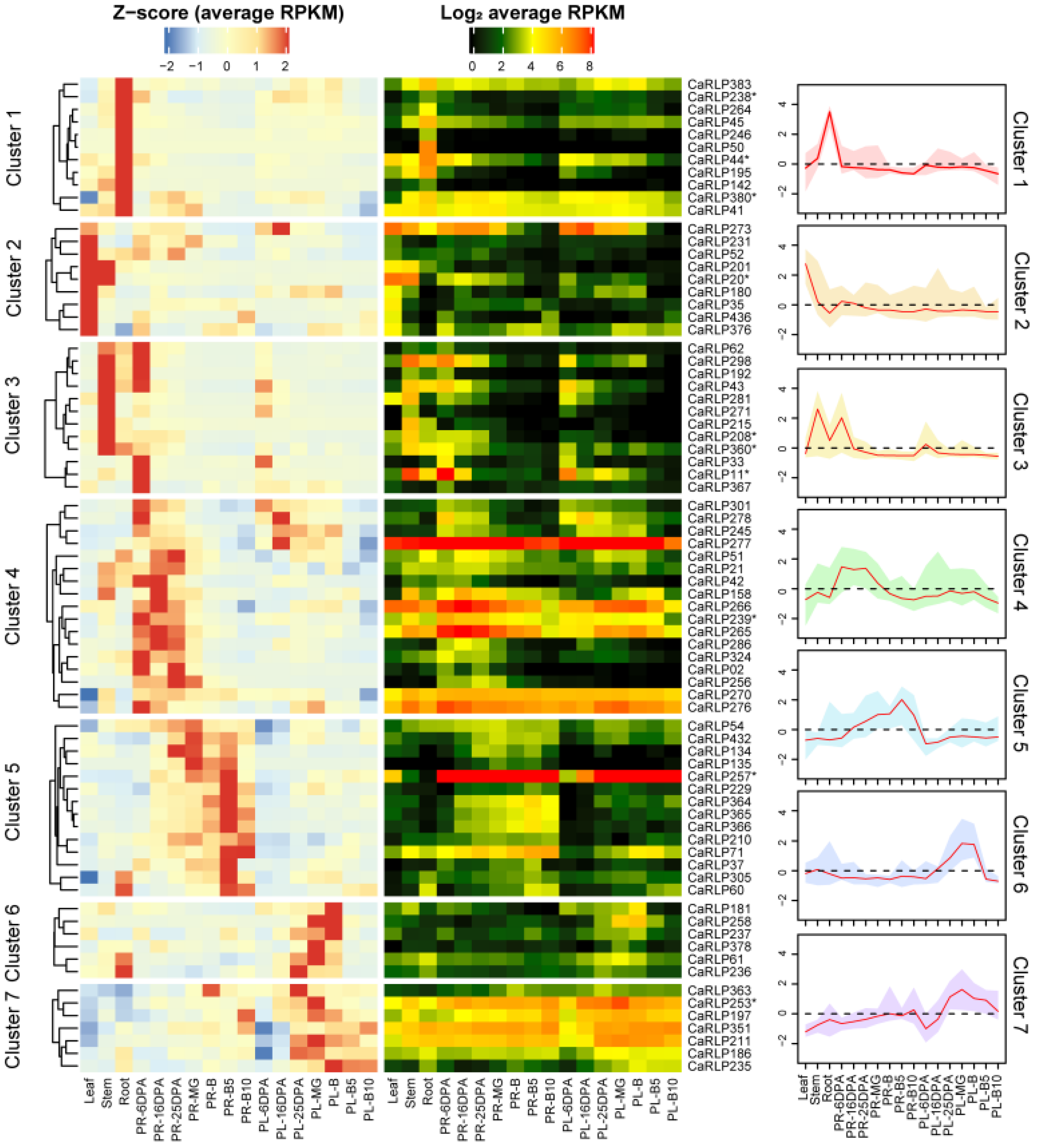
2.1. CaRLP Gene Expression Analysis in Various Organs and across Fruit Developmental Stages

To discover the biological function of CaRLPs in plant growth and development, we obtained the CaRLPs information and tissue-specific RNA-seq data from a previous study [26,27]. Expression analysis of a total of 438 CaRLPs was performed using RNA-seq datasets from the different pepper tissues (root, stem, leaf, pericarp, and placenta), and seven crucial fruit developmental stages. A total of 101 (23.1%) genes out of 438 pepper RLPs were not expressed in any of the five tissues and developmental stages in pepper and were excluded for expression analysis (Supplementary Table S1). We selected CaRLPs with RPKM values of > 5 of transcripts at least in one of all five tissues and developmental stages, and then eventually obtained 76 CaRLPs for further analysis. The expression values of 76 CaRLPs were visualized with Z-score and log2RPKM on a heat map (Figure 1). According to the Z-score and RPKM value, 76 CaRLP genes showed various expression patterns, with some CaRLPs evenly expressed in all tested tissues and some genes specifically expressed in only one or a few organs.
Hierarchical cluster analysis and Z-scores were used to understand the characteristics of 76 CaRLPs. The 76 CaRLPs were clustered into seven expression profiles along with a representative curve displaying each transcript pattern of Z-score (Figure 1). The transcript levels of CaRLPs detected in clusters 1, 2, and 3 were tissue-specific in particular vegetative tissues. Relatively high abundant transcripts in the root, leaf, and stem of CaRLPs clusters were detected compared to other tissues. To determine whether RLP gene members are involved in potential functions during pepper fruit development, we profiled transcripts of pepper RLP genes in the placenta and pericarp at seven crucial stages of fruit development. The CaRLPs in clusters 4 and 5 reached the highest level in early and late pericarp developmental stage, respectively. The 17 CaRLPs in cluster 4 and 14 CaRLPs in cluster 5 may be involved in the placenta developmental stage. In addition, the CaRLPs in clusters 6 and 7 were more highly expressed during the placenta developmental stage than in other tissues, suggesting they may play a role at the reproductive stage. Taken together, these results suggest that CaRLPs might be tissue-specific regulators in vegetative and reproductive tissues in pepper. To validate that the putative functions of CaRLPs expressed in pepper are tissue specific, we analyzed 76 CaRLPs sequences to perform loss-of-function analysis using virus-induced gene silencing (VIGS). Finally, we selected 10 CaRLPs carrying a specific nucleotide sequence in the genome for VIGS study. The information of selected CaRLPs is presented in Table 1.

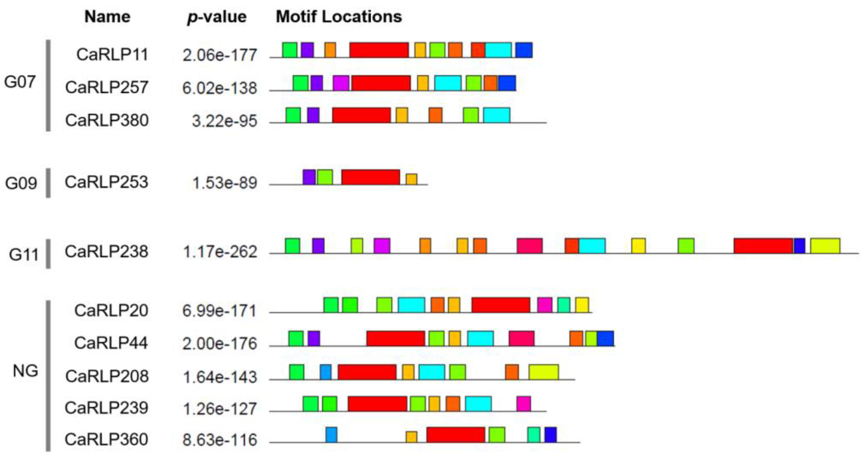
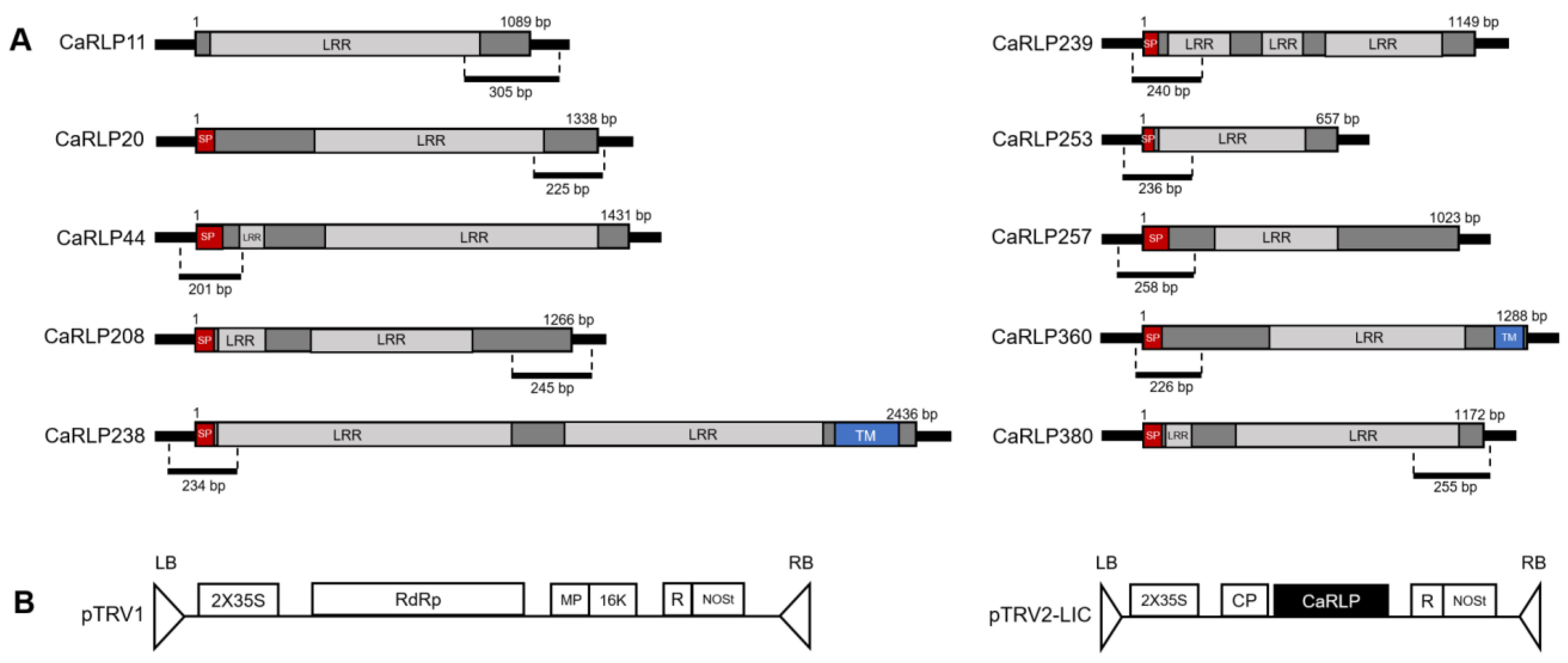
2.2. Structure Analysis of Selected CaRLPs

In a previous study, 10 selected CaRLPs were assigned to different pepper RLP groups (Table 1) [26]. Five of the genes were designated as the none-group, three genes constituted Group 07 (G07), one gene was assigned to Group 09 (G09), and one gene was put in Group 11 (G11), which contained the characterized RLP called CLAVATA2 (CLV2). To determine the structure similarities of RLPs, we conducted conserved motifs analyses with 10 CaRLPs. The motif analyses revealed that the 10 CaRLPs had various motif formations according to their group (Figure 2). As predicted, CaRLP11, 257, and 380 in G07 had similar motifs to one other, and there was low similarity of motif composition in the non-group to which five genes belonged.
Based on these motif analyses, the amino acid sequences of 10 CaRLPs were analyzed with the SMART program (http://smart.embl-heidelberg.de/, accessed on 25 March 2021) to predict putative domains and RLP structure. RLPs are cell surface receptors with an extracellular ligand binding domain that is composed of several distinct domains such as a signal peptide, a transmembrane domain, and an extracellular leucine-rich repeat region (LRR) [28]. All 10 CaRLPs contained LRR domains (Figure 3a). Only two CaRLPs (CaRLP238 and 360) had a full-type structure with three functional domains, such as signal peptide, transmembrane domains, and LRRs. Seven genes contained the two domains including signal peptide and LRRs. The remaining one gene (CaRLP11) had only a single domain with LRRs (partial-type RLP). Taken together, the selected CaRLP genes displayed similarity of motifs within the same groups and showed typical structures of RLPs.

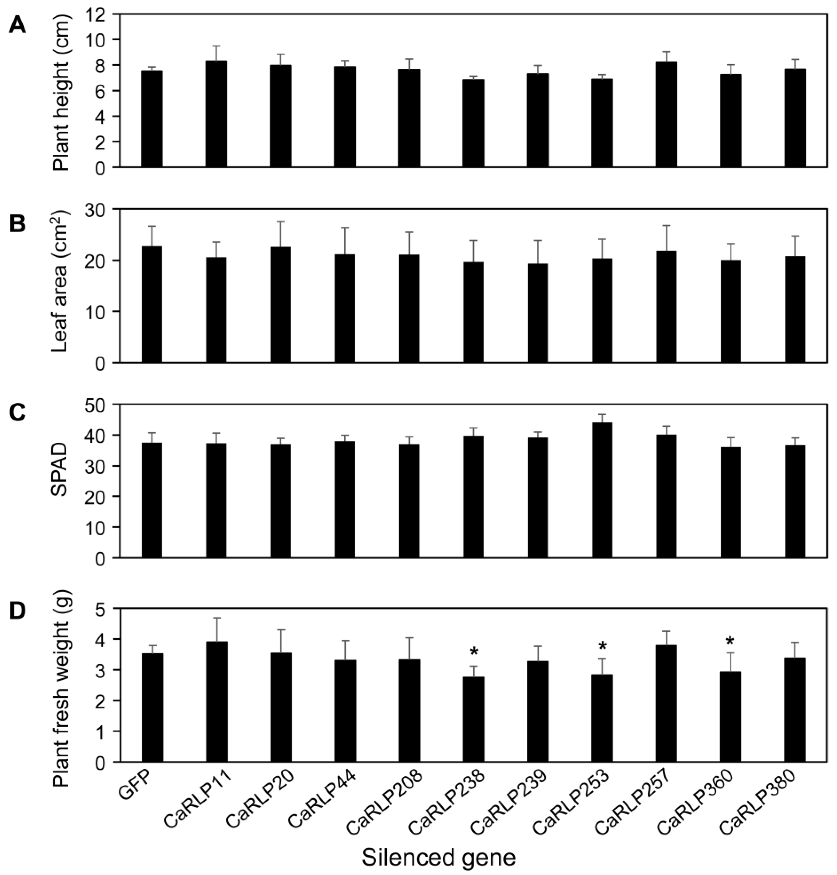
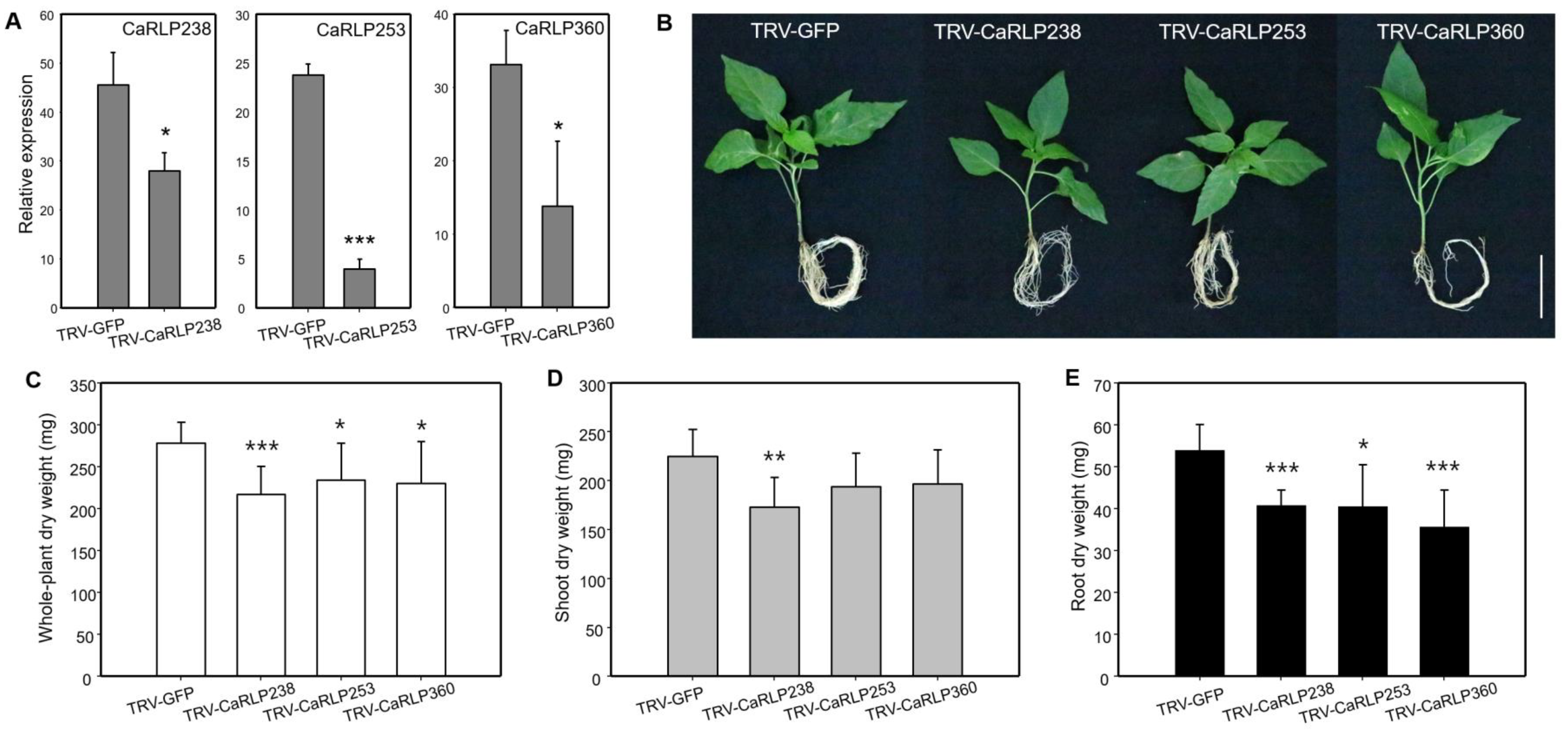
2.3. Phenotypic Alterations in Root Growth of CaRLP-Silenced Plants

To investigate the possible functions of CaRLPs in the development of peppers, 10 CaRLP genes were silenced using TRV-mediated VIGS (Figure 3b). Approximately, 200–300 bp cDNA fragments carrying the gene-specific sequences of each CaRLP gene were inserted in the TRV2 vector for the loss-of-function study (Figure 3a,b). We monitored plant growth and morphological changes, including plant height, chlorophyll contents, leaf area, and plant fresh weight, for six weeks after silencing. Even though silenced plants exhibited slight differences in the aforementioned phenotypes compared to control plants (TRV-GFP), eventually there were no significant differences in plant height, leaf area, and chlorophyll contents in CaRLP-silenced plants compare to controls (Figure 4a–c). However, plant fresh weight of three CaRLP-silenced plants (CaRLP238, CaRLP253, and CaRLP360) were significantly reduced compared to that of control plants. The level of fresh weight in these three CaRLP-silenced plants was decreased 0.7–0.85-fold compared with control plants (Figure 4d). These results indicate that the three CaRLP genes may be involved in plant biological processes in peppers.
To further understand a possible function, we observed the silencing effects of three CaRLPs (CaRLP238, CaRLP253, and CaRLP360) in the growth of shoots and roots. The expression level of each of the three CaRLPs, determined using semi-quantitative RT-PCR, was significantly lower in CaRLP-silenced plants than in the TRV2-GFP control plants (Figure 5a). Slight phenotypic alterations among the three CaRLP-silenced plants and TRV-GFP plants were observed. (Figure 5b). Even though significant differences were not detected in the plant height of CaRLP-silenced plants compared with control plants (Figure 3a), the growth of plant shoots in CaRLP-silenced plants was smaller than that of control plants (Figure 5b). The root phenotype of three CaRLP-silenced plants showed inhibition of root growth compared with control plants. In addition, we measured the dry weight of the whole plant, shoot, and root in three CaRLP-silenced plants and control plants. The whole-plant dry weight was significantly reduced in all three CaRLP-silenced plants compared to controls (Figure 5c). Interestingly, only CaRLP238-silenced plants had significantly decreased shoot dry weight compared with controls (Figure 5d). Root dry weights of all three CaRLP-silenced plants were significantly reduced compared to controls. Taken together, suppression of three genes resulted in retardation of shoot and/or root growth, suggesting that these genes may be involved in regulation of plant normal growth.

3. Discussion

The RLP gene family is one of the largest super families involved in various biological functions in plants, yet only a few RLP genes have been functionally characterized regarding biological processes. The RNA-seq has been widely applied to research including the analysis of gene expression and prediction of gene function since next-generation sequencing has been easily available to use. Several studies have reported the genome-wide identification of RLP genes in Arabidopsis, rice, tomato, Brassica, and legumes [24,29,30,31,32,33]. However, no previous study has reported the genome-wide transcriptional profiles in organ-specific of RLPs in pepper. Previously, we identified 438 pepper RLP genes and a comparative transcriptomic analysis revealed the role of core RLP regulators in disease resistance against various pathogens [26]. In the study reported here, we attempted to discover the additional function involved in plant growth by analyzing pepper tissue-specific transcriptome. A total of 76 CaRLPs had variable expression patterns whereby some were expressed evenly in all stages tested or specifically in one or a few stages (Supplementary Table S1). The hierarchical clustering of 76 CaRLPs was detected via categorization into seven clusters showing distinct tissue-specific expression patterns. Among them, ten CaRLP genes having specific sequences and distinguishable expressions were selected for further analysis.
Motif analysis of 10 CaRLP proteins revealed that CaRLPs had 20 motifs in total and shared conserved motifs in the same subgroup, which is consistent with a previous study [26]. The distribution of motifs also showed the conserved position of each gene in the same group (Figure 2). This result suggested that the genes in the same group might have similar evolutionary histories and possibly a similar function [34,35]. RLPs are pattern recognition receptors containing N-terminal signal peptides, transmembrane domains, and LRR [28]. However, some proteins without transmembrane domains or without signal peptides are considered as RLPs [29,31]. A previous study reported RLPs without a transmembrane domain attached to the extracellular side of the plasma membrane using a glycosylphosphatidylinositol (GPI) anchor [36]. In the case of the tomato, structure analysis of tomato RLPs show that 30.7% of genes of all tomato RLPs are considered full-type RLPs encompassing all three domains and 42.6% of genes contain two domains [30]. Therefore, even though some CaRLPs were predicted as partial-type RLPs (not having all three domains) in this study, they could be considered as a typical RLP gene.
Most biological processing genes are under purifying selection to maintain specific functions [37]. Therefore, developmental genes are less likely to be duplicated compared to gene families for stress adaptation, like resistance genes, and they are more structurally and functionally conserved among plant species. Moreover, analysis of Arabidopsis and rice RLP genes shows that developmental genes are less likely to be duplicated and undergo diversifying selection than disease-resistance genes [24]. For instance, CLV2 is involved in plant growth and development processes, including shoot meristem development, root apical meristem maintenance, and organ development [2,19]. The FASCIATED EAR2 (FEA2), an orthologue of CLV2 involved in meristem development, shows a similar mutant phenotype to CLV2 [25]. Previously, a small number of CaRLP genes were classified with TMM and CLV2 as Group 10 and 11, respectively. The CaRLPs in Group 10 and 11 were predicted to duplicate before tomato/pepper speciation, suggesting evolutionary pressure to conserve function [26]. Interestingly, CaRLP238, which shows most phenotypic changes in both root and shoot of silenced pepper plants in this study, was grouped with CLV2 in Group 10 (Table 1) based on a phylogenetic tree of sequence similarity. According to the aforementioned results, it can be predicted that genes belonging to groups like CLV2 and TMM could possibly function in relation to plant development. Therefore, functional analysis of other CaRLPs belonging to the same group as CLV2 and TMM, through further research, might provide clues to find biological roles in plant biological processes for the RLP gene family.
To understand their role in plant growth, 10 selected genes were silenced in pepper plants, and then three CaRLP genes (CaRLP238, CaRLP253, and CaRLP360) were characterized further. CaRLP238, highly expressed in the root (Figure 1), displayed morphological changes in growth after silencing such as reduced dry weight of whole plant, shoot, and root. Additionally, CaRLP253 and 360 displayed high expression levels in placenta and stem, respectively, but they showed significant differences in root dry weight when silenced. For these three CaRLP genes, although additional phenotypic analysis of other tissues was not performed, these CaRLPs might be involved in growth processes in peppers. However, in order to unveil the function, further studies including more morphological analysis and molecular biology analysis during growth and fruit development will be needed.
Although the expression level of silenced genes was significantly lower than control plants, one gene, CaRLP238, may not be down-regulated enough to compromise CaRLP function (Figure 5a). A VIGS assay could be a very useful tool for assessing gene functions and this tool has been developed in a wide variety of plant species [38]. However, VIGS has weaknesses, including partial silencing, that require consideration and prudence in its use. Partial silencing happens in an unpredictable manner. Therefore, less obvious phenotypes could impair the use of VIGS in the analysis. This problem can be addressed by increasing the sample size to include enough individuals [39,40]. In this study, we performed more than three independent assays for the VIGS that consisted of at least eight to ten plants for a replicate, totally more than thirty plants. In the case of silencing reduction, even though the silencing efficiency of CaRLP238 genes was approximately 40% lower compared to the control, we achieved the same stable results and phenotypic alteration-like significant differences in whole plant weights including both shoot and root compared to controls. In addition, a previous report showed that using VIGS, even target genes were reduced between 65 and 24% (p < 0.05) compared to WT plants and the silenced plants also exhibited clear phenotypic alterations [41]. Taken together, three CaRLPs (CaRLP238, 253, and 360) showed acceptable and reasonable ranges in expression reduction to phenotypic changes.

4. Materials and Methods

4.1. CaRLP Information, RNA-Seq Data, and Expression Profiling

We obtained gene information and sequences of CaRLP genes from a previous study [26]. The RNA-seq of five pepper tissues, including root, stem, leaf, pericarp, and placenta [42], were used for the expression analysis of CaRLPs. The pericarp and placenta RNA-seq contained seven developmental stages: 6, 16, and 25 days post anthesis; mature green (MG); breaker (B); and 5 and 10 days post breaker. All transcriptome data were analyzed using in-house pipelines [43,44]. The expression values of tissues were normalized using RPKM. To analyze expression profiles of CaRLPs, CaRLPs with RPKM values of >5 at least in one of all five tissues and developmental stages selected, and then, 76 CaRLPs were obtained. The expression patterns and hierarchical clustering of significant genes were visualized with Z-score and log2 RPKM using Heatmap by R package (http://bioconductor.org/, accessed on 25 March 2021). Of 76 CaRLPs, we selected 10 CaRLPs possessing a gene-specific sequence for further analysis.

4.2. Motif Analysis and Structure Analysis of CaRLP Genes

Identification of conserved motifs in 10 selected CaRLPs was performed using the MEME suite (http://meme-suite.org/tools/meme, accessed on 25 March 2021). The analysis parameters were as follows: maximum number of motifs, 20; minimum width of motifs, 15; maximum width of motifs, 200, and other details were used with default settings. To find functional domains of RLPs, 10 selected CaRLPs were analyzed using functional structure annotation with SMART (http://smart.embl-heidelberg.de/, accessed on 25 March 2021) with default values.

4.3. Gene Cloning for Virus-Induced Gene Silencing (VIGS) Assay

Capsicum annuum (‘Nockwang’) was used in this study for functional analysis. Seedlings were grown in a growth chamber at 25 °C under a 16 h light/8 h darkness cycle. The newly emerging cotyledons of germinating pepper seedlings were used for a VIGS assay.
To construct the plasmids for analysis of loss-of-function, Tobacco rattle virus (TRV)-based VIGS vectors containing pTRV1 and pTRV2-LIC were used. To find gene-specific sequences of CaRLPs for VIGS, 10 CaRLPs performed BLASTn against the C. annuum ‘CM334’ v. 1.55 genome. The gene-specific region, which is the 3′ or 5′ CDS region with untranslated region (UTR), of each CaRLP was amplified using SolgTM Pfu-X DNA polymerase (Solgent, Daejeon, Korea) with primers (Supplementary Table S2). The PCR products were cloned into the pTRV2-LIC vector according to a method used in a previous study [45,46]. The pTRV2-LIC-CaRLP plasmids were transformed into Agrobacterium tumefaciens strain GV3101 using the freeze-thaw method. Agrobacterium cultures, including pTRV1 and pTRV2-LIC-CaRLP, were mixed in a 1:1 ratio (OD600 = 0.5) and infiltrated into two fully expanded cotyledons of pepper seedlings (approximately 10 days after germination) using a needleless 1-mL syringe. In this experiment, TRV2-green fluorescent protein (GFP) and TRV2- phytoene desaturase (PDS) were used as a negative and positive control for VIGS, respectively.

4.4. Semi-Quantitative RT-PCR Analysis

The total RNA was extracted from pepper leaves using the TRIzol reagent (Thermo Fisher Scientific, Inc., Waltham, MA, USA) according to the manufacturer’s instructions. cDNA was synthesized from 2 µg of total RNA using oligo d(T) primer and M-MLV reverse transcriptase (Promega, Madison, WI, USA) following the manufacturer’s protocol. Each gene-specific primer was designed to detect the levels of silenced-CaRLP gene expression. For semi-quantitative RT-PCR reaction, we tested the RT-PCR cycles between 26–32 cycles with all samples to ensure the linearity of the results. Then, to determine efficiency of gene silencing, semi-quantitative RT-PCR was performed using rTaq polymerase (Takara, Shiga, Japan) on a MiniAmp™ Thermal Cycler (Thermo Fisher Scientific Inc., Waltham, MA, USA) under the following conditions: initial denaturation step was 94 °C for 2 min, followed by 30 cycles of 94 °C for 30 s, then an annealing step at 58 °C for 30 s, and an elongation step at 72 °C for 30 s. The pepper actin gene (CaActin) [47] was used as reference gene. All PCR reactions were repeated in triplicate using at least three independent samples. To determine quantification of RT-PCR products, images of the amplicon in ethidium bromide-stained agarose gel were obtained using WiseCaptureII software (Daihan Scientific Co., Ltd., Wonju, Korea). Quantification of the amplicon bands in agarose gel was measured using ImageJ software (https://imagej.nih.gov/ij/, accessed on 25 March 2021). Band intensity of CaActin of each cDNA sample was calculated to normalize for variation in sample concentration. Mean and standard deviation of each CaRLP expression level in silenced pepper plants and mock plants (TRV-GFP) were calculated after normalization to CaActin. Statistically significant differences (p < 0.05) of CaRLP expression levels in CaRLP-silenced plants were verified by comparison with those of the control plant (TRV-GFP). It was determined using one-way analysis of variance (ANOVA) and Student’s t-test using SAS software (version 9.4; SAS Inc., Cary, NC, USA).

4.5. Phenotypic Analysis of CaRLP-Silenced Pepper Plants

The morphological changes of each CaRLP-silenced pepper plant were measured, such as plant height (from soil surface to shoot growing point, cm), leaf area (3rd true leaf, cm2), plant fresh weight of whole plant (mg), and chlorophyll contents (SPAD value) at 6 weeks after silencing. Relative chlorophyll contents of the 3rd and 4th leaves of CaRLP-silenced plants were measured with a Chlorophyll Meter (Minolta Camera Co., Ltd., Japan). The silenced plants at 6 weeks after a VIGS assay were dried at 65 °C in an oven for two days, and then, dry weights of whole plants, shoot, and root were measured. All phenotypic analyses were performed using more than three independent experiments, with 8–10 plants for each experiment. Statistically significant differences (p < 0.05) of phenotypic analysis between control plants (TRV-GFP) and CaRLP-silenced plants (TRV-CaRLP) were determined using one-way ANOVA and Student’s t-test using SAS software (version 9.4; SAS Inc., Cary, NC, USA).

Supplementary Materials

The following are available online at https://www.mdpi.com/article/10.3390/plants10050972/s1, Table S1: Reads per kilobase of transcript per million mapped reads (RPKM) from tissue-specific RNA-seq of pepper RLP genes, Table S2: Primers used for VIGS (virus-induced gene silencing) analysis.

Author Contributions

W.-H.K., B.P. collected samples and performed experiments. J.L. generated RNA-seq data. W.-H.K., S.-I.Y. conceived and designed the experiments and organized and wrote the manuscript. S.-I.Y. supervised the project. All authors have read and agreed to the published version of the manuscript.

Funding

This research was supported by the National Research Foundation of Korea (NRF) and funded by the Korean Government (NRF-2015R1A6A1A03031413, 2017R1E1A1A01072843 and 2019R1C1C1007472).

Institutional Review Board Statement

Not applicable.

Informed Consent Statement

Not applicable.

Data Availability Statement

The data supporting the findings of this study are available within the article and its supplementary materials.

Conflicts of Interest

The authors declare no conflict of interest. The funders had no role in the design of the study; in the collection, analyses, or interpretation of data; in the writing of the manuscript, or in the decision to publish the results.

References

  1. He, Y.; Zhou, J.; Shan, L.; Meng, X. Plant cell surface receptor-mediated signaling—A common theme amid diversity. J. Cell Sci. 2018, 131, jcs209353. [Google Scholar] [CrossRef] [Green Version]
  2. Pan, L.; Lv, S.; Yang, N.; Lv, Y.; Liu, Z.; Wu, J.; Wang, G. The multifunction of CLAVATA2 in plant development and immunity. Front. Plant Sci. 2016, 7, 1573. [Google Scholar] [CrossRef] [Green Version]
  3. Bohm, H.; Albert, I.; Fan, L.; Reinhard, A.; Nurnberger, T. Immune receptor complexes at the plant cell surface. Curr. Opin. Plant Biol. 2014, 20, 47–54. [Google Scholar] [CrossRef] [PubMed]
  4. Jones, D.A.; Thomas, C.M.; Hammond-Kosack, K.E.; Balint-Kurti, P.J.; Jones, J.D. Isolation of the tomato Cf-9 gene for resistance to Cladosporium fulvum by transposon tagging. Science 1994, 266, 789–793. [Google Scholar] [CrossRef]
  5. Dixon, M.S.; Jones, D.A.; Keddie, J.S.; Thomas, C.M.; Harrison, K.; Jones, J.D. The tomato Cf-2 disease resistance locus comprises two functional genes encoding leucine-rich repeat proteins. Cell 1996, 84, 451–459. [Google Scholar] [CrossRef] [Green Version]
  6. Dixon, M.S.; Hatzixanthis, K.; Jones, D.A.; Harrison, K.; Jones, J.D. The tomato Cf-5 disease resistance gene and Six homologs show pronounced allelic variation in leucine-rich repeat copy number. Plant Cell 1998, 10, 1915–1925. [Google Scholar] [CrossRef] [Green Version]
  7. Thomas, C.M.; Jones, D.A.; Parniske, M.; Harrison, K.; Balint-Kurti, P.J.; Hatzixanthis, K.; Jones, J.D. Characterization of the tomato Cf-4 gene for resistance to Cladosporium fulvum identifies sequences that determine recognitional specificity in Cf-4 and Cf-9. Plant Cell 1997, 9, 2209–2224. [Google Scholar] [CrossRef] [Green Version]
  8. Takken, F.L.; Thomas, C.M.; Joosten, M.H.; Golstein, C.; Westerink, N.; Hille, J.; Nijkamp, H.J.; De Wit, P.J.; Jones, J.D. A second gene at the tomato Cf-4 locus confers resistance to cladosporium fulvum through recognition of a novel avirulence determinant. Plant J. 1999, 20, 279–288. [Google Scholar] [CrossRef]
  9. Ron, M.; Avni, A. The receptor for the fungal elicitor ethylene-inducing xylanase is a member of a resistance-like gene family in tomato. Plant Cell 2004, 16, 1604–1615. [Google Scholar] [CrossRef] [Green Version]
  10. Kawchuk, L.M.; Hachey, J.; Lynch, D.R.; Kulcsar, F.; van Rooijen, G.; Waterer, D.R.; Robertson, A.; Kokko, E.; Byers, R.; Howard, R.J.; et al. Tomato Ve disease resistance genes encode cell surface-like receptors. Proc. Natl. Acad. Sci. USA 2001, 98, 6511–6515. [Google Scholar] [CrossRef] [PubMed] [Green Version]
  11. Hegenauer, V.; Furst, U.; Kaiser, B.; Smoker, M.; Zipfel, C.; Felix, G.; Stahl, M.; Albert, M. Detection of the plant parasite Cuscuta reflexa by a tomato cell surface receptor. Science 2016, 353, 478–481. [Google Scholar] [CrossRef] [PubMed]
  12. Jehle, A.K.; Lipschis, M.; Albert, M.; Fallahzadeh-Mamaghani, V.; Furst, U.; Mueller, K.; Felix, G. The receptor-like protein ReMAX of Arabidopsis detects the microbe-associated molecular pattern eMax from Xanthomonas. Plant Cell 2013, 25, 2330–2340. [Google Scholar] [CrossRef] [PubMed] [Green Version]
  13. Shen, Y.; Diener, A.C. Arabidopsis thaliana resistance to fusarium oxysporum 2 implicates tyrosine-sulfated peptide signaling in susceptibility and resistance to root infection. PLoS Genet. 2013, 9, e1003525. [Google Scholar] [CrossRef] [PubMed] [Green Version]
  14. Albert, I.; Bohm, H.; Albert, M.; Feiler, C.E.; Imkampe, J.; Wallmeroth, N.; Brancato, C.; Raaymakers, T.M.; Oome, S.; Zhang, H.Q.; et al. An RLP23-SOBIR1-BAK1 complex mediates NLP-triggered immunity. Nat. Plants 2015, 1, 15140. [Google Scholar] [CrossRef] [PubMed]
  15. Larkan, N.J.; Lydiate, D.J.; Parkin, I.A.; Nelson, M.N.; Epp, D.J.; Cowling, W.A.; Rimmer, S.R.; Borhan, M.H. The Brassica napus blackleg resistance gene LepR3 encodes a receptor-like protein triggered by the Leptosphaeria maculans effector AVRLM1. New Phytol. 2013, 197, 595–605. [Google Scholar] [CrossRef]
  16. Vinatzer, B.A.; Patocchi, A.; Gianfranceschi, L.; Tartarini, S.; Zhang, H.B.; Gessler, C.; Sansavini, S. Apple contains receptor-like genes homologous to the Cladosporium fulvum resistance gene family of tomato with a cluster of genes cosegregating with Vf apple scab resistance. Mol. Plant Microbe Interact. 2001, 14, 508–515. [Google Scholar] [CrossRef] [Green Version]
  17. Du, J.; Verzaux, E.; Chaparro-Garcia, A.; Bijsterbosch, G.; Keizer, L.C.P.; Zhou, J.; Liebrand, T.W.H.; Xie, C.H.; Govers, F.; Robatzek, S.; et al. Elicitin recognition confers enhanced resistance to Phytophthora infestans in potato. Nat. Plants 2015, 1, 15034. [Google Scholar] [CrossRef] [PubMed]
  18. Nadeau, J.A.; Sack, F.D. Control of stomatal distribution on the Arabidopsis leaf surface. Science 2002, 296, 1697–1700. [Google Scholar] [CrossRef]
  19. Jeong, S.; Trotochaud, A.E.; Clark, S.E. The Arabidopsis CLAVATA2 gene encodes a receptor-like protein required for the stability of the CLAVATA1 receptor-like kinase. Plant Cell 1999, 11, 1925–1934. [Google Scholar] [CrossRef] [Green Version]
  20. Fiers, M.; Golemiec, E.; Xu, J.; van der Geest, L.; Heidstra, R.; Stiekema, W.; Liu, C.M. The 14-amino acid CLV3, CLE19, and CLE40 peptides trigger consumption of the root meristem in Arabidopsis through a CLAVATA2-dependent pathway. Plant Cell 2005, 17, 2542–2553. [Google Scholar] [CrossRef] [Green Version]
  21. Fiers, M.; Hause, G.; Boutilier, K.; Casamitjana-Martinez, E.; Weijers, D.; Offringa, R.; van der Geest, L.; van Lookeren Campagne, M.; Liu, C.M. Mis-expression of the CLV3/ESR-like gene CLE19 in Arabidopsis leads to a consumption of root meristem. Gene 2004, 327, 37–49. [Google Scholar] [CrossRef]
  22. Strabala, T.J.; O’Donnell, P.J.; Smit, A.M.; Ampomah-Dwamena, C.; Martin, E.J.; Netzler, N.; Nieuwenhuizen, N.J.; Quinn, B.D.; Foote, H.C.; Hudson, K.R. Gain-of-function phenotypes of many CLAVATA3/ESR genes, including four new family members, correlate with tandem variations in the conserved CLAVATA3/ESR domain. Plant Physiol. 2006, 140, 1331–1344. [Google Scholar] [CrossRef] [Green Version]
  23. Meng, L.; Feldman, L.J. CLE14/CLE20 peptides may interact with CLAVATA2/CORYNE receptor-like kinases to irreversibly inhibit cell division in the root meristem of Arabidopsis. Planta 2010, 232, 1061–1074. [Google Scholar] [CrossRef] [PubMed] [Green Version]
  24. Wang, G.D.; Ellendorff, U.; Kemp, B.; Mansfield, J.W.; Forsyth, A.; Mitchell, K.; Bastas, K.; Liu, C.M.; Woods-Tor, A.; Zipfel, C.; et al. A genome-wide functional investigation into the roles of receptor-like proteins in Arabidopsis. Plant Physiol. 2008, 147, 503–517. [Google Scholar] [CrossRef] [PubMed] [Green Version]
  25. Taguchi-Shiobara, F.; Yuan, Z.; Hake, S.; Jackson, D. The fasciated ear2 gene encodes a leucine-rich repeat receptor-like protein that regulates shoot meristem proliferation in maize. Genes Dev. 2001, 15, 2755–2766. [Google Scholar] [CrossRef] [PubMed] [Green Version]
  26. Kang, W.H.; Lee, J.; Koo, N.; Kwon, J.S.; Park, B.; Kim, Y.M.; Yeom, S.I. Universal gene co-expression network reveals receptor-like protein genes conferring broad-spectrum resistance in pepper (Capsicum annuum L.). bioRxiv 2021. [Google Scholar] [CrossRef]
  27. Kang, W.H.; Kim, S.; Lee, H.A.; Choi, D.; Yeom, S.I. Genome-wide analysis of Dof transcription factors reveals functional characteristics during development and response to biotic stresses in pepper. Sci. Rep. 2016, 6, 33332. [Google Scholar] [CrossRef] [PubMed] [Green Version]
  28. Jamieson, P.A.; Shan, L.B.; He, P. Plant cell surface molecular cypher: Receptor-like proteins and their roles in immunity and development. Plant Sci. 2018, 274, 242–251. [Google Scholar] [CrossRef] [PubMed]
  29. Fritz-Laylin, L.K.; Krishnamurthy, N.; Tor, M.; Sjolander, K.V.; Jones, J.D. Phylogenomic analysis of the receptor-like proteins of rice and Arabidopsis. Plant Physiol. 2005, 138, 611–623. [Google Scholar] [CrossRef] [PubMed] [Green Version]
  30. Kang, W.H.; Yeom, S.I. Genome-wide identification, classification, and expression analysis of the receptor-like protein family in tomato. Plant Pathol. J. 2018, 34, 435–444. [Google Scholar] [CrossRef] [PubMed]
  31. Petre, B.; Hacquard, S.; Duplessis, S.; Rouhier, N. Genome analysis of poplar LRR-RLP gene clusters reveals RISP, a defense-related gene coding a candidate endogenous peptide elicitor. Front. Plant Sci. 2014, 5, 111. [Google Scholar] [CrossRef] [PubMed] [Green Version]
  32. Restrepo-Montoya, D.; Brueggeman, R.; McClean, P.E.; Osorno, J.M. Computational identification of receptor-like kinases “RLK” and receptor-like proteins “RLP” in legumes. BMC Genom. 2020, 21, 459. [Google Scholar] [CrossRef] [PubMed]
  33. Yang, H.; Bayer, P.E.; Tirnaz, S.; Edwards, D.; Batley, J. Genome-wide identification and evolution of receptor-like kinases (RLKs) and receptor like proteins (RLPs) in Brassica juncea. Biology 2020, 10, 17. [Google Scholar] [CrossRef] [PubMed]
  34. Starr, T.N.; Picton, L.K.; Thornton, J.W. Alternative evolutionary histories in the sequence space of an ancient protein. Nature 2017, 549, 409–413. [Google Scholar] [CrossRef] [PubMed]
  35. Gabaldon, T.; Koonin, E.V. Functional and evolutionary implications of gene orthology. Nat. Rev. Genet. 2013, 14, 360–366. [Google Scholar] [CrossRef] [PubMed] [Green Version]
  36. Gong, B.Q.; Xue, J.; Zhang, N.N.; Xu, L.H.; Yao, X.R.; Yang, Q.J.; Yu, Y.; Wang, H.B.; Zhang, D.D.; Li, J.F. Rice chitin receptor OsCEBiP is not a transmembrane protein but targets the plasma membrane via a GPI Anchor. Mol. Plant 2017, 10, 767–770. [Google Scholar] [CrossRef]
  37. Kellogg, E.A. Evolution of developmental traits. Curr. Opin. Plant Biol. 2004, 7, 92–98. [Google Scholar] [CrossRef]
  38. Burch-Smith, T.M.; Anderson, J.C.; Martin, G.B.; Dinesh-Kumar, S.P. Applications and advantages of virus-induced gene silencing for gene function studies in plants. Plant J. 2004, 39, 734–746. [Google Scholar] [CrossRef]
  39. Lange, M.; Yellina, A.L.; Orashakova, S.; Becker, A. Virus-induced gene silencing (VIGS) in plants: An overview of target species and the virus-derived vector systems. Methods Mol. Biol. 2013, 975, 1–14. [Google Scholar] [CrossRef]
  40. Wege, S.; Scholz, A.; Gleissberg, S.; Becker, A. Highly efficient virus-induced gene silencing (VIGS) in California poppy (Eschscholzia californica): An evaluation of VIGS as a strategy to obtain functional data from non-model plants. Ann. Bot. 2007, 100, 641–649. [Google Scholar] [CrossRef] [Green Version]
  41. Raz, A.; Dahan-Meir, T.; Melamed-Bessudo, C.; Leshkowitz, D.; Levy, A.A. Redistribution of meiotic crossovers along wheat chromosomes by virus-induced gene silencing. Front. Plant Sci. 2020, 11, 635139. [Google Scholar] [CrossRef]
  42. Kim, S.; Park, M.; Yeom, S.I.; Kim, Y.M.; Lee, J.M.; Lee, H.A.; Seo, E.; Choi, J.; Cheong, K.; Kim, K.T.; et al. Genome sequence of the hot pepper provides insights into the evolution of pungency in Capsicum species. Nat. Genet. 2014, 46, 270–278. [Google Scholar] [CrossRef] [PubMed]
  43. Lee, J.; Nam, J.Y.; Jang, H.; Kim, N.; Kim, Y.M.; Kang, W.H.; Yeom, S.I. Comprehensive transcriptome resource for response to phytohormone-induced signaling in Capsicum annuum L. BMC Res. Notes 2020, 13, 40. [Google Scholar] [CrossRef]
  44. Kang, W.H.; Sim, Y.M.; Koo, N.; Nam, J.Y.; Lee, J.; Kim, N.; Jang, H.; Kim, Y.M.; Yeom, S.I. Transcriptome profiling of abiotic responses to heat, cold, salt, and osmotic stress of Capsicum annuum L. Sci. Data 2020, 7, 17. [Google Scholar] [CrossRef] [PubMed] [Green Version]
  45. Kang, W.H.; Lee, J.; Nam, J.Y.; Yeom, S.I. Identification of CaLOP regulating development and growth through virus-induced gene silencing in pepper. Hortic. Sci. Technol. 2018, 36, 292–301. [Google Scholar] [CrossRef]
  46. Dong, Y.; Burch-Smith, T.M.; Liu, Y.; Mamillapalli, P.; Dinesh-Kumar, S.P. A ligation-independent cloning tobacco rattle virus vector for high-throughput virus-induced gene silencing identifies Roles for NbMADS4-1 and -2 in floral development. Plant Physiol. 2007, 145, 1161–1170. [Google Scholar] [CrossRef] [Green Version]
  47. Kwon, J.S.; Nam, J.Y.; Yeom, S.I.; Kang, W.H. Leaf-to-whole plant spread bioassay for pepper and Ralstonia solanacearum interaction determines inheritance of resistance to bacterial wilt for further breeding. Int. J. Mol. Sci. 2021, 22, 2279. [Google Scholar] [CrossRef] [PubMed]
Figure 1. Tissue-specific expression patterns of CaRLPs (Capsicum annuum receptor-like proteins). The 76 RLP genes out of 438 genes were selected to construct the heat map. Heatmaps on left and right sides are exhibited using the Z-score and RPKM (reads per kilobase of transcript per million mapped reads), respectively. The selected 10 genes for VIGS (virus-induced gene silencing) assay were marked with asterisks in the right side of the heatmap. The graphs represent the Z-score of CaRLPs in each hierarchical cluster, and red lines indicate median Z-score within a cluster. Key: PR, pericarp; PL, placenta; MG, mature green stage; B, breaker stage; DPA, days post anthesis; B5 and 10, 5 and 10 days post-breaker.
Plants 10 00972 g001
Figure 2. Conserved motif analysis of 10 CaRLPs (Capsicum annuum receptor-like proteins). Conserved motifs of the 10 selected CaRLPs were analyzed using the MEME program. The left side of the gene name represents the CaRLP group to which each CaRLP belongs. Each color box in motif locations displays different motifs.
Plants 10 00972 g002
Figure 3. RLP (receptor-like protein) structure and schematic diagram of silenced region of each CaRLP (Capsicum annuum receptor-like proteins) and TRV (Tobacco rattle virus)-based VIGS (virus-induced gene silencing) vector. (A) RLP structure with the diagram of silencing region of CaRLPs. Partial fragment of the gene used for silencing are shown below full CDS (coding sequence) region of each CaRLP. LRR, leucine rich repeat; SP, signal peptide; TM, transmembrane domain. Dark gray region without domain name in CDS indicates non-LRR region. (B) Map of the TRV-based VIGS vector used in this study. Partial fragments of each CaRLP were cloned into black colored region (named “CaRLP”) of pTRV2-LIC. LB, left borders of the T-DNA; RB, right borders of the T-DNA; 2X35S, two copies of the Cauliflower mosaic virus 35S promoter; CP, coat protein; RdRp, RNA-dependent RNA polymerase; MP, movement protein; 16 K, 16 Kda protein; R, self-cleaving ribozyme; NOSt, nos-terminator.
Plants 10 00972 g003
Figure 4. Phenotype analysis of 10 CaRLP (Capsicum annuum receptor-like proteins)-silenced pepper plants. Comparison of plant height (A), leaf area (B), chlorophyll contents (C), and fresh weight (D) in control plants (GFP; green fluorescent protein) and CaRLP-silenced plants. CaRLP-silenced plants and control plants were used for phenotypic analysis at six weeks after silencing. The plant height was measured from the ground to the growth point of the shoot. The plant fresh weight included shoot and root weight. The leaf area and chlorophyll contents were measured on the third true leaf and third and fourth leaves, respectively. Data are shown as mean ± SD. Statistically significant differences compared with the TRV2-GFP control determined using Student’s t-test are indicated by asterisks (* p < 0.05).
Plants 10 00972 g004
Figure 5. Phenotypic characterization of CaRLP238, 253, and 360-silenced plants. (A) Semi-quantitative reverse transcription-polymerase chain reaction (Semi-quantitative RT-PCR) analyses of the each CaRLP (Capsicum annuum receptor-like proteins) in silenced pepper leaves. TRV-GFP (Tobacco rattle virus-green fluorescent protein)-treated mock plants were used for VIGS as a reference sample. Expression values were normalized to levels of CaActin gene expression. Data represent mean ± SD (Student’s t-test, * p < 0.05, ** p < 0.01, *** p < 0.0001). (B) Phenotype of CaRLP238, 253, and 360-silenced plant and control plants (TRV-GFP). The photo was taken at six weeks after agro-infiltration for a VIGS assay. Scale bar indicates 5 cm. (CE) Comparison of whole-plant dry weight, shoot dry weight, and root dry weight in control plant (TRV-GFP), and CaRLP238, 253, and 360 silenced plants. Data are shown as mean ± SD. Statistically significant differences compared with the TRV2-GFP control determined using Student’s t-test are indicated by asterisks (* p < 0.05, ** p < 0.01, *** p < 0.0001).
Plants 10 00972 g005
Table 1. Information for VIGS (virus-induced gene silencing) analysis of 10 selected CaRLPs (Capsicum annuum receptor-like proteins).
CaRLP IDLocationCDS Size (bp)Protein Size (aa)Group
CaRLP11Ch11089362G07
CaRLP20Ch21338445NG
CaRLP44Ch21431476NG
CaRLP208Ch71266421NG
CaRLP238Ch82436811G11
CaRLP239Ch81149382NG
CaRLP253Ch8657218G09
CaRLP257Ch91023340G07
CaRLP360Ch0 11288428NG
CaRLP380Ch01172382G07
1 Ch0 indicates unmapped scaffolds on chromosome.
Publisher’s Note: MDPI stays neutral with regard to jurisdictional claims in published maps and institutional affiliations.

