Article

Three Novel Biphenanthrene Derivatives and a New Phenylpropanoid Ester from Aerides multiflora and Their α-Glucosidase Inhibitory Activity

by
May Thazin Thant
1,2,
Boonchoo Sritularak
1,3,*,
Nutputsorn Chatsumpun
4,
Wanwimon Mekboonsonglarp
5,
Yanyong Punpreuk
6 and
Kittisak Likhitwitayawuid
1
1
Department of Pharmacognosy and Pharmaceutical Botany, Faculty of Pharmaceutical Sciences, Chulalongkorn University, Bangkok 10330, Thailand
2
Department of Pharmacognosy, University of Pharmacy, Yangon 11031, Myanmar
3
Natural Products for Ageing and Chronic Diseases Research Unit, Faculty of Pharmaceutical Sciences, Chulalongkorn University, Bangkok 10330, Thailand
4
Department of Pharmacognosy, Faculty of Pharmacy, Mahidol University, Bangkok 10400, Thailand
5
Scientific and Technological Research Equipment Centre, Chulalongkorn University, Bangkok 10330, Thailand
6
Department of Agriculture, Kasetsart University, Bangkok 10900, Thailand
*
Author to whom correspondence should be addressed.
Plants 2021, 10(2), 385; https://doi.org/10.3390/plants10020385
Submission received: 22 January 2021 / Revised: 15 February 2021 / Accepted: 16 February 2021 / Published: 17 February 2021
(This article belongs to the Special Issue Structural and Functional Analysis of Extracts in Plants II)

Abstract

:
A phytochemical investigation on the whole plants of Aerides multiflora revealed the presence of three new biphenanthrene derivatives named aerimultins A–C (13) and a new natural phenylpropanoid ester dihydrosinapyl dihydroferulate (4), together with six known compounds (510). The structures of the new compounds were elucidated by analysis of their spectroscopic data. All of the isolates were evaluated for their α-glucosidase inhibitory activity. Aerimultin C (3) showed the most potent activity. The other compounds, except for compound 4, also exhibited stronger activity than the positive control acarbose. Compound 3 showed non-competitive inhibition of the enzyme as determined from a Lineweaver–Burk plot. This study is the first phytochemical and biological investigation of A. multiflora.

1. Introduction

Diabetes mellitus (DM) is one of the main causes of global morbidity and mortality [1]. The disease is caused by insufficient insulin secretion and/or action. DM is associated with high blood glucose levels, and type 2 DM is the most common form, covering 90–95% of all diabetes cases [2]. Drugs currently used for treating DM can be classified into several classes following their chemical structures and modes of action, and some have limitations due to their adverse reactions or unpleasant effects [3]. α-Glucosidase is an enzyme located in the small intestine. It is responsible for converting starch and disaccharides into monosaccharides (glucose). Inhibition of this enzyme can significantly reduce postprandial hyperglycemia [4]. α-Glucosidase inhibitors (AGIs) have been widely used in combination with other anti-DM drugs in the management of type 2 DM [5,6,7,8].
However, AGIs can cause liver injuries and gastrointestinal side effects [9,10]. There has been a growing interest in developing antidiabetic drugs of botanical origin because they are perceived as possessing fewer undesired effects [11,12]. Several promising AGIs have been reported from some members of the family Orchidaceae, such as Dendrobium tortile [13], Bulbophyllum retusiusculum [14], and Arundina graminifolia [15].
Aerides is a small genus of epiphytes in the family Orchidaceae. It consists of approximately 21 species that are native to South and South-East Asia [16]. Some Aerides species have been used in traditional medicine. For example, Aerides falcata has been used for boosting the immune system, whereas Aerides odoratum has been known for its antibacterial properties [17]. Phytochemical screening of Aerides odoratum suggested the presence of alkaloids, glycosides, flavonoids, saponins, tannins, terpenoids, steroids, and anthroquinones [18]. Several phenanthrene derivatives have been identified from Aerides rosea [19] and Aerides crispum [20].
Aerides multiflora Roxb. (Figure 1) is commonly known as “The Multi-flowered Aerides” [21] and called “Malai Dang” in Thai [22]. It has several synonyms, including Aerides affinis, Aerides godefroyana, Aerides lobbii, Cleisostoma vacherotiana, and Epidendrum geniculatum [23]. The plant is indigenous to Bangladesh, India, Nepal, Myanmar, Thailand, Malaysia, Philippines, Laos, Cambodia, and Vietnam. A. multiflora has been traditionally used as a tonic [24]. It has also been used to treat cuts and wounds [17,25] and fractured and dislocated bones [26]. In an earlier study, its tubers showed an antibacterial effect in vitro [27]. As a continuation of our investigation of orchids for α-glucosidase inhibitors [28,29,30], a MeOH extract obtained from the whole plants of Aerides multiflora was evaluated and found to possess strong inhibitory property against the enzyme (82.4 ± 9.5% inhibition at 100 µg/mL). In this communication, we describe our findings on the chemical constituents of this plant and their α-glucosidase inhibitory activity.

2. Results and Discussion

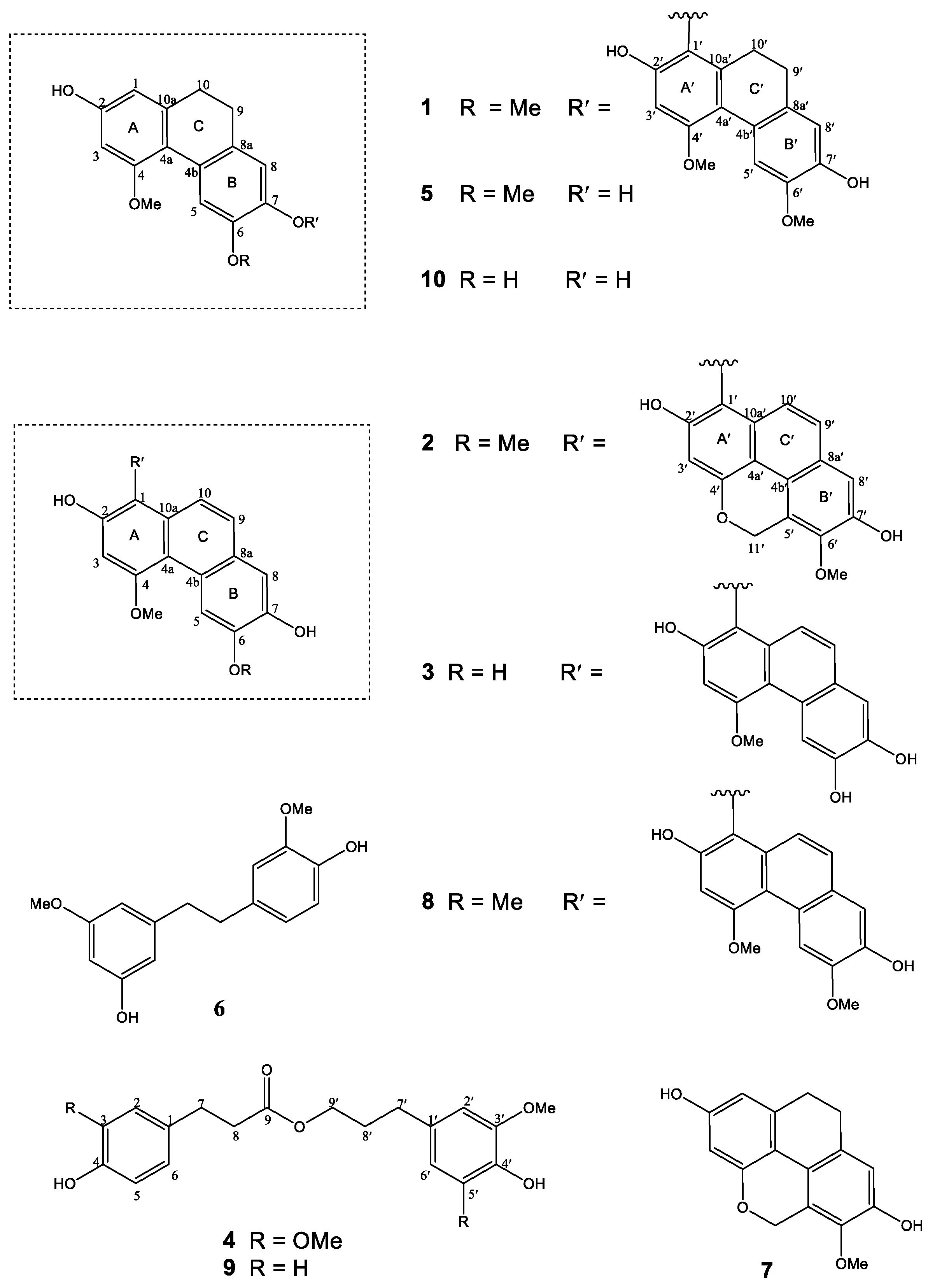
2.1. Structural Characterization

A total of 10 polyphenolic compounds were isolated from the MeOH extract of Aerides multiflora through solvent partition and repeated chromatography. They were characterized as three unknown compounds, named aerimultins A-C (13) and a new natural product, dihydrosinapyl dihydroferulate (4), together with six known compounds, i.e., 6-methoxycoelonin (5) [31], gigantol (6) [32], imbricatin (7) [33], agrostonin (8) [34], dihydroconiferyl dihydro-p-coumarate (9) [35] and 5-methoxy-9,10-dihydro- phenanthrene-2,3,7-triol (10) [36] (Figure 2).
Compound 1 was isolated as a whitish-brown amorphous solid. It showed a [M+Na]+ at m/z 565.1841 (calculated for C32H30O8Na, 565.1838) in the HR-ESI-MS. The IR spectrum showed absorption bands for hydroxyl (3350 cm−1), aromatic ring (2923, 1605 cm−1), methylene (1462 cm−1) and ether (1221 cm−1) groups. The UV absorptions at 265, 305, and 315 nm were indicative of a dihydrophenanthrene skeleton [37]. The 13C NMR and HSQC spectra revealed signals for twenty-four aromatic carbons, plus eight aliphatic carbons representing four methoxy and four methylene groups. The four CH2 carbons at 29.0 (C-9), 31.4 (C-10), 29.9 (C-9′), and 24.1 (C-10′) displayed HSQC correlations to the protons at δ 2.45 (2H, m, H2-9) and 2.56 (2H, m, H2-10) and 2.52 (4H, br s, H2-9′, H2-10′), respectively. These NMR signals suggested that 1 should be a dimeric compound consisting of two units of 9,10-dihydrophenanthrene (Table 1). The first unit of 1 (rings A, B, and C) should be derived from methoxycoelonin (5), a dihydrophenanthrene also obtained in this study, because its 1H and 13C NMR properties bore a close resemblance to those of 5 (Table 1). For example, in ring A of the first unit of 1, the proton at C-1 (δ 6.35, 1H, d, J = 2.5 Hz) exhibited HMBC correlation with C-10 (δ 31.4) and NOESY interaction with H2-10. H-3 (δ 6.46, 1H, d, J = 2.5 Hz) of 1 showed a NOESY cross peak with MeO-4 protons (δ 3.89, 3H, s). The hydroxyl proton at C-2 was observed at δ 8.35 (1H, s). For ring B of 1, the following 1H NMR signals were found: two one-proton singlets at δ 6.33 (1H, s, H-8) and 7.98 (1H, s, H-5), and a three-proton singlet at δ 3.92 (3H, s, MeO-6) which showed a NOESY cross-peak with H-5. The second unit of 1 (rings A′, B′, and C′) also exhibited 1H and 13C NMR data similar to those of 5 (Table 1). For instance, the 1H NMR spectrum of 1 exhibited two singlet proton signals at 6.66 (1H, s, H-8′) and 7.93 (1H, s, H-5′), two methoxy groups at δ 3.84 (3H, s, MeO-6′) and 3.91 (3H, s, MeO-4′), and two hydroxyl groups at δ 7.44 (s, HO-7′) and 8.25 (s, HO-2′). The HMBC spectrum of 1 showed correlation from H-3′ to C-1′ (δ 133.7) and C-4a′ (δ 117.1), and from H-5′ to C-4a′ (δ 117.1), C-8a′ (δ 131.4) and C-7′ (δ 145.6). H-8′ (δ 6.66, 1H, s) displayed HMBC correlation with C-9′ (δ 29.9) and NOESY interaction with H2-9′. The methoxyl protons at C-4′ (δ 155.3) and C-6′ (δ 146.1) showed NOESY correlations with H-3′ and H-5′, respectively. However, the second dihydrophenanthrene unit of 1 showed the absence of a H-1′ signal, with the signal for H-3′ appearing as a singlet at δ 6.65. Moreover, in the 13C NMR spectrum of 1, the signal for C-1′ of this unit was downfield shifted and observed as a quaternary carbon at δ 133.7, with HMBC correlations with H-3′ (δ 6.65, s), H2-10′ (δ 2.52, br s) and HO-2′ (δ 8.25, s). These NMR properties indicated that the structure of 1 consisted of two methoxycoelonin (5) units connected to each other through an ether linkage at C-7 and C-1′. This was also supported by the absence of a hydroxyl proton at C-7 (δ 146.6). Based on the above spectral data, compound 1 was characterized as a new dimeric 9,10-dihydrophenanthrene derivative and given the trivial name aerimultin A.
Compound 2, a brown amorphous solid, exhibited [M+Na]+ at m/z 559.1376 (calculated for C32H24O8Na, 559.1368) in the HR-ESI-MS, corresponding to the molecular formula C32H24O8. The IR spectrum showed absorption bands due to the presence of hydroxyl (3368 cm−1), aromatic ring (2919, 1587 cm−1) and ether (1259 cm−1) functionalities. The UV absorptions at 265, 315.5 and 370 nm were suggestive of a phenanthrene skeleton [30]. Compound 2 should be a dimeric phenanthrene, as suggested from the 1H NMR signals for two pairs of ortho-coupled doublets, representing H-9 (δ 7.36, 1H, d, J = 9.5 Hz), H-10 (δ 6.98, 1H, d, J = 9.5 Hz), H-9′ (δ 7.37, 1H, d, J = 9.0 Hz), and H-10′ (δ 6.92, 1H, d, J = 9.0 Hz) (Table 2). The first phenanthrene unit of 2 (rings A, B, and C) exhibited 1H and 13C NMR resonances similar to those of agrostonin (8), a biphenantherene also isolated from this plant (Table 2). These included three one-proton singlets at δ 6.99 (1H, s, H-3), 7.19 (1H, s, H-8) and 9.24 (1H, s, H-5), and two methoxy groups at δ 4.06 (3H, s, MeO-6) and 4.22 (3H, s, MeO-4). The proton at C-8 showed HMBC correlation with C-9 (δ 126.5). The protons H-3 and H-5 exhibited three-bond couplings to C-4a (δ 116.2) in the HMBC spectrum. The NOESY correlations of the MeO-4 and MeO-6 protons with H-3 and H-5, respectively, supported the attachment of these methoxy groups at C-4 and C-6. The quaternary carbon at δ 109.3 was assigned as C-1 according to its HMBC cross-peaks with H-3 and H-10. For the second phenanthrene unit (rings A′, B′, and C′), the presence of oxymethylene protons at δ 5.79 (2H, d, J = 1.5 Hz, H2-11′) indicated a phenanthropyran structure [38]. The 1H NMR spectrum also displayed two sharp one-proton singlets at δ 6.81 (1H, s, H-3′) and 7.21 (1H, s, H-8′), and a methoxy group at δ 3.95 (3H, s, MeO-6′). The assignments of H-8′ and H-3′ were supported by their HMBC correlations with C-9′ (δ 127.9) and C-1′ (δ 110.2), respectively. The HMBC correlations of C-6′ (δ 144.2) with MeO-6′ protons and H2-11′ indicated the location of the methoxy group at C-6′. The C-1′ of this second unit showed HMBC correlations with H-3′ and H-10′. The chemical shifts of C-1 (δ 109.3) and C-1′ (δ 110.2) suggested that they were not oxygenated, but, instead, they formed a C−C bridge linking the two monomers [39]. Therefore, it was concluded that 2 had the structure as shown, and the compound was given the trivial name aerimultin B.
Compound 3 was obtained as a brown amorphous solid. Its UV absorptions and IR absorption bands were similar to those of compound 2, indicating a phenanthrene derivative. The HR-ESI-MS exhibited [M+Na]+ at m/z 533.1218 (calculated for C30H22O8Na, 533.1212), suggesting the molecular formula C30H22O8. However, the 13C NMR spectrum showed only 15 carbon signals, suggesting that 3 should be a dimeric phenanthrene with two identical units. Comparison of the 1H and 13C NMR of 3 with those of agrostonin (8) (Table 2) revealed their structural similarity, except for the presence of a hydroxyl group at C-6/C-6′ in 3, instead of a methoxy group. Moreover, the two phenanthrene units were symmetrically linked to each other through a C−C bond between C-1 and C-1′ as supported by the HMBC correlations from C-1/C-1′ to H-3/H-3′, H-10/H-10′ and HO-2/HO-2′ [39]. On the basis of above spectral evidence, the structure of compound 3 was established as shown, and the trivial name aerimultin C was given to the compound.
Compound 4 was obtained as a yellow amorphous solid. The molecular formula was determined as C21H26O7 by HR-ESI-MS of [M+Na]+ at m/z 413.1584 (calculated for C21H26O7Na, 413.1576). The IR spectrum showed absorption bands for hydroxyl (3432 cm−1), aromatic ring (2937, 1608 cm−1), carbonyl ester (1723, 1208, 1111 cm−1) and methylene (1455 cm−1) groups. The UV spectrum exhibited maximum absorptions at 280 and 315 nm. The 1H NMR spectrum (Table 3) exhibited signals for a dihydroferulate structure [δ 2.59 (2H, t, J = 7.5 Hz, H2-8), 2.81 (2H, m, H2-7), 3.82 (3H, s, MeO-3), 6.68 (1H, dd, J = 8.1, 1.5 Hz, H-6), 6.73 (1H, d, J = 8.1 Hz, H-5), and 6.85 (1H, d, J = 1.5 Hz, H-2)] [40]. This was confirmed by the HMBC correlations of C-2 (δ 111.8), C-6 (δ 120.6) and C-9 (δ 172.2) with H2-7 (Table 3). The location of a MeO-3 group was supported by its NOESY correlation with H-2. The 1H NMR spectrum also showed signals for a dihydrosinapyl structure [δ 1.89 (2H, m, H2-8′), 2.57 (2H, t, J = 7.5 Hz, H2-7′), 3.80 (6H, s, MeO-3′, MeO-5′), 4.05 (2H, t, J = 7.5 Hz, H2-9′), and 6.49 (2H, s, H-2′, H-6′)] [41]. The HMBC correlations of C-2′/C-6′ (δ 105.8) and C-9′ (δ 63.2) with H2-7′ supported the presence of this unit.
The NOESY cross-peak between MeO-3′/MeO-5′ protons and H-2′/H-6′ confirmed the locations of the methoxy groups at C-3′/C-5′ (δ 147.7). The two phenylpropanoid units were connected by an ester bond at C-9 and C-9′, as determined by HMBC correlation of C-9 (δ 172.2) with H2-9′. Based on the above spectroscopic evidence, compound 4 was determined as dihydrosinapyl dihydroferulate. Prior to this study, the natural occurrence of 4 was unknown. However, the compound was earlier synthesized by acylation of the lignins from Arabidopsis thaliana [42].

2.2. Chemotaxonomic Significance

The presence of phenanthrene derivatives in Aerides multiflora is in line with the earlier findings in A. crispum and A. rosea [19,20]. In addition, the chemical profiles of these plants agreed with the conclusion from a molecular phylogenetic analysis that indicated their close relationships [43]. The family Orchidaceae is divided into 5 subfamilies, i.e., Epidendroideae, Orchidoideae, Vanilloideae, Cypripedioideae, and Apostasioideae [44]. So far, dimeric phenanthrenes have been reported from only two subfamilies, i.e., Epidendroideae (the genera Stanhopea, Bletilla, Pholidota, Pleione, Otochilus, Arundina, Bulbophyllum, Dendrobium, Monomeria, Cremastra, Agrostophyllum, Liparis, Cyrtopodium, Eria, Eulophia, Cirrhopetalum, Calanthe, Lusia, and Prosthechea) and Orchidoideae (the genera Spiranthes and Gymnadenia) [34,39,45,46,47,48,49,50,51,52,53,54,55,56,57,58,59,60,61,62,63]. No biphenanthrenes have been found outside these two subfamilies. Interestingly, a previous study revealed a monophyletic relationship between Epidendroideae and Orchidoideae [64]. Thus, from the currently available chemical data, the occurrence of biphenanthrenes could be taken as their chemotaxonomic marker. Nevertheless, additional chemical studies on the other three subfamilies are still needed to verify this postulation.
It should also be noted that the genus Aerides belongs to the same clade as Rhynchostylis [43]. Both genera have been called “the foxtail orchid” due to the erect or pendent inflorescences of closely packed flowers, and this has sometimes led to confusion. Up to the present, no reports on the secondary metabolites of the latter genus have appeared. Comparative chemical studies, in combination with detailed genetic analysis, may help shed light on the distinction between these two sister genera.

2.3. α-Glucosidase Inhibitory Activity

Yeast α-glucosidase enzyme was used in this study. In general, α-glucosidase enzymes can be obtained from several sources, for example, Saccharomyces cerevisiae, Rattus norvegicus, and GANC-human [65]. The enzyme derived from the yeast shows approximately 55% sequence homology with that obtained from mammalian sources [66], and therefore is widely employed in the investigations of natural compounds for α-glucosidase inhibitory potential [67,68].
All the isolated compounds (110) were initially tested for their α-glucosidase inhibitory activity at a concentration of 100 μg/mL. IC50 values were determined for compounds with more than 70% inhibition of the enzyme. As shown in Table 4, all compounds, except for dihydrosinapyl dihydroferulate (4), exhibited stronger activity (IC50 5.2−266.7 μM) than the drug acarbose (IC50 value of 514.4 ± 9.2 μM). It should be mentioned that biphenanthrenes with α-glucosidase inhibitory activity were isolated from the family Orchidaceae for the first time in this study.
Overall, the dimeric phenanthrenes (1, 2, 3, and 8) demonstrated higher activity than the monomers (5, 7, and 10), as indicated by their IC50 values (Table 4). Aerimultin C (3) was the most potent α-glucosidase inhibitor, with an IC50 value of 5.2 ± 0.7 μM. Replacing the phenolic groups at C-6 and C-6′ of this compound with methoxy groups reduced the activity by about seven-fold, as can be seen from the increased IC50 value (37.2 ± 4.5 μM) for agrostonin (8). The importance of free OH groups is also supported by the potent activity (IC50 2.08 ± 0.19 μM) earlier observed for a biphenanthrene (from Dioscorea bulbifera, Dioscoreaceae), the structure of which contains four free phenolic groups [69]. A molecular docking study on flavones with α-glucosidase inhibitory activity has also revealed that replacement of the hydroxyl groups with methoxy groups could lead to loss of activity [70].
Parallel observations were also obtained for the 3-phenylpropyl 3-propionate derivatives (4 and 9). Dihydroconiferyl dihydro-p-coumarate (9) showed appreciable activity (IC50 266.7 ± 8.6 μM). However, introducing methoxy groups to C-3 and C-5′ of 9 caused a total loss of activity, as seen in dihydrosinapyl dihydroferulate (4). A similar phenomenon, in which the presence of aromatic methoxy groups diminished α-glucosidase inhibitory activity, was earlier reported for p-coumarate esters of long-chain alcohols [71].
A kinetics study was then performed on compound 3 to analyze the mode of enzyme inhibition using various substrate concentrations (0.25–2.0 mM). From the Lineweaver–Burk plot in Figure 3A, the drug acarbose showed the intersection of the lines on the y-axis, indicating competitive type of inhibition. The Ki value of acarbose (190.57 μM) was obtained from the secondary plot by replotting the slopes of the lines against inhibitor concentrations. For compound 3, the increase in concentration (4 and 8 μM) decreased the Vmax from 0.10 to 0.035 but did not affect the Km value (Figure 3B). The results suggested non-competitive inhibition of the enzyme by 3. The Ki value of compound 3 (4.18 μM) was obtained from the secondary plot, as shown in Table 5.
Generally, non-competitive inhibitors have some advantages over competitive inhibitors [72]. Non-competitive inhibitors bind to the allosteric site of the enzyme, and thus do not depend upon the substrate concentration. Moreover, they require lower concentrations than competitive inhibitors to produce the same effect [73]. Compound 3, as a potent non-competitive inhibitor of α-glucosidase, provides a lead structure for the further design and development of AGI drugs.

3. Materials and Methods

3.1. Experimental

Optical rotations were determined with a PerkinElmer Polarimeter 341 (Boston, MA, USA). UV spectra were recorded on a Milton Roy Spectronic 3000 Array spectrophotometer (Rochester, Monroe, NY, USA). IR spectra were obtained with a PerkinElmer FT-IR 1760X spectrophotometer (Boston, MA, USA). Mass spectra were measured using a Bruker MicroTOF mass spectrometer (ESI-MS) (Billerica, MA, USA). NMR spectra were recorded on a Bruker Avance DPX-300FT NMR spectrometer or a Bruker Avance III HD 500 NMR spectrometer (Billerica, MA, USA). Yeast α-glucosidase enzyme and p-nitrophenol-α-d-glucopyranoside were obtained from Sigma Chemical, Inc. (St. Louis, MO, USA), and acarbose was purchased from Fluka Chemical (Buchs, Switzerland). Microtiter plate readings were carried out with a CLARIOstar apparatus (BMGLABTECH, Ortenberg, Germany).

3.2. Plant Material

The plant materials, whole plants of Aerides multiflora, were purchased from Chatuchak market in May 2019. Plant identification was performed by Mr. Yanyong Punpreuk, Department of Agriculture, Bangkok, Thailand. A voucher specimen BS-AM-052562 has been deposited at the Department of Pharmacognosy and Pharmaceutical Botany, Faculty of Pharmaceutical Sciences, Chulalongkorn University.

3.3. Extraction and Isolation

The dried powder from the whole plants of Aerides multiflora (6.1 kg) was macerated with MeOH (4 × 18 L). The MeOH extract, at a concentration of 100 µg/mL, showed 82.4 ± 9.5% inhibition of α-glucosidase. This MeOH extract (550 g) was then suspended in water and partitioned with EtOAc and butanol to give an EtOAc extract (201.1 g), a butanol extract (80.8 g), and an aqueous extract (150 g), respectively. The EtOAc extract exhibited 92.9 ± 3.2 inhibition at 100 µg/mL, whereas the others were devoid of activity (<50% inhibition). Therefore, the EtOAc extract was subjected to further investigation.
The EtOAc extract was first fractionated by vacuum liquid chromatography (silica gel, EtOAc–CH2Cl2, gradient) to give five fractions (A–E). Fraction B (11.4 g) was further fractionated on a silica gel column (EtOAc–hexane, gradient) to give 3 fractions (BA–BC). Fraction BA (1 g) was separated on Sephadex LH-20 (methanol) to yield fractions BAA, BAB, and BAC. Fraction BAA (200 mg) was further separated by column chromatography (CC, silica gel, EtOAc–CH2Cl2, gradient) to give 6-methoxycoelonin (5) (65.4 mg). Fraction BAB (300 mg) was subjected to CC (silica gel, EtOAc–CH2Cl2, gradient) to give fractions BAB1 and BAB2. Fraction BAB1 (160.2 mg) was separated by CC (silica gel, acetone-hexane, 3:7) to yield 1 (2.3 mg). Gigantol (6) (14.5 mg) was obtained from fraction BAB2 (100 mg) after purification on Sephadex LH-20 (acetone). Fraction BB (1 g) was separated on Sephadex LH-20 (acetone) to yield BBA and BBB fractions. Fraction BBA (195.8 mg) was subjected to CC (silica gel, EtOAc-CH2Cl2, gradient) to yield BBA1 and BBA2 fractions. Fraction BBA1 (132.2 mg) was subjected to CC (silica gel, acetone–hexane, 3:7) to produce imbricatin (7) (39 mg) and agrostonin (8) (7 mg). Fraction C (10.5 g) was fractionated on a silica gel column (EtOAc-CH2Cl2, gradient) to give 3 fractions (CA–CC). Fraction CB (500 mg) was further separated on Sephadex LH-20 (acetone) to yield CBA and CBB fractions. Fraction CBA (236.9 mg) was further separated by CC (silica gel, EtOAc–hexane, gradient) to give dihydroconiferyl dihydro-p-coumarate (9) (74.1 mg) and 5-methoxy-9,10-dihydrophenanthrene-2,3,7-triol (10) (9.2 mg). Fraction CC (100 mg) was separated on Sephadex LH-20 (acetone) to yield fractions CCA, CCB, and CCC. Fraction CCB (10 mg) was subjected to CC (silica gel, EtOAc–CH2Cl2, 0.2: 9.8) to yield 2 (3.9 mg). Fraction D (72 g) was chromatographed on a silica gel column (EtOAc-CH2Cl2, gradient) to give 3 fractions (DA–DC). Fraction DA (1 g) was separated on Sephadex LH-20 (methanol) to yield DAA and DAB fractions. Fraction DAA (300 mg) was re-separated on Sephadex LH20 (acetone) to yield DAA1 and DAA2 fractions. Fraction DAA1 (100 mg) was subjected to CC (silica gel, EtOAc–hexane, 3:7) to give 4 (4.2 mg). Fraction E (84.8 g) was subjected to Diaion HP-20 (water–methanol, gradient) to yield five fractions (EA–EE). Fraction EC (1.7 g) was separated on Sephadex LH-20 (methanol) to yield ECA, ECB and ECC fractions. Fraction ECC (40 mg) was subjected to CC (silica gel, methanol–CH2Cl2, 0.5:9.5) to produce 3 (8.8 mg).
Aerimultin A (1): whitish-brown amorphous powder; UV (MeOH) λmax (log ε) 265 (4.31), 305 (4.2), 315 (4.19); IR: νmax 3350, 2923, 2850, 1696, 1605, 1462, 1442, 1221, 1201 cm−1; HR-ESI-MS: [M+Na]+ at m/z 565.1841 (calculated for C32H30O8Na, 565.1838); 1H and 13C NMR data, see Table 1.
Aerimultin B (2): brown amorphous solid; [α] D20 −108 (c 0.005, MeOH); UV (MeOH) λmax (log ε) 265 (4.67), 315 (4.09), 370 (3.99); IR: νmax 3368, 2919, 2850, 1736, 1587, 1463, 1259 cm−1; HR-ESI-MS: [M+Na]+ at m/z 559.1376 (calculated for C32H24O8Na, 559.1368); 1H and 13C NMR data, see Table 2.
Aerimultin C (3): brown amorphous solid; [α] D20 +67.5 (c 0.008, MeOH); UV (MeOH) λmax (log ε) 265 (4.1), 315 (3.42), 355 (3.47), 370 (3.48); IR: νmax 3360, 2921, 2851, 1712, 1588, 1461, 1371 cm−1; HR-ESI-MS: [M+Na]+ at m/z 533.1218 (calculated for C30H22O8Na, 533.1212); 1H and 13C NMR data, see Table 2.
Dihydrosinapyl dihydroferulate (4): yellow amorphous solid; UV (MeOH) λmax (log ε) 280 (3.76), 315 (3.12); IR: νmax 3432, 2937, 2841, 1723, 1608, 1514, 1455, 1427, 1208, 1111 cm−1; HR-ESI-MS: [M+Na]+ at m/z 413.1584 (calculated for C21H26O7Na, 413.1576); 1H and 13C NMR data, see Table 3.

3.4. α-Glucosidase Inhibition Assay

The assays were performed following previous protocols [74]. The liberation of p-nitrophenol from the substrate p-nitrophenol-α-d-glucopyranoside (PNPG) was observed to determine the inhibition of the α-glucosidase enzyme. Each sample was initially dissolved in 50% DMSO. Then, 0.1 U/mL of α-glucosidase (40 μL) in phosphate buffer (pH 6.8) was added to each well of a 96-well plate which contained the sample solution (10 μL). The plate was pre-incubated at 37 °C for 10 min. Then, 2 mM p-nitrophenol-α-d-glucopyranoside (50 μL) was added, and the mixture was incubated again at 37 °C for 20 min. Finally, 1 M Na2CO3 solution (100 μL) was added to terminate the reaction. The absorbance of the mixture was measured at 405 nm using a microplate reader. Two-fold serial dilution was performed for IC50 determination. The drug acarbose was used as the positive control.
The mode of enzyme inhibition of the test compound was determined using the double reciprocal Lineweaver–Burk plot (1/V vs. 1/[S]). The experiment was performed by varying the PNPG concentrations (0.25, 0.5, 1.0, and 2.0 mM) in the absence or presence of compound 3 (4 μM and 8 μM) or acarbose (930 μM and 465 μM). The secondary plot was constructed by replotting the slopes of the lines against inhibitor concentrations, and the Ki was calculated from the line equation of the plot.

4. Conclusions

In this communication, ten compounds were isolated from Aerides multiflora, including three new compounds, namely, aerimultins A–C (13), the new natural product dihydrosinapyl dihydroferulate (4), and six known compounds (510). The structures of the new compounds were established by spectroscopic methods. The findings in this study suggested that biphenanthrenes might be taken as a chemotaxonomic marker for the subfamilies Epidendroideae and Orchidoideae within the family Orchidaceae. For the first time, the dimeric phenanthrenes obtained from this plant family were investigated for an α-glucosidase inhibitory activity. Among the isolates, the biphenathrene aerimultin (3) emerged as the most potent inhibitor, showing much higher potency than the drug acarbose. An enzyme kinetic study on this compound revealed a non-competitive type of inhibition and suggested that it could be a candidate structure for α-glucosidase inhibitor drug development.

Author Contributions

B.S. conceived, designed, and supervised the research project, as well as prepared and edited the manuscript; M.T.T. performed the experiments and prepared the manuscript; N.C. supervised the α-glucosidase inhibition assay; W.M. performed the NMR experiments; Y.P. performed the plant collection; K.L. provided comments and suggestions on the preparation of the manuscript. All authors have read and agreed to the published version of the manuscript.

Funding

This work was supported by the 90th Anniversary of Chulalongkorn University Fund (Ratchadaphiseksomphot Endowment Fund) 2021.

Institutional Review Board Statement

Not applicable.

Informed Consent Statement

Not applicable.

Data Availability Statement

All data presented in this study are available in the article.

Acknowledgments

M.T.T. is grateful to the Graduate School, Chulalongkorn University for a CUASEAN Ph.D. scholarship.

Conflicts of Interest

The authors declare no conflict of interest.

References

  1. Karunaratne, V.; Thadhani, V.M.; Khan, S.N.; Choudhary, M.I. Potent α-glucosidase inhibitors from the lichen Cladonia species from Sri Lanka. J. Natl. Sci. Found Sri. 2014, 42, 95–98. [Google Scholar] [CrossRef]
  2. Wu, J.H.; Micha, R.; Imamura, F.; Pan, A.; Biggs, M.L.; Ajaz, O.; Mozaffarian, D. Omega-3 fatty acids and incident type 2 diabetes: A systematic review and meta-analysis. Br. J. Nutr. 2012, 107, S214–S227. [Google Scholar] [CrossRef]
  3. Chaudhury, A.; Duvoor, C.; Reddy Dendi, V.S.; Kraleti, S.; Chada, A.; Ravilla, R.; Sasapu, A. Clinical review of antidiabetic drugs: Implications for type 2 diabetes mellitus management. Front. Endocrinol. 2017, 8, 1–12. [Google Scholar] [CrossRef] [PubMed] [Green Version]
  4. Nhiem, N.X.; Van Kiem, P.; Van Minh, C.; Ban, N.K.; Cuong, N.X.; Tung, N.H.; Kim, Y.H. α-glucosidase inhibition properties of cucurbitane-type triterpene glycosides from the fruits of Momordica charantia. Chem. Pharm. Bull. 2010, 58, 720–724. [Google Scholar] [CrossRef] [PubMed] [Green Version]
  5. Costa, T.M.; Mayer, D.A.; Siebert, D.A.; Micke, G.A.; Alberton, M.D.; Tavares, L.B.B.; De Oliveira, D. Kinetics analysis of the inhibitory effects of alpha-glucosidase and identification of compounds from Ganoderma lipsiense Mycelium. Appl. Biochem. Biotechnol. 2020, 1–14. [Google Scholar]
  6. Hung, H.Y.; Qian, K.; Morris-Natschke, S.L.; Hsu, C.S.; Lee, K.H. Recent discovery of plant-derived anti-diabetic natural products. Nat. Prod. Res. 2012, 29, 580–606. [Google Scholar] [CrossRef] [PubMed]
  7. Nashiru, O.; Koh, S.; Lee, S.Y.; Lee, D.S. Novel α-glucosidase from extreme thermophile Thermus caldophilus GK24. J. Biochem. Mol. Biol. 2001, 34, 347–354. [Google Scholar]
  8. Ernawati, T.; Radji, M.; Hanafi, M.; Munim, A.; Yanuar, A. Cinnamic acid derivatives as α-glucosidase inhibitor agents. Indones. J. Chem. 2017, 17, 151–160. [Google Scholar] [CrossRef]
  9. Yin, Z.; Zhang, W.; Feng, F.; Zhang, Y.; Kang, W. α-Glucosidase inhibitors isolated from medicinal plants. Food Sci. Hum. Well. 2014, 3, 136–174. [Google Scholar] [CrossRef] [Green Version]
  10. Kao, C.C.; Wu, P.C.; Wu, C.H.; Chen, L.K.; Chen, H.H.; Wu, M.S. Risk of liver injury after α-glucosidase inhibitor therapy in advanced chronic kidney disease patients. Sci. Rep. 2016, 6, 18996. [Google Scholar] [CrossRef] [Green Version]
  11. Babu, P.S.; Prabuseenivasan, S.; Ignacimuthu, S. Cinnamaldehyde—A potential antidiabetic agent. Phytomedicine 2007, 14, 15–22. [Google Scholar] [CrossRef]
  12. Liu, M.; Qi, C.; Sun, W.; Shen, L.; Wang, J.; Liu, J.; Zhang, Y. α-Glucosidase inhibitors from the coral-associated fungus Aspergillus terreus. Front. Chem. 2018, 6, 422. [Google Scholar] [CrossRef] [Green Version]
  13. Limpanit, R.; Chuanasa, T.; Likhitwitayawuid, K.; Jongbunprasert, V.; Sritularak, B. α-Glucosidase inhibitors from Dendrobium tortile. Rec. Nat. Prod. 2016, 10, 609–616. [Google Scholar]
  14. Sun, J.; Zhang, Y.; Chen, L.; Zhan, R.; Chen, Y. A new phenanthrene and a new 9,10-dihydrophenanthrene from Bulbophyllum retusiusculum. Nat. Prod. Res. 2018, 32, 2447–2451. [Google Scholar] [CrossRef] [PubMed]
  15. Auberon, F.; Olatunji, O.J.; Waffo-Teguo, P.; Adekoya, A.E.; Bonte, F.; Merillon, J.M.; Lobstein, A. New glucosyloxybenzyl 2R-benzylmalate derivatives from the underground parts of Arundina graminifolia (Orchidaceae). Fitoterapia 2019, 135, 33–43. [Google Scholar] [CrossRef] [PubMed]
  16. Kocyan, A.; de Vogel, E.F.; Conti, E.; Gravendeel, B. Molecular phylogeny of Aerides (Orchidaceae) based on one nuclear and two plastid markers: A step forward in understanding the evolution of the Aeridinae. Mol. Phylogenet. Evol. 2008, 48, 422–443. [Google Scholar] [CrossRef] [PubMed] [Green Version]
  17. Pant, B. Medicinal orchids and their uses: Tissue culture a potential alternative for conservation. Afr. J. Plant Sci. 2013, 7, 448–467. [Google Scholar] [CrossRef] [Green Version]
  18. Akter, M.; Huda, M.K.; Hoque, M.M. Investigation of secondary metabolites of nine medicinally important orchids of Bangladesh. J. Pharmacogn. Phytochem. 2018, 7, 602–606. [Google Scholar]
  19. Cakova, V.; Urbain, A.; Antheaume, C.; Rimlinger, N.; Wehrung, P.; Bonté, F.; Lobstein, A. Identification of phenanthrene derivatives in Aerides rosea (Orchidaceae) using the combined systems HPLC–ESI–HRMS/MS and HPLC–DAD–MS–SPE–UV–NMR. Phytochem. Anal. 2015, 26, 34–39. [Google Scholar] [CrossRef]
  20. Anuradha, V.; Rao, N.P. Aeridin: A phenanthropyran from Aerides crispum. Phytochemistry 1998, 48, 185–186. [Google Scholar] [CrossRef]
  21. Bhowmik, T.K.; Rahman, M.M. In vitro study of medicinally important orchid Aerides multiflora Roxb. from nodal and leaf explants. J. Pharmacog. Phytochem. 2020, 9, 179–184. [Google Scholar]
  22. Choon, K.K. Management of the Pha Taem Protected Forest Complex to Promote Cooperation for Transboundary Biodiversity Conservation between Thailand, Cambodia and Laos Phase I; Kasetsart University: Bangkok, Thailand, 2004. [Google Scholar]
  23. Schuiteman, A.; Bonnet, P.; Svengsuksa, B.; Barthélémy, D. An annotated checklist of the Orchidaceae of Laos. Nord. J. Bot. 2008, 26, 257–316. [Google Scholar] [CrossRef]
  24. Subedi, A.; Kunwar, B.; Choi, Y.; Dai, Y.; Van Andel, T.; Chaudhary, R.P.; De Boer, H.J.; Gravendeel, B. Collection and trade of wild-harvested orchids in Nepal. J. Ethnobiol. Ethnomed. 2013, 9, 1–10. [Google Scholar] [CrossRef] [Green Version]
  25. Gogoi, K.; Das, R.; Yonzone, R. Present ecological status, diversity, distribution and cultural significance of the genus Aerides Loureiro (Orchidaceae) in Tinsukia District (Assam) of North East India. J. Environ. Ecol. 2012, 30, 649–651. [Google Scholar]
  26. Rao, A.N. Medicinal orchid wealth of Arunachal Pradesh. Indian Med. Plants 2004, 1, 1–7. [Google Scholar]
  27. Ghanaksh, A.; Kaushik, P. Antibacterial effect of Aerides multiflora: A study in vitro. J. Orchid Soc. India 1999, 1, 65–68. [Google Scholar]
  28. Inthongkaew, P.; Chatsumpun, N.; Supasuteekul, C.; Kitisripanya, T.; Putalun, W.; Likhitwitayawuid, K.; Sritularak, B. α-Glucosidase and pancreatic lipase inhibitory activities and glucose uptake stimulatory effect of phenolic compounds from Dendrobium formosum. Rev. Bras. Farmacogn. 2017, 27, 480–487. [Google Scholar] [CrossRef]
  29. Sarakulwattana, C.; Mekboonsonglarp, W.; Likhitwitayawuid, K.; Rojsitthisak, P.; Sritularak, B. New bisbibenzyl and phenanthrene derivatives from Dendrobium scabrilingue and their α-glucosidase inhibitory activity. Nat. Prod. Res. 2020, 34, 1694–1710. [Google Scholar] [CrossRef] [PubMed]
  30. Thant, M.T.; Chatsumpun, N.; Mekboonsonglarp, W.; Sritularak, B.; Likhitwitayawuid, K. New fluorene derivatives from Dendrobium gibsonii and their α-glucosidase inhibitory activity. Molecules 2020, 25, 4931. [Google Scholar] [CrossRef]
  31. Leong, Y.W.; Kang, C.C.; Harrison, L.J.; Powell, A.D. Phenanthrenes, dihydrophenanthrenes and bibenzyls from the orchid Bulbophyllum vaginatum. Phytochemistry 1997, 44, 157–165. [Google Scholar] [CrossRef]
  32. Chen, Y.; Xu, J.; Yu, H.; Qing, C.; Zhang, Y.; Wang, L.; Liu, Y.; Wang, J. Cytotoxic phenolics from Bulbophyllum odoratissimum. Food Chem. 2008, 107, 169–173. [Google Scholar] [CrossRef]
  33. Simmler, C.; Antheaume, C.; Lobstein, A. Antioxidant biomarkers from Vanda coerulea stems reduce irradiated HaCaT PGE-2 production as a result of COX-2 inhibition. PLoS ONE 2010, 5, 1–9. [Google Scholar] [CrossRef] [PubMed] [Green Version]
  34. Majumder, P.L.; Banerjee, S.; Lahiri, S.; Mukhoti, N.; Sen, S. Dimeric phenanthrenes from two Agrostophyllum species. Phytochemistry 1998, 47, 855–860. [Google Scholar] [CrossRef]
  35. Zhang, X.; Gao, H.; Wang, N.; Yao, X. Phenolic components from Dendrobium nobile. Zhong Cao Yao 2006, 37, 652–655. [Google Scholar]
  36. Leong, Y.W.; Kang, C.C.; Harrison, L.J.; Powell, A.D. Phenanthrene and other aromatic constituents of Bulbophyllum vaginatum. Phytochemistry 1998, 50, 1237–1241. [Google Scholar] [CrossRef]
  37. Estrada, S.; Toscano, R.A.; Mata, R. New Phenanthrene derivatives from Maxillaria densa. J. Nat. Prod. 1999, 62, 1175–1178. [Google Scholar] [CrossRef]
  38. Majumder, P.L.; Sabzabadi, E. Agrostophyllin, a naturally occurring phenanthropyran derivative from Agrostophyllum khasiyanum. Phytochemistry 1988, 27, 1899–1901. [Google Scholar] [CrossRef]
  39. Liu, L.; Yin, Q.M.; Zhang, X.W.; Wang, W.; Dong, X.Y.; Yan, X.; Hu, R. Bioactivity−guided isolation of biphenanthrenes from Liparis nervosa. Fitoterapia 2016, 115, 15–18. [Google Scholar] [CrossRef]
  40. Beck, J.J.; Kim, J.H.; Campbell, B.C.; Chou, S.C. Fungicidal activities of dihydroferulic acid alkyl ester analogues. J. Nat. Prod. 2007, 70, 779–782. [Google Scholar] [CrossRef]
  41. Zhuo, J.X.; Wang, Y.H.; Su, X.L.; Mei, R.Q.; Yang, J.; Kong, Y.; Long, C.L. Neolignans from Selaginella moellendorffii. Nat. Prod. Bioprospect. 2016, 6, 161–166. [Google Scholar] [CrossRef] [Green Version]
  42. Sibout, R.; Le Bris, P.; Legee, F.; Cezard, L.; Renault, H.; Lapierre, C. Structural redesigning Arabidopsis lignins into alkali-soluble lignins through the expression of p-coumaroyl-CoA: Monolignol transferase PMT. Plant Physiol. 2016, 170, 1358–1366. [Google Scholar] [CrossRef] [Green Version]
  43. Zhang, G.Q.; Liu, K.W.; Chen, L.J.; Xiao, X.J.; Zhai, J.W.; Li, L.Q.; Liu, Z.J. A new molecular phylogeny and a new genus, Pendulorchis, of the Aerides–Vanda alliance (Orchidaceae: Epidendroideae). PLoS ONE 2013, 8, e60097. [Google Scholar] [CrossRef] [PubMed] [Green Version]
  44. Li, Y.X.; Li, Z.H.; Schuiteman, A.; Chase, M.W.; Li, J.W.; Huang, W.C.; Hidayat, A.; Wu, S.S.; Jin, X.H. Phylogenomics of Orchidaceae based on plastid and mitochondrial genomes. Mol. Phylogenet. Evol. 2019, 139, 106540. [Google Scholar] [CrossRef] [PubMed]
  45. Lucca, D.L.; Sa, G.P.; Polastri, L.R.; Ghiraldi, D.M.; Ferreira, N.P.; Chiavelli, L.U.; Pomini, A.M. Biphenanthrene from Stanhopea lietzei (Orchidaceae) and its chemophenetic significance within neotropical species of the Cymbidieae tribe. Biochem. Syst. Ecol. 2020, 89, 104014. [Google Scholar] [CrossRef]
  46. Qian, C.D.; Jiang, F.S.; Yu, H.S.; Shen, Y.; Fu, Y.H.; Cheng, D.Q.; Ding, Z.S. Antibacterial Biphenanthrenes from the fibrous roots of Bletilla striata. J. Nat. Prod. 2015, 78, 939–943. [Google Scholar] [CrossRef]
  47. Guo, X.Y.; Wang, J.; Wang, N.L.; Kitanaka, S.; Liu, H.W.; Yao, X.S. New stilbenoids from Pholidota yunnanensis and their inhibitory effects on nitric oxide production. Chem. Pharm. Bull. 2006, 54, 21–25. [Google Scholar] [CrossRef] [Green Version]
  48. Xu, J.; Yu, H.; Qing, C.; Zhang, Y.; Liu, Y.; Chen, Y. Two new biphenanthrenes with cytotoxic activity from Bulbophyllum odoratissimum. Fitoterapia 2009, 80, 381–384. [Google Scholar] [CrossRef] [PubMed]
  49. Zhang, G.N.; Zhong, L.Y.; Bligh, S.A.; Guo, Y.L.; Zhang, C.F.; Zhang, M.; Xu, L.S. Bi-bicyclic and bi-tricyclic compounds from Dendrobium thyrsiflorum. Phytochemistry 2005, 66, 1113–1120. [Google Scholar] [CrossRef]
  50. Yang, M.; Cai, L.; Tai, Z.; Zeng, X.; Ding, Z. Four new phenanthrenes from Monomeria barbata Lindl. Fitoterapia 2010, 81, 992–997. [Google Scholar] [CrossRef]
  51. Liu, L.; Li, J.; Zeng, K.W.; Jiang, Y.; Tu, P.F. Five new biphenanthrenes from Cremastra appendiculata. Molecules 2016, 21, 1089. [Google Scholar] [CrossRef] [Green Version]
  52. Auberon, F.; Olatunji, O.J.; Herbette, G.; Raminoson, D.; Antheaume, C.; Soengas, B.; Lobstein, A. Chemical constituents from the aerial parts of Cyrtopodium paniculatum. Molecules 2016, 21, 1418. [Google Scholar] [CrossRef] [Green Version]
  53. Wang, C.; Shao, S.Y.; Han, S.W.; Li, S. Atropisomeric bi (9, 10-dihydro) phenanthrene and phenanthrene/bibenzyl dimers with cytotoxic activity from the pseudobulbs of Pleione bulbocodioides. Fitoterapia 2019, 138, 104313. [Google Scholar] [CrossRef]
  54. Auberon, F.; Olatunji, O.J.; Krisa, S.; Antheaume, C.; Herbette, G.; Bonté, F.; Lobstein, A. Two new stilbenoids from the aerial parts of Arundina graminifolia (Orchidaceae). Molecules 2016, 21, 1430. [Google Scholar] [CrossRef] [Green Version]
  55. Shi, X.; Li, Y.; Liu, Y.; Jiang, J.; Wang, L.; Zhang, Y.; Chen, Y. A new 9, 10-dihydrophenanthropyran dimer and a new natural 9, 10-dihydrophenanthropyran from Otochilus porrectus. Biochem. Syst. Ecol. 2010, 38, 842–845. [Google Scholar] [CrossRef]
  56. Majumder, P.L.; Banerjee, S. Structure of flavanthrin, the first dimeric 9, 10-dihydrophenanthrene derivative from the orchid Eria flava. Tetrahedron 1988, 44, 7303–7308. [Google Scholar] [CrossRef]
  57. Tuchinda, P.; Udchachon, J.; Khumtaveeporn, K.; Taylor, W.C.; Engelhardt, L.M.; White, A.H. Phenanthrenes of Eulophia nuda. Phytochemistry 1988, 27, 3267–3271. [Google Scholar] [CrossRef]
  58. Majumder, P.L.; Pal, A.; Joardar, M. Cirrhopetalanthrin, a dimeric phenanthrene derivative from the orchid Cirrhopetalum maculosum. Phytochemistry 1990, 29, 271–274. [Google Scholar] [CrossRef]
  59. Lee, C.L.; Chang, F.R.; Yen, M.H.; Yu, D.; Liu, Y.N.; Bastow, K.F.; Morris-Natschke, S.L.; Wu, Y.C.; Lee, K.S. Cytotoxic phenanthrenequinones and 9,10-dihydrophenanthrenes from Calanthe arisanensis. J. Nat. Prod. 2009, 72, 210–213. [Google Scholar] [CrossRef] [Green Version]
  60. Majumder, P.L.; Lahiri, S. Volucrin, a new dimeric phenanthrene derivative from the orchid Lusia volucris. Tetrahedron 1990, 46, 3621–3626. [Google Scholar] [CrossRef]
  61. Gutierrez, R.M.P.; Gonzalez, A.M.N.; Baez, E.G.; Diaz, S.L. Studies on the constituents of bulbs of the orchid Prosthechea michuacana and antioxidant activity. Chem. Nat. Compd. 2010, 46, 554–561. [Google Scholar] [CrossRef]
  62. Li, C.Y.; Liu, J.; Su, X.H.; Yuan, Z.P.; Zhong, Y.J.; Li, Y.F.; Liang, B. New dimeric phenanthrene and flavone from Spiranthes sinensis. J. Asian Nat. Prod. Res. 2013, 15, 417–421. [Google Scholar] [CrossRef]
  63. Matsuda, H.; Morikawa, T.; Xie, H.; Yoshikawa, M. Antiallergic phenanthrenes and stilbenes from the tubers of Gymnadenia conopsea. Planta Med. 2004, 70, 847–855. [Google Scholar] [CrossRef]
  64. Niu, Z.; Xue, Q.; Zhu, S.; Sun, J.; Liu, W.; Ding, X. The complete plastome sequences of four orchid species: Insights into the evolution of the Orchidaceae and the utility of plastomic mutational hotspots. Front. Plant Sci. 2017, 8, 715. [Google Scholar] [CrossRef] [Green Version]
  65. Ernawati, T. In silico evaluation of molecular interactions between known α-glucosidase inhibitors and homologous α-glucosidase enzymes from Saccharomyces cerevisiae, Rattus norvegicus and GANC-human. Thai J. Pharm. Sci. 2018, 42. [Google Scholar]
  66. Dhanawansa, R.; Faridmoayer, A.; van der Merwe, G.; Li, Y.X.; Scaman, C.H. Overexpression, purification, and partial characterization of Saccharomyces cerevisiae processing alpha glucosidase I. J. Glycobiol. 2002, 12, 229–234. [Google Scholar] [CrossRef] [PubMed] [Green Version]
  67. Liu, J.L.; Kong, Y.C.; Miao, J.Y.; Mei, X.Y.; Wu, S.Y.; Yan, Y.C.; Cao, X.Y. Spectroscopy and molecular docking analysis reveal structural specificity of flavonoids in the inhibition of α-glucosidase activity. Int. J. Biol. Macromol. 2020, 152, 981–989. [Google Scholar] [CrossRef]
  68. Rosas-Ramírez, D.; Pereda-Miranda, R.; Escandón-Rivera, S.; Arreguín-Espinosa, R. Identification of α-glucosidase inhibitors from Ipomoea alba by affinity-directed fractionation-mass spectrometry. Rev. Bras. Farmacogn. 2020, 30, 336–345. [Google Scholar] [CrossRef]
  69. Zhao, Y.; Kongstad, K.T.; Liu, Y.; He, C.; Staerk, D. Unraveling the complexity of complex mixtures by combining high-resolution pharmacological, analytical and spectroscopic techniques: Antidiabetic constituents in Chinese medicinal plants. Faraday Discuss. 2019, 218, 202–218. [Google Scholar] [CrossRef] [PubMed]
  70. Proença, C.; Freitas, M.; Ribeiro, D.; Oliveira, E.F.; Sousa, J.L.; Tomé, S.M.; Fernandes, E. α-glucosidase inhibition by flavonoids: An in vitro and in silico structure–activity relationship study. J. Enzyme Inhib. Med. Chem. 2017, 32, 1216–1228. [Google Scholar] [CrossRef] [PubMed] [Green Version]
  71. San, H.T.; Boonsnongcheep, P.; Putalun, W.; Mekboonsonglarp, W.; Sritularak, B.; Likhitwitayawuid, K. α-glucosidase inhibitory and glucose uptake stimulatory effects of phenolic compounds from Dendrobium christyanum. Nat. Prod. Commun. 2020, 15. [Google Scholar] [CrossRef] [Green Version]
  72. Chougale, A.D.; Ghadyale, V.A.; Panaskar, S.N.; Arvindekar, A.U. Alpha glucosidase inhibition by stem extract of Tinospora cordifolia. J. Enzyme Inhib. Med. Chem. 2009, 24, 998–1001. [Google Scholar] [CrossRef] [PubMed]
  73. Ghadyale, V.; Takalikar, S.; Haldavnekar, V.; Arvindekar, A. Effective control of postprandial glucose level through inhibition of intestinal alpha glucosidase by Cymbopogon martinii (Roxb.). Evid. Based Complement. Altern. Med. 2012, 1–6. [Google Scholar] [CrossRef]
  74. Chatsumpun, N.; Sritularak, B.; Likhitwitayawuid, K. New biflavonoids with α-glucosidase and pancreatic lipase inhibitory activities from Boesenbergia rotunda. Molecules 2017, 22, 1862. [Google Scholar] [CrossRef] [PubMed] [Green Version]
Figure 1. Aerides multiflora Roxb.
Plants 10 00385 g001
Figure 2. Chemical structures of compounds 110 isolated from Aerides multiflora.
Plants 10 00385 g002
Figure 3. Lineweaver–Burk plots of (A) acarbose and (B) compound 3. The secondary plot of each compound is on the right.
Plants 10 00385 g003
Table 1. 1H (500 MHz) and 13C-NMR (125 MHz) spectral data of 1 and 5 in acetone-d6.
Position1 a 5 b
δH (Multiplicity,
J in Hz)
δCHMBC (Correlation
with 1H)
δH (Multiplicity, J in Hz)δC
16.35 (d, J = 2.5 Hz)108.33, 10, HO-26.39 (br s)107.4
2-157.81 *, 3 *, HO-2 *-156.5
36.46 (d, J = 2.5 Hz)99.11, HO-26.65 (br s)98.3
4-158.83 *, MeO-4-157.7
4a-115.91, 3, 5, 10-115.5
4b-127.85 *, 8, 9-124.7
57.98 (s)114.3-7.89 (s)112.2
6-147.48, MeO-6-145.1
7-146.65-144.3
86.33 (s)113.696.69 (s)114.0
8a-131.15, 10-130.7
92.45 (m)29.082.61 (m)28.9
102.56 (m)31.412.61 (m)30.7
10a-141.69, 10 *-140.5
1′-133.73′, 10′, HO-2′
2′-149.83′ *, HO-2′ *
3′6.65 (s)100.2HO-2′
4′-155.33′ *, MeO-4′
4a′-117.13′, 5′, 10′
4b′-125.25′ *, 8′, 9′
5′7.93 (s)113.4-
6′-146.18′, MeO-6′, HO-7′
7′-145.65′, HO-7′ *
8′6.66 (s)114.99′, HO-7′
8a′-131.45′, 10′
9′2.52 (br s)29.98′
10′2.52 (br s)24.1-
10a′-133.89′
MeO-43.89 (s)56.1-3.86 (s)55.5
MeO-63.92 (s)56.5-3.83 (s)54.9
MeO-4′3.91 (s)56.4-
MeO-6′3.84 (s)55.8-
HO-28.35 (s)--
HO-2′8.25 (s)--
HO-7′7.44 (s)--
a 1H (500 MHz) and 13C-NMR (125 MHz); b 1H (300 MHz) and 13C-NMR (75 MHz); * two-bond coupling.
Table 2. 1H and 13C-NMR spectral data of 2, 3 and 8 in acetone-d6.
Position 2 a3 b8 b
δH
(Multiplicity,
J in Hz)
δCHMBC
(Correlation with 1H)
δH
(Multiplicity,
J in Hz)
δCHMBC
(Correlation with 1H)
δH
(Multiplicity,
J in Hz)
δC
1-109.33, 10-108.83, 10, HO-2-108.9
2-155.03 *-154.13 *, HO-2 *-154.2
36.99 (s)100.0-6.95 (s)98.8HO-27.00 (s)99.2
4-160.23 *, MeO-4-159.43 *, MeO-4-159.3
4a-116.23, 5, 10-115.13, 5, 10-115.5
4b-125.88, 9-125.38, 9-124.9
59.24 (s)109.8-9.19 (s)112.7-9.26 (s)111.3
6-148.55 *, 8, MeO-6-145.38-147.6
7-146.05, 8 *-144.15-145.2
87.19 (s)112.297.19 (s)111.597.20 (s)112.3
8a-128.05, 10-126.75, 10-127.2
97.36
(d, J = 9.5 Hz)
126.587.31
(d, J = 9.0 Hz)
127.287.37
(d, J = 9.0 Hz)
127.1
106.98
(d, J = 9.5 Hz)
123.3-6.87
(d, J = 9.0 Hz)
121.8-6.95
(d, J = 9.0 Hz)
122.5
10a-135.49-134.69-134.6
1′-110.23′, 10′-108.83′, 10′, HO-2′-108.9
2′-156.33′ *-154.13′ *, HO-2′ *-154.2
3′6.81 (s)103.1-6.95 (s)98.8HO-2′7.00 (s)99.2
4′-153.73′ *, 11′-159.43′ *, MeO-4′-159.3
4a′-113.03′, 10′-115.13′, 5′, 10′-115.5
4b′-119.18′, 9′, 11′-125.38′, 9′-124.9
5′-120.611′ *9.19 (s)112.7-9.26 (s)111.3
6′-144.28′, 11′, MeO-6′-145.38′-147.6
7′-150.38′ *-144.15′-145.2
8′7.21 (s)111.69′7.19 (s)111.59′7.20 (s)112.3
8a′-126.210′-126.75′, 10′-127.2
9′7.37
(d, J = 9.0 Hz)
127.98′7.31
(d, J = 9.0 Hz)
127.28′7.37
(d, J = 9.0 Hz)
127.1
10′6.92
(d, J = 9.0 Hz)
124.6-6.87
(d, J = 9.0 Hz)
121.8-6.95
(d, J = 9.0 Hz)
122.5
10a′-132.29′-134.69′-134.6
11′5.79
(d, J = 1.5 Hz)
64.8------
MeO-44.22 (s)56.1-4.18 (s)55.0 4.24 (s)55.3
MeO-64.06 (s)56.0--- 4.08 (s)55.2
MeO-4′---4.18 (s)55.0 4.24 (s)55.3
MeO-6′3.95 (s)61.3--- 4.08 (s)55.2
HO-2---7.54 (s)- 7.61 (s)-
HO-2′---7.54 (s)- 7.61 (s)-
a 1H (500 MHz) and 13C-NMR (125 MHz); b 1H (300 MHz) and 13C-NMR (75 MHz); * two-bond coupling.
Table 3. 1H (300 MHz) and 13C-NMR (75 MHz) spectral data of 4 in acetone-d6.
PositionδH (Multiplicity,
J in Hz)
δCHMBC (Correlation with 1H)
1-132.15, 7 *, 8
26.85 (d, J = 1.5 Hz)111.86, 7
3-147.35, MeO-3, HO-4
4-144.92, 6, HO-4 *
56.73 (d, J = 8.1 Hz)114.8HO-4
66.68 (dd, J = 8.1, 1.5 Hz)120.62, 7
72.81 (m)30.42, 6, 8 *
82.59 (t, J = 7.5 Hz)35.87 *
9-172.27, 8 *, 9′
1′-131.78′
2′6.49 (s)105.86′, 7′
3′-147.72′ *, HO-4′, MeO-3′
4′-134.22′, 6′, HO-4′ *
5′-147.76′ *, HO-4′, MeO-5′
6′6.49 (s)105.82′, 7′
7′2.57 (t, J = 7.5 Hz)31.82′, 6′, 8′ *, 9′
8′1.89 (m)30.47′ *, 9′ *
9′4.05 (t, J = 7.5 Hz)63.27′, 8′ *
MeO-33.82 (s)55.3-
MeO-3′3.80 (s)55.7-
MeO-5′3.80 (s)55.7-
HO-47.35 (s)--
HO-4′6.94 (s)--
* Two-bond coupling.
Table 4. α-Glucosidase inhibitory activity of compounds 110.
CompoundsIC50 (μg/mL)IC50 (μM)
Aerimultin A (1)16.8 ± 1.030.9 ± 1.9
Aerimultin B (2)41.8 ± 1.377 ± 2.5
Aerimultin C (3)2.7 ± 0.45.2 ± 0.7
Dihydrosinapyl dihydroferulate (4)NANA
6-Methoxy coelonin (5)61.2 ± 2.2224.8 ± 7.8
Gigantol (6)52.5 ± 1.9191.3 ± 6.8
Imbricatin (7)44.9 ± 2.1165.9 ± 7.7
Agrostonin (8)20.1 ± 2.537.2 ± 4.5
Dihydroconiferyl dihydro-p-coumarate (9)88.1 ± 2.9266.7 ± 8.6
5-Methoxy-9,10-dihydrophenanthrene-2,3,7-triol (10)29.7 ± 2.3115.2 ± 9.1
Acarbose332.1 ± 5.9514.4 ± 9.2
NA = no inhibitory activity.
Table 5. Kinetic parameters of α-glucosidase inhibition in the presence of 3.
InhibitorsDose (μM)Vmax ∆OD/minKm (mM)Ki (μM)
None-0.101.22
380.0351.204.18
40.0551.22
Acarbose9300.116.47190.57
4650.104.17
Vmax, maximum rate of velocity; Km, Michaelis constant; Ki, inhibitor constant.
Publisher’s Note: MDPI stays neutral with regard to jurisdictional claims in published maps and institutional affiliations.

