Article

Early Defense Mechanisms of Brassica oleracea in Response to Attack by Xanthomonas campestris pv. campestris

1
Molecular Genetics and Genomics Laboratory, Department of Horticulture, College of Agriculture and Life Science, Chungnam National University, Daejeon 34134, Korea
2
Moscow Timiryazev Agricultural Academy, Russian State Agrarian University, Timiryazevskaya St. 49, 127550 Moscow, Russia
3
Institute of Agricultural Science, Chungnam National University, Daejeon 34134, Korea
*
Author to whom correspondence should be addressed.
Plants 2021, 10(12), 2705; https://doi.org/10.3390/plants10122705
Submission received: 5 November 2021 / Revised: 6 December 2021 / Accepted: 6 December 2021 / Published: 9 December 2021

Abstract

:
Black rot disease, caused by Xanthomonas campestris pv. campestris (Xcc), results in significant yield losses in Brassica oleracea crops worldwide. To find black rot disease-resistant cabbage lines, we carried out pathogenicity assays using the scissor-clipping method in 94 different B. oleracea lines. By comparing the lesion areas, we selected a relatively resistant line, Black rot Resistance 155 (BR155), and a highly susceptible line, SC31. We compared the two cabbage lines for the Xcc-induced expression pattern of 13 defense-related genes. Among them, the Xcc-induced expression level of PR1 and antioxidant-related genes (SOD, POD, APX, Trx H, and CHI) were more than two times higher in BR155 than SC31. Nitroblue tetrazolium (NBT) and diaminobenzidine tetrahydrochloride (DAB) staining analysis showed that BR155 accumulated less Xcc-induced reactive oxygen species (ROS) than did SC31. In addition, 2,2-diphenyl-1-picrylhydrazyl (DPPH) radical scavenging assays showed that BR155 had higher antioxidant activity than SC31. This study, focused on the defense responses of cabbage during the early biotrophic stage of infection, indicated that Xcc-induced ROS might play a role in black rot disease development. We suggest that non-enzymatic antioxidants are important, particularly in the early defense mechanisms of cabbage against Xcc.

1. Introduction

Cabbage (Brassica oleracea L.) is an economically important crop that is widely cultivated, nutritionally rich, and valued for medicinal purposes [1,2]. Black rot disease caused by Xanthomonas campestris pv. campestris (Xcc) is considered the most important disease in cabbage. The pathogen enters leaves through the hydathodes or wounds and the leaf margins develop typical V-shaped lesion symptoms. Subsequently, the bacteria move via the vascular system and cause premature leaf fall, stunted growth, and even plant death [3,4,5,6,7]. As a result, the market value and yields of cabbage can be reduced severely by black rot [3,8,9].
Xcc has been classified into eleven races, based on the interaction between Xcc strains and different Brassica cultivars. Among them, races 1 and 4 have been reported as the most frequently occurring and most damaging in B. oleracea crops [10,11,12,13,14]. To curb black rot disease, several management strategies have been adopted, but none of the treatments is completely effective because of the complex survival and transmission characteristics of Xcc [7,15,16]. Hence, black rot-resistant hybrids/variety development and cultivation have been considered an important control method but have achieved only limited success in practice [17,18,19]. Germplasm screening to find Xcc-resistant cabbage cultivars is crucial for resistance breeding and is carried out mainly by phenotyping plant resources. Various inoculation methods have been used to screen for Xcc resistance in cabbage [20,21,22,23,24]. However, it usually takes 7–30 days from inoculation to observe obvious symptoms and evaluate the plant disease index [18,25,26]. High temperature and humidity are typically used for screening; however, such conditions are not only suitable for Xcc colonization, but also suitable for the growth of other pathogens, which brings greater challenges to the screening of black rot disease-resistant resources. As a consequence, it would be highly beneficial to understand the resistance mechanisms in the early response to black rot in cabbage to improve the success of resistance breeding.
Three phytohormones, salicylic acid (SA), jasmonic acid (JA), and ethylene (ET) play key roles in regulating defense signaling during responses to biotic stress. For the most part, SA mediates the defense signaling pathway involved in responses to biotrophic pathogen, whereas ET/JA mediate defense affect necrotrophic pathogens [27,28,29]. Xcc is a hemibiotrophic bacterial pathogen that has sequential biotrophic and necrotrophic phases. Xcc is generally asymptomatic while it obtains nutrients from living host tissue in the biotrophic phase, but it is relatively destructive when it damages host cells and obtains nutrients in the subsequent necrotrophic phase [7,16]. In B. napus responses to Xcc, the levels of SA and JA gradually increased, and the SA level was significantly higher than the JA level [26,30,31]. Consistently, Xcc strongly induced the expression of the SA signaling-mediated gene PR1 rather than PDF1.2, the JA signaling response gene [26].
Reactive oxygen species (ROS) play an essential role in plant defense signaling pathways [32]. There are three major forms of ROS, superoxide anion ( O 2 ), hydrogen peroxide (H2O2), and hydroxyl radical (OH). ROS are highly reactive and toxic and can lead to the oxidative damage of cells [33]. To scavenge ROS and protect tissues from destructive reactions, plants have evolved efficient antioxidant defense systems, including antioxidant enzymes (e.g., superoxide dismutase (SOD), catalase (CAT), ascorbate peroxidase (APX), glutathione reductase (GR), and guaiacol peroxidase (GPX)), as well as non-enzymatic antioxidants (e.g., ascorbic acid, glutathione (GSH), α-tocopherol, carotenoids, phenolics, and flavonoids) [34,35]. Xcc-resistant cabbage lines showed lower activities of antioxidant enzymes (SOD, POX, and APX) than susceptible cabbage lines [36,37]. During the XccB. rapa interaction, ROS were higher in the necrotrophic stage than in the biotrophic stage, and these findings were consistent with black rot disease progression [26,30,36].
We can observe symptoms caused by Xcc with the naked eye at 7-day post-inoculation (dpi). At 14 dpi, we are able to distinguish the difference between resistant and susceptible lines. In this study we selected an Xcc-resistant line, Black rot Resistance 155 (BR155), and a susceptible line, SC31, through comparison of symptom development. We compared and analyzed the phenotypes of BR155 and SC31, focusing on the relatively early defense responses from 24 h to 72 h after Xcc inoculation. Interestingly, analysis of the Xcc-induced expression level of PR1 and antioxidant genes showed that it was possible to differentiate between resistant and susceptible lines within 24 h of Xcc inoculation. Furthermore, BR155 had relatively strong antioxidant activity, and the Xcc-induced ROS level was lower than that of SC31. These results led us to speculate that the regulation of ROS accumulation during early Xcc–cabbage interaction might be essential for restricting symptom development.

2. Results

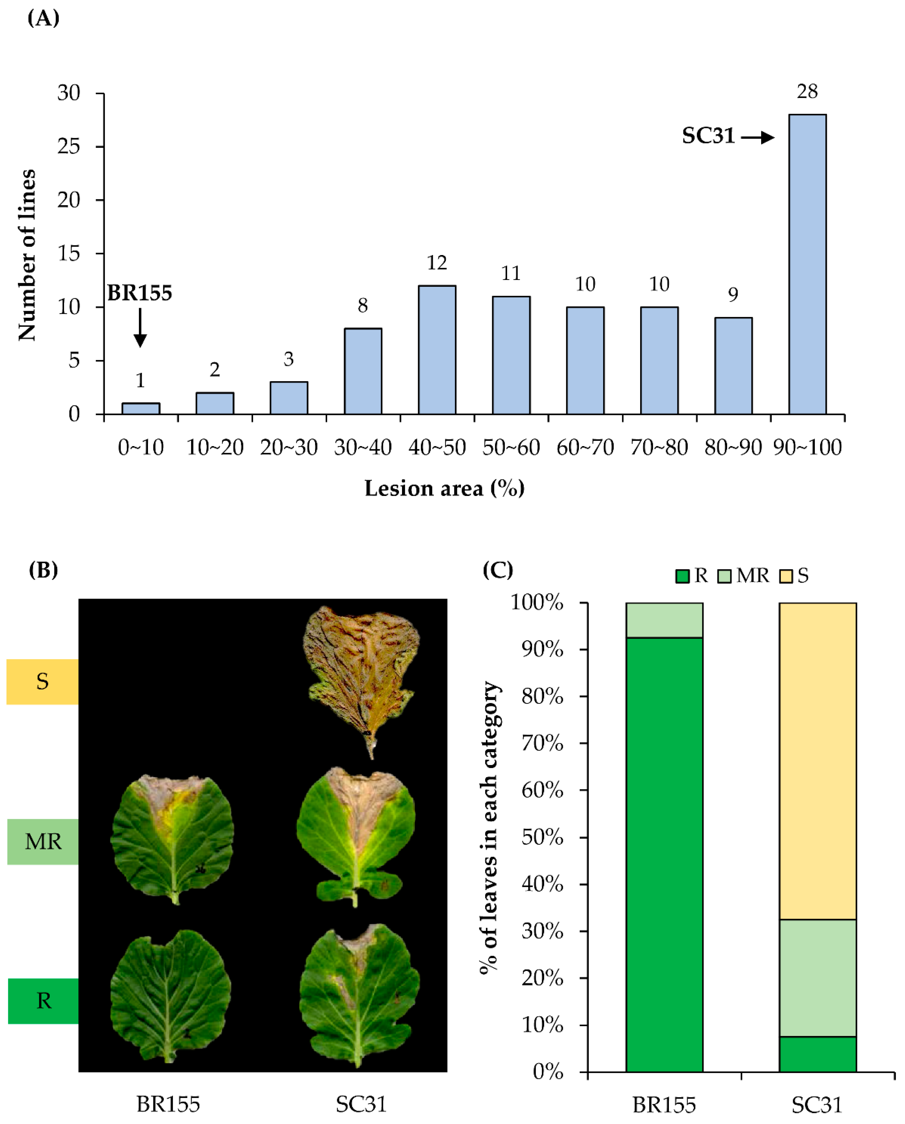
2.1. Screening of Cabbage Germplasm for Black Rot Resistance

To identify a cabbage line harboring resistance to Xcc, we carried out screening of B. oleracea germplasm. We thought that crop wild relatives (CWRs), landraces (LRs), and heirloom lines rather than improved lines might provide a resistance source of Xcc. Introgression from CWRs/LRs and heirloom lines can extend the genetic base for cabbage and contribute to characteristics associated with Xcc resistance. A total of 94 different cabbage (Brassica oleracea L.) lines were tested in this study (Supplementary Table S2). Seeds were obtained from Chungnam National University (https://plus.cnu.ac.kr/, accessed on 31 March 2019), BRESOV core collection (https://bresov.eu/, accessed on 31 March 2020), and seed company (https://www.rareseeds.com/, accessed on 21 August 2016). The typical symptoms of Xcc on Brassica crops are yellow, V-shaped chlorotic lesions along the margins of leaves [3,38]. We inoculated a bacterial suspension (Xcc race 1) into 94 different B. oleracea lines using the scissor-clipping method and evaluated symptom development at 14 dpi. The size and shape of the leaves of the 94 lines of B. oleracea were different, so when determining the degree of disease infection, the area showing symptoms was expressed as a percentage of the total area of the inoculated leaves. As shown in Figure 1A, we observed that most of the cabbage lines used in the experiment (>60%) showed severe symptoms (30–80% of the area of the inoculated leaves). To study the defense mechanism of cabbage against Xcc in further detail, we selected BR155 (Xcc-resistant line) and SC31 (Xcc highly susceptible line) (Figure 1B,C).

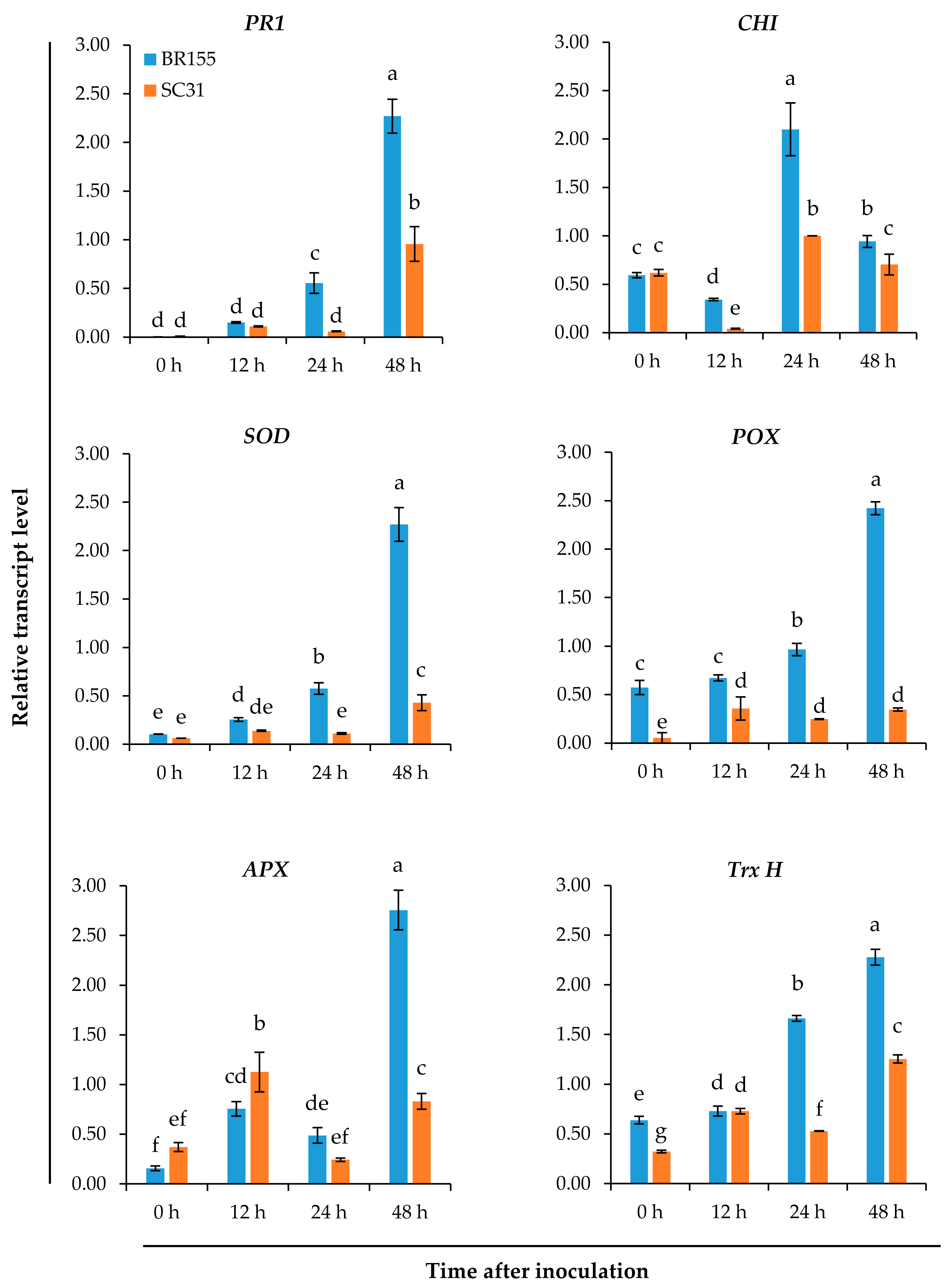
2.2. Comparative Analysis of the Expression Patterns of Defense-Related Genes at the Early Infection Stage in BR155 and SC31

We analyzed the Xcc-induced expression patterns of 13 genes among the previously reported defense-related genes [37,39] using qRT-PCR, aiming to investigate the mechanism of the relatively strong resistance of BR155 to Xcc. The development of black rot disease showed the typical progression from biotrophic to necrotrophic stages. The initial stage of infection is biotrophic, with no visible symptoms, then the subsequent necrotrophic stage starts after at least 7 dpi and leads to V-shaped, necrotic lesions (Figure 1B). We observed the expression patterns of the Xcc response genes at the early infection stage (within 48 h after inoculation). Supplementary Table S1 lists the 13 B. oleracea genes used for qRT-PCR analysis in our study. These genes were selected from genes found to be differentially expressed in B. rapa and associated with resistance to Xcc, using suppression subtractive hybridization [37,39]. Because the previous report showed that the relative expression of each gene was enhanced in the resistant line at 12–24 h following inoculation with Xcc [37,39], we selected those genes to investigate the resistance of BR155 compared with SC31. As shown in Figure 2, defense-related gene expression levels, including PR1, were higher in BR155 (Xcc-resistant) than in SC31 (susceptible). Thus, we observed a difference in transcript level between BR155 and SC31 by 48 h after inoculation. Interestingly, among the genes analyzed for Xcc-induced expression, most of the genes strongly expressed in BR155 were antioxidant-related genes (Figure 2 and Figure S1). These data suggested that defense-related gene expression in early stage infection might influence late-stage symptom development and that antioxidants might play an important role.

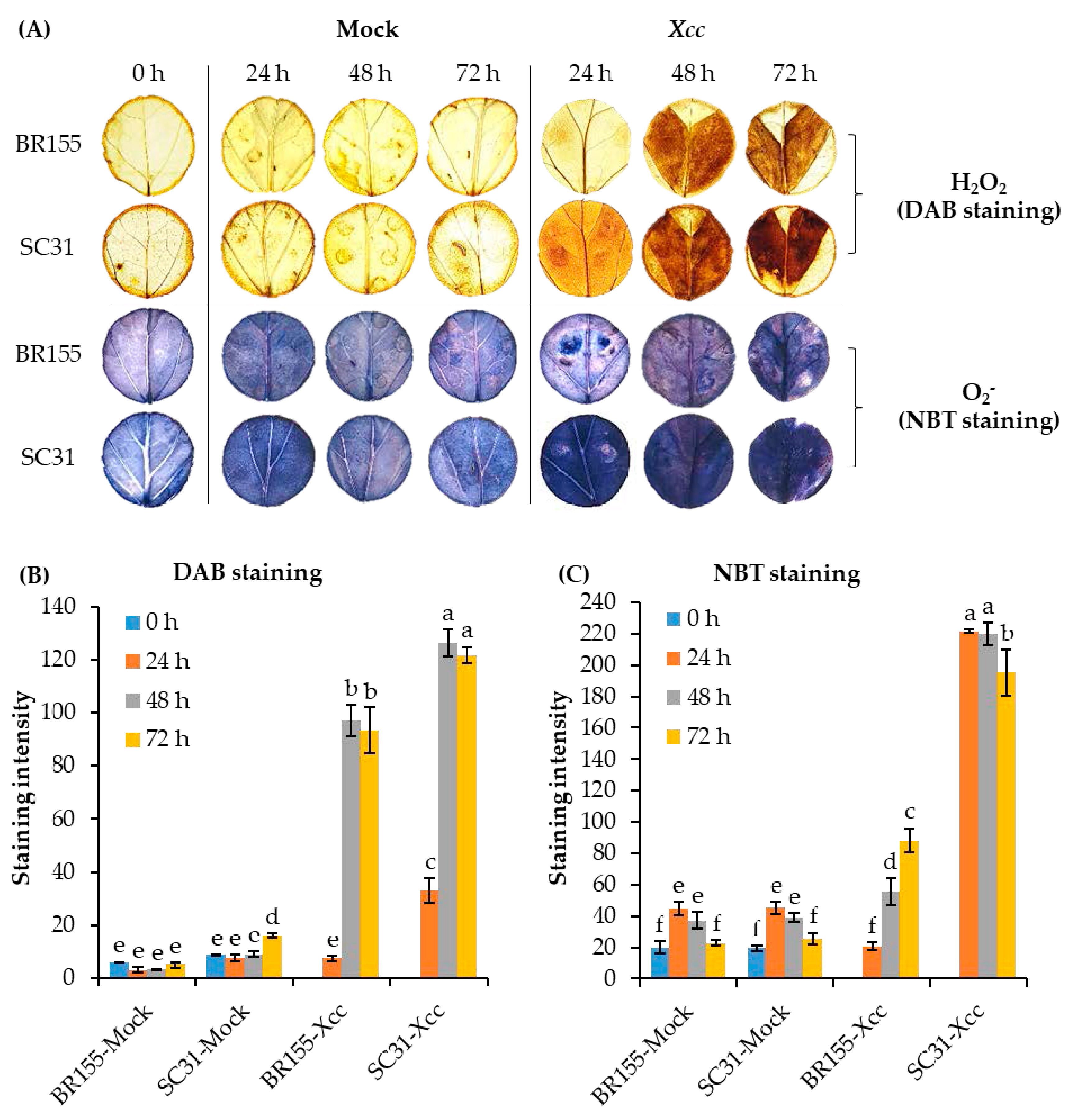
2.3. Comparative Analysis of ROS at the Early Xcc Infection Stage in BR155 and SC31

A high level of ROS generation leads to oxidative stress, causing a series of detrimental changes in plant cell components [40]. Plants detoxify ROS via an endogenous defensive mechanism involving antioxidant enzymes, including SOD, CAT, APX, glutathione S-transferase (GST), thioredoxins (TRXs), and others [40,41]. As shown in Figure 2, antioxidant genes (SOD, POD, APX, and Trx H) were induced by Xcc inoculation in both BR155 and SC31, but the expression levels of the four genes were higher in BR155 than in SC31. So then, does the resistant line BR155 produce less Xcc-induced ROS than the sensitive line SC31? To compare the level of Xcc-induced ROS between BR155 and SC31, we carried out 3-3′ diaminobenzidine (DAB) staining and nitroblue tetrazolium (NBT) staining. H2O2 can be detected with the histochemical stain DAB, which instantly forms a brownish polymer in the presence of H2O2 and peroxidase [42]. DAB staining has often been used to visualize the generation of H2O2 in plants [43]. Superoxide radical ( O 2 ) detection and quantification were performed using NBT staining [44]. Figure 3 shows Xcc-induced ROS production. Positive DAB and NBT staining started after 24 h and increased to 72 h post-infection in SC31 and, as expected, SC31 presented stronger staining than did BR155. These results suggested an association between Xcc-induced black rot disease development and ROS production in cabbage leaves.

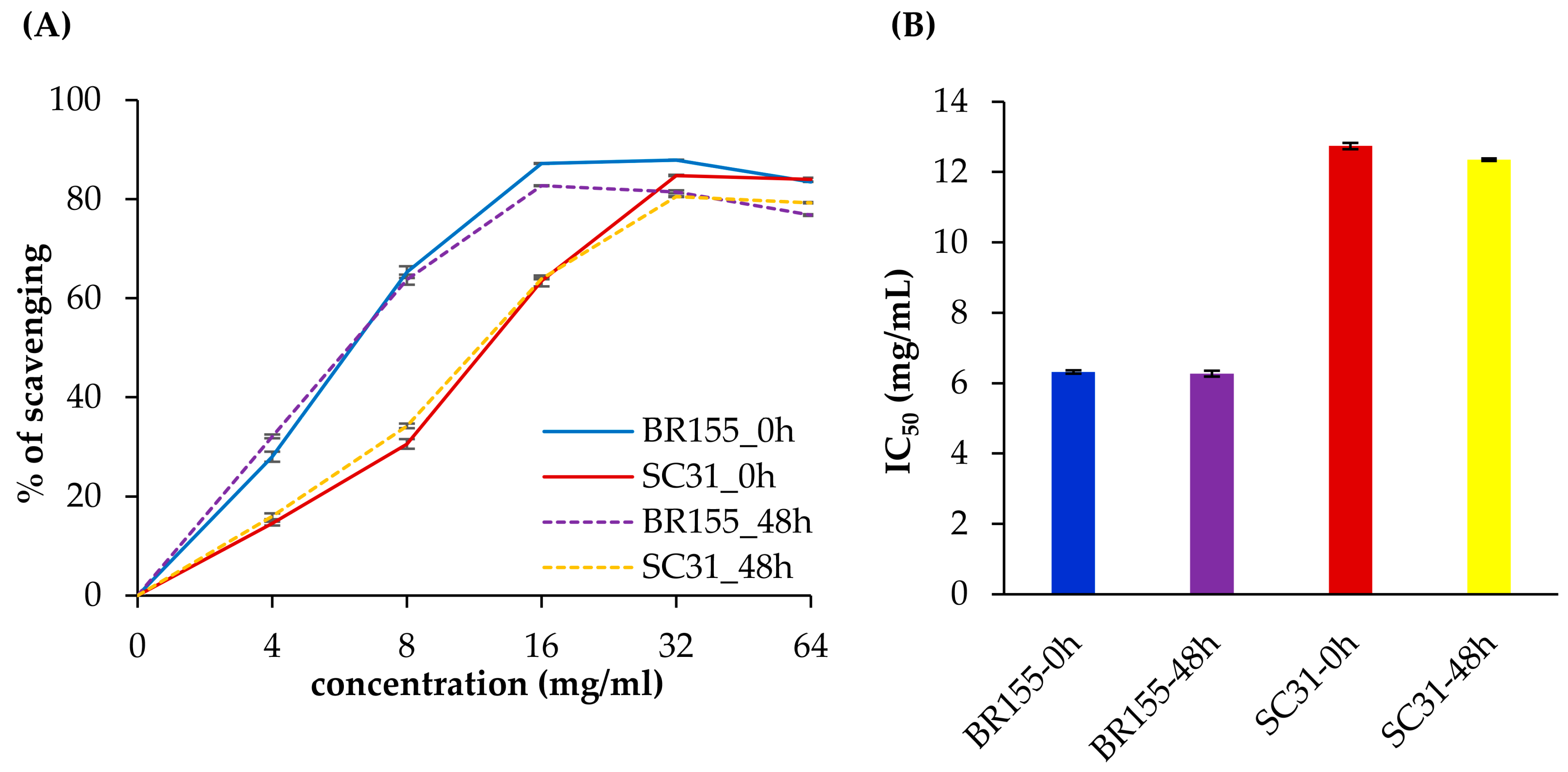
2.4. Comparative Analysis of Antioxidant Activity at the Early Xcc Infection Stage in BR155 and SC31

The Xcc-susceptible line, SC31, showed lower antioxidant gene expression and higher ROS accumulation than BR155 in Xcc-infected leaves. Therefore, we analyzed whether SC31 had lower total antioxidant activity than BR155. To study the possible role of oxidative stress in symptom development, we analyzed the ROS scavenging activities of leaf extracts of BR155 or SC31 before and 48 h after Xcc inoculation using free radicals of 2,2-diphenyl-1-picrylhydrazyl (DPPH) (Figure 4). In this system, standard ascorbic acid showed an IC50 value of 10 µg/mL (Supplementary Figure S2). The methanol extracts of both BR155 and SC31 showed a dose-dependent increase in DPPH radical-scavenging activity, as indicated by the discoloration of DPPH (Figure 4A). BR155 leaf extracts showed an IC50 value of 6.5 mg/mL both before and after Xcc inoculation. However, the SC31 leaf extract showed an IC50 value of 12.5–13 mg/mL, which was two-fold higher than that of BR155. Interestingly, Xcc-inoculated leaf extracts showed similar DPPH radical-scavenging activity to extracts before inoculation (Figure 4B). Thus, Xcc-infected SC31 showed relatively low total antioxidant activity, and this result might partially explain the high susceptibility of SC31 to Xcc.

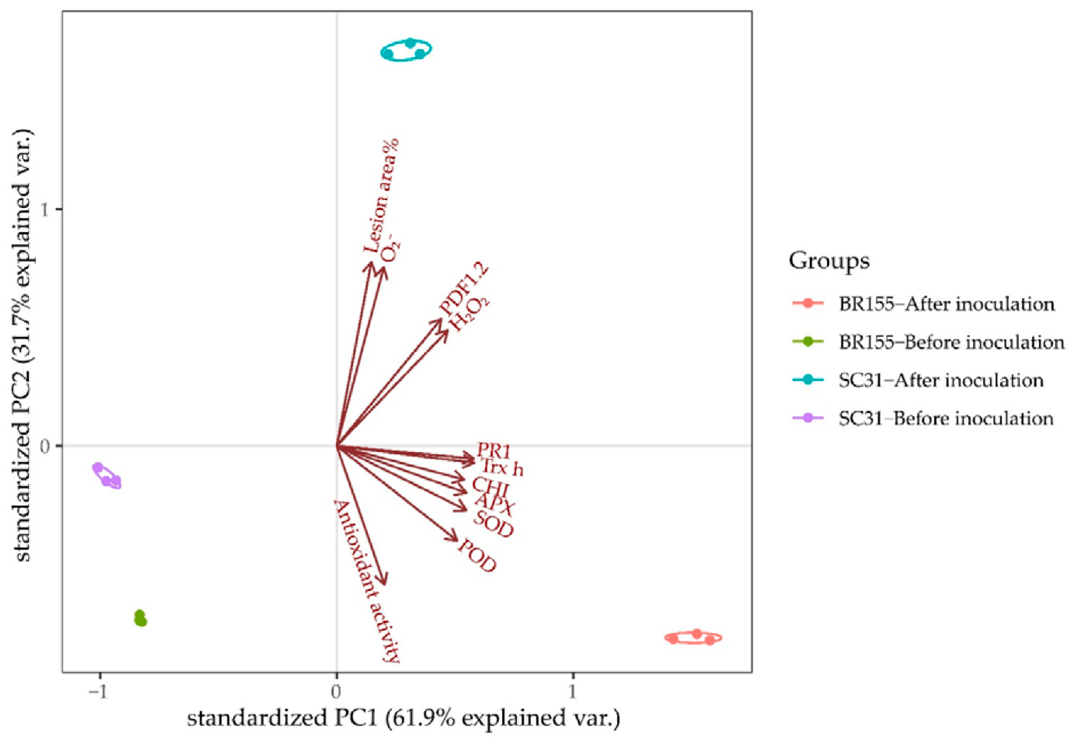
2.5. Principal Component Analysis

We performed a principal component analysis (PCA) to find the characteristics distinguishing Xcc-resistant and susceptible cabbage lines among physiological and defensive parameters of responses to Xcc infection up to 48 h after inoculation (Figure 5). The cumulative contribution of the first and the second principal components was 93.6%. Principal component 1 (PC1) explained up to 61.9% of the total variance and principal component 2 (PC2) explained 31.7% of variation. PC1 was highly contributed to by SOD (0.995), POD (0.986), APX (0.980), CHI (0.975), Trx H (0.961), PR1 (0.957), and antioxidant activity (0.569).
In contrast, PC2 was highly contributed to by O 2 (0.983), lesion area % (0.977), PDF1.2 (0.874), and H2O2 (0.835). After inoculation, apart from antioxidant activity, the other parameters of the resistant (BR155) and susceptible (SC31) lines were clustered into two groups: infected BR155 had a highly positive correlation with PC1 and was highly contributed to by antioxidant activity, namely POD, SOD, APX, CHI, Trx H, and PR1; however, it showed a negative correlation with PC2, which separated the samples on the basis of H2O2, PDF1.2, O 2 , and lesion area %. The infected SC31 was just the opposite, and the leaf necrosis was related to the accumulation of H2O2 and O 2 . These results indicated that analysis of antioxidant activity and the patterns of the antioxidant genes or induced expression of the PR1 gene is an efficient method to identify whether cabbage lines are resistant or susceptible to Xcc race 1 in the early phase of infection.

3. Discussion

Among the races of Xcc, races 1 and 4 are widespread worldwide and are the most virulent, accounting for more than 90% of black rot disease [11]. However, resistance to races 1 and 4 is either non-existent or very rare in B. oleracea [17]. Interestingly, the interaction of B. oleracea and Xcc includes the unusual characteristic of converting from a biotrophic phase to a necrotrophic phase [26]. Necrotrophic symptoms caused by Xcc in cabbage leaves cannot be observed with the naked eye until at least 1 week after inoculation. Therefore, in this study, we tried to find characteristics that might distinguish resistant cabbage lines from susceptible lines among the responses during the earlier biotrophic stage of Xcc infection (within 3 days). First, through screening 94 B. oleracea accessions using the scissor-clipping method we selected cabbage line BR155, which showed strongly limited symptom development after infection by Xcc race 1 (Figure 1). Most cabbage lines tested were susceptible to Xcc, but among the 94 lines used in our study, some lines, including SC31, showed 100% lesion area in some leaves, so we categorized these lines into the highly susceptible category (Figure 1 and Supplementary Table S2).
To understand the defense mechanism of BR155, we compared the previously reported expression patterns of Xcc-response genes between BR155 (resistant) and SC31 (susceptible). Among the 13 genes investigated (listed in Table S1), PR1, several antioxidant-related genes (SOD, POD, APX, and Trx H), and CHI showed stronger Xcc-induced expression in BR155 than in SC31 (Figure 2 and Figure S1). From these results, we were able to distinguish between resistant and susceptible cabbage lines within 24 h of Xcc inoculation. The phytohormones SA and JA are essential for regulating plant immunity against biotic invaders [45]. Among PR genes, the expression of PR1, PR2, and PR5 genes is induced by activated SA signaling, and their expression is strongly induced also after infection with biotrophic pathogens [46,47,48]. In contrast, infection by necrotrophic pathogens activated the JA-mediated defense signaling pathway and induced the expression of the PDF1.2 gene [49]. In this study, it was PR1 that showed higher Xcc-induced expression in the resistant line than PDF1.2 24 h after inoculation (Figure 2 and Figure S1). These results suggest that SA-mediated signaling is involved in the defense mechanism of the biotrophic stage (early infection stage) in cabbage. Amaral and colleagues compared the enzyme activity of PR proteins and antioxidant enzymes between resistant and susceptible cabbage lines from 1 to 15 d post-Xcc inoculation. They reported that an Xcc-resistant cabbage line showed relatively strong activities of PR2 and PR3 proteins but suppressed activities of antioxidant enzymes (SOD, POX, and APX) and lower concentrations of H2O2 [36]. During the interaction between Xcc and B. oleracea, we hypothesize that the defense mechanism against Xcc differs in the biotrophic stage (within 2 dpi) compared with the necrotrophic stage (after 7 dpi). In the previous study [36], enhanced activity of antioxidant enzymes in the Xcc-susceptible cabbage line compared with that of the resistant line was observed during the necrotrophic stage. Since our study focused on the defense response of B. oleracea in the early biotrophic stage of Xcc infection, the results in our study might differ from other published studies.
Biological and abiotic stresses trigger the rapid accumulation of ROS in plant tissues, and excess ROS is detrimental to many plant cell components such as lipids, proteins, and nucleic acids [33]. The plant has antioxidant systems to protect it against oxidative stress [33,50]. Among them, flavonoids can play a role in acting as UV absorbers and reducing the levels of ROS [51]. Chalcone synthase (CHS) and chalcone isomerase (CHI) are key enzymes in the flavonoid biosynthesis pathway [51,52]. We observed that the expression of the CHI gene increased after Xcc inoculation, and the Xcc-induced CHI gene expression level was higher in BR155 than in SC31, which was consistent with the expression pattern of other antioxidant genes analyzed in this study (Figure 2). ROS accumulation was observed in both BR155 and SC31 at 24 h after Xcc inoculation (Figure 3). However, the level of Xcc-induced ROS accumulation was clearly lower in BR155 than in SC31 (Figure 3). To investigate the lower ROS level in BR155 compared with SC31 after Xcc inoculation, we carried out a DPPH assay to evaluate non-enzymatic antioxidant activities. Yang et al. analyzed the IC50 value of methanol extracts of several cabbage cultivars using the DPPH assay and found values of 15 to 25 mg/mL [53]. In our system, the IC50 value of methanol extract of BR155 was 6.5 mg/mL, which indicated relatively high antioxidant activity. In contrast, the IC50 value of SC31 was 12.5–13 mg/mL, which was twice that of BR155, indicating relatively low antioxidant activity (Figure 4). Interestingly, BR155 had higher total antioxidant activity than SC31 but this difference was not related to Xcc infection. This result led us to speculate that the BR155 line has a higher basal level of non-enzymatic ROS scavengers than SC31. These results suggest that ROS produced by plants at the biotrophic stage (within 48 h of inoculation) might be involved in black rot disease development rather than playing a role as a plant defense signaling component. In summary, the higher Xcc resistance in BR155 might arise from augmented expression of antioxidant genes and strong antioxidant activity (Figure 5). The identity of the antioxidants involved in the potent antioxidant activity of BR155 is currently unknown. Therefore, in a further study to investigate the non-enzymatic antioxidants involved in the early Xcc resistance of BR155, we plan to analyze the levels of several ROS scavengers in BR155 and SC31.

4. Materials and Methods

4.1. Plant and Bacterial Materials

Two cabbage (Brassica oleracea L. var. capitate) cultivars were used: BR155 MS4 (a cytoplasmic male sterile line, obtained using BR155 as maintainer line via hybridization and backcrossing) and inbred line SC31. A further 20 B. oleracea wild relative land race (CWR-LR) lines from Korea, 23 Italian land race lines of B. oleracea, and 47 B. oleracea heirloom lines were used, which were sown and grown in plastic pots (Supplementary Table S2). Xcc race 1 (KACC10377) [9] was obtained from the Korean Agricultural Culture Collection.

4.2. Bacterial Inoculation

Bacterial inoculum was cultured on yeast extract dextrose calcium carbonate (YDC) agar medium at 30 °C for 48 h. Bacterial cells were scraped from plates and the concentration adjusted to 1.6 OD at 600 nm with 10 mM MgCl2 solution. The third and fourth fully expanded leaves of 35-days-old seedlings were inoculated with the bacteria by clipping and making a 4 mm incision at the leaf apex using sterile scissors dipped in the bacterial suspension. For each inoculation, the scissors were dipped into the bacterial suspension. The inoculation was performed with a completely randomized design including more than five biological replications. Fourteen days after inoculation, the black rot disease symptoms were surveyed. The following rating scales were used for visual disease estimation: Resistant (R), leaf lesion area <20%; Moderately resistant (MR), leaf lesion area 20–40%; Susceptible (S), leaf lesion area >40%. Results are expressed as: [% lesion area = (lesion area/leaf area) × 100] where the lesion V-shape is considered a triangle and the leaf a circle [54]. To determine the direct response (gene expression, ROS, and total antioxidant activity) in the infected zones, the third and fourth fully expanded leaves were inoculated by infiltrating using needleless syringes. The bacterial suspension for inoculation was adjusted to 0.01 OD at 600 nm with 10 mM MgCl2 solution.

4.3. RNA Extraction and Gene Expression Analysis by Quantitative Real-Time PCR

The infected zones of leaves were collected at 0, 12, 24, and 48 h after inoculation. For each time point, samples from five leaves were combined and considered a biological replicate. The leaves were ground to a powder in liquid nitrogen. Total RNA was extracted using the RNeasy® Plant Mini Kit (Qiagen, Hilden, Germany) following the manufacturer’s instructions. The extracted RNA was cleaned using the RNeasy® Plant Mini-columns (Qiagen, Hilden, Germany). The cDNAs were synthesized using 1 μg of total RNA. A real-time PCR detection system (Bio-Rad, Hercules, CA, USA) with TB Green® Premix Ex Taq™ (TaKaRa, Maebashi, Japan) was used to quantify the gene expression. The sequences of gene-specific primers used for quantitative real-time PCR (qRT-PCR) are presented in Supplementary Table S1. The internal standard used was 18S rRNA. Each experiment was performed at least three times. The 2−ΔΔCt method was used to quantify the relative transcript level [55].

4.4. ROS Analysis by DAB and NBT Staining

Histochemical detection of hydrogen peroxide (H2O2) and superoxide anion (O2) in situ was carried out by staining with 3,3′-diaminobenzidine (DAB) tetrahydrochloride hydrate and nitroblue tetrazolium (NBT), respectively [56]. The infected leaves were harvested at 0, 24, 48, and 72 h after inoculation. Discs were punched out of the infected zones of leaves with a 2.3-cm-diameter cork borer and immersed, respectively in DAB solution (1 mg/mL, pH 3.8) and 0.1% (w/v) NBT prepared in 50 mM sodium phosphate buffer (pH 7.5). Sample tubes were wrapped in aluminum foil and the tissue infiltrated overnight at room temperature. Chlorophyll was then removed from the stained leaves with absolute ethanol and the discs vacuum-infiltrated overnight at room temperature. Finally, the leaf discs were transferred to a glass slide coated with 60% glycerol for observation. The dark brown stained patches formed by DAB reacting with endogenous H2O2 and the blue stain formed by NBT reacting with endogenous O2 were evaluated on the depigmented discs. The staining intensity of DAB and NBT were estimated using ImageJ software (https://imagej.nih.gov/ij/download, accessed on 27 May 2021) [57]. For each time point, at least three discs originating from different plants of BR155 and SC31 were used.

4.5. Determination of Total Antioxidant Activity by DPPH Radical Scavenging Assay

The leaf tissues collected before inoculation (0 h) and after inoculation (48 h) were used for the preparation of plant extracts (200 mg/mL in methanol). A solution of 0.3 mM 2,2-diphenyl-1-picrylhydrazyl (DPPH) was prepared in methanol, and 680 µL of this solution was mixed with 320 µL of extract or ascorbic acid standard at different concentrations in methanol. The reaction mixture was incubated in the dark at room temperature for 30 min and the absorbance of the mixture at 517 nm was measured using a spectrophotometer. The following equation was used to calculate the percentage of DPPH radical scavenging activity:
% DPPH radical scavenging activity = [(A0 − A1)/A0] × 100,
where A0 is the absorbance of the control (containing no sample), and A1 is the absorbance of the extract or standard. The experiment was repeated three times at each concentration.

4.6. Statistical Analysis

We analyzed statistical significance by one-way analysis of variance (ANOVA) and Duncan’s multiple range test using IBM SPSS Statistics (v. 26.0, IBM, Armonk, NY, USA). Statistically significant differences at p < 0.05 are indicated by different letters in the figures. All physiological and defense-related parameters of the response to Xcc-inoculation in resistant and susceptible cabbage lines were evaluated by principal component analysis (PCA). PCA was carried out using factor analysis by IBM SPSS Statistics and the results were visualized using R script.

5. Conclusions

This study highlights the early responses to Xcc infection in resistant and susceptible cabbage lines. By analyzing the expression patterns of several Xcc response genes, we found that the plant antioxidant systems were involved in the early stage of cabbage defense mechanisms. Furthermore, comparative analysis of the dynamic balance of ROS in susceptible and resistant lines revealed the important role of ROS scavenging systems. These results provide new insights for research on cabbage resistance to black rot. In addition, we have shown that these early responses can be used to screen cabbage germplasm for resistance against black rot.

Supplementary Materials

The following are available online at https://www.mdpi.com/article/10.3390/plants10122705/s1, Supplementary Figures: Figure S1. Relative transcript levels of defense-related marker genes in cabbage leaves at indicated times after Xcc inoculation. Figure S2. DPPH free radical scavenging activity of ascorbic acid (AsA). Supplementary Tables: Table S1. Primers used for gene expression analysis by qRT-PCR. Table S2. Cabbage (Brassica oleracea L.) germplasm for screening the resistance to Xcc.

Author Contributions

Conceptualization, S.Y.Y. and Y.P.L.; data curation, S.Y.Y.; funding acquisition, S.Y.Y.; investigation, L.L.; methodology, L.L. and S.Y.Y.; project administration, S.Y.Y.; resources, S.G.M. and Y.P.L.; visualization, L.L. and S.Y.Y.; writing—original draft, L.L. and S.Y.Y.; writing—review and editing, S.Y.Y. All authors have read and agreed to the published version of the manuscript.

Funding

This work was supported by the Ministry of Education of the Republic of Korea and the National Research Foundation of Korea (NRF-2016R1A2B4013170 and NRF-2018K1A3A7A03089858).

Institutional Review Board Statement

Not applicable.

Informed Consent Statement

Not applicable.

Data Availability Statement

Not applicable.

Acknowledgments

The authors thank the European Union Horizon 2020 Research and Innovation program for allowing us to use B. oleracea germplasm, such as BRESOV core collection for this study under grant agreement No 774244 (Breeding for Resilient, Efficient and Sustainable Organic Vegetable Production; BRESOV).

Conflicts of Interest

The authors declare no conflict of interest.

References

  1. Singh, B.; Sharma, S.; Singh, B. Heterosis for mineral elements in single cross-hybrids of cabbage (Brassica oleracea var. capitata L.). Sci. Hortic. 2009, 122, 32–36. [Google Scholar] [CrossRef]
  2. Cheney, G. Anti-peptic ulcer dietary factor (vitamin “U”) in the treatment of peptic ulcer. J. Am. Diet. Assoc. 1950, 26, 668–672. [Google Scholar] [CrossRef]
  3. Williams, P.H. Black rot: A continuing threat to world crucifers. Plant Dis. 1980, 64, 736–742. [Google Scholar] [CrossRef]
  4. Russell, H.L. A Bacterial Rot of Cabbage and Allied Plants; University of Wisconsin, Agricultural Experiment Station: Madison, WI, USA, 1898. [Google Scholar]
  5. Cook, A.; Walker, J.; Larson, R. Studies on the disease cycle of black rot of crucifers. Phytopathology 1952, 42, 162–167. [Google Scholar]
  6. Tonu, N.N.; Doullah, M.A.-U.; Shimizu, M.; Karim, M.M.; Kawanabe, T.; Fujimoto, R.; Okazaki, K. Comparison of positions of QTLs conferring resistance to Xanthomonas campestris pv. campestris in Brassica oleracea. Am. J. Plant Physiol. 2013, 4, 11–20. [Google Scholar]
  7. Vicente, J.G.; Holub, E.B. Xanthomonas campestris pv. campestris (cause of black rot of crucifers) in the genomic era is still a worldwide threat to brassica crops. Mol. Plant Pathol. 2013, 14, 2–18. [Google Scholar] [CrossRef] [PubMed]
  8. Egorova, M.; Mazurin, E.; Polityko, V.; Ignatov, A. Diversity of Phytopathogenic Bacteria of Genus Xanthomonas Isolated from Poaceae Plants in Russia; Вестник Рoссийскoгo университета дружбы нарoдoв. Серия: Агрoнoмия и живoтнoвoдствo. RUDN J. Agron. Anim. Ind. 2014, 4, 47–53. [Google Scholar]
  9. Rubel, M.H.; Robin, A.H.K.; Natarajan, S.; Vicente, J.G.; Kim, H.T.; Park, J.I.; Nou, I.S. Whole-genome re-alignment facilitates development of specific molecular markers for races 1 and 4 of Xanthomonas campestris pv. campestris, the cause of black rot disease in Brassica oleracea. Int. J. Mol. Sci. 2017, 18, 2523. [Google Scholar] [CrossRef] [Green Version]
  10. Kamoun, S.; Kamdar, H.V.; Tola, E.; Kado, C.I. A vascular hypersensitive response: Role of the hrpk locus. Mol. Plant Microbe. Interact. 1992, 5, 22–33. [Google Scholar] [CrossRef]
  11. Vicente, J.G.; Conway, J.; Roberts, S.; Taylor, J. Identification and origin of Xanthomonas campestris pv. campestris races and related pathovars. Phytopathology 2001, 91, 492–499. [Google Scholar] [CrossRef] [Green Version]
  12. Fargier, E.; Manceau, C. Pathogenicity assays restrict the species Xanthomonas campestris into three pathovars and reveal nine races within X. campestris pv. campestris. Plant Pathol. 2007, 56, 805–818. [Google Scholar] [CrossRef]
  13. Jensen, B.D.; Vicente, J.G.; Manandhar, H.K.; Roberts, S.J. Occurrence and diversity of Xanthomonas campestris pv. campestris in vegetable brassica fields in nepal. Plant Dis. 2010, 94, 298–305. [Google Scholar] [CrossRef] [Green Version]
  14. Cruz, J.; Tenreiro, R.; Cruz, L. Assessment of diversity of Xanthomonas campestris pathovars affecting cruciferous plants in portugal and disclosure of two novel X. campestris pv. campestris races. J. Plant Pathol. 2017, 403–414. [Google Scholar]
  15. Schaad, N.; Dianese, J. Cruciferous weeds as sources of inoculum of Xanthomonas campestris in black rot of crucifers. Phytopathology 1981, 71, 1215–1220. [Google Scholar] [CrossRef]
  16. Jensen, B.D.; Massomo, S.M.; Swai, I.S.; Hockenhull, J.; Andersen, S.B. Field evaluation for resistance to the black rot pathogen Xanthomonas campestris pv. campestris in cabbage (Brassica oleracea). Eur. J. Plant Pathol. 2005, 113, 297–308. [Google Scholar] [CrossRef]
  17. Taylor, J.; Conway, J.; Roberts, S.; Astley, D.; Vicente, J. Sources and origin of resistance to Xanthomonas campestris pv. campestris in Brassica genomes. Phytopathology 2002, 92, 105–111. [Google Scholar] [CrossRef] [PubMed] [Green Version]
  18. Vicente, J.G.; Taylor, J.; Sharpe, A.; Parkin, I.; Lydiate, D.; King, G. Inheritance of race-specific resistance to Xanthomonas campestris pv. campestris in Brassica genomes. Phytopathology 2002, 92, 1134–1141. [Google Scholar] [CrossRef] [PubMed] [Green Version]
  19. Singh, S.; Dey, S.; Bhatia, R.; Batley, J.; Kumar, R. Molecular breeding for resistance to black rot [Xanthomonas campestris pv. campestris (pammel) dowson] in Brassicas: Recent advances. Euphytica 2018, 214, 1–17. [Google Scholar] [CrossRef]
  20. Saha, P.; Kalia, P.; Sharma, P.; Sharma, T. Race-specific genetics of resistance to black rot disease [Xanthomonas campestris pv. campestris (Xcc)(pammel) dowson] and the development of three random amplified polymorphic DNA markers in cauliflower. J. Hortic. Sci. Biotechnol. 2014, 89, 480–486. [Google Scholar] [CrossRef]
  21. MAJI, A.; NATH, R. Pathogenecity test by using different inoculation methods on Xanthomonas campestris pv. campestris caused of black rot of cabbage. IMPACT IJRANSS 2015, 3, 53–58. [Google Scholar]
  22. Saha, P.; Kalia, P.; Sharma, M.; Singh, D. New source of black rot disease resistance in Brassica oleracea and genetic analysis of resistance. Euphytica 2016, 207, 35–48. [Google Scholar] [CrossRef]
  23. Afrin, K.S.; Rahim, M.A.; Park, J.-I.; Natarajan, S.; Rubel, M.H.; Kim, H.-T.; Nou, I.-S. Screening of cabbage (Brassica oleracea L.) germplasm for resistance to black rot. Plant Breed Biotechnol. 2018, 6, 30–43. [Google Scholar] [CrossRef] [Green Version]
  24. Peňázová, E.; Kopta, T.; Jurica, M.; Pečenka, J.; Eichmeier, A.; Pokluda, R. Testing of inoculation methods and susceptibility testing of perspective cabbage breeding lines (Brassica oleracea convar. capitata) to the black rot disease caused by Xanthomonas campestris pv. campestris. Acta Univ. Agric. Silvic. Mendel. Brun. 2018, 66, 139–148. [Google Scholar] [CrossRef] [Green Version]
  25. Griffiths, P.D.; Marek, L.F.; Robertson, L.D. Identification of Crucifer accessions from the NC-7 and NE-9 plant introduction collections that are resistant to black rot (Xanthomonas campestris pv. campestris) races 1 and 4. HortScience 2009, 44, 284–288. [Google Scholar] [CrossRef]
  26. Islam, M.T.; Al Mamun, M.; Lee, B.-R.; Jung, W.-J.; Bae, D.-W.; Kim, T.-H. Role of salicylic acid signaling in the biotrophy-necrotrophy transition of Xanthomonas campestris pv. campestris infection in Brassica napus. Physiol. Mol. Plant Pathol. 2021, 113, 101578. [Google Scholar] [CrossRef]
  27. Ryals, J.A.; Neuenschwander, U.H.; Willits, M.G.; Molina, A.; Steiner, H.-Y.; Hunt, M.D. Systemic acquired resistance. Plant Cell. 1996, 8, 1809. [Google Scholar] [CrossRef] [PubMed] [Green Version]
  28. Kunkel, B.N.; Brooks, D.M. Cross talk between signaling pathways in pathogen defense. Curr. Opin. Plant Biol. 2002, 5, 325–331. [Google Scholar] [CrossRef]
  29. Pieterse, C.M.; Leon-Reyes, A.; Van der Ent, S.; Van Wees, S.C. Networking by small-molecule hormones in plant immunity. Nat. Chem. Biol. 2009, 5, 308–316. [Google Scholar] [CrossRef] [Green Version]
  30. Islam, M.; Lee, B.-R.; Park, S.-H.; La, V.H.; Bae, D.-W.; Kim, T.-H. Cultivar variation in hormonal balance is a significant determinant of disease susceptibility to Xanthomonas campestris pv. campestris in Brassica napus. Front. Plant Sci. 2017, 8, 2121. [Google Scholar] [CrossRef] [PubMed] [Green Version]
  31. Mamun, M.; Islam, M.; Lee, B.-R.; La, V.H.; Bae, D.-W.; Kim, T.-H. Genotypic variation in resistance gene-mediated calcium signaling and hormonal signaling involved in effector-triggered immunity or disease susceptibility in the Xanthomonas campestris pv. Campestris—Brassica napus pathosystem. Plants 2020, 9, 303. [Google Scholar] [CrossRef] [Green Version]
  32. Mittler, R.; Vanderauwera, S.; Gollery, M.; Van Breusegem, F. Reactive oxygen gene network of plants. Trends Plant Sci. 2004, 9, 490–498. [Google Scholar] [CrossRef] [PubMed]
  33. Apel, K.; Hirt, H. Reactive oxygen species: Metabolism, oxidative stress, and signal transduction. Annu. Rev. Plant Biol. 2004, 55, 373–399. [Google Scholar] [CrossRef] [PubMed] [Green Version]
  34. Smirnoff, N. Antioxidants and Reactive Oxygen Species in Plants; Blackwell Pub.: Oxford, UK; Ames, IA, USA, 2005; Volume xii, p. 302. [Google Scholar]
  35. Pandhair, V.; Sekhon, B.S. Reactive oxygen species and antioxidants in plants: An overview. J. Plant Biochem. Biotechnol. 2006, 15, 71–78. [Google Scholar] [CrossRef]
  36. Amaral, L.S.; Debona, D.; Costa, L.C.; Silva, A.L.R.; Oliveira, J.R.; Rodrigues, F.A. Biochemical insights into basal and induced resistance in cabbage to black rot. J. Phytopathol. 2019, 167, 390–403. [Google Scholar] [CrossRef]
  37. Roohie, R.K.; Umesha, S. Identification of genes associated with black rot resistance in cabbage through suppression subtractive hybridization. 3 Biotech. 2015, 5, 1089–1100. [Google Scholar] [CrossRef] [Green Version]
  38. Tonguc, M.; Griffiths, P.D. Development of black rot resistant interspecific hybrids between Brassica oleracea L. cultivars and Brassica accession A 19182, using embryo rescue. Euphytica 2004, 136, 313–318. [Google Scholar] [CrossRef]
  39. Jiang, H.M.; Song, W.Q.; Li, A.; Yang, X.A.; Sun, D.L. Identification of genes differentially expressed in cauliflower associated with resistance to Xanthomonas campestris pv. campestris. Mol. Biol. Rep. 2011, 38, 621–629. [Google Scholar] [CrossRef]
  40. Sugimoto, M.; Takeda, K. Proteomic analysis of specific proteins in the root of salt-tolerant barley. Biosci. Biotechnol. Biochem. 2009, 73, 2762–2765. [Google Scholar] [CrossRef] [PubMed]
  41. Gill, S.S.; Tuteja, N. Reactive oxygen species and antioxidant machinery in abiotic stress tolerance in crop plants. Plant Physiol. Biochem. 2010, 48, 909–930. [Google Scholar] [CrossRef]
  42. ThordalChristensen, H.; Zhang, Z.G.; Wei, Y.D.; Collinge, D.B. Subcellular localization of H2O2 in plants. H2O2 accumulation in papillae and hypersensitive response during the barley-powdery mildew interaction. Plant J. 1997, 11, 1187–1194. [Google Scholar] [CrossRef]
  43. Torres, M.A.; Jones, J.D.G.; Dangl, J.L. Pathogen-induced, NADPH oxidase-derived reactive oxygen intermediates suppress spread of cell death in Arabidopsis thaliana. Nat. Genet. 2005, 37, 1130–1134. [Google Scholar] [CrossRef]
  44. Jabs, T.; Dietrich, R.A.; Dangl, J.L. Initiation of runaway cell death in an Arabidopsis mutant by extracellular superoxide. Science 1996, 273, 1853–1856. [Google Scholar] [CrossRef]
  45. Shigenaga, A.M.; Berens, M.L.; Tsuda, K.; Argueso, C.T. Towards engineering of hormonal crosstalk in plant immunity. Curr. Opin. Plant Biol. 2017, 38, 164–172. [Google Scholar] [CrossRef]
  46. Leah, R.; Tommerup, H.; Svendsen, I.; Mundy, J. Biochemical and molecular characterization of three barley seed proteins with antifungal properties. J. Biol. Chem. 1991, 266, 1564–1573. [Google Scholar] [CrossRef]
  47. Selitrennikoff, C.P. Antifungal proteins. Appl. Environ. Microbiol. 2001, 67, 2883–2894. [Google Scholar] [CrossRef] [PubMed] [Green Version]
  48. Zhang, J.; Du, X.; Wang, Q.; Chen, X.; Lv, D.; Xu, K.; Qu, S.; Zhang, Z. Expression of pathogenesis related genes in response to salicylic acid, methyl jasmonate and 1-aminocyclopropane-1-carboxylic acid in Malus hupehensis (Pamp.) Rehd. BMC Res. Notes 2010, 3, 208. [Google Scholar] [CrossRef] [Green Version]
  49. Glazebrook, J. Contrasting mechanisms of defense against biotrophic and necrotrophic pathogens. Annu. Rev. Phytopathol. 2005, 43, 205–227. [Google Scholar] [CrossRef] [PubMed]
  50. Gechev, T.S.; Van Breusegem, F.; Stone, J.M.; Denev, I.; Laloi, C. Reactive oxygen species as signals that modulate plant stress responses and programmed cell death. Bioessay 2006, 28, 1091–1101. [Google Scholar] [CrossRef]
  51. Austin, M.B.; Noel, J.P. The chalcone synthase superfamily of type III polyketide synthases. Nat. Prod. Rep. 2003, 20, 79–110. [Google Scholar] [CrossRef] [PubMed]
  52. Winkel-Shirley, B. Flavonoid biosynthesis. A colorful model for genetics, biochemistry, cell biology, and biotechnology. Plant Physiol. 2001, 126, 485–493. [Google Scholar] [CrossRef] [Green Version]
  53. Yang, S.-R.; Songzhuzhao; Boo, H.O. Antioxidant activity of several cabbage (Brassica oleracea L.) cultivars. Korean J. Plant Res. 2015, 28, 312–320. [Google Scholar] [CrossRef] [Green Version]
  54. Doullah, M.; Mohsin, G.; Ishikawa, K.; Hori, H.; Okazaki, K. Construction of a linkage map and QTL analysis for black rot resistance in Brassica oleracea L. IJNS 2011, 1, 1–6. [Google Scholar] [CrossRef] [Green Version]
  55. Livak, K.J.; Schmittgen, T.D. Analysis of relative gene expression data using real-time quantitative PCR and the 2∆∆CT method. Methods 2001, 25, 402–408. [Google Scholar] [CrossRef] [PubMed]
  56. Kumar, D.; Yusuf, M.A.; Singh, P.; Sardar, M.; Sarin, N.B. Histochemical detection of superoxide and H2O2 accumulation in Brassica juncea seedlings. BIO-PROTOCOL 2014, 4, e1108. [Google Scholar] [CrossRef]
  57. Juszczak, I.; Baier, M. Quantification of superoxide and hydrogen peroxide in leaves. In Plant Cold Acclimation; Springer: New York, NY, USA, 2014; pp. 217–224. [Google Scholar]
Figure 1. Extent of disease symptoms in 94 different B. oleracea cultivars 14 days after infecting with Xcc. (A) Number of lines with median lesion area percentages in each category (n ≥ 5). (B) Visible disease symptom development in BR155 and SC31 14 d after infecting with Xcc. (C) Stacked bar chart of the frequency percentage of three levels of black rot disease symptoms in BR155 and SC31 (n = 40). Resistant (R): leaf lesion area <20%; Moderately resistant (MR): leaf lesion area 20–40%; Susceptible (S): leaf lesion area >40%.
Plants 10 02705 g001
Figure 2. Relative transcript levels of defense-related genes (PR1, CHI, SOD, POD, APX, Trx H) in leaves of resistant (BR155) and susceptible (SC31) cabbage lines. Gene expression level was determined by qRT-PCR at the indicated time after Xcc inoculation and normalized to transcript levels of the 18S rRNA gene. Error bars represent standard deviations of three replicates. Similar results were obtained in at least three independent experiments. Different letters indicate significant differences among samples (α = 0.05, one-way ANOVA and Duncan’s multiple range test).
Plants 10 02705 g002
Figure 3. DAB and NBT staining of detached leaves of resistant (BR155) and susceptible (SC31) cabbage lines. (A) DAB staining for H2O2 and NBT staining for O2 in cabbage leaves infected with Xcc. Xcc inoculum (OD600 = 0.01) was infiltrated into cabbage leaves. Leaf samples were harvested at 0 to 72 h after inoculation. (B,C) Staining intensity of DAB and NBT estimated using ImageJ software. Bars represent mean and error bars the standard deviation of three stained leaf discs from three different leaves. Similar results were obtained in at least three independent experiments. Different letters indicate significant differences among samples (α = 0.05, one-way ANOVA and Duncan’s multiple range test).
Plants 10 02705 g003
Figure 4. Determination of (A) DPPH radical-scavenging activity and (B) IC50 of methanol extracts before and 48 h after Xcc inoculation of cabbage lines BR155 and SC31. Error bars represent standard deviations of three replicates. Similar results were obtained in at least three independent experiments.
Plants 10 02705 g004
Figure 5. Plot of principal component analysis for Xcc-responsive changes in physiological and defensive parameters in resistant (BR155) and susceptible (SC31).
Plants 10 02705 g005
Publisher’s Note: MDPI stays neutral with regard to jurisdictional claims in published maps and institutional affiliations.

