
Screening for Selective Anticancer Activity of 65 Extracts of Plants Collected in Western Andalusia, Spain

 , ,
, ,
Abstract
:1. Introduction
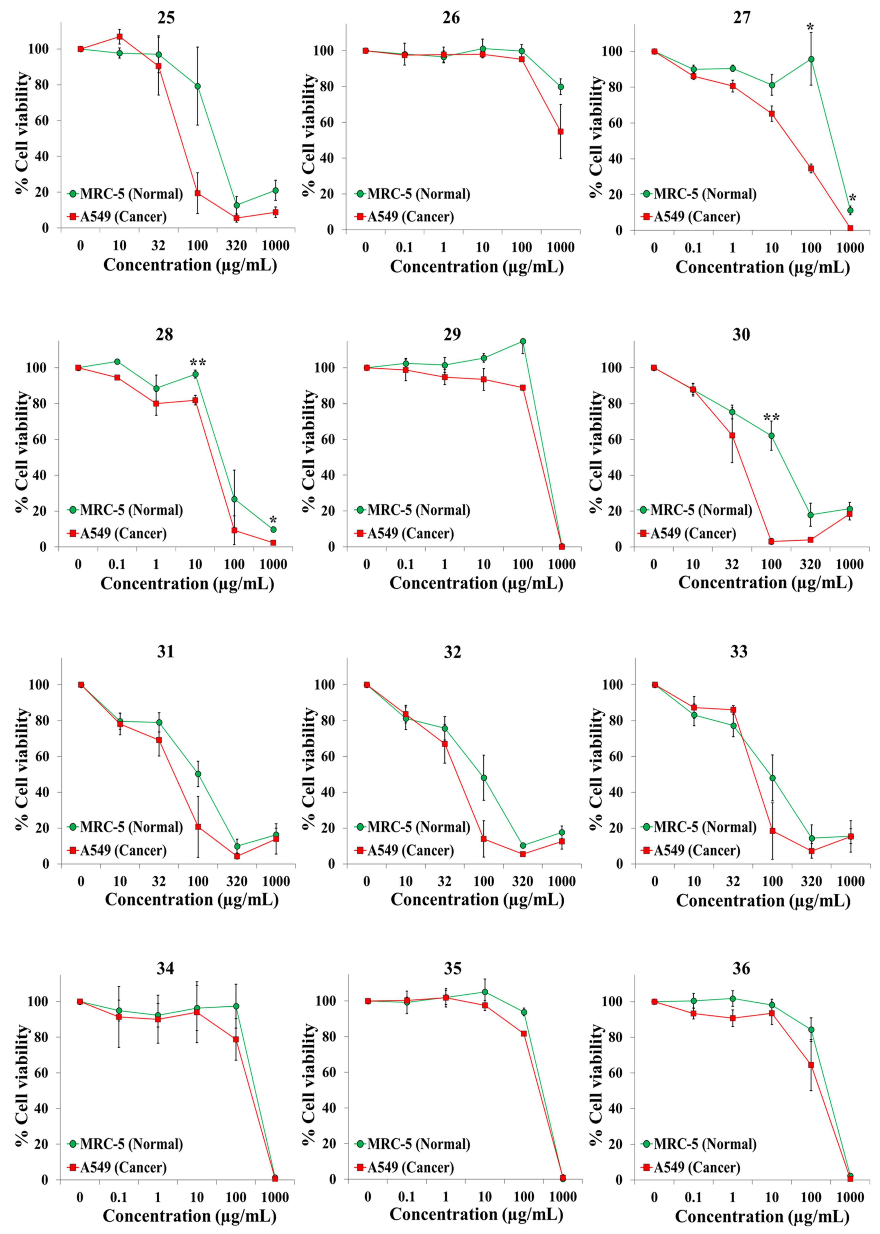
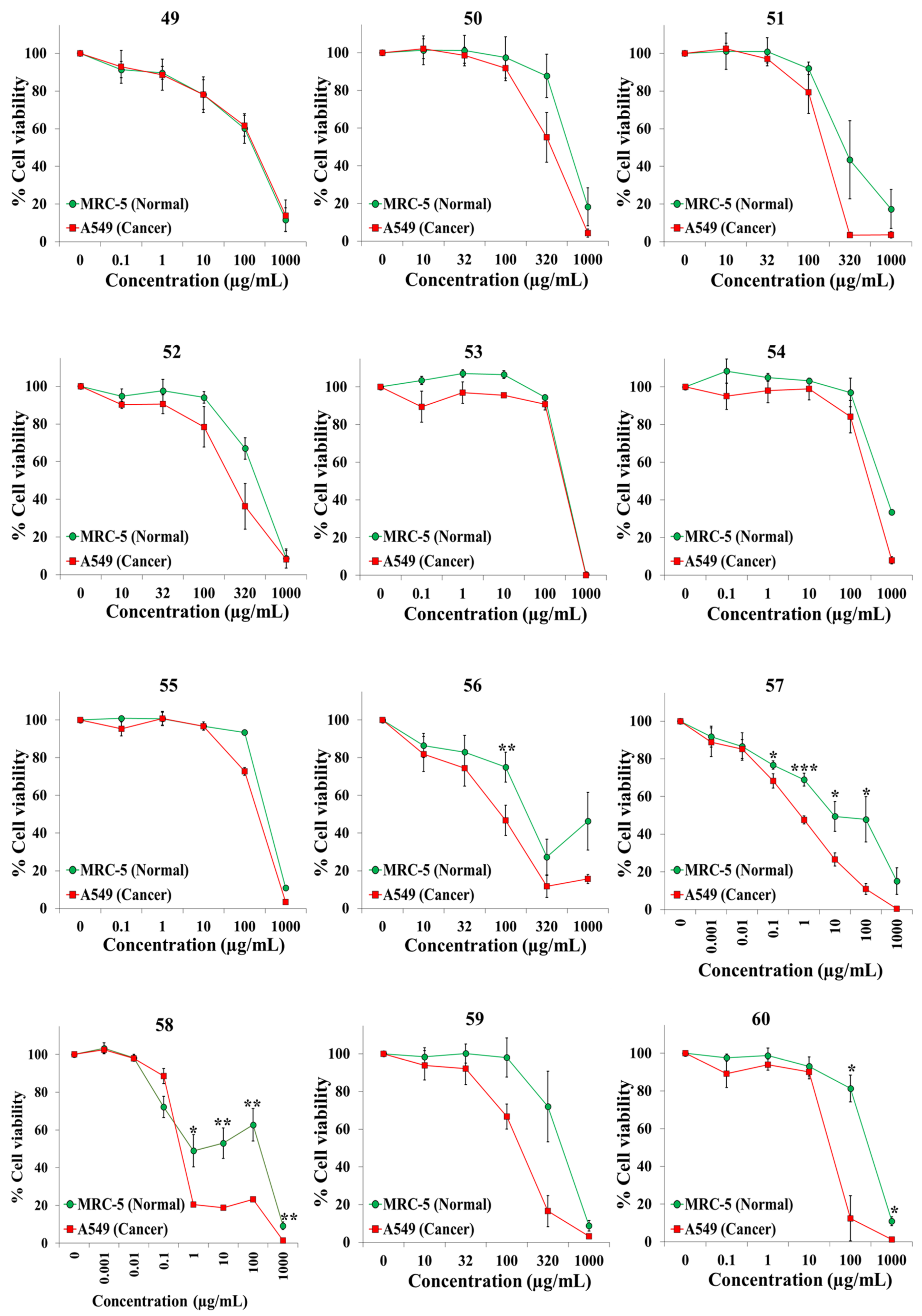
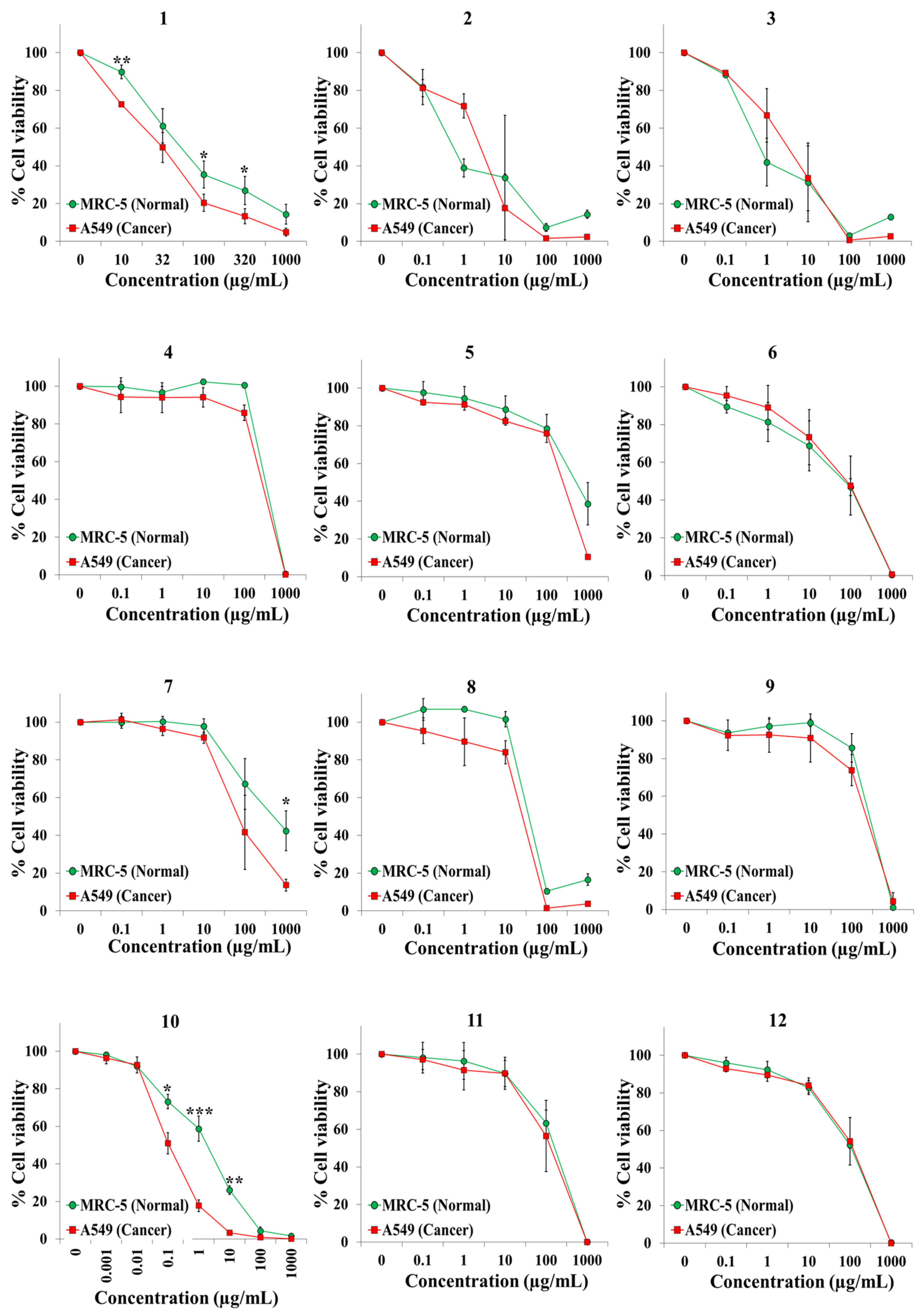
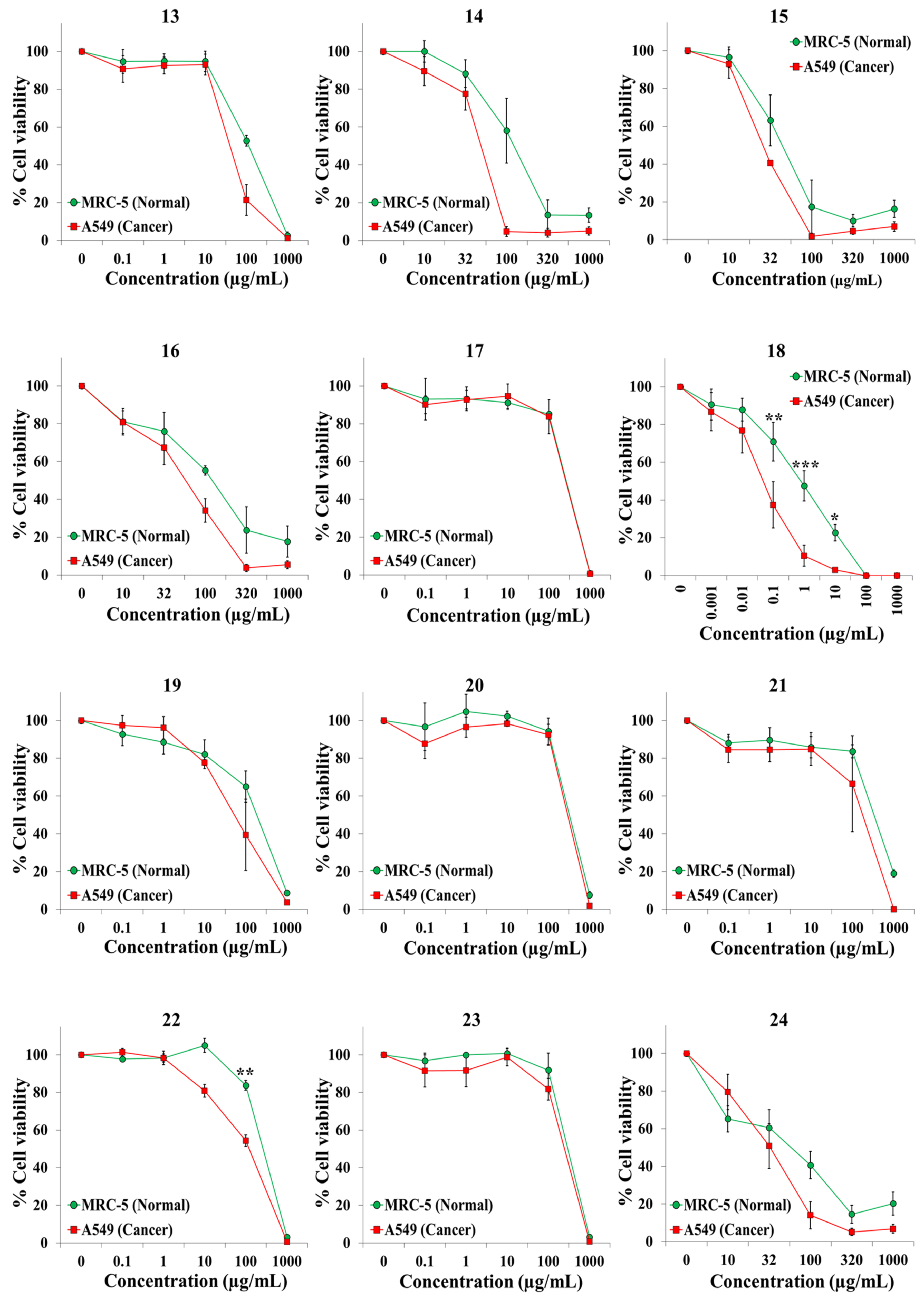
2. Results and Discussion
3. Materials and Methods
3.1. Plant Material
3.2. Preparation of the Extracts
3.3. Chemicals and Cell Lines
3.4. Cell Viability Assays
Supplementary Materials
Author Contributions
Funding
Institutional Review Board Statement
Informed Consent Statement
Data Availability Statement
Acknowledgments
Conflicts of Interest
References
- Siegel, R.L.; Miller, K.D.; Fuchs, H.E.; Jemal, A. Cancer Statistics, 2021. CA Cancer J. Clin. 2021, 71, 7–33. [Google Scholar] [CrossRef]
- López-Lázaro, M. A simple and reliable approach for assessing anticancer activity in vitro. Curr. Med. Chem. 2015, 22, 1324–1334. [Google Scholar] [CrossRef] [PubMed]
- López-Lázaro, M. Two preclinical tests to evaluate anticancer activity and to help validate drug candidates for clinical trials. Oncoscience 2015, 2, 91–98. [Google Scholar] [CrossRef] [PubMed]
- López-Lázaro, M. How many times should we screen a chemical library to discover an anticancer drug? Drug Discov. Today 2015, 20, 167–169. [Google Scholar] [CrossRef] [PubMed]
- Cragg, G.M.; Grothaus, P.G.; Newman, D.J. Impact of natural products on developing new anti-cancer agents. Chem. Rev. 2009, 109, 3012–3043. [Google Scholar] [CrossRef]
- Newman, D.J.; Cragg, G.M. Natural Products as Sources of New Drugs over the Nearly Four Decades from 01/1981 to 09/2019. J. Nat. Prod. 2020, 83, 770–803. [Google Scholar] [CrossRef]
- Kingston, D.G.I.; Newman, D.J. The search for novel drug leads for predominately antitumor therapies by utilizing mother nature’s pharmacophoric libraries. Curr. Opin. Drug Discov. Devel. 2005, 8, 207–227. [Google Scholar] [PubMed]
- Wani, M.C.; Taylor, H.L.; Wall, M.E.; Coggon, P.; Mcphail, A.T. Plant Antitumor Agents.VI.The Isolation and Structure of Taxol, a Novel Antileukemic and Antitumor Agent from Taxus brevifolia. J. Am. Chem. Soc. 1971, 54, 2347–2360. [Google Scholar]
- Ruiz-Ceja, K.A.; Chirino, Y.I. Current FDA-approved treatments for non-small cell lung cancer and potential biomarkers for its detection. Biomed. Pharmacother. 2017, 90, 24–37. [Google Scholar] [CrossRef]
- Calderón-Montaño, J.M.; Martínez-Sánchez, S.M.; Burgos-Morón, E.; Guillén-Mancina, E.; Jiménez-Alonso, J.J.; García, F.; Aparicio, A.; López-Lázaro, M. Screening for selective anticancer activity of plants from Grazalema Natural Park, Spain. Nat. Prod. Res. 2019, 33, 3454–3458. [Google Scholar] [CrossRef]
- Valderrama, J.A.; Delgado, V.; Sepúlveda, S.; Benites, J.; Theoduloz, C.; Buc Calderon, P.; Muccioli, G.G. Synthesis and Cytotoxic Activity on Human Cancer Cells of Novel Isoquinolinequinone-Amino Acid Derivatives. Molecules 2016, 21, 1199. [Google Scholar] [CrossRef] [PubMed] [Green Version]
- Motadi, L.R.; Choene, M.S.; Mthembu, N.N. Anticancer Properties of Tulbaghia Violacea Regulate the Expression of P53-Dependent Mechanisms in Cancer Cell Lines. Sci. Rep. 2020, 10, 1–11. [Google Scholar] [CrossRef] [PubMed]
- Gao, J.; Luo, T.; Wang, J. Gene Interfered-Ferroptosis Therapy for Cancers. Nat. Commun. 2021, 12, 1–16. [Google Scholar] [CrossRef]
- Hahn, W.C.; Counter, C.M.; Lundberg, A.S.; Beijersbergen, R.L.; Brooks, M.W.; Weinberg, R.A. Creation of human tumour cells with defined genetic elements. Nature 1999, 400, 464–468. [Google Scholar] [CrossRef]
- Calderón-Montaño, J.M.; Burgos-Morón, E.; Orta, M.L.; Mateos, S.; López-Lázaro, M. A hydroalcoholic extract from the leaves of Nerium oleander inhibits glycolysis and induces selective killing of lung cancer cells. Planta Med. 2013, 79, 1017–1023. [Google Scholar] [CrossRef] [PubMed] [Green Version]
- López-Lázaro, M.; Palma De La Peña, N.; Pastor, N.; Martín-Cordero, C.; Navarro, E.; Cortés, F.; Ayuso, M.J.; Toro, M.V. Anti-tumour activity of Digitalis purpurea L. subsp. heywoodii. Planta Med. 2003, 69, 701–704. [Google Scholar] [PubMed]
- López-Lázaro, M.; Pastor, N.; Azrak, S.S.; Ayuso, M.J.; Austin, C.A.; Cortés, F. Digitoxin inhibits the growth of cancer cell lines at concentrations commonly found in cardiac patients. J. Nat. Prod. 2005, 68, 1642–1645. [Google Scholar] [CrossRef]
- López-Lázaro, M. Digitoxin as an anticancer agent with selectivity for cancer cells: Possible mechanisms involved. Expert Opin. Ther. Targets 2007, 11, 1043–1053. [Google Scholar] [CrossRef]
- Calderón-Montaño, J.M.; Burgos-Morón, E.; Orta, M.L.; Maldonado-Navas, D.; García-Domínguez, I.; López-Lázaro, M. Evaluating the cancer therapeutic potential of cardiac glycosides. Biomed. Res. Int. 2014, 2014, 794930. [Google Scholar] [CrossRef]
- Ramos-Silva, A.; Tavares-Carreón, F.; Figueroa, M.; De la Torre-Zavala, S.; Gastelum-Arellanez, A.; Rodríguez-García, A.; Galán-Wong, L.J.; Avilés-Arnaut, H. Anticancer potential of Thevetia peruviana fruit methanolic extract. BMC Complement. Altern. Med. 2017, 17, 241. [Google Scholar] [CrossRef]
- Calderón-Montaño, J.M.; Burgos-Morón, E.; López-Lázaro, M. The in vivo antitumor activity of cardiac glycosides in mice xenografted with human cancer cells is probably an experimental artifact. Oncogene 2014, 33, 2947–2948. [Google Scholar] [CrossRef] [PubMed] [Green Version]
- Fujino, T.; Kuroda, M.; Matsuo, Y.; Kubo, S.; Tamura, C.; Sakamoto, N.; Mimaki, Y.; Hayakawa, M. Cardenolide glycosides from the seeds of Digitalis purpurea exhibit carcinoma-specific cytotoxicity toward renal adenocarcinoma and hepatocellular carcinoma cells. Biosci. Biotechnol. Biochem. 2015, 79, 177–184. [Google Scholar] [CrossRef] [PubMed]
- Zhu, J.-J.; Zhang, X.-X.; Miao, Y.-Q.; He, S.-F.; Tian, D.-M.; Yao, X.-S.; Tang, J.-S.; Gan, Y. Delivery of acetylthevetin B, an antitumor cardiac glycoside, using polymeric micelles for enhanced therapeutic efficacy against lung cancer cells. Acta Pharmacol. Sin. 2017, 38, 290–300. [Google Scholar] [CrossRef] [PubMed]
- Youssef, D.T.; Frahm, A.W. Alkaloids of the flowers of Pancratium maritimum. Planta Med. 1998, 64, 669–670. [Google Scholar] [CrossRef] [PubMed]
- Ibrahim, S.; Mohamed, G.; Shaala, L.; Youssef, D.; El Sayed, K. New Alkaloids from Pancratium maritimum. Planta Med. 2013, 79, 1480–1484. [Google Scholar] [CrossRef] [Green Version]
- Witherup, K.M.; Look, S.A.; Stasko, M.W.; Ghiorzi, T.J.; Muschik, G.M.; Cragg, G.M. Taxus spp. needles contain amounts of taxol comparable to the bark of Taxus brevifolia: Analysis and isolation. J. Nat. Prod. 1990, 53, 1249–1255. [Google Scholar] [CrossRef] [PubMed]
- Duquesnoy, E.; Paoli, M.; Castola, V.; Bighelli, A.; Casanova, J. Identification of taxanes in extracts from leaves of Taxus baccata L. using (13)C-NMR spectroscopy. Phytochem. Anal. 2009, 20, 246–252. [Google Scholar] [CrossRef]
- Azémard, C.; Ménager, M.; Vieillescazes, C. On the tracks of sandarac, review and chemical analysis. Environ. Sci. Pollut. Res. 2017, 24, 27746–27754. [Google Scholar] [CrossRef]
- Jlizi, S.; Lahmar, A.; Zardi-Bergaoui, A.; Ascrizzi, R.; Flamini, G.; Harrath, A.H.; Chekir-Ghedira, L.; Ben Jannet, H. Chemical Composition and Cytotoxic Activity of the Fractionated Trunk Bark Essential Oil from Tetraclinis articulata (Vahl) Mast. Growing in Tunisia. Molecules 2021, 26, 1110. [Google Scholar] [CrossRef]
- El Jemli, M.; Kamal, R.; Marmouzi, I.; Doukkali, Z.; Bouidida, E.H.; Touati, D.; Nejjari, R.; El Guessabi, L.; Cherrah, Y.; Alaoui, K. Chemical composition, acute toxicity, antioxidant and anti-inflammatory activities of Moroccan Tetraclinis articulata L. J. Tradit. Complement. Med. 2017, 7, 281–287. [Google Scholar] [CrossRef] [Green Version]
- Ho, S.T.; Tung, Y.T.; Kuo, Y.H.; Lin, C.C.; Wu, J.H. Ferruginol inhibits non-small cell lung cancer growth by inducing caspase-associated apoptosis. Integr. Cancer Ther. 2015, 14, 86–97. [Google Scholar] [CrossRef]
- Yu, X.; Lin, H.; Wang, Y.; Lv, W.; Zhang, S.; Qian, Y.; Deng, X.; Feng, N.; Yu, H.; Qian, B. D-limonene exhibits antitumor activity by inducing autophagy and apoptosis in lung cancer. Onco Targets Ther. 2018, 11, 1833–1847. [Google Scholar] [CrossRef] [Green Version]
- Chung, K.S.; Hong, J.Y.; Lee, J.H.; Lee, H.J.; Park, J.Y.; Choi, J.H.; Park, H.J.; Hong, J.; Lee, K.T. Β-Caryophyllene in the essential oil from chrysanthemum boreale induces G1 phase cell cycle arrest in human lung cancer cells. Molecules 2019, 24, 3754. [Google Scholar] [CrossRef] [PubMed] [Green Version]
- Sirakanyan, S.N.; Spinelli, D.; Geronikaki, A.; Hakobyan, E.K.; Sahakyan, H.; Arabyan, E.; Zakaryan, H.; Nersesyan, L.E.; Aharonyan, A.S.; Danielyan, I.S.; et al. Synthesis, Antitumor Activity, and Docking Analysis of New Pyrido [3,2:4,5] furo (thieno)[3,2-d]pyrimidin-8-amines. Molecules 2019, 24, 3952. [Google Scholar] [CrossRef] [PubMed] [Green Version]
- Zhao, J.; Li, Q.Q.; Zou, B.; Wang, G.; Li, X.; Kim, J.E.; Cuff, C.F.; Huang, L.; Reed, E.; Gardner, K. In vitro combination characterization of the new anticancer plant drug β-elemene with taxanes against human lung carcinoma. Int. J. Oncol. 2007, 31, 241–252. [Google Scholar] [CrossRef] [PubMed] [Green Version]
- Kaczirek, K.; Schindl, M.; Weinhäusel, A.; Scheuba, C.; Passler, C.; Prager, G.; Raderer, M.; Hamilton, G.; Mittlböck, M.; Siegl, V.; et al. Cytotoxic Activity of Camptothecin and Paclitaxel in Newly Established Continuous Human Medullary Thyroid Carcinoma Cell Lines. J. Clin. Endocrinol. Metab. 2004, 89, 2397–2401. [Google Scholar] [CrossRef] [Green Version]
- Martín-Banderas, L.; Muñoz-Rubio, I.; Prados, J.; Álvarez-Fuentes, J.; Calderón-Montaño, J.M.; López-Lázaro, M.; Arias, J.L.; Leiva, M.C.; Holgado, M.A.; Fernández-Arévalo, M. In vitro and in vivo evaluation of Delta (9)-tetrahidrocannabinol/PLGA nanoparticles for cancer chemotherapy. Int. J. Pharm. 2015, 487, 205–212. [Google Scholar] [CrossRef] [PubMed]


| Extract | Plant Name (Family) | Part Used | Voucher Number (SEV) | Origin | IC50 (MTT) (Mean ± SEM, µg/mL) | S.I. (Mean ± SEM) | |
|---|---|---|---|---|---|---|---|
| A549 (Cancer) | MRC-5 (Normal) | ||||||
| 1 | Acis autumnalis (L.) Sweet (Amaryllidaceae) | Whole plant | 284654 | Seville | 32.9 ± 9.6 | 59.1 ± 18.0 | 1.9 ± 0.6 |
| 2 | Anagallis monelli L. (Primulaceae) | Aerial Parts | 284675 | Cádiz | 3.2 ± 1.4 | 0.6 ± 0.2 | 0.3 ± 0.2 |
| 3 | Anagallis monelli L. (Primulaceae) | Root | 284675 | Cádiz | 7.4 ± 5.2 | 0.9 ± 0.5 | 0.3 ± 0.3 |
| 4 | Anthyllis hamosa Desf. (Leguminosae) | Whole plant | 284685 | Huelva | 260.9 ± 14.7 | 317.7 ± 3.1 | 1.2 ± 0.1 |
| 5 | Aristolochia baetica L. (Aristolochiaceae) | Fruits | 284674 | Seville | 249.1 ± 2.5 | 649.9 ± 345.0 | 2.6 ± 1.4 |
| 6 | Aristolochia baetica L. (Aristolochiaceae) | Leaves | 284674 | Seville | 91.8 ± 70.5 | 66.0 ± 39.9 | 0.9 ± 0.3 |
| 7 | Armeria pungens (Link) Hoffmanns. & Link (Plumbaginaceae) | Flowering aerial parts | 284687 | Huelva | 143.4 ± 64.3 | 550.7 ± 352.2 | 3.6 ± 1.1 |
| 8 | Armeria velutina Welw. ex Boiss. & Reut. (Plumbaginaceae) | Whole plant | 284689 | Huelva | 25.7 ± 1.9 | 36.7 ± 0.9 | 1.4 ± 0.1 |
| 9 | Campanula lusitanica L. (Campanulaceae) | Whole plant | 284667 | Seville | 223.4 ± 51.2 | 263.7 ± 34.2 | 1.3 ± 0.4 |
| 10 | Cascabela thevetia (L.) Lippold (Apocynaceae) | Leaves | 284662 | Seville | 0.14 ± 0.02 | 1.6 ± 0.5 | 26.6 ± 11.6 |
| 11 | Centaurea sphaerocephala L. (Compositae) | Whole plant | 284683 | Huelva | 134.2 ± 82.4 | 160.4 ± 33.8 | 1.7 ± 0.8 |
| 12 | Centaurea sphaerocephala L. (Compositae) | Flowers | 284676 | Cádiz | 139.1 ± 40.1 | 108.7 ± 7.7 | 1.7 ± 1.1 |
| 13 | Centaurea sphaerocephala L. (Compositae) | Leaves | 284676 | Cádiz | 43.1 ± 11.1 | 114.7 ± 14.4 | 3.0 ± 0.8 |
| 14 | Cistus crispus L. (Cistaceae) | Leaves | 284660 | Seville | 52.4 ± 5.1 | 126.4 ± 34.3 | 2.5 ± 0.7 |
| 15 | Cistus crispus L. (Cistaceae) | Root | 284660 | Seville | 24.9 ± 0.7 | 58.8 ± 14.8 | 2.1 ± 0.7 |
| 16 | Cistus salviifolius L. (Cistaceae) | Leaves | 284653 | Seville | 56.6 ± 12.6 | 142.7 ± 20.4 | 3.0 ± 0.9 |
| 17 | Cleome violacea L. (Cleomaceae) | Aerial Parts | 284668 | Seville | 253.9 ± 39.9 | 261.1 ± 3.4 | 1.1 ± 0.2 |
| 18 | Digitalis purpurea L. (Plantaginaceae) | Leaves | 284691 | Huelva | 0.17 ± 0.15 | 1.06 ± 0.69 | 9.3 ± 0.5 |
| 19 | Dorycnium rectum (L.) Ser. (Leguminosae) | Flowers | 284690 | Seville | 102.1 ± 40.2 | 182.9 ± 46.1 | 4.3 ± 3.2 |
| 20 | Dorycnium rectum (L.) Ser. (Leguminosae) | Leaves | 284690 | Seville | 293.6 ± 21.2 | 322.1 ± 27.4 | 1.1 ± 0.2 |
| 21 | Echium gaditanum Boiss. (Boraginaceae) | Aerial Parts | 284684 | Huelva | 196.1 ± 107.2 | 325.6 ± 44.0 | 1.1 ± 0.1 |
| 22 | Elaeoselinum foetidum (L.) Boiss. (Apiaceae) | Flowers | 284670 | Seville | 119.0 ± 16.2 | 262.1 ± 8.7 | 2.3 ± 0.3 |
| 23 | Elaeoselinum foetidum (L.) Boiss. (Apiaceae) | Leaves | 284670 | Seville | 246.5 ± 25.5 | 295.0 ± 36.4 | 1.2 ± 0.0 |
| 24 | Erica arborea L. (Ericaceae) | Bark | 284657 | Seville | 32.2 ± 5.6 | 62.1 ± 28.9 | 1.8 ± 0.7 |
| 25 | Erica arborea L. (Ericaceae) | Leaves | 284657 | Seville | 45.7 ± 7.4 | 158.4 ± 43.6 | 3.9 ± 1.4 |
| 26 | Erophaca baetica (L.) Boiss. (Leguminosae) | Leaves | 284673 | Seville | >1000 | >1000 | N.D. |
| 27 | Frangula alnus Mill. (Rhamnaceae) | Bark | 284680 | Huelva | 32.8 ± 7.4 | 339.3 ± 74.1 | 12.4 ± 3.3 |
| 28 | Frangula alnus Mill. (Rhamnaceae) | Leaves | 284680 | Huelva | 28.6 ± 3.5 | 74.0 ± 38.5 | 2.4 ± 1.0 |
| 29 | Genista hirsuta M.Vahl (Leguminosae) | Aereal Parts | 284671 | Seville | 273.6 ± 5.8 | 363.9 ± 27.9 | 1.3 ± 0.1 |
| 30 | Halimium calycinum (L.) K.Koch (Cistaceae) | Leaves | 284656 | Seville | 47.4 ± 10.0 | 135.4 ± 23.6 | 4.3 ± 1.9 |
| 31 | Halimium calycinum (L.) K.Koch (Cistaceae) | Root | 284656 | Seville | 60.1 ± 17.7 | 101.1 ± 13.6 | 2.2 ± 0.8 |
| 32 | Halimium halimifolium (L.) Willk. (Cistaceae) | Leaves | 284659 | Seville | 47.9 ± 10.9 | 105.9 ± 21.4 | 2.7 ± 0.9 |
| 33 | Halimium halimifolium (L.) Willk. (Cistaceae) | Root | 284659 | Seville | 67.5 ± 16.0 | 110.2 ± 33.4 | 1.8 ± 0.8 |
| 34 | Hedysarum coronarium L. (Leguminosae) | Flowers | 284677 | Cádiz | 230.5 ± 54.6 | 306.9 ± 55.1 | 1.4 ± 0.1 |
| 35 | Hedysarum coronarium L. (Leguminosae) | Fruits | 284677 | Cádiz | 247.0 ± 3.2 | 294.5 ± 0.4 | 1.2 ± 0.0 |
| 36 | Hedysarum coronarium L. (Leguminosae) | Leaves | 284677 | Cádiz | 173.9 ± 60.6 | 262.0 ± 32.3 | 2.1 ± 0.9 |
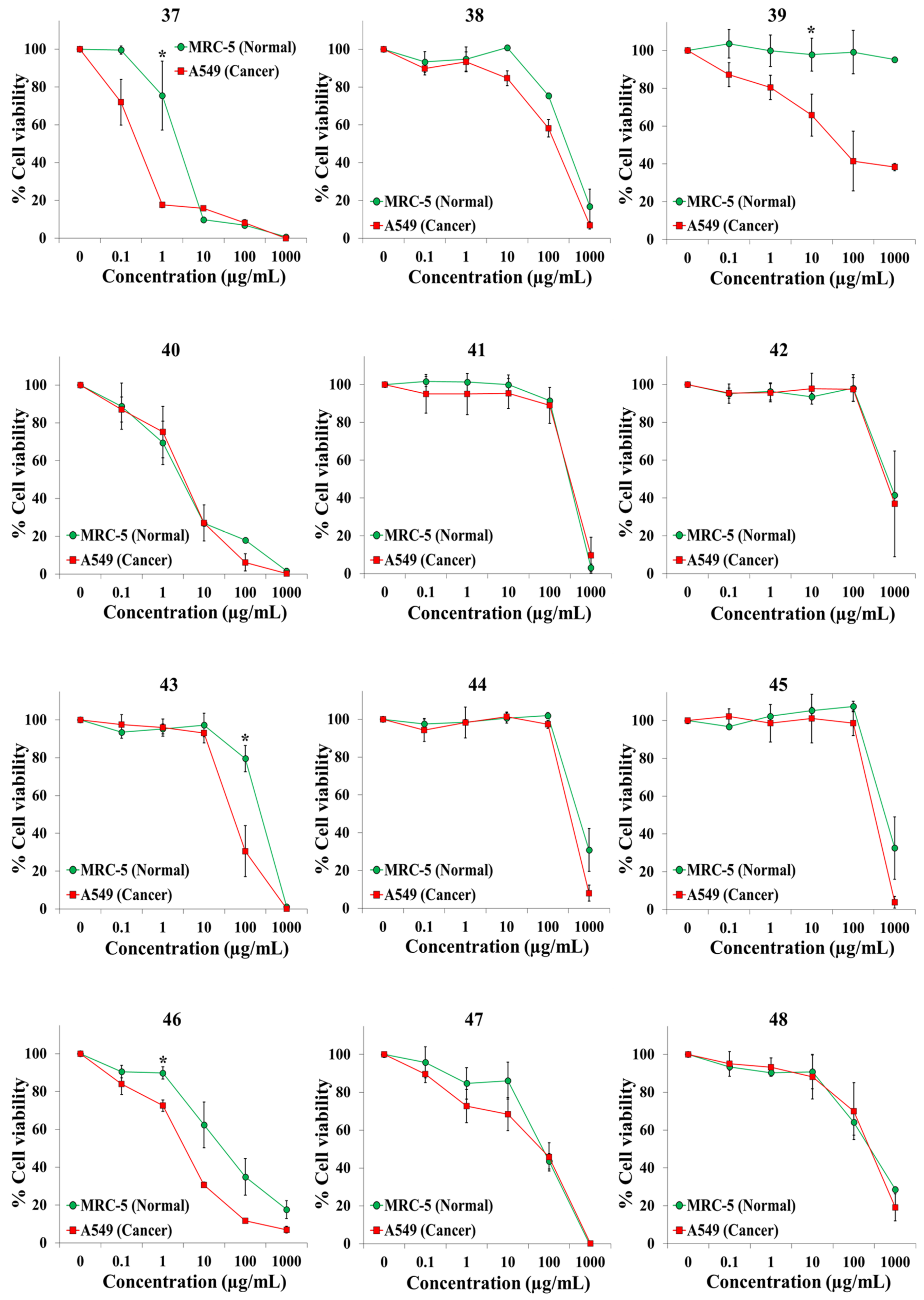
| 37 | Iberis ciliata subsp. contracta (Pers.) Moreno (Brassicaceae) | Whole plant | 284688 | Huelva | 0.31 ± 0.06 | 2.31 ± 0.88 | 13.0 ± 4.7 |
| 38 | Jasione montana L. (Campanulaceae) | Whole plant | 284666 | Seville | 145.0 ± 26.0 | 301.6 ± 69.2 | 2.0 ± 0.1 |
| 39 | Juniperus macrocarpa Sm. (Cupressaceae) | Monosperma cones | 284682 | Huelva | 146.1 ± 125.9 | >1000 | >20 |
| 40 | Juniperus macrocarpa Sm. (Cupressaceae) | Aerial Parts | 284682 | Huelva | 3.7 ± 1.9 | 2.8 ± 1.0 | 0.8 ± 0.1 |
| 41 | Malcolmia lacera (L.) DC. (Brassicaceae) | Whole plant | 284664 | Seville | 322.3 ± 86.7 | 295.0 ± 5.9 | 1.0 ± 0.3 |
| 42 | Malva hispanica L. (Malvaceae) | Aerial Parts | 284669 | Seville | >1000 | 703.1 ± 51.1 | N.D. |
| 43 | Ononis subspicata Lag. (Leguminosae) | Whole plant | 284695 | Huelva | 63.9 ± 23.6 | 232.3 ± 29.5 | 4.6 ± 1.4 |
| 44 | Ornithopus compressus L. (Leguminosae) | Whole plant | 284693 | Seville | 340.7 ± 28.4 | 583.8 ± 150.0 | 1.8 ± 0.6 |
| 45 | Ornithopus sativus Brot. (Leguminosae) | Whole plant | 284692 | Seville | 326.6 ± 37.5 | 699.0 ± 262.9 | 2.3 ± 1.1 |
| 46 | Pancratium maritimum L. (Amaryllidaceae) | Bulb | 284681 | Huelva | 3.4 ± 0.2 | 74.1 ± 56.1 | 19.7 ± 14.6 |
| 47 | Pycnocomon rutifolium (Vahl) Hoffmanns. & Link (Caprifoliaceae) | Leaves | 284678 | Cádiz | 71.4 ± 40.4 | 70.6 ± 18.1 | 2.2 ± 1.4 |
| 48 | Pycnocomon rutifolium (Vahl) Hoffmanns. & Link (Caprifoliaceae) | Root | 284678 | Cádiz | 262.5 ± 131,9 | 242.9 ± 56.7 | 1.1 ± 0.3 |
| 49 | Ranunculus peltatus Schrank (Ranunculaceae) | Whole plant | 284672 | Seville | 186.7 ± 54.6 | 170.1 ± 58.8 | 0.9 ± 0.1 |
| 50 | Rhamnus lycioides subsp. oleoides (L.) Jahand. & Maire (Rhamnaceae) | Bark | 284655 | Seville | 332.2 ± 61.9 | 599.4 ± 122.1 | 2.1 ± 0.8 |
| 51 | Rhamnus lycioides subsp. oleoides (L.) Jahand. & Maire (Rhamnaceae) | Leaves | 284655 | Seville | 151.5 ± 16.7 | 421.3 ± 176.5 | 3.1 ± 1.6 |
| 52 | Rhamnus lycioides subsp. oleoides (L.) Jahand. & Maire (Rhamnaceae) | Root | 284655 | Seville | 257.0 ± 76.6 | 442.3 ± 35.1 | 2.3 ± 0.8 |
| 53 | Scrophularia frutescens L. (Scrophulariaceae) | Whole plant | 284686 | Huelva | 281.5 ± 9.2 | 297.6 ± 8.6 | 1.1 ± 0.1 |
| 54 | Stauracanthus genistoides (Brot.) G. Sampaio (Leguminosae) | Aerial Parts | 284679 | Huelva | 278.4 ± 32.9 | 546.5 ± 53.5 | 2.0 ± 0.4 |
| 55 | Tamarix canariensis Willd. (Tamaricaceae) | Flowers | 284650 | Seville | 212.8 ± 10.2 | 336.3 ± 3.6 | 1.6 ± 0.1 |
| 56 | Tamarix canariensis Willd. (Tamaricaceae) | Leaves | 284650 | Seville | 92.7 ± 24.6 | 192.1 ± 33.0 | 2.2 ± 0.3 |
| 57 | Taxus baccata L. (Taxaceae) | Leaves | 284621 | Seville | 0.86 ± 0.27 | 146.9 ± 87.8 | 157.3 ± 110.6 |
| 58 | Tetraclinis articulata (Vahl) Mast. (Cupressaceae) | Leaves | 284663 | Seville | 0.37 ± 0.03 | 129.5 ± 64.0 | 378.3 ± 178.1 |
| 59 | Teucrium fruticans L. (Lamiaceae) | Leaves | 284658 | Seville | 157.1 ± 30.5 | 433.0 ± 112.3 | 2.8 ± 0.7 |
| 60 | Thymus mastichina (L.) L. (Lamiaceae) | Whole plant | 284694 | Huelva | 36.8 ± 7.7 | 277.7 ± 40.6 | 8.2 ± 2.0 |
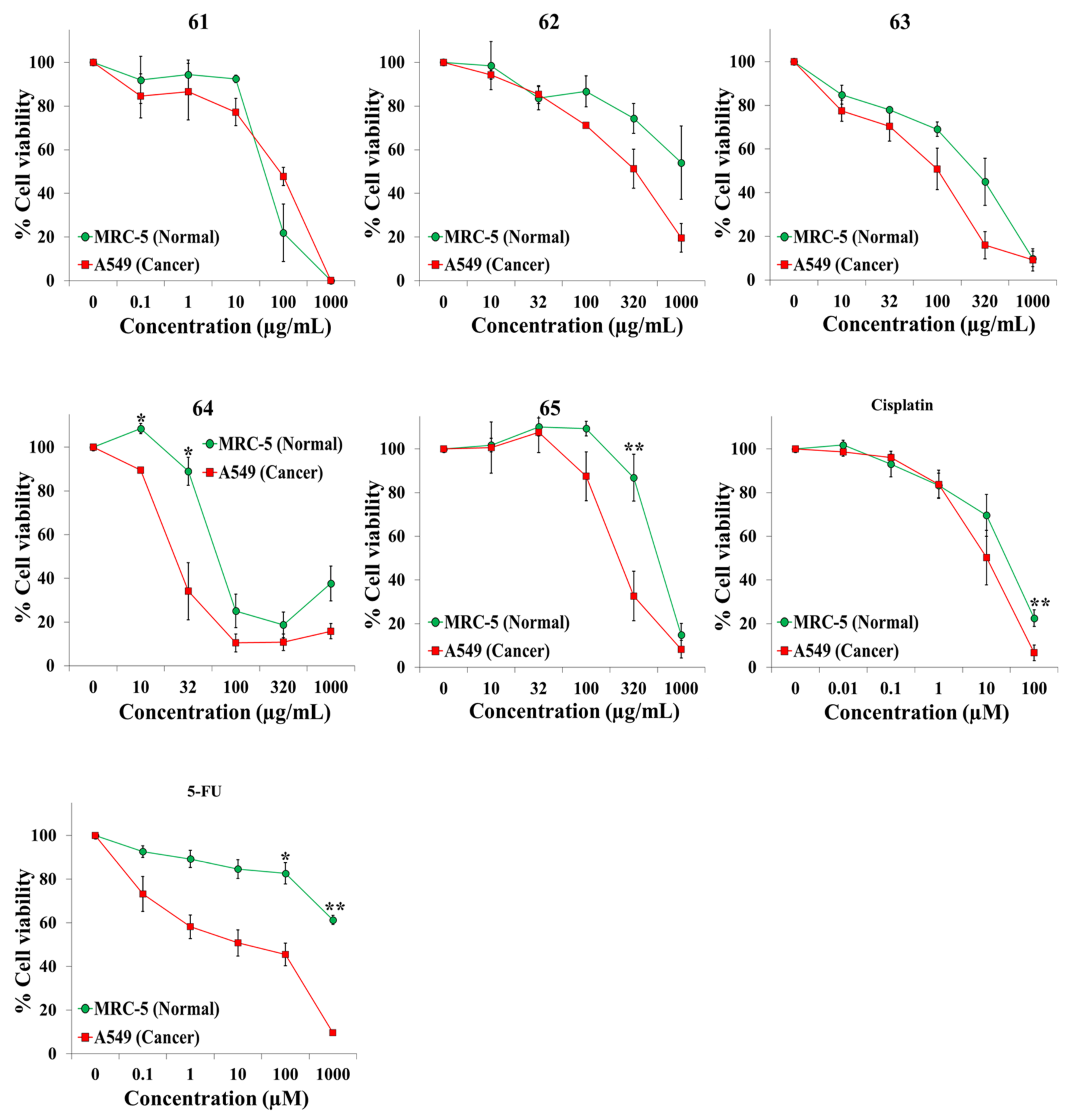
| 61 | Tolpis barbata (L.) Gaertn. (Compositae) | Whole plant | 284665 | Seville | 86.5 ± 28.3 | 43.7 ± 12.2 | 0.5 ± 0.0 |
| 62 | Ulex parviflorus Pourr. subsp. parviflorus (Leguminosae) | Flowers | 284652 | Seville | 361.7 ± 89.6 | 876.1 ± 426.1 | 4.0 ± 2.9 |
| 63 | Ulex parviflorus Pourr. subsp. parviflorus (Leguminosae) | Leaves | 284652 | Seville | 99.6 ± 34.8 | 270.4 ± 85.3 | 3.4 ± 1.2 |
| 64 | Viburnum tinus L. (Adoxaceae) | Fruits | 284651 | Seville | 26.6 ± 6.5 | 65.4 ± 8.6 | 2.6 ± 0.3 |
| 65 | Viburnum tinus L. (Adoxaceae) | Leaves | 284651 | Seville | 234.8 ± 58.7 | 568.2 ± 73.0 | 2.6 ± 0.3 |
| Cisplatin (Standard anticancer drug) | 10.5 ± 5.5 (µM) | 25.4 ± 7.4 (µM) | 4.2 ± 2.2 | ||||
| 5-Fluorouracil (Standard anticancer drug) | 101.8 ± 7.7 (µM) | >1000 (µM) | >9.9 | ||||
| IC50 (Resazurin) (Mean ± SEM, µg/mL) | IC50 (Resazurin) (Mean ± SEM, µM) | |||||||
|---|---|---|---|---|---|---|---|---|
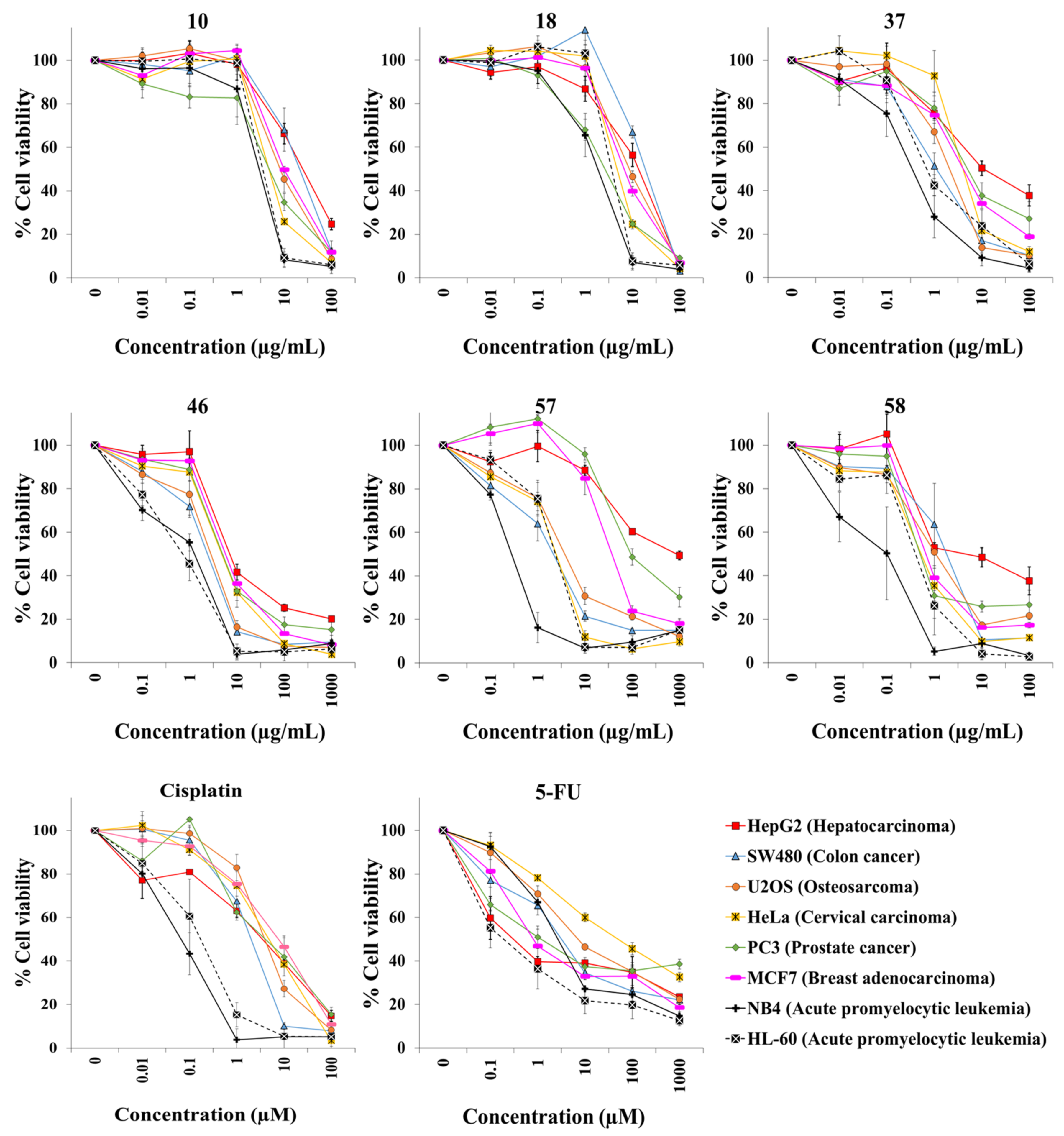
| Cell Line | 10 | 18 | 37 | 46 | 57 | 58 | Cisplatin | 5-FU |
| HepG2 | 24.6 ± 4.1 | 13.2 ± 3.0 | 19.4 ± 10.4 | 7.0 ± 1.3 | 632.6 ± 106.1 | 18.4 ± 17.1 | 3.5 ± 0.8 | 0.3 ± 0.2 |
| SW480 | 20.7 ± 5.6 | 18.3 ± 1.2 | 1.2 ± 0.3 | 2.1 ± 0.4 | 2.2 ± 0.7 | 2.1 ± 0.8 | 2.0 ± 0.5 | 3.4 ± 1.0 |
| U2OS | 9.2 ± 1.5 | 8.8 ± 0.8 | 1.9 ± 0.2 | 2.8 ± 0.7 | 4.0 ± 1.2 | 2.1 ± 0.9 | 4.0 ± 1.0 | 7.2 ± 1.0 |
| HeLa | 4.7 ± 0.1 | 4.9 ± 0.2 | 3.5 ± 0.5 | 5.4 ± 0.4 | 2.5 ± 0.4 | 1.3 ± 0.6 | 4.8 ± 0.1 | 66.5 ± 27.0 |
| PC3 | 4.7 ± 1.5 | 2.6 ± 0.4 | 5.6 ± 2.1 | 5.4 ± 1.2 | 123.8 ± 42.7 | 0.50 ± 0.04 | 7.3 ± 4.6 | 1.5 ± 0.8 |
| MCF7 | 10.0 ± 0.5 | 6.5 ± 1.0 | 4.0 ± 0.9 | 5.9 ± 0.8 | 37.1 ± 4.9 | 0.7 ± 0.1 | 8.0 ± 3.0 | 1.1 ± 0.8 |
| NB4 | 2.9 ± 0.7 | 1.8 ± 0.5 | 0.5 ± 0.2 | 1.2 ± 0.2 | 2.8 ± 0.2 | 0.2 ± 0.1 | 0.08 ± 0.04 | 2.7 ± 0.3 |
| HL-60 | 3.5 ± 0.4 | 3.6 ± 0.3 | 0.8 ± 0.2 | 1.0 ± 0.3 | 3.3 ± 0.2 | 0.7 ± 0.3 | 0.2 ± 0.1 | 0.6 ± 0.5 |
| BJ-hTERT | 3.7 ± 0.4 | 2.8 ± 0.3 | 3.4 ± 0.5 | 3.4 ± 0.1 | 4.0 ± 0.6 | 0.8 ± 0.3 | 1.2 ± 0.4 | 2.6 ± 0.6 |
| BJ-SV40T | 2.9 ± 0.4 | 2.1 ± 0.4 | 2.9 ± 0.4 | 3.2 ± 0.4 | 2.8 ± 0.2 | 1.0 ± 0.4 | 0.7 ± 0.2 | 5.8 ± 0.7 |
| BJ-RASV12 | 2.9 ± 0.4 | 1.8 ± 0.7 | 4.0 ± 1.4 | 2.0 ± 0.7 | 3.3 ± 0.2 | 0.6 ± 0.2 | 1.1 ± 0.1 | 1.7 ± 0.8 |
| Cell Line | IC50 (Resazurin) (Media ± SEM, µg/mL) |
|---|---|
| 58 | |
| A64-CLS | 4.7 ± 0.3 |
| AN3Ca | 4.5 ± 0.9 |
| Calu-1 | 4.7 ± 0.2 |
| GAMG | 4.5 ± 0.6 |
| HNO97 | 4.5 ± 0.3 |
| HT29 | 4.4 ± 0.4 |
| KATO III | 4.4 ± 0.7 |
| MDA-MB-231 | 4.7 ± 0.2 |
| MeWo | 4.3 ± 0.2 |
| PC-3 | 4.5 ± 1.0 |
| Sk-Br-3 | 4.9 ± 0.0 |
| Sk-OV-3 | 4.7 ± 0.1 |
| T24 | 4.5 ± 0.3 |
| UACC-62 | 4.5 ± 0.2 |
| Extract | Plant Name | Collection Coordinates |
|---|---|---|
| 1 | Acis autumnalis (L.) Sweet | 37°14′22.06″ N 6°11′37.85″ W |
| 2–3 | Anagallis monelli L. | 36°36′15.24″ N 6°16′2.76″ W |
| 4 | Anthyllis hamosa Desf. | 37°04′25.2″ N 6°41′19.68″ W |
| 5–6 | Aristolochia baetica L. | 37°14′16.68″ N 6°11′48.38″ W |
| 7 | Armeria pungens (Link) Hoffmanns. & Link | 37°04′13.73″ N 6°41′16.97″ W |
| 8 | Armeria velutina Welw. ex Boiss. & Reut. | 37°02′33.33″ N 6°35′53.85″ W |
| 9 | Campanula lusitanica L. | 37°14′14.46″ N 6°11′55.8″ W |
| 10 | Cascabela thevetia (L.) Lippold | 37°22′59.8″ N 5°59′27.36″ W |
| 11–13 | Centaurea sphaerocephala L. | 37°05′40.18″ N 6°43′37.9″ W |
| 14–15 | Cistus crispus L. | 37°14′22.06″ N 6°11′37.85″ W |
| 16 | Cistus salvifolius L. | 37°14′19.74″ N 6°11′40.71″ W |
| 17 | Cleome violacea L. | 37°14′23.37″ N 6°11′52.72″ W |
| 18 | Digitalis purpurea L. | 37°27′30.78″ N 6°41′20.3″ W |
| 19–20 | Dorycnium rectum (L.) Ser. | 37°20′14.71″ N 5°51′27.13″ W |
| 21 | Echium gaditanum Boiss. | 37°04′11.69″ N 6°41′17.16″ W |
| 22–23 | Elaeoselinum foetidum (L.) Boiss. | 37°14′15.72″ N 6°11′50.86″ W |
| 24–25 | Erica arborea L. | 37°14′38.35″ N 6°11′49.92″ W |
| 26 | Erophaca baetica (L.) Boiss. | 37°14′17.72″ N 6°11′46.41″ W |
| 27–28 | Frangula alnus Mill. | 37°05′40.46″ N 6°43′34.47″ W |
| 29 | Genista hirsuta M. Vahl | 37°14′16.70 “N 6°11′49.05″ W |
| 30–31 | Halimium calycinum (L.) K. Koch | 37°14′21.27N 6°11′38.12″ W |
| 32–33 | Halimium halimifolium (L.) Willk. | 37°14′23.43″ N 6°11′38.77″ W |
| 34–36 | Hedysarum coronarium L. | 36°36′39.92″ N 6°16′46.6″ W |
| 37 | Iberis ciliata subsp. contracta (Pers.) Moreno | 37°04′47.25″ N 6°41’13.82″ W |
| 38 | Jasione montana L. | 37°13′45.76″ N 6°9′16.08″ W |
| 39–40 | Juniperus macrocarpa Sm. | 37°04′13.53″ N 6°41′16.34″ W |
| 41 | Malcolmia lacera (L.) DC. | 37°13′45.76″ N 6°9′16.08″ W |
| 42 | Malva hispanica L. | 37°14′16.09″ N 6°11′51.89″ W |
| 43 | Ononis subspicata Lag. | 37°04′45.90″ N 6°41′14.14″ W |
| 44 | Ornithopus compressus L. | 37°13′45.76″ N 6°9′16.08″ W |
| 45 | Ornithopus sativus Brot. | 37°13′45.76″ N 6°9′16.08″ W |
| 46 | Pancratium maritimum L. | 37°04′11.14″ N 6°41′16.29″ W |
| 47–48 | Pycnocomon rutifolium (Vahl) Hoffmanns. & Link | 36°36′12.69″ N 6°15′54.45″ W |
| 49 | Ranunculus peltatus Schrank | 37°13′45.76″ N 6°9′16.08″ W |
| 50–52 | Rhamnus lycioides subsp. oleoides (L.) Jahand. & Maire | 37°14′25.18″ N 6°11′38.35″ W |
| 53 | Scrophularia frutescens L. | 37°04′47.25″ N 6°41′13.82″ W |
| 54 | Stauracanthus genistoides (Brot.) G. Sampaio | 37°04′44.55″ N 6°41′16.05″ W |
| 55–56 | Tamarix canariensis Willd. | 37°15′45.64″ N 5°59′50.9″ W |
| 57 | Taxus baccata L. | 37°22′27″ N 5°59′19″ W |
| 58 | Tetraclinis articulata (Vahl) Mast. | 37°22′22.18″ N 5°59′10.75″ W |
| 59 | Teucrium fruticans L. | 37°14′42.60″ N 6°11′52.78″ W |
| 60 | Thymus mastichina (L.) L. | 37°02′33.33″ N 6°35′53.85″ W |
| 61 | Tolpis barbata (L.) Gaertn. | 37°13′45.76″ N 6°9′16.08″ W |
| 62–63 | Ulex parviflorus Pourr. subsp. parviflorus | 37°13′48.06″ N 6°1′31.4″ W |
| 64–65 | Viburnum tinus L. | 37°22′27″ N 5°59′19″ W |
Publisher’s Note: MDPI stays neutral with regard to jurisdictional claims in published maps and institutional affiliations. |
© 2021 by the authors. Licensee MDPI, Basel, Switzerland. This article is an open access article distributed under the terms and conditions of the Creative Commons Attribution (CC BY) license (https://creativecommons.org/licenses/by/4.0/).
Share and Cite
Calderón-Montaño, J.M.; Martínez-Sánchez, S.M.; Jiménez-González, V.; Burgos-Morón, E.; Guillén-Mancina, E.; Jiménez-Alonso, J.J.; Díaz-Ortega, P.; García, F.; Aparicio, A.; López-Lázaro, M. Screening for Selective Anticancer Activity of 65 Extracts of Plants Collected in Western Andalusia, Spain. Plants 2021, 10, 2193. https://doi.org/10.3390/plants10102193
Calderón-Montaño JM, Martínez-Sánchez SM, Jiménez-González V, Burgos-Morón E, Guillén-Mancina E, Jiménez-Alonso JJ, Díaz-Ortega P, García F, Aparicio A, López-Lázaro M. Screening for Selective Anticancer Activity of 65 Extracts of Plants Collected in Western Andalusia, Spain. Plants. 2021; 10(10):2193. https://doi.org/10.3390/plants10102193
Chicago/Turabian StyleCalderón-Montaño, José Manuel, Sara María Martínez-Sánchez, Víctor Jiménez-González, Estefanía Burgos-Morón, Emilio Guillén-Mancina, Julio José Jiménez-Alonso, Patricia Díaz-Ortega, Felipe García, Abelardo Aparicio, and Miguel López-Lázaro. 2021. "Screening for Selective Anticancer Activity of 65 Extracts of Plants Collected in Western Andalusia, Spain" Plants 10, no. 10: 2193. https://doi.org/10.3390/plants10102193
APA StyleCalderón-Montaño, J. M., Martínez-Sánchez, S. M., Jiménez-González, V., Burgos-Morón, E., Guillén-Mancina, E., Jiménez-Alonso, J. J., Díaz-Ortega, P., García, F., Aparicio, A., & López-Lázaro, M. (2021). Screening for Selective Anticancer Activity of 65 Extracts of Plants Collected in Western Andalusia, Spain. Plants, 10(10), 2193. https://doi.org/10.3390/plants10102193