Article

Triterpenoid and Steroidal Saponins Differentially Influence Soil Bacterial Genera

1
Research Institute for Sustainable Humanosphere, Kyoto University, Gokasho, Uji 611-0011, Japan
2
Tohoku Medical Megabank Organization, Tohoku University, Sendai 980-8573, Japan
*
Author to whom correspondence should be addressed.
Plants 2021, 10(10), 2189; https://doi.org/10.3390/plants10102189
Submission received: 7 September 2021 / Revised: 30 September 2021 / Accepted: 11 October 2021 / Published: 15 October 2021
(This article belongs to the Special Issue Transport of Specialized Metabolites)

Abstract

:
Plant specialized metabolites (PSMs) are secreted into the rhizosphere, i.e., the soil zone surrounding the roots of plants. They are often involved in root-associated microbiome assembly, but the association between PSMs and microbiota is not well characterized. Saponins are a group of PSMs widely distributed in angiosperms. In this study, we compared the bacterial communities in field soils treated with the pure compounds of four different saponins. All saponin treatments decreased bacterial α-diversity and caused significant differences in β-diversity when compared with the control. The bacterial taxa depleted by saponin treatments were higher than the ones enriched; two families, Burkholderiaceae and Methylophilaceae, were enriched, while eighteen families were depleted with all saponin treatments. Sphingomonadaceae, which is abundant in the rhizosphere of saponin-producing plants (tomato and soybean), was enriched in soil treated with α-solanine, dioscin, and soyasaponins. α-Solanine and dioscin had a steroid-type aglycone that was found to specifically enrich Geobacteraceae, Lachnospiraceae, and Moraxellaceae, while soyasaponins and glycyrrhizin with an oleanane-type aglycone did not specifically enrich any of the bacterial families. At the bacterial genus level, the steroidal-type and oleanane-type saponins differentially influenced the soil bacterial taxa. Together, these results indicate that there is a relationship between the identities of saponins and their effects on soil bacterial communities.

1. Introduction

At least one million diverse plant specialized metabolites (PSMs) [1] exist that have a wide range of bioactivities and contribute to a plant’s ability to adapt to its environment and protect against pathogens and herbivores [2]. PSMs are also secreted into the rhizosphere, i.e., the zone of soil surrounding the roots, which impacts plant responses to nutrient deficiencies and their interactions with soil-borne organisms, such as symbiosis, attraction, and repelling [3,4,5,6]. Metabolites secreted from roots are called root exudates, and these account for up to 40% of the carbon fixed during photosynthesis [7]. Rhizosphere microbiomes assembled by root exudates promote plant growth and help the host plants overcome biotic and abiotic stresses [8,9]. In the last decade, multiple studies have revealed that PSMs are involved in the formation of the rhizosphere and root microbiome [10,11]. PSM-deficient mutants of certain plant species, such as thale cress (Arabidopsis thaliana) and maize (Zea mays), show that di-, sester-, tri-terpenoids, coumarins, and benzoxazinoids modulate the root-associated microbiome [12,13,14,15,16,17,18]. The treatment of soils with authentic compounds has also helped to reveal the roles of PSMs such as flavonoids (daidzein and quercetin), alkaloids (nicotine and gramine), benzoxazinoid, and opine (santhopine) in modulating the soil microbiome [19,20,21,22].
Saponins are a group of PSMs widely distributed in angiosperm plants. They exhibit biological and pharmacological activities, including antibacterial, antifungal, hemolytic, and cytotoxic properties [23]. They consist of two parts: a hydrophobic skeleton, which is the aglycone unit, and a hydrophilic saccharide, which is the glycosidic unit. Based on aglycone structure, saponins are classified into triterpenoid saponins, steroidal saponins, and steroidal glycoalkaloids (SGAs; Figure 1). Glycyrrhizin is an oleanane-type triterpenoid saponin that is used as a natural sweetener and is found only in licorice (Glycyrrhiza spp.) [24]. Soyasaponins are oleanane-type triterpenoid saponins found in several legume plants, including barrel medic (Medicago truncatula) and soybean (Glycine max). Dioscin is a spirostane-type steroidal saponin used for the semi-synthetic production of pharmaceutical steroidal drugs in Dioscorea plants [25]. α-Tomatine and α-solanine are spirosolane- and solanidane-type SGAs in tomato (Solanum lycopersicum) and potato (S. tuberosum), respectively [26]. All saponins are biosynthesized from 2,3-oxidosqualene—their last common precursor subjected to cyclization, oxidation, and glycosylation [27,28]. This cyclization step catalyzed by oxidosqualene cyclases (OSCs) is the branch point of their biosynthesis. Cyclization to form dammarenediol-II, cucurbitadienol, and β-amyrin leads to triterpenoid saponins, while cyclization to form cycloartenol leads to steroidal saponins and SGAs through cholesterol [28,29,30]. Furthermore, a nitrogen atom is also incorporated into the hydrophobic skeleton during SGA biosynthesis.
Recent studies have shown that saponins are secreted from plant roots into the rhizosphere [31,32,33,34]. Previously, we demonstrated that soyasaponins and α-tomatine were secreted from soybean and tomato roots in both hydroponic and field conditions [35,36,37]. Furthermore, both soyasaponins and α-tomatine were found to alter the bacterial communities and increase Sphingomonadaceae. Soyasaponins enriched Novosphingobium, whereas tomatine enriched Sphingobium at the genus level [35,37]. Based on these differential effects of the saponins in the soil bacterial communities, we hypothesized a link between saponin identity and soil bacterial community. In this study, we compared bacterial communities in field soils treated with steroidal-type and oleanane-type saponins. We propose that the correlation of rhizosphere microbiome formation with the chemical structure of PSMs may be a factor for microbiome variation among plant species.

2. Materials and Methods

2.1. Chemicals and Soils

α-Solanine, dioscin, a saponin mixture from soybeans constituted by soyasaponins (mainly group B), and glycyrrhizin were purchased from Extrasynthese (Genay, France), Cayman Chemical (Ann Arbor, MI, USA), FUJIFILM Wako Pure Chemical Corporation (Osaka, Japan), and the Tokyo Chemical Industry Co., Ltd. (Tokyo, Japan), respectively. The soil used was the same as that described in a previous study [20]. Briefly, before plant cultivation, soils were collected from five different points across a field at the Kyoto University of Advanced Science, Kameoka, Kyoto, Japan (34°99′38″ N, 135°55′14″ E), where crops had been cultivated for more than twelve years and soybean had been grown the previous five years. The soils were combined and air-dried in a greenhouse. The soils that passed through a 2 mm sieve were used in the following experiments. Soil chemical and physical properties were measured at the Tokachi Federation of Agricultural Cooperatives [20].

2.2. Treatment of Field Soil with Saponins

α-Solanin, dioscin, soyasaponins, and glycyrrhizin were dissolved in methanol and 20, 100, and 500 nmol of each was then dried in 5 mL tubes. Field soil (2 g) and distilled water (600 μL) were added to each tube in biological quadruplicate and then sealed, vortexed, and incubated at 28 °C in the dark. The control was untreated soil that was added to an empty tube. Each soil sample was transferred to a new tube containing the relevant compound once every 3 days over a 15-day incubation period. Tubes were sealed during incubation. After incubation, the tubes were stored at −30 °C until they were used for DNA extraction. The saponin concentrations were determined according to previous works; soyasaponins and α-tomatine in rhizosphere soils of soybean and tomato were approximately 20–80 and 300–1000 nmol g soil−1 at the different growth stages, respectively [35,37]. They were equivalent to the low, middle, or high concentrations of saponins treated in this study (i.e., 50, 250, or 1250 nmol g soil−1). We previously also treated field soil with a pure compound of α-tomatine in a similar manner to this study [37].

2.3. DNA Extraction and 16S rRNA Amplicon Sequencing

DNA was extracted from the saponin-treated soils using a DNeasy PowerSoil Kit (QIAGEN K.K., Tokyo, Japan) and according to the manufacturer’s instructions. The DNA concentrations were quantified using a Qubit dsDNA HS Assay Kit and Qubit 2.0 Fluorometer (Thermo Fisher Scientific, Waltham, MA, USA). The V4 region of the bacterial 16S rRNA was PCR-amplified in technical triplicate using KOD FX Neo (TOYOBO, Osaka, Japan) with the following primer set: 515F (5′-ACACTCTTTCCCTACACGACGCTCTTCCGATCT-GTGCCAGCMGCCGCGGTAA-3′) and 806R (5′-GTGACTGGAGTTCAGACGTGTGCTCTTCCGATCT-GGACTACHVGGGTWTCTAAT-3′) [38]. PCR cycling was carried out at 94 °C for 2 min and 20 cycles at 98 °C for 10 s, 50 °C for 30 s, and 68 °C for 30 s. The PCR products were purified using AMPure XP (Beckman Coulter, Danvers, MA, USA). PCR amplification for attachment of the MiSeq adapters (Illumina, San Diego, CA, USA) was performed with the purified DNA as a template in technical duplicate using KOD FX Neo and primers provided by FASMAC Co. Ltd. (Kanagawa, Japan). PCR cycling was carried out at 94 °C for 2 min and 10 cycles of 98 °C for 10 s, 59 °C for 30 s, and 68 °C for 30 s. The PCR products were purified, and the DNA concentrations were quantified as described above. The purified DNA was mixed in equal amounts and used for 2 × 250-bp paired-end sequencing with MiSeq (Illumina) by FASMAC Co. Ltd. The 16S rRNA amplicon dataset supporting the results of this study has been registered to the DNA Data Bank of Japan (https://www.ddbj.nig.ac.jp, accessed on 13 October 2021) and is to be publicly available (the accession number DRA012729).

2.4. Sequence Data Analysis

The acquired sequence data were analyzed using the QIIME2 pipeline (version 2019.7) [39]. The bases, other than the 21st to 200th of the paired-end sequences, were trimmed, and error-corrected amplicon sequence variants (ASVs) were constructed using DADA2 [40] with the q2-dada2 plugin in QIIME2. Multiple alignments of the obtained ASV sequences were created using MAFFT [41], and phylogenetic trees were constructed using FastTree [42] in the q2-phylogeny plugin. Taxonomic assignment of the ASVs was performed using the Naïve Bayes classifier with the Silva rRNA database release 132 [43,44]. ASV dataset with 19,105–65,734 reads per sample was obtained (Table S1). Shannon’s diversity indices as α-diversity and both weighted and unweighted UniFrac distances as β-diversity were calculated from a subsampled ASV dataset with 19,000 sequences per sample using the core-metrics-phylogenetic pipeline in the q2-diversity plugin within QIIME2 (Figure S2). The relative abundances of the bacterial taxa were calculated from the ASV-read abundances.

2.5. Statistical Analysis

The Welch’s t-test, corrected by the Benjamini–Hochberg method, was carried out using the pairwise.t.test function of R package “stats”. The principal coordinate analysis (PCoA) plots based on the weighted and unweighted UniFrac distances for each saponin-treated soil were generated using the cmdscale function in the R package “stats”. Hierarchical clustering was performed using the complete-linkage method with the hclust function that was also in the R package “stats”. Permutational multivariate analysis of variance (PERMANOVA) was performed using the adonis function in the R package “vegan” [45]. The linear discriminant analysis (LDA) effect size (LEfSe) method [46] was applied using default parameters to detect differentially abundant taxa at the family and genus levels. An adjusted p value of <0.05 for Kruskal–Wallis and an LDA score of >2 was used to define significant differences. Low abundant taxa (the mean relative abundance of all samples <0.1%) were filtered out for the LEfSe method.

3. Results

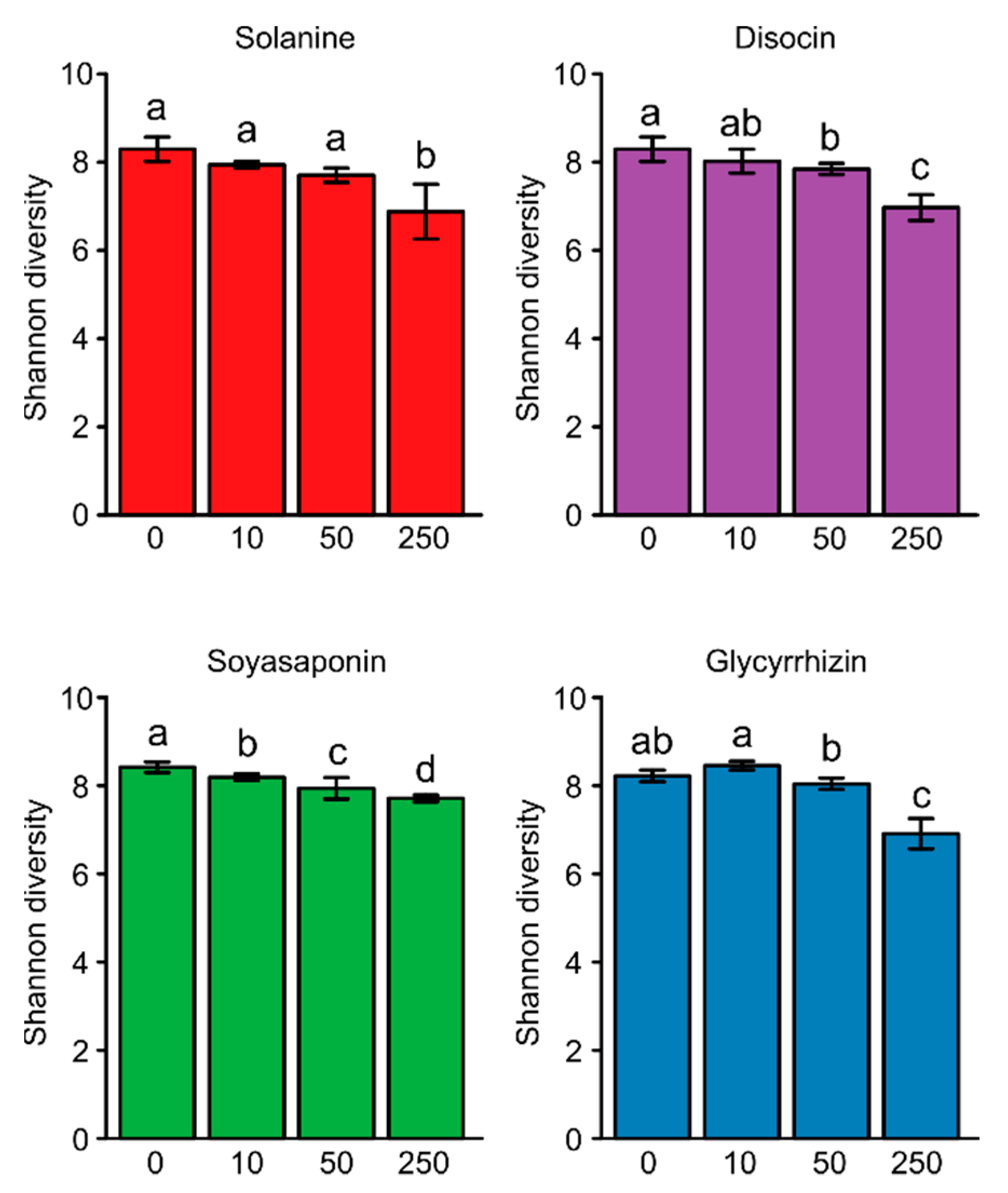
3.1. Bacterial Diversity in the Saponin-Treated Soils

We treated field soil with α-solanine, dioscin, soyasaponins, and glycyrrhizin compounds at doses of 10, 50, and 250 nmol g soil−1. α-Diversity in untreated soils was slightly higher than 8, and the high concentration of each saponin treatment (250 nmol g soil−1) significantly reduced the α-diversity compared with control (p < 0.05; Figure 2). The PCoA, based on the weighted UniFrac distance metric (β-diversity), showed that the bacterial communities of all saponin-treated soils were distinct from those of the untreated soil (Figure 3). The PERMANOVA analysis confirmed these significant differences (α-solanine, R2 = 0.67, p < 0.01; dioscin, R2 = 0.69, p < 0.01, soyasaponins, R2 = 0.62, p < 0.01, and glycyrrhizin, R2 = 0.80, p < 0.01). The PCoA, based on the unweighted UniFrac distance metric, also showed their clear distinction (Figure S2 and Table S2). These results indicate that all the saponins tested modified the bacterial communities in the soil.

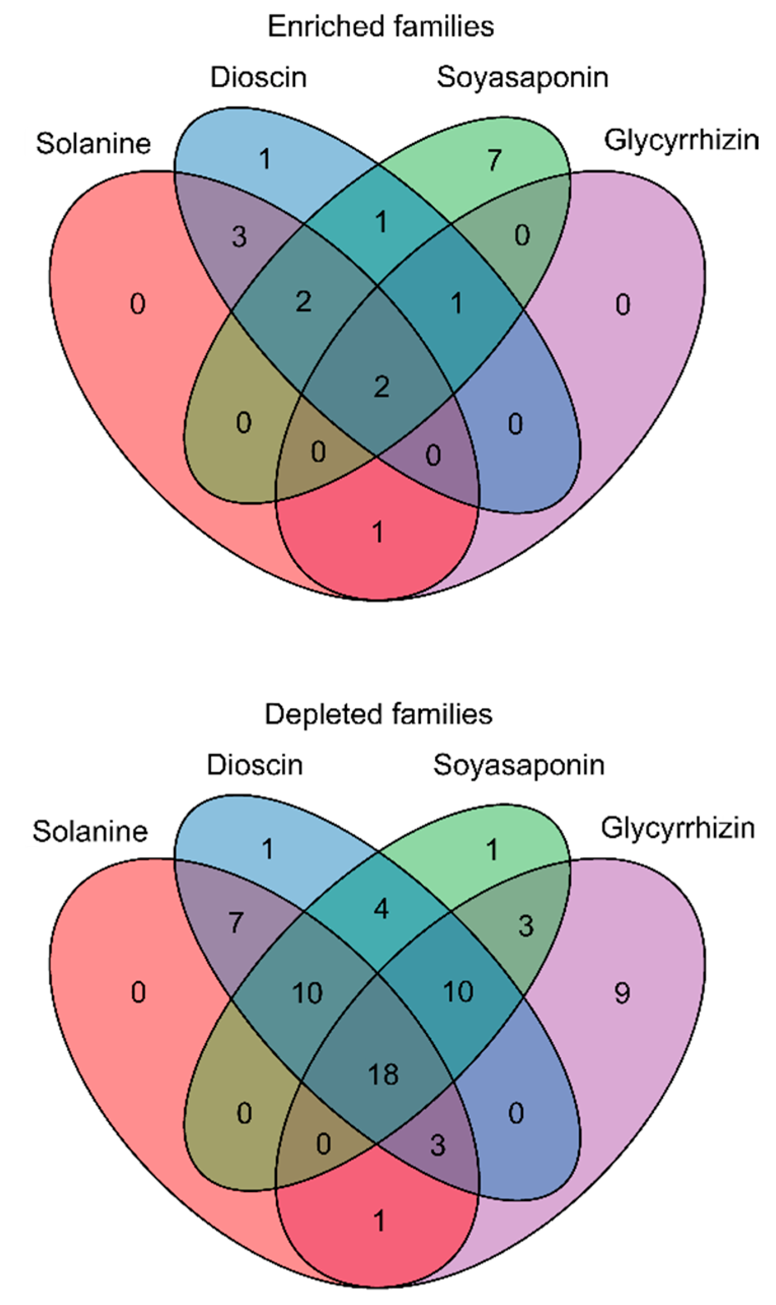
3.2. Effects of the Saponin Treatments on Bacterial Families in the Soil

We compared the abundance of the bacterial taxa between the saponin-treated and untreated soils at the bacterial family level. The increase and decrease in the relative abundances of taxa in soil samples were defined as “enrichment” and “depletion”, respectively. α-Solanine, dioscin, soyasaponins, and glycyrrhizin significantly enriched 8, 10, 13, and 4 families but depleted 39, 53, 46, and 44 families, respectively (Figure 4 and Table S3). Among these, Burkholderiaceae and Methylophilaceae were enriched in all treatments, and 18 families were depleted in all the treatments (Figure 4 and Figure 5, and Table S3). Except for α-solanine, the saponins enriched Caulobacteraceae, and, except for glycyrrhizin, they enriched P3OB-42 and Sphingomonadaceae (Figure 4 and Figure 5, and Table S3). The α-solanine and dioscin, which have a steroid-type aglycone, specifically enriched Geobacteraceae, Lachnospiraceae, and Moraxellaceae, while soyasaponins and glycyrrhizin, which have an oleanane-type aglycone, did not specifically increase any bacterial family (Figure 4 and Figure 5, and Table S3).

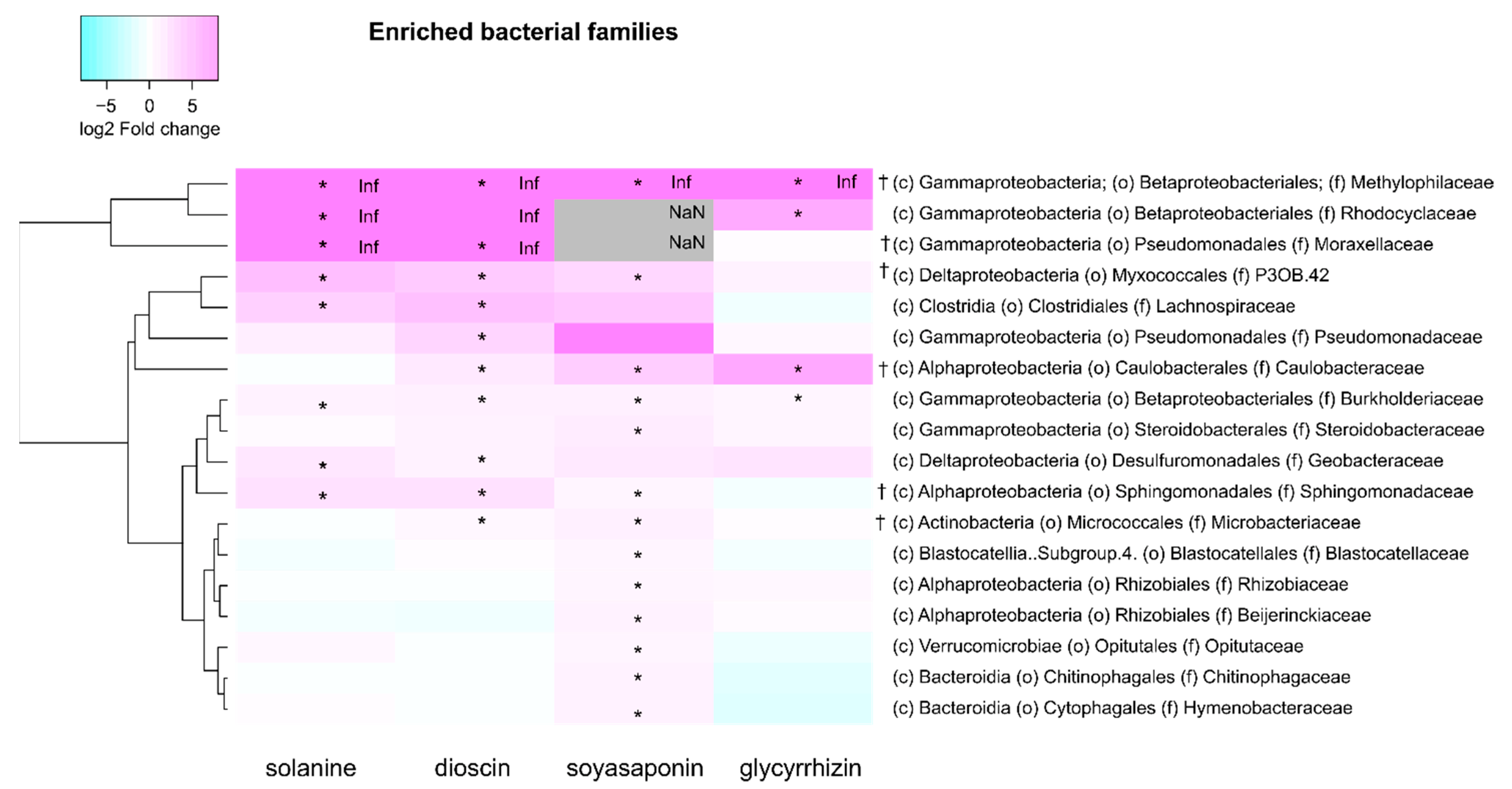
3.3. Taxonomic Composition of the Bacterial Families Enriched by the Saponin Treatments

The bacterial families that were significantly enriched by the saponin treatments were further investigated at the genus level. Among the Burkholderiaceae, which were commonly enriched in all the saponin-treated soils, the Azohydromonas, Cupriavidus, Ramlibacter, and uncultured genera were differently altered with the various saponin treatments, while the unknown genus was increased in all the saponin treatments (Figure 6 and Table S4). In contrast to the Burkholderiaceae, a single genus was markedly abundant in Methylophilaceae, Caulobacteraceae, and Geobacteraceae. The MM2 genus within the Methylophilaceae was dominant in all the saponin-treated soils, but its relative abundance in the glycyrrhizin-treated soils was lower when compared with the other saponins (Figure 6 and Table S4). Phenylobacterium was strongly enriched by the oleanane-type saponins (soyasaponins and glycyrrhizin) but not by the steroidal-type saponins (α-solanine and dioscin) in Caulobacteraceae. Among the Geobacteraceae, Geobacter was significantly increased only by the steroid-type saponin treatments (Figure 6 and Table S4). Novosphingobium was dominant in the treatment with oleanane-type saponins, whereas Sphingobium was abundant in the treatment of steroidal-type saponins within the genus belonging to Sphingomonadaceae (Figure 6 and Table S4). At the ASV level of the Sphingomonadaceae, the relative abundances of multiple ASVs were increased in the oleanane-type saponin-treated soils, while that of one ASV (ASV 4) was particularly rich in steroid-type saponin-treated soils (Figure 7 and Table S5), which was the same ASV that was accumulated in α-tomatine- and tomatidine-treated soils and the tomato rhizosphere soil [37].

4. Discussion

Treating soil with pure compounds has revealed the strong influences PSMs have in shaping the microbiota [19,20,21,22,35,37,47]. In this study, we treated soil with saponins that had either steroid- or oleanane-type aglycones. The bacterial α-diversity was found to decrease with all four saponin treatments, which was consistent with previous results for benzoxazolin-2(3H)-one (BOA), quercetin, daidzein, flavonoid mixture, α-tomatine, and tomatidine [20,22,37,47]. The decreases found in the bacterial α-diversity were in accordance with previous observations made for the rhizosphere when affected by root exudates [48,49]. In contrast, the treatment of barley indole-alkaloid gramine did not reduce α-diversity, although it did modify the bacterial community structures [19,22]. Intriguingly, the α-diversities in the rhizospheres of the mutants deficient in benzoxazinoids, coumarins, and α-tomatine were comparable with those in wildtypes of maize, thale cress, and tomato, respectively [13,14,37,50,51]. On the other hand, the thale cress triterpenoid mutants harbored root bacterial communities with lower α-diversities than the wildtype [15]. The accumulation of intermediates or the effects of other metabolites on the mutants may alleviate the decrease in the α-diversity by the PSMs.
In accordance with our previous studies with daidzein and α-tomatine [20,37], we found that the bacterial taxa depleted by the saponin treatments were greater than the enriched ones. In the treatments with gramine or quercetin, however, the results were inconsistent [22]. These variations may be caused by differences in the concentrations of the PSMs between our study and that of Schütz et al. [22] (the former: 1250 nmol g soil−1 and the latter: 33 nmol g soil−1) and the promotion and inhibition effects of the PSMs on the growth of soil microbes. There are some taxa commonly enriched by the PSM treatments and in the host plant rhizosphere and roots, such as: Comamonadaceae and Microbacteriaceae by daidzein and soybean root; Arthrobacter of Micrococcaceae by santhopine, nicotine, and tobacco roots; Sphingobium of Sphingomonadaceae by α-tomatine, tomatidine, and tomato root; and Caulobacteraceae and Novosphingobium of Sphingomonadaceae by soyasaponin Bb and soybean root [20,21,35,47]. It has also been reported that the bacterial communities in the soils treated with daidzein, α-tomatine, nicotine, and santhopine are closer to those of the rhizosphere or endosphere of the respective host plants than the bulk soils, defined as a soil that does not adhere to plant roots [20,21,47]. Rhizosphere and root microbiota have been reported in several saponin-synthesizing plants. Bradyrhizobiaceae, Rhizobiaceae, and Sphingomonadaceae are highly abundant in the taproots of sugar beet (Beta vulgaris), which accumulates triterpenoid saponins in its roots when grown in disease-suppressive soils [52,53]. Bacillaceae, Burkholderiaceae, Mycobacteriaceae, Rhizobiaceae, and Sphingomonadaceae are abundant in the roots of Chinese ginseng (Panax notoginseng) where ginsenosides are abundant when compared to its leaves and stems [54]. The abundant bacterial families in the rhizosphere and rhizoplane of α-solanine-producing potato are composed of Sphingomonadaceae, Flavobacteriaceae, Pseudomonadaceae, Sphingobacteriaceae, Oxalobacteraceae, Moraxellaceae, and Comamonadaceae [55]. Comamonadaceae is assigned to Burkholderiaceae when the Silva rRNA database release 132 is used as a taxonomy classifier. It is to be noted that identical sequences can be annotated to different taxonomy, depending on the database. Burkholderiaceae has also been identified as a predominant member in the phyllosphere of a yam species (D. bulbifera) [56], which contains several steroidal saponins, including dioscin [57]. Streptomyces and Sphingobium are dominant in the root-associated compartments of switchgrass (Panicum virgatum)-containing steroidal saponins [58,59]. To the best of our knowledge, there have only been a few microbiome analyses of Glycyrrhiza plants outside of the culture-dependent identification of their endophytic bacteria [60]. Consistent with these findings, all four saponins analyzed in this study enriched Burkholderiaceae, and, except for glycyrrhizin, the other three saponins enriched Sphingomonadaceae. Among the Sphingomonadaceae, Sphingobium and Novosphingobium were specifically enriched by the steroid- and oleanane-type saponin treatments, respectively. Moraxellaceae, which was enriched in the potato rhizoplane [55], was only increased in steroid-type saponin-treated soils. Therefore, our results partially reflect the rhizosphere microbiome of field-grown plants. Confirmation of saponin secretion from diverse plants and analysis of saponin-producing plants’ rhizosphere microbiome will reinforce our findings.
Some bacteria that can survive in the presence of saponins with anti-microbial activities are thought to have resistance to them or the ability to degrade or assimilate them. Metagenome analysis of the steroid degradation pathway from diverse environments revealed that Alphaproteobacteria and Actinobacteria were predominant in the rhizosphere and that most of the Alphaproteobacteria were Sphingomonadaceae and Rhizobiales [61]. In accordance with this, it has been reported that the Sphingobium of Sphingomonadaceae that were isolated from α-tomatine-treated soil degraded α-tomatine and tomatidine, and that Nocardia and Arthrobacter of Actinobacteria modified tomatidine [37,62,63]. Some genera, including Novosphingobium of Sphingomonadaceae, Caulobacter, and Burkholderia, displayed β-glucosidase activities toward ginsenoside [64,65,66]. Phenylobacterium, Amycolatopsis, Sediminibacterium, and Ochrobactrum were the predominant taxa correlated with tea saponin degradation in the gut of the camellia weevil [67]. However, bacteria capable of metabolizing aglycones were not isolated from soils, and the saponin catabolic pathway in bacteria remains unclear, especially in the rhizosphere [34]. To elucidate the saponin metabolism and its significance in microbiome formation, it is necessary to isolate bacterial strains belonging to taxa enriched by saponin treatments and to investigate their abilities to assimilate saponins and colonize the rhizosphere of saponin-producing plants.
Rhizosphere and root microbiomes modified by PSMs can be beneficial for plant growth and defense. For example, in maize, soil microbial communities assembled by benzoxazinoids suppressed herbivore growth in the next generation of plants and Oxalobacteraceae enriched by flavones promoted plant growth and nitrogen acquisition [9,14]. Members of the family Burkholderiaceae are reportedly involved in plant–pathogen suppression via the upregulation of induced systemic resistance-associated genes and the production of sulfurous volatile compounds and siderophores [68,69,70]. Members of Sphingomonadaceae have been found to promote plant growth via phytohormone production, alleviation of heavy metal toxicity and drought stress, and pathogen suppression [71,72,73,74,75,76,77]. It is plausible that saponin-producing plants may benefit from attracting those bacterial families to their rhizospheres and roots. A better understanding of interactions between plant and rhizosphere microbiota mediated by saponins would contribute to the elucidation of the mechanisms by which plants shape their microbiota and the effects of root and rhizosphere microbiota on plant growth and health. PSMs, including saponins, have the potential to be used as biostimulants to manipulate the rhizosphere microbiome for improving crop growth and yield. Knowledge of the association between PSMs and the microbiome would benefit us in optimally designing crop rotations in the field.

Supplementary Materials

The following are available online at https://www.mdpi.com/article/10.3390/plants10102189/s1: Figure S1, Rarefaction curves for observed ASVs richness of saponin-treated soils; Figure S2, Unweighted UniFrac-based principal coordinate analysis (PCoA) of the bacterial communities in the soils treated with saponins, 10, 50, and 250 nmol (low, middle, and high concentrations) g soil−1; Table S1, Sequencing depth for each sample analyzed to assess the bacterial communities; Table S2, Results of Permutational multivariate analysis of variance (PERMANOVA) in Figure S2; Table S3, Significantly enriched or depleted bacterial families in the soils treated with the saponins, 250 nmol (high concentration) g soil−1 when compared with the control soil; Table S4, Significantly enriched or depleted bacterial genera in the soils treated with the saponins, 250 nmol (high concentration) g soil−1 when compared with control soil; Table S5, DNA sequences of the amplicon sequence variants (ASVs) detected in the Novosphingobium and Sphingobium samples analyzed for bacterial communities.

Author Contributions

M.N. and A.S. conceived and designed the research; K.Y. and A.S. supervised the experiments; M.N. performed the experiments on microbial communities; M.N., S.Y. and Y.A. analyzed the data. M.N., S.Y. and A.S. wrote the article with contributions from all the authors; A.S. agreed to serve as the author responsible for contact and ensured communication. All authors have read and agreed to the published version of the manuscript.

Funding

This research was funded in part by grants from the Core Research for Evolutional Science and Technology, Japan Science and Technology Agency (CREST, JST; JPMJCR17O2 to A.S.); and from the Research Institute for Sustainable Humanosphere (Mission 1).

Institutional Review Board Statement

Not applicable.

Informed Consent Statement

Not applicable.

Data Availability Statement

All data are available in the paper.

Acknowledgments

The authors thank Keiko Kanai for technical assistance and Hisabumi Takase for soil sampling.

Conflicts of Interest

The authors declare no conflict of interest.

References

  1. Afendi, F.M.; Okada, T.; Yamazaki, M.; Hirai-Morita, A.; Nakamura, Y.; Nakamura, K.; Ikeda, S.; Takahashi, H.; Altaf-Ul-Amin, M.; Darusman, L.K.; et al. KNApSAcK Family Databases: Integrated Metabolite-Plant Species Databases for Multifaceted Plant Research. Plant Cell Physiol. 2012, 53, e1. [Google Scholar] [CrossRef] [PubMed] [Green Version]
  2. Pichersky, E.; Lewinsohn, E. Convergent Evolution in Plant Specialized Metabolism. Annu. Rev. Plant Biol. 2011, 62, 549–566. [Google Scholar] [CrossRef] [PubMed] [Green Version]
  3. Guerrieri, A.; Dong, L.M.; Bouwmeester, H.J. Role and exploitation of underground chemical signaling in plants. Pest Manag. Sci. 2019, 75, 2455–2463. [Google Scholar] [CrossRef] [PubMed] [Green Version]
  4. Massalha, H.; Korenblum, E.; Tholl, D.; Aharoni, A. Small molecules below-ground: The role of specialized metabolites in the rhizosphere. Plant J. 2017, 90, 788–807. [Google Scholar] [CrossRef] [PubMed] [Green Version]
  5. Hartmann, A.; Rothballer, M.; Schmid, M. Lorenz Hiltner, a pioneer in rhizosphere microbial ecology and soil bacteriology research. Plant Soil 2008, 312, 7–14. [Google Scholar] [CrossRef]
  6. Hiltner, L. Uber nevere Erfahrungen und Probleme auf dem Gebiet der Boden Bakteriologie und unter besonderer Beurchsichtigung der Grundungung und Broche. Arbeit. Deut. Landw. Ges. Berl. 1904, 98, 59–78. [Google Scholar]
  7. Badri, D.V.; Vivanco, J.M. Regulation and function of root exudates. Plant Cell Environ. 2009, 32, 666–681. [Google Scholar] [CrossRef]
  8. Mendes, R.; Garbeva, P.; Raaijmakers, J.M. The rhizosphere microbiome: Significance of plant beneficial, plant pathogenic, and human pathogenic microorganisms. FEMS Microbiol. Rev. 2013, 37, 634–663. [Google Scholar] [CrossRef]
  9. Yu, P.; He, X.; Baer, M.; Beirinckx, S.; Tian, T.; Moya, Y.A.T.; Zhang, X.; Deichmann, M.; Frey, F.P.; Bresgen, V.; et al. Plant flavones enrich rhizosphere Oxalobacteraceae to improve maize performance under nitrogen deprivation. Nat. Plants 2021, 7, 481–499. [Google Scholar] [CrossRef] [PubMed]
  10. Pascale, A.; Proietti, S.; Pantelides, I.S.; Stringlis, I.A. Modulation of the Root Microbiome by Plant Molecules: The Basis for Targeted Disease Suppression and Plant Growth Promotion. Front. Plant Sci. 2020, 10, 1741. [Google Scholar] [CrossRef] [PubMed]
  11. Jacoby, R.P.; Koprivova, A.; Kopriva, S. Pinpointing secondary metabolites that shape the composition and function of the plant microbiome. J. Exp. Bot. 2021, 72, 57–69. [Google Scholar] [CrossRef]
  12. Chen, Q.W.; Jiang, T.; Liu, Y.X.; Liu, H.L.; Zhao, T.; Liu, Z.X.; Gan, X.C.; Hallab, A.; Wang, X.M.; He, J.; et al. Recently duplicated sesterterpene (C25) gene clusters in Arabidopsis thaliana modulate root microbiota. Sci. China-Life Sci. 2019, 62, 947–958. [Google Scholar] [CrossRef]
  13. Harbort, C.J.; Hashimoto, M.; Inoue, H.; Niu, Y.; Guan, R.; Rombola, A.D.; Kopriva, S.; Voges, M.J.E.E.E.; Sattely, E.S.; Garrido-Oter, R.; et al. Root-Secreted Coumarins and the Microbiota Interact to Improve Iron Nutrition in Arabidopsis. Cell Host Microbe 2020, 28, 825–837. [Google Scholar] [CrossRef]
  14. Hu, L.F.; Robert, C.A.M.; Cadot, S.; Zhang, X.; Ye, M.; Li, B.B.; Manzo, D.; Chervet, N.; Steinger, T.; van der Heijden, M.G.A.; et al. Root exudate metabolites drive plant-soil feedbacks on growth and defense by shaping the rhizosphere microbiota. Nat. Commun. 2018, 9, 13. [Google Scholar] [CrossRef] [Green Version]
  15. Huang, A.C.C.; Jiang, T.; Liu, Y.X.; Bai, Y.C.; Reed, J.; Qu, B.Y.; Goossens, A.; Nutzmann, H.W.; Bai, Y.; Osbourn, A. A specialized metabolic network selectively modulates Arabidopsis root microbiota. Science 2019, 364, eaau6389. [Google Scholar] [CrossRef] [PubMed]
  16. Murphy, K.M.; Edwards, J.; Louie, K.B.; Bowen, B.P.; Sundaresan, V.; Northen, T.R.; Zerbe, P. Bioactive diterpenoids impact the composition of the root-associated microbiome in maize (Zea mays). Sci. Rep. 2021, 11, 333. [Google Scholar] [CrossRef]
  17. Stringlis, I.A.; Yu, K.; Feussner, K.; de Jonge, R.; van Bentum, S.; Van Verk, M.C.; Berendsen, R.L.; Bakker, P.; Feussner, I.; Pieterse, C.M.J. MYB72-dependent coumarin exudation shapes root microbiome assembly to promote plant health. Proc. Natl. Acad. Sci. USA 2018, 115, E5213–E5222. [Google Scholar] [CrossRef] [PubMed] [Green Version]
  18. Voges, M.; Bai, Y.; Schulze-Lefert, P.; Sattely, E.S. Plant-derived coumarins shape the composition of an Arabidopsis synthetic root microbiome. Proc. Natl. Acad. Sci. USA 2019, 116, 12558–12565. [Google Scholar] [CrossRef] [PubMed] [Green Version]
  19. Maver, M.; Escudero-Martinez, C.; Abbott, J.; Morris, J.; Hedley, P.E.; Mimmo, T.; Bulgarelli, D. Applications of the indole-alkaloid gramine shape the prokaryotic microbiota thriving at the barley root-soil interface. bioRxiv 2021. [Google Scholar] [CrossRef]
  20. Okutani, F.; Hamamoto, S.; Aoki, Y.; Nakayasu, M.; Nihei, N.; Nishimura, T.; Yazaki, K.; Sugiyama, A. Rhizosphere modelling reveals spatiotemporal distribution of daidzein shaping soybean rhizosphere bacterial community. Plant Cell Environ. 2020, 43, 1036–1046. [Google Scholar] [CrossRef]
  21. Shimasaki, T.; Masuda, S.; Garrido-Oter, R.; Kawasaki, T.; Aoki, Y.; Shibata, A.; Suda, W.; Shirasu, K.; Yazaki, K.; Nakano, R.T.; et al. Tobacco Root Endophytic Arthrobacter Harbors Genomic Features Enabling the Catabolism of Host-Specific Plant Specialized Metabolites. mBio 2021, 12, e0084621. [Google Scholar] [CrossRef]
  22. Schütz, V.; Frindte, K.; Cui, J.; Zhang, P.; Hacquard, S.; Schulze-Lefert, P.; Knief, C.; Schulz, M.; Doermann, P. Differential Impact of Plant Secondary Metabolites on the Soil Microbiota. Front. Microbiol. 2021, 12. [Google Scholar] [CrossRef] [PubMed]
  23. Sparg, S.G.; Light, M.E.; van Staden, J. Biological activities and distribution of plant saponins. J. Ethnopharmacol. 2004, 94, 219–243. [Google Scholar] [CrossRef] [PubMed]
  24. Kitagawa, I. Licorice root. A-natural sweetener and an important ingredient in Chinese medicine. Pure Appl. Chem. 2002, 74, 1189–1198. [Google Scholar] [CrossRef] [Green Version]
  25. Sautour, M.; Mitaine-Offer, A.-C.; Lacaille-Dubois, M.-A. The Dioscorea genus: A review of bioactive steroid saponins. J. Nat. Med. 2007, 61, 91–101. [Google Scholar] [CrossRef]
  26. Friedman, M. Potato glycoalkaloids and metabolites: Roles in the plant and in the diet. J. Agric. Food Chem. 2006, 54, 8655–8681. [Google Scholar] [CrossRef] [PubMed]
  27. Cardenas, P.D.; Almeida, A.; Bak, S. Evolution of Structural Diversity of Triterpenoids. Front. Plant Sci. 2019, 10, 10. [Google Scholar] [CrossRef] [PubMed]
  28. Moses, T.; Papadopoulou, K.K.; Osbourn, A. Metabolic and functional diversity of saponins, biosynthetic intermediates and semi-synthetic derivatives. Crit. Rev. Biochem. Mol. Biol. 2014, 49, 439–462. [Google Scholar] [CrossRef] [Green Version]
  29. Sawai, S.; Saito, K. Triterpenoid biosynthesis and engineering in plants. Front. Plant Sci. 2011, 2, 25. [Google Scholar] [CrossRef] [Green Version]
  30. Seki, H.; Tamura, K.; Muranaka, T. P450s and UGTs: Key Players in the Structural Diversity of Triterpenoid Saponins. Plant Cell Physiol. 2015, 56, 1463–1471. [Google Scholar] [CrossRef]
  31. Kirwa, H.K.; Murungi, L.K.; Beck, J.J.; Torto, B. Elicitation of Differential Responses in the Root-Knot Nematode Meloidogyne incognita to Tomato Root Exudate Cytokinin, Flavonoids, and Alkaloids. J. Agric. Food Chem. 2018, 66, 11291–11300. [Google Scholar] [CrossRef]
  32. Rial, C.; Gomez, E.; Varela, R.M.; Molinillo, J.M.G.; Macias, F.A. Ecological Relevance of the Major Allelochemicals in Lycopersicon esculentum Roots and Exudates. J. Agric. Food Chem. 2018, 66, 4638–4644. [Google Scholar] [CrossRef]
  33. Korenblum, E.; Dong, Y.; Szymanski, J.; Panda, S.; Jozwiak, A.; Massalha, H.; Meir, S.; Rogachev, I.; Aharoni, A. Rhizosphere microbiome mediates systemic root metabolite exudation by root-to-root signaling. Proc. Natl. Acad. Sci. USA 2020, 117, 3874–3883. [Google Scholar] [CrossRef]
  34. Sugiyama, A. Flavonoids and saponins in plant rhizospheres: Roles, dynamics, and the potential for agriculture. Biosci. Biotechnol. Biochem. 2021. [Google Scholar] [CrossRef] [PubMed]
  35. Fujimatsu, T.; Endo, K.; Yazaki, K.; Sugiyama, A. Secretion dynamics of soyasaponins in soybean roots and effects to modify the bacterial composition. Plant Direct 2020, 4, e00259. [Google Scholar] [CrossRef]
  36. Tsuno, Y.; Fujimatsu, T.; Endo, K.; Sugiyama, A.; Yazaki, K. Soyasaponins: A New Class of Root Exudates in Soybean (Glycine max). Plant Cell Physiol. 2018, 59, 366–375. [Google Scholar] [CrossRef] [PubMed] [Green Version]
  37. Nakayasu, M.; Ohno, K.; Takamatsu, K.; Aoki, Y.; Yamazaki, S.; Takase, H.; Shoji, T.; Yazaki, K.; Sugiyama, A. Tomato roots secrete tomatine to modulate the bacterial assemblage of the rhizosphere. Plant Physiol. 2021, 186, 270–284. [Google Scholar] [CrossRef] [PubMed]
  38. Caporaso, J.G.; Lauber, C.L.; Walters, W.A.; Berg-Lyons, D.; Lozupone, C.A.; Turnbaugh, P.J.; Fierer, N.; Knight, R. Global patterns of 16S rRNA diversity at a depth of millions of sequences per sample. Proc. Natl. Acad. Sci. USA 2011, 108, 4516–4522. [Google Scholar] [CrossRef] [Green Version]
  39. Bolyen, E.; Rideout, J.R.; Dillon, M.R.; Bokulich, N.; Abnet, C.C.; Al-Ghalith, G.A.; Alexander, H.; Alm, E.J.; Arumugam, M.; Asnicar, F.; et al. Reproducible, interactive, scalable and extensible microbiome data science using QIIME 2. Nat. Biotechnol. 2019, 37, 852–857. [Google Scholar] [CrossRef]
  40. Callahan, B.J.; McMurdie, P.J.; Rosen, M.J.; Han, A.W.; Johnson, A.J.A.; Holmes, S.P. DADA2: High-resolution sample inference from Illumina amplicon data. Nat. Methods 2016, 13, 581–583. [Google Scholar] [CrossRef] [Green Version]
  41. Katoh, K.; Standley, D.M. MAFFT Multiple Sequence Alignment Software Version 7: Improvements in Performance and Usability. Mol. Biol. Evol. 2013, 30, 772–780. [Google Scholar] [CrossRef] [Green Version]
  42. Price, M.N.; Dehal, P.S.; Arkin, A.P. FastTree: Computing Large Minimum Evolution Trees with Profiles instead of a Distance Matrix. Mol. Biol. Evol. 2009, 26, 1641–1650. [Google Scholar] [CrossRef]
  43. Bokulich, N.A.; Kaehler, B.D.; Rideout, J.R.; Dillon, M.; Bolyen, E.; Knight, R.; Huttley, G.A.; Caporaso, J.G. Optimizing taxonomic classification of marker-gene amplicon sequences with QIIME 2′s q2-feature-classifier plugin. Microbiome 2018, 6, 90. [Google Scholar] [CrossRef]
  44. Quast, C.; Pruesse, E.; Yilmaz, P.; Gerken, J.; Schweer, T.; Yarza, P.; Peplies, J.; Glockner, F.O. The SILVA ribosomal RNA gene database project: Improved data processing and web-based tools. Nucleic Acids Res. 2013, 41, D590–D596. [Google Scholar] [CrossRef] [PubMed]
  45. Oksanen, J.; Blanchet, F.; Friendly, M.; Kindt, R.; Legendre, P.; McGlinn, D.; Minchin, P.; O’Hara, R.; Simpson, G.; Solymos, P.; et al. Vegan: Community Ecology Package. R Package Version 2.5-7. 2020. Available online: https://CRAN.R-project.org/package=vegan (accessed on 13 October 2021).
  46. Segata, N.; Izard, J.; Waldron, L.; Gevers, D.; Miropolsky, L.; Garrett, W.S.; Huttenhower, C. Metagenomic biomarker discovery and explanation. Genome Biol. 2011, 12, R60. [Google Scholar] [CrossRef] [Green Version]
  47. Liu, Y.; Ma, B.; Chen, W.; Schlaeppi, K.; Erb, M.; Stirling, E.; Hu, L.; Wang, E.; Zhang, Y.; Zhao, K. The Symbiotic Capacity of Rhizobium Shapes Root-Associated Microbiomes. Res. Sq. 2020. Available online: https://assets.researchsquare.com/files/rs-41035/v1/de4ef2de-8b8d-4fea-bc73-2eaa09424d40.pdf?c=1631845632 (accessed on 13 October 2021).
  48. Bulgarelli, D.; Schlaeppi, K.; Spaepen, S.; van Themaat, E.V.L.; Schulze-Lefert, P. Structure and Functions of the Bacterial Microbiota of Plants. Annu. Rev. Plant Biol. 2013, 64, 807–838. [Google Scholar] [CrossRef] [Green Version]
  49. Duran, P.; Thiergart, T.; Garrido-Oter, R.; Agler, M.; Kemen, E.; Schulze-Lefert, P.; Hacquard, S. Microbial Interkingdom Interactions in Roots Promote Arabidopsis Survival. Cell 2018, 175, 973–983. [Google Scholar] [CrossRef] [PubMed]
  50. Cotton, T.E.A.; Petriacq, P.; Cameron, D.D.; Al Meselmani, M.; Schwarzenbacher, R.; Rolfe, S.A.; Ton, J. Metabolic regulation of the maize rhizobiome by benzoxazinoids. ISME J. 2019, 13, 1647–1658. [Google Scholar] [CrossRef] [Green Version]
  51. Kudjordjie, E.N.; Sapkota, R.; Steffensen, S.K.; Fomsgaard, I.S.; Nicolaisen, M. Maize synthesized benzoxazinoids affect the host associated microbiome. Microbiome 2019, 7, 59. [Google Scholar] [CrossRef] [PubMed]
  52. Tsurumaru, H.; Okubo, T.; Okazaki, K.; Hashimoto, M.; Kakizaki, K.; Hanzawa, E.; Takahashi, H.; Asanome, N.; Tanaka, F.; Sekiyama, Y.; et al. Metagenomic Analysis of the Bacterial Community Associated with the Taproot of Sugar Beet. Microbes Environ. 2015, 30, 63–69. [Google Scholar] [CrossRef] [PubMed] [Green Version]
  53. Mikolajczyk-Bator, K.; Blaszczyk, A.; Czyzniejewski, M.; Kachlicki, P. Identification of saponins from sugar beet (Beta vulgaris) by low and high-resolution HPLC-MS/MS. J. Chromatogr. B-Anal. Technol. Biomed. Life Sci. 2016, 1029, 36–47. [Google Scholar] [CrossRef] [PubMed]
  54. Wei, G.; Chen, Z.; Wang, B.; Wei, F.; Zhang, G.; Wang, Y.; Zhu, G.; Zhou, Y.; Zhao, Q.; He, M.; et al. Endophytes isolated from Panax notoginseng converted ginsenosides. Microb. Biotechnol. 2021, 14, 1730–1746. [Google Scholar] [CrossRef]
  55. Mardanova, A.; Lutfullin, M.; Hadieva, G.; Akosah, Y.; Pudova, D.; Kabanov, D.; Shagimardanova, E.; Vankov, P.; Vologin, S.; Gogoleva, N.; et al. Structure and variation of root-associated microbiomes of potato grown in alfisol. World J. Microbiol. Biotechnol. 2019, 35, 181. [Google Scholar] [CrossRef]
  56. Herpell, J.B.; Schindler, F.; Bejtovic, M.; Fragner, L.; Diallo, B.; Bellaire, A.; Kublik, S.; Foesel, B.U.; Gschwendtner, S.; Kerou, M.; et al. The Potato Yam Phyllosphere Ectosymbiont Paraburkholderia sp. Msb3 Is a Potent Growth Promotor in Tomato. Front. Microbiol. 2020, 11, 581. [Google Scholar] [CrossRef] [Green Version]
  57. Liu, H.; Chou, G.-X.; Wu, T.; Guo, Y.-L.; Wang, S.-C.; Wang, C.-H.; Wang, Z.-T. Steroidal Sapogenins and Glycosides from the Rhizomes of Dioscorea bulbifera. J. Nat. Prod. 2009, 72, 1964–1968. [Google Scholar] [CrossRef]
  58. Lee, S.T.; Mitchell, R.B.; Wang, Z.; Heiss, C.; Gardner, D.R.; Azadi, P. Isolation, Characterization, and Quantification of Steroidal Saponins in Switchgrass (Panicum virgatum L.). J. Agric. Food Chem. 2009, 57, 2599–2604. [Google Scholar] [CrossRef]
  59. Singer, E.; Bonnette, J.; Kenaley, S.C.; Woyke, T.; Juenger, T.E. Plant compartment and genetic variation drive microbiome composition in switchgrass roots. Environ. Microbiol. Rep. 2019, 11, 185–195. [Google Scholar] [CrossRef] [Green Version]
  60. Li, L.; Mohamad, O.A.A.; Ma, J.; Friel, A.D.; Su, Y.; Wang, Y.; Musa, Z.; Liu, Y.; Hedlund, B.P.; Li, W. Synergistic plant-microbe interactions between endophytic bacterial communities and the medicinal plant Glycyrrhiza uralensis F. Antonie Van Leeuwenhoek Int. J. Gen. Mol. Microbiol. 2018, 111, 1735–1748. [Google Scholar] [CrossRef]
  61. Holert, J.; Cardenas, E.; Bergstrand, L.H.; Zaikova, E.; Hahn, A.S.; Hallam, S.J.; Mohn, W.W. Metagenomes Reveal Global Distribution of Bacterial Steroid Catabolism in Natural, Engineered, and Host Environments. Mbio 2018, 9, e02345-17. [Google Scholar] [CrossRef] [PubMed] [Green Version]
  62. Belic, I.; Hirslpintaric, V.; Socic, H.; Vranjek, B. Microbial cleavage of tomatidine spiroketal sidechain. J. Steroid Biochem. Mol. Biol. 1975, 6, 1211–1212. [Google Scholar] [CrossRef]
  63. Belic, I.; Socic, H. Microbial dehydrogenation of tomatidine. J. Steroid Biochem. 1972, 3, 843–846. [Google Scholar] [CrossRef]
  64. Cheng, L.-Q.; Kim, M.K.; Lee, J.-W.; Lee, Y.-J.; Yang, D.-C. Conversion of major ginsenoside Rb-1 to ginsenoside F-2 by Caulobacter leidyia. Biotechnol. Lett. 2006, 28, 1121–1127. [Google Scholar] [CrossRef]
  65. Kim, J.-K.; He, D.; Liu, Q.-M.; Park, H.-Y.; Jung, M.-S.; Yoon, M.-H.; Kim, S.-C.; Im, W.-T. Novosphingobium ginsenosidimutans sp nov., with the Ability to Convert Ginsenoside. J. Microbiol. Biotechnol. 2013, 23, 444–450. [Google Scholar] [CrossRef] [Green Version]
  66. Fu, Y.; Yin, Z.H.; Yin, C.Y. Biotransformation of ginsenoside Rb1 to ginsenoside Rg3 by endophytic bacterium Burkholderia sp GE 17-7 isolated from Panax ginseng. J. Appl. Microbiol. 2017, 122, 1579–1585. [Google Scholar] [CrossRef]
  67. Zhang, S.; Shu, J.; Xue, H.; Zhang, W.; Zhang, Y.; Liu, Y.; Fang, L.; Wang, Y.; Wang, H. The Gut Microbiota in Camellia Weevils Are Influenced by Plant Secondary Metabolites and Contribute to Saponin Degradation. Msystems 2020, 5, e00692-19. [Google Scholar] [CrossRef] [Green Version]
  68. Carrion, V.J.; Cordovez, V.; Tyc, O.; Etalo, D.W.; de Bruijn, I.; de Jager, V.C.L.; Medema, M.H.; Eberl, L.; Raaijmakers, J.M. Involvement of Burkholderiaceae and sulfurous volatiles in disease-suppressive soils. ISME J. 2018, 12, 2307–2321. [Google Scholar] [CrossRef] [PubMed] [Green Version]
  69. Gu, S.; Wei, Z.; Shao, Z.; Friman, V.-P.; Cao, K.; Yang, T.; Kramer, J.; Wang, X.; Li, M.; Mei, X.; et al. Competition for iron drives phytopathogen control by natural rhizosphere microbiomes. Nat. Microbiol. 2020, 5, 1002–1010. [Google Scholar] [CrossRef] [PubMed]
  70. Zhang, Y.; Xu, J.; Riera, N.; Jin, T.; Li, J.; Wang, N. Huanglongbing impairs the rhizosphere-to-rhizoplane enrichment process of the citrus root-associated microbiome. Microbiome 2017, 5, 97. [Google Scholar] [CrossRef] [PubMed]
  71. Innerebner, G.; Knief, C.; Vorholt, J.A. Protection of Arabidopsis thaliana against Leaf-Pathogenic Pseudomonas syringae by Sphingomonas Strains in a Controlled Model System. Appl. Environ. Microbiol. 2011, 77, 3202–3210. [Google Scholar] [CrossRef] [PubMed] [Green Version]
  72. Vogel, C.; Innerebner, G.; Zingg, J.; Guder, J.; Vorholt, J.A. Forward Genetic In Planta Screen for Identification of Plant-Protective Traits of Sphingomonas sp Strain Fr1 against Pseudomonas syringae DC3000. Appl. Environ. Microbiol. 2012, 78, 5529–5535. [Google Scholar] [CrossRef] [Green Version]
  73. Khan, A.L.; Waqas, M.; Kang, S.M.; Al-Harrasi, A.; Hussain, J.; Al-Rawahi, A.; Al-Khiziri, S.; Ullah, I.; Ali, L.; Jung, H.Y.; et al. Bacterial Endophyte Sphingomonas sp LK11 Produces Gibberellins and IAA and Promotes Tomato Plant Growth. J. Microbiol. 2014, 52, 689–695. [Google Scholar] [CrossRef]
  74. Pan, F.S.; Meng, Q.; Wang, Q.; Luo, S.; Chen, B.; Khan, K.Y.; Yang, X.E.; Feng, Y. Endophytic bacterium Sphingomonas SaMR12 promotes cadmium accumulation by increasing glutathione biosynthesis in Sedum alfredii Hance. Chemosphere 2016, 154, 358–366. [Google Scholar] [CrossRef]
  75. Asaf, S.; Khan, A.L.; Khan, M.A.; Imran, Q.M.; Yun, B.W.; Lee, I.J. Osmoprotective functions conferred to soybean plants via inoculation with Sphingomonas sp LK11 and exogenous trehalose. Microbiol. Res. 2017, 205, 135–145. [Google Scholar] [CrossRef] [PubMed]
  76. Santiago, C.D.; Yagi, S.; Ijima, M.; Nashimoto, T.; Sawada, M.; Ikeda, S.; Asano, K.; Orikasa, Y.; Ohwada, T. Bacterial Compatibility in Combined Inoculations Enhances the Growth of Potato Seedlings. Microbes Environ. 2017, 32, 14–23. [Google Scholar] [CrossRef] [PubMed] [Green Version]
  77. Luo, Y.; Wang, F.; Huang, Y.L.; Zhou, M.; Gao, J.L.; Yan, T.Z.; Sheng, H.M.; An, L.Z. Sphingomonas sp. Cra20 Increases Plant Growth Rate and Alters Rhizosphere Microbial Community Structure of Arabidopsis thaliana Under Drought Stress. Front. Microbiol. 2019, 10, 19. [Google Scholar] [CrossRef] [PubMed] [Green Version]
Figure 1. Chemical structures and biosynthesis of the plant saponins tested in this study: BAS, β-amyrin synthase; CAS, cycloartenol synthase.
Plants 10 02189 g001
Figure 2. Shannon diversity indices of the bacterial communities in the soils treated with saponins, 10, 50, and 250 nmol (low, middle, and high concentrations) g soil−1. Error bars indicate standard deviation (n = 4). Different letters (a–d) indicate statistically significant differences (p < 0.05) by Welch's t-test, corrected by the Benjamini–Hochberg method. Each saponin-0 indicates untreated soils.
Plants 10 02189 g002
Figure 3. Weighted UniFrac-based principal coordinate analysis (PCoA) of the bacterial communities in the soils treated with saponins, 10, 50, and 250 nmol (low, middle, and high concentrations) g soil−1. Each saponin−0 indicates untreated soils.
Plants 10 02189 g003
Figure 4. Venn diagrams to illustrate the overlap of the significantly enriched or depleted bacterial families in the soils treated with saponins, 250 nmol (high concentration) g soil−1 compared with untreated soils using LEfSe method (an adjusted p-value < 0.05). The numbers of bacterial families are shown in the ellipses.
Plants 10 02189 g004
Figure 5. Clustered heatmaps showing the ratio of the relative abundances of the bacterial families (the mean of all samples >0.1%) in the saponin-treated soils to untreated soils. The color of the heatmaps indicates log2 fold changes in the relative abundances. Asterisks indicate statistically significant differences in the soils treated with saponins, 250 nmol (high concentrations) g soil−1 using the LEfSe method (an adjusted p−value < 0.05). The daggers indicate families significantly enriched in tomatine-treated soil in previous work (Nakayasu et al., 2021). c, class; f, family; o, order; NaN; not a number; Inf, infinity (drawn by substituting eight for log2 fold changes).
Plants 10 02189 g005
Figure 6. The mean relative abundances for the bacterial genera found in the representative families enriched in the soils treated with saponins, 10, 50, and 250 nmol (low, middle, and high concentrations) g soil−1 (n = 4). Asterisks and red letters indicate significantly enriched genera (the mean of all samples >0.1%) in soils treated with saponins, 250 nmol (high concentrations) g soil−1 using the LEfSe method (an adjusted p-value < 0.05).
Plants 10 02189 g006
Figure 7. The mean relative abundances of amplicon sequence variants (ASVs) found in the genera Novosphingobium and Sphingobium of Sphingomonadaceae in soils treated with 10, 50, and 250 nmol (low, middle, and high concentrations) of saponins g soil−1 (n = 4).
Plants 10 02189 g007
Publisher’s Note: MDPI stays neutral with regard to jurisdictional claims in published maps and institutional affiliations.

