Article

Chemical Composition, Antibacterial and Antifungal Activity of the Essential Oil from Cistus ladanifer L.

1
Research Team of Chemistry Bioactive Molecules and the Environment, Laboratoire des Matériaux Innovants et Biothenologie des Ressources Naturelles, Faculty of Sciences, University Moulay Ismaïl of Meknes, BP 11201, Zitoune, Meknes 50003, Morocco
2
Department of Pharmaceutical Sciences, College of Pharmacy, Princess Nourah Bint Abdulrahman University, Riyadh 11564, Saudi Arabia
3
Laboratory of Biotechnology, Environment, Agri-Food, and Health, Faculty of Sciences Dhar El Mahraz, University Sidi Mohamed Ben Abdellah, P.O. Box 1796, Fez 30000, Morocco
*
Authors to whom correspondence should be addressed.
Plants 2021, 10(10), 2068; https://doi.org/10.3390/plants10102068
Submission received: 24 August 2021 / Revised: 24 September 2021 / Accepted: 24 September 2021 / Published: 30 September 2021
(This article belongs to the Special Issue Updates on African Traditional Medicinal Plants Research)

Abstract

:
Cistus ladanifer L. is a plant widely used in folk medicine to treat various illnesses. This study aims to evaluate the effect of the plant flourishing time harvest on the chemical composition and the antimicrobial effect of its essential oil. Chemical analysis of the essential oil was carried out using gas chromatography-mass spectrometry (GC-MS). The antibacterial and antifungal proprieties were tested against four selected bacteria (Staphylococcus aureus, Salmonella Typhi, Escherichia coli, and Acinetobacter baumannii) and nine fungi (Yeasts (Candida tropicalis, Candida glabrata, Candida dubliniensis, Candida sp., Rhodotorula rubra, Cryptococcus neoformans) and molds (Penicillium sp. (P), Fusarium sp. (F), Aspergillus niger (A. niger)), respectively. The essential oil of C. ladanifer demonstrated a powerful antibacterial activity with an inhibition zone of 55 ± 0.22 mm for Staphylococcus aureus, 42 ± 0.11 mm for Escherichia coli, 35 ± 0.27 mm for Acinetobacter baumannii (Full resistant to antibiotics) and 30 ± 0.25 mm for Salmonella Typhi. It also inhibited all tested bacteria at 10 µL/mL. For the antifungal activity test, C. tropicalis and C. neoformans appeared to be the most sensitive strains to the essential oil with an inhibition zone of 13 mm, followed by R. rubra and Penicillium sp. (12 mm), then C. dubliniensis and C. glabrata (11 mm). The chemical analysis of the essential oil by GC-MS revealed that the major components of the essential oil were viridiflorol (17.64%), pinocarveol (11.02%), bornylacetate (9.38%), and ledol (8.85%). C. ladanifer exhibited a remarkable antimicrobial activity that could be more exploited to develop targeted natural remedies against specific diseases.

1. Introduction

Aromatic and medicinal plants (AMPs) have long been a part of man’s everyday existence for a variety of purposes. AMPs play a crucial and fundamental role in traditional medicine and play a very significant role in drug discovery [1]. Natural substances account for around 25% of all medicines available for the treatment of illnesses (plants, animals, bacteria, and fungi) [2]. Cistus ladanifer L. is one of the medicinal plants, belonging to the Cistaceae family, the latter represented by seven genera (Cistus, Fumane, Halimium, Tuberaria, Helianthemum, Hudsonia and Lechea), the genus Cistus alone encompass 16 species particularly distributed in the Mediterranean region [3,4], this kind is widespread in Portugal, Spain, Italy, Algeria, and Morocco [5]. The most common species are C. ladanifer (Gum Cistus), C. monspeliensis (Montpellier Cistus), C. salviifolius (Sage Cistus), C. laurifolius (Laurel Cistus), C. creticus (Cretan Cistus), and C. albidus (Cottony Cistus). C. ladanifer is represented in Morocco by two varieties that differ mainly by the color of the petals of flowers: C. ladanifer var. albiflorus Dun with completely white petals and C. ladanifer var. maculatus Dun with petals spotted with crimson. C. ladanifer is a much-exploited plant in Morocco, it is known as ‘Touzzalt’ in Amazigh, and in Arabic, it is called kastousse, Bouzegzaw, ftah, Targla, Touzzala’ [6]. It is a very fragrant spontaneous shrub; it has sticky branches up to 2 m tall, large white flowers (64 mm in diameter) that appear during spring (March–May) and have a three-day lifespan. Its seeds appear between July and October [7]. This species grows in very diverse climates; it is extremely resistant to cold stress, drought, and high temperatures [8].
In Oulmes (Middle Atlas, Morocco), the local population uses C. ladanifer traditionally to treat various diseases and health issues due to its antioxidant, gastric, anti-inflammatory, antitumor, antimicrobial, and antiviral properties [9,10,11]. This plant is usually harvested in May in this area (flowering time) [12]. The leaves of all Cistus species secrete essential oils [13]. Essential oils from different species of medicinal plants have been documented to possess antimicrobial propriety with strong activity against Gram-negative and Gram-positive bacteria and also fungi [14].
C. ladanifer essential oil is characterized by a large number of sesquiterpenes (viridiflorol and ledol) and monoterpenoids (bornyl acetate and pinocarveol) [15] that are maybe behind several reported activities such as analgesic, anti-inflammatory [16], antiplatelet [17], antioxidant [10,18], antidiarrheal, antispasmodic, anti-acid [19,20], antiulcer [21], antitumor and gastroprotective activities [22]. Sosa et al. confirmed that aqueous Cistus extract could be considered an inhibitor of calcium transport in skeletal muscles [23]. Belmoukhtar et al. and Aziz et al. showed that aqueous extract of C. Ladanifer has also been considered as a curative and preventive treatment for hypertension and the possibility of using it in the treatment of gastrointestinal disorders [20,24]. Amensour et al. reported that C. ladanifer is a source of natural antioxidants that can be exploited in the food industry given the high levels of existing flavonoids and phenolic compounds [25]. Finally, Andrade et al. and Barrajón-Catalánet al. have also reported its cytotoxic potential against several human cancer cells [10,11]. Due to bacterial and fungal resistance and the side effects of antibiotics, great interest has been given to biologically active molecules isolated from plant species [26], in particular, essential oils [19]. The objective of this work is to study for the first time the effect of C. ladanifer essential oil at its flourishing time (April) and the collection region (Oulmès region, Middle Atlas) on the chemical composition and antimicrobial effect of C. ladanifer var. maculatus Dun.

2. Results

2.1. C. Ladanifer Moisture Content and Its Essential Oil Yield Percentage

The moisture content of C. ladanifer essential oil was 13.2%. The yield extraction of the essential oil was around 0.21 ± 0.01%. These results were quite high compared to those obtained by several other researchers. In France, Robles reported a yield of 0.119 ± 0.016% [27]. In Algeria, Bechlaghem reported a yield of 0.08% [28]. Zidane et al. from Morocco obtained a yield of 0.14% [29]. Grech et al., also from Morocco found a yield of 0.3 to 0.4% in the northern region [30]. Thus, we find that the C. ladanifer essential oil yield, although highly variable, remains relatively low regardless of the region and the time of harvest.

2.2. Mineral (ash) and Organic Matter Content in the C. Ladanifer Essential Oil

The mineral content obtained was 3.7%, which was considered quite important. The organic content was around 1.35%. This variation can be explained by the mineral reserves of the soil, the efficiency of their root capture, and their movement towards the aerial organs of the C. ladanifer.

2.3. Refractive Index and Brix Index of the C. Ladanifer Essential Oil

The refractive index and Brix index are qualitative identification characteristics that may be used to evaluate the purity of essential oils [31]. Each substance has its specific refractive index. The purity of a product is determined by how near its refractive index is to the anticipated value. The refractive index of our studied essential oil is 1.45. Mrabet et al. reported a refractive index of 1.49 for the essential oil of C. ladanifer var maculatus from northern Morocco [32]. The refractive index values of C. ladanifer essential oil extracted by hydrodistillation are comparable to those of standards, indicating that our extracts are of excellent purity confirmed also with the low Brix index (1.33) which is an indicator of the concentration (%) of all solids dissolved in the essential oil (Table 1).

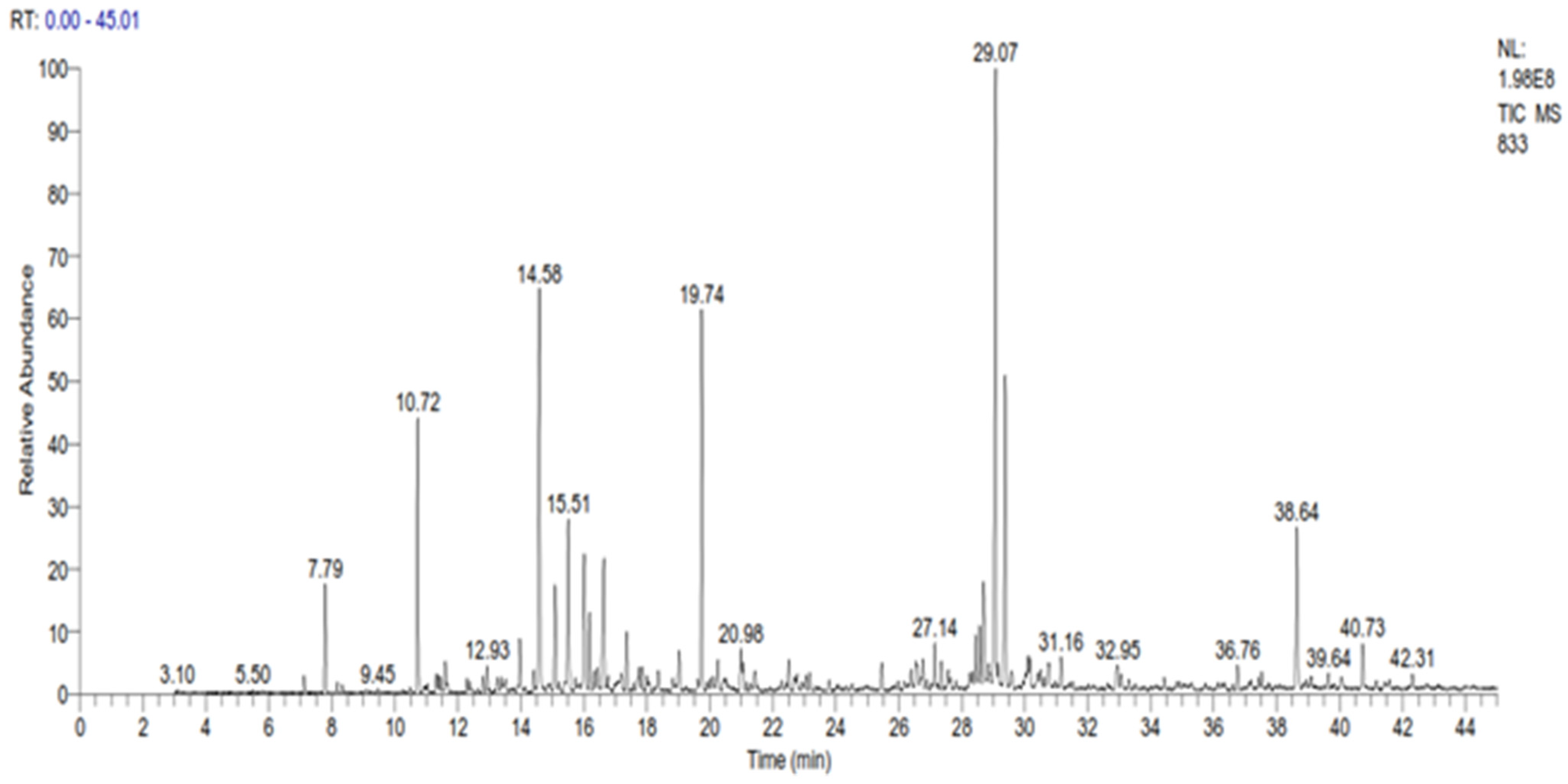
2.4. GC-SM Analysis of the Essential Oil of C. Ladanifer

The GC-MS analysis revealed the presence of 35 compounds in the essential oil of C. ladanifer (Figure 1). These compounds were divided into oxygenated sesquiterpenes (34.02%), oxygenated monoterpenes (33.14%), linear esters (10.38%), monoterpenes (9.11%), and sesquiterpenes (4.29%). The major constituents present in this EO were viridiflorol (17.64%), trans-pinocarveol (11.02%), bornylacetate (9.38%), and ledol (8.85%) (Table 2). The percentage of these constituents is higher than those found by Boukil et al. (oxygenated hydrocarbons (13.27%), oxygenated sesquiterpenes (2.57%), and monoterpenic ester (5.86%)) [19]. The same authors found that the main components from the fresh leaves of C. ladanifer were verticiol (18.16%), camphene (17.70%), n-butylcyclohexane (5.95%), and 3-carene (5.23%) [19]. These findings indicate that the time of harvest has a significant impact on the chemical composition obtained. The results of various chemical analysis studies on C. ladanifer essential oil carried out previously showed that the main constituents of C. ladanifer leaf essential oil from Northern Morocco were viridiflorol (19.6%), bornyl acetate (16.7%), and camphene (12.3%) [30]. Zidane et al. characterized C. ladanifer from Eastern Morocco and indicated the presence of camphene (15.5%), borneol (11.1%), 2,2,6-trimethylcyclohexanol (7.3%), 4-terpineol (6.3%), and α-pinene (4.2%) as the major compounds in the essential oil of this plant [29]. In Algeria, it was found that the main constituents of this oil were 5,7-di-epi-α-eudesmol (13.6%), borneol (12.5%), camphene (12.2%), δ-cadinene (7.6%), α-eudesmol (6.4%)%), 4-terpineol (5.7%) and α-pinene (4.2%) [28]. In France, Verdeguer et al. characterized the chemical composition of the oil extracted from the leaves and stems of C. ladanifer of Spanish origin but cultivated in Corsica by the presence of pinene (39%), viridiflorol (11.8%), ledol (3.3%) and bornyl acetate (3.1%) [33]. In Portugal, the chemical composition of C. ladanifer oil shows the presence of three sesquiterpenes alcohols, viridiflorol (13.6–17.4%), globulol (3.1–5.0%), and an unknown alcohol sesquiterpene (2.7–6.0%), as well as diterpene alcohol 15-nor-labdan-8-ol (1.7–5.2%) [34]. In Spain, the composition of the essential oil of C. ladanifer cultivated in central Spain, revealed its richness in oxygenated compounds, with trans-pinocarveol (20.00%), bornyl acetate (7.03%), and terpinen-4-ol (6.37%) as the main monoterpene compounds. Viridiflorol (13.59%) and ledol (4.36%) were the main constituents of the oxygenated sesquiterpene fraction. Large amounts of α-pinene (4.70%) were found in the hydrocarbon fractions. From this comparison, it seems that Moroccan C. ladanifer essential oil composition is closer to that of Corsica with Spanish origin. The chemical composition of C. ladanifer essential oil varies considerably depending on the source, plant material, and extraction method. As a result, based on the intended product during the exploitation of the species, a selection of organs, vegetative stage, and area proves to be extremely helpful in promoting the acquisition of very accurate chemotypes.

2.5. Antibacterial Activity

In this study, the antibacterial activity was evaluated using two methods: the agar disk diffusion method and the dilution in a liquid environment. The aim of the tests is to highlight the inhibitory power of the essential oil vis-à-vis the tested bacteria after 24 h of incubation at an adequate temperature of 37 °C.

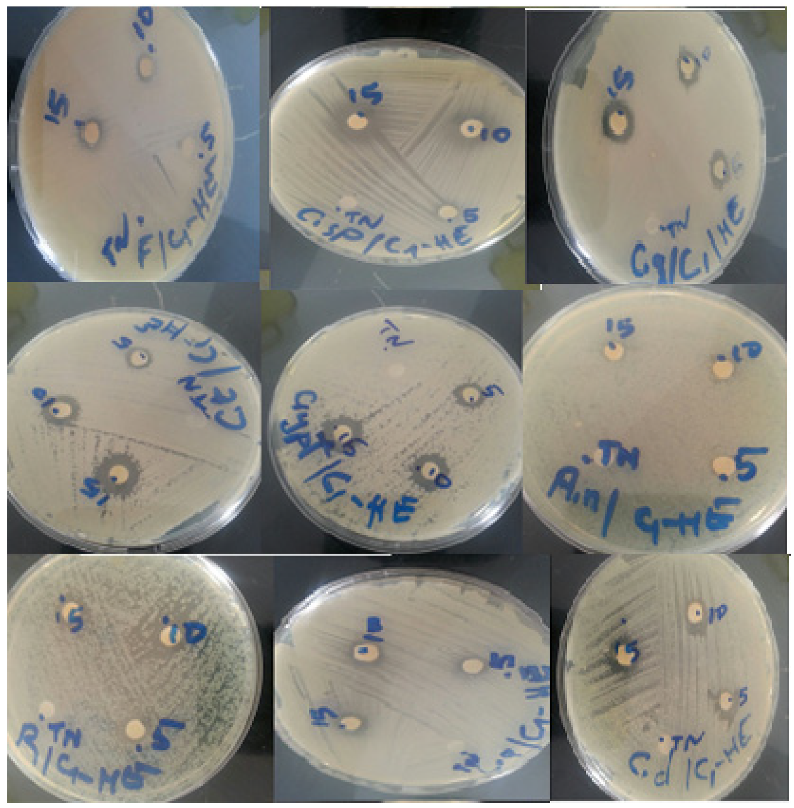
2.5.1. The Antibiotic Sensitivity Test

The antibiotic sensitivity profiles of the strains are developed according to the recommendations of the Committee on Antibiotic susceptibility of the French Society for Microbiology (CA-SFM) and presented in Table 3. From the table, it can be concluded that the strains of S. aureus and S. Typhi were sensitive to all antibiotics, while E. coli was only resistant to ticarcillin. At the same time, A. baumannii demonstrated complete resistance to all tested antibiotics. This may be due to the higher resistance of Gram-negative bacteria due to the complexity of their cell wall, containing a double membrane in opposition to the single glycoprotein/teichoic acid membrane of Gram-positive bacteria [35]

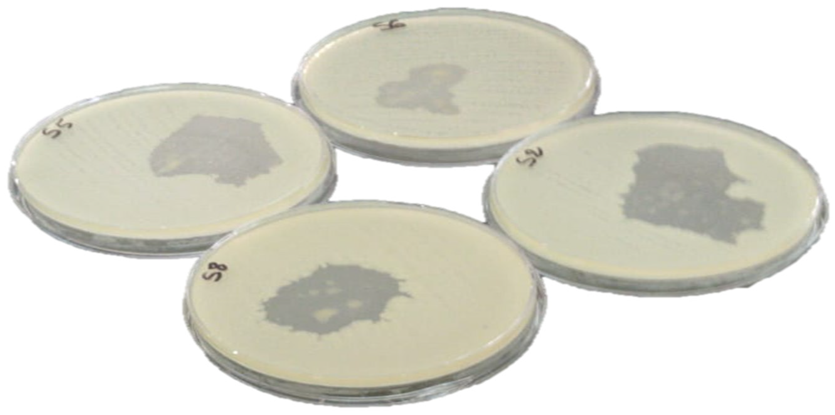
2.5.2. The Agar Disk-Diffusion Method for C. ladanifer Essential Oil

Table 4 and Figure 2 represent the results of the agar disk-diffusion method for C. ladanifer essential oil against the selected bacterial strains after 24 h at 37 °C. The agar disk diffusion method results indicated that the essential oil of C. ladanifer has a remarkable antibacterial activity compared to the concentration used (5 μL). From the analysis of the results obtained (Table 4), it was noted that the four microorganisms studied are sensitive except S. Typhi which demonstrated an inhibition zone of 30 ± 0.25 mm when the EO of C. ladanifer was used. The best inhibition diameter was against S. aureus (55 ± 0.22 mm) and E. coli (42 ± 0.11). It was also noted that contrary to the sensitivity test when A. baumannii demonstrated full resistance to the tested antibiotics, it demonstrated an inhibition diameter of 35 ± 0.27 when the EO was used, indicating a good and promising effect. Our results are superior to those found by Benayad et al. (S. aureus (28 mm), A. Baumannii (24 mm), and E. coli (18 mm)) (plant harvested in May) [11]. Same note for results obtained by Boukil et al. (S. aureus (14 mm), and E. coli (9 mm)) (plant harvested in August) [19]. As an outcome, harvesting the plant at the flowering stage is correlated to a potent antibacterial power.

2.5.3. Minimum Inhibitory Concentrations (MIC) and Minimum Bactericidal Concentrations (MBC)

During this investigation, the determination of the MIC was evaluated by assessing the inhibitory power of the plant’s essential oil at different concentrations against the selected bacteria (Table 5). In total accordance with the previous results obtained in the agar disk diffusion method and the observed inhibition zone, the results obtained indicated that the C. ladanifer essential oil MIC and MBC for all strains were 10 μL/mL. Regarding the MBC/MIC activity ratio, our results indicate that it was equal to 1 for all strains. This value allows us to affirm that the essential oil of C. ladanifer is bactericidal. This antimicrobial activity of this essential oil can only be explained by its chemical profile rich in 34.02% of oxygenated sesquiterpenes and oxygenated monoterpenes, and 33.14% of monoterpenes which are known as versatile anti-infective agents (antibacterial and antifungal) [36]. The structures of the functional groupings of the constituents of essential oils could play a crucial role in determining the antibacterial power of essential oils [37]. Nevertheless, minority compounds can interact directly, or in a synergistic or in an antagonistic way, to create a mixture with biological activity. Guinoiseau et al. [38], Rossi et al. [39], and Vieira et al. [40] demonstrated that essential oil from C. ladanifer has antimicrobial activity against Gram-positive and negative pathogens of clinical importance such as Staphylococcus aureus, E. coli, Streptococcus pneumonia, Pseudomonas aeruginosa, Enterobacter aerogenes, and Campylobacter jejuni.

2.6. Antifungal Activity

The disc diffusion technique enabled us to demonstrate the antifungal activity of C. ladanifer essential oil against 10 different fungus strains (Figure 3). The antifungal effect of a volume of 5 μL, 10 μL, and 15 μL of C. ladanifer essential oil is presented in Table 6. The essential oil demonstrated a good inhibitory activity with a slight dose-dependent activity. Fluconazole used as a positive control presented the best inhibition zone diameters against all the selected strains. In relation to the essential oil activity, C. tropicalis and C. neoformans were the most sensitive with an inhibition zone of 13 mm, followed by R. rubra and Penicillium sp., which have the same inhibition diameter of 12 mm, then C. dubliniensis and C. glabrata with inhibition zones of 11 mm. No studies on the antifungal activity of C. ladanifer of our study area (Middle Atlas, Morocco) have been carried out, except for a few attempts that have been reported by Boukil et al. [16].

Minimum Inhibitory Concentrations (MIC) and Minimum Fungicide Concentrations (MFC)

Table 7 summarizes the values of the MIC and MFC of the essential oil determined in a liquid medium against the ten tested fungal strains. The essential oil of C. ladanifer showed an antifungal activity on all fungal strains tested with concentrations ranging between 16 and 64 μL/mL.
The results of the MFC were all compared to those of MIC except for Candida sp. strain. Those obtained results were interesting and in agreement with those obtained by Guinoiseau et al., Rossi et al., Vieira et al. which indicate that the essential oil from C. ladanifer has an antimicrobial activity on fungi (such as Aspergillus niger, Botrytis cinerea, Mucorracemosus, and Verticilliumalboatrum) [38,39,40].

3. Discussion

This study, among others, focused on revealing the potential bioactivity of the essential oil of C. ladanifer. The study’s novelty was that plant material was collected during its time of flowering and in a different region (Middle Atlas), which is known for its semi-arid climate. The edaphic and climate parameters and others have influenced the variation of the composition revealed in the chromatographic analysis. The essential oil major components were viridiflorol (17.64%), trans-pinocarveol (11.02%), bornyl acetate (9.38%), and ledol (8.85%) indicating the domination of the oxygenated sesquiterpenes (34.02%), oxygenated monoterpenes (33.14%) in the overall composition.
The oxygenated sesquiterpenes and monoterpenes are a well-known group of compounds with antibacterial and antifungal proprieties [41,42] and their presence among the EO composition explains the majority of the outstanding outcomes obtained.
The strains chosen for this research are of great interest in the areas of clinical and public health. Their increasing resistance to conventional drugs has prompted further research into new, more effective options, particularly natural products [43,44]. S. aureus (Gram-positive) is a member of the indigenous human microflora and may be found asymptomatically in a variety of bodily locations. Diseases caused by transmission from these locations are both endemic and epidemic [45]. Infection with S. aureus is a leading cause of skin, soft tissue, respiratory, bone, joint, and endovascular diseases. Many strains of S. aureus are becoming resistant to current antibacterial treatments, posing a significant issue in medical microbiology [46].
S. Typhi, one of the representatives of the Salmonella family, is the direct causative organism of typhoid fever [47] (accompanied by weakness, headaches, mild vomiting, abdominal pain, and constipation). Symptoms may persist for weeks or months if not treated [48].
E. coli usually colonizes human babies’ gastrointestinal tracts within a few hours after birth. E. coli and its human host often live in excellent health and mutual benefit for decades [48]. These commensal E. coli strains seldom cause illness unless the host is immunocompromised or the usual gastrointestinal barriers are broken, as in peritonitis. Diarrhea induced by E. coli infection is a growing issue in both the developing and developed worlds, with significant rates of death in newborn infants and animals [49]. Although most commensal representatives found in human and animal gut flora are non-pathogenic, certain strains are very dangerous.
The genus Acinetobacter, over the past 30 years, has experienced considerable taxonomic evolution. Its most prominent example, A. baumannii, has emerged as one of the most problematic infections for healthcare facilities worldwide [50]. A. baumannii strains resistant to all known antibiotics have now been discovered, indicating a sentinel occurrence that should be addressed by the worldwide health care community as soon as possible. It often attacks the most susceptible hospitalized patients, those who are severely sick and have compromised skin integrity and airway protection [51].
In terms of fungus, non-albicans candida species are increasingly being reported as both colonizers and pathogens causing nosocomial fungal bloodstream infections, accounting for nearly half of all non-superficial candida infections, with C. glabrata, C. tropicalis, and C. dubliniensis being the most common [52].
One other life-threatening fungi exploited in this study is C. neoformans which is responsible for cryptococcal meningitis, the most prevalent type of cryptococcosis, often chronic and deadly if left untreated [53]. This virulence is none less than those presented by other fungi such as Aspergillus niger, a fungus that causes the “black mold” on certain fruits and vegetables (contaminant of food) which its consumption (as it secretes ochratoxins –mycotoxins) causes nephrotoxicity and renal tumors [54]. The EO of C. ladanifer and through this study demonstrated a strong and real potential that could be better exploited to fight against the threats presented by all the microbial strains studied with slight differences in terms of efficacy.

4. Materials and Methods

4.1. Materials and Reagents

Ticarcillin (TIC), ceftazidim (CAZ), Meropenem 10 (MEM10), and Ticarcilline85 (TIM85), were purchased from Sigma Aldrich (St-Quentin Fallavier, France). All chemicals and solvents were of highly analytical grade and were used as received from the supplier without further purification.

4.2. Plant Material

C. ladanifer was harvested in April 2019 (time of flowering) in El Harcha forest, which is located in the province of Khemisset, circle of Oulmes, 150 km south-east of Rabat, it extends entirely over the rural commune of Teddas (Middle Atlas region with semi-arid climate). The plant material was then dried in the shade, at room temperature, and stored for various uses. Professor Mohammed Fennan, a botanical expert from Mohammed V University’s scientific institute, identified this plant where a specimen of the voucher was set under collection number RTCBME10.

4.3. Extraction and Chemical Analysis of C. Ladanifer Essential Oil

4.3.1. Moisture Content Determination of the Plant Material

The moisture content was calculated after drying three samples (5 g) of the plant material at 100 °C in an oven until a constant weight was obtained. The calculation formula was as follows:
% MC = w 1 w 2 w 1 × 100
where %MC: Moisture content of the plant material, w1: Initial mass of plant material in g introduced into the oven at the time (t1) and w2: Final mass of plant material in g removed from the oven at the time (t2).

4.3.2. Essential Oil Extraction

Extraction of the essential oil was carried out by hydrodistillation using a Clevenger-type apparatus (VWR, Pennsylvania, United States). The essential oils were recovered in small opaque bottles and stored at 4 °C until further use. The yield extraction is expressed as a percentage of the volume of essential oil (mL) about the mass of the vegetable material (g). The calculation formula is as follows:
YE = V w ( w × MC )
where YE: Efficiency of extraction of essential oil expressed as volume of essential oil per mass of vegetable material (V/m), w: Mass of vegetable material used for oil extraction, V: Volume of essential oil collected (in mL) and MC: Moisture content of the plant material.

4.3.3. Mineral (ash) and Organic Matters Content of C. Ladanifer Essential Oil

The method used is that described by the “Association Française de Normalisation”
(AFNOR) standard [55]. The raw ash is obtained after the destruction of the organic matter by incineration. In a desiccator, nickel crucibles were dried in the oven for 1 h and were deposited and tarnished. After cooling, each crucible was weighed at 0.1 mg. Approximately 3 g of sample was crushed at 1 mm and weighed in the crucibles at 0.1 mg. The solid crucibles are fed into the muffle furnace under the exhaust hood and their contents are burnt for 4 h at 550 °C. After this time, we let the temperature go down to 100 °C and the crucibles were taken out, cooled in a desiccator, and then weighed at 0.1 g. Total ash was calculated as follows:
% MW = ( w 1 w 0 MT ) × 100
where %MW: Mineral matter (ash content) expressed as a percentage of the crude product, w1: Mass of crucible containing dry residue, in grams, w0: Empty crucible mass, in grams, and MT: Mass of the test sample, in grams.
The organic matter content (OM) of the sample is the difference between the dry matter mass (DM) and the mineral matter mass expressed as a percentage:
OM = DM MW

4.3.4. Refractive Index

The refractive index of chemical compounds is important because it indicates characteristic physical properties. The refractive index of the essential oil was measured at 20 °C using a refractometer (ATAGO, Tokyo, Japan) [56].

4.3.5. Brix Degree

The Brix degree gives an idea of the concentration (%) of all solids dissolved in the essential oil (sugar, salts, protein, fatty acid, etc.). Indeed, a few drops of the essential oil was deposited on the refractometer tank (ATAGO). The light beam deflection through the sample (the essential oil) was varied and indicated by a colored delimitation of the degree Brix.

4.3.6. GC-SM Analysis

The chromatographic analysis was carried out on a HP 6890 series gas chromatograph (Hewlett Packard, Palo Alto, CA, USA), equipped with a DB-5 (5% phenylmethylsiloxane) capillary column (30 m × 0.25 mm × 0.25 µm film thickness), an FID detector set at 250 °C and lied by a mixture of H2/air gas. The injection mode is split; the carrier gas used is nitrogen with a flow rate of 1.7 mL/min. The column temperature was programmed with a rise of 4 °C/min from 50 °C to 200 °C for 5 min. The device was controlled by a HP Chemstation computer system managing the operation of the device and allowing to follow the evolution of chromatographic analyses. The GC-MS was coupled to a mass spectrometer (HP 5973 series). The fragmentation was carried out by electronic impact at 70 eV. The column used is a capillary type DB-5SM (30 m × 0.25 mm × 0.25 µm). The column temperature was programmed at a rate of 4 °C/min from 50 to 200 °C for 5 min. The carrier gas used is helium with a flow rate of 1.7 mL/min. The injection mode is split. The identification of the constituents of the essential oils studied was carried out both by the index identification method of Kovàts [57], and Adams [58], and from the mass spectral database.

4.4. Study of C. Ladanifer L. Antimicrobial Activity

4.4.1. Microorganisms, Medium, and Antibiotics

In this study, four bacterial strains and nine fungal strains were collected from the Mohammed V University Hospital in Rabat-Morocco (Table 8). Microbial strains were seeded in petri dishes containing nutrient agar, the incubation time and temperature were 24 h and 37 °C for bacteria, 48 h and 28 °C for yeast, 7 days and 28 °C for molds to obtain well-isolated colonies. Isolated colonies were collected from young cultures obtained using an inoculator. The colonies were placed in 5 mL of sterile physiological water with NaCl (0.9%) so that the solution contained about 10 g/mL at a density ranging from 0.08 to 0.1 to 625 nm [59]. The inoculum is adjusted either by adding culture if it is too weak or sterile physiological water if it is too strong. Seeding was done directly after the preparation of the inoculum. The antibiotics used for bacterial testing were ticarcillin (TIC), ceftazidime (CEF), vancomycin (VAN), tetracycline (TET), cefalexin (CEF); colistin (COL) and amikacin (AMI).

4.4.2. Diffusion Method on Agar Medium

This test was carried out on Petri dishes (90 mm) by the diffusion method in the agar media of Mueller-Hinton and Sabouraud by determining the diameter of the zone of inhibition [60,61]. Disks of 6 mm in diameter, filter paper, were cut and sterilized, then deposited delicately in the center and on the peripheries of the surface of a medium previously inoculated with the microbial inoculum (this inoculum was obtained in the form of a suspension in saline solution). Then, volumes of 5 μL of essential oil of C. ladanifer relating to the antibacterial tests and volumes of 5, 10 and 15 μL relating to the fungal tests were deposited on these discs. A sterile disc flooded with 6 μL of sterile physiological water was used as a negative control as well as another negative control disc impregnated with 2 μL of DMSO to check the growth of different bacterial and fungal strains. The inhibition diameters were measured around the disc after incubation in an oven, each test was repeated three times to obtain as precise an estimate as possible of the true experimental value sought.

4.4.3. Liquid Dilution Method

This method was used to determine the minimum inhibitory concentration (MIC) and the minimum bactericidal (MBC) and fungicidal (MFC) concentration of the essential oil of C. ladanifer L. [62,63]. An amount of 2 µL of DMSO was added to the 10 tubes. Hemolysis containing the culture medium (1 mL) for each bacterial strain. Each volume of essential oil (2, 4, 6, 10, 12, 14, 16, 18, 20 and 22 µL) was introduced into a hemolysis tube. Then, a volume of 6 μL of the bacterial suspension with a concentration of 105 CFU/mL was taken and then was deposited in each of the preceding tubes and then 2 μL of DMSO was added for each microbial strain. Two sets were made, one containing the culture medium (1 mL) plus a strain (bacterial or fungal) and the other containing the culture medium (1 mL) plus 2 µL of essential oil alone. After an incubation of 24 h at 37 °C for bacteria, 48 h at 28 °C for yeasts, and 7 days for molds, the MIC of the essential oil was determined. We then inoculated the surface of a Mueller Hinton agar cast in a Petri dish with 100 μL of the contents of the tubes having a concentration greater than or equal to the MIC to determine the CMB and CMF.

5. Conclusions

In this study, it was demonstrated that the essential oil of C. ladanifer is high in oxygenated sesquiterpenes, which mediated the activities seen. The results of the antimicrobial activity showed that this essential oil was able to inhibit the growth of a wide variety of bacterial and fungal strains. The observed activity was correlated to the harvest time of this plant done in May, which influenced the phytochemical profile of the EO in comparison to its harvest in other months as seen in other studies from the literature. The essential oil demonstrated an excellent antimicrobial activity and could serve as an alternative to some medicine and antibiotics. More studies should be done to determine the toxicity and the therapeutic margin for a more controlled and oriented application.

Author Contributions

Conceptualization, J.E.K. and T.Z.; validation, T.Z.; formal analysis, I.A.; investigation, S.A. and S.A.I.; data curation, H.M. and O.M.A.K.; writing—original draft preparation, A.K. and M.B. supervision, T.Z.; All authors have read and agreed to the published version of the manuscript.

Funding

This research was funded by the Deanship of Scientific Research at Princess Nourah bint Abdulrahman University through the Fast-track Research Funding Program.

Institutional Review Board Statement

Not applicable.

Informed Consent Statement

Not applicable.

Data Availability Statement

Data are available upon request.

Acknowledgments

The authors are thankful to the Deanship of Scientific Research at Princess Nourah bint Abdulrahman University for the support of this research through the Fast-track Research Funding Program.

Conflicts of Interest

The authors declare no conflict of interest.

References

  1. Es-safi, I.; Mechchate, H.; Amaghnouje, A.; Elbouzidi, A.; Bouhrim, M.; Bencheikh, N.; Hano, C.; Bousta, D. Assessment of Antidepressant-Like, Anxiolytic Effects and Impact on Memory of Pimpinella anisum L. Total Extract on Swiss Albino Mice. Plants 2021, 10, 1573. [Google Scholar] [CrossRef]
  2. Bencheikh, N.; Elbouzidi, A.; Kharchoufa, L.; Ouassou, H.; Alami Merrouni, I.; Mechchate, H.; Es-safi, I.; Hano, C.; Addi, M.; Bouhrim, M.; et al. Inventory of Medicinal Plants Used Traditionally to Manage Kidney Diseases in North-Eastern Morocco: Ethnobotanical Fieldwork and Pharmacological Evidence. Plants 2021, 10, 1966. [Google Scholar] [CrossRef] [PubMed]
  3. Herrera, C.M. Tipos Morfológicos y Funcionales En Plantas Del Matorral Mediterráneo Del Sur de España. Stud. Oecologica 1984, 5, 7–34. [Google Scholar]
  4. Nuñez, E. Ecología Del Jaral de Cistus ladanifer L. Ph.D. Thesis, Facultad de Ciencias, Universidad de Extremadura, Badajoz, Spain, 1989. [Google Scholar]
  5. Mariotti, J.P.; Tomi, F.; Casanova, J.; Costa, J.; Bernardini, A.F. Composition of the Essential Oil of Cistus ladaniferus L. Cultivated in Corsica (France). Flavour Fragr. J. 1997, 12, 147–151. [Google Scholar] [CrossRef]
  6. Bellakhdar, J. Medecine arabe ancienne et savoirs populaires. In La Pharmacopée Traditionnelle; Ibis Press: Paris, France, 1997. [Google Scholar]
  7. Talavera, S.; Gibbs, P.E.; Herrera, J. Reproductive Biology of Cistus ladanifer (Cistaceae). Plant Syst. Evol. 1993, 186, 123–134. [Google Scholar] [CrossRef]
  8. Devesa Alcaraz, J.A. Vegetación y Flora de Extremadura; Universitas Editorial: Badajoz, Spain, 1995. [Google Scholar]
  9. Pomponio, R.; Gotti, R.; Santagati, N.A.; Cavrini, V. Analysis of Catechins in Extracts of Cistus Species by Microemulsion Electrokinetic Chromatography. J. Chromatogr. A 2003, 990, 215–223. [Google Scholar] [CrossRef]
  10. Barrajón-Catalán, E.; Fernández-Arroyo, S.; Saura, D.; Guillén, E.; Fernández-Gutiérrez, A.; Segura-Carretero, A.; Micol, V. Cistaceae Aqueous Extracts Containing Ellagitannins Show Antioxidant and Antimicrobial Capacity, and Cytotoxic Activity against Human Cancer Cells. Food Chem. Toxicol. 2010, 48, 2273–2282. [Google Scholar] [CrossRef]
  11. Andrade, D.; Gil, C.; Breitenfeld, L.; Domingues, F.; Duarte, A.P. Bioactive Extracts from Cistus ladanifer and Arbutus unedo L. Ind. Crops Prod. 2009, 30, 165–167. [Google Scholar] [CrossRef]
  12. Benayad, N.; Mennane, Z.; Charof, R.; Hakiki, A.; Mosaddak, M. Antibacterial Activity of Essential Oil and Some Extracts of Cistus ladaniferus from Oulmes in Morocco. J. Mater. Environ. Sci. 2013, 4, 1066–1071. [Google Scholar]
  13. Bonnier, G.; Douin, R.; Poinsot, J.; Palese, R.; Aeschimann, D. La Grande Flore en Couleurs de Gaston Bonnier: France, Suisse, Belgique et Pays Voisins; P. Parey: Paris, France; Hamburg, Germany; Berlin, Germany, 1990; ISBN 978-2-7011-1300-5. [Google Scholar]
  14. Di Pasqua, R.; De Feo, V.; Villani, F.; Mauriello, G. In Vitro Antimicrobial Activity of Essential Oils from Mediterranean Apiaceae, Verbenaceae and Lamiaceae against Foodborne Pathogens and Spoilage Bacteria. Ann. Microbiol. 2005, 55, 139–143. [Google Scholar]
  15. Oller-López, J.L.; Rodríguez, R.; Cuerva, J.M.; Oltra, J.E.; Bazdi, B.; Dahdouh, A.; Lamarti, A.; Mansour, A.I. Composition of the Essential Oils of Cistus ladaniferus and C. monspeliensis from Morocco. J. Essent. Oil Res. 2005, 17, 553–555. [Google Scholar] [CrossRef]
  16. Youbi, A.; El Mansouri, L.; Boukhira, S.; Daoudi, A.; Bousta, D. In Vivo Anti-Inflammatory and Analgesic Effects of Aqueous Extract of Cistus ladanifer L. Moroc. Am. J. Ther. 2016, 23, e1554–e1559. [Google Scholar] [CrossRef]
  17. Mekhfi, H.; El Haouari, M.; Legssyer, A.; Bnouham, M.; Aziz, M.; Atmani, F.; Remmal, A.; Ziyyat, A. Platelet Anti-Aggregant Property of Some Moroccan Medicinal Plants. J. Ethnopharmacol. 2004, 94, 317–322. [Google Scholar] [CrossRef]
  18. Guimarães, R.; Sousa, M.J.; Ferreira, I.C.F.R. Contribution of Essential Oils and Phenolics to the Antioxidant Properties of Aromatic Plants. Ind. Crops Prod. 2010, 32, 152–156. [Google Scholar] [CrossRef]
  19. Mohammed, B.; Said, C.; Fouzia, F.R.; Kawtar, F.B.; Zoubida, H.; Abdelilah, O.; Mohammed, E.; Ghizlane, E. Chemical Composition and Antimicrobial Activity of the Essential Oil of Cistus ladanifer Var. Maculatus Dun. J. Microbiol. Biotechnol. Food Sci. 2021, 2021, 925–930. [Google Scholar]
  20. Aziz, M.; Tab, N.; Karim, A.; Mekhfi, H.; Bnouham, M.; Ziyyat, A.; Melhaoui, A.; Legssyer, A. Relaxant Effect of Aqueous Extract of Cistus ladaniferus on Rodent Intestinal Contractions. Fitoterapia 2006, 77, 425–428. [Google Scholar] [CrossRef]
  21. Attaguile, G.; Perticone, G.; Mania, G.; Savoca, F.; Pennisi, G.; Salomone, S. Cistus incanus and Cistus monspeliensis Inhibit the Contractile Response in Isolated Rat Smooth Muscle. J. Ethnopharmacol. 2004, 92, 245–250. [Google Scholar] [CrossRef]
  22. Skorić, M.; Todorović, S.; Gligorijević, N.; Janković, R.; Živković, S.; Ristić, M.; Radulović, S. Cytotoxic Activity of Ethanol Extracts of in Vitro Grown Cistus creticus Subsp. creticus L. on Human Cancer Cell Lines. Ind. Crops Prod. 2012, 38, 153–159. [Google Scholar]
  23. Sosa, T.; Chaves, N.; Alias, J.C.; Escudero, J.C.; Henao, F.; Gutiérrez-Merino, C. Inhibition of Mouth Skeletal Muscle Relaxation by Flavonoids of Cistus ladanifer L.: A Plant Defense Mechanism against Herbivores. J. Chem. Ecol. 2004, 30, 1087–1101. [Google Scholar] [CrossRef]
  24. Belmokhtar, M.; Bouanani, N.E.; Ziyyat, A.; Mekhfi, H.; Bnouham, M.; Aziz, M.; Matéo, P.; Fischmeister, R.; Legssyer, A. Antihypertensive and Endothelium-Dependent Vasodilator Effects of Aqueous Extract of Cistus Ladaniferus. Biochem. Biophys. Res. Commun. 2009, 389, 145–149. [Google Scholar] [CrossRef]
  25. Amensour, M.; Sendra, E.; Pérez-Alvarez, J.A.; Skali-Senhaji, N.; Abrini, J.; Fernández-López, J. Antioxidant Activity and Chemical Content of Methanol and Ethanol Extracts from Leaves of Rockrose (Cistus ladaniferus). Plant Foods Hum. Nutr. 2010, 65, 170–178. [Google Scholar] [CrossRef]
  26. Lahcen, S.A.; El Hattabi, L.; Benkaddour, R.; Chahboun, N.; Ghanmi, M.; Satrani, B.; Tabyaoui, M.; Zarrouk, A. Chemical Composition, Antioxidant, Antimicrobial and Antifungal Activity of Moroccan Cistus creticus Leaves. Chem. Data Collect. 2020, 26, 100346. [Google Scholar] [CrossRef]
  27. Robles, C.; Bousquet-Mélou, A.; Garzino, S.; Bonin, G. Comparison of Essential Oil Composition of Two Varieties of Cistus ladanifer. Biochem. Syst. Ecol. 2003, 31, 339–343. [Google Scholar] [CrossRef]
  28. Bechlaghem, K.; Allali, H.; Benmehdi, H.; Aissaoui, N.; Flamini, G. Chemical Analysis of the Essential Oils of Three Cistus Species Growing in North-West of Algeria. Agric. Conspec. Sci. 2019, 84, 283–293. [Google Scholar]
  29. Zidane, H.; Elmiz, M.; Aouinti, F.; Tahani, A.; Wathelet, J.; Sindic, M.; Elbachiri, A. Chemical Composition and Antioxidant Activity of Essential Oil, Various Organic Extracts of Cistus ladanifer and Cistus libanotis Growing in Eastern Morocco. Afr. J. Biotechnol. 2013, 12, 5314–5320. [Google Scholar]
  30. Greche, H.; Mrabet, N.; Zrira, S.; Ismaili-Alaoui, M.; Benjilali, B.; Boukir, A. The Volatiles of the Leaf Oil of Cistus ladanifer L. Var. Albiflorus and Labdanum Extracts of Moroccan Origin and Their Antimicrobial Activities. J. Essent. Oil Res. 2009, 21, 166–173. [Google Scholar] [CrossRef]
  31. Hellal, Z. Contribution à l’étude Des Propriétés Antibactériennes et Antioxydantes de Certaines Huiles Essentielles Extraites Des Citrus. Application Sur La Sardine (Sardina pilchardus). Doctoral Dissertation, Université Mouloud Mammeri, Tizi Ouzou, Algeria, 2011. [Google Scholar]
  32. Mrabkt, N.; Lahlou, H.; Benjilali, B. Effet de Quelques Extraits Du Ciste Ladanifère Du Maroc (Cistus ladaniferus L.) Sur La Croissance de Quatre Champignons. Cryptogam. Mycol. 1999, 20, 23–33. [Google Scholar]
  33. Verdeguer, M.; Blázquez, M.A.; Boira, H. Chemical Composition and Herbicidal Activity of the Essential Oil from a Cistus ladanifer L. Population from Spain. Nat. Prod. Res. 2012, 26, 1602–1609. [Google Scholar] [CrossRef]
  34. Gomes, P.B.; Mata, V.G.; Rodrigues, A.E. Characterization of the Portuguese-Grown Cistus Ladanifer Essential Oil. J. Essent. Oil Res. 2005, 17, 160–165. [Google Scholar] [CrossRef]
  35. Sadeq, O.; Mechchate, H.; Es-safi, I.; Bouhrim, M.; Zahra Jawhari, F.; Ouassou, H.; Kharchoufa, L.; AlZain, M.N.; Alzamel, N.M.; Mohamed Al kamaly, O.; et al. Phytochemical Screening, Antioxidant and Antibacterial Activities of Pollen Extracts from Micromeria Fruticosa, Achillea Fragrantissima, and Phoenix Dactylifera. Plants 2021, 10, 676. [Google Scholar] [CrossRef]
  36. Daferera, D.J.; Ziogas, B.N.; Polissiou, M.G. The Effectiveness of Plant Essential Oils on the Growth of Botrytis cinerea, Fusarium sp. and Clavibacter Michiganensis subsp. Michiganensis. Crop Prot. 2003, 22, 39–44. [Google Scholar] [CrossRef]
  37. Verdeguer, M.; Sánchez-Moreiras, A.M.; Araniti, F. Phytotoxic Effects and Mechanism of Action of Essential Oils and Terpenoids. Plants 2020, 9, 1571. [Google Scholar] [CrossRef]
  38. Guinoiseau, E.; Lorenzi, V.; Luciani, A.; Tomi, F.; Casanova, J.; Berti, L. Susceptibility of the Multi-Drug Resistant Strain of Enterobacter Aerogenes EA289 to the Terpene Alcohols from Cistus ladaniferus Essential Oil. Nat. Prod. Commun. 2011, 6, 1159–1162. [Google Scholar] [CrossRef] [Green Version]
  39. Rossi, P.-G.; Berti, L.; Panighi, J.; Luciani, A.; Maury, J.; Muselli, A.; de Rocca Serra, D.; Gonny, M.; Bolla, J.-M. Antibacterial Action of Essential Oils from Corsica. J. Essent. Oil Res. 2007, 19, 176–182. [Google Scholar] [CrossRef]
  40. Vieira, M.; Bessa, L.J.; Martins, M.R.; Arantes, S.; Teixeira, A.P.S.; Mendes, Â.; Martins da Costa, P.; Belo, A.D.F. Chemical Composition, Antibacterial, Antibiofilm and Synergistic Properties of Essential Oils from Eucalyptus globulus Labill. and Seven Mediterranean Aromatic Plants. Chem. Biodivers. 2017, 14, e1700006. [Google Scholar] [CrossRef] [Green Version]
  41. da Costa, J.S.; de Figueiredo, R.O.; Setzer, W.N.; da Silva, J.K.R.; Maia, J.G.S.; Figueiredo, P.L.B. Monoterpenes and Sesquiterpenes of Essential Oils from Psidium Species and Their Biological Properties. Molecules 2021, 26, 965. [Google Scholar]
  42. Meng, J.C.; Zhu, Q.X.; Tan, R.X. New Antimicrobial Mono-and Sesquiterpenes from Soroseris hookeriana subsp. erysimoides. Planta Med. 2000, 66, 541–544. [Google Scholar] [CrossRef]
  43. Mechchate, H.; Costa de Oliveira, R.; Es-safi, I.; Vasconcelos Mourão, E.M.; Bouhrim, M.; Kyrylchuk, A.; Soares Pontes, G.; Bousta, D.; Grafov, A. Antileukemic Activity and Molecular Docking Study of a Polyphenolic Extract from Coriander Seeds. Pharmaceuticals 2021, 14, 770. [Google Scholar] [CrossRef]
  44. Bouhrim, M.; Ouassou, H.; Boutahiri, S.; Daoudi, N.E.; Mechchate, H.; Gressier, B.; Eto, B.; Imtara, H.; Alotaibi, A.A.; Al-zharani, M.; et al. Opuntia dillenii (Ker Gawl.) Haw., Seeds Oil Antidiabetic Potential Using In Vivo, In Vitro, In Situ, and Ex Vivo Approaches to Reveal Its Underlying Mechanism of Action. Molecules 2021, 26, 1677. [Google Scholar] [CrossRef]
  45. Fa, W. Staphylococcus Aureus (Including Toxic Shock Syndrome). In Principles and Practices of Infectious Diseases, 4th ed.; Churchill Livingstone Elsevier: Philadelphia, PA, USA, 1995; Volume 4, pp. 1754–1783. [Google Scholar]
  46. Chambers, H.F. Methicillin Resistance in Staphylococci: Molecular and Biochemical Basis and Clinical Implications. Clin. Microbiol. Rev. 1997, 10, 781–791. [Google Scholar] [CrossRef]
  47. Arshad, R.; Pal, K.; Sabir, F.; Rahdar, A.; Bilal, M.; Shahnaz, G.; Kyzas, G.Z. A Review of the Nanomaterials Use for the Diagnosis and Therapy of Salmonella Typhi. J. Mol. Struct. 2021, 1230, 129928. [Google Scholar] [CrossRef]
  48. da Cruz, L.F.; Souza, I.L.A.; de Souza, L.D.; de Freitas Araújo, M.G.; Granjeiro, P.A. The Importance of Intestinal Microbiota and Its Role in the Nosocomial Infection. Res. Soc. Dev. 2021, 10, e489101019166. [Google Scholar] [CrossRef]
  49. Radu, S.; Ling, O.W.; Rusul, G.; Karim, M.I.A.; Nishibuchi, M. Detection of Escherichia coli O157: H7 by Multiplex PCR and Their Characterization by Plasmid Profiling, Antimicrobial Resistance, RAPD and PFGE Analyses. J. Microbiol. Methods 2001, 46, 131–139. [Google Scholar] [CrossRef]
  50. Fournier, P.E.; Richet, H.; Weinstein, R.A. The Epidemiology and Control of Acinetobacter Baumannii in Health Care Facilities. Clin. Infect. Dis. 2006, 42, 692–699. [Google Scholar] [CrossRef] [Green Version]
  51. Nie, D.; Hu, Y.; Chen, Z.; Li, M.; Hou, Z.; Luo, X.; Mao, X.; Xue, X. Outer Membrane Protein A (OmpA) as a Potential Therapeutic Target for Acinetobacter Baumannii Infection. J. Biomed. Sci. 2020, 27, 1–8. [Google Scholar] [CrossRef] [Green Version]
  52. Ann Chai, L.Y.; Denning, D.W.; Warn, P. Candida Tropicalis in Human Disease. Crit. Rev. Microbiol. 2010, 36, 282–298. [Google Scholar] [CrossRef]
  53. Zhao, Y.; Lin, J.; Fan, Y.; Lin, X. Life Cycle of Cryptococcus Neoformans. Annu. Rev. Microbiol. 2019, 73, 17–42. [Google Scholar] [CrossRef]
  54. Cairns, T.C.; Nai, C.; Meyer, V. How a Fungus Shapes Biotechnology: 100 Years of Aspergillus Niger Research. Fungal Biol. Biotechnol. 2018, 5, 1–14. [Google Scholar] [CrossRef] [Green Version]
  55. Despinasse, Y.; Moja, S.; Soler, C.; Jullien, F.; Pasquier, B.; Bessière, J.-M.; Noûs, C.; Baudino, S.; Nicolè, F. Structure of the Chemical and Genetic Diversity of the True Lavender over Its Natural Range. Plants 2020, 9, 1640. [Google Scholar] [CrossRef]
  56. Kruse, E.B.; Revolinski, S.; Aplin, J.; Skinner, D.Z.; Murray, T.D.; Edwards, C.G.; Carter, A.H. Carbohydrate Accumulation and Differential Transcript Expression in Winter Wheat Lines with Different Levels of Snow Mold and Freezing Tolerance after Cold Treatment. Plants 2020, 9, 1416. [Google Scholar] [CrossRef]
  57. Kovats, E.S. Gas Chromatographic Characterization of Organic Substances in the Retention Index System. Adv. Chromatogr. 1965, 1, 229–247. [Google Scholar]
  58. Adams, R.P. Identification of Essential Oil Components by Gas Chromatography/Mass Spectrometry; Allured Publishing Corporation: Carol Stream, IL, USA, 2007; Volume 456. [Google Scholar]
  59. Mohammedi, Z. Etude Du Pouvoir Antimicrobien et Antioxydant Des Huiles Essentielles et Flavonoïdes de Quelques Plantes de La Région de Tlemcen; Mémoire de Magister; University Abou Bakr Belkaïd Tlemcen: Chetouane, Algeria, 2006; 105p. [Google Scholar]
  60. Gupta, V.K.; Roy, A.; Nigam, V.K.; Mukherjee, K. Antimicrobial Activity of Spondias Pinnata Resin. J. Med. Plants Res. 2010, 4, 1656–1661. [Google Scholar]
  61. De Billerbeck, V.-G. Huiles Essentielles et Bactéries Résistantes Aux Antibiotiques. Phytothérapie 2007, 5, 249–253. [Google Scholar] [CrossRef]
  62. Biyiti, L.F.; Meko’o, D.J.L.; Tamzc, V.; Amvam Zollo, P.H. Recherche de l’activité Antibactérienne de Quatre Plantes Médicinales Camerounaises. Pharm. Med. Trad. Afr. 2004, 13, 11–20. [Google Scholar]
  63. Doughari, J.H.; Manzara, S. In Vitro Antibacterial Activity of Crude Leaf Extracts of Mangifera Indica Linn. Afr. J. Microbiol. Res. 2008, 2, 67–72. [Google Scholar]
Figure 1. GC Chromatogram of the essential oil of C. ladanifer.
Plants 10 02068 g001
Figure 2. Bacterial strain growth inhibition zone of C. ladanifer essential oil.
Plants 10 02068 g002
Figure 3. Fungal strains zone of inhibition diameters of essential oil of C. ladanifer.
Plants 10 02068 g003
Table 1. Refractive index and Brix index of the C. ladanifer essential oil.
PlantRefractive IndexBrix Index
C. ladanifer L. essential oil1.451.33556
Table 2. The compounds identified in the essential oil of C. ladanifer after analysis by GC/MS on column DB-5.
No.CompoundsFormulasPercentageIK CalculatedIK (ADAMS)
1α-PineneC10H162.43922939
2p-CymeneC10H146.1110041024
3(Z)-vertocitral CC9H14O0.6310281080
4p-CymeneneC10H140.5710651091
5α-CampholenalC10H16O1.3410931126
6trans-Pinocarveol C10H16O11.0211101139
7PinocarvoneC10H14O2.7211251164
8BorneolC10H18O4.8011371169
9Terpinen-4-olC10H18O4.0911511177
10MyrtenalC10H14O1.7611561195
11MyrtenolC10H16O4.0211681195
12trans-CarveolC10H16O1.4411891216
13CarvoneC10H14O0.5412031243
14(Z)-β-Damascone C13H20O 0.9712381387
15BornylacetateC12H20O29.3812591285
16CarvacrolC10H14O0.8012741084
17MyrtenylacetateC12H18O2 1.0012961326
182,4,6-trimethoxytolueneC10H14O30.6112981483
19(E)-Trimenal C13H22O 0.7713431421
20AromadendreneC15H240.6714381441
21ViridifloreneC15H240.9514731496
222,3-Dihydro-1,1,4,5,6-pentamethyl 1H-indene C14H200.8114811522
23cis-CalameneneC15H221.1714931529
24δ-Cadinene C15H240.6815001523
25PalustrolC15H26O1.1415381568
26SpathulenolC15H24O1.4115421578
27Caryophyllene oxideC15H24O2.8515461583
28ViridiflorolC15H26O17.7415601592
29LedolC15H26O8.8515701602
301,10-di-epi-Cubenol C15H26O0.7515951619
31Caryophylla-4 (12), 8 (13)-dien-5α-olC15H24O0.6415981640
32CadaleneC15H180.8216341676
3314-Hydroxy-4,5-dihydro-
caryophyllene
C15H26O0.6418481706
34SclareolC20H36O2 4.6019242223
3513-epi-Dolabradiene C20H321.2820132000
Oxygenated sesquiterpenes 34.02
Oxygenated monoterpenes 33.14
Linear esters 10.38
Monoterpenes 9.11
Sesquiterpenes 4.29
Others 9.06
Total 100
Table 3. The test for the sensitivity of bacterial strains to certain antibiotics.
ATBA. baumanniiATBS. aureus
TIC 75 μgRCIP 5 μgS
CEF 30 μgRVAN 30 μgS
MEM 10 μgRTET 30 μgS
TIM 85 μgRCEF 15 μgS
ATBE. coliATBS. Typhi
COL 50 μgSCOL 50 μgS
MEM 10 μgSMEM 10 μgS
TIC 75 μgRTIC 75 μgS
AMI 30 μgSAMI 30 μgS
S: Sensitive; I: Intermediate; A: Resistant; ATB: Antibiotics; TIC: Ticarcillin; CEF: Ceftazidime; VAN: Vancomycin; TET: Tetracycline; CEF: Cefalexin; COL: Colistin; AMI: Amikacin.
Table 4. Diameter of the inhibition zone of C. ladanifer essential oil against the four pathogenic strains.
S. aureusE. coliA. baumanniiS. Typhi
C. ladanifer 5 μL (EO)55 ± 0.2242 ± 0.1135 ± 0.2730 ± 0.25
The inhibition diameters were expressed in millimeters (mm): Mean ± SD.
Table 5. MIC and MBC of C. ladanifer essential oil against selected bacterial strains.
Bacterial StrainsConcentrations (μL/mL) DMSO
2 μL/mL
21020304050MBC/MIC
S. aureusMIC+1+
MBC++
Acinetobacter baumanniiMIC+1+
MBC++
E. coliMIC+1+
MBC++
Salmonella TyphiMIC+1+
MBC++
+: Presence; −: Absence.
Table 6. Values of the inhibition diameters zone of the essential oil of C. ladanifer.
Fungal StrainsGrowth Inhibition Diameter (GID) (mm) Fluconazole GID (mm)
5 μL10 μL15 μL150 mg
C. albicans781024.7
C. tropicalis9111325
C. glabrata9111123.7
C. dubliniensis8101119.3
Candida sp.781024
R. rubra8101218.7
A. niger-7813.7
C. neoformans9111329.3
Penicillium sp.7101213.8
Fusarium sp.-7816.3
Table 7. The effect of the C. ladanifer essential oil on the MIC and MFC of the selected fungi strains.
Fungal Strains
MICMFCMFC/MIC
Candida albicans32321
Candida tropicalis64641
Candida glabrata32321
Candida dubliniensis32321
Candida sp.16624
Rhodotorula rubra32321
Cryptoccocus neoformans64641
Penicillium sp. 64641
Fusarium sp. 64641
Aspergillus niger32321
Table 8. List of microbial strains tested.
Microbial Strains
Bacterial StrainsFungal Strains
YeastsMolds
Staphylococcus aureusCandida tropicalisPenicillium sp.
Salmonella TyphiCandida glabrataFusarium sp.
Escherichia coliCandida dubliniensisAspergillus niger
Acinetobacter baumanniiCandida sp.
Rhodotorula rubra
Cryptoccocus neoformans
Publisher’s Note: MDPI stays neutral with regard to jurisdictional claims in published maps and institutional affiliations.

