1. Introduction
The global demographic transition is causing a rapid increase in the proportion of elderly individuals within the overall population. This structural change presents significant challenges to healthcare systems, particularly in maintaining autonomy, mobility, and quality of life among aging citizens [
1,
2]. Preserving the functional independence of elderly people requires structured physical activity programs that are designed to sustain muscle strength, joint mobility, and neuromuscular coordination. Evidence in the large literature highlights the effectiveness of resistance-based and functional training interventions in mitigating age-related decline and reducing the risk of frailty, as pointed out, for example, in [
3,
4]. Practical guidelines also emphasize the importance of implementing accessible exercise routines in both community and home settings to ensure continuity of care and adherence [
5].
Frailty is a multidimensional clinical condition that is characterized by increased vulnerability to adverse outcomes, such as falls, hospitalization, and loss of independence [
6]. Identifying and quantifying frailty indicators are essential for customizing motion exercise, rehabilitation interventions, and monitoring progress over time. Therefore, technological solutions that assist movement while enabling measurable and controlled exercise execution are becoming increasingly important, especially for elderly populations who need safe, lightweight, low-cost, and user-friendly devices. The increasing clinical demand for effective upper-limb rehabilitation and motion assistance has driven extensive research into wearable robotic systems, particularly exoskeletons and exosuits [
7,
8,
9,
10]. Over the past decade, a substantial amount of work has explored design methodologies, actuation strategies, control paradigms, and human–robot interaction approaches specifically for upper-limb rehabilitation devices. Comprehensive review studies provide valuable insights into existing developments and the key challenges that continue to shape this field, as discussed in [
7,
8,
9,
10]. Recent reviews have highlighted the diversity of mechanical architectures and interactive design strategies that are employed in upper-limb exoskeletons. Dhatrak et al. [
7] present a comprehensive analysis of more than thirty existing upper-limb exoskeleton prototypes, focusing on interactive design principles, human-in-the-loop control, and challenges related to ergonomics, adaptability, and usability. Their work underscores the multidisciplinary nature of exoskeleton development and emphasizes the need for systems that better integrate physiotherapy requirements with user-centered design. In addition, increasing attention has been devoted to soft robotic wearable devices that can be designed to overcome the limitations associated with rigid exoskeletons. A systematic review by Bardi et al. [
8] examines upper-limb exosuits that are based on cable-driven and pneumatic actuation, covering more than fifty systems. The authors highlight the advantages of soft, lightweight solutions in terms of comfort, portability, and preservation of natural joint motion, while also identifying challenges that are related to force transmission, control accuracy, and repeatability in rehabilitation contexts. Focusing specifically on the elbow joint, Supriyono et al. [
9] provide a comprehensive review of elbow exoskeletons, classifying devices based on mechanical structure, actuation methods, and sensing technologies. Their analysis reveals recurring trade-offs among mechanical simplicity, assistance capability, and user comfort, emphasizing the importance of aligning device kinematics with physiological joint motion. Complementing this, Gonçalves et al. [
10] review robotic interfaces developed for post-stroke upper-limb rehabilitation, examining assistance strategies, actuation methods, and control mechanisms. Their review identifies a clear trend toward lightweight, modular, and portable solutions, while also highlighting persistent barriers to clinical translation and widespread adoption. Collectively, these reviews demonstrate strong and sustained research interest in upper-limb and elbow assisting devices while also highlighting unresolved challenges related to portability, adaptability, and integration within physiotherapy-oriented rehabilitation frameworks, although applications specifically targeting elderly healthcare have not yet been thoroughly considered [
2]. These gaps underscore the need for continued development and experimental validation of compact, lightweight, and user-centered assisting devices capable of supporting functional rehabilitation beyond traditional clinical settings. Moreover, recent biomechanical works suggest that upper-limb assistive and prosthetic systems may influence whole-body movement patterns beyond the assisted limb itself. For instance, the use of customized upper-limb prostheses has been shown to modify gait symmetry, plantar pressure distribution, and compensatory loading strategies, highlighting the complex interactions between upper-limb assistance and overall motor function [
11]. These findings further motivate the need for comprehensive experimental evaluations of assistive devices within rehabilitation-oriented frameworks.
In response to specific needs, extensive research has focused on developing upper-limb rehabilitation robots and exoskeletons. Early clinically oriented systems, such as ARMin, introduced patient-cooperative arm therapy through semi-exoskeleton kinematics combined with multimodal feedback, demonstrating the potential of robot-assisted intensive training [
12]. Highly articulated rigid exoskeletons, such as ANYexo 2.0, offer full joint actuation and enable joint-oriented, task-based training across various rehabilitation stages [
13]. Self-aligning shoulder mechanisms have been proposed to enhance kinematic compatibility and to reduce the effects of joint misalignment [
14], while clinically evaluated systems like AGREE have demonstrated therapeutic effectiveness through randomized controlled trials [
15]. Lightweight anthropomorphic exoskeletons that integrate series elastic actuators and Bowden cable transmissions, such as CURER [
16], aim to improve compliance and comfort while maintaining torque accuracy. Additionally, hybrid drive solutions combining cable and linkage mechanisms have been introduced to enhance force transmission and decoupling in cable-driven architectures like the one in [
17].
In addition to rigid-link systems, cable-driven and wire-based devices have been explored to reduce inertia and enhance intrinsic safety. CAREX demonstrated the early feasibility of cable-based force-field generation for neural rehabilitation [
18], while Bowdendriven soft exoskeletons focused on minimizing interaction forces through optimized structural configurations [
19]. Earlier wire-driven robots, such as NeReBot and its evolution MariBot, emphasized lightweight construction and an enlarged workspace while maintaining safe human–robot interaction [
20]. More recent developments include soft and hybrid exosuits that reduce muscle activation during assisted tasks [
21,
22,
23], underactuated systems for adaptive arm de-weighting [
24], and accessible open-source actuator solutions such as OpenSEA [
25]. Affordable, elbow-focused devices like ARDEL further demonstrate the interest in compact and low-cost active-assist solutions [
26]. Current accessible solutions also include compact and low-cost elbow rehabilitation tools fabricated through 3D printing technologies, such as the ERT device, which integrates basic actuation and motion analysis functionalities for structured exercise programs [
27]. Collectively, these contributions reveal sustained efforts toward balancing biomechanical performance, portability, compliance, and user comfort. Recent studies have also emphasized the importance of objective motion assessment and sensor integration in upper-limb assistive technologies. For example, inertial-magnetic measurement units have been employed to quantify movement quality and intersegmental coordination during activities of daily living, revealing compensatory strategies and reduced movement smoothness in prosthesis users [
28]. Similarly, sensorized and individualized upper-limb prostheses fabricated through additive manufacturing have demonstrated the value of integrating force and motion measurements for functional assessment and iterative design improvements [
29].
Within this context, the team at the LARM2 Laboratory of Robotics and Mechatronics has worked out activities to develop new cable-driven elbow assisting devices. The CADEL mechanism introduced a portable, low-cost elbow assistance concept that is based on a cable-driven parallel architecture, whose performance was validated through both simulation and experimental testing [
30]. Subsequent refinements resulted in the L-CADEL series, featuring structural redesign, lightweight construction, and digital interfacing to better support elderly-oriented home exercise applications [
31]. Laboratory evaluations of intermediate prototypes demonstrated satisfactory efficiency while highlighting areas needing ergonomic and functional improvements [
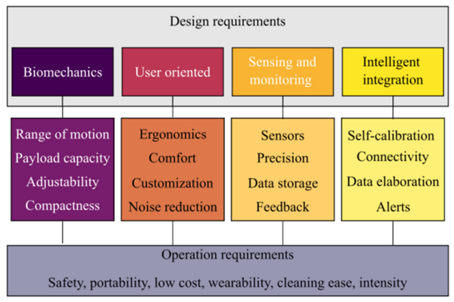
32]. A systematic analysis of requirements for elderly motion-assisting systems further clarified critical factors such as modularity, ease of wearing, adaptability, and safe interaction [
2,
33]. Related developments within the same research framework include the AlmatyExoElbow device, a lightweight antagonistic cable-driven exoskeleton integrating sensor-based motion tracking and experimental validation, demonstrating the feasibility of compact and modular elbow assistance architectures [
34].
These findings and further challenges motivated the structured research activities of the ASSIST project [
35], aimed to develop modular, portable cable-driven exoskeletons both for elderly exercise and rehabilitation in home use that were validated through significant experimental campaigns. Within this framework, the ASSIST-FEE device family was designed for elbow flexion–extension assistance and evolved through three successive prototypes [
36]. The progression from versions v1 and v2 to ASSIST-FEEv3 involved refining cable routing patterns, upgrading actuators, and iteratively repositioning the extension actuation point, resulting in improved torque transmission and smoother operation. A distinctive feature of ASSIST-FEEv3 is its extremely low wearable mass of approximately 100 g, significantly lighter than earlier cable-driven systems [
18,
19] and substantially lighter than other multi-DOF rigid exoskeletons [
13]. The ASSIST-FEEv3 device has undergone a structured testing campaign with healthy volunteers under physiotherapist supervision [
37,
38], extending preliminary investigations and providing a broader assessment of usability and assisted motion performance. The originality of the solution is further recognized through patent registration [
39].
Despite extensive research in upper-limb rehabilitation robotics, many studies primarily focus on mechanical design, actuation capabilities, or control strategies, while structured experimental validation under physiotherapy-guided protocols remains relatively limited. Specifically, lightweight cable-driven elbow devices often lack comprehensive evaluations that incorporate therapist-supervised exercise execution, usability within structured rehabilitation sessions, and preliminary assessments of user interaction. Consequently, there is a need for studies that integrate engineering validation with physiotherapy considerations to evaluate feasibility, operational reliability, and suitability for both structured rehabilitation and home-based exercise scenarios.
This work presents the experimental validation of the ASSIST-FEEv3 elbow assisting device, incorporating explicit physiotherapy considerations. It aims to provide systematic evidence of the device’s performance, usability, and potential applicability in supervised rehabilitation settings and elderly home use.
3. Results
From a clinical rehabilitation perspective, the possibility of objectively quantifying acceleration and power output during exercise may represent a valuable tool for physiotherapists, providing quantitative metrics for monitoring patient performance, exercise adherence, and progression throughout rehabilitation programs.
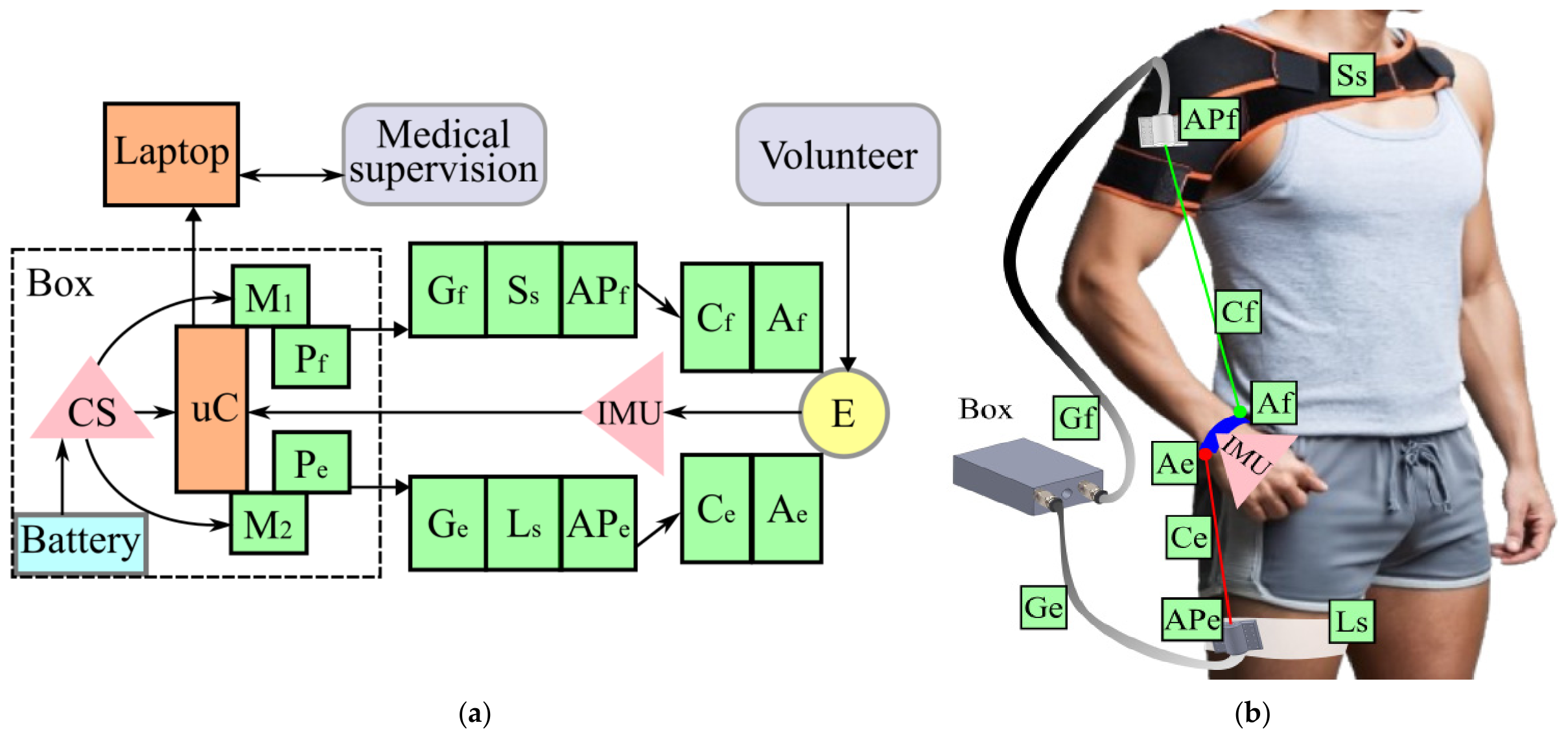
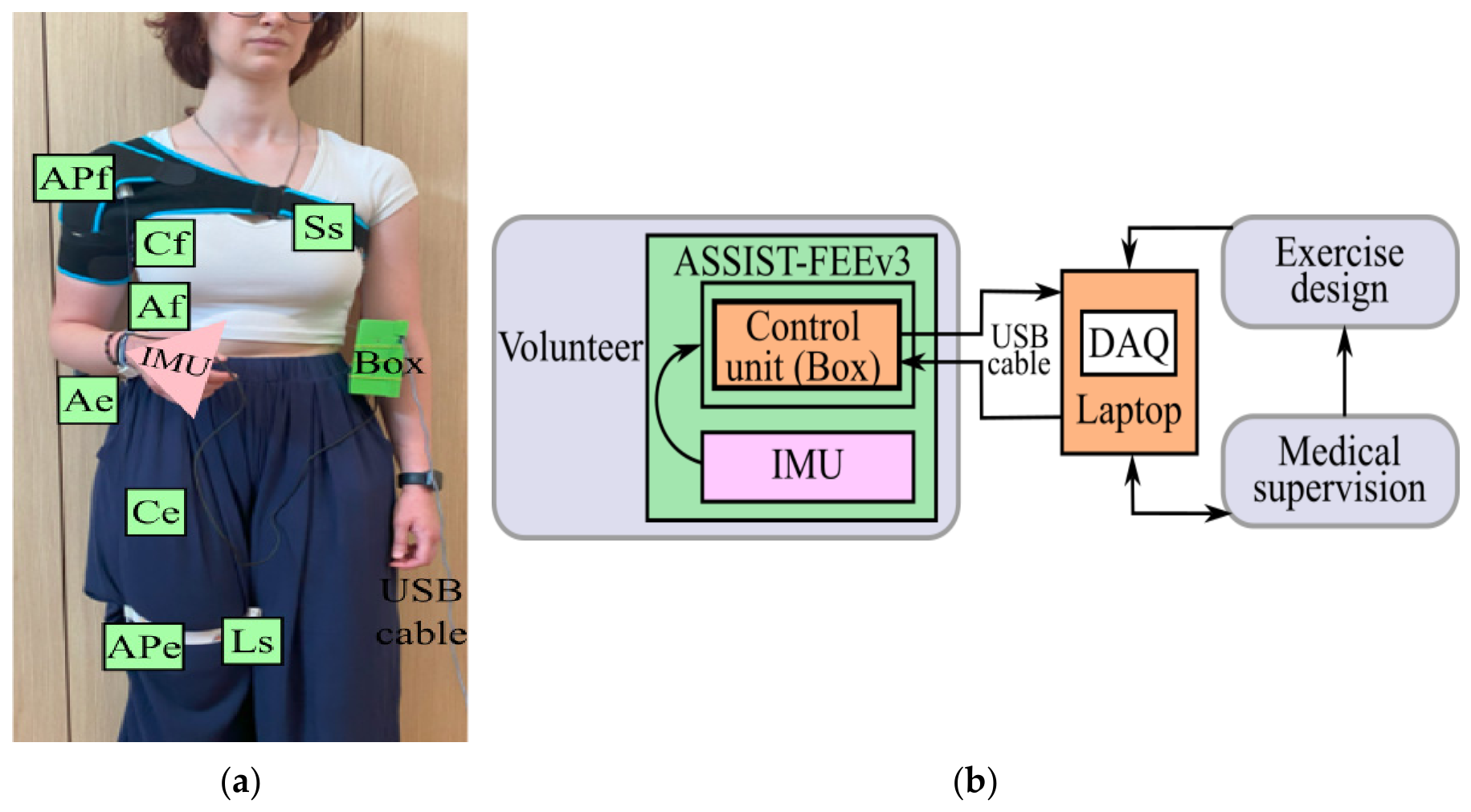
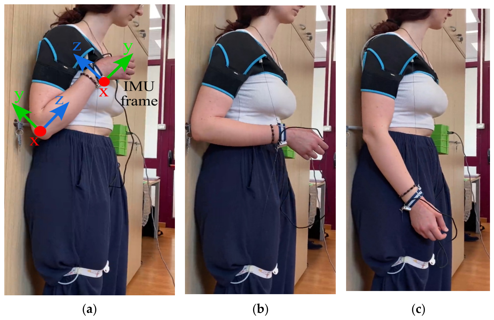
Figure 5a–c present snapshots of a laboratory test with the ASSIST-FEEv3 device worn by a healthy volunteer. Constructed using commercially available components, as reported in [
36,
37], the device effectively assists elbow motion. Its performance is experimentally evaluated through the motion, acceleration, and power-related results whose illustrative representative outcomes are presented in
Figure 6,
Figure 7,
Figure 8 and
Figure 9. The testing campaign has been planned as per a proof-of-concept validation with 25 healthy student volunteers to check the feasibility of the device and to characterize its operation from the physiotherapist’s perspective. The limited number of volunteers and tests can still be considered suitable for the above targets, as referred to in the physiotherapist testing practice [
41].
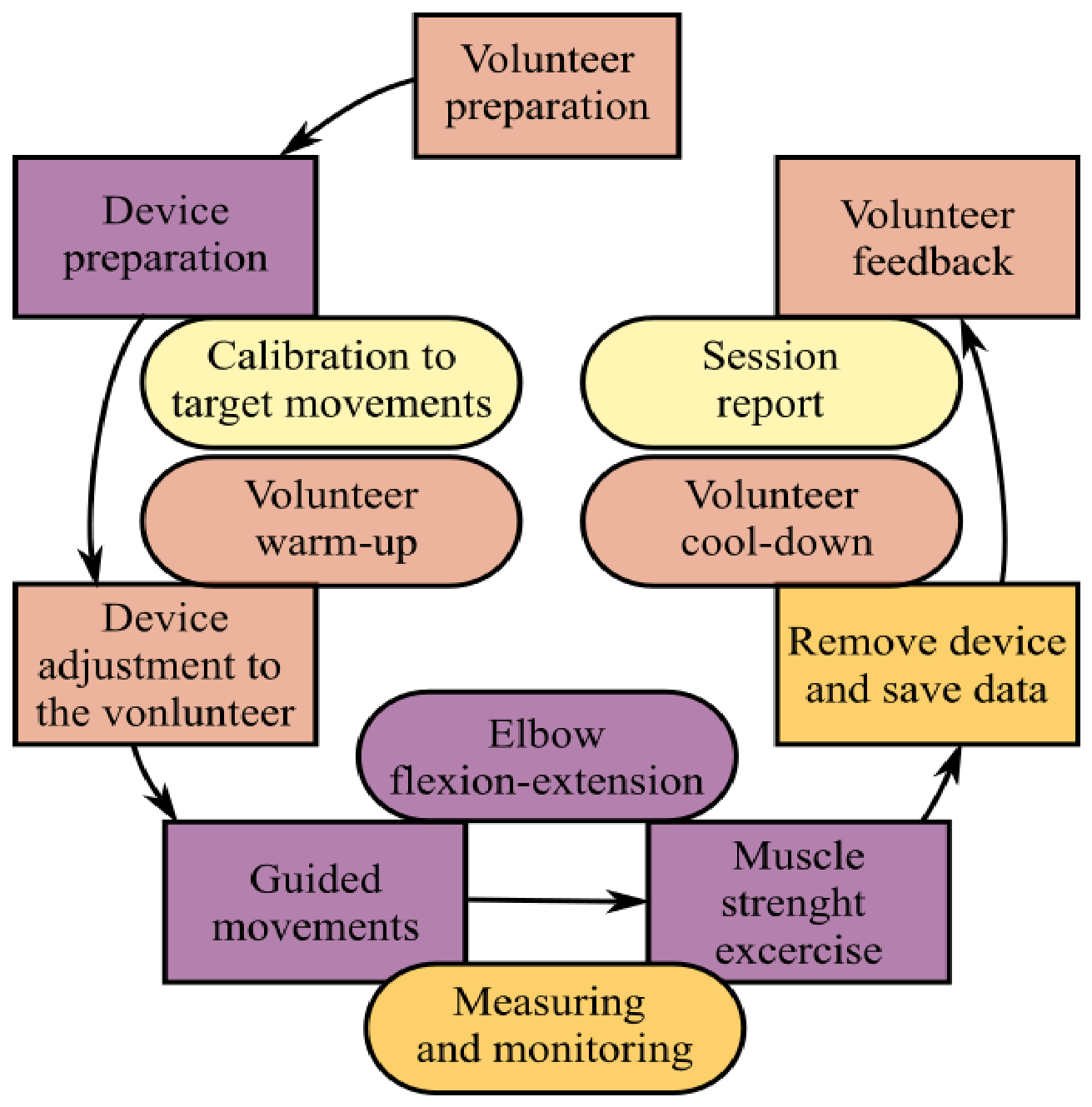
According to physiotherapy recommendations, the duration for one complete flexion–extension repetition was established at approximately 28 s to ensure a controlled and safe exercise-oriented movement. During the experiments, the volunteer performed the experiments in a standing position, keeping their back close to the wall and following the physiotherapist’s instructions. Throughout the experiments, the arm remained passive, while the ASSIST-FEEv3 device was solely responsible for moving it up and down. This posture was selected to guarantee a proper body configuration for all the volunteers during the exercise and to minimize undesired compensatory movements while using the ASSIST-FEEv3 device.
Figure 5a shows the initial configuration corresponding to the full flexion position, just before the assisting motion starts. The figure also illustrates the IMU reference frame adopted in this work, where the roll angle is measured about the x-axis, indicated in red color, and the pitch angle is measured about the y-axis, indicated in green color. This reference frame configuration is maintained as a reference for testing to acquire different data, as in
Figure 6,
Figure 7 and
Figure 8.
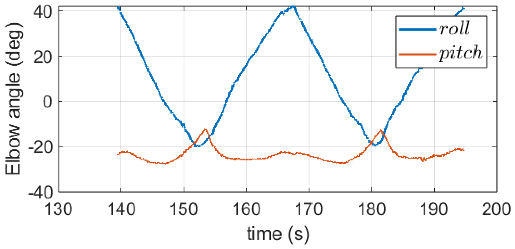
Figure 6 shows the time-domain data of the elbow motion during two extension-flexion repetitions, corresponding to a section of a test duration of approximately 1 min. The measurements were obtained from the IMU mounted on the ASSIST-FEEv3 device, using the reference frame illustrated in
Figure 5a, and are presented for a selected volunteer as representative experimental evidence of the obtained motion data. The results describe the range of motion (ROM) that is achieved during the assisted motion exercise, from full flexion to full extension of the elbow joint. In particular, the roll angle exhibits the main variation associated with the flexion–extension motion, ranging approximately from −24.34° to 48°, showing a smooth and continuous assisted motion throughout the exercise repetitions. Although the pitch angle presents comparatively small variations, its value is not negligible. This effect indicates that the elbow joint is allowed to rotate freely during supination–pronation movements while the assistance is provided. Such kinematic adaptability is important because it enables the ASSIST-FEEv3 device to accommodate natural arm motion according to the specific movement needs of the volunteer during the rehabilitation task.
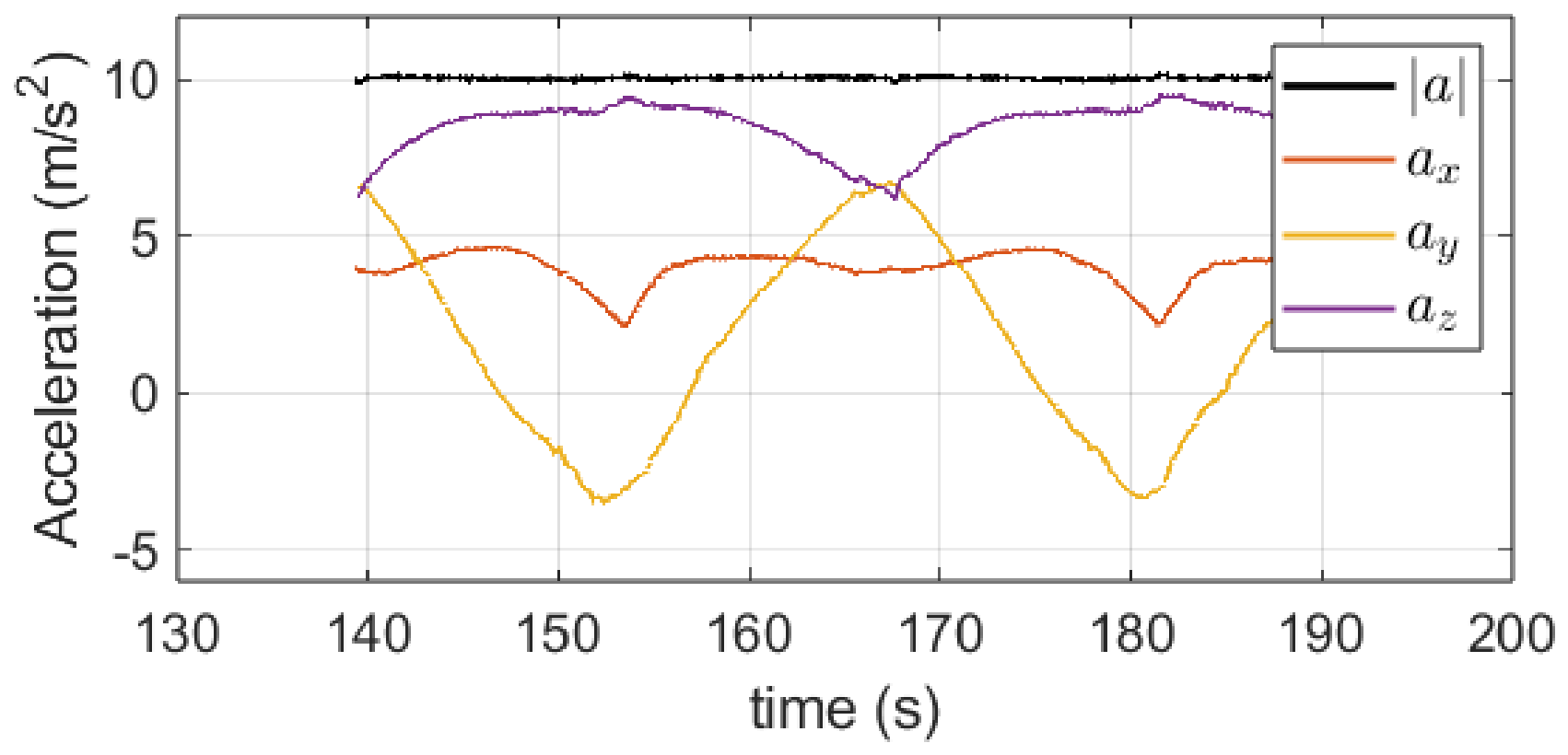
Figure 7 shows the measured linear acceleration components at the wrist together with the corresponding acceleration magnitude as a function of time during the assisted motion. The acceleration signals were obtained according to the IMU reference frame shown in
Figure 5a. The reported data further confirm the smooth and continuous motion behavior recorded with data in
Figure 6, as indicated by the relatively low acceleration levels throughout the two exercise cycles.
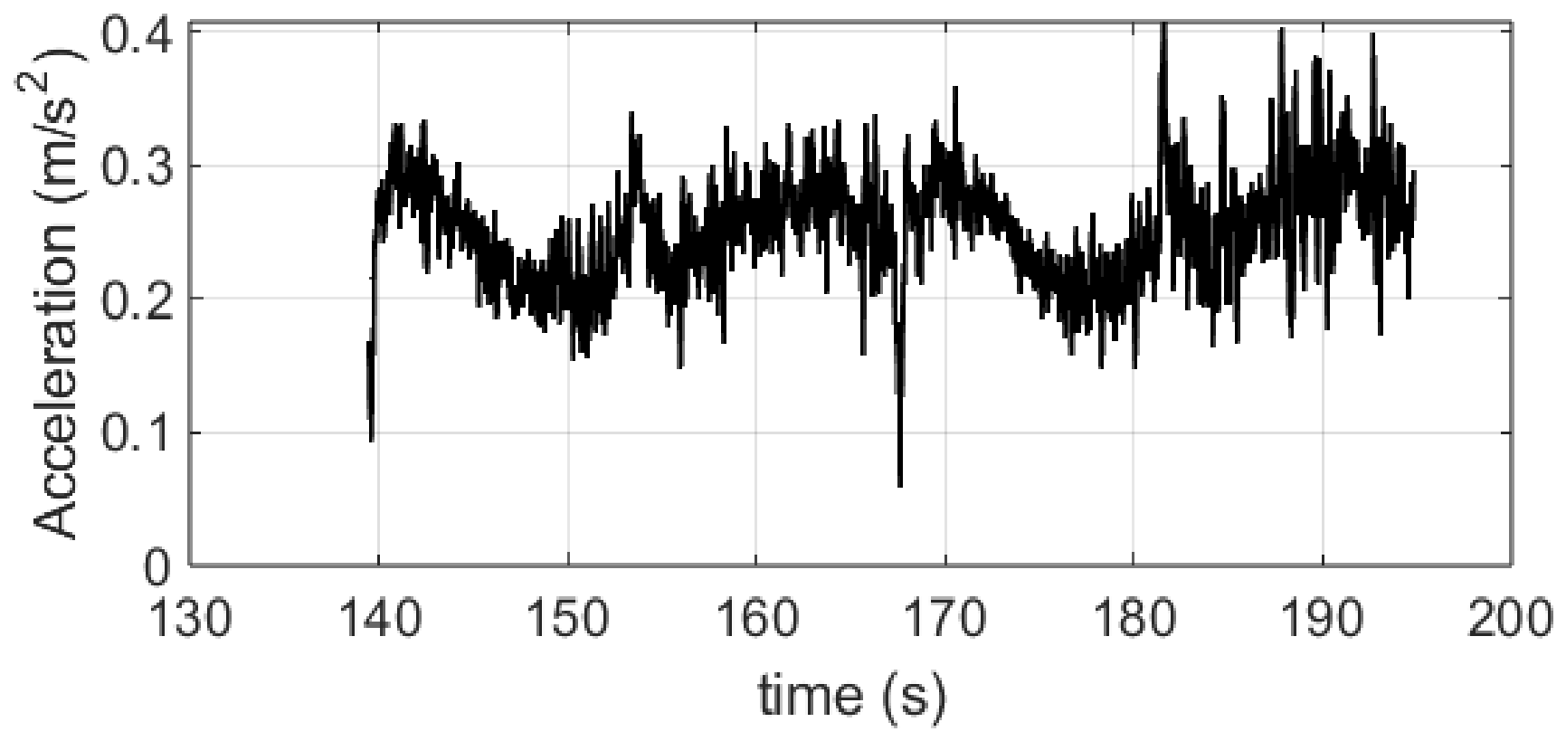
A more representative assessment of the motion quality can be obtained from the free-of-gravity acceleration magnitude, denoted as
, which is also reported in
Figure 8. This parameter allows an evaluation of the dynamic contribution that is associated only with the arm motion by compensating for the gravitational component. The quantity
is computed as
where
,
, and
are the measured acceleration components along the IMU axes, and g is the gravitational acceleration. The relatively low values of
indicate that the assisted motion is performed without abrupt changes in acceleration, which is desirable for physiotherapy-oriented rehabilitation exercises and contributes to user comfort and safe interaction with the ASSIST-FEEv3 device.
The acceleration trends shown in
Figure 8 correlate with the changes in motion direction occurring during the flexion and extension phases that are shown in
Figure 5, providing additional evidence of the smoothness and stability of the assisted movement. The free-of-gravity acceleration does not reach zero values because the arm motion is continuously maintained throughout the assisting task.
These acceleration values correspond to instantaneous measurements acquired by the inertial sensor and cannot be interpreted as a constant acceleration acting over the entire experiment duration, giving motion displacement, but an evaluation of the motion variability and smoothness. Most of the observed acceleration peaks occur at the transition instants where the movement changes direction from flexion to extension, or vice versa. These variations can also be associated with the concentric and eccentric phases of the upper-limb motion, reflecting the subject’s ability to maintain coordinated and rhythmic motion control during repetitive exercise. Between these transition phases, the acceleration behavior remains quasi-stationary with relatively low values, which can be associated with proper assisted motion performance and adequate recovery of muscle strength while maintaining smooth arm motion.
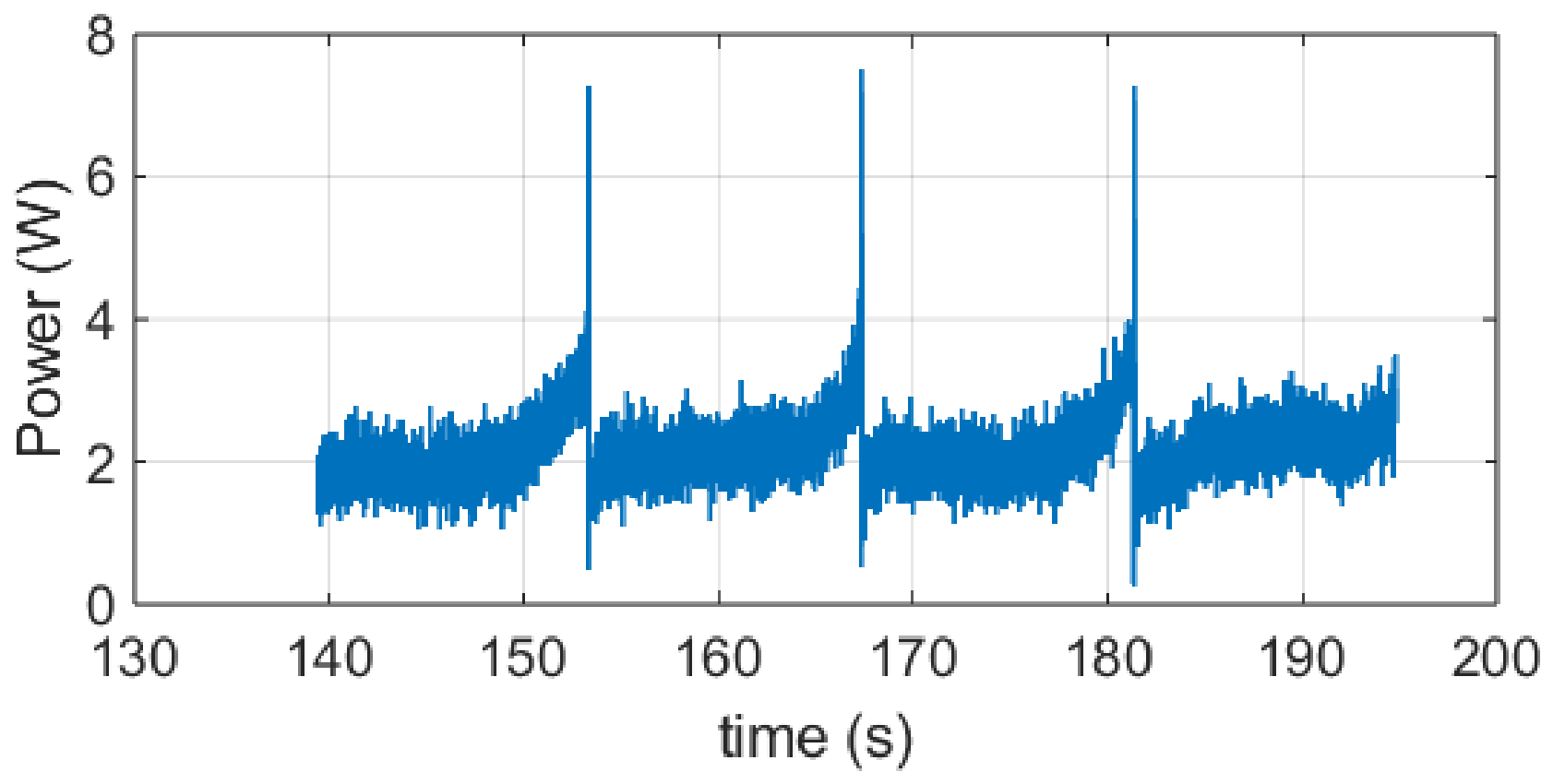
Figure 9 shows the computed power consumption that is calculated from the measured current supplied to the two servomotors actuating the cable transmission system. The observed power peaks correspond to the inversion points between flexion and extension, where the largest torque is required. The small and acceptable level of noise visible in the acquired signals is very likely attributable to minor physiological tremors of the arm, sensor resolution limits, and mechanical backlash within the transmission mechanism.
Overall, test results from a testing campaign with 25 volunteers demonstrate that the device is capable of assisting a wide range of elbow motion while maintaining low power consumption, with an average current demand of approximately 0.26 A. Such a low current requirement allows operation using lightweight commercial batteries, supporting system portability. These characteristics confirm that ASSIST-FEEv3 is a suitable solution for elbow exercise applications in both home-based and clinical environments.
4. Discussion
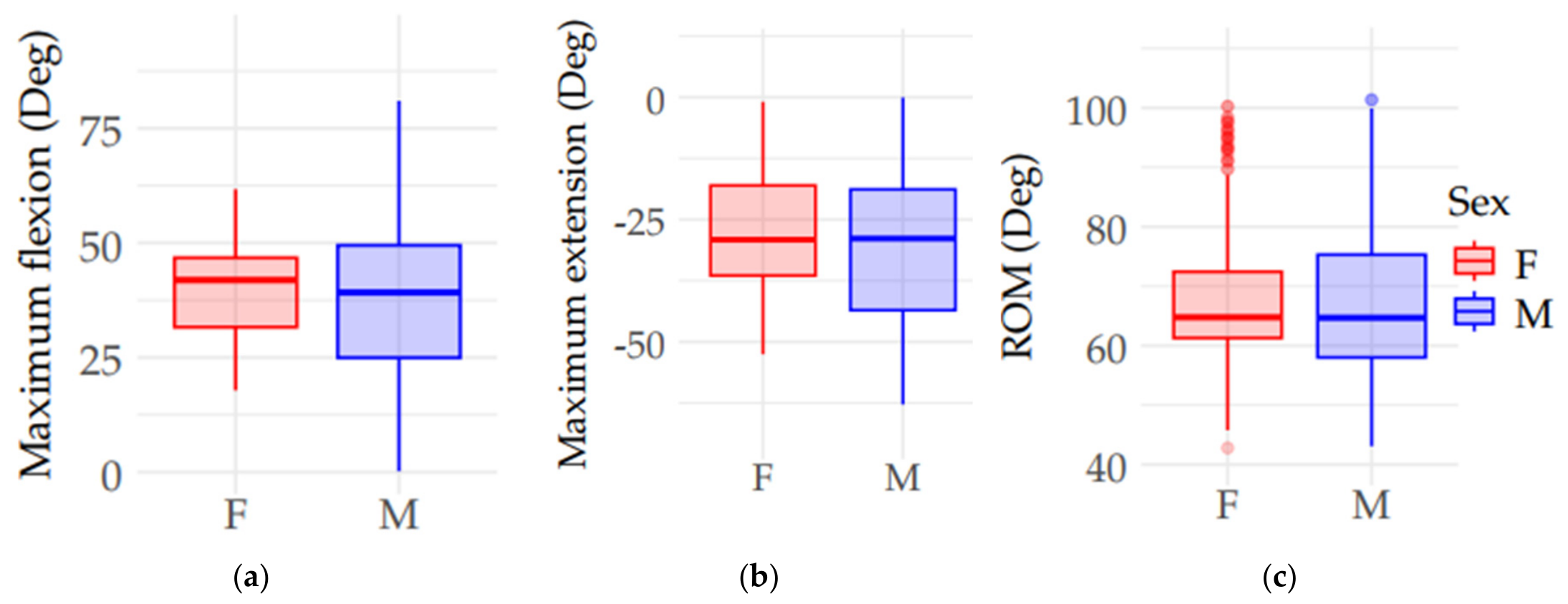
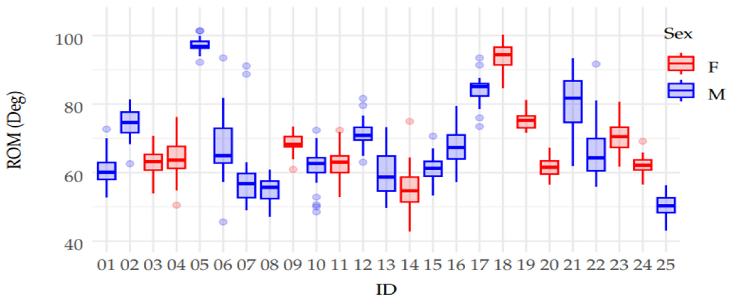
Figure 10 shows statistical results from the testing campaign using the ASSIST-FEEv3 device, showing the distribution of flexion–extension repetitions across all participants, grouped by sex. Each panel illustrates the variability and spread of the measurements collected during the study. Each box represents the distribution of repetitions recorded for participants of the corresponding sex, as elaborated from test results with 283 repetitions for female participants (F) and 436 repetitions for male participants (M). The boxes are colored according to participant sex, with red representing females and blue representing males, and the y-axis indicates the corresponding measurement in degrees.
Figure 10a shows the maximum flexion values,
Figure 10b shows the maximum extension values, and
Figure 10c shows the range of motion (ROM), which is calculated as the difference between maximum flexion and extension. The data indicate that female participants exhibited smaller variability when compared with results by males, which is clearly observed in the smaller interquartile ranges (IQRs) across all three plots. The Shapiro–Wilk test indicated that the assumption of normality was not satisfied for at least one of the groups, and non-parametric comparisons between sexes were performed using the Mann–Whitney U test. The analyses were conducted using R version 4.6.0. The results show no statistically significant differences between female and male participants for maximum flexion (
p = 0.14), maximum extension (
p = 0.071), or ROM (
p = 0.35). Therefore, despite the large variability observed among male participants, the central tendency of the kinematic variables remained statistically similar between female and male tested volunteers, suggesting similar functional elbow mobility across sexes. Therefore, the results do not show statistically significant differences between female and male participants. This study, conducted with healthy young adults, serves to validate and characterize the data acquisition and analysis procedures before using the device in elderly populations in future clinical testing campaigns.
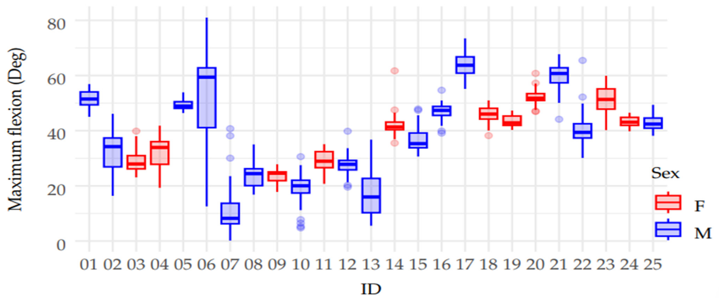
Figure 11 shows the distribution of maximum elbow flexion values for each participant, as a function of individual ID and color-coded sex, red for females and blue for males. Each box represents the spread of flexion–extension repetitions recorded for a single participant. According to the testing protocol defined together with the collaborating physiotherapists, each participant was expected to perform a total of 30 repetitions, derived from three separate sessions of 10 repetitions each. This figure provides a visual summary of the variability in maximum flexion performance across all volunteers. The height of each distribution reflects the maximum values achieved, where large distributions indicate better performance, since they correspond to large elbow flexion angles. The plot shows participant-specific differences in both performance and variability, which may reflect individual physical characteristics, engagement levels, or temporary conditions during testing.
Notably, participant ID 06 exhibits a substantially larger interquartile range (IQR) when compared with other participants, indicating larger variability and a different behavior pattern during the flexion measurements. Overall,
Figure 11 not only shows individual performance in terms of flexion but also highlights the presence of inter-subject variability within a standardized test campaign.
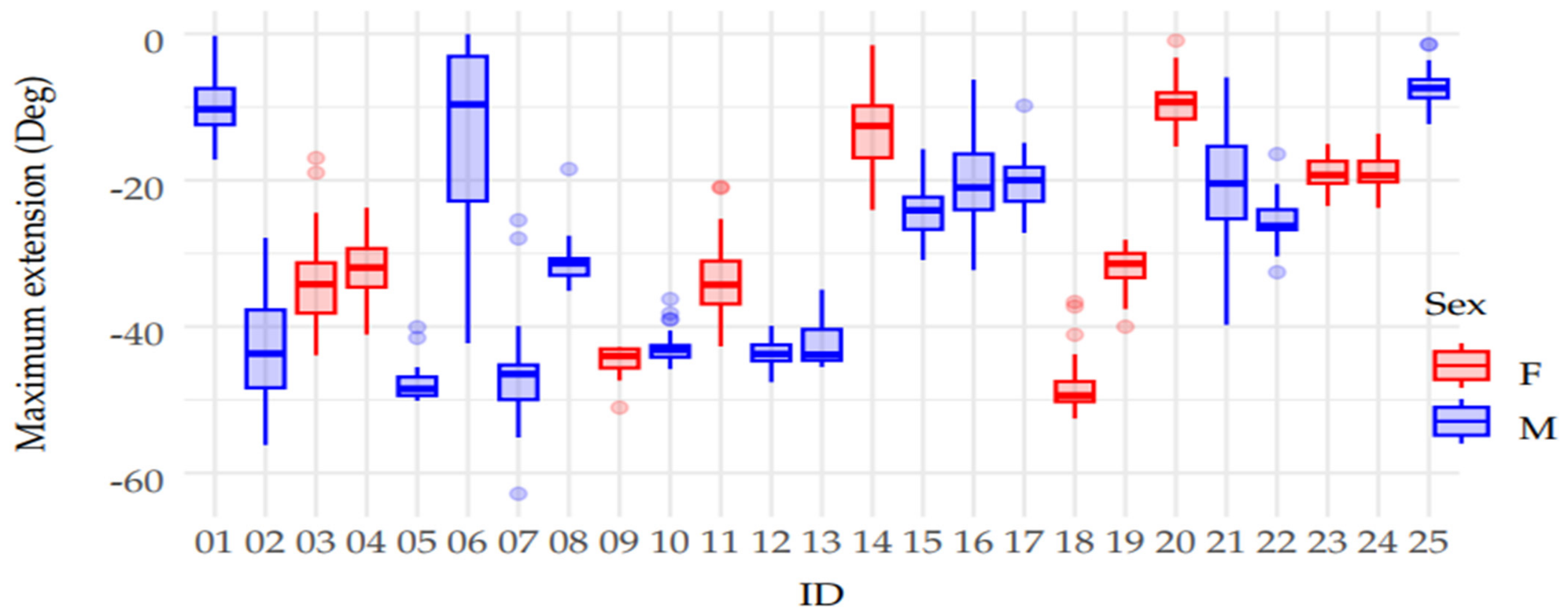
Figure 12 shows the distribution of maximum elbow extension values for each participant, as a function of individual ID and sex, red for female and blue for male participants. Each box represents the distribution of flexion–extension repetitions recorded for a single individual. As defined in the experimental protocol developed in collaboration with physiotherapists, each participant was expected to complete 30 repetitions, derived from three sessions of 10 repetitions each. This figure shows the variability in extension performance across all volunteers. Smaller distributions are indicative of better performance, as they reflect a greater ability to fully extend the elbow joint, closer to its anatomical limit. The figure highlights individual differences in extension range and consistency, with some participants demonstrating tightly clustered values and others showing greater variability. As observed previously in
Figure 11, participant ID 06 again exhibits a notably wider distribution and reduced extension values when compared with other participants, indicating larger variability in the measured performance. Nevertheless, in
Figure 13, the ROM values for this participant remain within the overall tendency observed across the study population, suggesting that the atypical flexion and extension distributions did not substantially alter the overall functional range of motion.
Figure 13 shows the distribution of elbow joint range of motion (ROM) values for each participant as a function of individual ID and sex, red for female and blue for male participants. Each boxplot reflects the variability in ROM across 30 flexion–extension repetitions, following the standardized protocol of three sessions with 10 repetitions each, as established in collaboration with physiotherapists. The ROM was calculated as the difference between maximum flexion and maximum extension values for each repetition, representing the total movement achieved by the elbow joint.
The collected evidence suggests a significant potential clinical impact of the device. The combination of an extremely low-burden wearable system, smooth and repeatable assistance, and the ability to acquire objective movement parameters (ROM, maximum flexion/extension, and acceleration profiles) enables the integration of ASSIST-FEEv3 into standard rehabilitation protocols without increasing the physical or cognitive load on the patient.
These characteristics are particularly relevant for older adults, post-surgical patients, and individuals with reduced muscular endurance, for whom safety, portability, and the possibility of performing controlled exercises outside the hospital environment are key factors for therapeutic adherence. Moreover, the availability of objective and repeatable movement measurements can support physiotherapists in monitoring clinical progression, tailoring exercise intensity, and evaluating treatment effectiveness over time.
5. Conclusions
The functionality of the ASSIST-FEEv3 elbow assisting device has been demonstrated through characterization tests that were conducted within a medical physiotherapy context, with results confirming both its technically feasible performance and user-centered design. The device employs a cable-driven mechanism to guide the forearm through controlled elbow flexion–extension exercises in the sagittal plane. This motion is generated by two coordinated cables that are actuated by servomotors housed within a lightweight module positioned on the user’s body. Experimental results indicate that the ASSIST-FEEv3 device reliably supports the full range of elbow motion during repeated cyclic exercises while maintaining low energy consumption, high operational comfort, and positive acceptance among physiotherapy practitioners.
The proposed device also enables objective monitoring of elbow kinematics while ensuring safe, comfortable, and repeatable exercise execution. These characteristics support its potential application in supervised rehabilitation, elderly exercise programs, and home-based therapeutic scenarios. In particular, the obtained results confirm that ASSISTFEEv3 is a valid and reliable platform for collecting and analyzing biomechanical data related to elbow motion. Tests conducted with healthy volunteers validated the proposed operational protocols, demonstrating consistency and repeatability in the measured data. Minor deviations observed during the experiments were mainly attributed to transient individual variations rather than intrinsic limitations of the device.
From physiotherapeutic perspectives, the developed system represents a significant step toward more objective, sensitive, and reproducible assessment methods for upper-limb motion assistance. Its capability to continuously monitor joint kinematics and motion-related dynamic parameters provides valuable support for evaluating functional recovery, elderly exercise performance, and the adaptation of rehabilitation programs according to patient progress. This aspect is particularly relevant for elderly users and individuals with neuromotor impairments, for whom systematic monitoring and the possibility of supervised home-based rehabilitation may contribute to improved therapeutic outcomes and exercise adherence.
From an operational perspective, patient safety, comfort, and adaptability remain fundamental requirements for practical clinical implementation. Features such as physiotherapy-friendly setup, portability, and rapid session configuration enhance the suitability of ASSISTFEEv3 for outdoor patient clinics, home-based care, and low-intensity rehabilitation facilities. Furthermore, the integration of non-invasive sensing technology and compatibility with clinical reporting and monitoring tools increase its applicability within existing rehabilitation workflows, supporting efficient, data-driven, and patient-centered physiotherapeutic practices.




 ,
,