


Abstract
In recent years, the role of non-coding RNAs in human physiology and pathology has emerged as an essential avenue of investigation. Among others, studies identifying the biological role of microRNAs (miRNAs) have paved the way for future inquiries on the importance and biological significance of non-coding RNAs. In this review, we provide an overview of miRNAs’ biology and their contribution to the pathogenesis of diabetic retinopathy (DR). This complication of diabetes is the leading cause of blindness in adults, affecting more than 4 million people in the US alone and over 103 million people worldwide. Despite the tremendous efforts of the scientific community and the pharmaceutical industry, the development of new, more effective therapeutic and diagnostic tools for DR to date remains an unmet need. More than a decade from the initial work assessing miRNA expression profiles in diabetic patients, we have learned the impact of these signaling molecules in DR and garnered knowledge of their complexity and their potential as new diagnostic and therapeutic targets for this potentially blinding condition.
1. Introduction
The complexity of the genetic code has been evident since the first identification of DNA in 1860 by Johann Friedrich Miescher and its subsequent characterization in 1950 by James Watson, Francis Crick, and Rosalind Franklin [1]. However, the more recent discovery and characterization of non-coding RNA (ncRNA) species have added a significant layer of complexity underpinning the biological importance of fine-tuned gene regulation and epigenetics.
Dysregulated non-coding RNA expression has been evidenced in pathological conditions and linked these molecular players to the onset and progression of several human pathologies, including cancer, cardiovascular, and neurodegenerative diseases [2].
MicroRNAs (miRNAs) are a single-stranded subset of ncRNAs classified by their length (21–23 nucleotides) and characterized by their gene-silencing ability [3].
The recognition of Viktor Ambros and Gary Ruvkun with the 2024 Nobel Prize in Physiology or Medicine for the discovery of miRNAs highlights the profound significance of these regulatory molecules in shaping our understanding of human biology, physiology, and disease mechanisms [4,5].
In this review, we present an overview of the recent research progress, delving into the role of miRNAs in diabetic retinopathy (DR), a neurovascular complication of diabetes [6], and their potential as diagnostic and therapeutic tools for this potentially blinding condition.
2. Diabetic Retinopathy (DR)
2.1. Epidemiology
Diabetic retinopathy (DR) is a late complication of Type 1 and Type 2 diabetes (T1D and T2D, respectively) the leading cause of preventable blindness in working-age adults aged 20–74 years old [7]. The development of other diabetic complications, such as diabetic nephropathy, peripheral neuropathy, and cardiovascular events, can be associated with the development and degree of DR [8,9].
The onset of DR correlates with the duration of diabetes, with approximately all patients with T1DM and over 70% of patients with T2DM developing DR by 20 years into the disease. Recent advances in diabetes management have improved glycemic control and are expected to delay disease onset and progression; nevertheless, DR remains a major cause of vision loss worldwide [10,11,12]. Retinopathy is pervasive among patients with T1DM and T2DM in the United States, with a prevalence of 40% (estimated 4 million people) and 80% (estimated 750 thousand people), respectively [13,14]. A high prevalence of DR has been reported in other countries, such as the United Kingdom, with those of South Asian ethnicity being affected in higher proportions than white Europeans [15]. People of Chinese descent are also disproportionately affected by DR, with rates as high as 43%, and 1.2 million of whom have vision-threatening retinopathy [16]. DR incidence and related vision loss is higher in non-Hispanic Black and Hispanic patients compared to Caucasian patients [17]. The National Eye Institute indicates that over 800,000 African Americans currently have DR, with this number expected to rise by 2030 [18].
Recent epidemiological estimates highlight the increasing global burden of DR in parallel with the rising prevalence of diabetes worldwide [19]. The International Diabetes Federation estimates that more than 500 million adults currently live with diabetes, a number projected to substantially increase over the coming decades, contributing to a continued rise in diabetes-associated complications, including DR [20]. Recent meta-analyses estimate that approximately one-third of individuals with diabetes develop some degree of DR, with a substantial proportion progressing to vision-threatening forms, including diabetic macular edema and proliferative diabetic retinopathy [21]. Despite advances in glycemic control and retinal therapies, DR remains a leading cause of preventable vision impairment among working-age adults and represents an increasing public health challenge worldwide [22,23]. Clearly, DR is a significant public health problem not only in the United States but also around the world in high-income and rural areas.
2.2. DR Pathogenesis
The pathogenesis of DR is complex, and the molecular mechanisms underlying this pathology are not completely understood. Chronic hyperglycemia affects both vascular and neural tissue in the retina. Persistent hyperglycemia-induced changes in cellular metabolism contribute to oxidative injury of the retinal microvasculature [24,25]. Early changes include loss of retinal supporting pericytes and basement membrane thickening in retinal capillaries [26]. Oxidative damage to pericytes contributes to endothelial dysfunction and increased capillary permeability, leading to consequent macular edema [27]. Weakened vessel walls are more susceptible to forming microaneurysms that are prone to rupture into a dot-blot hemorrhage, and even microvascular thrombosis leading to retinal vessel occlusion and retinal nerve infarction [28].
As the disease progresses, hypoxia-induced growth factors, such as vascular endothelial growth factor (VEGF), are stimulated by reduced blood flow and chronic retinal ischemia due to capillary occlusion [29,30]. The release of these growth factors stimulates retinal neovascularization and the growth of aberrant, fragile blood vessels that tend to hemorrhage. These misdirected blood vessels can grow into the vitreous and hemorrhage or cause tractional retinal detachment, both of which can lead to sudden vision loss [31]. Inflammatory markers such as IL-1β, IL-18, and caspase-1 are important for sterile inflammation via the NLRP3 inflammasome and are elevated in the vitreous of diabetic rat models. Protein expression levels of adhesion molecules ICAM-1 and VCAM-1 and the inflammatory cytokine HMGB1 are higher in the ganglion cell and retinal nerve fiber layer of diabetic patients compared to normoglycemic controls, indicating increased leukocyte infiltration and inflammation [32].
The goal of DR screening is the timely detection of retinal damage and intervention with laser photocoagulation or intraocular glucocorticoids and anti-vascular endothelial growth factor (VEGF). Current screening methods for diabetic DR include direct visualization of signs of microaneurysms, exudates, and intraretinal hemorrhages through dilated fundus exams or retinal photography [33,34]. Additional tests to detect vision-threatening macular edema include fluorescein angiography and optical coherence tomography. Unfortunately, these methods require the expertise of a trained eye care specialist with specialized equipment, and they are only able to diagnose DR once retinal damage has already occurred. Emerging research suggests that several miRNAs may be involved in the development of DR, and these could represent biomarkers to enable better screening and treatment for diabetic individuals at risk of retinopathy [35,36,37].
3. MicroRNAs (MiRNAs)
3.1. Biogenesis and Function
MiRNAs are small, noncoding RNAs that play a regulatory role in every cell process, including cell development, signaling, proliferation, and differentiation [38,39]. By binding complementary sequences in the 3′ untranslated region of target mRNA, miRNAs can decrease the translation of expressed transcripts [40]. In addition to the miRNA seed sequence, a prominent distinguishing feature of mRNA targets is that miRNAs have imperfect complementarities to the sequence of their target mRNA [41]. This imperfect complementary pairing allows a single miRNA to have numerous, even hundreds of mRNA targets, thus, globally, potentially regulating the expression of most human proteins [42].
MiRNA biogenesis comprises a multistep processing pathway [43] (Figure 1). Most miRNAs are transcribed from their genes, although some are encoded within other RNA molecules or introns and further processed to produce their mature form [44,45]. The first transcript produces pri-miRNA, a stem–loop precursor consisting of several thousand nucleotides. The initial processing to form pre-miRNA is completed in the nucleus by a Drosha protein, a ribonuclease, and the DiGeorge Syndrome critical region 8 homolog (DGCR8) complex protein [46,47]. Following pre-miRNA formation, exportin 5 transports pre-miRNA to the cytoplasm, where a Dicer/TRBP complex cleaves the loop structure of pre-miRNA to produce a double-stranded molecule of about 20 bp. One of the miRNA strands (usually the strand with the less stable 5′-end) is subsequently degraded, while the other is preserved to bind with argonaute (AGO) and other proteins to form the RNA-Induced Silencing Complex (RISC). This RISC complex then functions to degrade or suppress mRNA transcripts, thereby silencing certain genes in order to carefully regulate gene expression [45].
Figure 1.
MicroRNA biogenesis diagram illustrating the principal steps of miRNA expression, maturation, and function. Abbreviations in text. Created in BioRender. Mahrous, M. (2026) https://BioRender.com/l41mcpz, access on 17 May 2026.
Interest in miRNAs has grown in recent literature due to their involvement in paracrine and endocrine secretion, and intercellular signaling [48,49]. After secretion from a cell via the exosomal pathway, miRNAs have been found to transcriptionally regulate gene expression in remote cells. Various pathways have been hypothesized to regulate miRNA sorting into exosomes for secretion, involving sphingomyelinase 2, heterogeneous nuclear ribonucleoprotein A2B1, 3′ post-translational modification of miRNA, and even the RISC itself [50]. Regardless of the mechanism of production, intercellular miRNA communication is key to normal biological function.
3.2. Exosomal miRNAs
Exosomes are extracellular vesicles generated by the endosomal pathway [51] (Figure 2). Traditionally, a receptor–ligand interaction on the cell surface triggers an invagination of the plasma membrane, resulting in the formation of an early endosome. These vesicles can then combine with an intraluminal vesicle (such as a lysosomal vesicle) to form an intermediate structure called a multivesicular body (MVB) [52]. MVB formation is regulated by a protein multicomplex termed Endosomal Sorting Complex Request for Transport (ESCRT) [53]. MVBs can subsequently undergo two fates. One pathway is to fuse with other MVBs, late endosomes, and/or lysosomes. The other is to migrate to the cell membrane, fuse, and release its contents. Vesicles undergoing this latter pathway are deemed to be exosomes. The exosomal pathway is seen in nearly all cell types [50] (Figure 2).
Figure 2.
Exosome pathway diagram illustrating exosome formation and release. Abbreviations in text. Created in BioRender. Mahrous, M. (2026) https://BioRender.com/75l8qnm, access on 17 May 2026.
A unique feature of exosomes is their ability to transport many RNA species, including mRNA, miRNA, circular RNA, and long non-coding RNA. Interestingly, the most numerous cargo molecules in the exosome are, in fact, miRNAs [54,55,56]. Cells utilize the exosomal pathway to transport miRNA molecules to regulate gene expression in nearby cells [50]. Recent research has taken advantage of this property as a method for the therapeutic delivery of drugs [57,58].
3.3. MiRNA and the Eye
MiRNAs are essential for post-transcriptional gene regulation in many tissues throughout the body, including the eye (Figure 3). Many miRNAs serve a crucial role in the development and maintenance of the retina and are conserved across multiple animal models (Table 1). Among others, miR-124, miR-132, miR-204, and miR-211 have been shown to play important discrete roles in retinal development and homeostasis [59].
Figure 3.
The anatomy of the eye and relevant miRNAs. The schematic diagram illustrates the main structures of the human eye. The schematic representation of the cell types in the neural retina depicts their cellular connections (including ganglion cells, amacrine cells, bipolar cells, and horizontal cells, as well as rod and cone photoreceptors) and supporting cells (Müller cells and RPE), the laminar organization of the nuclear layers (GCL, INL, and ONL), the retinal vasculature, and segments of photoreceptors (IS/OS). The RPE monolayer, with Bruch’s membrane underneath, is located between the neural retina and the choroid complex. MicroRNAs (MiRNAs) that regulate the physiological functions or pathological conditions related to each retinal neuronal and vessel layer, and RPE, are listed next to their histological structures; retinal vessels; GCL, ganglion cell layer; INL, inner nuclear layer; Int, intermediate layer of retinal vessels; IS/OS, inner/outer segments; ONL, outer nuclear layer; RPE, retinal pigment epithelium. Created in BioRender. Mahrous, M. (2026) https://BioRender.com/be6tk51, access on 17 May 2026.
MiR-124 is expressed in all retinal neuronal cell layers and is involved in the localization and maintenance of cone photoreceptors through suppression of Lhx2 transcription [59]. MiR-124 may play a role in the pathogenesis of age-related macular degeneration (AMD) because its levels are found to be depleted from the outer nuclear layer (ONL) and the inner nuclear layer (INL) of the retina in later stages of the disease [60]. Intravitreal administration of miR-124 in mice retinas after photo-oxidative damage not only decreased retinal inflammation and photoreceptor cell death but also improved retinal function [61]. MiR-132, essential for neural synaptic growth in the brain [62], is also expressed in the ganglion cell layer (GCL) and INL of the retina and promotes retinal ganglion cell (RGC) axon formation through downregulation of p250 GTPase-activating protein (p250GAP) [63]. MiR-132 is essential for retinal plasticity, as demonstrated in a lack- and gain-of-function study in mice [64]. Because of its proposed use for ischemic stroke [65], this could also apply to ischemic retinal pathologies. The two most abundant miRNAs in the retinal pigment epithelium (RPE) are miR-204 and miR-211 [66]. They are both found in high amounts in the GCL, ONL, and INL and are critical for directing RPE differentiation and maintaining the RPE epithelial phenotype and function [66]. MiR-204 regulates multiple aspects of eye development, including lens formation, dorso-ventral patterning of the retina, and regulation of eye size and iris shape, as demonstrated in medaka fish [67]. It also regulates differentiation of the RPE, as demonstrated in human fetal RPE (hfRPE) cells [66,67]. Its close relative, miR-211, is crucial for cone cell development, maintenance, and function [68]. Inactivation of miR-211 in mice leads to progressive cone dysfunction and cone loss, without the involvement of rods [68]. The conserved roles of these miRNAs across species demonstrate their crucial role in eye development.
Table 1.
Essential miRNAs in eye development.
Dysregulation of specific miRNAs may contribute to abnormalities in proper retinal cell development, maintenance, and function, including retinal blood vessels [74]. Identifying these miRNAs may help uncover the mechanism of angiogenesis of specific retinal vascular pathologies.
Angiogenesis, the formation of new blood vessels from pre-existing ones, is crucial for many tissues’ physiological development and metabolic homeostasis. The mature retina is a highly metabolically active tissue supplied by a complex vascular network of retinal blood vessels and the choriocapillaris [73]. In late embryogenesis, transient retinal hypoxia induces vascular endothelial-derived factor (VEGF) secretion from retinal cells to promote the growth and proliferation of retinal vessels from the optic disc to the retinal periphery [75]. This process has been studied extensively across different animal models [76,77,78,79] and the human fetal retina [80,81,82].
Several mechanisms are responsible for the regulation of retinal vascularization [83], including specific miRNAs. A subset of miRNAs, termed “angiomiRs”, are highly expressed in human vascular endothelial cells and are key regulators of angiogenesis [84]. AngiomiRs affect angiogenesis in many tissues, including the retina, choroid, heart, and lungs. Depending on the angiomiR, it can be categorized as either a pro-angiomiR, which functions by inhibiting negative regulators of angiogenic signaling pathways to promote vessel growth and sprouting, or as an anti-angiomiR, which inhibits positive regulators of the angiogenic process [84]. Some prominent angiomiRs include the miR-17-92 cluster [85], miR-21 [86], miR-23, miR-27 [87], miR-31, miR-126, miR-130a, miR-200 family [88], miR-210, miR-296, miR-378, miR-27b, the let-7 family [89],miR-24 [90], miR-221/222, and miR-320 [59,84].
4. MiRNA and Ischemic Retinopathies
Ischemic retinopathies are characterized by abnormal retinal and choroidal vascular growth. These ocular ailments comprise the leading cause of blindness, such as proliferative diabetic retinopathy (PDR), exudative age-related macular degeneration (wAMD), and retinopathy of prematurity (ROP) [91,92,93]. There is increasing evidence of miRNA dysregulation in ischemic retinopathies, and specific miRNAs may serve as biomarkers for disease [94,95].
Among other common pathogenic changes, ischemic retinopathies are all characterized by tissue hypoxia that stimulates retinal VEGF secretion, resulting in vascular hyperpermeability (edema) and abnormal vascular growth [73]. This abnormal neovascular process leads to insufficient oxygen and nutrient supply, as well as waste products [59]. These fragile vessels are also prone to bleeding and predispose to retinal hemorrhages. New vessel growth into the vitreous and inner layers of the retina may also lead to tractional retinal detachment and vision loss [59]. There is increasing evidence of miRNA dysregulation in ischemic retinopathies, and specific miRNAs may serve as biomarkers for disease. In this review, we will focus specifically on those relevant to DR.
Specific angiomiRs are termed “hypoxiamiRs”, as they have been found to disrupt levels of the transcriptional regulator hypoxia-inducible factor (HIF), a VEGF transcriptional regulator that is upregulated in hypoxic conditions. These hypoxia-miRs modulate the HIF/VEGF axis by downregulating HIF1α and VEGF transcription in hypoxic and ischemic conditions [67,78]. Since VEGF stimulates endothelial cell migration, survival, and proliferation, these angiomiRs are crucial for proper vascular growth [78]. Hypoxia and ischemia change the expression profiles of many miRNAs, but only a limited number serve a functional role in angiogenesis [86,87].
Of the angiomiRs mentioned above, those that are also hypoxiamiRs include specific members of the miR-17-92 cluster, miR-31, and miR-126. MiR-17, miR-18a, and miR-20a [67] of the miR-17-92 cluster target HIF1A and VEGF [88,89]; miR-31 targets HIF1A [96]; and miR-126 targets VEGF-A [97]. The miR-17-92 cluster has been studied extensively in the context of lymphoproliferative diseases and is linked to the proliferation, differentiation, and activation of B-cells, T-cells, and monocytes/macrophages mainly through targeting the phosphate and tensin homolog (PTEN) and the transcription factor E2 family [98]. In a mouse model of acute kidney injury, members of this cluster were found to promote angiogenesis after renal ischemia–reperfusion injury through direct repression of thrombospondin 1 (TSP1), a potent antiangiogenic factor [99]. Serum levels of the miR-17-92 cluster have also been found to be elevated in retinoblastoma and proposed as a biomarker for retinoblastoma progression [100].
MiR-31 is a highly conserved miRNA involved in many normal physiological processes such as vascular development, spermatogenesis, embryo implantation, embryonic development, myogenesis, bone homeostasis, autoimmunity, and wound healing [101]. MiR-126 has been studied extensively in autoimmune disorders [102], cancers [103], and atherosclerotic disease [104] due to its cell-specific and strand-specific angiogenic functions [105]. Silencing of the miR-126-3p strand represses but overexpression of the miR-126-5p strand enhances angiogenesis and laser-induced choroidal neovascularization (CNV) in mice [105].
In experimental models of ischemic retinopathies (in vivo and in vitro), these hypoxiamiRs were overexpressed, leading to a decrease in HIF-1α and VEGF levels and a decrease in retinal and choroidal neovascularization. This supports the notion that hypoxic and ischemic conditions alter the delicate balance of tissue-specific and strand-specific miRNAs, potentially contributing to pathologic neovascularization (NV), such as that seen in many ischemic retinopathies [106].
5. MiRNAs and Diabetic Retinopathy
To date, several studies have shown that expression and activities of multiple miRNAs are dysregulated in diabetes and DR. These miRNAs are summarized in Table 2 and Table 3 along with the pathways affected. Notably, members of the miR-200 family and the let-7 family may play a role in the pathogenesis of diabetes [107]. The miR-200 family consists of five miRNAs: miR-200a, miR-200b, miR-200c, miR-141, and miR-429, which can be categorized into miR-141/200a and miR-200b/200c/429 based on the homology of their seed sequences [108,109]. In vivo evidence suggests miR-200a induces beta-cell apoptosis by downregulating anti-apoptotic genes caspase inhibitor X-linked inhibitor of apoptosis protein (XIAP) and β-cell chaperone p58IPK, thereby suppressing insulin production [109]. MiR-200 was elevated in diabetic conditions, and its ablation conferred protection against oxidative stress, DNA damage stress, and ER stress-induced beta-cell apoptosis [109]. Let-7 was the first miRNA discovered in humans and is known most notably for its role in tissue differentiation [110,111]. The Let-7 family consists of 12 members in humans (let-7a, let-7b, let-7d, let-7e, let-7c, let-7f, let-7g, let-7i, miR-98, miR-202) and is highly conserved across species in sequence and function. It is involved in a variety of functions throughout the body, including the regulation of insulin production, release, and sensitivity, making it a key player in the pathogenesis of diabetes [112]. Overexpression of let-7 has been shown to lead to features of non-proliferative diabetic retinopathy (NPDR) but also inhibits angiogenesis and choroidal neovascularization in mice, suggesting that let-7 plays a role in early but not late stages of DR [89].
Multiple miRNAs have also been linked to the VEGF pathway and the development of retinal neovascularization in DR (Proliferative Diabetic Retinopathy = PDR). A study on murine models and diabetic patients showed that hyperglycemia and diabetes were associated with the downregulation of plasma levels of miR-200b, which is an inhibitor of VEGF-mediated angiogenesis [113,114]. Downregulated levels in DR lead to the growth of aberrant capillaries with increased vascular permeability, contributing to fluid accumulation in the retinal tissue [113]. Intravitreal injection of an miR-200b mimic prevented VEGF upregulation and VEGF-mediated angiogenesis of leaky blood vessels [113]. Anti-angiogenic miRNAs (miR-106a-5p, miR-20a-3p, miR-20b, and miR-20a-5p) are downregulated in retinal photoreceptor cells under hyperglycemic conditions, a mechanism correlating with aberrant angiogenesis and retinal damage [115].
Furthermore, diabetes has been shown to decrease retinal levels of miR-126, potentially contributing to retinal endothelial injury and diabetic vascular inflammation [32]. A recent study found a positive correlation between circulating levels of miR-15a and severity of type 2 diabetes. In hyperglycemic conditions, there is elevated serum miR-15a due to excessive exosomal secretion by pancreatic beta cells. Retinal Muller cells are the principal glial cells that form the architectural support of the retina. Transport of exosomal miR-15a through the circulation to retinal capillary beds leads to accumulation of miR-15a in retinal cells and promotes apoptotic cell death through the Akt3 pathway [99].
Inflammation also plays an essential role in diabetes, influencing the pathogenesis of various complications, including DR [116,117]. Numerous miRNAs have been identified as key growth factors and cytokine regulators, contributing to retinal inflammation. Notably, miR-146b-3p, miR-152, and miR-200b are associated with this inflammatory process [118,119,120]. MiR-152 is particularly significant as it modulates pro-renin levels, thereby impacting inflammation through the renin–angiotensin system (RAS) [119]. Additionally, miR-152 regulates the expression of VEGF and transforming growth factor-beta (TGF-β) as an effect downstream of the pro-renin receptor (PCR2) complex [119]. The latter, in turn, regulates the levels of miR-200b, which further exacerbates retinal inflammation [121].
Recently, miR-873-5p has been shown to be involved in DR. This miRNA is known to regulate HMOX1, the rate-limiting enzyme in the heme oxygenase reaction pathway [122]. MiR-624-5p and miR-542-3p target prostaglandin-endoperoxide synthase (PTGS2), a rate-limiting enzyme in the arachidonic acid synthesis of prostaglandins [123]. The anti-angiogenic factor Thrombospondin-1 (TSP-1) and its receptor CD36 on endothelial cells are regulated by PTGS2, which may impact DR [124]. Studies revealed that PTGS2 might have a role in ferroptosis-related DR process, which is a new mode of programmed cell death involving lipid peroxide buildup that compromises the integrity of the cell membrane [123]. Pyroptosis, also known as sterile inflammation, is a mechanism of programmed cell death distinguished by reliance on inflammatory caspases and the subsequent release of a multitude of pro-inflammatory factors [125,126,127]. Pyroptosis has been shown to play an important role in DR [125,128]. Studies have identified approximately 20 miRNAs that regulate key pyroptosis-associated genes, including CASP3, TLR4, and GBP2 [129], Given the established role of pyroptosis in DR, these findings warrant further investigation into the potential contribution of these miRNAs to disease pathogenesis.
Recent studies have shown that diabetes-induced vascular senescence is involved in DR induction and progression [130]. MiR-34a is a known regulator of cellular senescence. Studies have demonstrated its key role in promoting vascular senescence in the diabetic retina [131]. This effect was secondary to miR-34a-dependent loss of SIRT1 (silent mating type information regulation 2 homolog) activity and expression [132] and downregulation of mitochondrial endogenous antioxidants, such as thioredoxin reductase 2 (TrxR2) and superoxide dismutase 2 (SOD2), thus directly linking this miRNA to increased retinal endothelial cell susceptibility to oxidative damage and senescence [131,133].
Table 2.
MiRNA levels in the diabetic retina and in retinal cells exposed to diabetes-relevant stimuli.
5.1. miRNA Polymorphisms and Genetic Susceptibility in DR
miR-related polymorphisms contribute to individual susceptibility to DR by altering miRNA biogenesis, stability, or target recognition. Variants may occur within miRNA genes or miR-binding sites in target genes, affecting pathways involved in inflammation, angiogenesis, oxidative stress, and vascular dysfunction [155]. The miR-146a polymorphism rs2910164 has been associated with susceptibility to sight-threatening DR and may alter NF-κB-mediated inflammatory signaling and VEGF regulation [155]. Similarly, the miR-126 rs4636297 variant has been linked to severe DR, likely through effects on endothelial integrity and angiogenesis [156]. In addition, SNPs within VEGF 3′UTR regions may alter miRNA binding efficiency, influencing VEGF expression and pathological neovascularization [157].
5.2. Mechanistic Role of miRNAs in DR Pathogenesis
Chronic inflammation is a major contributor to DR, promoting leukostasis, endothelial dysfunction, blood–retinal barrier (BRB) breakdown, and neuronal injury. Hyperglycemia-induced oxidative stress activates inflammatory pathways including NF-κB, NLRP3 inflammasome, HMGB1 signaling, adhesion molecules, and pro-inflammatory cytokines. miRNAs critically regulate these mechanisms [23,158] as summarized in Figure 4.
Figure 4.
Mechanistic pathways regulated by miRNAs in diabetic retinopathy.
MiR-146a/b acts as a key anti-inflammatory regulator in DR by suppressing IRAK1 and TRAF6 expression, thereby inhibiting NF-κB signaling and reducing the production of pro-inflammatory cytokines such as IL-1β and TNF-α [159]. It also attenuates ICAM-1 expression, decreases leukostasis, and suppresses inflammasome activation [160]. Reduced miR-146a/b expression contributes to persistent retinal inflammation, vascular permeability, and progressive retinal injury in DR.
MiR-21 primarily functions as a pro-inflammatory miRNA in DR. It enhances NF-κB-associated inflammatory signaling, promotes microglial activation, and increases TNF-α-mediated inflammatory responses, thereby contributing to retinal inflammation, vascular dysfunction, and neuronal injury [121].
miR-126 exerts vasculoprotective, anti-inflammatory, and anti-angiogenic effects in DR by maintaining endothelial homeostasis and preserving blood–retinal barrier (BRB) integrity. It suppresses endothelial activation, leukostasis, vascular permeability, and inflammatory signaling while regulating VEGF/HIF-1α and PI3K/Akt pathways. Reduced miR-126 expression contributes to BRB breakdown, vascular leakage, inflammation, and endothelial dysfunction [161].
miR-30a attenuates retinal inflammation and cellular injury in DR by suppressing NLRP3 inflammasome activation and reducing oxidative stress- and apoptosis-associated signaling pathways. Through these effects, miR-30a limits IL-1β and IL-18 production and protects against cytokine-mediated retinal damage [162].
miR-152 appears to function as an anti-inflammatory regulator in DR, as decreased miR-152 expression is associated with enhanced NLRP3 inflammasome activation and increased retinal inflammation [163].
miR-34a is a major pro-oxidative and pro-apoptotic miRNA in DR. It promotes ROS accumulation, mitochondrial dysfunction, apoptosis, and ferroptosis by suppressing antioxidant and survival pathways, particularly through inhibition of the SIRT1/FOXO signaling axis. miR-34a also indirectly reduces SOD2-mediated antioxidant defense, thereby exacerbating oxidative stress, mitochondrial fragmentation, and retinal cell injury [133].
miR-383 contributes to oxidative stress and inflammatory injury in DR by enhancing ROS accumulation and ferroptotic signaling pathways. Dysregulated miR-383 is associated with increased oxidative damage and retinal inflammation [148].
miR-451a functions as an antioxidant and cytoprotective miRNA in DR by preserving mitochondrial homeostasis, reducing oxidative stress, and enhancing antioxidant defense mechanisms. It may improve SOD2 and TrxR2 activity and exerts anti-ferroptotic effects that protect retinal cells from oxidative injury [150].
miR-26a-5p exerts antioxidant and anti-ferroptotic effects in DR by enhancing SIRT1-associated antioxidant signaling and strengthening cellular resistance to oxidative stress. It supports mitochondrial protection and reduces oxidative retinal injury [164].
miR-200b acts as a potent anti-angiogenic miRNA in DR by directly targeting VEGF mRNA and suppressing VEGF/HIF-1α-mediated pathological angiogenesis and vascular hyperpermeability. It also modulates Notch-associated angiogenic signaling and helps preserve endothelial barrier function [165].
miR-92a negatively regulates endothelial repair and angiogenic homeostasis in DR. Increased miR-92a expression impairs PI3K/Akt-associated endothelial repair mechanisms, exacerbates vascular dysfunction, and promotes BRB breakdown and retinal vascular leakage [166].
5.3. MiRNAs as Biomarkers in DR
MiRNAs can be classified as intracellular or extracellular. Multiple studies have been conducted that measured miRNA levels in retinal cells, endothelial cells, aqueous humor, vitreous humor, and serum of diabetic animal models as well as diabetic patients [94,167,168,169]. These studies support the idea that miRNAs act as hormone-like molecules that travel through the bloodstream or other biological fluids via exosomes to promote maladaptive responses to hyperglycemia; therefore, they have the potential to serve as novel biomarkers of disease progression. Additionally, their role in paracrine signaling makes them a potential candidate to serve as treatment options via delivery by exosomes and/or intraocular injection.
Intracellular miRNAs that are dysregulated in DR are summarized in Table 2. Studies examining miRNA levels in retinal and endothelial cells were mostly done on animal models due to the difficulty of performing retina biopsy on diabetic patients. MiRNAs that were found to be downregulated in animal models with DR included miR-106a-5p, miR-20a-3p, miR-20a-5p, miR-20b [170], miR-200b [171], miR-126 [172,173], miR-222 [174], miR-7a [139], and miR-139-5p [175]. Most of these miRNAs target the VEGF pathways and have been shown to play a role in regulating angiogenesis. Therefore, their downregulation could contribute to abnormal neovascularization, as seen in PDR [121]. MiRNAs that were upregulated in DR included miR-21 [176], miR-29b-3p [137], miR-34a [132,177] and miR-183 [178,179]. Upregulation of miR-21 and miR-29b-3p was associated with endothelial cell dysfunction and retinal cell apoptosis [94,137], while upregulation of miR-183 was associated with increased angiogenesis [178].
Due to the present difficulty in assessing intracellular miRNAs in clinical practice, it is important to identify extracellular miRNAs that may serve as biomarkers for DR, as these are more easily collected and analyzed. Multiple studies have been conducted on patients with DR to identify dysregulated miRNAs (summarized in Table 3). Most of the identified miRNAs play a role in pathways that regulate angiogenesis and endothelial cell dysfunction. Some notable miRNAs include miR-221, which multiple studies have shown to be upregulated in the plasma of patients with DR [95,180,181,182,183]. MiR-210 was also elevated in the plasma of patients with DR, while miR-126, miR-146a, and miR-200b were downregulated in serum [113,114,184,185]. In terms of intraocular miRNAs, miR-16, miR-92, miR-146a, and miR-146b-3p were found to be downregulated [94,120,141,186,187], while miR-21, miR-93, and miR-423-5p were upregulated in vitreous humor of DR patients [95,135,188,189,190]. Studies have shown that miR-16 provided over 90% sensitivity and specificity in distinguishing patients with DR from patients without DR [141,186]. Lastly, certain miRNAs such as let-7b, let-7a-5p, miR-320b, miR-762, and miR-4488 are dysregulated in all 3 fluid compartments [186]. MiR-let-7b was shown to be upregulated in aqueous and vitreous while being downregulated in plasma; miR-320b was upregulated in all three fluid compartments; and miR-762 and miR-4488 were either upregulated or downregulated in aqueous humor, vitreous humor, and plasma depending on the severity of DR [186]. Moreover, there is significant and progressive downregulation of circulating and intraocular miR-150-5p and miR-329 in patients with DR [186,191,192]. Interpretation of circulating and intraocular miRNA profiles is further complicated by methodological heterogeneity among studies. Differences in sample source (serum, plasma, vitreous, aqueous humor), RNA isolation protocols, sequencing platforms, normalization methods, patient demographics, diabetes duration, and DR stage may significantly influence reported miRNA expression patterns. Standardization of experimental and analytical approaches will therefore be essential before miRNAs can be reliably translated into clinical biomarkers.
Table 3.
Extracellular miRNA in Diabetic Retinopathy.
5.4. MiRNAs with Beneficial Effects on DR
Some exosomal miRNAs secreted by mesenchymal stem cells (MSCs) have been shown to attenuate the aberrant changes in DR. Current studies show that delivery of miR-216a, miR-200b, miR-126, and miR-222 can reduce angiogenesis and inflammation, and promote retinal repair in animal models [32,113,174,214] (Table 4). An experiment by Zhang et al. demonstrated that miR-126 delivery via MCS-derived exosomes effectively downregulated the expression of the inflammatory cytokine HMGB1, dampened the inflammatory signaling pathways that led to NLRP3 inflammasome activation, and reduced NF-κB/P65 protein levels in diabetic rats and human retinal endothelial cells (HRECs) cultured in high-glucose media [32]. This supports that miR-126 may be essential in maintaining retinal endothelial cell vascular integrity and ameliorating the inflammatory response to hyperglycemia [32]. Another recent study found that retinal miR-222 is significantly decreased in streptozotocin-induced diabetic rabbits, and low levels were associated with the development of severe retinal damage consistent with extensive hemorrhages, retinal detachment, and macular edema [174]. Administration of miR-222 via exosomes from adipose-derived mesenchymal stem cells (MSC) via intraocular and subcutaneous injection facilitated retinal tissue repair and restored the cellular components of the retina to normal over a 12-week period [174]. These studies provide promising support for the use of miRNAs in the treatment of DR.
Table 4.
Beneficial miRNA in Diabetic Retinopathy.
6. Potential Clinical Applications
To date, DR remains the leading cause of blindness among adults, thus urging the need to identify new diagnostic and therapeutic tools. There is a significant amount of work supporting the role of miRNAs in DR pathogenesis; however, their relative impact on DR diagnosis or treatment is still under investigation. Using miRNAs as a biomarker for DR may improve its screening and diagnosis in areas with a shortage of eye care specialists or regions that lack complex imaging modalities needed to visualize the retina. However, current studies involve small sample sizes, and future research involving larger studies needs to be conducted to identify possible markers [217].
Current treatments for DR include laser photocoagulation therapy to ablate aberrant blood vessels, intravitreal anti-VEGF treatment and intraocular corticosteroid implants to treat macular edema, and intraocular surgery to remove scar tissue that is obscuring vision. Limitations of current treatment options include unsatisfactory efficacy and high treatment burden [218]. The intravitreal anti-VEGF injection is commonly used among the various non-surgical treatments for DR; however, about 15–25% of patients with DR do not respond well to anti-VEGF therapy [219,220,221]. Therefore, there is a growing need for alternative treatment options that provide a more targeted approach.
MiRNAs present an intriguing therapeutic intervention option because they can control the expression of several genes, are simple to produce, and have low toxicity [211]. MiRNA manipulation generally follows two principal strategies, depending on whether the goal is to suppress an overexpressed miRNA (loss of function) or to restore the function of a downregulated one (gain of function).
Loss-of-function modulation of miRNAs is primarily accomplished through:
(i) Antagomirs (anti-miRs) that are chemically synthesized single-stranded oligonucleotides designed to bind and inactivate specific miRNAs [222]. They have been successfully utilized in preclinical models of DR to silence upregulated miRNAs that drive inflammation and vascular problems. For example, inhibiting miR-21, which is associated with proinflammatory and profibrotic signals, has been shown to reduce NF-κB activation and retinal vascular leakage in streptozotocin (STZ)-induced diabetic rats [223]. (ii) miRNA sponges are synthetic transcripts containing multiple tandem binding sites that are complementary to a particular miRNA or miRNA family, functioning as competitive inhibitors [224]. These constructs have been delivered via adeno-associated virus (AAV) vectors in ocular disease models [225,226]. In experimental DR, sponge-based inhibition of miR-15a/16, which is involved in endothelial apoptosis and angiogenesis, led to improved retinal vascular integrity [227]. (iii) A miRNA mask is a specially designed single-stranded molecule that blocks miRNA from binding to its target messenger RNA (mRNA). This effect results by directly binding of the mask to the target mRNA at the miRNA attachment site, effectively “masking” that region [228]. This prevents the miRNA from suppressing protein production, allowing the mRNA to be translated more efficiently and increasing the expression of the target gene. (iv) Small-molecule inhibitors, identified through bioinformatics or experimental screening of active compounds, work by disrupting proteins involved in miRNA biogenesis or by preventing miR–target interactions through binding to specific secondary structures of the miRNA [229].
Restoration of function strategies mainly depend on using either chemically synthesized double-stranded miRNA mimics or viral vectors to enhance miRNA expression. These synthetic mimics are not identical to endogenous miRNA duplexes; the guide (sense) strand is modified to be fully complementary to the mature miRNA for increased stability [229]. Additionally, chemical modifications are made to inactivate the sense strand, as it no longer reflects the natural sequence and could otherwise cause unintended effects. miRNA mimics can be designed to silence a specific mRNA target or include multiple miRNA sequences, allowing the simultaneous regulation of different genes [230]. In contrast, viral vector-based miRNA overexpression involves using integrating vectors that carry short hairpin RNAs (shRNAs) under the control of RNA polymerase III promoters [231]. Once delivered, these vectors enable stable and long-term expression of the desired miRNA. Mesenchymal stem cells are multipotent cells that have the potential to transform into many cell types and can be isolated from a variety of tissues such as umbilical cord, bone marrow, and adipose tissue, making them a feasible source for experimentation and possible clinical application [198]. Exosomes are currently recognized as an important shuttle for cellular communication, as they can encapsulate and deliver various genetic materials (mRNAs, miRNAs, and proteins) to receptor cells to regulate various cellular processes. The use of MSC-derived exosomes enriched with beneficial miRNAs such as MiR-126 and MiR-222 may be a new strategy for the treatment of DR [26,121].
Several miRNAs such as miR-16, miR-216a, miR-200b, miR-126, miR-222, and miR-329 have been shown to reduce angiogenesis, control inflammation and promote retinal cell repair, making them potential therapies. The use of miR-200 mimics to downregulate VEGF release in ischemic retinopathies may be a potential treatment option for managing DR and macular degeneration, and retinopathy of prematurity. MiR-329 has also been reported to have anti-angiogenic characteristics by inhibiting the production of CD146, an adhesion protein that functions as a VEGF co-receptor. As a result, miR-329-based therapy demonstrated dramatically reduced retinal neovascularization in animal models of pathological angiogenesis [215]. Currently, treatments for DR focus on mitigating neovascularization. The ability of miR-222 to reverse retinal damage provides the potential to develop a revolutionary treatment that would not only control but also reverse retinal damage. Subcutaneous and intraocular administration of MSC exosomes may represent a better route of delivery, as these routes were associated with improved retinal repair in STZ-induced diabetic rabbits [174]. While these are promising studies, they were performed mostly in animal models. Therefore, more work needs to be conducted using human-derived MSC exosomes to cargo-specific miRNAs in human ocular tissues.
7. Limitations of the Therapeutic Use of miRNAs
Although pre-clinical studies have shown promising outcomes, miR-based therapies have struggled to move into clinical use due to several ongoing challenges. Only a few of the many miR-targeting compounds have progressed to clinical trials. Key hurdles include identifying the right molecular targets, ensuring that miRNA inhibitors remain stable in biological fluids, and improving their precision to avoid off-target effects and unintended changes in gene expression [232]. For instance, MRX34, a synthetic miR-34a mimic, was used in a clinical trial, and due to its broad activity and lack of tumor specificity, led to serious off-target effects, highlighting the need for better delivery systems and tighter control of miRNA mimic activity in future therapies [233]. The inherent pleiotropy of miRNAs also raises concerns about off-target effects, underscoring the importance of rigorous safety evaluations, particularly for long-term use. Moreover, effective delivery across ocular barriers such as the inner blood–retinal barrier remains a critical hurdle. To address these issues, advanced delivery strategies, including nanoparticle-based carriers [234], exosomes, and tissue-specific viral vectors [235], are currently being explored to enhance retinal targeting while limiting systemic exposure. Nanotechnology has significantly advanced drug delivery by offering new methods to boost effectiveness and reduce side effects. Central to these innovations are nanoparticles, tiny structures between 1 and 100 nanometers in size that can carry therapeutic agents like miRNAs directly to the retina and avoid contact with other eye tissues [236]. Their unique properties allow drugs to be encapsulated and delivered with greater precision. Exosomes, often lipid-based, offer a promising platform for drug and gene delivery. They can be loaded with miRNAs, are readily absorbed by cells, do not trigger immune responses, and can cross critical barriers such as the blood–brain and blood–retinal barriers [94,237]. As, nanoscale delivery systems, engineered exosomes are gaining significant attention for targeted miRNA therapy. Given their broad regulatory roles across physiological and pathological pathways, miRNA-based therapies continue to hold substantial therapeutic potential, provided these delivery and safety challenges can be effectively addressed [238]. Despite substantial preclinical evidence supporting the therapeutic potential of miRNAs modulation, to date no miRNA-based therapy has yet demonstrated clinical efficacy in late-stage trials or received regulatory approval for DR. Consequently, miRNA-based interventions remain experimental, and significant challenges must be overcome before clinical translation can be achieved.
An additional challenge for the clinical translation of miRNA-based biomarkers and therapeutics is the tissue- and disease stage-specific nature of miRNA expression. The expression and functional effects of individual miRNAs may differ among retinal cell types, circulating biofluids, and the various stages of DR, due to the dynamic progression of the disease. Consequently, careful consideration of tissue specificity and disease stage will be essential for the identification of reliable biomarkers and the development of personalized miRNA-based therapeutic strategies.
8. Conclusions
Dysregulated miRNAs expression and function are closely associated with diabetes pathogenesis and the induction and progression of its complications, including DR. Therefore, miRNAs can be a novel and potentially valuable biomarker for early recognition and treatment of many diseases, including DR. They participate in various gene regulatory pathways to modulate disease progression. The stability of these molecules in biological fluids such as serum, plasma, vitreous, tears, and aqueous humor allows for easy collection and analysis in the various clinical specimens. Several miRNAs are dysregulated in patients with DR, making these molecules potential disease biomarkers. Additionally, miRNAs have been shown to regulate target genes responsible for inflammation and angiogenic factors involved in the pathogenesis of DR and its complications (diabetic macular edema and proliferative diabetic retinopathy).
Interpretation of miRNA dysregulation in DR remains challenging due to substantial variability among studies. Differences in species, duration of diabetes, glycemic severity, retinal cell types analyzed, sequencing platforms, normalization strategies, and sample processing may all contribute to inconsistent findings across reports. Additionally, some miRNAs exhibit tissue-specific or stage-specific expression patterns, complicating direct comparisons between studies.
Future treatment strategies can focus on delivering beneficial miRNAs exogenously through exosomes, allowing a more targeted approach for retinopathy treatment compared to current methods. Although much more research needs to be conducted on the use of miRNAs as biomarkers and the development of exosome-based drug delivery systems, exosomal miRNAs hold promising potential to aid in the prevention, diagnosis, and treatment of DR.
Author Contributions
Conceptualization, M.B. and S.N.; software, M.S.G. and M.M.; investigation, S.Y., K.Y., C.T., S.N., M.S.G. and M.M.; resources, M.B.; writing—original draft preparation, S.N., M.S.G., S.Y., K.Y., C.T. and M.M.; writing—review and editing, M.S.G. and M.B.; visualization, M.M. and M.S.G.; supervision, M.B.; funding acquisition, M.B. All authors have read and agreed to the published version of the manuscript.
Funding
This work is supported by NIH-RO1 grant EY034684 and the NIH core grant P30EY031631 to the Vision Discovery Institute at Medical College of Georgia, Augusta University, Augusta, GA, USA.
Institutional Review Board Statement
Not applicable.
Informed Consent Statement
Not applicable.
Data Availability Statement
No new data were created or analyzed in this study.
Conflicts of Interest
The authors declare no conflicts of interest.
References
- Betz, U.A.K. (Ed.) Science: An Amazing Success Story. In Science and Religion United: The Salvation Machine; Springer Nature: Cham, Switzerland, 2024; pp. 5–89. [Google Scholar]
- Rupaimoole, R.; Slack, F.J. MicroRNA therapeutics: Towards a new era for the management of cancer and other diseases. Nat. Rev. Drug Discov. 2017, 16, 203–222. [Google Scholar] [CrossRef] [Scilit] [PubMed]
- Bartel, D.P. MicroRNAs: Genomics, biogenesis, mechanism, and function. Cell 2004, 116, 281–297. [Google Scholar] [CrossRef] [Scilit] [PubMed]
- Wightman, B. It’s about time: The heterochronic background for the 2024 Nobel Prize in Physiology or Medicine. Dis. Model. Mech. 2024, 17, dmm052187. [Google Scholar] [CrossRef] [Scilit]
- Calin, G.A.; Hube, F.; Ladomery, M.R.; Delihas, N.; Ferracin, M.; Poliseno, L.; Agnelli, L.; Alahari, S.K.; Yu, A.M.; Zhong, X.B. The 2024 Nobel Prize in Physiology or Medicine: MicroRNA Takes Center Stage. Noncoding RNA 2024, 10, 62. [Google Scholar] [CrossRef] [Scilit]
- Ji, L.; Tian, H.; Webster, K.A.; Li, W. Neurovascular regulation in diabetic retinopathy and emerging therapies. Cell. Mol. Life Sci. 2021, 78, 5977–5985. [Google Scholar] [CrossRef] [Scilit] [PubMed]
- Das, A.; McGuire, P.G. Retinal and choroidal angiogenesis: Pathophysiology and strategies for inhibition. Prog. Retin. Eye Res. 2003, 22, 721–748. [Google Scholar] [CrossRef] [Scilit] [PubMed]
- He, F.; Xia, X.; Wu, X.F.; Yu, X.Q.; Huang, F.X. Diabetic retinopathy in predicting diabetic nephropathy in patients with type 2 diabetes and renal disease: A meta-analysis. Diabetologia 2013, 56, 457–466. [Google Scholar] [CrossRef] [Scilit] [PubMed]
- Mottl, A.K.; Pajewski, N.; Fonseca, V.; Ismail-Beigi, F.; Chew, E.; Ambrosius, W.T.; Greven, C.; Schubart, U.; Buse, J. The degree of retinopathy is equally predictive for renal and macrovascular outcomes in the ACCORD Trial. J. Diabetes Complicat. 2014, 28, 874–879. [Google Scholar] [CrossRef] [Scilit] [PubMed]
- Hagg, S.; Thorn, L.M.; Putaala, J.; Liebkind, R.; Harjutsalo, V.; Forsblom, C.M.; Gordin, D.; Tatlisumak, T.; Groop, P.H.; FinnDiane Study Group. Incidence of stroke according to presence of diabetic nephropathy and severe diabetic retinopathy in patients with type 1 diabetes. Diabetes Care 2013, 36, 4140–4146. [Google Scholar] [CrossRef] [Scilit] [PubMed]
- Lee, R.; Wong, T.Y.; Sabanayagam, C. Epidemiology of diabetic retinopathy, diabetic macular edema and related vision loss. Eye Vis. 2015, 2, 17. [Google Scholar] [CrossRef] [Scilit] [PubMed]
- Yau, J.W.; Rogers, S.L.; Kawasaki, R.; Lamoureux, E.L.; Kowalski, J.W.; Bek, T.; Chen, S.J.; Dekker, J.M.; Fletcher, A.; Grauslund, J.; et al. Global prevalence and major risk factors of diabetic retinopathy. Diabetes Care 2012, 35, 556–564. [Google Scholar] [CrossRef] [Scilit] [PubMed]
- Kempen, J.H.; O’Colmain, B.J.; Leske, M.C.; Haffner, S.M.; Klein, R.; Moss, S.E.; Taylor, H.R.; Hamman, R.F.; West, S.K.; Wang, J.J.; et al. The prevalence of diabetic retinopathy among adults in the United States. Arch. Ophthalmol. 2004, 122, 552–563. [Google Scholar] [CrossRef] [Scilit] [PubMed]
- Roy, M.S.; Klein, R.; O’Colmain, B.J.; Klein, B.E.; Moss, S.E.; Kempen, J.H. The prevalence of diabetic retinopathy among adult type 1 diabetic persons in the United States. Arch. Ophthalmol. 2004, 122, 546–551. [Google Scholar] [CrossRef] [Scilit] [PubMed]
- Raymond, N.T.; Varadhan, L.; Reynold, D.R.; Bush, K.; Sankaranarayanan, S.; Bellary, S.; Barnett, A.H.; Kumar, S.; O’Hare, J.P.; UK Asian Diabetes Study Retinopathy Study Group. Higher prevalence of retinopathy in diabetic patients of South Asian ethnicity compared with white Europeans in the community: A cross-sectional study. Diabetes Care 2009, 32, 410–415. [Google Scholar] [CrossRef] [Scilit] [PubMed]
- Wang, F.H.; Liang, Y.B.; Zhang, F.; Wang, J.J.; Wei, W.B.; Tao, Q.S.; Sun, L.P.; Friedman, D.S.; Wang, N.L.; Wong, T.Y. Prevalence of diabetic retinopathy in rural China: The Handan Eye Study. Ophthalmology 2009, 116, 461–467. [Google Scholar] [CrossRef] [Scilit] [PubMed]
- Roy, M.S. Diabetic retinopathy in African Americans with type 1 diabetes: The New Jersey 725: II. Risk factors. Arch. Ophthalmol. 2000, 118, 105–115. [Google Scholar] [CrossRef] [Scilit] [PubMed]
- Barsegian, A.; Kotlyar, B.; Lee, J.; Salifu, M.O.; McFarlane, S.I. Diabetic Retinopathy: Focus on Minority Populations. Int. J. Clin. Endocrinol. Metab. 2017, 3, 34–45. [Google Scholar] [CrossRef] [Scilit] [PubMed]
- Ngumbi, S.; Gad, M.S.; Mahrous, M.; Ijaz, A.; Orecchioni, M.; Bollinger, K.; Bartoli, M. Vascular Deletion of HDAC6 Ameliorates Diabetic Retinal Microangiopathy and Diabetic Retinopathy in an Experimental Model of Type 1 Diabetes. Cells 2026, 15, 1244. [Google Scholar] [CrossRef] [Scilit] [PubMed]
- Pradeepa, R.; Mohan, V. Epidemiology of chronic complications of diabetes: A global perspective. In Chronic Complications of Diabetes Mellitus; Elsevier: Amsterdam, The Netherlands, 2024; pp. 11–23. [Google Scholar]
- Wang, T.-W.; Luo, W.-T.; Tu, Y.-K.; Chou, Y.-B.; Wu, Y.-T. Prospective validation of deep-learning algorithms for diabetic retinopathy screening: A systematic review and meta-analysis. Surv. Ophthalmol. 2026, 71, 827–846. [Google Scholar] [CrossRef] [Scilit] [PubMed]
- Shi, Y.; Wei, J.; Jin, Y. The burden of diabetic retinopathy-related blindness and visual impairment in the global labor force from 1990 to 2021 and projections to 2050: Analysis based on the 2021 global burden of disease study. J. Diabetes Metab. Disord. 2026, 25, 189. [Google Scholar] [CrossRef] [Scilit] [PubMed]
- Gad, M.S.; Elsherbiny, N.M.; El-Bassouny, D.R.; Omar, N.M.; Mahmoud, S.M.; Al-Shabrawey, M.; Tawfik, A. Exploring the role of Müller cells-derived exosomes in diabetic retinopathy. Microvasc. Res. 2024, 154, 104695. [Google Scholar] [CrossRef] [Scilit] [PubMed]
- Kang, Q.; Yang, C. Oxidative stress and diabetic retinopathy: Molecular mechanisms, pathogenetic role and therapeutic implications. Redox Biol. 2020, 37, 101799. [Google Scholar] [CrossRef] [Scilit] [PubMed]
- Yumnamcha, T.; Guerra, M.; Singh, L.P.; Ibrahim, A.S. Metabolic Dysregulation and Neurovascular Dysfunction in Diabetic Retinopathy. Antioxidants 2020, 9, 1244. [Google Scholar] [CrossRef] [Scilit] [PubMed]
- Kowluru, R.A.; Mishra, M. Oxidative stress, mitochondrial damage and diabetic retinopathy. Biochim. Biophys. Acta 2015, 1852, 2474–2483. [Google Scholar] [CrossRef] [Scilit] [PubMed]
- Ferland-McCollough, D.; Slater, S.; Richard, J.; Reni, C.; Mangialardi, G. Pericytes, an overlooked player in vascular pathobiology. Pharmacol. Ther. 2017, 171, 30–42. [Google Scholar] [CrossRef] [Scilit] [PubMed]
- Brownlee, M. The pathobiology of diabetic complications: A unifying mechanism. Diabetes 2005, 54, 1615–1625. [Google Scholar] [CrossRef] [Scilit] [PubMed]
- Nishijima, K.; Ng, Y.S.; Zhong, L.; Bradley, J.; Schubert, W.; Jo, N.; Akita, J.; Samuelsson, S.J.; Robinson, G.S.; Adamis, A.P.; et al. Vascular endothelial growth factor-A is a survival factor for retinal neurons and a critical neuroprotectant during the adaptive response to ischemic injury. Am. J. Pathol. 2007, 171, 53–67. [Google Scholar] [CrossRef] [Scilit] [PubMed]
- Yang, J.; Liu, Z. Mechanistic Pathogenesis of Endothelial Dysfunction in Diabetic Nephropathy and Retinopathy. Front. Endocrinol. 2022, 13, 816400. [Google Scholar] [CrossRef] [Scilit] [PubMed]
- Cheung, N.; Mitchell, P.; Wong, T.Y. Diabetic retinopathy. Lancet 2010, 376, 124–136. [Google Scholar] [CrossRef] [Scilit] [PubMed]
- Zhang, W.; Wang, Y.; Kong, Y. Exosomes Derived From Mesenchymal Stem Cells Modulate miR-126 to Ameliorate Hyperglycemia-Induced Retinal Inflammation Via Targeting HMGB1. Investig. Ophthalmol. Vis. Sci. 2019, 60, 294–303. [Google Scholar] [CrossRef] [Scilit] [PubMed]
- Davidson, J.A.; Ciulla, T.A.; McGill, J.B.; Kles, K.A.; Anderson, P.W. How the diabetic eye loses vision. Endocrine 2007, 32, 107–116. [Google Scholar] [CrossRef] [Scilit] [PubMed]
- Abidalkareem, A.J.; Abd, M.A.; Ibrahim, A.K.; Zhuang, H.; Altaher, A.S.; Muhamed Ali, A. Diabetic Retinopathy (DR) Severity Level Classification Using Multimodel Convolutional Neural Networks. In 2020 42nd Annual International Conference of the IEEE Engineering in Medicine & Biology Society (EMBC); IEEE: New York, NY, USA, 2020; pp. 1404–1407. [Google Scholar] [CrossRef] [Scilit] [PubMed]
- Ma, L.; Wen, Y.; Li, Z.; Wu, N.; Wang, Q. Circulating MicroRNAs as Potential Diagnostic Biomarkers for Diabetic Retinopathy: A Meta-Analysis. Front. Endocrinol. 2022, 13, 929924. [Google Scholar] [CrossRef] [Scilit] [PubMed]
- Satari, M.; Aghadavod, E.; Mobini, M.; Asemi, Z. Association between miRNAs expression and signaling pathways of oxidative stress in diabetic retinopathy. J. Cell. Physiol. 2019, 234, 8522–8532. [Google Scholar] [CrossRef] [Scilit] [PubMed]
- He, X.; Kuang, G.; Wu, Y.; Ou, C. Emerging roles of exosomal miRNAs in diabetes mellitus. Clin. Transl. Med. 2021, 11, e468. [Google Scholar] [CrossRef] [Scilit] [PubMed]
- Saliminejad, K.; Khorram Khorshid, H.R.; Soleymani Fard, S.; Ghaffari, S.H. An overview of microRNAs: Biology, functions, therapeutics, and analysis methods. J. Cell. Physiol. 2019, 234, 5451–5465. [Google Scholar] [CrossRef] [Scilit] [PubMed]
- Tafrihi, M.; Hasheminasab, E. MiRNAs: Biology, Biogenesis, their Web-based Tools, and Databases. Microrna 2019, 8, 4–27. [Google Scholar] [CrossRef] [Scilit] [PubMed]
- Hammond, S.M. An overview of microRNAs. Adv. Drug Deliv. Rev. 2015, 87, 3–14. [Google Scholar] [CrossRef] [Scilit] [PubMed]
- Wang, X. Composition of seed sequence is a major determinant of microRNA targeting patterns. Bioinformatics 2014, 30, 1377–1383. [Google Scholar] [CrossRef] [Scilit] [PubMed]
- Peterson, S.M.; Thompson, J.A.; Ufkin, M.L.; Sathyanarayana, P.; Liaw, L.; Congdon, C.B. Common features of microRNA target prediction tools. Front. Genet. 2014, 5, 23. [Google Scholar] [CrossRef] [Scilit] [PubMed]
- Lee, Y.; Jeon, K.; Lee, J.T.; Kim, S.; Kim, V.N. MicroRNA maturation: Stepwise processing and subcellular localization. EMBO J. 2002, 21, 4663–4670. [Google Scholar] [CrossRef] [Scilit] [PubMed]
- Liu, B.; Shyr, Y.; Cai, J.; Liu, Q. Interplay between miRNAs and host genes and their role in cancer. Brief. Funct. Genom. 2018, 18, 255–266. [Google Scholar] [CrossRef] [Scilit] [PubMed]
- O’Brien, J.; Hayder, H.; Zayed, Y.; Peng, C. Overview of MicroRNA Biogenesis, Mechanisms of Actions, and Circulation. Front. Endocrinol. 2018, 9, 402. [Google Scholar] [CrossRef] [Scilit] [PubMed]
- Gong, Q.; Su, G. Roles of miRNAs and long noncoding RNAs in the progression of diabetic retinopathy. Biosci. Rep. 2017, 37, BSR20171157. [Google Scholar] [CrossRef] [Scilit] [PubMed]
- Michlewski, G.; Caceres, J.F. Post-transcriptional control of miRNA biogenesis. RNA 2019, 25, 1–16. [Google Scholar] [CrossRef] [Scilit] [PubMed]
- Mori, M.A.; Ludwig, R.G.; Garcia-Martin, R.; Brandao, B.B.; Kahn, C.R. Extracellular miRNAs: From Biomarkers to Mediators of Physiology and Disease. Cell Metab. 2019, 30, 656–673. [Google Scholar] [CrossRef] [Scilit] [PubMed]
- Peng, C.; Wang, Y.L. Editorial: MicroRNAs as New Players in Endocrinology. Front. Endocrinol. 2018, 9, 459. [Google Scholar] [CrossRef] [Scilit] [PubMed]
- Zheng, D.; Huo, M.; Li, B.; Wang, W.; Piao, H.; Wang, Y.; Zhu, Z.; Li, D.; Wang, T.; Liu, K. The Role of Exosomes and Exosomal MicroRNA in Cardiovascular Disease. Front. Cell Dev. Biol. 2020, 8, 616161. [Google Scholar] [CrossRef] [Scilit] [PubMed]
- Bayraktar, R.; Van Roosbroeck, K.; Calin, G.A. Cell-to-cell communication: MicroRNAs as hormones. Mol. Oncol. 2017, 11, 1673–1686. [Google Scholar] [CrossRef] [Scilit] [PubMed]
- Arya, S.B.; Collie, S.P.; Parent, C.A. The ins-and-outs of exosome biogenesis, secretion, and internalization. Trends Cell Biol. 2024, 34, 90–108. [Google Scholar] [CrossRef] [Scilit] [PubMed]
- Henne, W.M.; Buchkovich, N.J.; Emr, S.D. The ESCRT pathway. Dev. Cell 2011, 21, 77–91. [Google Scholar] [CrossRef] [Scilit] [PubMed]
- Bhome, R.; Del Vecchio, F.; Lee, G.H.; Bullock, M.D.; Primrose, J.N.; Sayan, A.E.; Mirnezami, A.H. Exosomal microRNAs (exomiRs): Small molecules with a big role in cancer. Cancer Lett. 2018, 420, 228–235. [Google Scholar] [CrossRef] [Scilit] [PubMed]
- Sun, Z.; Shi, K.; Yang, S.; Liu, J.; Zhou, Q.; Wang, G.; Song, J.; Li, Z.; Zhang, Z.; Yuan, W. Effect of exosomal miRNA on cancer biology and clinical applications. Mol. Cancer 2018, 17, 147. [Google Scholar] [CrossRef] [Scilit] [PubMed]
- Bayat, M.; Sadri Nahand, J. Exosomal miRNAs: The tumor’s trojan horse in selective metastasis. Mol. Cancer 2024, 23, 167. [Google Scholar] [CrossRef] [Scilit] [PubMed]
- Cully, M. Exosome-based candidates move into the clinic. Nat. Rev. Drug Discov. 2021, 20, 6–7. [Google Scholar] [CrossRef] [Scilit] [PubMed]
- Moghassemi, S.; Dadashzadeh, A.; Sousa, M.J.; Vlieghe, H.; Yang, J.; Leon-Felix, C.M.; Amorim, C.A. Extracellular vesicles in nanomedicine and regenerative medicine: A review over the last decade. Bioact. Mater. 2024, 36, 126–156. [Google Scholar] [CrossRef] [Scilit] [PubMed]
- Liu, C.H.; Huang, S.; Britton, W.R.; Chen, J. MicroRNAs in Vascular Eye Diseases. Int. J. Mol. Sci. 2020, 21, 649. [Google Scholar] [CrossRef] [Scilit] [PubMed]
- Zuzic, M.; Rojo Arias, J.E.; Wohl, S.G.; Busskamp, V. Retinal miRNA Functions in Health and Disease. Genes 2019, 10, 377. [Google Scholar] [CrossRef] [Scilit] [PubMed]
- Chen, Y.; Schlotterer, A.; Kurowski, L.; Li, L.; Dannehl, M.; Hammes, H.-P.; Lin, J. miRNA-124 Prevents Rat Diabetic Retinopathy by Inhibiting the Microglial Inflammatory Response. Int. J. Mol. Sci. 2023, 24, 2291. [Google Scholar] [CrossRef] [Scilit] [PubMed]
- Qian, Y.; Song, J.; Ouyang, Y.; Han, Q.; Chen, W.; Zhao, X.; Xie, Y.; Chen, Y.; Yuan, W.; Fan, C. Advances in Roles of miR-132 in the Nervous System. Front. Pharmacol. 2017, 8, 770. [Google Scholar] [CrossRef] [Scilit] [PubMed]
- Marler, K.J.; Suetterlin, P.; Dopplapudi, A.; Rubikaite, A.; Adnan, J.; Maiorano, N.A.; Lowe, A.S.; Thompson, I.D.; Pathania, M.; Bordey, A.; et al. BDNF promotes axon branching of retinal ganglion cells via miRNA-132 and p250GAP. J. Neurosci. 2014, 34, 969–979. [Google Scholar] [CrossRef] [Scilit] [PubMed]
- Tognini, P.; Putignano, E.; Coatti, A.; Pizzorusso, T. Experience-dependent expression of miR-132 regulates ocular dominance plasticity. Nat. Neurosci. 2011, 14, 1237–1239. [Google Scholar] [CrossRef] [Scilit] [PubMed]
- Majdi, A.; Mahmoudi, J.; Sadigh-Eteghad, S.; Farhoudi, M.; Shotorbani, S.S. The interplay of microRNAs and post-ischemic glutamate excitotoxicity: An emergent research field in stroke medicine. Neurol. Sci. 2016, 37, 1765–1771. [Google Scholar] [CrossRef] [Scilit] [PubMed]
- Adijanto, J.; Castorino, J.J.; Wang, Z.X.; Maminishkis, A.; Grunwald, G.B.; Philp, N.J. Microphthalmia-associated transcription factor (MITF) promotes differentiation of human retinal pigment epithelium (RPE) by regulating microRNAs-204/211 expression. J. Biol. Chem. 2012, 287, 20491–20503. [Google Scholar] [CrossRef] [Scilit] [PubMed]
- Conte, I.; Carrella, S.; Avellino, R.; Karali, M.; Marco-Ferreres, R.; Bovolenta, P.; Banfi, S. miR-204 is required for lens and retinal development via Meis2 targeting. Proc. Natl. Acad. Sci. USA 2010, 107, 15491–15496. [Google Scholar] [CrossRef] [Scilit] [PubMed]
- Barbato, S.; Marrocco, E.; Intartaglia, D.; Pizzo, M.; Asteriti, S.; Naso, F.; Falanga, D.; Bhat, R.S.; Meola, N.; Carissimo, A.; et al. MiR-211 is essential for adult cone photoreceptor maintenance and visual function. Sci. Rep. 2017, 7, 17004. [Google Scholar] [CrossRef] [Scilit] [PubMed]
- Sanuki, R.; Onishi, A.; Koike, C.; Muramatsu, R.; Watanabe, S.; Muranishi, Y.; Irie, S.; Uneo, S.; Koyasu, T.; Matsui, R.; et al. miR-124a is required for hippocampal axogenesis and retinal cone survival through Lhx2 suppression. Nat. Neurosci. 2011, 14, 1125–1134. [Google Scholar] [CrossRef] [Scilit] [PubMed]
- Conte, I.; Hadfield, K.D.; Barbato, S.; Carrella, S.; Pizzo, M.; Bhat, R.S.; Carissimo, A.; Karali, M.; Porter, L.F.; Urquhart, J.; et al. MiR-204 is responsible for inherited retinal dystrophy associated with ocular coloboma. Proc. Natl. Acad. Sci. USA 2015, 112, E3236–E3245. [Google Scholar] [CrossRef] [Scilit] [PubMed]
- Karali, M.; Peluso, I.; Gennarino, V.A.; Bilio, M.; Verde, R.; Lago, G.; Dolle, P.; Banfi, S. miRNeye: A microRNA expression atlas of the mouse eye. BMC Genom. 2010, 11, 715. [Google Scholar] [CrossRef] [Scilit] [PubMed]
- Karali, M.; Peluso, I.; Marigo, V.; Banfi, S. Identification and characterization of microRNAs expressed in the mouse eye. Investig. Ophthalmol. Vis. Sci. 2007, 48, 509–515. [Google Scholar] [CrossRef] [Scilit] [PubMed]
- Plastino, F.; Pesce, N.A.; Andre, H. MicroRNAs and the HIF/VEGF axis in ocular neovascular diseases. Acta Ophthalmol. 2021, 99, e1255–e1262. [Google Scholar] [CrossRef] [Scilit] [PubMed]
- Pawlick, J.S.; Zuzic, M.; Pasquini, G.; Swiersy, A.; Busskamp, V. MiRNA Regulatory Functions in Photoreceptors. Front. Cell Dev. Biol. 2020, 8, 620249. [Google Scholar] [CrossRef] [Scilit] [PubMed]
- Selvam, S.; Kumar, T.; Fruttiger, M. Retinal vasculature development in health and disease. Prog. Retin. Eye Res. 2018, 63, 1–19. [Google Scholar] [CrossRef] [Scilit] [PubMed]
- Connolly, S.E.; Hores, T.A.; Smith, L.E.; D’Amore, P.A. Characterization of vascular development in the mouse retina. Microvasc. Res. 1988, 36, 275–290. [Google Scholar] [CrossRef] [Scilit] [PubMed]
- Chan-Ling, T.L.; Halasz, P.; Stone, J. Development of retinal vasculature in the cat: Processes and mechanisms. Curr. Eye Res. 1990, 9, 459–478. [Google Scholar] [CrossRef] [Scilit] [PubMed]
- Stone, J.; Itin, A.; Alon, T.; Pe’er, J.; Gnessin, H.; Chan-Ling, T.; Keshet, E. Development of retinal vasculature is mediated by hypoxia-induced vascular endothelial growth factor (VEGF) expression by neuroglia. J. Neurosci. 1995, 15, 4738–4747. [Google Scholar] [CrossRef] [Scilit] [PubMed]
- Gariano, R.F.; Iruela-Arispe, M.L.; Hendrickson, A.E. Vascular development in primate retina: Comparison of laminar plexus formation in monkey and human. Investig. Ophthalmol. Vis. Sci. 1994, 35, 3442–3455. [Google Scholar]
- Hughes, S.; Yang, H.; Chan-Ling, T. Vascularization of the human fetal retina: Roles of vasculogenesis and angiogenesis. Investig. Ophthalmol. Vis. Sci. 2000, 41, 1217–1228. [Google Scholar]
- Nishimura, M.; Taniguchi, Y. Retinal vascular patterns in the macula and the perimacular area in premature and full-term infants. Ophthalmologica 1982, 185, 147–157. [Google Scholar] [CrossRef] [Scilit] [PubMed]
- Ashton, N. Retinal angiogenesis in the human embryo. Br. Med. Bull. 1970, 26, 103–106. [Google Scholar] [CrossRef] [Scilit] [PubMed]
- Penn, J.S.; Madan, A.; Caldwell, R.B.; Bartoli, M.; Caldwell, R.W.; Hartnett, M.E. Vascular endothelial growth factor in eye disease. Prog. Retin. Eye Res. 2008, 27, 331–371. [Google Scholar] [CrossRef] [Scilit] [PubMed]
- Wang, S.; Olson, E.N. AngiomiRs--key regulators of angiogenesis. Curr. Opin. Genet. Dev. 2009, 19, 205–211. [Google Scholar] [CrossRef] [Scilit] [PubMed]
- Chamorro-Jorganes, A.; Lee, M.Y.; Araldi, E.; Landskroner-Eiger, S.; Fernandez-Fuertes, M.; Sahraei, M.; Quiles Del Rey, M.; van Solingen, C.; Yu, J.; Fernandez-Hernando, C.; et al. VEGF-Induced Expression of miR-17-92 Cluster in Endothelial Cells Is Mediated by ERK/ELK1 Activation and Regulates Angiogenesis. Circ. Res. 2016, 118, 38–47. [Google Scholar] [CrossRef] [Scilit] [PubMed]
- Gutsaeva, D.R.; Thounaojam, M.; Rajpurohit, S.; Powell, F.L.; Martin, P.M.; Goei, S.; Duncan, M.; Bartoli, M. STAT3-mediated activation of miR-21 is involved in down-regulation of TIMP3 and neovascularization in the ischemic retina. Oncotarget 2017, 8, 103568–103580. [Google Scholar] [CrossRef] [Scilit] [PubMed]
- Zhou, Q.; Gallagher, R.; Ufret-Vincenty, R.; Li, X.; Olson, E.N.; Wang, S. Regulation of angiogenesis and choroidal neovascularization by members of microRNA-23~27~24 clusters. Proc. Natl. Acad. Sci. USA 2011, 108, 8287–8292. [Google Scholar] [CrossRef] [Scilit] [PubMed]
- Pecot, C.V.; Rupaimoole, R.; Yang, D.; Akbani, R.; Ivan, C.; Lu, C.; Wu, S.; Han, H.D.; Shah, M.Y.; Rodriguez-Aguayo, C.; et al. Tumour angiogenesis regulation by the miR-200 family. Nat. Commun. 2013, 4, 2427. [Google Scholar] [CrossRef] [Scilit] [PubMed]
- Zhou, Q.; Frost, R.J.A.; Anderson, C.; Zhao, F.; Ma, J.; Yu, B.; Wang, S. let-7 Contributes to Diabetic Retinopathy but Represses Pathological Ocular Angiogenesis. Mol. Cell. Biol. 2017, 37, e00001-17. [Google Scholar] [CrossRef] [Scilit] [PubMed]
- Fiedler, J.; Jazbutyte, V.; Kirchmaier, B.C.; Gupta, S.K.; Lorenzen, J.; Hartmann, D.; Galuppo, P.; Kneitz, S.; Pena, J.T.; Sohn-Lee, C.; et al. MicroRNA-24 regulates vascularity after myocardial infarction. Circulation 2011, 124, 720–730. [Google Scholar] [CrossRef] [Scilit] [PubMed]
- Lee, D.; Tomita, Y.; Negishi, K.; Kurihara, T. Retinal ischemic diseases and promising therapeutic molecular targets. Histol. Histopathol. 2025, 40, 11–20. [Google Scholar] [CrossRef] [Scilit] [PubMed]
- Kermorvant-Duchemin, E.; Sapieha, P.; Sirinyan, M.; Beauchamp, M.; Checchin, D.; Hardy, P.; Sennlaub, F.; Lachapelle, P.; Chemtob, S. Understanding ischemic retinopathies: Emerging concepts from oxygen-induced retinopathy. Doc. Ophthalmol. 2010, 120, 51–60. [Google Scholar] [CrossRef] [Scilit] [PubMed]
- Bertelli, P.M.; Pedrini, E.; Guduric-Fuchs, J.; Peixoto, E.; Pathak, V.; Stitt, A.W.; Medina, R.J. Vascular Regeneration for Ischemic Retinopathies: Hope from Cell Therapies. Curr. Eye Res. 2020, 45, 372–384. [Google Scholar] [CrossRef] [Scilit] [PubMed]
- Smit-McBride, Z.; Morse, L.S. MicroRNA and diabetic retinopathy-biomarkers and novel therapeutics. Ann. Transl. Med. 2021, 9, 1280. [Google Scholar] [CrossRef] [Scilit] [PubMed]
- Martinez, B.; Peplow, P.V. MicroRNAs as biomarkers of diabetic retinopathy and disease progression. Neural Regen. Res. 2019, 14, 1858–1869. [Google Scholar] [CrossRef] [Scilit] [PubMed]
- Shen, J.; Yang, X.; Xie, B.; Chen, Y.; Swaim, M.; Hackett, S.F.; Campochiaro, P.A. MicroRNAs regulate ocular neovascularization. Mol. Ther. 2008, 16, 1208–1216. [Google Scholar] [CrossRef] [Scilit] [PubMed]
- Sanguineti, R.; Puddu, A.; Nicolo, M.; Traverso, C.E.; Cordera, R.; Viviani, G.L.; Maggi, D. miR-126 Mimic Counteracts the Increased Secretion of VEGF-A Induced by High Glucose in ARPE-19 Cells. J. Diabetes Res. 2021, 2021, 6649222. [Google Scholar] [CrossRef] [Scilit] [PubMed]
- Hu, T.; Chong, Y.; Qin, H.; Kitamura, E.; Chang, C.S.; Silva, J.; Ren, M.; Cowell, J.K. The miR-17/92 cluster is involved in the molecular etiology of the SCLL syndrome driven by the BCR-FGFR1 chimeric kinase. Oncogene 2018, 37, 1926–1938. [Google Scholar] [CrossRef] [Scilit] [PubMed]
- Chiba, T.; Cerqueira, D.M.; Li, Y.; Bodnar, A.J.; Mukherjee, E.; Pfister, K.; Phua, Y.L.; Shaikh, K.; Sanders, B.T.; Hemker, S.L.; et al. Endothelial-Derived miR-17 approximately 92 Promotes Angiogenesis to Protect against Renal Ischemia-Reperfusion Injury. J. Am. Soc. Nephrol. 2021, 32, 553–562. [Google Scholar] [CrossRef] [Scilit] [PubMed]
- Beta, M.; Venkatesan, N.; Vasudevan, M.; Vetrivel, U.; Khetan, V.; Krishnakumar, S. Identification and Insilico Analysis of Retinoblastoma Serum microRNA Profile and Gene Targets Towards Prediction of Novel Serum Biomarkers. Bioinform. Biol. Insights 2013, 7, 21–34. [Google Scholar] [CrossRef] [Scilit] [PubMed]
- Stepicheva, N.A.; Song, J.L. Function and regulation of microRNA-31 in development and disease. Mol. Reprod. Dev. 2016, 83, 654–674. [Google Scholar] [CrossRef] [Scilit] [PubMed]
- Casciaro, M.; Di Salvo, E.; Brizzi, T.; Rodolico, C.; Gangemi, S. Involvement of miR-126 in autoimmune disorders. Clin. Mol. Allergy 2018, 16, 11. [Google Scholar] [CrossRef] [Scilit] [PubMed]
- Jalil, A.T.; Abdulhadi, M.A.; Al-Ameer, L.R.; Abbas, H.A.; Merza, M.S.; Zabibah, R.S.; Fadhil, A.A. The emerging role of microRNA-126 as a potential therapeutic target in cancer: A comprehensive review. Pathol. Res. Pract. 2023, 248, 154631. [Google Scholar] [CrossRef] [Scilit] [PubMed]
- Theofilis, P.; Oikonomou, E.; Vogiatzi, G.; Sagris, M.; Antonopoulos, A.S.; Siasos, G.; Iliopoulos, D.C.; Perrea, D.; Vavouranakis, M.; Tsioufis, K.; et al. The Role of MicroRNA-126 in Atherosclerotic Cardiovascular Diseases. Curr. Med. Chem. 2023, 30, 1902–1921. [Google Scholar] [CrossRef] [Scilit] [PubMed]
- Zhou, Q.; Anderson, C.; Hanus, J.; Zhao, F.; Ma, J.; Yoshimura, A.; Wang, S. Strand and Cell Type-specific Function of microRNA-126 in Angiogenesis. Mol. Ther. 2016, 24, 1823–1835. [Google Scholar] [CrossRef] [Scilit] [PubMed]
- Nallamshetty, S.; Chan, S.Y.; Loscalzo, J. Hypoxia: A master regulator of microRNA biogenesis and activity. Free Radic. Biol. Med. 2013, 64, 20–30. [Google Scholar] [CrossRef] [Scilit] [PubMed]
- Kozłowska, M.; Śliwińska, A. The Link between Diabetes, Pancreatic Tumors, and miRNAs—New Players for Diagnosis and Therapy? Int. J. Mol. Sci. 2023, 24, 10252. [Google Scholar] [CrossRef] [Scilit] [PubMed]
- Humphries, B.; Yang, C. The microRNA-200 family: Small molecules with novel roles in cancer development, progression and therapy. Oncotarget 2015, 6, 6472–6498. [Google Scholar] [CrossRef] [Scilit] [PubMed]
- Belgardt, B.F.; Ahmed, K.; Spranger, M.; Latreille, M.; Denzler, R.; Kondratiuk, N.; von Meyenn, F.; Villena, F.N.; Herrmanns, K.; Bosco, D.; et al. The microRNA-200 family regulates pancreatic beta cell survival in type 2 diabetes. Nat. Med. 2015, 21, 619–627. [Google Scholar] [CrossRef] [Scilit] [PubMed]
- Boyerinas, B.; Park, S.M.; Hau, A.; Murmann, A.E.; Peter, M.E. The role of let-7 in cell differentiation and cancer. Endocr. Relat. Cancer 2010, 17, F19–F36. [Google Scholar] [CrossRef] [Scilit] [PubMed]
- Reinhart, B.J.; Slack, F.J.; Basson, M.; Pasquinelli, A.E.; Bettinger, J.C.; Rougvie, A.E.; Horvitz, H.R.; Ruvkun, G. The 21-nucleotide let-7 RNA regulates developmental timing in Caenorhabditis elegans. Nature 2000, 403, 901–906. [Google Scholar] [CrossRef] [Scilit] [PubMed]
- Frost, R.J.; Olson, E.N. Control of glucose homeostasis and insulin sensitivity by the Let-7 family of microRNAs. Proc. Natl. Acad. Sci. USA 2011, 108, 21075–21080. [Google Scholar] [CrossRef] [Scilit] [PubMed]
- McArthur, K.; Feng, B.; Wu, Y.; Chen, S.; Chakrabarti, S. MicroRNA-200b regulates vascular endothelial growth factor-mediated alterations in diabetic retinopathy. Diabetes 2011, 60, 1314–1323. [Google Scholar] [CrossRef] [Scilit] [PubMed]
- Li, E.H.; Huang, Q.Z.; Li, G.C.; Xiang, Z.Y.; Zhang, X. Effects of miRNA-200b on the development of diabetic retinopathy by targeting VEGFA gene. Biosci. Rep. 2017, 37, BSR20160572. [Google Scholar] [CrossRef] [Scilit] [PubMed]
- Banik, A.; Chatterjee, S.; Banerjee, R.; Das, S. A Consecutive Nano-carrier Based Approach for the Management of Diabetic Retinopathy (DR): An Overview. Res. J. Pharm. Technol. 2026, 19, 961–969. [Google Scholar] [CrossRef] [Scilit]
- Gomulka, K.; Ruta, M. The Role of Inflammation and Therapeutic Concepts in Diabetic Retinopathy-A Short Review. Int. J. Mol. Sci. 2023, 24, 1024. [Google Scholar] [CrossRef] [Scilit] [PubMed]
- Yue, T.; Shi, Y.; Luo, S.; Weng, J.; Wu, Y.; Zheng, X. The role of inflammation in immune system of diabetic retinopathy: Molecular mechanisms, pathogenetic role and therapeutic implications. Front. Immunol. 2022, 13, 1055087. [Google Scholar] [CrossRef] [Scilit] [PubMed]
- Su, Z.; Wu, F. Inflammatory Factors Induce Thrombosis through the miR-146b-3p/p38MAPK/COX-2 Pathway. Biomed. Res. Int. 2020, 2020, 8718321. [Google Scholar] [CrossRef] [Scilit] [PubMed]
- Haque, R.; Hur, E.H.; Farrell, A.N.; Iuvone, P.M.; Howell, J.C. MicroRNA-152 represses VEGF and TGFbeta1 expressions through post-transcriptional inhibition of (Pro)renin receptor in human retinal endothelial cells. Mol. Vis. 2015, 21, 224–235. [Google Scholar] [PubMed]
- Fulzele, S.; El-Sherbini, A.; Ahmad, S.; Sangani, R.; Matragoon, S.; El-Remessy, A.; Radhakrishnan, R.; Liou, G.I. MicroRNA-146b-3p regulates retinal inflammation by suppressing adenosine deaminase-2 in diabetes. BioMed Res. Int. 2015, 2015, 846501. [Google Scholar] [CrossRef] [Scilit] [PubMed]
- Kaur, P.; Kotru, S.; Singh, S.; Munshi, A. miRNA signatures in diabetic retinopathy and nephropathy: Delineating underlying mechanisms. J. Physiol. Biochem. 2022, 78, 19–37. [Google Scholar] [CrossRef] [Scilit] [PubMed]
- Li, H.; Liu, B.; Lian, L.; Zhou, J.; Xiang, S.; Zhai, Y.; Chen, Y.; Ma, X.; Wu, W.; Hou, L. High dose expression of heme oxigenase-1 induces retinal degeneration through ER stress-related DDIT3. Mol. Neurodegener. 2021, 16, 16. [Google Scholar] [CrossRef] [Scilit] [PubMed]
- Huang, Y.; Peng, J.; Liang, Q. Identification of key ferroptosis genes in diabetic retinopathy based on bioinformatics analysis. PLoS ONE 2023, 18, e0280548. [Google Scholar] [CrossRef] [Scilit] [PubMed]
- Sennlaub, F.; Valamanesh, F.; Vazquez-Tello, A.; El-Asrar, A.M.; Checchin, D.; Brault, S.; Gobeil, F.; Beauchamp, M.H.; Mwaikambo, B.; Courtois, Y.; et al. Cyclooxygenase-2 in human and experimental ischemic proliferative retinopathy. Circulation 2003, 108, 198–204. [Google Scholar] [CrossRef] [Scilit] [PubMed]
- Wu, X.; Li, Y.; Zhang, S.; Zhou, X. Ferroptosis as a novel therapeutic target for cardiovascular disease. Theranostics 2021, 11, 3052–3059. [Google Scholar] [CrossRef] [Scilit] [PubMed]
- Al Mamun, A.; Mimi, A.A.; Zaeem, M.; Wu, Y.; Monalisa, I.; Akter, A.; Munir, F.; Xiao, J. Role of pyroptosis in diabetic retinopathy and its therapeutic implications. Eur. J. Pharmacol. 2021, 904, 174166. [Google Scholar] [CrossRef] [Scilit] [PubMed]
- Rao, Z.; Zhu, Y.; Yang, P.; Chen, Z.; Xia, Y.; Qiao, C.; Liu, W.; Deng, H.; Li, J.; Ning, P.; et al. Pyroptosis in inflammatory diseases and cancer. Theranostics 2022, 12, 4310–4329. [Google Scholar] [CrossRef] [Scilit] [PubMed]
- Vasudevan, S.O.; Behl, B.; Rathinam, V.A. Pyroptosis-induced inflammation and tissue damage. Semin. Immunol. 2023, 69, 101781. [Google Scholar] [CrossRef] [Scilit] [PubMed]
- Zheng, X.; Wan, J.; Tan, G. The mechanisms of NLRP3 inflammasome/pyroptosis activation and their role in diabetic retinopathy. Front. Immunol. 2023, 14, 1151185. [Google Scholar] [CrossRef] [Scilit] [PubMed]
- Shi, J.; Gao, W.; Shao, F. Pyroptosis: Gasdermin-Mediated Programmed Necrotic Cell Death. Trends Biochem. Sci. 2017, 42, 245–254. [Google Scholar] [CrossRef] [Scilit] [PubMed]
- Wang, N.; Ding, L.; Liu, D.; Zhang, Q.; Zheng, G.; Xia, X.; Xiong, S. Molecular investigation of candidate genes for pyroptosis-induced inflammation in diabetic retinopathy. Front. Endocrinol. 2022, 13, 918605. [Google Scholar] [CrossRef] [Scilit] [PubMed]
- Liao, Y.L.; Fang, Y.F.; Sun, J.X.; Dou, G.R. Senescent endothelial cells: A potential target for diabetic retinopathy. Angiogenesis 2024, 27, 663–679. [Google Scholar] [CrossRef] [Scilit] [PubMed]
- Thounaojam, M.C.; Jadeja, R.N.; Warren, M.; Powell, F.L.; Raju, R.; Gutsaeva, D.; Khurana, S.; Martin, P.M.; Bartoli, M. MicroRNA-34a (miR-34a) Mediates Retinal Endothelial Cell Premature Senescence through Mitochondrial Dysfunction and Loss of Antioxidant Activities. Antioxidants 2019, 8, 328. [Google Scholar] [CrossRef] [Scilit] [PubMed]
- Lamoke, F.; Shaw, S.; Yuan, J.; Ananth, S.; Duncan, M.; Martin, P.; Bartoli, M. Increased Oxidative and Nitrative Stress Accelerates Aging of the Retinal Vasculature in the Diabetic Retina. PLoS ONE 2015, 10, e0139664. [Google Scholar] [CrossRef] [Scilit] [PubMed]
- Thounaojam, M.C.; Bartoli, M. MicroRNA-34a and vascular senescence in diabetes. Aging 2019, 11, 11799–11800. [Google Scholar] [CrossRef] [Scilit] [PubMed]
- Ruiz, M.A.; Feng, B.; Chakrabarti, S. Polycomb repressive complex 2 regulates MiR-200b in retinal endothelial cells: Potential relevance in diabetic retinopathy. PLoS ONE 2015, 10, e0123987. [Google Scholar] [CrossRef] [Scilit] [PubMed]
- Roy, D.; Modi, A.; Khokhar, M.; Sankanagoudar, S.; Yadav, D.; Sharma, S.; Purohit, P.; Sharma, P. MicroRNA 21 Emerging Role in Diabetic Complications: A Critical Update. Curr. Diabetes Rev. 2021, 17, 122–135. [Google Scholar] [CrossRef] [Scilit] [PubMed]
- Chen, Q.; Qiu, F.; Zhou, K.; Matlock, H.G.; Takahashi, Y.; Rajala, R.V.S.; Yang, Y.; Moran, E.; Ma, J.X. Pathogenic Role of microRNA-21 in Diabetic Retinopathy Through Downregulation of PPARalpha. Diabetes 2017, 66, 1671–1682. [Google Scholar] [CrossRef] [Scilit] [PubMed]
- Zeng, Y.; Cui, Z.; Liu, J.; Chen, J.; Tang, S. MicroRNA-29b-3p Promotes Human Retinal Microvascular Endothelial Cell Apoptosis via Blocking SIRT1 in Diabetic Retinopathy. Front. Physiol. 2019, 10, 1621. [Google Scholar] [CrossRef] [Scilit] [PubMed]
- Wang, S.S.; Liao, X.; Liu, F.; Zhang, Q.; Qiu, J.J.; Fu, S.H. miR-132 mediates cell permeability and migration by targeting occludin in high-glucose -induced ARPE-19 cells. Endocr. J. 2021, 68, 531–541. [Google Scholar] [CrossRef] [Scilit] [PubMed]
- Ji, Z.; Luo, J.; Su, T.; Chen, C.; Su, Y. miR-7a Targets Insulin Receptor Substrate-2 Gene and Suppresses Viability and Invasion of Cells in Diabetic Retinopathy Mice via PI3K-Akt-VEGF Pathway. Diabetes Metab. Syndr. Obes. 2021, 14, 719–728. [Google Scholar] [CrossRef] [Scilit] [PubMed]
- Yang, Y.; Liu, Y.; Li, Y.; Chen, Z.; Xiong, Y.; Zhou, T.; Tao, W.; Xu, F.; Yang, H.; Yla-Herttuala, S.; et al. MicroRNA-15b Targets VEGF and Inhibits Angiogenesis in Proliferative Diabetic Retinopathy. J. Clin. Endocrinol. Metab. 2020, 105, 3404–3415. [Google Scholar] [CrossRef] [Scilit] [PubMed]
- Ye, E.A.; Steinle, J.J. miR-15b/16 protects primary human retinal microvascular endothelial cells against hyperglycemia-induced increases in tumor necrosis factor alpha and suppressor of cytokine signaling 3. J. Neuroinflamm. 2015, 12, 44. [Google Scholar] [CrossRef] [Scilit] [PubMed]
- Zhu, K.; Hu, X.; Chen, H.; Li, F.; Yin, N.; Liu, A.L.; Shan, K.; Qin, Y.W.; Huang, X.; Chang, Q.; et al. Downregulation of circRNA DMNT3B contributes to diabetic retinal vascular dysfunction through targeting miR-20b-5p and BAMBI. eBioMedicine 2019, 49, 341–353. [Google Scholar] [CrossRef] [Scilit] [PubMed]
- Ma, Y.; Dong, C.; Chen, X.; Zhu, R.; Wang, J. Silencing of miR-20b-5p Exerts Inhibitory Effect on Diabetic Retinopathy via Inactivation of THBS1 Gene Induced VEGF/Akt/PI3K Pathway. Diabetes Metab. Syndr. Obes. 2021, 14, 1183–1193. [Google Scholar] [CrossRef] [Scilit] [PubMed]
- Qiu, A.W.; Wang, N.Y.; Yin, W.J.; Zhu, Z.Q.; Liu, Q.H.; Zhang, W.W. Retinal Muller Cell-Released Exosomal MiR-92a-3p Delivers Interleukin-17A Signal by Targeting Notch-1 to Promote Diabetic Retinopathy. Investig. Ophthalmol. Vis. Sci. 2025, 66, 1. [Google Scholar] [CrossRef] [Scilit] [PubMed]
- Yao, J.; Wang, J.; Yao, Y.; Wang, K.; Zhou, Q.; Tang, Y. miR-133b regulates proliferation and apoptosis in high-glucose-induced human retinal endothelial cells by targeting ras homolog family member A. Int. J. Mol. Med. 2018, 42, 839–850. [Google Scholar] [CrossRef] [Scilit] [PubMed]
- Jiang, Y.; Sang, Y.; Qiu, Q. microRNA-383 mediates high glucose-induced oxidative stress and apoptosis in retinal pigment epithelial cells by repressing peroxiredoxin 3. Am. J. Transl. Res. 2017, 9, 2374–2383. [Google Scholar] [PubMed]
- Chen, P.; Miao, Y.; Yan, P.; Wang, X.J.; Jiang, C.; Lei, Y. MiR-455-5p ameliorates HG-induced apoptosis, oxidative stress and inflammatory via targeting SOCS3 in retinal pigment epithelial cells. J. Cell. Physiol. 2019, 234, 21915–21924. [Google Scholar] [CrossRef] [Scilit] [PubMed]
- Shao, Y.; Dong, L.J.; Takahashi, Y.; Chen, J.; Liu, X.; Chen, Q.; Ma, J.X.; Li, X.R. miRNA-451a regulates RPE function through promoting mitochondrial function in proliferative diabetic retinopathy. Am. J. Physiol. Endocrinol. Metab. 2019, 316, E443–E452. [Google Scholar] [CrossRef] [Scilit] [PubMed]
- Bian, J.; Ge, W.; Jiang, Z. miR-26a-5p Attenuates Oxidative Stress and Inflammation in Diabetic Retinopathy through the USP14/NF-kappaB Signaling Pathway. J. Ophthalmol. 2024, 2024, 1470898. [Google Scholar] [CrossRef] [Scilit] [PubMed]
- Shi, R.; Liu, D.D.; Cao, Y.; Xue, Y.S. microRNA-26a-5p Prevents Retinal Neuronal Cell Death in Diabetic Mice by Targeting PTEN. Curr. Eye Res. 2022, 47, 409–417. [Google Scholar] [CrossRef] [Scilit] [PubMed]
- Zhang, X.; Yang, Y.; Feng, Z. Suppression of microRNA-495 alleviates high-glucose-induced retinal ganglion cell apoptosis by regulating Notch/PTEN/Akt signaling. Biomed. Pharmacother. 2018, 106, 923–929. [Google Scholar] [CrossRef] [Scilit] [PubMed]
- Wang, J.; Zhang, J.; Chen, X.; Yang, Y.; Wang, F.; Li, W.; Awuti, M.; Sun, Y.; Lian, C.; Li, Z.; et al. miR-365 promotes diabetic retinopathy through inhibiting Timp3 and increasing oxidative stress. Exp. Eye Res. 2018, 168, 89–99. [Google Scholar] [CrossRef] [Scilit] [PubMed]
- Zheng, K.; Wang, N.; Shen, Y.; Zhang, Z.; Gu, Q.; Xu, X.; Qin, Q.; Liu, Y. Pro-apoptotic effects of micro-ribonucleic acid-365 on retinal neurons by targeting insulin-like growth factor-1 in diabetic rats: An in vivo and in vitro study. J. Diabetes Investig. 2018, 9, 1041–1051. [Google Scholar] [CrossRef] [Scilit] [PubMed]
- Xiao, Q.; Zhao, Y.; Sun, H.; Xu, J.; Li, W.; Gao, L. MiR-423-5p activated by E2F1 promotes neovascularization in diabetic retinopathy by targeting HIPK2. Diabetol. Metab. Syndr. 2021, 13, 152. [Google Scholar] [CrossRef] [Scilit] [PubMed]
- Kaidonis, G.; Gillies, M.C.; Abhary, S.; Liu, E.; Essex, R.W.; Chang, J.H.; Pal, B.; Sivaprasad, S.; Pefkianaki, M.; Daniell, M. A single-nucleotide polymorphism in the MicroRNA-146a gene is associated with diabetic nephropathy and sight-threatening diabetic retinopathy in Caucasian patients. Acta Diabetol. 2016, 53, 643–650. [Google Scholar] [CrossRef] [Scilit] [PubMed]
- Massignam, E.T.; Dieter, C.; Pellenz, F.M.; Assmann, T.S.; Crispim, D. Involvement of miR-126 rs4636297 and miR-146a rs2910164 polymorphisms in the susceptibility for diabetic retinopathy: A case–control study in a type 1 diabetes population. Acta Ophthalmol. 2021, 99, e461–e469. [Google Scholar] [CrossRef] [Scilit] [PubMed]
- Mohammad, H.M.; Abdelghany, A.A.; Al Ageeli, E.; Kattan, S.W.; Hassan, R.; Toraih, E.A.; Fawzy, M.S.; Mokhtar, N. Long non-coding RNAs gene variants as molecular markers for diabetic retinopathy risk and response to anti-VEGF therapy. Pharmacogenom. Pers. Med. 2021, 2021, 997–1014. [Google Scholar] [CrossRef] [Scilit] [PubMed]
- Sahajpal, N.S.; Goel, R.K.; Chaubey, A.; Aurora, R.; Jain, S.K. Pathological perturbations in diabetic retinopathy: Hyperglycemia, AGEs, oxidative stress and inflammatory pathways. Curr. Protein Pept. Sci. 2019, 20, 92–110. [Google Scholar] [CrossRef] [Scilit] [PubMed]
- Cowan, C.; Muraleedharan, C.K.; O’Donnell, J.J.; Singh, P.K.; Lum, H.; Kumar, A.; Xu, S. MicroRNA-146 inhibits thrombin-induced NF-κB activation and subsequent inflammatory responses in human retinal endothelial cells. Investig. Ophthalmol. Vis. Sci. 2014, 55, 4944–4951. [Google Scholar] [CrossRef] [Scilit] [PubMed]
- Yin, Y.; Li, F.; Shi, J.; Li, S.; Cai, J.; Jiang, Y. MiR-146a regulates inflammatory infiltration by macrophages in polymyositis/dermatomyositis by targeting TRAF6 and affecting IL-17/ICAM-1 pathway. Cell. Physiol. Biochem. 2016, 40, 486–498. [Google Scholar] [CrossRef] [Scilit] [PubMed]
- Akaray, I.; Ozal, S.A.; Sancar, H.; Ozal, E.; Ayaz, L. miR-124, miR-126-3p, and miR-200b: Potential therapeutic targets for VEGF-mediated complications in proliferative diabetic retinopathy. Indian J. Ophthalmol. 2025, 73, 886–892. [Google Scholar] [CrossRef] [Scilit] [PubMed]
- Dong, N.; Wang, Y. MiR-30a regulates S100A12-induced retinal microglial activation and inflammation by targeting NLRP3. Curr. Eye Res. 2019, 44, 1236–1243. [Google Scholar] [CrossRef] [Scilit] [PubMed]
- Li, H.; Liu, X.; Zhong, H.; Fang, J.; Li, X.; Shi, R.; Yu, Q. Research progress on the pathogenesis of diabetic retinopathy. BMC Ophthalmol. 2023, 23, 372. [Google Scholar] [CrossRef] [Scilit] [PubMed]
- Zheng, X.; Zhang, C. The regulation of ferroptosis by noncoding RNAs. Int. J. Mol. Sci. 2023, 24, 13336. [Google Scholar] [CrossRef] [Scilit] [PubMed]
- Gou, L.; Zhao, L.; Song, W.; Wang, L.; Liu, J.; Zhang, H.; Huang, Y.; Lau, C.W.; Yao, X.; Tian, X.Y. Inhibition of miR-92a suppresses oxidative stress and improves endothelial function by upregulating heme oxygenase-1 in db/db mice. Antioxid. Redox Signal. 2018, 28, 358–370. [Google Scholar] [CrossRef] [Scilit] [PubMed]
- Gui, F.; You, Z.; Fu, S.; Wu, H.; Zhang, Y. Endothelial Dysfunction in Diabetic Retinopathy. Front. Endocrinol. 2020, 11, 591. [Google Scholar] [CrossRef] [Scilit] [PubMed]
- Zhao, X.; Ling, F.; Zhang, G.W.; Yu, N.; Yang, J.; Xin, X.Y. The Correlation Between MicroRNAs and Diabetic Retinopathy. Front. Immunol. 2022, 13, 941982. [Google Scholar] [CrossRef] [Scilit] [PubMed]
- Bielska, A.; Niemira, M.; Kretowski, A. Recent Highlights of Research on miRNAs as Early Potential Biomarkers for Cardiovascular Complications of Type 2 Diabetes Mellitus. Int. J. Mol. Sci. 2021, 22, 3153. [Google Scholar] [CrossRef] [Scilit] [PubMed]
- Platania, C.B.M.; Maisto, R.; Trotta, M.C.; D’Amico, M.; Rossi, S.; Gesualdo, C.; D’Amico, G.; Balta, C.; Herman, H.; Hermenean, A.; et al. Retinal and circulating miRNA expression patterns in diabetic retinopathy: An in silico and in vivo approach. Br. J. Pharmacol. 2019, 176, 2179–2194. [Google Scholar] [CrossRef] [Scilit] [PubMed]
- Pang, B.; Ni, Q.; Di, S.; Du, L.J.; Qin, Y.L.; Li, Q.W.; Li, M.; Tong, X.L. Luo Tong Formula Alleviates Diabetic Retinopathy in Rats Through Micro-200b Target. Front. Pharmacol. 2020, 11, 551766. [Google Scholar] [CrossRef] [Scilit] [PubMed]
- Pramanik, S.; Saha, C.; Chowdhury, S.; Bose, C.; Bhattacharyya, N.P.; Mondal, L.K. Decreased Levels of miR-126 and miR-132 in Plasma and Vitreous Humor of Non-Proliferative Diabetic Retinopathy Among Subjects with Type-2 Diabetes Mellitus. Diabetes Metab. Syndr. Obes. 2022, 15, 345–358. [Google Scholar] [CrossRef] [Scilit] [PubMed]
- Fang, S.; Ma, X.; Guo, S.; Lu, J. MicroRNA-126 inhibits cell viability and invasion in a diabetic retinopathy model via targeting IRS-1. Oncol. Lett. 2017, 14, 4311–4318. [Google Scholar] [CrossRef] [Scilit] [PubMed]
- Safwat, A.; Sabry, D.; Ragiae, A.; Amer, E.; Mahmoud, R.H.; Shamardan, R.M. Adipose mesenchymal stem cells-derived exosomes attenuate retina degeneration of streptozotocin-induced diabetes in rabbits. J. Circ. Biomark. 2018, 7, 1849454418807827. [Google Scholar] [CrossRef] [Scilit] [PubMed]
- Shao, K.; Chen, G.; Xia, L.; Chen, C.; Huang, S. MicroRNA-139-5p Alleviates High Glucose-Triggered Human Retinal Pigment Epithelial Cell Injury by Targeting LIM-Only Factor 4. Mediat. Inflamm. 2021, 2021, 1629783. [Google Scholar] [CrossRef] [Scilit] [PubMed]
- Qing, S.; Yuan, S.; Yun, C.; Hui, H.; Mao, P.; Wen, F.; Ding, Y.; Liu, Q. Serum miRNA biomarkers serve as a fingerprint for proliferative diabetic retinopathy. Cell. Physiol. Biochem. 2014, 34, 1733–1740. [Google Scholar] [CrossRef] [Scilit] [PubMed]
- Ji, Q.; Han, J.; Wang, L.; Liu, J.; Dong, Y.; Zhu, K.; Shi, L. MicroRNA-34a promotes apoptosis of retinal vascular endothelial cells by targeting SIRT1 in rats with diabetic retinopathy. Cell Cycle 2020, 19, 2886–2896. [Google Scholar] [CrossRef] [Scilit] [PubMed]
- Zhang, Z.Z.; Qin, X.H.; Zhang, J. MicroRNA-183 inhibition exerts suppressive effects on diabetic retinopathy by inactivating BTG1-mediated PI3K/Akt/VEGF signaling pathway. Am. J. Physiol. Endocrinol. Metab. 2019, 316, E1050–E1060. [Google Scholar] [CrossRef] [Scilit] [PubMed]
- Wu, J.H.; Gao, Y.; Ren, A.J.; Zhao, S.H.; Zhong, M.; Peng, Y.J.; Shen, W.; Jing, M.; Liu, L. Altered microRNA expression profiles in retinas with diabetic retinopathy. Ophthalmic Res. 2012, 47, 195–201. [Google Scholar] [CrossRef] [Scilit] [PubMed]
- Chen, S.; Yuan, M.; Liu, Y.; Zhao, X.; Lian, P.; Chen, Y.; Liu, B.; Lu, L. Landscape of microRNA in the aqueous humour of proliferative diabetic retinopathy as assessed by next-generation sequencing. Clin. Exp. Ophthalmol. 2019, 47, 925–936. [Google Scholar] [CrossRef] [Scilit] [PubMed]
- Liang, Z.; Gao, K.P.; Wang, Y.X.; Liu, Z.C.; Tian, L.; Yang, X.Z.; Ding, J.Y.; Wu, W.T.; Yang, W.H.; Li, Y.L.; et al. RNA sequencing identified specific circulating miRNA biomarkers for early detection of diabetes retinopathy. Am. J. Physiol. Endocrinol. Metab. 2018, 315, E374–E385. [Google Scholar] [CrossRef] [Scilit] [PubMed]
- Liu, H.N.; Cao, N.J.; Li, X.; Qian, W.; Chen, X.L. Serum microRNA-211 as a biomarker for diabetic retinopathy via modulating Sirtuin 1. Biochem. Biophys. Res. Commun. 2018, 505, 1236–1243. [Google Scholar] [CrossRef] [Scilit] [PubMed]
- Mammadzada, P.; Bayle, J.; Gudmundsson, J.; Kvanta, A.; Andre, H. Identification of Diagnostic and Prognostic microRNAs for Recurrent Vitreous Hemorrhage in Patients with Proliferative Diabetic Retinopathy. J. Clin. Med. 2019, 8, 2217. [Google Scholar] [CrossRef] [Scilit] [PubMed]
- Qin, L.L.; An, M.X.; Liu, Y.L.; Xu, H.C.; Lu, Z.Q. MicroRNA-126: A promising novel biomarker in peripheral blood for diabetic retinopathy. Int. J. Ophthalmol. 2017, 10, 530–534. [Google Scholar] [CrossRef] [Scilit] [PubMed]
- Bhatt, K.; Lanting, L.L.; Jia, Y.; Yadav, S.; Reddy, M.A.; Magilnick, N.; Boldin, M.; Natarajan, R. Anti-Inflammatory Role of MicroRNA-146a in the Pathogenesis of Diabetic Nephropathy. J. Am. Soc. Nephrol. 2016, 27, 2277–2288. [Google Scholar] [CrossRef] [Scilit] [PubMed]
- Smit-McBride, Z.; Nguyen, A.T.; Yu, A.K.; Modjtahedi, S.P.; Hunter, A.A.; Rashid, S.; Moisseiev, E.; Morse, L.S. Unique molecular signatures of microRNAs in ocular fluids and plasma in diabetic retinopathy. PLoS ONE 2020, 15, e0235541. [Google Scholar] [CrossRef] [Scilit] [PubMed]
- Liu, H.N.; Li, X.; Wu, N.; Tong, M.M.; Chen, S.; Zhu, S.S.; Qian, W.; Chen, X.L. Serum microRNA-221 as a biomarker for diabetic retinopathy in patients associated with type 2 diabetes. Int. J. Ophthalmol. 2018, 11, 1889–1894. [Google Scholar] [CrossRef] [Scilit] [PubMed]
- Fiorentino, L.; Cavalera, M.; Mavilio, M.; Conserva, F.; Menghini, R.; Gesualdo, L.; Federici, M. Regulation of TIMP3 in diabetic nephropathy: A role for microRNAs. Acta Diabetol. 2013, 50, 965–969. [Google Scholar] [CrossRef] [Scilit] [PubMed]
- Lightell, D.J., Jr.; Moss, S.C.; Woods, T.C. Upregulation of miR-221 and -222 in response to increased extracellular signal-regulated kinases 1/2 activity exacerbates neointimal hyperplasia in diabetes mellitus. Atherosclerosis 2018, 269, 71–78. [Google Scholar] [CrossRef] [Scilit] [PubMed]
- Saleh, A.A.; El-Hefnawy, S.M.; Kasemy, Z.A.; Alhagaa, A.A.; Nooh, M.Z.; Arafat, E.S. Mi-RNA-93 and Mi-RNA-152 in the Diagnosis of Type 2 Diabetes and Diabetic Retinopathy. Br. J. Biomed. Sci. 2022, 79, 10192. [Google Scholar] [CrossRef] [Scilit] [PubMed]
- Becker, K.; Klein, H.; Simon, E.; Viollet, C.; Haslinger, C.; Leparc, G.; Schultheis, C.; Chong, V.; Kuehn, M.H.; Fernandez-Albert, F.; et al. In-depth transcriptomic analysis of human retina reveals molecular mechanisms underlying diabetic retinopathy. Sci. Rep. 2021, 11, 10494. [Google Scholar] [CrossRef] [Scilit] [PubMed]
- Mazzeo, A.; Beltramo, E.; Lopatina, T.; Gai, C.; Trento, M.; Porta, M. Molecular and functional characterization of circulating extracellular vesicles from diabetic patients with and without retinopathy and healthy subjects. Exp. Eye Res. 2018, 176, 69–77. [Google Scholar] [CrossRef] [Scilit] [PubMed]
- Luo, Q.; Hajrasouliha, A.R.; Bhatwadekar, A.D. miR-92a and integrin expression in fibrovascular membranes in proliferative diabetic retinopathy. Front. Ophthalmol. 2023, 3, 1116838. [Google Scholar] [CrossRef] [Scilit] [PubMed]
- Han, N.; Xu, H.; Yu, N.; Wu, Y.; Yu, L. MiR-203a-3p inhibits retinal angiogenesis and alleviates proliferative diabetic retinopathy in oxygen-induced retinopathy (OIR) rat model via targeting VEGFA and HIF-1alpha. Clin. Exp. Pharmacol. Physiol. 2020, 47, 85–94. [Google Scholar] [CrossRef] [Scilit] [PubMed]
- Bao, X.Y.; Cao, J. MiRNA-138-5p protects the early diabetic retinopathy by regulating NOVA1. Eur. Rev. Med. Pharmacol. Sci. 2019, 23, 7749–7756. [Google Scholar] [CrossRef] [Scilit] [PubMed]
- Yin, C.; Lin, X.; Sun, Y.; Ji, X. Dysregulation of miR-210 is involved in the development of diabetic retinopathy and serves a regulatory role in retinal vascular endothelial cell proliferation. Eur. J. Med. Res. 2020, 25, 20. [Google Scholar] [CrossRef] [Scilit] [PubMed]
- Collado, A.; Jiao, T.; Kontidou, E.; Carvalho, L.; Chernogubova, E.; Yang, J.; Zaccagnini, G.; Zhao, A.; Tengbom, J.; Zheng, X.; et al. miR-210 as a therapeutic target in diabetes-associated endothelial dysfunction. Br. J. Pharmacol. 2025, 182, 417–431. [Google Scholar] [CrossRef] [Scilit] [PubMed]
- Wang, C.; Lin, Y.; Fu, Y.; Zhang, D.; Xin, Y. MiR-221-3p regulates the microvascular dysfunction in diabetic retinopathy by targeting TIMP3. Pflug. Arch. 2020, 472, 1607–1618. [Google Scholar] [CrossRef] [Scilit] [PubMed]
- Zheng, Y.; Liu, Y.; Wang, L.; Xu, H.; Lu, Z.; Xuan, Y.; Meng, W.; Ye, L.; Fang, D.; Zhou, Y.; et al. MicroRNA-126 suppresses the proliferation and migration of endothelial cells in experimental diabetic retinopathy by targeting polo-like kinase 4. Int. J. Mol. Med. 2021, 47, 151–160. [Google Scholar] [CrossRef] [Scilit] [PubMed]
- Wang, Y.; Yan, H. MicroRNA-126 contributes to Niaspan treatment induced vascular restoration after diabetic retinopathy. Sci. Rep. 2016, 6, 26909. [Google Scholar] [CrossRef] [Scilit] [PubMed]
- Wang, H.; Su, X.; Zhang, Q.Q.; Zhang, Y.Y.; Chu, Z.Y.; Zhang, J.L.; Ren, Q. MicroRNA-93-5p participates in type 2 diabetic retinopathy through targeting Sirt1. Int. Ophthalmol. 2021, 41, 3837–3848. [Google Scholar] [CrossRef] [Scilit] [PubMed]
- Jiang, Q.; Lyu, X.M.; Yuan, Y.; Wang, L. Plasma miR-21 expression: An indicator for the severity of Type 2 diabetes with diabetic retinopathy. Biosci. Rep. 2017, 37, BSR20160589. [Google Scholar] [CrossRef] [Scilit] [PubMed]
- Zampetaki, A.; Willeit, P.; Burr, S.; Yin, X.; Langley, S.R.; Kiechl, S.; Klein, R.; Rossing, P.; Chaturvedi, N.; Mayr, M. Angiogenic microRNAs Linked to Incidence and Progression of Diabetic Retinopathy in Type 1 Diabetes. Diabetes 2016, 65, 216–227. [Google Scholar] [CrossRef] [Scilit] [PubMed]
- Du, H.; Zhao, Y.; Yin, Z.; Wang, D.W.; Chen, C. The role of miR-320 in glucose and lipid metabolism disorder-associated diseases. Int. J. Biol. Sci. 2021, 17, 402–416. [Google Scholar] [CrossRef] [Scilit] [PubMed]
- Lv, Y.N.; Ou-Yang, A.J.; Fu, L.S. MicroRNA-27a Negatively Modulates the Inflammatory Response in Lipopolysaccharide-Stimulated Microglia by Targeting TLR4 and IRAK4. Cell Mol. Neurobiol. 2017, 37, 195–210. [Google Scholar] [CrossRef] [Scilit] [PubMed]
- Yu, F.; Ko, M.L.; Ko, G.Y. Decreased MicroRNA-150 Exacerbates Neuronal Apoptosis in the Diabetic Retina. Biomedicines 2021, 9, 1135. [Google Scholar] [CrossRef] [Scilit] [PubMed]
- Ko, G.Y.; Yu, F.; Bayless, K.J.; Ko, M.L. MicroRNA-150 (miR-150) and Diabetic Retinopathy: Is miR-150 Only a Biomarker or Does It Contribute to Disease Progression? Int. J. Mol. Sci. 2022, 23, 12099. [Google Scholar] [CrossRef] [Scilit] [PubMed]
- Shaker, O.G.; Abdelaleem, O.O.; Mahmoud, R.H.; Abdelghaffar, N.K.; Ahmed, T.I.; Said, O.M.; Zaki, O.M. Diagnostic and prognostic role of serum miR-20b, miR-17-3p, HOTAIR, and MALAT1 in diabetic retinopathy. IUBMB Life 2019, 71, 310–320. [Google Scholar] [CrossRef] [Scilit] [PubMed]
- Li, W.; Jin, L.Y.; Cui, Y.B.; Xie, N. Human umbilical cord mesenchymal stem cells-derived exosomal microRNA-17-3p ameliorates inflammatory reaction and antioxidant injury of mice with diabetic retinopathy via targeting STAT1. Int. Immunopharmacol. 2021, 90, 107010. [Google Scholar] [CrossRef] [Scilit] [PubMed]
- Shi, R.; Chen, L.; Wang, W.; Deng, Y.; Liu, Y.; Zhou, H.; Lin, R. Plasma miR-26a-5p is a biomarker for retinal neurodegeneration of early diabetic retinopathy. Eye 2021, 35, 1587–1599. [Google Scholar] [CrossRef] [Scilit] [PubMed]
- Santovito, D.; Toto, L.; De Nardis, V.; Marcantonio, P.; D’Aloisio, R.; Mastropasqua, A.; De Cesare, D.; Bucci, M.; Paganelli, C.; Natarelli, L.; et al. Plasma microRNA signature associated with retinopathy in patients with type 2 diabetes. Sci. Rep. 2021, 11, 4136. [Google Scholar] [CrossRef] [Scilit] [PubMed]
- Kamalden, T.A.; Macgregor-Das, A.M.; Kannan, S.M.; Dunkerly-Eyring, B.; Khaliddin, N.; Xu, Z.; Fusco, A.P.; Yazib, S.A.; Chow, R.C.; Duh, E.J.; et al. Exosomal MicroRNA-15a Transfer from the Pancreas Augments Diabetic Complications by Inducing Oxidative Stress. Antioxid. Redox Signal. 2017, 27, 913–930. [Google Scholar] [CrossRef] [Scilit] [PubMed]
- Wang, Q.; Navitskaya, S.; Chakravarthy, H.; Huang, C.; Kady, N.; Lydic, T.A.; Chen, Y.E.; Yin, K.J.; Powell, F.L.; Martin, P.M.; et al. Dual Anti-Inflammatory and Anti-Angiogenic Action of miR-15a in Diabetic Retinopathy. eBioMedicine 2016, 11, 138–150. [Google Scholar] [CrossRef] [Scilit] [PubMed]
- Liu, Y.; Xiao, J.; Zhao, Y.; Zhao, C.; Yang, Q.; Du, X.; Wang, X. microRNA-216a protects against human retinal microvascular endothelial cell injury in diabetic retinopathy by suppressing the NOS2/JAK/STAT axis. Exp. Mol. Pathol. 2020, 115, 104445. [Google Scholar] [CrossRef] [Scilit] [PubMed]
- Wang, P.; Luo, Y.; Duan, H.; Xing, S.; Zhang, J.; Lu, D.; Feng, J.; Yang, D.; Song, L.; Yan, X. MicroRNA 329 suppresses angiogenesis by targeting CD146. Mol. Cell. Biol. 2013, 33, 3689–3699. [Google Scholar] [CrossRef] [Scilit] [PubMed]
- Zhuang, P.; Muraleedharan, C.K.; Xu, S. Intraocular Delivery of miR-146 Inhibits Diabetes-Induced Retinal Functional Defects in Diabetic Rat Model. Investig. Ophthalmol. Vis. Sci. 2017, 58, 1646–1655. [Google Scholar] [CrossRef] [Scilit] [PubMed]
- Upadhayay, P.; Sikka, R.; Gupta, H. Circulating microRNAs as diagnostic biomarkers and pathogenic mediators in type 2 diabetic retinopathy: A systematic review. Acta Diabetol. 2026, 1–17. [Google Scholar] [CrossRef] [Scilit] [PubMed]
- Wykoff, C.C.; Elman, M.J.; Regillo, C.D.; Ding, B.; Lu, N.; Stoilov, I. Predictors of Diabetic Macular Edema Treatment Frequency with Ranibizumab During the Open-Label Extension of the RIDE and RISE Trials. Ophthalmology 2016, 123, 1716–1721. [Google Scholar] [CrossRef] [Scilit] [PubMed]
- Busch, C.; Fraser-Bell, S.; Iglicki, M.; Lupidi, M.; Couturier, A.; Chaikitmongkol, V.; Giancipoli, E.; Rodriguez-Valdes, P.J.; Gabrielle, P.H.; Lains, I.; et al. Real-world outcomes of non-responding diabetic macular edema treated with continued anti-VEGF therapy versus early switch to dexamethasone implant: 2-year results. Acta Diabetol. 2019, 56, 1341–1350. [Google Scholar] [CrossRef] [Scilit] [PubMed]
- Chen, Y.P.; Wu, A.L.; Chuang, C.C.; Chen, S.N. Factors influencing clinical outcomes in patients with diabetic macular edema treated with intravitreal ranibizumab: Comparison between responder and non-responder cases. Sci. Rep. 2019, 9, 10952. [Google Scholar] [CrossRef] [Scilit] [PubMed]
- Shah, A.R.; Yonekawa, Y.; Todorich, B.; Van Laere, L.; Hussain, R.; Woodward, M.A.; Abbey, A.M.; Wolfe, J.D. Prediction of Anti-VEGF Response in Diabetic Macular Edema After 1 Injection. J. VitreoRetin. Dis. 2017, 1, 169–174. [Google Scholar] [CrossRef] [Scilit] [PubMed]
- Seyhan, A.A. Trials and Tribulations of MicroRNA Therapeutics. Int. J. Mol. Sci. 2024, 25, 1469. [Google Scholar] [CrossRef] [Scilit] [PubMed]
- de Azevedo, J.C.V.; de Medeiros Fernandes, T.A.A.; Cavalcante, G.A.; de Medeiros, I.A.C.M.; Lanza, D.C.; de Araújo, J.M.G.; Bezerra, F.L.; Fernandes, J.V. Biology and natural history of type 1 diabetes mellitus. Curr. Pediatr. Rev. 2023, 19, 253–275. [Google Scholar] [CrossRef] [Scilit] [PubMed]
- Ebert, M.S.; Neilson, J.R.; Sharp, P.A. MicroRNA sponges: Competitive inhibitors of small RNAs in mammalian cells. Nat. Methods 2007, 4, 721–726. [Google Scholar] [CrossRef] [Scilit] [PubMed]
- Karali, M.; Guadagnino, I.; Marrocco, E.; De Cegli, R.; Carissimo, A.; Pizzo, M.; Casarosa, S.; Conte, I.; Surace, E.M.; Banfi, S. AAV-miR-204 Protects from Retinal Degeneration by Attenuation of Microglia Activation and Photoreceptor Cell Death. Mol. Ther. Nucleic Acids 2020, 19, 144–156. [Google Scholar] [CrossRef] [Scilit] [PubMed]
- Mead, B.; Cullather, E.; Nakaya, N.; Niu, Y.; Kole, C.; Ahmed, Z.; Tomarev, S. Viral delivery of multiple miRNAs promotes retinal ganglion cell survival and functional preservation after optic nerve crush injury. Exp. Eye Res. 2020, 197, 108071. [Google Scholar] [CrossRef] [Scilit] [PubMed]
- Paraszkiewicz, K.; Bernat, P.; Kusmierska, A.; Chojniak, J.; Plaza, G. Structural identification of lipopeptide biosurfactants produced by Bacillus subtilis strains grown on the media obtained from renewable natural resources. J. Environ. Manag. 2018, 209, 65–70. [Google Scholar] [CrossRef] [Scilit] [PubMed]
- Wang, Z. The principles of MiRNA-masking antisense oligonucleotides technology. In MicroRNA and Cancer; Methods in Molecular Biology; Springer Nature: Cham, Switzerland, 2011; Volume 676, pp. 43–49. [Google Scholar] [CrossRef] [Scilit] [PubMed]
- Garzon, R.; Marcucci, G.; Croce, C.M. Targeting microRNAs in cancer: Rationale, strategies and challenges. Nat. Rev. Drug Discov. 2010, 9, 775–789. [Google Scholar] [CrossRef] [Scilit] [PubMed]
- Wang, Z. The guideline of the design and validation of MiRNA mimics. In MicroRNA and Cancer; Methods in Molecular Biology; Springer Nature: Cham, Switzerland, 2011; Volume 676, pp. 211–223. [Google Scholar] [CrossRef] [Scilit] [PubMed]
- Xia, X.G.; Zhou, H.; Xu, Z. Multiple shRNAs expressed by an inducible pol II promoter can knock down the expression of multiple target genes. Biotechniques 2006, 41, 64–68. [Google Scholar] [CrossRef] [Scilit] [PubMed]
- Luo, Y.; Li, C. Advances in Research Related to MicroRNA for Diabetic Retinopathy. J. Diabetes Res. 2024, 2024, 8520489. [Google Scholar] [CrossRef] [Scilit] [PubMed]
- Hong, D.S.; Kang, Y.K.; Borad, M.; Sachdev, J.; Ejadi, S.; Lim, H.Y.; Brenner, A.J.; Park, K.; Lee, J.L.; Kim, T.Y.; et al. Phase 1 study of MRX34, a liposomal miR-34a mimic, in patients with advanced solid tumours. Br. J. Cancer 2020, 122, 1630–1637. [Google Scholar] [CrossRef] [Scilit] [PubMed]
- Lu, H.; He, N.; Zhang, L.; You, Y.; Lv, Z. Revolutionizing Retinal Therapy: The Role of Nanoparticle Drug Carriers in Managing Vascular Retinal Disorders. Clin. Ophthalmol. 2025, 19, 1565–1582. [Google Scholar] [CrossRef] [Scilit] [PubMed]
- Alsalloum, A.; Gornostal, E.; Mingaleva, N.; Pavlov, R.; Kuznetsova, E.; Antonova, E.; Nadzhafova, A.; Kolotova, D.; Kadyshev, V.; Mityaeva, O.; et al. A Comparative Analysis of Models for AAV-Mediated Gene Therapy for Inherited Retinal Diseases. Cells 2024, 13, 1706. [Google Scholar] [CrossRef] [Scilit] [PubMed]
- Liu, Y.; Wu, N. Progress of Nanotechnology in Diabetic Retinopathy Treatment. Int. J. Nanomed. 2021, 16, 1391–1403. [Google Scholar] [CrossRef] [Scilit] [PubMed]
- Chen, Y.-X.; Wu, Y.-S.; Yu, R.-D.; Dong, Y.-Y.; Zhao, J.-X.; Zhang, Y.-F. Exosome-mediated cell-cell communication: A new perspective on the mechanisms and therapeutic potential of diabetic microvascular complications. Front. Pharmacol. 2026, 17, 1839895. [Google Scholar] [CrossRef] [Scilit] [PubMed]
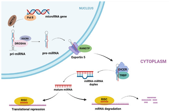
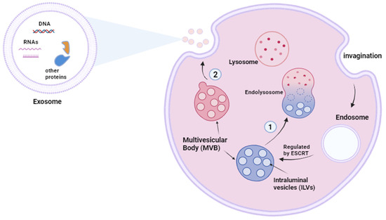
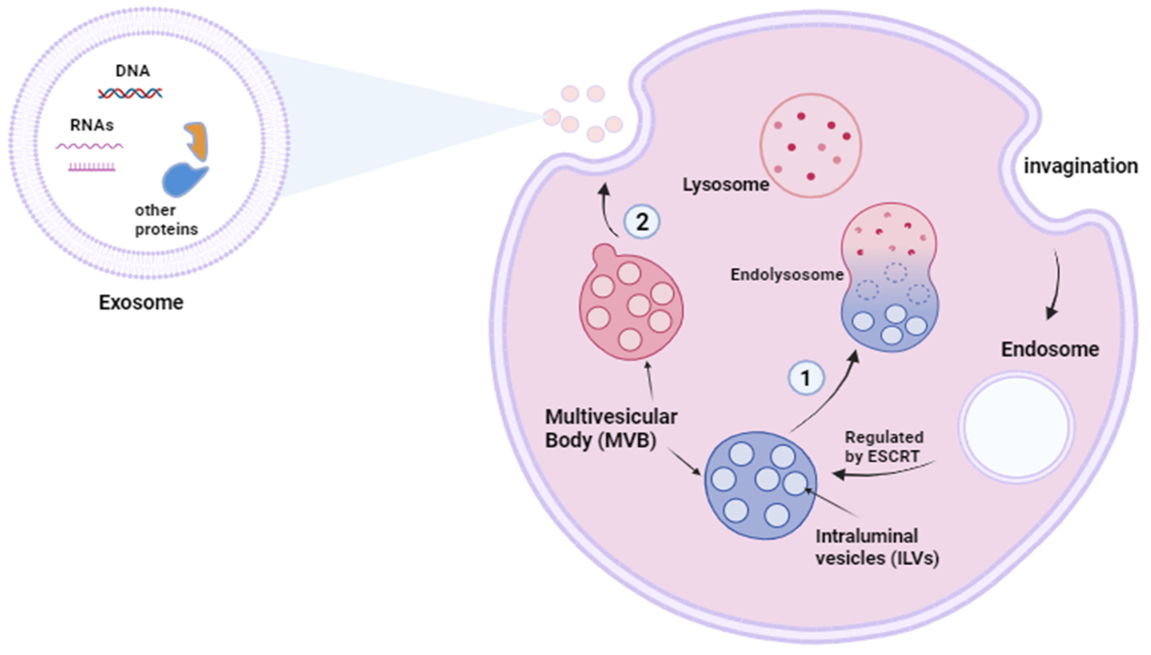
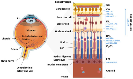
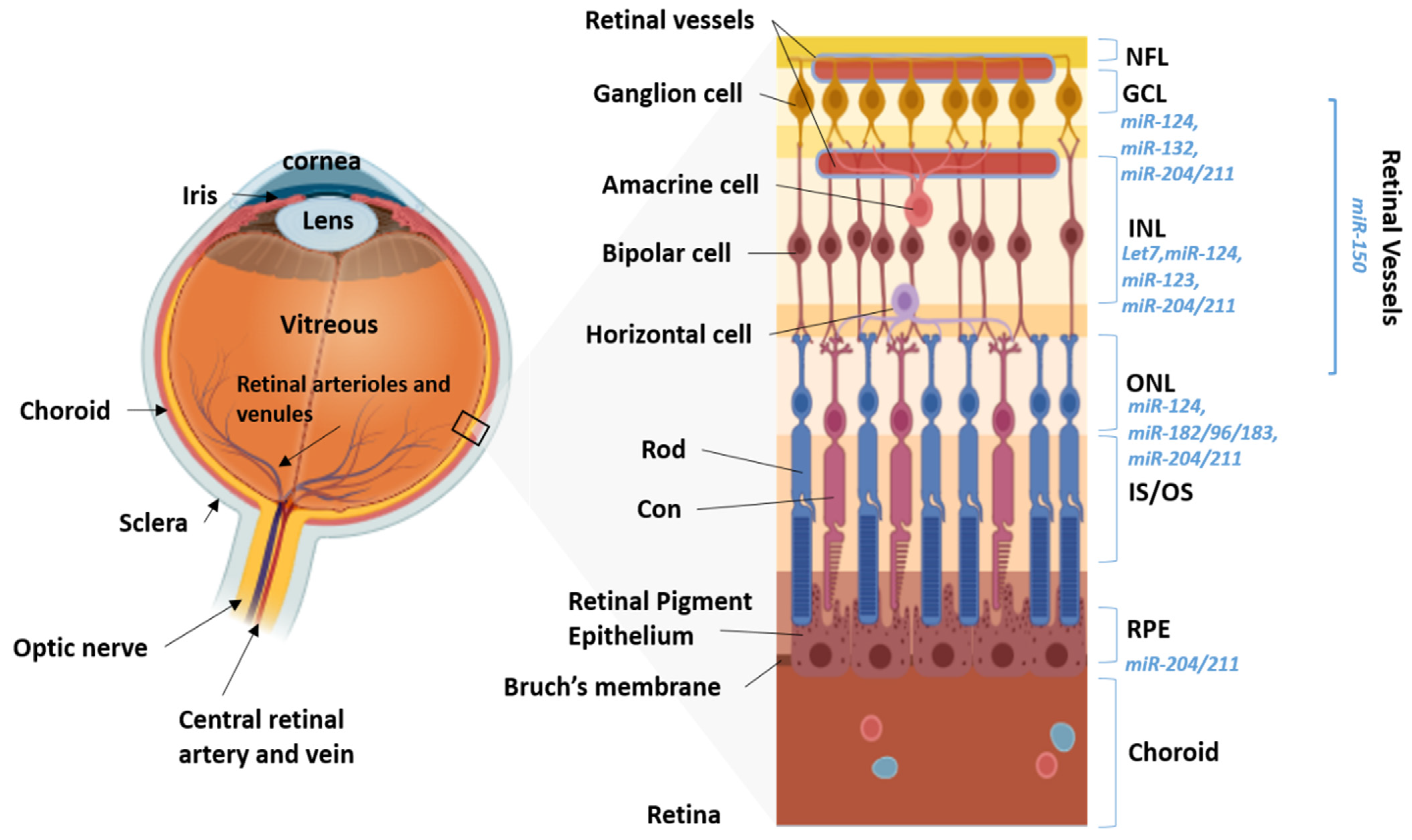
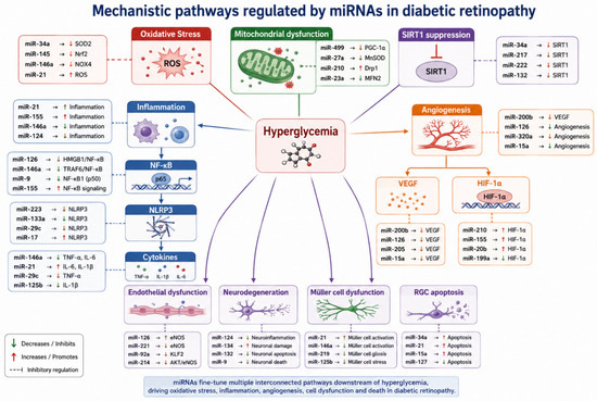
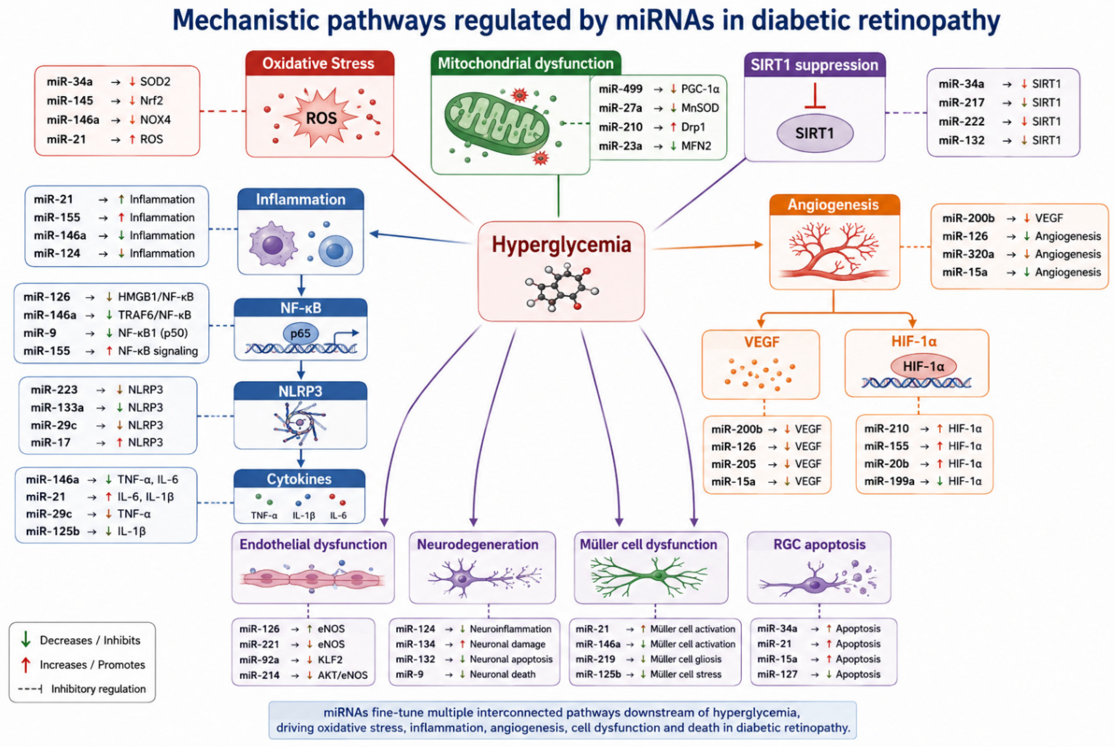

Disclaimer/Publisher’s Note: The statements, opinions and data contained in all publications are solely those of the individual author(s) and contributor(s) and not of MDPI and/or the editor(s). MDPI and/or the editor(s) disclaim responsibility for any injury to people or property resulting from any ideas, methods, instructions or products referred to in the content. |
© 2026 by the authors. Licensee MDPI, Basel, Switzerland. This article is an open access article distributed under the terms and conditions of the Creative Commons Attribution (CC BY) license.