1. Introduction
The boundary between the atmosphere and the aqueous phase of the tear film is stabilized by the tear film lipid layer (TFLL), a viscoelastic duplex architecture with a thickness of approximately 100 nm [
1,
2]. Structurally, it comprises a basal monolayer of amphiphilic polar lipids (PL) in direct contact with the aqueous tear (AT), overlaid by a thicker, exterior bulk phase-like stratum of nonpolar lipids (NPL) [
3,
4]. This protective barrier is primarily composed of meibum (MGS)—the secretory output of the meibomian glands. Chemically, this secretion is dominated by NPL (exceeding 90%), including various triglycerides, wax esters, and cholesteryl esters, whereas the PL fraction (under 10%) is characterized largely by (O-acyl)-ω-hydroxy fatty acids [
5,
6].
The integrity of the tear film is frequently undermined by adverse external conditions, such as depressed ambient temperatures, low relative humidity (RH), and the presence of fine particulate matter (PM2.5). These stressors are recognized drivers in the pathogenesis of dry eye disease (DED) [
7,
8,
9,
10]. As the most superficial component of the ocular surface, the tear film lipid layer (TFLL) serves as the primary shield against such environmental challenges. As we recently showed, despite their distinct nature, these adverse ambient conditions appear to influence MGS films in a similar fashion, by disturbing the uniform spreading, the morphological integrity, and the viscoelastic properties of meibum [
11]. Indeed, decreased RH is expected to facilitate the evaporative cooling of TFLL (due to the AT exsiccation to the air), thus sharing a common physical mechanism with the impact of low environmental temperature.
Polyanionic mucomimetic polymers, such as hyaluronic acid (HA), polyvinylpyrrolidone (PVP), and chondroitin sulfate (CHS), play a sophisticated role in stabilizing the tear film lipid layer (TFLL) by enhancing its structural integrity and spreading dynamics. Rather than merely increasing bulk viscosity, these polymers—particularly the high-molecular-weight variants of HA—interact with the polar lipid subphase (e.g., phospholipids) to reinforce the interfacial “scaffold” upon which the non-polar lipids reside [
12,
13,
14].
Research indicates that these polymers can integrate into the lipid-aqueous interface, reducing surface tension and promoting the formation of a more cohesive and continuous lipid film during the blink cycle in vitro and in vivo [
12,
13,
14,
15,
16]. This interaction mitigates the formation of “lipid-free” gaps, thereby effectively reducing the rate of aqueous evaporation and increasing tear film break-up time (TFBUT). Furthermore, the mucomimetic properties of CHS and HA allow for superior retention on the ocular surface through non-covalent bonding with membrane-associated mucins, providing a synergistic lubricating effect that minimizes friction-induced damage to the corneal epithelium [
12,
13,
14].
The primary goal of this investigation is to decipher the synergistic relationship between mucomimetic agents—specifically hyaluronic acid (HA), polyvinylpyrrolidone (PVP), and chondroitin sulfate (CHS)—and the meibomian gland secretion (MGS). We seek to determine whether these polymers stabilize the tear film’s lipid component when subjected to environmental stressors, such as particulate matter (PM2.5) contamination, reduced thermal states (20 °C), and arid conditions (20% relative humidity). Gaining insight into the interplay among these molecules at the air–liquid interface is essential for identifying the biophysical pathways through which the polymers can shield the ocular surface from instability. To probe these complex interactions, we utilized a Langmuir trough to generate artificial tear film models [
12,
17,
18,
19,
20]. The properties of these interfacial layers were analyzed both in isolation and when formed over the polymer-enriched subphase via analysis of their surface pressure area isocycles, dilatational rheology (probed with stress relaxation technique) and film morphology visualized with Brewster Angle Microscopy (BAM).
4. Discussion
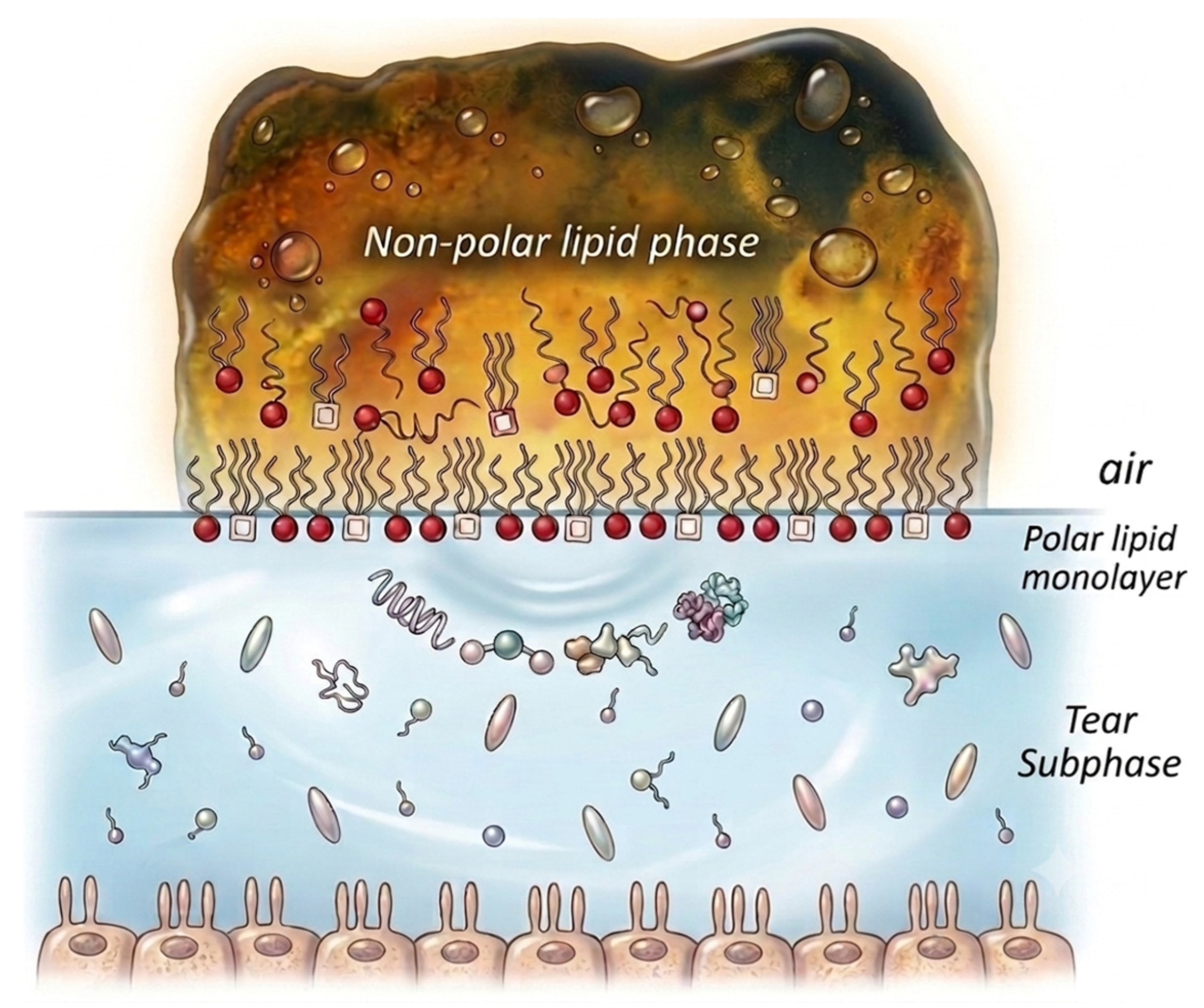
The integrity of the ocular surface and the stability of the tear film are fundamentally dependent on the sophisticated, stratified arrangement (see
Figure 1 and
Figure 4) of the meibomian gland secretion (MGS) [
1,
2,
39]. This interfacial architecture is characterized by a dual-layered composition: a distal nonpolar region, primarily comprised of cholesterol and wax esters, which is theorized to inhibit the evaporation of the aqueous tear (AT) phase; and a proximal polar lipid layer—rich in (O-acyl)-ω-hydroxy fatty acids and phospholipids—that facilitates the adhesion of the lipid film to the underlying aqueous subphase. Such a highly ordered configuration ensures efficient modulation of surface tension, promotes rapid lateral spreading during the blink cycle, and maintains the viscoelastic flexibility of the film [
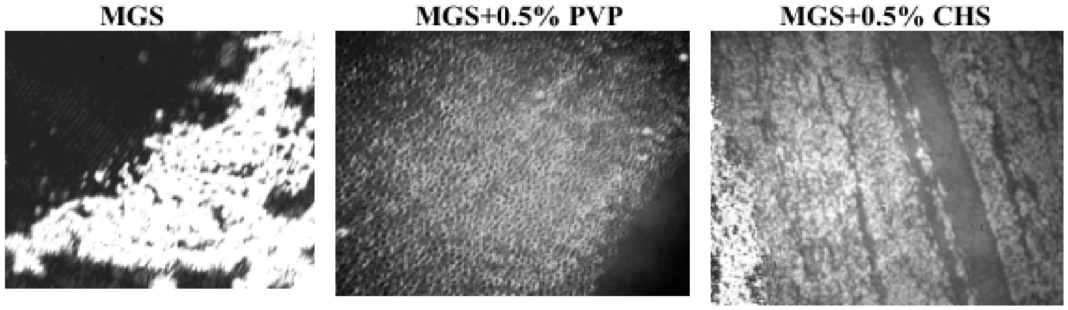
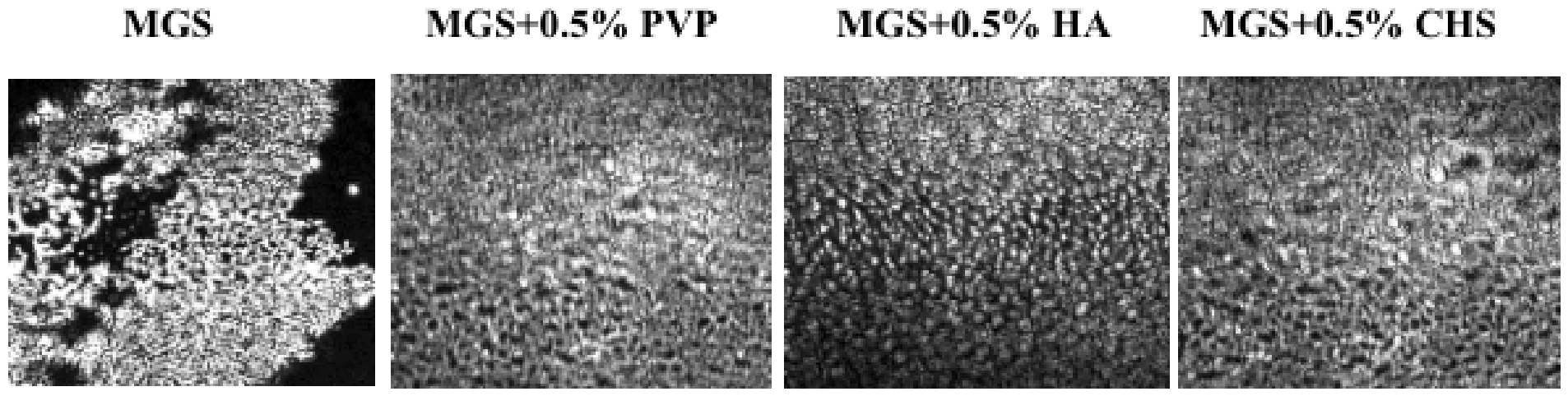
40]. However, when external stressors—including particulate matter or fluctuations in ambient temperature and humidity—interfere with this molecular alignment, the film’s ability to act as a cohesive barrier is impaired. This structural degradation (as in left panel of
Figure 7) results in accelerated tear fluid loss and diminished film persistence. Consequently, maintaining the native multilayered morphology of the MGS is vital for protecting the biophysical and physiological health of the eye.
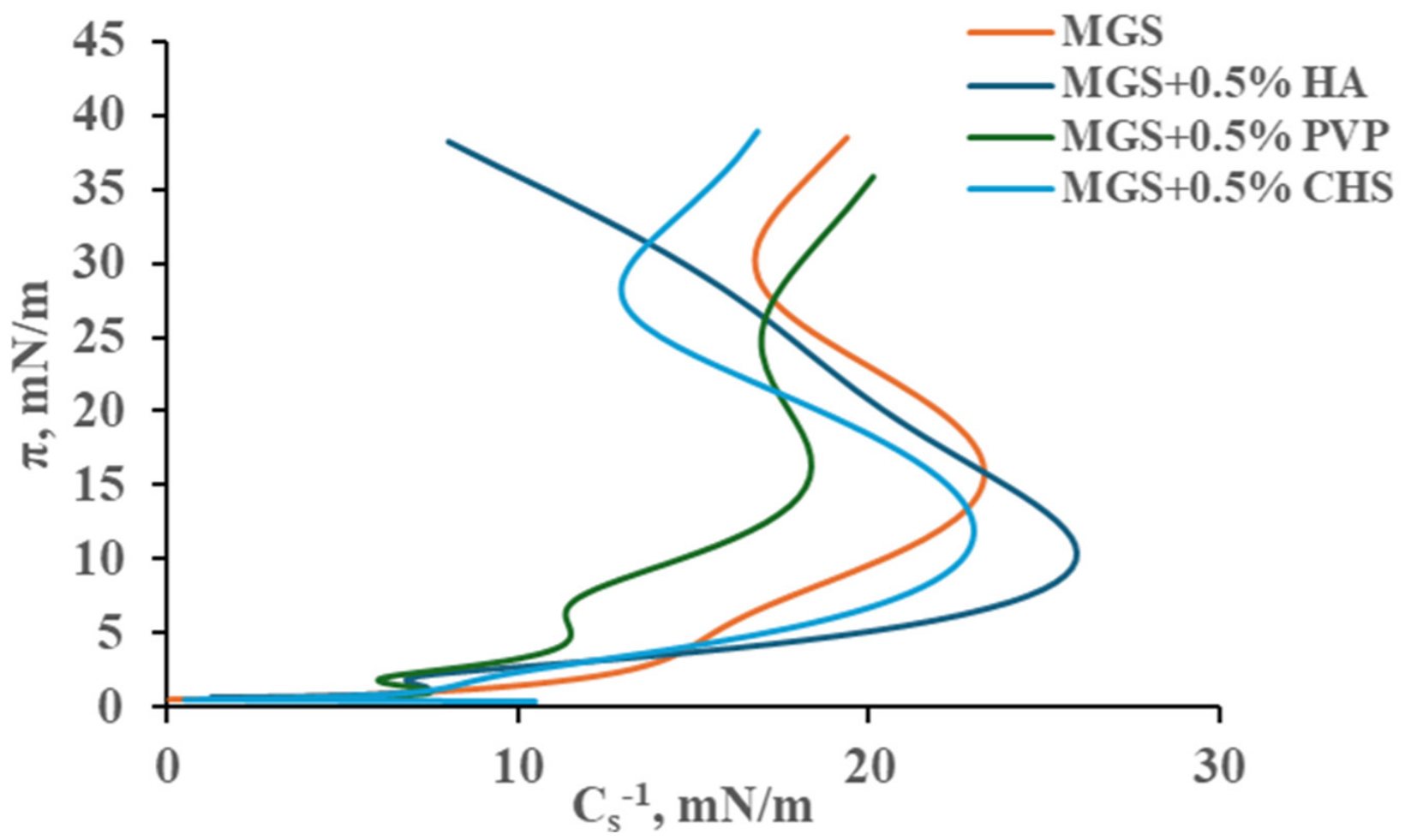
The current investigation examines the in vitro morphological features, mechanical compressibility (Cs
−1), and viscoelastic dynamics—determined by the equilibrium between diffusional and viscoelastic contributions to stress relaxation—of lipidic interfaces. By utilizing a Langmuir trough at the air–water interface, this model decouples meibomian gland secretion (MGS) films from auxiliary tear components and ignores physiological variables like tear replenishment, blink rates, and epithelial crosstalk. Although such simplifications may reduce direct translatability to in vivo scenarios, they allow for rigorous experimental standardization. This approach isolates MGS properties from confounding biological factors to identify fundamental correlations with clinical ocular surface performance. Prior studies involving both healthy cohorts and individuals with Dry Eye Disease (DED) substantiate that these laboratory metrics serve as robust proxies for the real-world behavior of the human tear film (TF) and its lipid layer (TFLL) [
1,
12,
41,
42,
43,
44,
45,
46,
47]. Clinical data mirror these in vitro findings: DED-derived samples typically show compromised surfactant functionality and an inability to sustain a stable duplex architecture under compression, whereas healthy controls remain cohesive [
1,
12]. On a macroscopic scale, the elasticity of the TFLL is evidenced by its restorative capacity post-deformation, specifically quantified through (i) the velocity of lateral spreading following eyelid opening and (ii) the rate of structural attrition during consecutive blink cycles [
42,
43]. A certain limitation of our study stems from the restriction inherently set by the tiny amount of meibum that can be collected from volunteers, which in turn requires its careful utilization across experiments. Therefore, each polymer (with mean molecular weight 1000 kDa) and each environmental stressor (low RH, low temperature, PM2.5) is tested at a single, fixed condition. The choice of environmental stressors conditions is explained elsewhere [
20] while the polymer molecular weight and inclusion at 0.5% are selected based on their current and intended implementation in ophthalmic formulations. It is of significant practical interest and target of future studies to probe the impact of environmental stressors in a broader range as it will allow for assessment of thresholds or nonlinear effects and for predictive understanding of how various levels of these conditions will impact the performance of TFLL. The same is valid for evaluating the influence of varying the molecular weight of polymers, which is known to differentially affect the physicochemical properties and stability of tear film in vivo [
48,
49,
50].
Provided that other parameters remain unchanged, a more elastic Tear Film Lipid Layer (TFLL) is anticipated to undergo more accelerated expansion across the air–aqueous interface following a blink. This spreading phase is clinically indispensable; as the TFLL migrates superiorly, it generates a mechanical traction that draws the underlying aqueous phase upward. This mechanism ensures homogenous corneal lubrication, bolsters tear film stability, and mitigates localized break-up [
44,
45,
46,
47,
51]. Investigations by Yokoi et al. [
43] substantiate that TFLL propagation is notably impaired in subjects with Dry Eye Disease (DED) relative to healthy cohorts. Additionally, Goto and Tseng [
46] characterized specific anomalies in patients with Meibomian Gland Dysfunction (MGD): in addition to a significantly delayed spreading duration (3.54 ± 1.86 s vs. 0.36 ± 0.22 s), the expansion morphology lacked horizontal symmetry. Rather than advancing as a uniform linear front, MGD-derived films exhibited vertical striations that migrated at disparate velocities, resulting in a fragmented boundary. Furthermore, the topographical roughness of the lipid layer—a proxy for thickness non-uniformity—was substantially elevated in these individuals (lipid map uniformity = 125 nm
2) compared to healthy controls (lipid map uniformity = 14 nm
2) [
51]. This correlates with the heightened structural heterogeneity observed (
Figure 7) via Brewster Angle Microscopy (BAM) in meibum subjected to environmental stressors [
47].
The structural resilience of the Tear Film Lipid Layer (TFLL) can also be gauged by the velocity of its morphological deterioration during the inter-blink interval. Enhanced elasticity generally provides superior structural stability; conversely, research by Georgiev et al. [
1,
12] indicates that the rate of pattern breakdown is significantly more pronounced in individuals with Meibomian Gland Dysfunction (MGD). This suggests that their lipid interfaces are characterized by a predominant dissipative viscous behavior rather than the robust elastic nature typical of healthy eyes [
41]. These observations are corroborated by findings from the Goto research group [
46]. Accordingly, the capacity of mucomimetic polymers to bolster the expansion dynamics and structural persistence of meibum films under simulated environmental stressors in vitro acts as a reliable indicator of their clinical effectiveness in vivo [
41].
This research indicates that reduced thermal conditions, arid environments, and atmospheric particulates induce analogous deleterious shifts in the interfacial characteristics and structural cohesion of meibomian gland secretion (MGS) layers. These stressors promote heightened film stiffness, perturbed molecular packing, and diminished interfacial stability—factors that collectively undermine the tear film’s defensive capacity [
8,
9,
10,
52]. Such observations imply that atmospheric conditions typical of dry or contaminated regions facilitate tear film collapse and contribute to the etiology of evaporative dry eye disease.
The impact of low humidity and cold temperatures aligns with fundamental thermodynamic principles. In low-humidity environments, accelerated evaporation of the aqueous subphase triggers evaporative cooling at the air–lipid boundary [
24], potentially decreasing the TFLL temperature by 5–10 °C [
53]. This thermal drop shifts the thermodynamic equilibrium, inducing phase transitions toward higher molecular ordering and rigidity [
51]. As the lipid layer loses its requisite fluidity, it becomes susceptible to structural failure and less capable of stabilizing the ocular surface [
41,
54,
55,
56].
Concurrently, exposure to fine particulate matter, specifically PM2.5 facilitates the aggregation of MGS lipids, resulting in increased film heterogeneity [
57,
58]. This clustering disrupts homogenous lateral spreading, producing a non-uniform, patchy morphology with compromised durability. While the biochemical pathways of particulate interaction differ from the purely physicochemical effects of temperature and humidity, the macroscopic impact on film integrity is strikingly congruent. Such aggregation likely stems from direct particle–lipid interactions that recalibrate intermolecular forces and promote localized nucleation [
59,
60]. Precisely defining the mechanistic pathways for PM2.5 remains difficult due to the heterogeneous chemical profile of these aerosols, which include water-soluble ions, inorganic elements, and varied carbonaceous compounds [
61]. Specifically, the concentration of polycyclic aromatic hydrocarbons (PAHs)—which are prone to partitioning into the non-polar stratum of the MGS—varies significantly in environmental samples. This variability, alongside fluctuations in metallic or microplastic content, suggests that different particulate compositions may elicit distinct impacts on TFLL functionality compared to those observed in this controlled study.
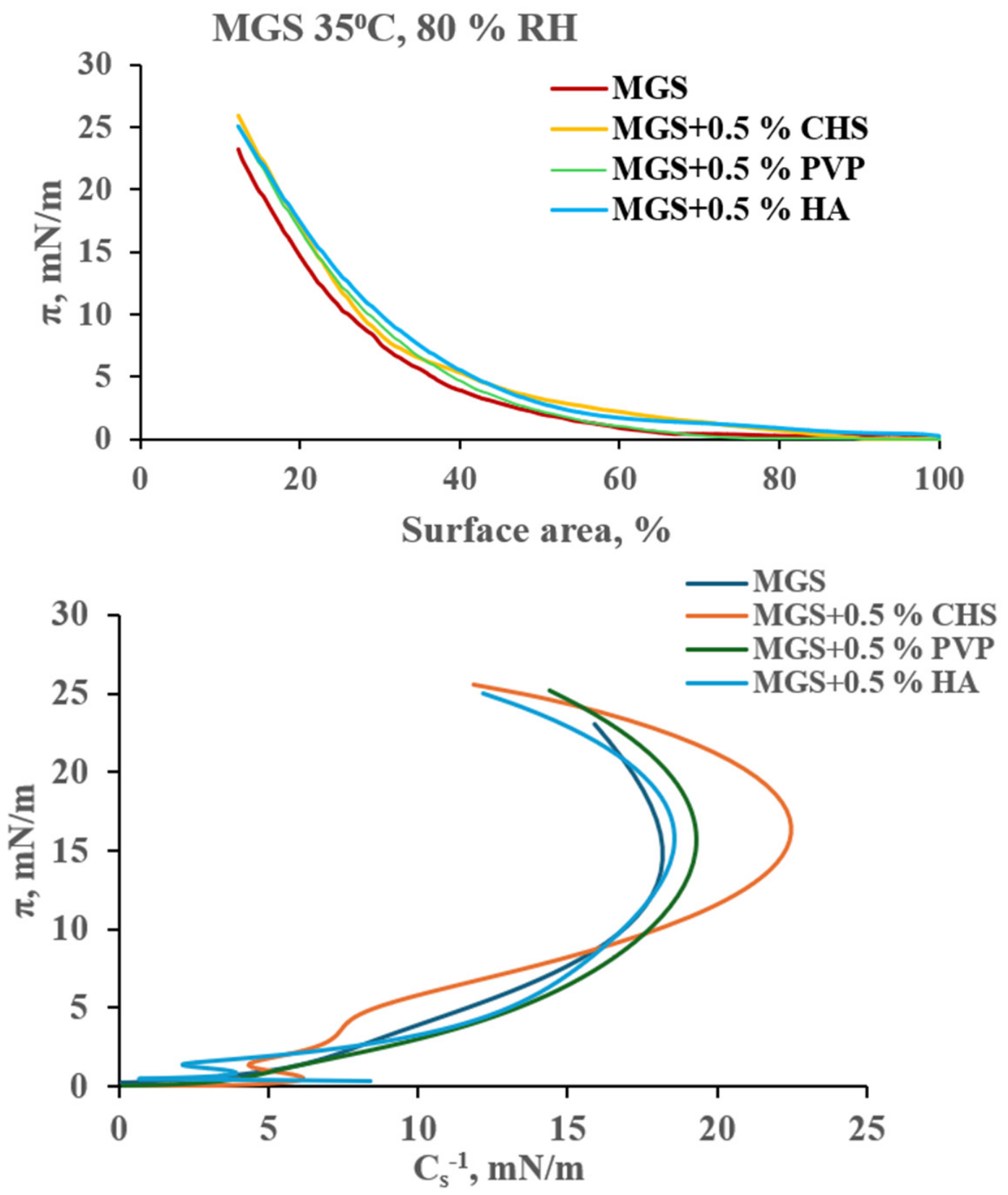
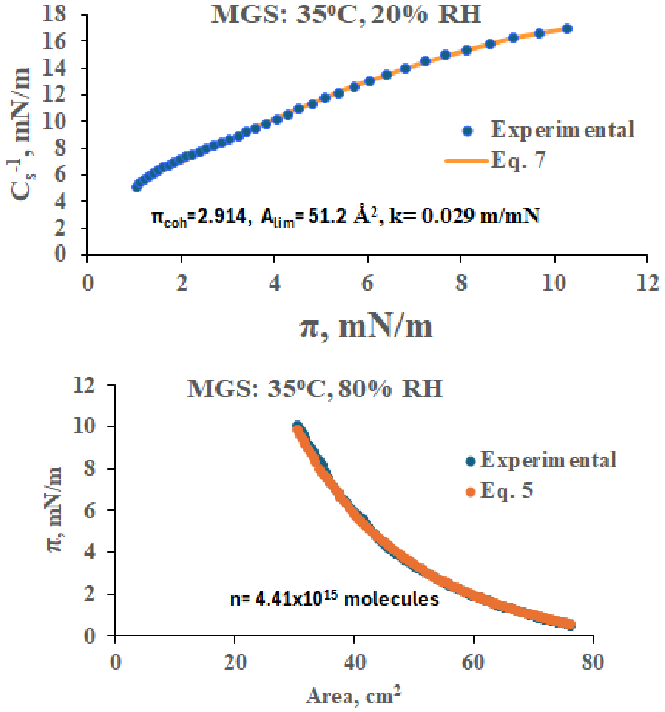
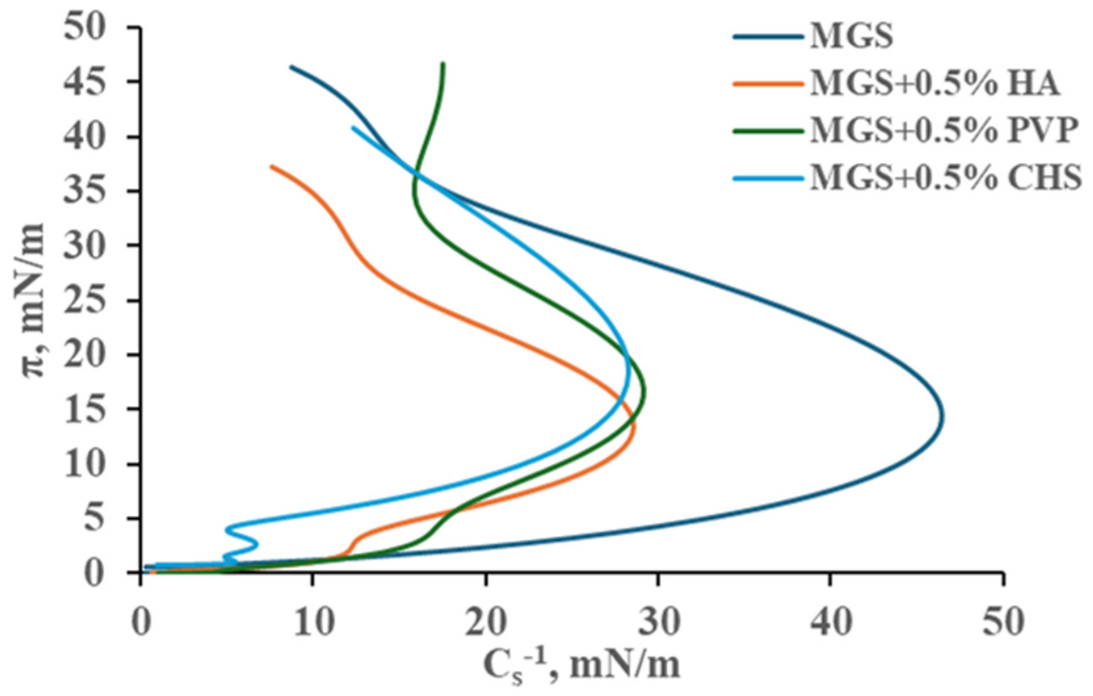
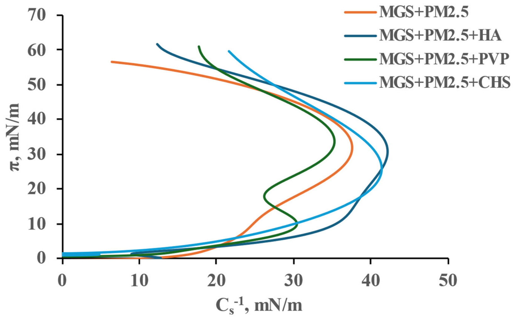
In the current study, a sophisticated analytical apparatus was developed to analyze the impact of polymers on the surface properties of MGS duplex multilayers: (i) a Volmer equation-based version of 2D-VES (Equation (5)) which (together with Equation (7)) makes it possible to probe the apparent number of molecules located at the MGS/aqueous interface as well as their unique molecular properties (i.e., limiting area per molecule, molecular compressibility, cohesion pressure, etc.) and (ii) a combined Maxwell viscoelastic and diffusion-relaxation model (Equation (10)) to quantify the transient behavior of the dilatational relaxation modulus. Apart from the exposure to decreased RH where a complex picture is observed (probably due to in-depth incorporation of dehydrated polymer chain residues in the MGS films), at most ambient conditions it can be seen that the supplementation of polymers resulted in more compressible (higher value of k) films and in a drop of πcoh (i.e., improved spreading) which is typical for polymer-containing films. In line with this, the presence of PVP resulted in an increase of the apparent values of Alim and decrease of the apparent number of molecules at the film/aqueous interface which is also indicative for the inclusion of polymers in the molecular packing. At 35 °C, 80% RH there is a somewhat unique behavior of CHS which results in an increase of the apparent number of molecules at the MGS/aqueous interface. This suggests a distinct mode of action at normal ambient conditions compared to the rest of the polymers. As per MGS and in many cases MGS + 0.5% CHS layers the Alim are typical for different degrees of compression of OAHFA, the intrinsic amphiphilic species of MGS.
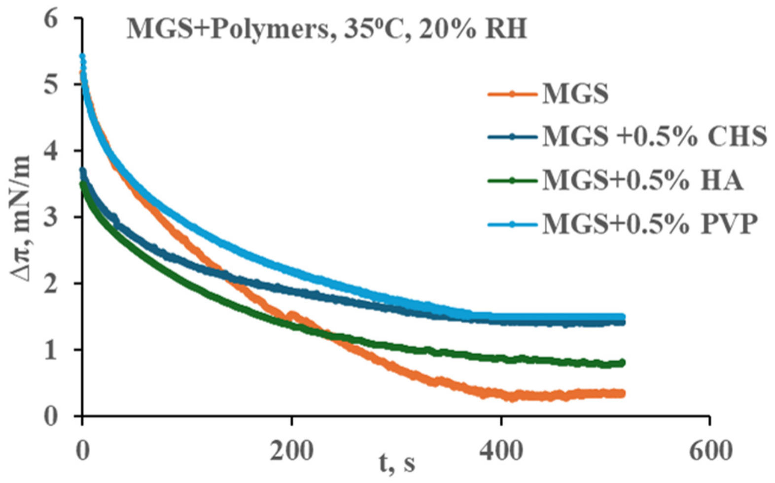
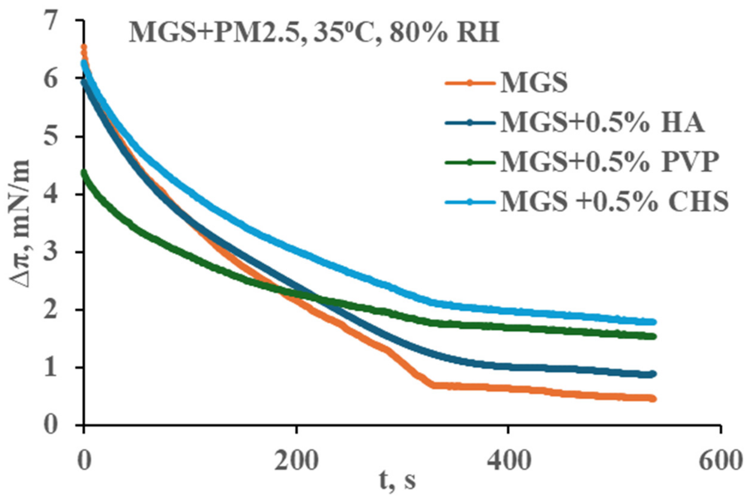
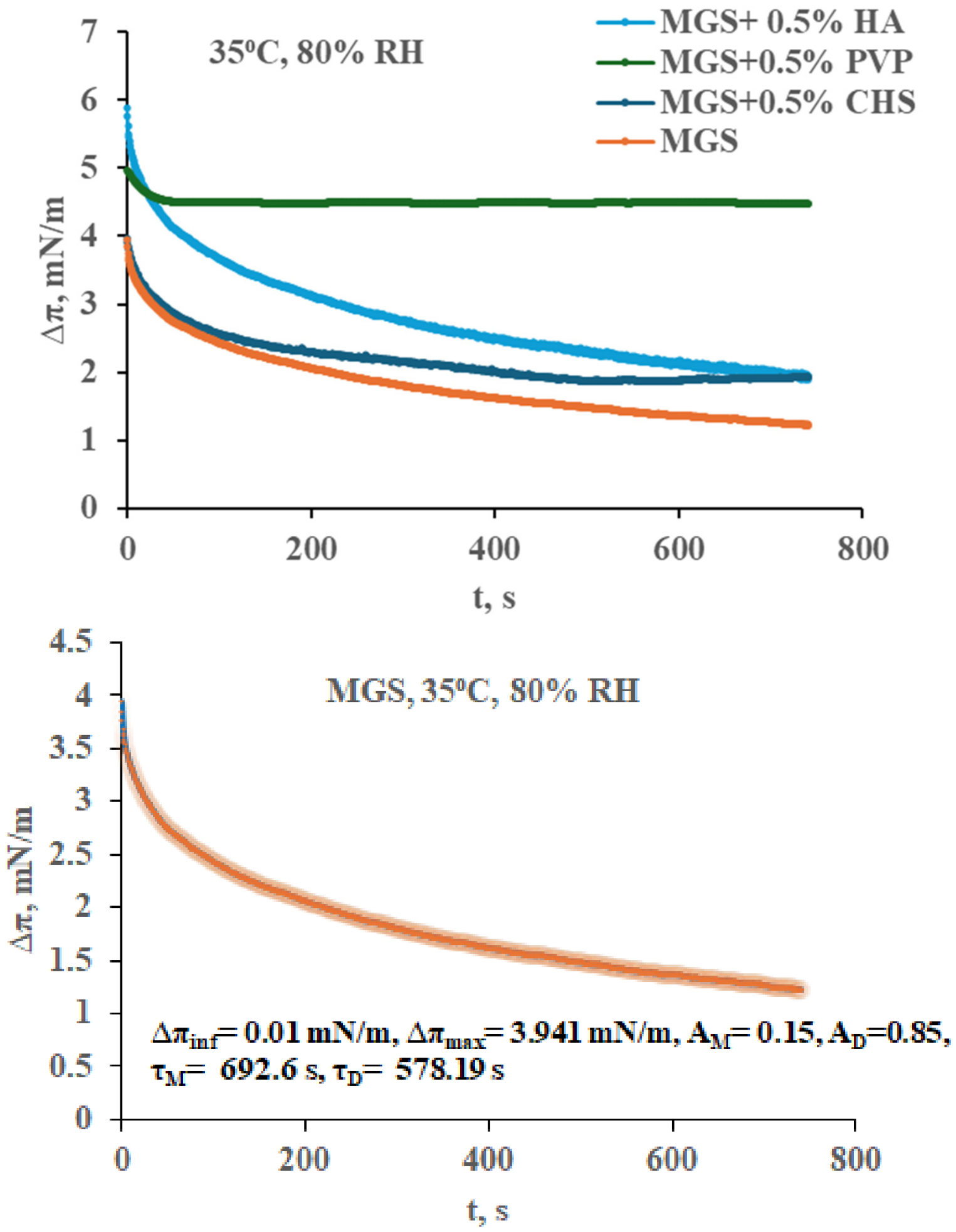
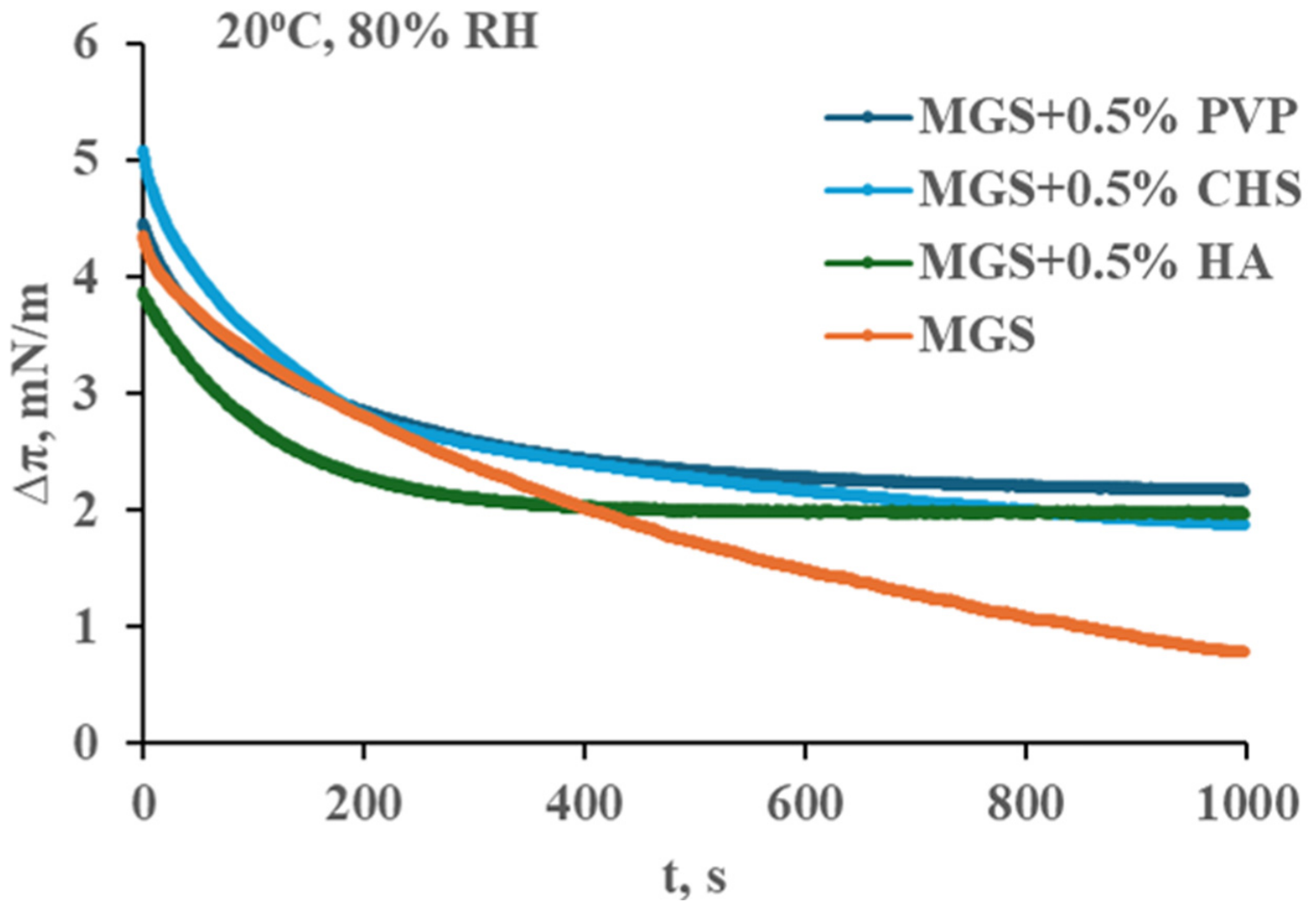
The analysis of stress relaxation transients (
Figure 5,
Figure 9,
Figure 12 and
Figure 15) reveals that mucomimetic polymers altered the relative contributions of viscoelastic and diffusion processes in the films and invariably resulted in higher ∆π
inf suggesting enhanced elastic response of the layers upon dilatational deformation. Apart from shifting the relaxation process towards higher ∆π
inf and the supplementation of polymers changed the balance between the viscoelastic and diffusion processes with an overall rise of the contribution of viscoelasticity compared to the one of diffusional rearrangement of molecules across the strata of the MGS duplex film. These in vitro results are consistent with documented clinical evidence regarding ocular surface therapeutic interventions. Specifically, the topical administration of 0.1% hyaluronic acid (HA) has been demonstrated to augment the thickness of the tear film lipid layer (TFLL) in lipid-deficient patients, increasing it from sub-60 nm to 75 nm within 15 min post-instillation [
15]. Furthermore, these data mirror the observed effects of a 3% diquafosol ophthalmic solution (i.e., a secretagogue known to stimulate rapid MUC5AC mucin release) which has been shown to sustain increased TFLL thickness in healthy subjects for over an hour following application [
16]. The observed MGS-modulating properties of water-soluble mucomimetic polymers appear to stem from the establishment of an interfacial, gel-like polymer matrix. This structural assembly is primarily driven by an extensive network of hydrogen bonds occurring both inter-polymerically and between the polymer chains and the polar lipid headgroups. Consequently, this consolidation facilitates elevated film viscosity, enhanced lateral homogeneity of the lipid monolayer, and improved aqueous integration within the tear film structure [
1,
12]. Operating on a physiologically significant timescale of mere seconds, this rapid kinetic mechanism reveals that the distinct strata of the tear film function not as isolated compartments, but as an integrated, dynamic continuum characterized by perpetual interlayer crosstalk and systemic interdependence.
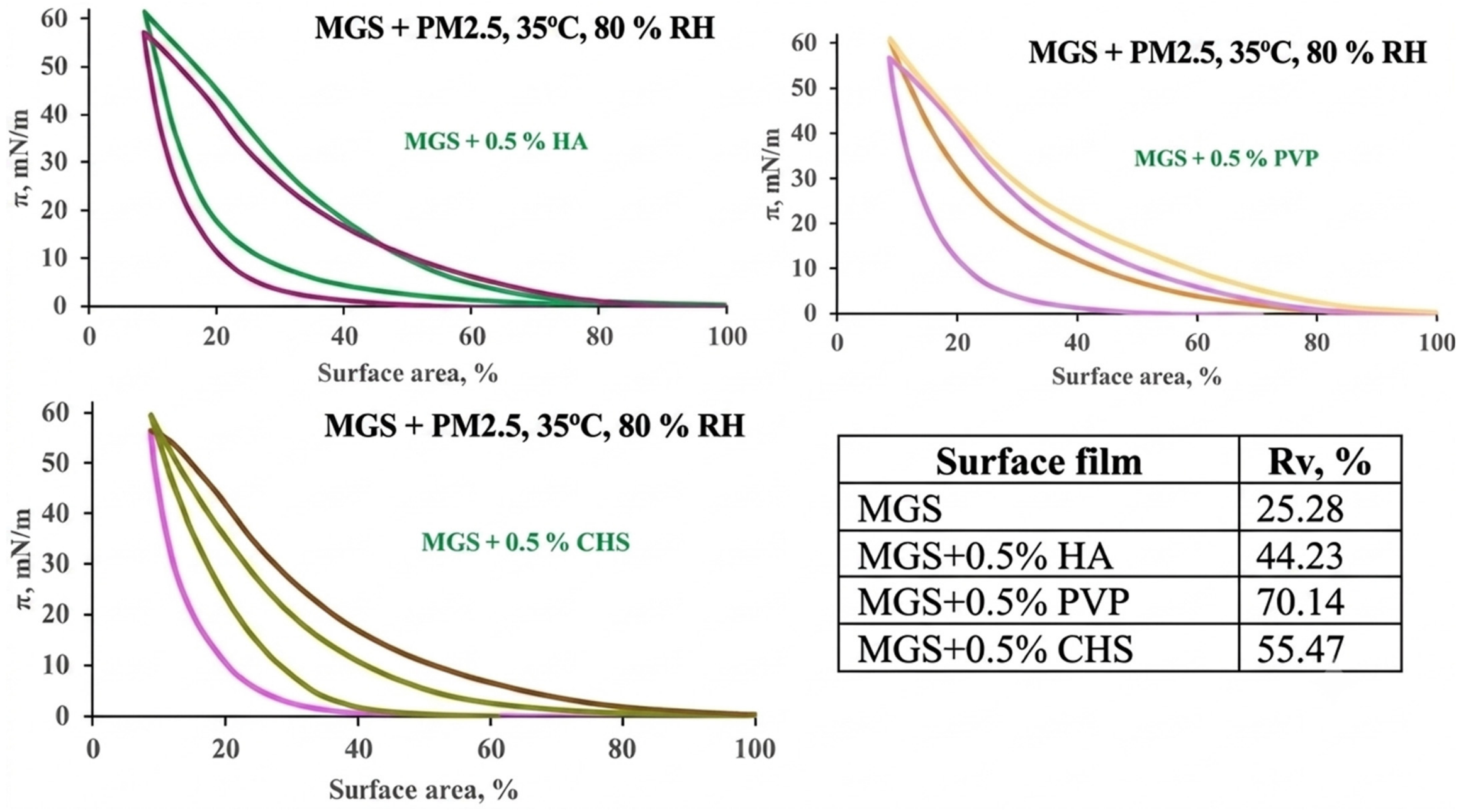
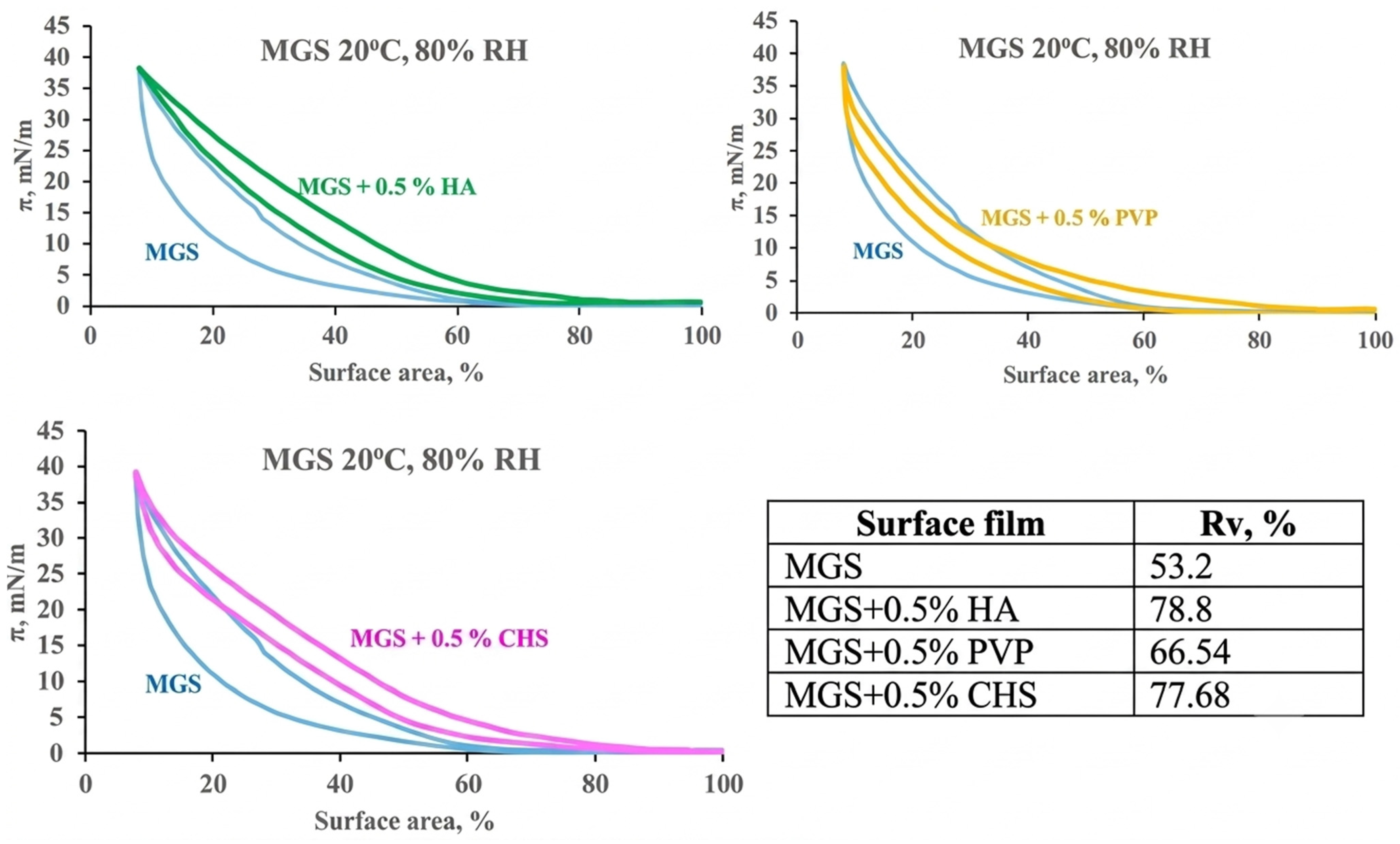
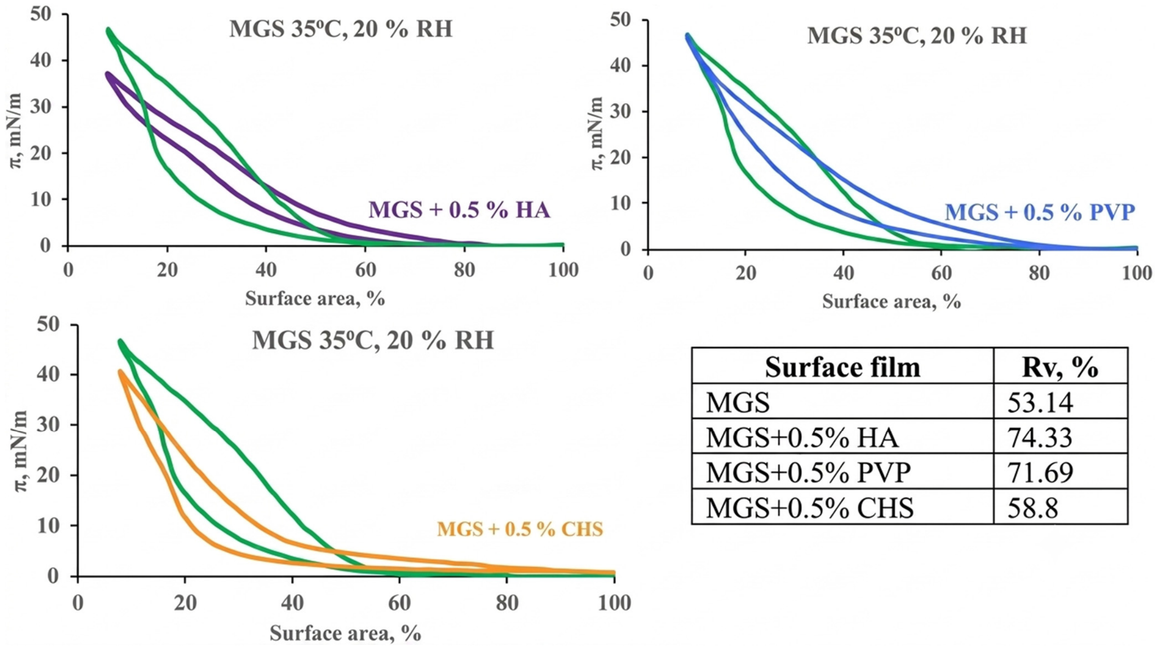
The biophysical and mechanistic effects are thought to be the primary route of action of polymeric eyedrops in the restoration of the tear film and ocular surface integrity and represent the main focus of the current study. Although the performance of the polymers is certainly dependent on the environmental stressors, still some general patterns are identifiable. Polymers improve the spreading of lipid films, as manifested by the decrease of πcoh and of isothermal reversibility. PVP, CHS, and HA facilitate uniform lipid distribution and also partially integrate into the existing film, thereby helping to restore duplex multilayer organization disrupted by low humidity, low temperature, or particulate matter. At 35 °C and 80% RH, CHS shows characteristic effect, resulting in an increase of the apparent number of molecules at the MGS/aqueous interface. This suggests a distinct impact at normal ambient conditions compared to the other polymers. As observed for MGS and, in many cases, MGS + 0.5% CHS layers, the Alim values are typical for different degrees of compression of OAHFA, the intrinsic amphiphilic species of MGS. Also, it should be noted that the presence of HA tended to result in a strong increase of the Alim value and a corresponding decrease in the apparent mean molecular number of molecules (as calculated with Equations (5) and (7)), which suggests a distinct mode of interaction of HA with MGS films. The results, as well as the multiple numerical estimates obtainable via the approaches described here, may be beneficial for the design of artificial tears for the treatment of dry eye. To draw a definitive conclusion, it is necessary to establish statistical correlations between the in vitro estimates and the in vivo clinical performance of the eyedrop formulations, which is a subject of our subsequent research.
Apart from the biophysical and mechanistic implementations, the biochemical interactions may also significantly contribute, especially in cases where long-term residence time of the polymers at the ocular surface is achievable [
41]. High-molecular-weight hyaluronic acid (HA) variants have undergone extensive research due to their widespread formulation in lubricating eye drops designed for dry eye therapy [
62]. The substantial chain length and viscosity of HA not only bolster corneal and conjunctival hydration but also lower shear stress against the eyelid wiper during blinking [
62]. Concurrently, HA mitigates inflammatory responses and downregulates immune activation, which accelerates ocular tissue regeneration [
63]. In vitro and in vivo models demonstrate that HA scavenges reactive oxygen species and offers cytoprotection by buffering the cellular toxicity of common ophthalmic preservatives [
64,
65]. These physiological responses are mediated by transmembrane signaling cascades triggered when HA binds to an array of specific cellular receptors, including CD44, lymphatic vessel endothelial receptor 1 (LYVE-1), receptor for hyaluronan-mediated motility (RHAMM), and toll-like receptor 4 (TLR-4) [
66]. Through these receptor interactions, HA signaling critically modulates angiogenesis by directing endothelial cell dynamics [
63]. Crucially, the biological outcome of these pathways is highly sensitive to the polymer’s molecular weight. While large HA chains (Mw ≥ 200 kDa) promote cell survival and tissue homeostasis, fragmented, low-molecular-weight HA behaves as a strong mediator of inflammation. This phenomenon is attributed to the different manner of interaction with the HA receptors, CD44 and RHAMM in particular [
67]. It is thought that these diverse effects stem from the impact of the molecular weight on the HA conformation: large >MDa HA is a random coil, while very small (e.g., 10 kDa) HA behaves like a rod. Size exclusion chromatography and multiangle light scattering studies revealed that HA mass-to-diameter ratio showed a transition in the 150–250 kDa size range (~65 nm). Hence, the HA rod-to-coil transition occurs in the size range that specifically activates cell signaling by the receptors discussed above. Thus, size-specific signaling could be due to (i) unique external receptor/HA conformation changes enabling transmembrane-mediated activation of cytoplasmic domains, or (ii) transition-size HA may enable multiple receptors to bind the same HA, creating new internal signal-competent cytoplasmic domain complexes [
68,
69]. Although relatively less studied, similar effects are reported for chondroitin sulfate, demonstrating its (i) high biocompatibility and biodegradability, (ii) propensity to create a favorable environment for tissue repair by reduction of inflammatory processes and oxidative stress, and (iii) binding to CD44 and syndecan cell receptors, enhancing cellular uptake and activating pathways for cell migration, differentiation, and proliferation [
70]. While PVP biochemical activity is generally considered limited, it forms a protective polymeric shield over the ocular surface that preserves epithelial integrity by physically sealing micro-gaps between damaged cells. Furthermore, when combined, PVP and HA work synergistically to reduce perilimbal conjunctival erythema (redness) and optimize epithelial repair better than either polymer alone, a cooperative pattern that may be observed with other polymer combinations as well [
71,
72,
73,
74,
75,
76,
77,
78].




 ,
,