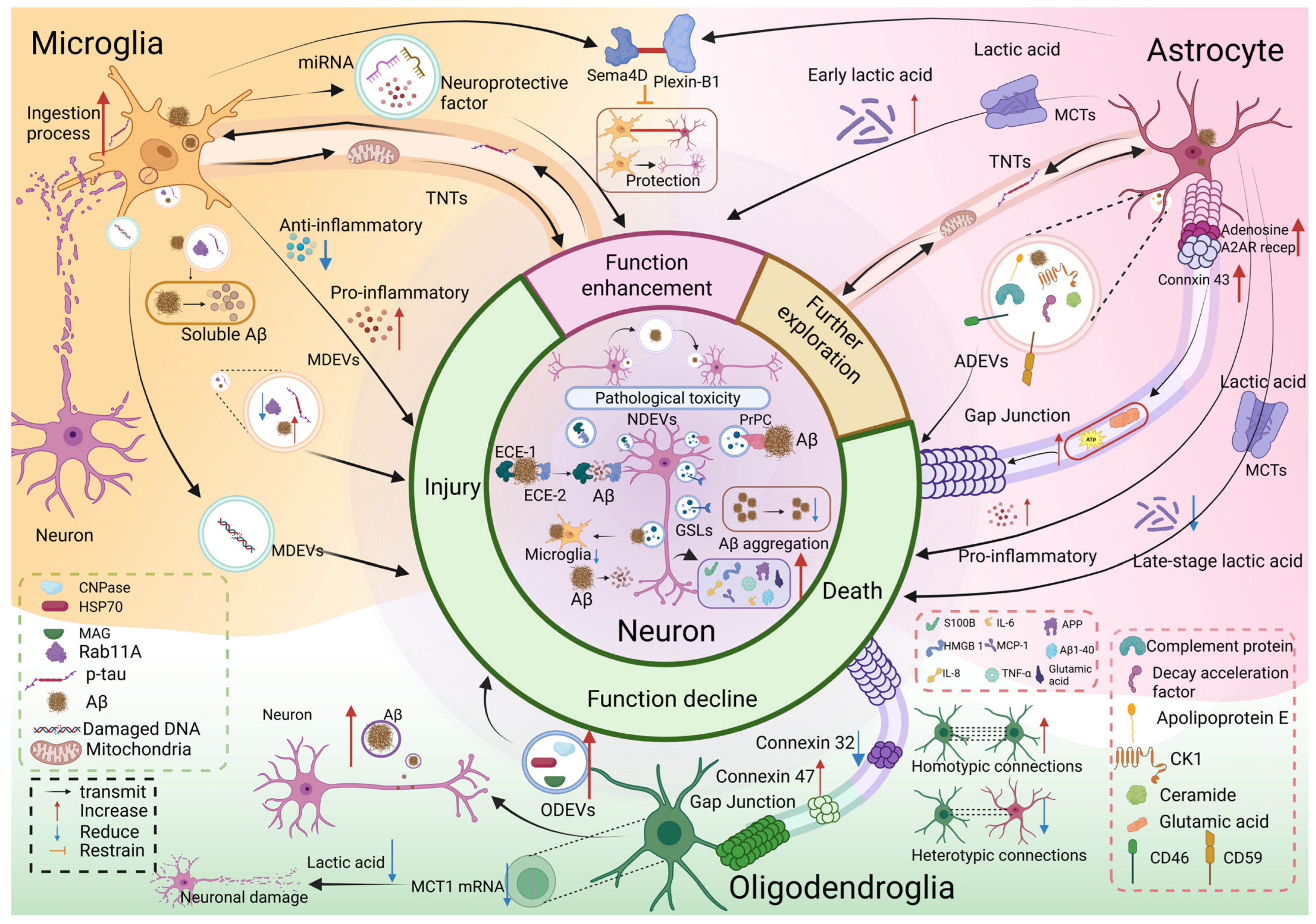
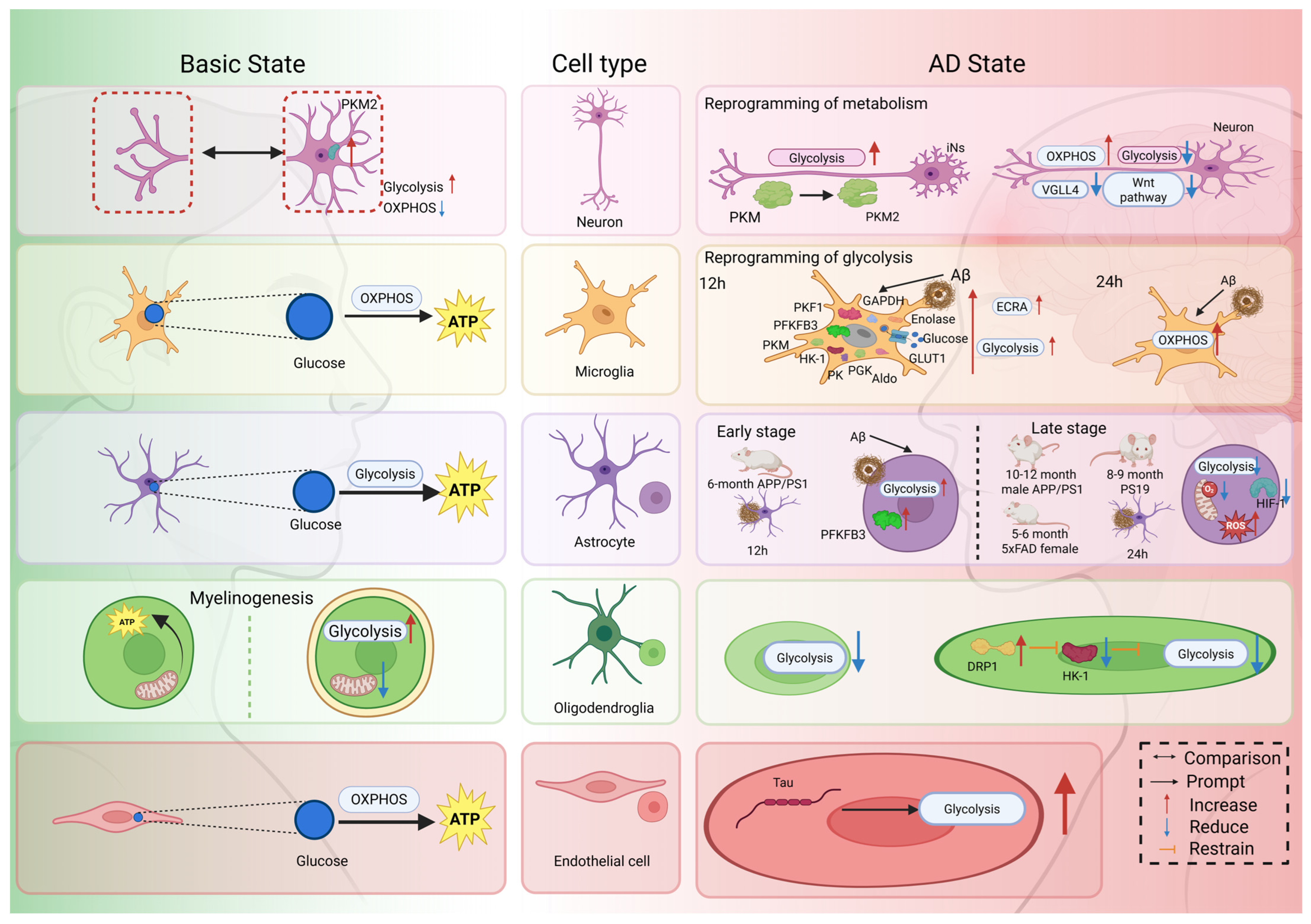
Glycolysis: The Bridge Between Cellular Interaction and Alzheimer’s Disease

Abstract
1. Introduction
2. Cellular Interactions and Alterations in Glycolysis in AD
2.1. Cellular Interactions in AD
2.1.1. Non-Direct Interactions
Soluble Secretory Factors
Extracellular Vesicles (EVs)
2.1.2. Direct Interaction
Tunneling Nanotubes (TNTs)
Gap Junctions (GJs)
Contact-Dependent Interactions
2.2. Cell Type-Specific Changes in Glycolytic Flux in AD
2.2.1. Neurons
2.2.2. Microglia
2.2.3. Astrocytes
2.2.4. Oligodendrocytes and Endothelial Cells
3. Glycolysis Regulates Cellular Interactions
3.1. Glycolysis–Lactate Coupling
3.2. Glycolysis-Inflammation Coupling
3.3. Glycolysis-Oxidative Stress Coupling
3.4. Glycolysis-Exosomes Coupling
3.5. Glycolysis-Phagocytosis Coupling
3.6. The Interplay of the Above Mechanisms
3.6.1. Glycolysis–Lactate–Inflammation Cross-Talk
3.6.2. Glycolysis–Lactate–Exosome Cross-Talk
4. Prospective Treatment
4.1. Strategies for Inhibiting Glycolysis
4.1.1. Purine/Pyrimidine Compounds
4.1.2. Uncarin
4.2. Strategies for Promoting Glycolysis
4.2.1. α-Adrenergic Receptor Antagonist
4.2.2. IDO1 Inhibitor
4.2.3. GLP-1 Receptor Agonists
4.2.4. Cannabidiol (CBD)
4.2.5. Probiotics
4.2.6. Ketones/Ketogenic Diet
4.3. Supplementary Metabolites Related to Glycolysis
L-Serine
5. Conclusions
Author Contributions
Funding
Institutional Review Board Statement
Informed Consent Statement
Data Availability Statement
Acknowledgments
Conflicts of Interest
Abbreviations
| AD | Alzheimer’s disease |
| TNTs | Tunneling nanotubes |
| CNS | Central nervous system |
| PNS | Peripheral nervous system |
| Aβ | β-amyloid plaques |
| Tau | Microtubule-associated protein Tau |
| ROS | Reactive oxygen species |
| GJs | Gap junctions |
| EVs | Extracellular vesicles |
| HMGB-1 | High mobility group box protein-1 |
| MCP-1 | Monocyte chemoattractant protein-1 |
| IL-6 | Interleukin-6 |
| IL-8 | Interleukin-8 |
| TNF-α | Tumour necrosis factor-α |
| IL-1β | Interleukin-1β |
| IL-18 | Interleukin-18 |
| iNOS | Inducible Nitric Oxide Synthase |
| COX-2 | Cyclooxygenase-2 |
| IL-10 | Interleukin-10 |
| APP | Amyloid Precursor Protein |
| ANLS | Astrocyte-neuron lactate shuttle |
| MCTs | Monocarboxylate transporters |
| mRNA | Messenger ribonucleic acid |
| ncRNA | Non-coding RNA |
| NDEVs | Neuron-derived extracellular vesicles |
| ECE-1 | Endothelin-converting enzyme-1 |
| ECE-2 | Endothelin-converting enzyme-2 |
| GSLs | Glycosphingolipids |
| PrPC | Prion protein |
| MDEVs | Microglia-derived extracellular vesicles |
| Rab11A | Ras-related protein 11A |
| GDP | Guanosine diphosphate |
| GTP | Guanosine triphosphate |
| dsDNA | Double-stranded deoxyribonucleic acid |
| DNA | Deoxyribonucleic acid |
| IGF2 | Insulin-like growth factor 2 |
| ITM2B | Integral membrane protein 2B |
| CASPR-1 | Cysteine aspartate protease 1 |
| miRNAs | MicroRNAs |
| ADEVs | Astrocyte-derived extracellular vesicles |
| APOE | Apolipoprotein E |
| CK1 | Casein Kinase 1 |
| DAF | Decay-accelerating factor |
| ODEVs | Oligodendrocytes-derived extracellular vesicles |
| HSP70 | Heat shock protein 70 |
| MAG | Myelin-associated glycoprotein |
| CNPase | 2′,3′-cyclic nucleotide 3′-phosphodiesterase |
| ATP | Adenosine triphosphate |
| cAMP | Cyclic adenosine monophosphate |
| Cx43 | Connexin 43 |
| A2AR | Adenosine A2A receptor |
| OPCs | Oligodendrocyte precursor cells |
| 5xFAD | An early-onset AD mouse model |
| Sema4D | Semaphorin-4D |
| MCT1 | Monocarboxylate transporter 1 |
| OXPHOS | Oxidative phosphorylation |
| TCA | Tricarboxylic acid |
| PK | Pyruvate kinase |
| PKM2 | Pyruvate kinase muscle isozyme 2 |
| iNs | Induced neurons |
| VGLL4 | Vestigial-like family member 4 |
| LDHA | Lactate dehydrogenase A |
| HK-1 | Hexokinase-1 |
| PFKFB3 | Phosphoenolpyruvate carboxylase 3 |
| PGK | Phosphoglycerate kinase |
| GAPDH | Glyceraldehyde-3-phosphate dehydrogenase |
| PFK1 | Phosphofructokinase 1 |
| GLUT1 | Glucose transporter type 1 |
| ECAR | Extracellular acidification rate |
| HIF-1 | Hypoxia-inducible factor 1 |
| mROS | Mitochondrial ROS |
| DRP1 | Dynamin-related protein 1 |
| MS | Multiple sclerosis |
| BBB | Blood–brain barrier |
| eNOS | Endothelial Nitric Oxide Synthase |
| H4K12 | Histone H4 at lysine 12 |
| H3K18 | Histone H3 at lysine 18 |
| MPC | Mitochondrial pyruvate carrier |
| PSEN-1 | Presenilin-1 |
| EAAT2 | Excitatory amino acid transporter 2 |
| WT | Wild-type |
| fAD | Familial AD |
| NOS2 | Nitric Oxide Synthase 2 |
| HK-2 | Hexokinase-2 |
| 3PO | 3-(3-pyridinyl)-1-(4-pyridinyl)-2-propen-1-one |
| LDH | Lactate dehydrogenase |
| MGO | Methylglyoxal |
| PPP | Pentose phosphate pathway |
| G6PD | Glucose-6-phosphate dehydrogenase |
| NOX | Nicotinamide adenine dinucleotide phosphate oxidase |
| GLP-1 | Glucagon-like peptide-1 |
| AβOs | Amyloid β protein oligomers |
| H4K12la | H4K12 lactylation |
| H3K18la | H3K18 lactylation |
| RCTs | Randomised controlled trials |
| iPA | N6-isopentenyladenosine |
| UR | Uncarin |
| Tz | Terazosin |
| Dz | Doxazosin |
| Az | Alfuzosin |
| PD | Parkinson’s disease |
| IDO1 | Indoleamine 2,3-dioxygenase |
| Kyn | Kynurenine |
| AhR | Aromatic hydrocarbon receptor |
| OGT | Oren-gedoku-to |
| CBD | Cannabidiol |
| CB1 | Cannabinoid receptor type 1 |
| GLUT3 | Glucose transporter type 3 |
| KE | Ketone esters |
References
- McKhann, G.M.; Knopman, D.S.; Chertkow, H.; Hyman, B.T.; Jack, C.R., Jr.; Kawas, C.H.; Klunk, W.E.; Koroshetz, W.J.; Manly, J.J.; Mayeux, R.; et al. The diagnosis of dementia due to Alzheimer’s disease: Recommendations from the National Institute on Aging-Alzheimer’s Association workgroups on diagnostic guidelines for Alzheimer’s disease. Alzheimer’s Dement. 2011, 7, 263–269. [Google Scholar] [CrossRef]
- Tarawneh, R.; Holtzman, D.M. The clinical problem of symptomatic Alzheimer disease and mild cognitive impairment. Cold Spring Harb. Perspect. Med. 2012, 2, a006148. [Google Scholar] [CrossRef] [PubMed]
- Shen, Q.; Guo, H.; Yan, Y. Photobiomodulation for Neurodegenerative Diseases: A Scoping Review. Int. J. Mol. Sci. 2024, 25, 1625. [Google Scholar] [CrossRef]
- Temple, S. Advancing cell therapy for neurodegenerative diseases. Cell Stem Cell 2023, 30, 512–529. [Google Scholar] [CrossRef]
- O’Brien, R.J.; Wong, P.C. Amyloid precursor protein processing and Alzheimer’s disease. Annu. Rev. Neurosci. 2011, 34, 185–204. [Google Scholar] [CrossRef]
- Alzheimer’s disease facts and figures. Alzheimer’s Dement. 2023, 19, 1598–1695. [CrossRef]
- Nandi, A.; Counts, N.; Chen, S.; Seligman, B.; Tortorice, D.; Vigo, D.; Bloom, D.E. Global and regional projections of the economic burden of Alzheimer’s disease and related dementias from 2019 to 2050: A value of statistical life approach. eClinicalMedicine 2022, 51, 101580. [Google Scholar] [CrossRef]
- Su, J.; Song, Y.; Zhu, Z.; Huang, X.; Fan, J.; Qiao, J.; Mao, F. Cell-cell communication: New insights and clinical implications. Signal Transduct. Target. Ther. 2024, 9, 196. [Google Scholar] [CrossRef]
- Armingol, E.; Officer, A.; Harismendy, O.; Lewis, N.E. Deciphering cell-cell interactions and communication from gene expression. Nat. Rev. Genet. 2021, 22, 71–88. [Google Scholar] [CrossRef]
- Dimitrov, D.; Turei, D.; Garrido-Rodriguez, M.; Burmedi, P.L.; Nagai, J.S.; Boys, C.; Ramirez Flores, R.O.; Kim, H.; Szalai, B.; Costa, I.G.; et al. Comparison of methods and resources for cell-cell communication inference from single-cell RNA-Seq data. Nat. Commun. 2022, 13, 3224. [Google Scholar] [CrossRef] [PubMed]
- Shen, Y.; Timsina, J.; Heo, G.; Beric, A.; Ali, M.; Wang, C.; Yang, C.; Wang, Y.; Western, D.; Liu, M.; et al. CSF proteomics identifies early changes in autosomal dominant Alzheimer’s disease. Cell 2024, 187, 6309–6326.e15. [Google Scholar] [CrossRef]
- Miyoshi, E.; Morabito, S.; Henningfield, C.M.; Das, S.; Rahimzadeh, N.; Shabestari, S.K.; Michael, N.; Emerson, N.; Reese, F.; Shi, Z.; et al. Spatial and single-nucleus transcriptomic analysis of genetic and sporadic forms of Alzheimer’s disease. Nat. Genet. 2024, 56, 2704–2717. [Google Scholar] [CrossRef]
- Zeng, J.; Zhang, R.; Xu, H.; Zhang, C.; Lu, L. Integrative single-cell RNA sequencing and mendelian randomization analysis reveal the potential role of synaptic vesicle cycling-related genes in Alzheimer’s disease. J. Prev. Alzheimer’s Dis. 2025, 12, 100097. [Google Scholar] [CrossRef]
- Huang, Y.; Wang, M.; Ni, H.; Zhang, J.; Li, A.; Hu, B.; Junqueira Alves, C.; Wahane, S.; Rios de Anda, M.; Ho, L.; et al. Regulation of cell distancing in peri-plaque glial nets by Plexin-B1 affects glial activation and amyloid compaction in Alzheimer’s disease. Nat. Neurosci. 2024, 27, 1489–1504. [Google Scholar] [CrossRef] [PubMed]
- Scheiblich, H.; Eikens, F.; Wischhof, L.; Opitz, S.; Jüngling, K.; Cserép, C.; Schmidt, S.V.; Lambertz, J.; Bellande, T.; Pósfai, B.; et al. Microglia rescue neurons from aggregate-induced neuronal dysfunction and death through tunneling nanotubes. Neuron 2024, 112, 3106–3125.e8. [Google Scholar] [CrossRef]
- Faust, T.E.; Lee, Y.H.; O’Connor, C.D.; Boyle, M.A.; Gunner, G.; Durán-Laforet, V.; Ferrari, L.L.; Murphy, R.E.; Badimon, A.; Sakers, K.; et al. Microglia-astrocyte crosstalk regulates synapse remodeling via Wnt signaling. Cell 2025, 188, 5212–5230.e21. [Google Scholar] [CrossRef]
- Chang, M.; Krüssel, S.; Parajuli, L.K.; Kim, J.; Lee, D.; Merodio, A.; Kwon, J.; Okabe, S.; Kwon, H.B. Intercellular communication in the brain through a dendritic nanotubular network. Science 2025, 390, eadr7403. [Google Scholar] [CrossRef] [PubMed]
- Kaminsky, Y.G.; Reddy, V.P.; Ashraf, G.M.; Ahmad, A.; Benberin, V.V.; Kosenko, E.A.; Aliev, G. Age-related defects in erythrocyte 2,3-diphosphoglycerate metabolism in dementia. Aging Dis. 2013, 4, 244–255. [Google Scholar] [CrossRef] [PubMed]
- Sivanesan, S.; Mundugaru, R.; Rajadas, J. Possible Clues for Brain Energy Translation via Endolysosomal Trafficking of APP-CTFs in Alzheimer’s Disease. Oxidative Med. Cell Longev. 2018, 2018, 2764831. [Google Scholar] [CrossRef]
- Baik, S.H.; Kang, S.; Lee, W.; Choi, H.; Chung, S.; Kim, J.I.; Mook-Jung, I. A Breakdown in Metabolic Reprogramming Causes Microglia Dysfunction in Alzheimer’s Disease. Cell Metab. 2019, 30, 493–507.e6. [Google Scholar] [CrossRef]
- Chen, H.; Li, N.; Liu, N.; Zhu, H.; Ma, C.; Ye, Y.; Shi, X.; Luo, G.; Dong, X.; Tan, T.; et al. Photobiomodulation modulates mitochondrial energy metabolism and ameliorates neurological damage in an APP/PS1 mousmodel of Alzheimer’s disease. Alzheimer’s Res. Ther. 2025, 17, 72. [Google Scholar] [CrossRef]
- Fairley, L.H.; Lai, K.O.; Wong, J.H.; Chong, W.J.; Vincent, A.S.; D’Agostino, G.; Wu, X.; Naik, R.R.; Jayaraman, A.; Langley, S.R.; et al. Mitochondrial control of microglial phagocytosis by the translocator protein and hexokinase 2 in Alzheimer’s disease. Proc. Natl. Acad. Sci. USA 2023, 120, e2209177120. [Google Scholar] [CrossRef]
- Traxler, L.; Herdy, J.R.; Stefanoni, D.; Eichhorner, S.; Pelucchi, S.; Szücs, A.; Santagostino, A.; Kim, Y.; Agarwal, R.K.; Schlachetzki, J.C.M.; et al. Warburg-like metabolic transformation underlies neuronal degeneration in sporadic Alzheimer’s disease. Cell Metab. 2022, 34, 1248–1263.e6. [Google Scholar] [CrossRef]
- Zheng, J.; Xie, Y.; Ren, L.; Qi, L.; Wu, L.; Pan, X.; Zhou, J.; Chen, Z.; Liu, L. GLP-1 improves the supportive ability of astrocytes to neurons by promoting aerobic glycolysis in Alzheimer’s disease. Mol. Metab. 2021, 47, 101180. [Google Scholar] [CrossRef] [PubMed]
- Soto-Heredero, G.; Gomez de Las Heras, M.M.; Gabande-Rodriguez, E.; Oller, J.; Mittelbrunn, M. Glycolysis—A key player in the inflammatory response. FEBS J. 2020, 287, 3350–3369. [Google Scholar] [CrossRef] [PubMed]
- Minhas, P.S.; Jones, J.R.; Latif-Hernandez, A.; Sugiura, Y.; Durairaj, A.S.; Wang, Q.; Mhatre, S.D.; Uenaka, T.; Crapser, J.; Conley, T.; et al. Restoring hippocampal glucose metabolism rescues cognition across Alzheimer’s disease pathologies. Science 2024, 385, eabm6131. [Google Scholar] [CrossRef] [PubMed]
- Yang, X.; Chen, Y.H.; Liu, L.; Gu, Z.; You, Y.; Hao, J.R.; Sun, N.; Gao, C. Regulation of glycolysis-derived L-lactate production in astrocytes rescues the memory deficits and Aβ burden in early Alzheimer’s disease models. Pharmacol. Res. 2024, 208, 107357. [Google Scholar] [CrossRef]
- Wu, Y.; Yang, L.; Jiang, W.; Zhang, X.; Yao, Z. Glycolytic dysregulation in Alzheimer’s disease: Unveiling new avenues for understanding pathogenesis and improving therapy. Neural Regen. Res. 2025, 20, 2264–2278. [Google Scholar] [CrossRef]
- Pan, R.Y.; He, L.; Zhang, J.; Liu, X.; Liao, Y.; Gao, J.; Liao, Y.; Yan, Y.; Li, Q.; Zhou, X.; et al. Positive feedback regulation of microglial glucose metabolism by histone H4 lysine 12 lactylation in Alzheimer’s disease. Cell Metab. 2022, 34, 634–648.e6. [Google Scholar] [CrossRef]
- Chen, Z.; Zhong, C. Oxidative stress in Alzheimer’s disease. Neurosci. Bull. 2014, 30, 271–281. [Google Scholar] [CrossRef]
- Wei, Y.; Miao, Q.; Zhang, Q.; Mao, S.; Li, M.; Xu, X.; Xia, X.; Wei, K.; Fan, Y.; Zheng, X.; et al. Aerobic glycolysis is the predominant means of glucose metabolism in neuronal somata, which protects against oxidative damage. Nat. Neurosci. 2023, 26, 2081–2089. [Google Scholar] [CrossRef]
- Vizuete, A.F.K.; Froes, F.; Seady, M.; Zanotto, C.; Bobermin, L.D.; Roginski, A.C.; Wajner, M.; Quincozes-Santos, A.; Goncalves, C.A. Early effects of LPS-induced neuroinflammation on the rat hippocampal glycolytic pathway. J. Neuroinflammation 2022, 19, 255. [Google Scholar] [CrossRef]
- Rubio-Araiz, A.; Finucane, O.M.; Keogh, S.; Lynch, M.A. Anti-TLR2 antibody triggers oxidative phosphorylation in microglia and increases phagocytosis of beta-amyloid. J. Neuroinflammation 2018, 15, 247. [Google Scholar] [CrossRef]
- Ma, X.; Zhang, Y.; Gou, D.; Ma, J.; Du, J.; Wang, C.; Li, S.; Cui, H. Metabolic Reprogramming of Microglia Enhances Proinflammatory Cytokine Release through EphA2/p38 MAPK Pathway in Alzheimer’s Disease. J. Alzheimer’s Dis. 2022, 88, 771–785. [Google Scholar] [CrossRef]
- Wei, L.; Yang, X.; Wang, J.; Wang, Z.; Wang, Q.; Ding, Y.; Yu, A. H3K18 lactylation of senescent microglia potentiates brain aging and Alzheimer’s disease through the NFκB signaling pathway. J. Neuroinflammation 2023, 20, 208. [Google Scholar] [CrossRef] [PubMed]
- Tong, M.K.; Thakur, A.; Yang, T.; Wong, S.K.; Li, W.K.; Lee, Y. Amyloid-β oligomer-induced neurotoxicity by exosomal interactions between neuron and microglia. Biochem. Biophys. Res. Commun. 2024, 727, 150312. [Google Scholar] [CrossRef] [PubMed]
- Ceyzériat, K.; Badina, A.M.; Petrelli, F.; Montessuit, S.; Nicolaides, A.; Millet, P.; Savioz, A.; Martinou, J.C.; Tournier, B.B. Inhibition of the mitochondrial pyruvate carrier in astrocytes reduces amyloid and tau accumulation in the 3xTgAD mouse model of Alzheimer’s disease. Neurobiol. Dis. 2024, 200, 106623. [Google Scholar] [CrossRef]
- Wang, Y.; Feng, B.; Hong, R.; Qiu, D.; Zhao, J.; Zhao, L. Glial lactate metabolism and transport in Alzheimer’s disease. Front. Aging Neurosci. 2026, 18, 1731089. [Google Scholar] [CrossRef] [PubMed]
- An, Y.; Varma, V.R.; Varma, S.; Casanova, R.; Dammer, E.; Pletnikova, O.; Chia, C.W.; Egan, J.M.; Ferrucci, L.; Troncoso, J.; et al. Evidence for brain glucose dysregulation in Alzheimer’s disease. Alzheimer’s Dement. 2018, 14, 318–329. [Google Scholar] [CrossRef]
- Polyakova, N.; Kalashnikova, M.; Belyavsky, A. Non-Classical Intercellular Communications: Basic Mechanisms and Roles in Biology and Medicine. Int. J. Mol. Sci. 2023, 24, 6455. [Google Scholar] [CrossRef]
- Ariazi, J.; Benowitz, A.; De Biasi, V.; Den Boer, M.L.; Cherqui, S.; Cui, H.; Douillet, N.; Eugenin, E.A.; Favre, D.; Goodman, S.; et al. Tunneling Nanotubes and Gap Junctions-Their Role in Long-Range Intercellular Communication during Development, Health, and Disease Conditions. Front. Mol. Neurosci. 2017, 10, 333. [Google Scholar] [CrossRef]
- Kim, H.; Kim, K.E.; Madan, E.; Martin, P.; Gogna, R.; Rhee, H.W.; Won, K.J. Unveiling contact-mediated cellular crosstalk. Trends Genet. 2024, 40, 868–879. [Google Scholar] [CrossRef]
- Fernandes, A.; Ribeiro, A.R.; Monteiro, M.; Garcia, G.; Vaz, A.R.; Brites, D. Secretome from SH-SY5Y APP(Swe) cells trigger time-dependent CHME3 microglia activation phenotypes, ultimately leading to miR-21 exosome shuttling. Biochimie 2018, 155, 67–82. [Google Scholar] [CrossRef] [PubMed]
- Sokolova, A.; Hill, M.D.; Rahimi, F.; Warden, L.A.; Halliday, G.M.; Shepherd, C.E. Monocyte chemoattractant protein-1 plays a dominant role in the chronic inflammation observed in Alzheimer’s disease. Brain Pathol. 2009, 19, 392–398. [Google Scholar] [CrossRef] [PubMed]
- Eltom, K.; Mothes, T.; Libard, S.; Ingelsson, M.; Erlandsson, A. Astrocytic accumulation of tau fibrils isolated from Alzheimer’s disease brains induces inflammation, cell-to-cell propagation and neuronal impairment. Acta Neuropathol. Commun. 2024, 12, 34. [Google Scholar] [CrossRef]
- Liu, Y.; Peng, H.; Liu, Q.; Hao, J.; Tang, C.; Yan, H. Differential Expression of GABA Receptor-Related Genes in Alzheimer’s Disease and the Positive Regulatory Role of Aerobic Exercise-From Genetic Screening to D-gal-induced AD-like Pathology Model. NeuroMolecular Med. 2024, 27, 1. [Google Scholar] [CrossRef] [PubMed]
- Goetzl, E.J.; Schwartz, J.B.; Abner, E.L.; Jicha, G.A.; Kapogiannis, D. High complement levels in astrocyte-derived exosomes of Alzheimer disease. Ann. Neurol. 2018, 83, 544–552. [Google Scholar] [CrossRef]
- Salcedo, C.; Pozo Garcia, V.; García-Adán, B.; Ameen, A.O.; Gegelashvili, G.; Waagepetersen, H.S.; Freude, K.K.; Aldana, B.I. Increased glucose metabolism and impaired glutamate transport in human astrocytes are potential early triggers of abnormal extracellular glutamate accumulation in hiPSC-derived models of Alzheimer’s disease. J. Neurochem. 2024, 168, 822–840. [Google Scholar] [CrossRef]
- Dias, C.; Fernandes, E.; Barbosa, R.M.; Laranjinha, J.; Ledo, A. Astrocytic aerobic glycolysis provides lactate to support neuronal oxidative metabolism in the hippocampus. Biofactors 2023, 49, 875–886. [Google Scholar] [CrossRef]
- Pellerin, L.; Magistretti, P.J. Glutamate uptake into astrocytes stimulates aerobic glycolysis: A mechanism coupling neuronal activity to glucose utilization. Proc. Natl. Acad. Sci. USA 1994, 91, 10625–10629. [Google Scholar] [CrossRef]
- Bélanger, M.; Allaman, I.; Magistretti, P.J. Brain energy metabolism: Focus on astrocyte-neuron metabolic cooperation. Cell Metab. 2011, 14, 724–738. [Google Scholar] [CrossRef]
- Sun, Y.; Wang, Y.; Chen, S.T.; Chen, Y.J.; Shen, J.; Yao, W.B.; Gao, X.D.; Chen, S. Modulation of the Astrocyte-Neuron Lactate Shuttle System contributes to Neuroprotective action of Fibroblast Growth Factor 21. Theranostics 2020, 10, 8430–8445. [Google Scholar] [CrossRef] [PubMed]
- Saito, E.R.; Miller, J.B.; Harari, O.; Cruchaga, C.; Mihindukulasuriya, K.A.; Kauwe, J.S.K.; Bikman, B.T. Alzheimer’s disease alters oligodendrocytic glycolytic and ketolytic gene expression. Alzheimer’s Dement. 2021, 17, 1474–1486. [Google Scholar] [CrossRef]
- Dong, Y.X.; Zhang, H.Y.; Li, H.Y.; Liu, P.H.; Sui, Y.; Sun, X.H. Association between Alzheimer’s disease pathogenesis and early demyelination and oligodendrocyte dysfunction. Neural Regen. Res. 2018, 13, 908–914. [Google Scholar] [CrossRef] [PubMed]
- Dutta, S.; Hornung, S.; Taha, H.B.; Bitan, G. Biomarkers for parkinsonian disorders in CNS-originating EVs: Promise and challenges. Acta Neuropathol. 2023, 145, 515–540. [Google Scholar] [CrossRef] [PubMed]
- Muraoka, S.; Jedrychowski, M.P.; Iwahara, N.; Abdullah, M.; Onos, K.D.; Keezer, K.J.; Hu, J.; Ikezu, S.; Howell, G.R.; Gygi, S.P.; et al. Enrichment of Neurodegenerative Microglia Signature in Brain-Derived Extracellular Vesicles Isolated from Alzheimer’s Disease Mouse Models. J. Proteome Res. 2021, 20, 1733–1743. [Google Scholar] [CrossRef]
- Ghosh, M.; Pearse, D.D. The Yin and Yang of Microglia-Derived Extracellular Vesicles in CNS Injury and Diseases. Cells 2024, 13, 1834. [Google Scholar] [CrossRef]
- Cohn, W.; Melnik, M.; Huang, C.; Teter, B.; Chandra, S.; Zhu, C.; McIntire, L.B.; John, V.; Gylys, K.H.; Bilousova, T. Multi-Omics Analysis of Microglial Extracellular Vesicles from Human Alzheimer’s Disease Brain Tissue Reveals Disease-Associated Signatures. Front. Pharmacol. 2021, 12, 766082. [Google Scholar] [CrossRef]
- Aharon, A.; Spector, P.; Ahmad, R.S.; Horrany, N.; Sabbach, A.; Brenner, B.; Aharon-Peretz, J. Extracellular Vesicles of Alzheimer’s Disease Patients as a Biomarker for Disease Progression. Mol. Neurobiol. 2020, 57, 4156–4169. [Google Scholar] [CrossRef]
- Goetzl, E.J.; Abner, E.L.; Jicha, G.A.; Kapogiannis, D.; Schwartz, J.B. Declining levels of functionally specialized synaptic proteins in plasma neuronal exosomes with progression of Alzheimer’s disease. FASEB J. 2018, 32, 888–893. [Google Scholar] [CrossRef]
- Miyoshi, E.; Bilousova, T.; Melnik, M.; Fakhrutdinov, D.; Poon, W.W.; Vinters, H.V.; Miller, C.A.; Corrada, M.; Kawas, C.; Bohannan, R.; et al. Exosomal tau with seeding activity is released from Alzheimer’s disease synapses, and seeding potential is associated with amyloid beta. Lab. Investig. 2021, 101, 1605–1617. [Google Scholar] [CrossRef]
- Pan, W.; Teng, Y.; Han, X.; Liu, S.; Pang, X.; Wang, L.; Zhao, M. Value of blood neural cell-derived small extracellular vesicles in the diagnosis and prediction of Alzheimer’s disease: A systematic review. J. Prev. Alzheimer’s Dis. 2025, 12, 100193. [Google Scholar] [CrossRef]
- Li, T.R.; Yao, Y.X.; Jiang, X.Y.; Dong, Q.Y.; Yu, X.F.; Wang, T.; Cai, Y.N.; Han, Y. β-Amyloid in blood neuronal-derived extracellular vesicles is elevated in cognitively normal adults at risk of Alzheimer’s disease and predicts cerebral amyloidosis. Alzheimer’s Res. Ther. 2022, 14, 66. [Google Scholar] [CrossRef]
- Boyer, E.; Deltenre, L.; Dourte, M.; Colmant, L.; Paître, E.; Sleegers, K.; Suelves, N.; Hanseeuw, B.; Kienlen-Campard, P. Comparison of plasma soluble and extracellular vesicles-associated biomarkers in Alzheimer’s disease patients and cognitively normal individuals. Alzheimer’s Res. Ther. 2024, 16, 141. [Google Scholar] [CrossRef]
- Yuyama, K.; Sun, H.; Usuki, S.; Sakai, S.; Hanamatsu, H.; Mioka, T.; Kimura, N.; Okada, M.; Tahara, H.; Furukawa, J.; et al. A potential function for neuronal exosomes: Sequestering intracerebral amyloid-β peptide. FEBS Lett. 2015, 589, 84–88. [Google Scholar] [CrossRef]
- An, K.; Klyubin, I.; Kim, Y.; Jung, J.H.; Mably, A.J.; O’Dowd, S.T.; Lynch, T.; Kanmert, D.; Lemere, C.A.; Finan, G.M.; et al. Exosomes neutralize synaptic-plasticity-disrupting activity of Aβ assemblies in vivo. Mol. Brain 2013, 6, 47. [Google Scholar] [CrossRef]
- Pacheco-Quinto, J.; Clausen, D.; Pérez-González, R.; Peng, H.; Meszaros, A.; Eckman, C.B.; Levy, E.; Eckman, E.A. Intracellular metalloprotease activity controls intraneuronal Aβ aggregation and limits secretion of Aβ via exosomes. FASEB J. 2019, 33, 3758–3771. [Google Scholar] [CrossRef] [PubMed]
- Yuyama, K.; Sun, H.; Mitsutake, S.; Igarashi, Y. Sphingolipid-modulated exosome secretion promotes clearance of amyloid-β by microglia. J. Biol. Chem. 2012, 287, 10977–10989. [Google Scholar] [CrossRef] [PubMed]
- Agosta, F.; Dalla Libera, D.; Spinelli, E.G.; Finardi, A.; Canu, E.; Bergami, A.; Bocchio Chiavetto, L.; Baronio, M.; Comi, G.; Martino, G.; et al. Myeloid microvesicles in cerebrospinal fluid are associated with myelin damage and neuronal loss in mild cognitive impairment and Alzheimer disease. Ann. Neurol. 2014, 76, 813–825. [Google Scholar] [CrossRef] [PubMed]
- Gouwens, L.K.; Ismail, M.S.; Rogers, V.A.; Zeller, N.T.; Garrad, E.C.; Amtashar, F.S.; Makoni, N.J.; Osborn, D.C.; Nichols, M.R. Aβ42 Protofibrils Interact with and Are Trafficked through Microglial-Derived Microvesicles. ACS Chem. Neurosci. 2018, 9, 1416–1425. [Google Scholar] [CrossRef]
- Gabrielli, M.; Prada, I.; Joshi, P.; Falcicchia, C.; D’Arrigo, G.; Rutigliano, G.; Battocchio, E.; Zenatelli, R.; Tozzi, F.; Radeghieri, A.; et al. Microglial large extracellular vesicles propagate early synaptic dysfunction in Alzheimer’s disease. Brain 2022, 145, 2849–2868. [Google Scholar] [CrossRef]
- Ruan, Z.; Delpech, J.C.; Venkatesan Kalavai, S.; Van Enoo, A.A.; Hu, J.; Ikezu, S.; Ikezu, T. P2RX7 inhibitor suppresses exosome secretion and disease phenotype in P301S tau transgenic mice. Mol. Neurodegener. 2020, 15, 47. [Google Scholar] [CrossRef]
- Asai, H.; Ikezu, S.; Tsunoda, S.; Medalla, M.; Luebke, J.; Haydar, T.; Wolozin, B.; Butovsky, O.; Kugler, S.; Ikezu, T. Depletion of microglia and inhibition of exosome synthesis halt tau propagation. Nat. Neurosci. 2015, 18, 1584–1593. [Google Scholar] [CrossRef]
- Joshi, P.; Turola, E.; Ruiz, A.; Bergami, A.; Libera, D.D.; Benussi, L.; Giussani, P.; Magnani, G.; Comi, G.; Legname, G.; et al. Microglia convert aggregated amyloid-beta into neurotoxic forms through the shedding of microvesicles. Cell Death Differ. 2014, 21, 582–593. [Google Scholar] [CrossRef]
- Mignogna, G.; Fabrizi, C.; Correani, V.; Giorgi, A.; Maras, B. Rab11A Depletion in Microglia-Derived Extracellular Vesicle Proteome upon Beta-Amyloid Treatment. Cell Biochem. Biophys. 2023, 81, 337–347. [Google Scholar] [CrossRef]
- Arvanitaki, E.S.; Goulielmaki, E.; Gkirtzimanaki, K.; Niotis, G.; Tsakani, E.; Nenedaki, E.; Rouska, I.; Kefalogianni, M.; Xydias, D.; Kalafatakis, I.; et al. Microglia-derived extracellular vesicles trigger age-related neurodegeneration upon DNA damage. Proc. Natl. Acad. Sci. USA 2024, 121, e2317402121. [Google Scholar] [CrossRef] [PubMed]
- Casciati, A.; Pasquali, E.; De Stefano, I.; Braga-Tanaka, I.; Tanaka, S.; Mancuso, M.; Antonelli, F.; Pazzaglia, S. Role of Apolipoprotein E in the Hippocampus and Its Impact following Ionizing Radiation Exposure. Cells 2024, 13, 899. [Google Scholar] [CrossRef] [PubMed]
- Soelter, T.M.; Howton, T.C.; Clark, A.D.; Oza, V.H.; Lasseigne, B.N. Altered glia-neuron communication in Alzheimer’s Disease affects WNT, p53, and NFkB Signaling determined by snRNA-seq. Cell Commun. Signal. 2024, 22, 317. [Google Scholar] [CrossRef] [PubMed]
- Nikitidou, E.; Khoonsari, P.E.; Shevchenko, G.; Ingelsson, M.; Kultima, K.; Erlandsson, A. Increased Release of Apolipoprotein E in Extracellular Vesicles Following Amyloid-β Protofibril Exposure of Neuroglial Co-Cultures. J. Alzheimer’s Dis. 2017, 60, 305–321. [Google Scholar] [CrossRef]
- Huang, Y.A.; Zhou, B.; Wernig, M.; Südhof, T.C. ApoE2, ApoE3, and ApoE4 Differentially Stimulate APP Transcription and Aβ Secretion. Cell 2017, 168, 427–441.e21. [Google Scholar] [CrossRef]
- Goetzl, E.J.; Mustapic, M.; Kapogiannis, D.; Eitan, E.; Lobach, I.V.; Goetzl, L.; Schwartz, J.B.; Miller, B.L. Cargo proteins of plasma astrocyte-derived exosomes in Alzheimer’s disease. FASEB J. 2016, 30, 3853–3859. [Google Scholar] [CrossRef]
- Li, Z.; Moniruzzaman, M.; Dastgheyb, R.M.; Yoo, S.W.; Wang, M.; Hao, H.; Liu, J.; Casaccia, P.; Nogueras-Ortiz, C.; Kapogiannis, D.; et al. Astrocytes deliver CK1 to neurons via extracellular vesicles in response to inflammation promoting the translation and amyloidogenic processing of APP. J. Extracell. Vesicles 2020, 10, e12035. [Google Scholar] [CrossRef]
- Söllvander, S.; Nikitidou, E.; Brolin, R.; Söderberg, L.; Sehlin, D.; Lannfelt, L.; Erlandsson, A. Accumulation of amyloid-β by astrocytes result in enlarged endosomes and microvesicle-induced apoptosis of neurons. Mol. Neurodegener. 2016, 11, 38. [Google Scholar] [CrossRef]
- Crivelli, S.M.; Quadri, Z.; Vekaria, H.J.; Zhu, Z.; Tripathi, P.; Elsherbini, A.; Zhang, L.; Sullivan, P.G.; Bieberich, E. Inhibition of acid sphingomyelinase reduces reactive astrocyte secretion of mitotoxic extracellular vesicles and improves Alzheimer’s disease pathology in the 5xFAD mouse. Acta Neuropathol. Commun. 2023, 11, 135. [Google Scholar] [CrossRef]
- Winston, C.N.; Goetzl, E.J.; Schwartz, J.B.; Elahi, F.M.; Rissman, R.A. Complement protein levels in plasma astrocyte-derived exosomes are abnormal in conversion from mild cognitive impairment to Alzheimer’s disease dementia. Alzheimer’s Dement 2019, 11, 61–66. [Google Scholar] [CrossRef]
- Fruhbeis, C.; Frohlich, D.; Kuo, W.P.; Amphornrat, J.; Thilemann, S.; Saab, A.S.; Kirchhoff, F.; Mobius, W.; Goebbels, S.; Nave, K.A.; et al. Neurotransmitter-triggered transfer of exosomes mediates oligodendrocyte-neuron communication. PLoS Biol. 2013, 11, e1001604. [Google Scholar] [CrossRef] [PubMed]
- Van den Broek, B.; Wuyts, C.; Sisto, A.; Pintelon, I.; Timmermans, J.P.; Somers, V.; Timmerman, V.; Hellings, N.; Irobi, J. Oligodendroglia-derived extracellular vesicles activate autophagy via LC3B/BAG3 to protect against oxidative stress with an enhanced effect for HSPB8 enriched vesicles. Cell Commun. Signal. 2022, 20, 58. [Google Scholar] [CrossRef] [PubMed]
- Frohlich, D.; Kuo, W.P.; Fruhbeis, C.; Sun, J.J.; Zehendner, C.M.; Luhmann, H.J.; Pinto, S.; Toedling, J.; Trotter, J.; Kramer-Albers, E.M. Multifaceted effects of oligodendroglial exosomes on neurons: Impact on neuronal firing rate, signal transduction and gene regulation. Philos. Trans. R. Soc. Lond. B Biol. Sci. 2014, 369, 20130510. [Google Scholar] [CrossRef]
- Chamberlain, K.A.; Huang, N.; Xie, Y.; LiCausi, F.; Li, S.; Li, Y.; Sheng, Z.H. Oligodendrocytes enhance axonal energy metabolism by deacetylation of mitochondrial proteins through transcellular delivery of SIRT2. Neuron 2021, 109, 3456–3472.e8. [Google Scholar] [CrossRef] [PubMed]
- de la Monte, S.M.; Tong, M. Alzheimer’s Disease-Associated Molecular Abnormalities in White Matter Glia and Related Pathologies Detected in Unfractionated and O4-Selected Serum Exosomes Using a Liquid Biopsy Approach. Biomedicines 2026, 14, 251. [Google Scholar] [CrossRef]
- Dagar, S.; Subramaniam, S. Tunneling Nanotube: An Enticing Cell-Cell Communication in the Nervous System. Biology 2023, 12, 1288. [Google Scholar] [CrossRef]
- Palacios-Prado, N.; Bukauskas, F.F. Modulation of metabolic communication through gap junction channels by transjunctional voltage; synergistic and antagonistic effects of gating and ionophoresis. Biochim. Biophys. Acta 2012, 1818, 1884–1894. [Google Scholar] [CrossRef]
- Rouach, N.; Koulakoff, A.; Abudara, V.; Willecke, K.; Giaume, C. Astroglial metabolic networks sustain hippocampal synaptic transmission. Science 2008, 322, 1551–1555. [Google Scholar] [CrossRef]
- Chakraborty, R.; Nonaka, T.; Hasegawa, M.; Zurzolo, C. Tunnelling nanotubes between neuronal and microglial cells allow bi-directional transfer of alpha-Synuclein and mitochondria. Cell Death Dis. 2023, 14, 329. [Google Scholar] [CrossRef]
- Giaume, C.; Koulakoff, A.; Roux, L.; Holcman, D.; Rouach, N. Astroglial networks: A step further in neuroglial and gliovascular interactions. Nat. Rev. Neurosci. 2010, 11, 87–99. [Google Scholar] [CrossRef]
- Madeira, D.; Domingues, J.; Lopes, C.R.; Canas, P.M.; Cunha, R.A.; Agostinho, P. Modification of astrocytic Cx43 hemichannel activity in animal models of AD: Modulation by adenosine A(2A) receptors. Cell. Mol. Life Sci. 2023, 80, 340. [Google Scholar] [CrossRef]
- McManus, R.M.; Komes, M.P.; Griep, A.; Santarelli, F.; Schwartz, S.; Ramón Perea, J.; Schlachetzki, J.C.M.; Bouvier, D.S.; Khalil, M.A.; Lauterbach, M.A.; et al. NLRP3-mediated glutaminolysis controls microglial phagocytosis to promote Alzheimer’s disease progression. Immunity 2025, 58, 326–343.e11. [Google Scholar] [CrossRef]
- Pampuscenko, K.; Jankeviciute, S.; Morkuniene, R.; Sulskis, D.; Smirnovas, V.; Brown, G.C.; Borutaite, V. S100A9 protein activates microglia and stimulates phagocytosis, resulting in synaptic and neuronal loss. Neurobiol. Dis. 2025, 206, 106817. [Google Scholar] [CrossRef]
- Depp, C.; Sun, T.; Sasmita, A.O.; Spieth, L.; Berghoff, S.A.; Nazarenko, T.; Overhoff, K.; Steixner-Kumar, A.A.; Subramanian, S.; Arinrad, S.; et al. Myelin dysfunction drives amyloid-beta deposition in models of Alzheimer’s disease. Nature 2023, 618, 349–357. [Google Scholar] [CrossRef]
- Guan, F.; Wu, X.; Zhou, J.; Lin, Y.; He, Y.; Fan, C.; Zeng, Z.; Xiong, W. Mitochondrial transfer in tunneling nanotubes-a new target for cancer therapy. J. Exp. Clin. Cancer Res. 2024, 43, 147. [Google Scholar] [CrossRef]
- Zurzolo, C. Tunneling nanotubes: Reshaping connectivity. Curr. Opin. Cell Biol. 2021, 71, 139–147. [Google Scholar] [CrossRef]
- Chastagner, P.; Loria, F.; Vargas, J.Y.; Tois, J.; Marc, I.D.; Okafo, G.; Brou, C.; Zurzolo, C. Fate and propagation of endogenously formed Tau aggregates in neuronal cells. EMBO Mol. Med. 2020, 12, e12025. [Google Scholar] [CrossRef]
- Kleopa, K.A.; Sargiannidou, I. Connexins, gap junctions and peripheral neuropathy. Neurosci. Lett. 2015, 596, 27–32. [Google Scholar] [CrossRef]
- Bruzzone, R.; White, T.W.; Paul, D.L. Connections with connexins: The molecular basis of direct intercellular signaling. Eur. J. Biochem. 1996, 238, 1–27. [Google Scholar] [CrossRef]
- Clasadonte, J.; Scemes, E.; Wang, Z.; Boison, D.; Haydon, P.G. Connexin 43-Mediated Astroglial Metabolic Networks Contribute to the Regulation of the Sleep-Wake Cycle. Neuron 2017, 95, 1365–1380.e5. [Google Scholar] [CrossRef]
- Angeli, S.; Kousiappa, I.; Stavrou, M.; Sargiannidou, I.; Georgiou, E.; Papacostas, S.S.; Kleopa, K.A. Altered Expression of Glial Gap Junction Proteins Cx43, Cx30, and Cx47 in the 5XFAD Model of Alzheimer’s Disease. Front. Neurosci. 2020, 14, 582934. [Google Scholar] [CrossRef]
- Yi, C.; Mei, X.; Ezan, P.; Mato, S.; Matias, I.; Giaume, C.; Koulakoff, A. Astroglial connexin43 contributes to neuronal suffering in a mouse model of Alzheimer’s disease. Cell Death Differ. 2016, 23, 1691–1701. [Google Scholar] [CrossRef]
- Madeira, D.; Dias, L.; Santos, P.; Cunha, R.A.; Canas, P.M.; Agostinho, P. Association Between Adenosine A(2A) Receptors and Connexin 43 Regulates Hemichannels Activity and ATP Release in Astrocytes Exposed to Amyloid-β Peptides. Mol. Neurobiol. 2021, 58, 6232–6248. [Google Scholar] [CrossRef]
- Pechlivanidou, M.; Kousiappa, I.; Angeli, S.; Sargiannidou, I.; Koupparis, A.M.; Papacostas, S.S.; Kleopa, K.A. Glial Gap Junction Pathology in the Spinal Cord of the 5xFAD Mouse Model of Early-Onset Alzheimer’s Disease. Int. J. Mol. Sci. 2022, 23, 15597. [Google Scholar] [CrossRef]
- Moore, S.; Meschkat, M.; Ruhwedel, T.; Trevisiol, A.; Tzvetanova, I.D.; Battefeld, A.; Kusch, K.; Kole, M.H.P.; Strenzke, N.; Mobius, W.; et al. A role of oligodendrocytes in information processing. Nat. Commun. 2020, 11, 5497. [Google Scholar] [CrossRef]
- Meyer, N.; Richter, N.; Fan, Z.; Siemonsmeier, G.; Pivneva, T.; Jordan, P.; Steinhäuser, C.; Semtner, M.; Nolte, C.; Kettenmann, H. Oligodendrocytes in the Mouse Corpus Callosum Maintain Axonal Function by Delivery of Glucose. Cell Rep. 2018, 22, 2383–2394. [Google Scholar] [CrossRef]
- Fünfschilling, U.; Supplie, L.M.; Mahad, D.; Boretius, S.; Saab, A.S.; Edgar, J.; Brinkmann, B.G.; Kassmann, C.M.; Tzvetanova, I.D.; Möbius, W.; et al. Glycolytic oligodendrocytes maintain myelin and long-term axonal integrity. Nature 2012, 485, 517–521. [Google Scholar] [CrossRef]
- Lee, Y.; Morrison, B.M.; Li, Y.; Lengacher, S.; Farah, M.H.; Hoffman, P.N.; Liu, Y.; Tsingalia, A.; Jin, L.; Zhang, P.W.; et al. Oligodendroglia metabolically support axons and contribute to neurodegeneration. Nature 2012, 487, 443–448. [Google Scholar] [CrossRef]
- Chen, J.F.; Liu, K.; Hu, B.; Li, R.R.; Xin, W.; Chen, H.; Wang, F.; Chen, L.; Li, R.X.; Ren, S.Y.; et al. Enhancing myelin renewal reverses cognitive dysfunction in a murine model of Alzheimer’s disease. Neuron 2021, 109, 2292–2307 e2295. [Google Scholar] [CrossRef]
- Chandel, N.S. Glycolysis. Cold Spring Harb. Perspect. Biol. 2021, 13, a040535. [Google Scholar] [CrossRef]
- Zhang, X.; Alshakhshir, N.; Zhao, L. Glycolytic Metabolism, Brain Resilience, and Alzheimer’s Disease. Front. Neurosci. 2021, 15, 662242. [Google Scholar] [CrossRef]
- Li, H.; Guglielmetti, C.; Sei, Y.J.; Zilberter, M.; Le Page, L.M.; Shields, L.; Yang, J.; Nguyen, K.; Tiret, B.; Gao, X.; et al. Neurons require glucose uptake and glycolysis in vivo. Cell Rep. 2023, 42, 112335. [Google Scholar] [CrossRef]
- Demetrius, L.A.; Simon, D.K. An inverse-Warburg effect and the origin of Alzheimer’s disease. Biogerontology 2012, 13, 583–594. [Google Scholar] [CrossRef]
- Cisternas, P.; Zolezzi, J.M.; Martinez, M.; Torres, V.I.; Wong, G.W.; Inestrosa, N.C. Wnt-induced activation of glucose metabolism mediates the in vivo neuroprotective roles of Wnt signaling in Alzheimer disease. J. Neurochem. 2019, 149, 54–72. [Google Scholar] [CrossRef]
- Bergau, N.; Maul, S.; Rujescu, D.; Simm, A.; Navarrete Santos, A. Reduction of Glycolysis Intermediate Concentrations in the Cerebrospinal Fluid of Alzheimer’s Disease Patients. Front. Neurosci. 2019, 13, 871. [Google Scholar] [CrossRef]
- Tian, Q.; Li, J.; Wu, B.; Wang, J.; Xiao, Q.; Tian, N.; Yi, L.; Luo, M.; Li, Z.; Pang, Y.; et al. Hypoxia-sensing VGLL4 promotes LDHA-driven lactate production to ameliorate neuronal dysfunction in a cellular model relevant to Alzheimer’s disease. FASEB J. 2023, 37, e23290. [Google Scholar] [CrossRef]
- Zhang, S.S.; Zhu, L.; Peng, Y.; Zhang, L.; Chao, F.L.; Jiang, L.; Xiao, Q.; Liang, X.; Tang, J.; Yang, H.; et al. Long-term running exercise improves cognitive function and promotes microglial glucose metabolism and morphological plasticity in the hippocampus of APP/PS1 mice. J. Neuroinflammation 2022, 19, 34. [Google Scholar] [CrossRef] [PubMed]
- Choi, H.; Choi, Y.; Lee, E.J.; Kim, H.; Lee, Y.; Kwon, S.; Hwang, D.W.; Lee, D.S. Hippocampal glucose uptake as a surrogate of metabolic change of microglia in Alzheimer’s disease. J. Neuroinflammation 2021, 18, 190. [Google Scholar] [CrossRef]
- Stojiljkovic, M.R.; Schmeer, C.; Witte, O.W. Senescence and aging differentially alter key metabolic pathways in murine brain microglia. Neurosci. Lett. 2024, 828, 137751. [Google Scholar] [CrossRef] [PubMed]
- Jayasena, T.; Poljak, A.; Braidy, N.; Smythe, G.; Raftery, M.; Hill, M.; Brodaty, H.; Trollor, J.; Kochan, N.; Sachdev, P. Upregulation of glycolytic enzymes, mitochondrial dysfunction and increased cytotoxicity in glial cells treated with Alzheimer’s disease plasma. PLoS ONE 2015, 10, e0116092. [Google Scholar] [CrossRef]
- Holland, R.; McIntosh, A.L.; Finucane, O.M.; Mela, V.; Rubio-Araiz, A.; Timmons, G.; McCarthy, S.A.; Gun’ko, Y.K.; Lynch, M.A. Inflammatory microglia are glycolytic and iron retentive and typify the microglia in APP/PS1 mice. Brain Behav. Immun. 2018, 68, 183–196. [Google Scholar] [CrossRef]
- Wang, L.; Pavlou, S.; Du, X.; Bhuckory, M.; Xu, H.; Chen, M. Glucose transporter 1 critically controls microglial activation through facilitating glycolysis. Mol. Neurodegener. 2019, 14, 2. [Google Scholar] [CrossRef] [PubMed]
- Miao, J.; Chen, L.; Pan, X.; Li, L.; Zhao, B.; Lan, J. Microglial Metabolic Reprogramming: Emerging Insights and Therapeutic Strategies in Neurodegenerative Diseases. Cell. Mol. Neurobiol. 2023, 43, 3191–3210. [Google Scholar] [CrossRef]
- Qiu, Z.; Bai, X.; Ji, X.; Wang, X.; Han, X.; Wang, D.; Jiang, F.; An, Y. The significance of glycolysis index and its correlations with immune infiltrates in Alzheimer’s disease. Front. Immunol. 2022, 13, 960906. [Google Scholar] [CrossRef]
- Liu, X.; Jiang, N.; Zhou, W. Various Energetic Metabolism of Microglia in Response to Different Stimulations. Molecules 2023, 28, 4501. [Google Scholar] [CrossRef]
- Le Douce, J.; Maugard, M.; Veran, J.; Matos, M.; Jégo, P.; Vigneron, P.A.; Faivre, E.; Toussay, X.; Vandenberghe, M.; Balbastre, Y.; et al. Impairment of Glycolysis-Derived l-Serine Production in Astrocytes Contributes to Cognitive Deficits in Alzheimer’s Disease. Cell Metab. 2020, 31, 503–517.e8. [Google Scholar] [CrossRef]
- Dematteis, G.; Vydmantaite, G.; Ruffinatti, F.A.; Chahin, M.; Farruggio, S.; Barberis, E.; Ferrari, E.; Marengo, E.; Distasi, C.; Morkuniene, R.; et al. Proteomic analysis links alterations of bioenergetics, mitochondria-ER interactions and proteostasis in hippocampal astrocytes from 3xTg-AD mice. Cell Death Dis. 2020, 11, 645. [Google Scholar] [CrossRef] [PubMed]
- Schubert, D.; Soucek, T.; Blouw, B. The induction of HIF-1 reduces astrocyte activation by amyloid beta peptide. Eur. J. Neurosci. 2009, 29, 1323–1334. [Google Scholar] [CrossRef]
- Ryu, W.I.; Bormann, M.K.; Shen, M.; Kim, D.; Forester, B.; Park, Y.; So, J.; Seo, H.; Sonntag, K.C.; Cohen, B.M. Brain cells derived from Alzheimer’s disease patients have multiple specific innate abnormalities in energy metabolism. Mol. Psychiatry 2021, 26, 5702–5714. [Google Scholar] [CrossRef]
- Zysk, M.; Beretta, C.; Naia, L.; Dakhel, A.; Pavenius, L.; Brismar, H.; Lindskog, M.; Ankarcrona, M.; Erlandsson, A. Amyloid-beta accumulation in human astrocytes induces mitochondrial disruption and changed energy metabolism. J. Neuroinflammation 2023, 20, 43. [Google Scholar] [CrossRef] [PubMed]
- Farmer, B.C.; Williams, H.C.; Devanney, N.A.; Piron, M.A.; Nation, G.K.; Carter, D.J.; Walsh, A.E.; Khanal, R.; Young, L.E.A.; Kluemper, J.C.; et al. APOEpsilon4 lowers energy expenditure in females and impairs glucose oxidation by increasing flux through aerobic glycolysis. Mol. Neurodegener. 2021, 16, 62. [Google Scholar] [CrossRef]
- Fu, W.; Shi, D.; Westaway, D.; Jhamandas, J.H. Bioenergetic mechanisms in astrocytes may contribute to amyloid plaque deposition and toxicity. J. Biol. Chem. 2015, 290, 12504–12513. [Google Scholar] [CrossRef]
- Bell, S.M.; Wareing, H.; Capriglia, F.; Hughes, R.; Barnes, K.; Hamshaw, A.; Adair, L.; Shaw, A.; Olejnik, A.; De, S.; et al. Increasing hexokinase 1 expression improves mitochondrial and glycolytic functional deficits seen in sporadic Alzheimer’s disease astrocytes. Mol. Psychiatry 2025, 30, 1369–1382. [Google Scholar] [CrossRef] [PubMed]
- Narayan, P.; Holmström, K.M.; Kim, D.H.; Whitcomb, D.J.; Wilson, M.R.; St George-Hyslop, P.; Wood, N.W.; Dobson, C.M.; Cho, K.; Abramov, A.Y.; et al. Rare individual amyloid-β oligomers act on astrocytes to initiate neuronal damage. Biochemistry 2014, 53, 2442–2453. [Google Scholar] [CrossRef]
- Raha, S.; Ghosh, A.; Dutta, D.; Patel, D.R.; Pahan, K. Activation of PPARα enhances astroglial uptake and degradation of β-amyloid. Sci. Signal. 2021, 14, eabg4747. [Google Scholar] [CrossRef]
- Meyer, N.; Rinholm, J.E. Mitochondria in Myelinating Oligodendrocytes: Slow and Out of Breath? Metabolites 2021, 11, 359. [Google Scholar] [CrossRef]
- Simons, M.; Nave, K.A. Oligodendrocytes: Myelination and Axonal Support. Cold Spring Harb. Perspect. Biol. 2015, 8, a020479. [Google Scholar] [CrossRef] [PubMed]
- Bao, M.Y.; Li, X.Q.; Sun, Q.Q.; He, Y.; Yin, Y.J.; Li, S.H.; Du, R.Y.; Ma, G.X.; Feng, C.Y.; Han, B.; et al. Oligodendrocyte-encoded lactate dehydrogenase A couples glycolysis to remyelination via protein lactylation. Neuron 2026, S0896-6273(26)00137-6. [Google Scholar] [CrossRef]
- Rao, V.T.S.; Khan, D.; Cui, Q.L.; Fuh, S.C.; Hossain, S.; Almazan, G.; Multhaup, G.; Healy, L.M.; Kennedy, T.E.; Antel, J.P. Distinct age and differentiation-state dependent metabolic profiles of oligodendrocytes under optimal and stress conditions. PLoS ONE 2017, 12, e0182372. [Google Scholar] [CrossRef] [PubMed]
- Zhang, X.; Wang, R.; Hu, D.; Sun, X.; Fujioka, H.; Lundberg, K.; Chan, E.R.; Wang, Q.; Xu, R.; Flanagan, M.E.; et al. Oligodendroglial glycolytic stress triggers inflammasome activation and neuropathology in Alzheimer’s disease. Sci. Adv. 2020, 6, eabb8680. [Google Scholar] [CrossRef]
- Narine, M.; Azmi, M.A.; Umali, M.; Volz, A.; Colognato, H. The AMPK activator metformin improves recovery from demyelination by shifting oligodendrocyte bioenergetics and accelerating OPC differentiation. Front. Cell. Neurosci. 2023, 17, 1254303. [Google Scholar] [CrossRef] [PubMed]
- Kurz, C.; Walker, L.; Rauchmann, B.S.; Perneczky, R. Dysfunction of the blood-brain barrier in Alzheimer’s disease: Evidence from human studies. Neuropathol. Appl. Neurobiol. 2022, 48, e12782. [Google Scholar] [CrossRef]
- Andjelkovic, A.V.; Situ, M.; Citalan-Madrid, A.F.; Stamatovic, S.M.; Xiang, J.; Keep, R.F. Blood-Brain Barrier Dysfunction in Normal Aging and Neurodegeneration: Mechanisms, Impact, and Treatments. Stroke 2023, 54, 661–672. [Google Scholar] [CrossRef]
- Wu, Y.C.; Bogale, T.A.; Koistinaho, J.; Pizzi, M.; Rolova, T.; Bellucci, A. The contribution of β-amyloid, Tau and α-synuclein to blood-brain barrier damage in neurodegenerative disorders. Acta Neuropathol. 2024, 147, 39. [Google Scholar] [CrossRef] [PubMed]
- Hussong, S.A.; Banh, A.Q.; Van Skike, C.E.; Dorigatti, A.O.; Hernandez, S.F.; Hart, M.J.; Ferran, B.; Makhlouf, H.; Gaczynska, M.; Osmulski, P.A.; et al. Soluble pathogenic tau enters brain vascular endothelial cells and drives cellular senescence and brain microvascular dysfunction in a mouse model of tauopathy. Nat. Commun. 2023, 14, 2367. [Google Scholar] [CrossRef]
- Thomas, T.; Thomas, G.; McLendon, C.; Sutton, T.; Mullan, M. beta-Amyloid-mediated vasoactivity and vascular endothelial damage. Nature 1996, 380, 168–171. [Google Scholar] [CrossRef]
- Take, Y.; Chikai, Y.; Shimamori, K.; Kuragano, M.; Kurita, H.; Tokuraku, K. Amyloid β aggregation induces human brain microvascular endothelial cell death with abnormal actin organization. Biochem. Biophys. Rep. 2022, 29, 101189. [Google Scholar] [CrossRef]
- Tarafdar, A.; Wolska, N.; Krisp, C.; Schlüter, H.; Pula, G. The amyloid peptide β disrupts intercellular junctions and increases endothelial permeability in a NADPH oxidase 1-dependent manner. Redox Biol. 2022, 52, 102287. [Google Scholar] [CrossRef]
- Guzmán-Hernández, R.; Fossati, S. Fibrillar tau alters cerebral endothelial cell metabolism, vascular inflammatory activation, and barrier function in vitro and in vivo. Alzheimer’s Dement. 2025, 21, e70077. [Google Scholar] [CrossRef]
- Zhao, Y.; Xu, H. Microglial lactate metabolism as a potential therapeutic target for Alzheimer’s disease. Mol. Neurodegener. 2022, 17, 36. [Google Scholar] [CrossRef]
- Philips, T.; Mironova, Y.A.; Jouroukhin, Y.; Chew, J.; Vidensky, S.; Farah, M.H.; Pletnikov, M.V.; Bergles, D.E.; Morrison, B.M.; Rothstein, J.D. MCT1 Deletion in Oligodendrocyte Lineage Cells Causes Late-Onset Hypomyelination and Axonal Degeneration. Cell Rep. 2021, 34, 108610. [Google Scholar] [CrossRef] [PubMed]
- Codocedo, J.F.; Mera-Reina, C.; Bor-Chian Lin, P.; Fallen, P.B.; Puntambekar, S.S.; Casali, B.T.; Jury-Garfe, N.; Martinez, P.; Lasagna-Reeves, C.A.; Landreth, G.E. Therapeutic targeting of immunometabolism reveals a critical reliance on hexokinase 2 dosage for microglial activation and Alzheimer’s progression. Cell Rep. 2024, 43, 114488. [Google Scholar] [CrossRef] [PubMed]
- Donato, R.; Sorci, G.; Riuzzi, F.; Arcuri, C.; Bianchi, R.; Brozzi, F.; Tubaro, C.; Giambanco, I. S100B’s double life: Intracellular regulator and extracellular signal. Biochim. Biophys. Acta 2009, 1793, 1008–1022. [Google Scholar] [CrossRef]
- Stepanov, Y.V.; Golovynska, I.; Zhang, R.; Golovynskyi, S.; Stepanova, L.I.; Gorbach, O.; Dovbynchuk, T.; Garmanchuk, L.V.; Ohulchanskyy, T.Y.; Qu, J. Near-infrared light reduces β-amyloid-stimulated microglial toxicity and enhances survival of neurons: Mechanisms of light therapy for Alzheimer’s disease. Alzheimer’s Res. Ther. 2022, 14, 84. [Google Scholar] [CrossRef] [PubMed]
- Wang, D.; Gu, X.; Ma, X.; Chen, J.; Zhang, Q.; Yu, Z.; Li, J.; Hu, M.; Tan, X.; Tang, Y.; et al. Nanopolyphenol rejuvenates microglial surveillance of multiple misfolded proteins through metabolic reprogramming. Acta Pharm. Sin. B 2023, 13, 834–851. [Google Scholar] [CrossRef]
- Li, W.; Wang, S.; Zhang, H.; Li, B.; Xu, L.; Li, Y.; Kong, C.; Jiao, H.; Wang, Y.; Pang, Y.; et al. Honokiol Restores Microglial Phagocytosis by Reversing Metabolic Reprogramming. J. Alzheimer’s Dis. 2021, 82, 1475–1485. [Google Scholar] [CrossRef]
- McIntosh, A.; Mela, V.; Harty, C.; Minogue, A.M.; Costello, D.A.; Kerskens, C.; Lynch, M.A. Iron accumulation in microglia triggers a cascade of events that leads to altered metabolism and compromised function in APP/PS1 mice. Brain Pathol. 2019, 29, 606–621. [Google Scholar] [CrossRef]
- Liu, X.; Wang, H.; Tian, X.; Luo, Y.; Ma, M.; Zheng, Z.; Wang, Y.; Feng, S.; Wang, Q.; Xu, Z.; et al. Depression exacerbates AD pathology through lactate-dependent activation of microglial Kv1.3 to promote Aβ-containing exosome spreading. J. Neuroinflammation 2025, 22, 166. [Google Scholar] [CrossRef]
- Zhang, D.; Tang, Z.; Huang, H.; Zhou, G.; Cui, C.; Weng, Y.; Liu, W.; Kim, S.; Lee, S.; Perez-Neut, M.; et al. Metabolic regulation of gene expression by histone lactylation. Nature 2019, 574, 575–580. [Google Scholar] [CrossRef]
- Yang, J.; Ruchti, E.; Petit, J.M.; Jourdain, P.; Grenningloh, G.; Allaman, I.; Magistretti, P.J. Lactate promotes plasticity gene expression by potentiating NMDA signaling in neurons. Proc. Natl. Acad. Sci. USA 2014, 111, 12228–12233. [Google Scholar] [CrossRef]
- Wang, Y.; Zhao, J.; Zhao, L. L-Lactate Administration Improved Synaptic Plasticity and Cognition in Early 3xTg-AD Mice. Int. J. Mol. Sci. 2025, 26, 1486. [Google Scholar] [CrossRef] [PubMed]
- Roosterman, D.; Cottrell, G.S. Astrocytes and neurons communicate via a monocarboxylic acid shuttle. AIMS Neurosci. 2020, 7, 94–106. [Google Scholar] [CrossRef] [PubMed]
- Späte, E.; Zhou, B.; Sun, T.; Kusch, K.; Asadollahi, E.; Siems, S.B.; Depp, C.; Werner, H.B.; Saher, G.; Hirrlinger, J.; et al. Downregulated expression of lactate dehydrogenase in adult oligodendrocytes and its implication for the transfer of glycolysis products to axons. Glia 2024, 72, 1374–1391. [Google Scholar] [CrossRef] [PubMed]
- Fodelianaki, G.; Lansing, F.; Bhattarai, P.; Troullinaki, M.; Zeballos, M.A.; Charalampopoulos, I.; Gravanis, A.; Mirtschink, P.; Chavakis, T.; Alexaki, V.I. Nerve Growth Factor modulates LPS—Induced microglial glycolysis and inflammatory responses. Exp. Cell Res. 2019, 377, 10–16. [Google Scholar] [CrossRef]
- Cheng, J.; Zhang, R.; Xu, Z.; Ke, Y.; Sun, R.; Yang, H.; Zhang, X.; Zhen, X.; Zheng, L.T. Early glycolytic reprogramming controls microglial inflammatory activation. J. Neuroinflammation 2021, 18, 129. [Google Scholar] [CrossRef]
- Vallee, K.J.; Fields, J.A. Caloric Restriction Mimetic 2-Deoxyglucose Reduces Inflammatory Signaling in Human Astrocytes: Implications for Therapeutic Strategies Targeting Neurodegenerative Diseases. Brain Sci. 2022, 12, 308. [Google Scholar] [CrossRef]
- de Bari, L.; Scirè, A.; Minnelli, C.; Cianfruglia, L.; Kalapos, M.P.; Armeni, T. Interplay among Oxidative Stress, Methylglyoxal Pathway and S-Glutathionylation. Antioxidants 2020, 10, 19. [Google Scholar] [CrossRef]
- Liu, M.; Zhou, X.; Li, Y.; Ma, S.; Pan, L.; Zhang, X.; Zheng, W.; Wu, Z.; Wang, K.; Ahsan, A.; et al. TIGAR alleviates oxidative stress in brain with extended ischemia via a pentose phosphate pathway-independent manner. Redox Biol. 2022, 53, 102323. [Google Scholar] [CrossRef]
- Cordani, M.; Rumio, C.; Bontempi, G.; Strippoli, R.; Marcucci, F. Oxidative and Glycolytic Metabolism: Their Reciprocal Regulation and Dysregulation in Cancer. Cells 2025, 14, 1177. [Google Scholar] [CrossRef]
- Butterfield, D.A.; Halliwell, B. Oxidative stress, dysfunctional glucose metabolism and Alzheimer disease. Nat. Rev. Neurosci. 2019, 20, 148–160. [Google Scholar] [CrossRef]
- Shaheen, H.; Singh, S.; Melnik, R. A Neuron-Glial Model of Exosomal Release in the Onset and Progression of Alzheimer’s Disease. Front. Comput. Neurosci. 2021, 15, 653097. [Google Scholar] [CrossRef]
- Sardar Sinha, M.; Ansell-Schultz, A.; Civitelli, L.; Hildesjö, C.; Larsson, M.; Lannfelt, L.; Ingelsson, M.; Hallbeck, M. Alzheimer’s disease pathology propagation by exosomes containing toxic amyloid-beta oligomers. Acta Neuropathol. 2018, 136, 41–56. [Google Scholar] [CrossRef]
- Zhao, Y.; Liu, B.; Wang, J.; Xu, L.; Yu, S.; Fu, J.; Yan, X.; Su, J. Aβ and Tau Regulate Microglia Metabolism via Exosomes in Alzheimer’s Disease. Biomedicines 2022, 10, 1800. [Google Scholar] [CrossRef]
- Ribeiro, G.; Rodrigues, F.R.; Pasqualotto, E.; Dantas, J.M.; Di Luca, D.G. Exposure to Glycolysis-Enhancing Drugs and Risk of Parkinson’s Disease: A Meta-Analysis. J. Park. Dis. 2024, 14, 1237–1242. [Google Scholar] [CrossRef]
- Guyatt, G.H.; Oxman, A.D.; Vist, G.E.; Kunz, R.; Falck-Ytter, Y.; Alonso-Coello, P.; Schünemann, H.J. GRADE: An emerging consensus on rating quality of evidence and strength of recommendations. BMJ 2008, 336, 924–926. [Google Scholar] [CrossRef]
- Percie du Sert, N.; Hurst, V.; Ahluwalia, A.; Alam, S.; Avey, M.T.; Baker, M.; Browne, W.J.; Clark, A.; Cuthill, I.C.; Dirnagl, U.; et al. The ARRIVE guidelines 2.0: Updated guidelines for reporting animal research. PLoS Biol. 2020, 18, e3000410. [Google Scholar] [CrossRef]
- Hooijmans, C.R.; Rovers, M.M.; de Vries, R.B.; Leenaars, M.; Ritskes-Hoitinga, M.; Langendam, M.W. SYRCLE’s risk of bias tool for animal studies. BMC Med. Res. Methodol. 2014, 14, 43. [Google Scholar] [CrossRef]
- Chandel, N.S. Nucleotide Metabolism. Cold Spring Harb. Perspect. Biol. 2021, 13, a040592. [Google Scholar] [CrossRef]
- Kundu, D.; Dubey, V.K. Purines and Pyrimidines: Metabolism, Function and Potential as Therapeutic Options in Neurodegenerative Diseases. Curr. Protein Pept. Sci. 2021, 22, 170–189. [Google Scholar] [CrossRef]
- Drewry, D.H.; Annor-Gyamfi, J.K.; Wells, C.I.; Pickett, J.E.; Dederer, V.; Preuss, F.; Mathea, S.; Axtman, A.D. Identification of Pyrimidine-Based Lead Compounds for Understudied Kinases Implicated in Driving Neurodegeneration. J. Med. Chem. 2022, 65, 1313–1328. [Google Scholar] [CrossRef]
- Yao, H.; Uras, G.; Zhang, P.; Xu, S.; Yin, Y.; Liu, J.; Qin, S.; Li, X.; Allen, S.; Bai, R.; et al. Discovery of Novel Tacrine-Pyrimidone Hybrids as Potent Dual AChE/GSK-3 Inhibitors for the Treatment of Alzheimer’s Disease. J. Med. Chem. 2021, 64, 7483–7506. [Google Scholar] [CrossRef]
- Pagano, C.; Coppola, L.; Navarra, G.; Avilia, G.; Savarese, B.; Torelli, G.; Bruzzaniti, S.; Piemonte, E.; Galgani, M.; Laezza, C.; et al. N6-isopentenyladenosine inhibits aerobic glycolysis in glioblastoma cells by targeting PKM2 expression and activity. FEBS Open Bio 2024, 14, 843–854. [Google Scholar] [CrossRef]
- Yang, W.; Ip, S.P.; Liu, L.; Xian, Y.F.; Lin, Z.X. Uncaria rhynchophylla and its Major Constituents on Central Nervous System: A Review on Their Pharmacological Actions. Curr. Vasc. Pharmacol. 2020, 18, 346–357. [Google Scholar] [CrossRef]
- Matsumoto, K.; Zhao, Q.; Niu, Y.; Fujiwara, H.; Tanaka, K.; Sasaki-Hamada, S.; Oka, J. Kampo formulations, chotosan, and yokukansan, for dementia therapy: Existing clinical and preclinical evidence. J. Pharmacol. Sci. 2013, 122, 257–269. [Google Scholar] [CrossRef]
- Ndagijimana, A.; Wang, X.; Pan, G.; Zhang, F.; Feng, H.; Olaleye, O. A review on indole alkaloids isolated from Uncaria rhynchophylla and their pharmacological studies. Fitoterapia 2013, 86, 35–47. [Google Scholar] [CrossRef]
- Kuang, Y.; Zhu, M.; Gu, H.; Tao, Y.; Huang, H.; Chen, L. Alkaloids in Uncaria rhynchophylla improves AD pathology by restraining CD4(+) T cell-mediated neuroinflammation via inhibition of glycolysis in APP/PS1 mice. J. Ethnopharmacol. 2024, 331, 118273. [Google Scholar] [CrossRef]
- Yuan, D.; Ma, B.; Yang, J.Y.; Xie, Y.Y.; Wang, L.; Zhang, L.J.; Kano, Y.; Wu, C.F. Anti-inflammatory effects of rhynchophylline and isorhynchophylline in mouse N9 microglial cells and the molecular mechanism. Int. Immunopharmacol. 2009, 9, 1549–1554. [Google Scholar] [CrossRef]
- Xian, Y.F.; Lin, Z.X.; Mao, Q.Q.; Chen, J.N.; Su, Z.R.; Lai, X.P.; Ip, P.S. Isorhynchophylline Protects PC12 Cells Against Beta-Amyloid-Induced Apoptosis via PI3K/Akt Signaling Pathway. Evid.-Based Complement. Altern. Med. 2013, 2013, 163057. [Google Scholar] [CrossRef]
- Katsouri, L.; Vizcaychipi, M.P.; McArthur, S.; Harrison, I.; Suárez-Calvet, M.; Lleo, A.; Lloyd, D.G.; Ma, D.; Sastre, M. Prazosin, an α(1)-adrenoceptor antagonist, prevents memory deterioration in the APP23 transgenic mouse model of Alzheimer’s disease. Neurobiol. Aging 2013, 34, 1105–1115. [Google Scholar] [CrossRef]
- Coelho, B.P.; Gaelzer, M.M.; Dos Santos Petry, F.; Hoppe, J.B.; Trindade, V.M.T.; Salbego, C.G.; Guma, F. Dual Effect of Doxazosin: Anticancer Activity on SH-SY5Y Neuroblastoma Cells and Neuroprotection on an In Vitro Model of Alzheimer’s Disease. Neuroscience 2019, 404, 314–325. [Google Scholar] [CrossRef]
- Pan, X.; Lei, Z.; Chen, J.; Jia, C.; Deng, J.; Liu, Y.; Luo, X.; Wang, L.; Zi, D.; Wang, Z.; et al. Blocking α(1) Adrenergic Receptor as a Novel Target for Treating Alzheimer’s Disease. ACS Chem. Neurosci. 2024, 15, 3724–3734. [Google Scholar] [CrossRef]
- Cai, R.; Zhang, Y.; Simmering, J.E.; Schultz, J.L.; Li, Y.; Fernandez-Carasa, I.; Consiglio, A.; Raya, A.; Polgreen, P.M.; Narayanan, N.S.; et al. Enhancing glycolysis attenuates Parkinson’s disease progression in models and clinical databases. J. Clin. Investig. 2019, 129, 4539–4549. [Google Scholar] [CrossRef]
- Simmering, J.E.; Welsh, M.J.; Liu, L.; Narayanan, N.S.; Pottegård, A. Association of Glycolysis-Enhancing α-1 Blockers With Risk of Developing Parkinson Disease. JAMA Neurol. 2021, 78, 407–413. [Google Scholar] [CrossRef]
- Hart, A.; Aldridge, G.; Zhang, Q.; Narayanan, N.S.; Simmering, J.E. Association of Terazosin, Doxazosin, or Alfuzosin Use and Risk of Dementia With Lewy Bodies in Men. Neurology 2024, 103, e209570. [Google Scholar] [CrossRef]
- Riley, M.J.; Mitchell, C.C.; Ernst, S.E.; Taylor, E.B.; Welsh, M.J. A model for stimulation of enzyme activity by a competitive inhibitor based on the interaction of terazosin and phosphoglycerate kinase 1. Proc. Natl. Acad. Sci. USA 2024, 121, e2318956121. [Google Scholar] [CrossRef]
- Simmering, J.E.; Welsh, M.J.; Schultz, J.; Narayanan, N.S. Use of Glycolysis-Enhancing Drugs and Risk of Parkinson’s Disease. Mov. Disord. 2022, 37, 2210–2216. [Google Scholar] [CrossRef]
- Duan, Z.; Zhang, S.; Liang, H.; Xing, Z.; Guo, L.; Shi, L.; Du, L.; Kuang, C.; Takikawa, O.; Yang, Q. Amyloid beta neurotoxicity is IDO1-Kyn-AhR dependent and blocked by IDO1 inhibitor. Signal Transduct. Target. Ther. 2020, 5, 96. [Google Scholar] [CrossRef]
- Duan, Z.; Shi, L.; He, Z.N.T.; Kuang, C.; Han, T.; Yang, Q. The Protective Effect of IDO1 Inhibition in Aβ-Treated Neurons and APP/PS1 Mice. Am. J. Alzheimer’s Dis. Other Demen. 2023, 38, 15333175231214861. [Google Scholar] [CrossRef]
- Li, Y.; Zhang, S.; Wang, R.; Cui, M.; Liu, W.; Yang, Q.; Kuang, C. Synthesis of novel tryptanthrin derivatives as dual inhibitors of indoleamine 2,3-dioxygenase 1 and tryptophan 2,3-dioxygenase. Bioorg. Med. Chem. Lett. 2020, 30, 127159. [Google Scholar] [CrossRef]
- Yang, D.; Zhang, S.; Fang, X.; Guo, L.; Hu, N.; Guo, Z.; Li, X.; Yang, S.; He, J.C.; Kuang, C.; et al. N-Benzyl/Aryl Substituted Tryptanthrin as Dual Inhibitors of Indoleamine 2,3-Dioxygenase and Tryptophan 2,3-Dioxygenase. J. Med. Chem. 2019, 62, 9161–9174. [Google Scholar] [CrossRef]
- Pan, S.; Zhou, Y.; Wang, Q.; Wang, Y.; Tian, C.; Wang, T.; Huang, L.; Nan, J.; Li, L.; Yang, S. Discovery and structure-activity relationship studies of 1-aryl-1H-naphtho[2,3-d][1,2,3]triazole-4,9-dione derivatives as potent dual inhibitors of indoleamine 2,3-dioxygenase 1 (IDO1) and trytophan 2,3-dioxygenase (TDO). Eur. J. Med. Chem. 2020, 207, 112703. [Google Scholar] [CrossRef]
- Lu, X.; He, S.Y.; Li, Q.; Yang, H.; Jiang, X.; Lin, H.; Chen, Y.; Qu, W.; Feng, F.; Bian, Y.; et al. Investigation of multi-target-directed ligands (MTDLs) with butyrylcholinesterase (BuChE) and indoleamine 2,3-dioxygenase 1 (IDO1) inhibition: The design, synthesis of miconazole analogues targeting Alzheimer’s disease. Bioorg. Med. Chem. 2018, 26, 1665–1674. [Google Scholar] [CrossRef]
- Yu, C.J.; Zheng, M.F.; Kuang, C.X.; Huang, W.D.; Yang, Q. Oren-gedoku-to and its constituents with therapeutic potential in Alzheimer’s disease inhibit indoleamine 2, 3-dioxygenase activity in vitro. J. Alzheimer’s Dis. 2010, 22, 257–266. [Google Scholar] [CrossRef]
- Yao, Y.; Liang, H.; Fang, X.; Zhang, S.; Xing, Z.; Shi, L.; Kuang, C.; Seliger, B.; Yang, Q. What is the prospect of indoleamine 2,3-dioxygenase 1 inhibition in cancer? Extrapolation from the past. J. Exp. Clin. Cancer Res. 2021, 40, 60. [Google Scholar] [CrossRef]
- Fujiwara, Y.; Kato, S.; Nesline, M.K.; Conroy, J.M.; DePietro, P.; Pabla, S.; Kurzrock, R. Indoleamine 2,3-dioxygenase (IDO) inhibitors and cancer immunotherapy. Cancer Treat. Rev. 2022, 110, 102461. [Google Scholar] [CrossRef]
- Cheng, D.; Yang, S.; Zhao, X.; Wang, G. The Role of Glucagon-Like Peptide-1 Receptor Agonists (GLP-1 RA) in Diabetes-Related Neurodegenerative Diseases. Drug Des. Dev. Ther. 2022, 16, 665–684. [Google Scholar] [CrossRef]
- Li, Z.; Zhang, Y.; Meng, X.; Li, M.; Cao, W.; Yang, J.; Xu, X.; Liu, W.; Li, W.; Cai, Q.; et al. A novel DPP-4 inhibitor Gramcyclin A attenuates cognitive deficits in APP/PS1/tau triple transgenic mice via enhancing brain GLP-1-dependent glucose uptake. Phytother. Res. 2022, 36, 1297–1309. [Google Scholar] [CrossRef]
- Xie, Y.; Zheng, J.; Li, S.; Li, H.; Zhou, Y.; Zheng, W.; Zhang, M.; Liu, L.; Chen, Z. GLP-1 improves the neuronal supportive ability of astrocytes in Alzheimer’s disease by regulating mitochondrial dysfunction via the cAMP/PKA pathway. Biochem. Pharmacol. 2021, 188, 114578. [Google Scholar] [CrossRef]
- Hölscher, C. Novel dual GLP-1/GIP receptor agonists show neuroprotective effects in Alzheimer’s and Parkinson’s disease models. Neuropharmacology 2018, 136, 251–259. [Google Scholar] [CrossRef]
- Edison, P.; Femminella, G.D.; Ritchie, C.; Nowell, J.; Holmes, C.; Walker, Z.; Ridha, B.; Raza, S.; Livingston, N.R.; Frangou, E.; et al. Liraglutide in mild to moderate Alzheimer’s disease: A phase 2b clinical trial. Nat. Med. 2026, 32, 353–361. [Google Scholar] [CrossRef] [PubMed]
- Jha, S.K.; Nelson, V.K.; Suryadevara, P.R.; Panda, S.P.; Pullaiah, C.P.; Nuli, M.V.; Kamal, M.; Imran, M.; Ausali, S.; Abomughaid, M.M.; et al. Cannabidiol and neurodegeneration: From molecular mechanisms to clinical benefits. Ageing Res. Rev. 2024, 100, 102386. [Google Scholar] [CrossRef] [PubMed]
- Trojan, V.; Landa, L.; Sulcova, A.; Sliva, J.; Hrib, R. The Main Therapeutic Applications of Cannabidiol (CBD) and Its Potential Effects on Aging with Respect to Alzheimer’s Disease. Biomolecules 2023, 13, 1446. [Google Scholar] [CrossRef]
- Marques, B.L.; Campos, A.C. Cannabidiol and Alzheimer’s disease. Int. Rev. Neurobiol. 2024, 177, 121–134. [Google Scholar] [CrossRef]
- Chen, L.; Sun, Y.; Li, J.; Liu, S.; Ding, H.; Wang, G.; Li, X. Assessing Cannabidiol as a Therapeutic Agent for Preventing and Alleviating Alzheimer’s Disease Neurodegeneration. Cells 2023, 12, 2672. [Google Scholar] [CrossRef] [PubMed]
- Ibork, H.; Idrissi, S.E.; Zulu, S.S.; Miller, R.; Hajji, L.; Morgan, A.M.; Taghzouti, K.; Abboussi, O. Effect of Cannabidiol in LPS-Induced Toxicity in Astrocytes: Possible Role for Cannabinoid Type-1 Receptors. Neurotox. Res. 2023, 41, 615–626. [Google Scholar] [CrossRef]
- Kreilaus, F.; Przybyla, M.; Ittner, L.; Karl, T. Cannabidiol (CBD) treatment improves spatial memory in 14-month-old female TAU58/2 transgenic mice. Behav. Brain Res. 2022, 425, 113812. [Google Scholar] [CrossRef]
- Snigdha, S.; Ha, K.; Tsai, P.; Dinan, T.G.; Bartos, J.D.; Shahid, M. Probiotics: Potential novel therapeutics for microbiota-gut-brain axis dysfunction across gender and lifespan. Pharmacol. Ther. 2022, 231, 107978. [Google Scholar] [CrossRef]
- Bonfili, L.; Cecarini, V.; Gogoi, O.; Berardi, S.; Scarpona, S.; Angeletti, M.; Rossi, G.; Eleuteri, A.M. Gut microbiota manipulation through probiotics oral administration restores glucose homeostasis in a mouse model of Alzheimer’s disease. Neurobiol. Aging 2020, 87, 35–43. [Google Scholar] [CrossRef]
- Pawlosky, R.J.; Kemper, M.F.; Kashiwaya, Y.; King, M.T.; Mattson, M.P.; Veech, R.L. Effects of a dietary ketone ester on hippocampal glycolytic and tricarboxylic acid cycle intermediates and amino acids in a 3xTgAD mouse model of Alzheimer’s disease. J. Neurochem. 2017, 141, 195–207. [Google Scholar] [CrossRef]
- Pawlosky, R.J.; Kashiwaya, Y.; King, M.T.; Veech, R.L. A Dietary Ketone Ester Normalizes Abnormal Behavior in a Mouse Model of Alzheimer’s Disease. Int. J. Mol. Sci. 2020, 21, 1044. [Google Scholar] [CrossRef] [PubMed]
- Takeishi, J.; Tatewaki, Y.; Nakase, T.; Takano, Y.; Tomita, N.; Yamamoto, S.; Mutoh, T.; Taki, Y. Alzheimer’s Disease and Type 2 Diabetes Mellitus: The Use of MCT Oil and a Ketogenic Diet. Int. J. Mol. Sci. 2021, 22, 12310. [Google Scholar] [CrossRef] [PubMed]
- Broom, G.M.; Shaw, I.C.; Rucklidge, J.J. The ketogenic diet as a potential treatment and prevention strategy for Alzheimer’s disease. Nutrition 2019, 60, 118–121. [Google Scholar] [CrossRef] [PubMed]
- Norwitz, N.G.; Jaramillo, J.G.; Clarke, K.; Soto, A. Ketotherapeutics for neurodegenerative diseases. Int. Rev. Neurobiol. 2020, 155, 141–168. [Google Scholar] [CrossRef]
- Taylor, M.K.; Swerdlow, R.H.; Sullivan, D.K. Dietary Neuroketotherapeutics for Alzheimer’s Disease: An Evidence Update and the Potential Role for Diet Quality. Nutrients 2019, 11, 1910. [Google Scholar] [CrossRef]
- Suissa, L.; Kotchetkov, P.; Guigonis, J.M.; Doche, E.; Osman, O.; Pourcher, T.; Lindenthal, S. Ingested Ketone Ester Leads to a Rapid Rise of Acetyl-CoA and Competes with Glucose Metabolism in the Brain of Non-Fasted Mice. Int. J. Mol. Sci. 2021, 22, 524. [Google Scholar] [CrossRef]
- Chen, X.; Calandrelli, R.; Girardini, J.; Yan, Z.; Tan, Z.; Xu, X.; Hiniker, A.; Zhong, S. PHGDH expression increases with progression of Alzheimer’s disease pathology and symptoms. Cell Metab. 2022, 34, 651–653. [Google Scholar] [CrossRef]
- Inoue, R.; Hashimoto, K.; Harai, T.; Mori, H. NMDA- and beta-amyloid1-42-induced neurotoxicity is attenuated in serine racemase knock-out mice. J. Neurosci. 2008, 28, 14486–14491. [Google Scholar] [CrossRef]
- Bonvento, G.; Oliet, S.H.R.; Panatier, A. Glycolysis-derived L-serine levels versus PHGDH expression in Alzheimer’s disease. Cell Metab. 2022, 34, 654–655. [Google Scholar] [CrossRef]
- Zerbini, E.; Riva, D.; Maffioli, E.; Tedeschi, G.; Sacchi, S.; Pollegioni, L. Post-translational regulation of human D-3-phosphoglycerate dehydrogenase in Alzheimer’s disease. Protein Sci. 2026, 35, e70505. [Google Scholar] [CrossRef]
- Murtas, G.; Marcone, G.L.; Sacchi, S.; Pollegioni, L. L-serine synthesis via the phosphorylated pathway in humans. Cell. Mol. Life Sci. 2020, 77, 5131–5148. [Google Scholar] [CrossRef]



| Functional Coupling Types | Cellular Interactions Involved | Glycolysis-Mediated Alterations | Potential Mechanism | References |
|---|---|---|---|---|
| Glycolysis–lactate coupling | Microglia–Cellular microenvironment–Neurons | Aβ stimulates microglia to release excess lactate into the extracellular microenvironment | [155] | |
| Astrocytes–Neurons | Reduced lactate release from astrocytes leads to a decrease in the metabolic substrates available to neurons | [131,138] | ||
| Astrocytes–Neurons | Inhibition of glutamate uptake by astrocytes disrupts nitrogen homeostasis in the extracellular microenvironment | Promote the secretion of S100B | [32] | |
| Astrocytes exhibit reduced expression of excitatory amino acid transporter 2 (EAAT2) | [48] | |||
| Oligodendrocytes–Neurons | Reduced levels of lactate, an energy substrate transported by oligodendrocytes to axons | Glycolysis is inhibited in oligodendrocytes, and there is a deficiency of monocarboxylate transporter 1 (MCT1) on their cell membranes | [54,156] | |
| Glycolysis-inflammation coupling | Microglia–Neurons | Increased secretion of various pro-inflammatory mediators in microglia, including IL-1β, TNF-α, IL-6, IL-8, and Nitric Oxide Synthase 2 (NOS2) | Increased expression of hexokinase 2 (HK-2) promotes the nuclear translocation of NF-κB and enhances the transcription of inflammation-related genes | [157] |
| Promotes the assembly of the NLRP3 inflammasome, leading to the cleavage of IL-1β into its active form | [32] | |||
| Astrocytes–Neurons | Promotes the expression of pro-inflammatory genes in astrocytes, such as complement component 3, IL-1β, IL-6, and TNF-α | Promotes the secretion of the paracrine molecule S100B | [32,158] | |
| Oligodendrocytes–Neurons | Triggers autoinflammation and pyroptosis in oligodendrocytes, leading to myelin damage and white matter degeneration, thereby exacerbating neuronal damage | Promotes NLRP3 inflammasome activation | [145] | |
| Glycolysis-oxidative stress coupling | Microglia–Neurons | Increased ROS production leads to greater neuronal damage | Increased levels of the glycolytic enzyme glucose-6-phosphate dehydrogenase (G6PD), enhanced activity of the pentose phosphate pathway (PPP), and activation of nicotinamide adenine dinucleotide phosphate oxidase (NOX) | [159] |
| Astrocytes–Neurons | Increased ROS production leads to greater neuronal damage | The shift in glucose metabolism from glycolysis to oxidative phosphorylation (OXPHOS) leads to increased ROS production | [24] | |
| Glycolysis-exosome coupling | Microglia–Neurons | Increased exosome secretion leads to enhanced neurotoxicity | Increased expression of monocarboxylate transporter 1 (MCT1) and its chaperone protein CD147 promotes exosome biogenesis and release | [36] |
| Glycolysis-Phagocytosis Coupling | Microglia–Cellular microenvironment–Neurons | Impairs Aβ clearance, leading to neuronal and synaptic damage | The low energy output of glycolysis is insufficient to meet the high energy demands of phagocytosis | [22,33,159,160,161,162] |
| Glycolysis–lactate–inflammation cross-talk | Microglia–Neurons | Increased histone lactylation and enhanced neuroinflammation in microglia | Increased H4K12 lactylation (H4K12la) further enhances glycolysis and promotes increased secretion of inflammatory cytokines by microglia | [29] |
| Increased H3K18 lactylation (H3K18la) stimulates the NF-κB signalling pathway, leading to increased secretion of pro-inflammatory factors | [35] | |||
| Glycolysis–lactate–exosome cross-talk | Microglia–Neurons | Upregulation of glycolysis promotes the production of large amounts of lactate and increases the release of exosomes | Lactic acid upregulates Kv1.3 channel activity, reduces intracellular potassium concentrations, and promotes the release of Aβ-laden exosomes and the spread of toxicity | [163] |
| Treatment Strategies | Highest Standard of Proof | Evidence Specific to AD | Main Limitations | References |
|---|---|---|---|---|
| Purine/pyrimidine compounds | Level 4 (research not involving AD animal models) | Yes | Genotoxicity risk, lack of validation of specific targets | [115,183,184,185,186,187] |
| Uncarin (UR) | Level 3 (AD animal model research) | Yes | Unclear mechanism, multiple targets | [188,189,190,191,192,193] |
| α-adrenergic receptor antagonist | Level 2 (Clinical Trials) | very few | The inference that Parkinson’s disease may lead to AD is invalid | [179,194,195,196,197,198,199,200,201] |
| IDO1 inhibitor | Level 3 (AD animal model research) | Yes | The clinical benefits and safety of the drug also remain to be assessed, and the risks associated with clinical translation are high | [26,202,203,204,205,206,207,208,209,210] |
| GLP-1 receptor agonists | Level 2 (Clinical Trials) | Yes (but negative) | Clinical RCTs failed to demonstrate therapeutic efficacy | [24,211,212,213,214,215] |
| Cannabidiol (CBD) | Level 3 (AD animal model research) | Yes | No clinical trials are available, and the pathological basis is confined to astrocytes | [216,217,218,219,220,221] |
| Probiotics | Level 3 (AD animal model research) | Yes | A lack of exploration into the underlying mechanisms | [222,223] |
| Ketones/Ketogenic diet | Level 3 (AD animal model research) | Yes | The relevant mechanisms remain unclear | [224,225,226,227,228,229,230] |
| L-serine | Level 3 (AD animal model research) | Yes | Contradictory evidence and questionable therapeutic efficacy | [131,231,232,233,234] |
Disclaimer/Publisher’s Note: The statements, opinions and data contained in all publications are solely those of the individual author(s) and contributor(s) and not of MDPI and/or the editor(s). MDPI and/or the editor(s) disclaim responsibility for any injury to people or property resulting from any ideas, methods, instructions or products referred to in the content. |
© 2026 by the authors. Licensee MDPI, Basel, Switzerland. This article is an open access article distributed under the terms and conditions of the Creative Commons Attribution (CC BY) license.
Share and Cite
Lei, Q.; He, Y.; Fang, B. Glycolysis: The Bridge Between Cellular Interaction and Alzheimer’s Disease. Biomolecules 2026, 16, 796. https://doi.org/10.3390/biom16060796
Lei Q, He Y, Fang B. Glycolysis: The Bridge Between Cellular Interaction and Alzheimer’s Disease. Biomolecules. 2026; 16(6):796. https://doi.org/10.3390/biom16060796
Chicago/Turabian StyleLei, Qian, Yinghan He, and Bo Fang. 2026. "Glycolysis: The Bridge Between Cellular Interaction and Alzheimer’s Disease" Biomolecules 16, no. 6: 796. https://doi.org/10.3390/biom16060796
APA StyleLei, Q., He, Y., & Fang, B. (2026). Glycolysis: The Bridge Between Cellular Interaction and Alzheimer’s Disease. Biomolecules, 16(6), 796. https://doi.org/10.3390/biom16060796