Role of Microbial Toxins in Neurodegenerative Diseases: Insights and Future Perspectives




Abstract
1. Introduction
2. Gut Microbiota and Toxin Production
3. Microbial Toxins Implicated in Neurodegenerative Diseases and Their Action Mechanisms


| Microbial Toxins | Sources | Mechanism in Neurodegeneration | Associated Neurodegenerative Diseases | Level of Evidence | References |
|---|---|---|---|---|---|
| Lipopolysaccharide (LPS) | Gram-negative bacteria | Induces neuroinflammation and oxidative stress via microglial activation | Alzheimer’s disease, Parkinson’s disease | Moderate (preclinical + associative human evidence) | [69] |
| Beta-amyloid | Produced by bacteria and fungi | Aggregates into plaques, disrupts synaptic function | Alzheimer’s disease | Moderate evidence | [70] |
| Mycotoxins (e.g., aflatoxins) | Aspergillus, Penicillium | Disrupts cellular membranes’ mitochondrial function, inducing apoptosis | Neurotoxicity leading to cognitive impairment | Moderate evidence | [71,72] |
| Prions | Various sources, including fungi | Induce misfolding of host proteins, propagate protein aggregation | Prion diseases (e.g., Creutzfeldt-Jakob disease) | High evidence | [73] |
| Botulinum toxin | Clostridium botulinum | Inhibits neurotransmitter release, leading to muscle paralysis | Botulism | High evidence | [74] |
| Tetanus toxin | Clostridium tetani | Blocks inhibitory neurotransmission, causing muscle rigidity and spasms | hyperactivity and dysfunction in motor neurons, | High evidence | [75] |
| Staphylococcal enterotoxins | Staphylococcus aureus | Activates immune response, exacerbates neuroinflammation | Multiple sclerosis, Alzheimer’s disease | Moderate evidence | [76,77] |
| Shiga toxins | Shigella dysenteriae | Induces apoptosis in neurons, disrupts protein synthesis | Hemolytic-uremic syndrome, neurological sequelae | High evidence | [78] |
| α-Synuclein-like proteins | E. coli | Antibacterial proteins imitate or engage with human αSyn, encouraging its accumulation and dissemination. | Parkinson’s Disease | High evidence | [79] |
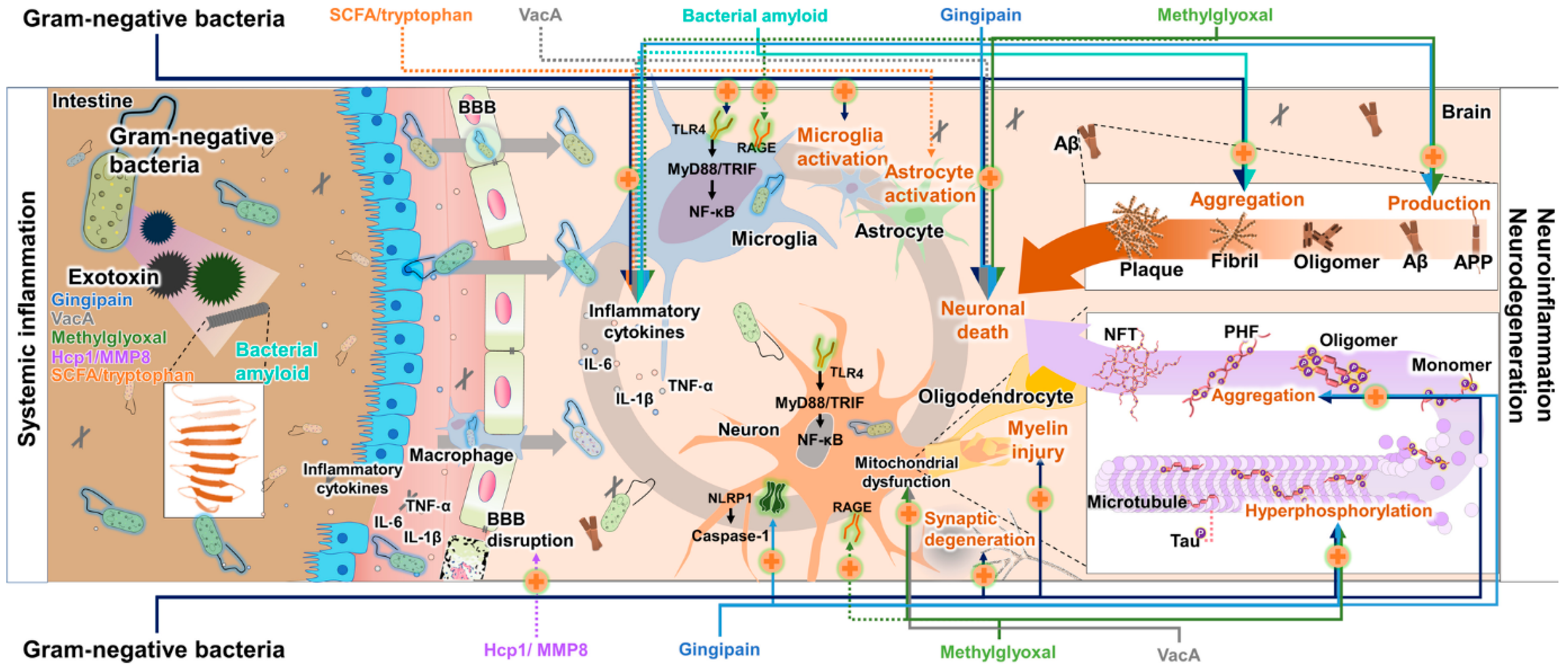
3.1. Neuroinflammation: Contribution of Microbial Toxins to Neuroinflammatory Processes

3.2. Oxidative Stress: Impact of Toxins on Oxidative Stress Pathways in Neurodegeneration
3.3. Toxin-Induced Protein Misfolding and Aggregation in Neurodegeneration
3.4. Protein Aggregation
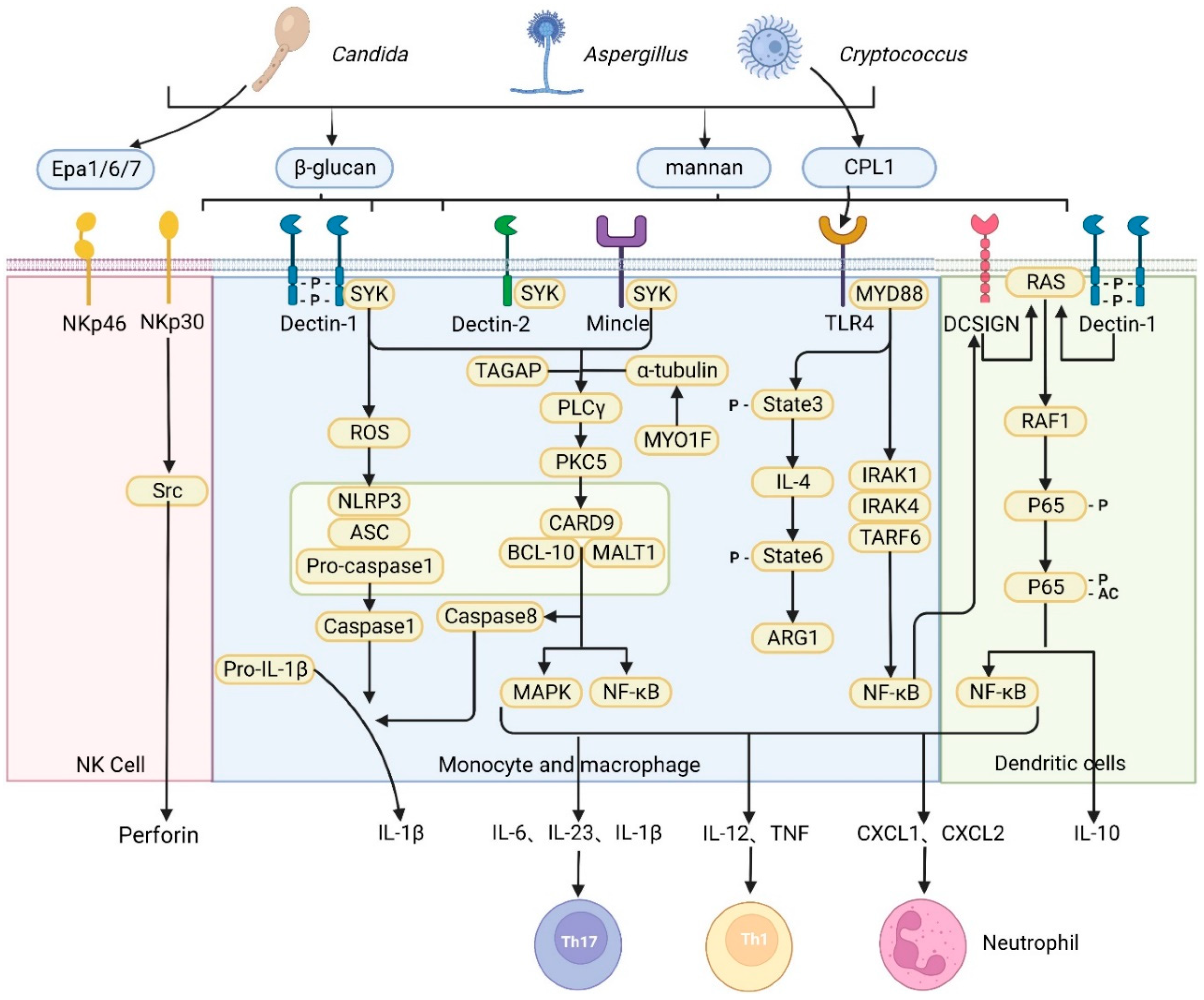
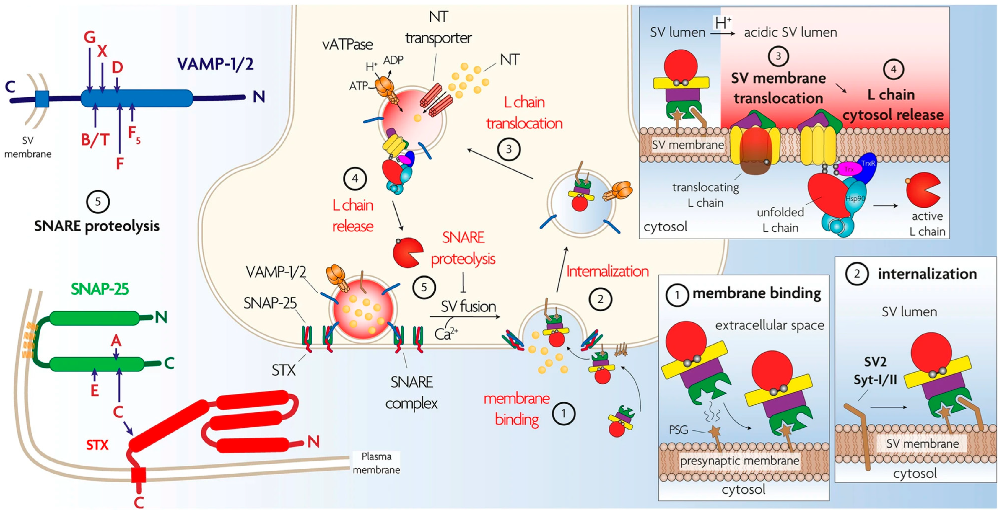
3.5. Direct Neurotoxicity
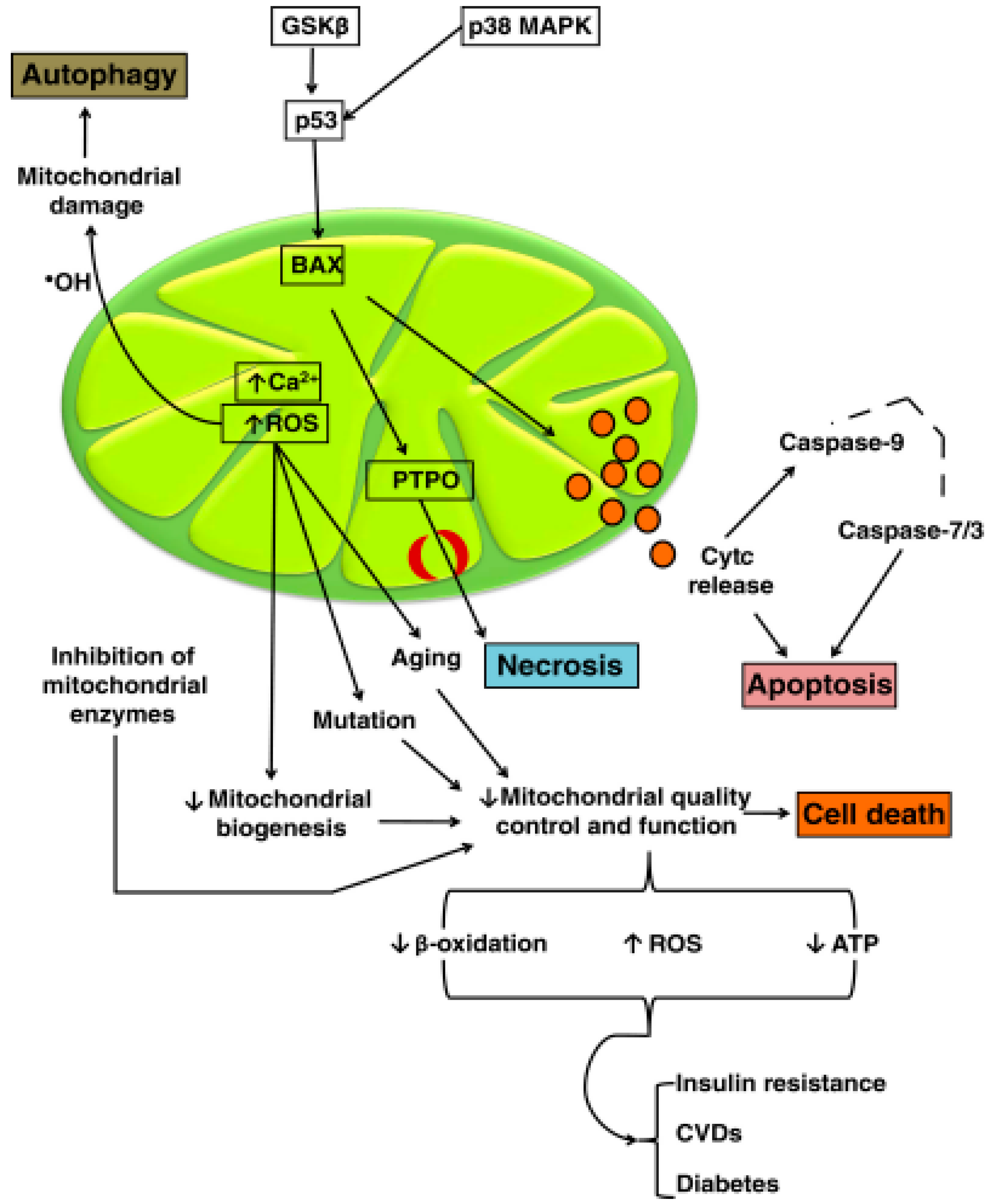
3.6. Mitochondrial Dysfunction and Apoptosis
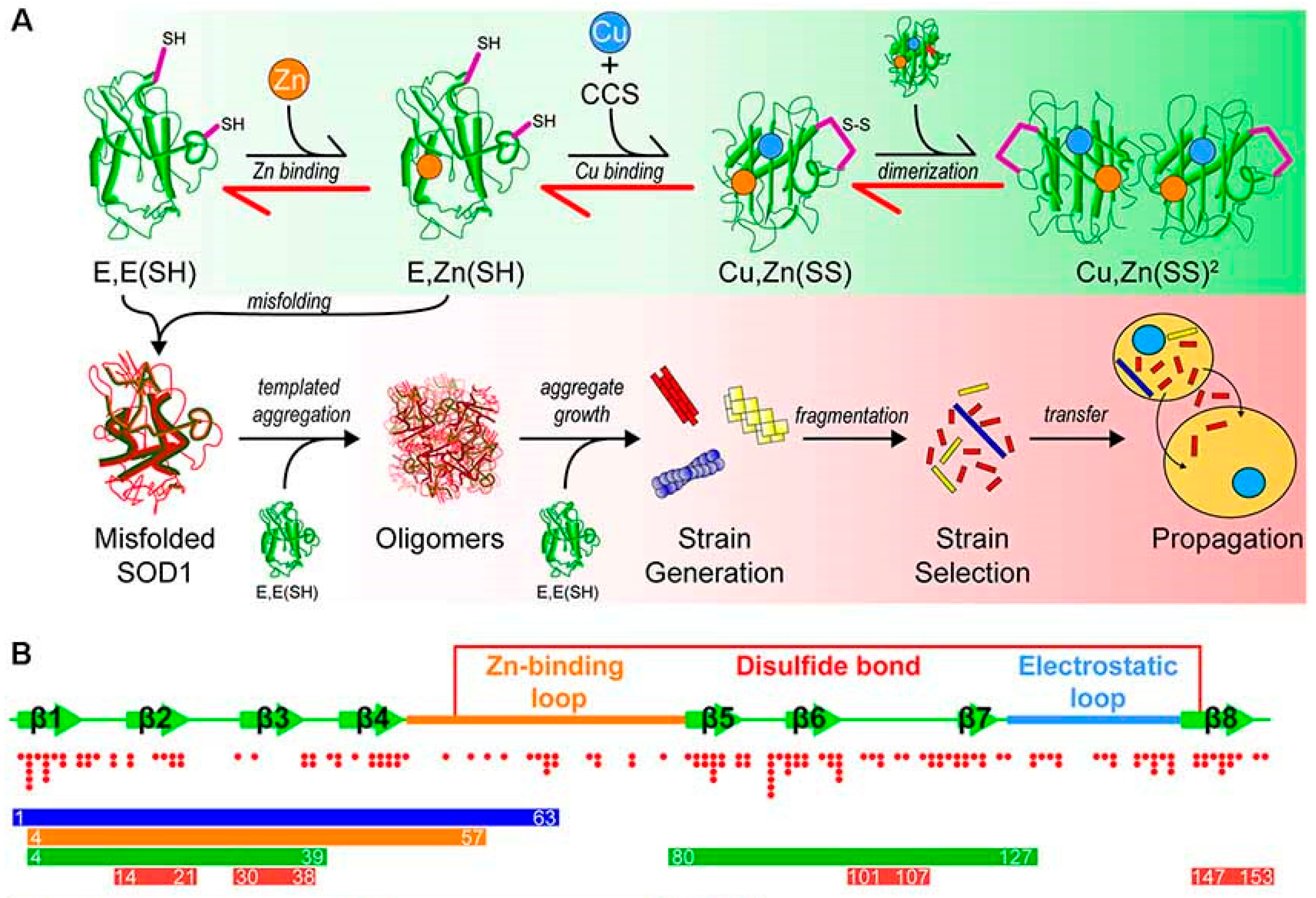
3.7. Prion-like Behavior
4. Conclusions and Future Perspectives
Author Contributions
Funding
Institutional Review Board Statement
Informed Consent Statement
Data Availability Statement
Conflicts of Interest
Abbreviations
| LPS | Lipopolysaccharide |
| BBB | blood–brain barrier |
| AD | Alzheimer’s disease |
| PD | Parkinson’s disease |
| HD | Huntington’s disease |
| ALS | amyotrophic lateral sclerosis |
| PRRs | express pattern recognition receptors |
| TLRs | toll-like receptors |
| ROS | reactive oxygen species |
| GI | gastrointestinal |
| α-Syn | Alpha-synuclein |
| ASD | Autism spectrum disorder |
| IL-10 | Interleukin-10 |
| IL-6 | interleukin-6 |
| IL-17 | interleukin-17 |
| IFN-γ | interferon-gamma |
| MS | Multiple sclerosis |
| LTA | lipoteichoic acid |
| TLR | toll-like receptor |
| 6-OHDA | 6-hydroxy-dopamine |
| MDMA | 4-methylenedioxymethamphetamine |
| DA | dopamine |
| CNS | central nervous system |
| O2 | Superoxide |
| H2O2 | hydrogen peroxide |
| NO | nitric oxide |
| PN | peroxynitrite |
| NADPH | Nicotinamide Adenine Dinucleotide Phosphate Hydrogen |
| APP | amyloid precursor protein |
| TLR2 | Toll-like receptor 2 |
| TLR1 | Toll-like receptor 1 |
| CD14 | cluster of differentiation 14 |
| MG | methylglyoxal |
| FB1 | fumonisin B1 |
| CSF | cerebrospinal fluid |
| PCR | polymerase chain reaction |
| TDP-43 | transactive response DNA binding protein 43 |
| Cu/Zn-SOD1 | Copper/Zinc Superoxide Dismutase 1 |
| OTA | Ochratoxin A |
| 3-NP | 3-Nitropropionic |
| PRRs | Pattern recognition receptors |
| PAMPs | pathogen-associated molecular patterns |
| CARD9 | Caspase Recruitment Domain-containing protein 9 |
| CLRs | C-type lectin receptors |
| SYK | Spleen Tyrosine Kinase |
| DC-SIGN | Dendritic Cell-Specific Intercellular adhesion molecule-3-Grabbing Non-integrin |
| NKp30 | Natural Killer Cell p30-Related Protein |
| NKp46 | Natural Killer Cell p46-Related Protein |
| BoNTs | botulinum neurotoxins |
| NMJ | neuromuscular junction |
| Ach | acetylcholine |
| PNS | peripheral nervous systems |
| CGRP | Calcitonin Gene-Related Peptide |
| SNARE | Soluble NSF Attachment Protein Receptor |
| TeNT | tetanus neurotoxins |
| ETC | electron transport chain |
| ATP | adenosine-5′-triphosphate |
| ACTH | Adrenocorticotropic Hormone |
| Aox1 | alternative oxidase |
| BAX | Bcl-2-associated X protein |
| CVDs | cardiovascular diseases |
| GSK3β | glycogen synthase kinase-3 beta |
| MAPK | mitogen-activated protein kinase |
| PTPO | permeability transition pore |
| TSEs | transmissible spongiform encephalopathies |
| PrPC | cellular prion protein |
| PrPSc | scrapie prion protein |
| GPI | glycosylphosphatidylinositol |
| eRF3 | Eukaryotic Polypeptide Chain Release Factor 3 |
| SOD1 | superoxide dismutase 1 |
References
- Zahra, W.; Rai, S.N.; Birla, H.; Singh, S.S.; Dilnashin, H.; Rathore, A.S.; Singh, S.P. The global economic impact of neurodegenerative diseases: Opportunities and challenges. In Bioeconomy for Sustainable Development; Springer Nature: Singapore, 2020; pp. 333–345. [Google Scholar]
- Pasko, V.I.; Churkina, A.S.; Shakhov, A.S.; Kotlobay, A.A.; Alieva, I.B. Modeling of neurodegenerative diseases: ‘step by step’ and ‘network’ organization of the complexes of model systems. Int. J. Mol. Sci. 2022, 24, 604. [Google Scholar] [CrossRef] [Scilit] [PubMed]
- Seo, D.-o.; Holtzman, D.M. Current understanding of the Alzheimer’s disease-associated microbiome and therapeutic strategies. Exp. Mol. Med. 2024, 56, 86–94. [Google Scholar] [CrossRef] [Scilit]
- Wang, Y.; Du, W.; Hu, X.; Yu, X.; Guo, C.; Jin, X.; Wang, W. Targeting the blood–brain barrier to delay aging-accompanied neurological diseases by modulating gut microbiota, circadian rhythms, and their interplays. Acta Pharm. Sin. B 2023, 13, 4667–4687. [Google Scholar] [CrossRef] [Scilit]
- Martin, C.R.; Osadchiy, V.; Kalani, A.; Mayer, E.A. The brain-gut-microbiome axis. Cell. Mol. Gastroenterol. Hepatol. 2018, 6, 133–148. [Google Scholar] [CrossRef] [Scilit] [PubMed]
- Navale, V.; Vamkudoth, K.R.; Ajmera, S.; Dhuri, V. Aspergillus derived mycotoxins in food and the environment: Prevalence, detection, and toxicity. Toxicol. Rep. 2021, 8, 1008–1030. [Google Scholar] [CrossRef] [Scilit]
- Popoff, M.R.; Poulain, B. Bacterial toxins and the nervous system: Neurotoxins and multipotential toxins interacting with neuronal cells. Toxins 2010, 2, 683–737. [Google Scholar] [CrossRef] [Scilit]
- Lamptey, R.N.; Chaulagain, B.; Trivedi, R.; Gothwal, A.; Layek, B.; Singh, J. A review of the common neurodegenerative disorders: Current therapeutic approaches and the potential role of nanotherapeutics. Int. J. Mol. Sci. 2022, 23, 1851. [Google Scholar] [CrossRef] [Scilit]
- Buchman, A.S.; Bennett, D.A. Loss of motor function in preclinical Alzheimer’s disease. Expert Rev. Neurother. 2011, 11, 665–676. [Google Scholar] [CrossRef] [Scilit] [PubMed]
- Rajmohan, R.; Reddy, P.H. Amyloid-beta and phosphorylated tau accumulations cause abnormalities at synapses of Alzheimer’s disease neurons. J. Alzheimer’s Dis. 2017, 57, 975–999. [Google Scholar] [CrossRef] [Scilit]
- Sharma, A.; Nair, A.; Dedhia, D. Parkinson’s disease: Epidemiology, pathophysiology, diagnosis, and treatment. In Proteostasis: Investigating Molecular Dynamics in Neurodegenerative Disorders; Springer: Berlin/Heidelberg, Germany, 2025; pp. 73–91. [Google Scholar]
- Tiryaki, E.; Horak, H.A. ALS and other motor neuron diseases. Contin. Lifelong Learn. Neurol. 2014, 20, 1185–1207. [Google Scholar] [CrossRef] [Scilit]
- Houldsworth, A. Role of oxidative stress in neurodegenerative disorders: A review of reactive oxygen species and prevention by antioxidants. Brain Commun. 2024, 6, fcad356. [Google Scholar] [CrossRef] [Scilit]
- Chen, Y.; Wu, X.; Xu, C.; Huang, J.; Zhang, L.; Qiu, P.; Zheng, D.; Chen, W.; Zhang, S. Pathogen virulence genes: Advances, challenges and future directions in infectious disease research. Int. J. Mol. Med. 2025, 56, 173. [Google Scholar] [CrossRef] [Scilit]
- Huangfu, B.; Chen, Y.; Xu, T.; Huang, K.; Liang, Z.; He, X. Mycotoxins and non-communicable diseases: Exacerbation and susceptibility. Crit. Rev. Food Sci. Nutr. 2025, 22, 1–17. [Google Scholar] [CrossRef] [Scilit]
- Poulain, B.; Molgó, J.; Popoff, M.R. Clostridial neurotoxins: From the cellular and molecular mode of action to their therapeutic use. In The Comprehensive Sourcebook of Bacterial Protein Toxins; Elsevier: Amsterdam, The Netherlands, 2015; pp. 287–336. [Google Scholar]
- Mello, F.D.; Braidy, N.; Marçal, H.; Guillemin, G.; Nabavi, S.M.; Neilan, B.A. Mechanisms and effects posed by neurotoxic products of cyanobacteria/microbial eukaryotes/dinoflagellates in algae blooms: A review. Neurotox. Res. 2018, 33, 153–167. [Google Scholar] [CrossRef] [Scilit]
- Kalyan, M.; Tousif, A.H.; Sonali, S.; Vichitra, C.; Sunanda, T.; Praveenraj, S.S.; Ray, B.; Gorantla, V.R.; Rungratanawanich, W.; Mahalakshmi, A.M. Role of endogenous lipopolysaccharides in neurological disorders. Cells 2022, 11, 4038. [Google Scholar] [CrossRef] [Scilit] [PubMed]
- Yan, J.; Fu, Q.; Cheng, L.; Zhai, M.; Wu, W.; Huang, L.; Du, G. Inflammatory response in Parkinson’s disease. Mol. Med. Rep. 2014, 10, 2223–2233. [Google Scholar] [CrossRef] [Scilit] [PubMed]
- Di Gregorio, S.E.; Duennwald, M.L. Yeast as a model to study protein misfolding in aged cells. FEMS Yeast Res. 2018, 18, foy054. [Google Scholar] [CrossRef] [Scilit] [PubMed]
- Tulisiak, C.T.; Mercado, G.; Peelaerts, W.; Brundin, L.; Brundin, P. Can infections trigger alpha-synucleinopathies? Prog. Mol. Biol. Transl. Sci. 2019, 168, 299–322. [Google Scholar] [PubMed]
- Lehnardt, S. Innate immunity and neuroinflammation in the CNS: The role of microglia in Toll-like receptor-mediated neuronal injury. Glia 2010, 58, 253–263. [Google Scholar] [CrossRef] [Scilit]
- Fischer, R.; Maier, O. Interrelation of oxidative stress and inflammation in neurodegenerative disease: Role of TNF. Oxidative Med. Cell. Longev. 2015, 2015, 610813. [Google Scholar] [CrossRef] [Scilit]
- Yang, N.J.; Chiu, I.M. Bacterial signaling to the nervous system through toxins and metabolites. J. Mol. Biol. 2017, 429, 587–605. [Google Scholar] [CrossRef] [Scilit]
- Dai, C.; Xiao, X.; Sun, F.; Zhang, Y.; Hoyer, D.; Shen, J.; Tang, S.; Velkov, T. T-2 toxin neurotoxicity: Role of oxidative stress and mitochondrial dysfunction. Arch. Toxicol. 2019, 93, 3041–3056. [Google Scholar] [CrossRef] [Scilit] [PubMed]
- Shahnawaz, M.; Park, K.-W.; Mukherjee, A.; Diaz-Espinoza, R.; Soto, C. Prion-like characteristics of the bacterial protein Microcin E492. Sci. Rep. 2017, 7, 45720. [Google Scholar] [CrossRef] [Scilit] [PubMed]
- Fang, P.; Kazmi, S.; Jameson, K.; Hsiao, E. The microbiome as a modifier of neurodegenerative disease risk. Cell Host Microbe 2020, 28, 201–222. [Google Scholar] [CrossRef] [Scilit] [PubMed]
- Niazi, S.K. Non-invasive drug delivery across the blood–brain barrier: A prospective analysis. Pharmaceutics 2023, 15, 2599. [Google Scholar] [CrossRef] [Scilit]
- Rinninella, E.; Raoul, P.; Cintoni, M.; Franceschi, F.; Miggiano, G.A.D.; Gasbarrini, A.; Mele, M.C. What is the healthy gut microbiota composition? A changing ecosystem across age, environment, diet, and diseases. Microorganisms 2019, 7, 14. [Google Scholar] [CrossRef] [Scilit]
- Alvarenga, L.; Kemp, J.A.; Baptista, B.G.; Ribeiro, M.; Lima, L.S.; Mafra, D. Production of Toxins by the Gut Microbiota: The Role of Dietary Protein. Curr. Nutr. Rep. 2024, 13, 340–350. [Google Scholar] [CrossRef] [Scilit]
- Forsyth, C.B.; Shannon, K.M.; Kordower, J.H.; Voigt, R.M.; Shaikh, M.; Jaglin, J.A.; Estes, J.D.; Dodiya, H.B.; Keshavarzian, A. Increased intestinal permeability correlates with sigmoid mucosa alpha-synuclein staining and endotoxin exposure markers in early Parkinson’s disease. PLoS ONE 2011, 6, e28032. [Google Scholar] [CrossRef] [Scilit]
- Chen, Y.-Y.; Chen, D.-Q.; Chen, L.; Liu, J.-R.; Vaziri, N.D.; Guo, Y.; Zhao, Y.-Y. Microbiome–metabolome reveals the contribution of gut–kidney axis on kidney disease. J. Transl. Med. 2019, 17, 5. [Google Scholar] [CrossRef] [Scilit]
- Nedungadi, P.; Shah, S.M.; Stokes, M.A.; Kumar Nair, V.; Moorkoth, A.; Raman, R. Mapping autism’s research landscape: Trends in autism screening and its alignment with sustainable development goals. Front. Psychiatry 2024, 14, 1294254. [Google Scholar] [CrossRef] [Scilit]
- Mair, R.D.; Sirich, T.L.; Plummer, N.S.; Meyer, T.W. Characteristics of colon-derived uremic solutes. Clin. J. Am. Soc. Nephrol. 2018, 13, 1398–1404. [Google Scholar] [CrossRef] [Scilit]
- Kikuchi, M.; Ueno, M.; Itoh, Y.; Suda, W.; Hattori, M. Uremic toxin-producing gut microbiota in rats with chronic kidney disease. Nephron 2017, 135, 51–60. [Google Scholar] [CrossRef] [Scilit] [PubMed]
- Ramezani, A.; Massy, Z.A.; Meijers, B.; Evenepoel, P.; Vanholder, R.; Raj, D.S. Role of the gut microbiome in uremia: A potential therapeutic target. Am. J. Kidney Dis. 2016, 67, 483–498. [Google Scholar] [CrossRef] [Scilit]
- Gryp, T.; De Paepe, K.; Vanholder, R.; Kerckhof, F.-M.; Van Biesen, W.; Van de Wiele, T.; Verbeke, F.; Speeckaert, M.; Joossens, M.; Couttenye, M.M. Gut microbiota generation of protein-bound uremic toxins and related metabolites is not altered at different stages of chronic kidney disease. Kidney Int. 2020, 97, 1230–1242. [Google Scholar] [CrossRef] [Scilit]
- Popkov, V.A.; Zharikova, A.A.; Demchenko, E.A.; Andrianova, N.V.; Zorov, D.B.; Plotnikov, E.Y. Gut microbiota as a source of uremic toxins. Int. J. Mol. Sci. 2022, 23, 483. [Google Scholar] [CrossRef] [Scilit]
- Hida, M.; Aiba, Y.; Sawamura, S.; Suzuki, N.; Satoh, T.; Koga, Y. Inhibition of the accumulation of uremic toxins in the blood and their precursors in the feces after oral administration of Lebenin®, a lactic acid bacteria preparation, to uremic patients undergoing hemodialysis. Nephron 1996, 74, 349–355. [Google Scholar] [CrossRef] [Scilit]
- Wong, J.; Piceno, Y.M.; DeSantis, T.Z.; Pahl, M.; Andersen, G.L.; Vaziri, N.D. Expansion of urease-and uricase-containing, indole-and p-cresol-forming and contraction of short-chain fatty acid-producing intestinal microbiota in ESRD. Am. J. Nephrol. 2014, 39, 230–237. [Google Scholar] [CrossRef] [Scilit]
- Hatch, M.; Freel, R.W.; Vaziri, N. Intestinal excretion of oxalate in chronic renal failure. J. Am. Soc. Nephrol. 1994, 5, 1339–1343. [Google Scholar] [CrossRef] [Scilit] [PubMed]
- Dunn, S.R.; Gabuzda, G.M.; Superdock, K.R.; Kolecki, R.S.; Schaedler, R.W.; Simenhoff, M.L. Induction of creatininase activity in chronic renal failure: Timing of creatinine degradation and effect of antibiotics. Am. J. Kidney Dis. 1997, 29, 72–77. [Google Scholar] [CrossRef] [Scilit] [PubMed]
- Miller, A.W.; Dearing, D. The metabolic and ecological interactions of oxalate-degrading bacteria in the mammalian gut. Pathogens 2013, 2, 636–652. [Google Scholar] [CrossRef] [Scilit]
- Park, J.; Kim, C.H. Regulation of common neurological disorders by gut microbial metabolites. Exp. Mol. Med. 2021, 53, 1821–1833. [Google Scholar] [CrossRef] [Scilit] [PubMed]
- Wasilewska, J.; Klukowski, M. Gastrointestinal symptoms and autism spectrum disorder: Links and risks–a possible new overlap syndrome. Pediatr. Health Med. Ther. 2015, 6, 153–166. [Google Scholar] [CrossRef] [Scilit]
- Teskey, G.; Anagnostou, E.; Mankad, D.; Smile, S.; Roberts, W.; Brian, J.; Bowdish, D.M.; Foster, J.A. Intestinal permeability correlates with behavioural severity in very young children with ASD: A preliminary study. J. Neuroimmunol. 2021, 357, 577607. [Google Scholar] [CrossRef] [Scilit]
- Hsiao, E.Y.; McBride, S.W.; Hsien, S.; Sharon, G.; Hyde, E.R.; McCue, T.; Codelli, J.A.; Chow, J.; Reisman, S.E.; Petrosino, J.F. Microbiota modulate behavioral and physiological abnormalities associated with neurodevelopmental disorders. Cell 2013, 155, 1451–1463. [Google Scholar] [CrossRef] [Scilit]
- Oyovwi, M.O.; Ajayi, A.F. A comprehensive review on immunological mechanisms and gut-brain pathways linking gut health and neurological disorders. Discov. Med. 2025, 2, 245. [Google Scholar] [CrossRef] [Scilit]
- Sharon, G.; Cruz, N.J.; Kang, D.-W.; Gandal, M.J.; Wang, B.; Kim, Y.-M.; Zink, E.M.; Casey, C.P.; Taylor, B.C.; Lane, C.J. Human gut microbiota from autism spectrum disorder promote behavioral symptoms in mice. Cell 2019, 177, 1600–1618.e17. [Google Scholar] [CrossRef] [Scilit] [PubMed]
- Krajmalnik-Brown, R.; Lozupone, C.; Kang, D.-W.; Adams, J.B. Gut bacteria in children with autism spectrum disorders: Challenges and promise of studying how a complex community influences a complex disease. Microb. Ecol. Health Dis. 2015, 26, 26914. [Google Scholar] [CrossRef] [Scilit]
- Xu, M.; Xu, X.; Li, J.; Li, F. Association between gut microbiota and autism spectrum disorder: A systematic review and meta-analysis. Front. Psychiatry 2019, 10, 473. [Google Scholar] [CrossRef] [Scilit] [PubMed]
- Parracho, H.M.; Bingham, M.O.; Gibson, G.R.; McCartney, A.L. Differences between the gut microflora of children with autistic spectrum disorders and that of healthy children. J. Med. Microbiol. 2005, 54, 987–991. [Google Scholar]
- de Araújo Boleti, A.P.; de Oliveira Cardoso, P.H.; Frihling, B.E.F.; de Moraes, L.F.R.N.; Nunes, E.A.C.; Mukoyama, L.T.H.; Biembengute, M.E.F.; de Melo, V.C.B.; Morales, M.F.; de Castro, A.P. Immune dysregulation and gut microbiota: Connection to health and disease development. Neural Regen. Res. 2026, 21, 3908–3918. [Google Scholar] [CrossRef] [Scilit]
- Wang, R.-x.; Afzal, A.; Jing, X.-y.; Zhou, Y.; Feng, J.-x.; Chen, Z.-x.; Cao, D.-z.; Liu, X.-a. Intergenerational effects of the microbiota on neurodevelopment: Mechanisms and therapeutic perspectives. Acta Pharmacol. Sin. 2026, 1–23. [Google Scholar] [CrossRef] [Scilit]
- Kang, D.-W.; Park, J.G.; Ilhan, Z.E.; Wallstrom, G.; LaBaer, J.; Adams, J.B.; Krajmalnik-Brown, R. Reduced incidence of Prevotella and other fermenters in intestinal microflora of autistic children. PLoS ONE 2013, 8, e68322. [Google Scholar] [CrossRef] [Scilit] [PubMed]
- Sandler, R.H.; Finegold, S.M.; Bolte, E.R.; Buchanan, C.P.; Maxwell, A.P.; Väisänen, M.-L.; Nelson, M.N.; Wexler, H.M. Short-term benefit from oral vancomycin treatment of regressive-onset autism. J. Child. Neurol. 2000, 15, 429–435. [Google Scholar] [CrossRef] [Scilit]
- Kiu, R.; Hall, L.J. An update on the human and animal enteric pathogen Clostridium perfringens. Emerg. Microbes Infect. 2018, 7, 1–15. [Google Scholar] [CrossRef] [Scilit]
- Nagahama, M.; Sakurai, J. Distribution of labeled Clostridium perfringens epsilon toxin in mice. Toxicon 1991, 29, 211–217. [Google Scholar] [CrossRef] [Scilit]
- Mirabbasi, S.; Ghane, M. Cytotoxic effects of epsilon toxin from Clostridium perfringens on colon cancer cell lines (HT-29) and (Caco2) in terms of oxidative stress and inflammatory cytokines. Toxicol. Res. 2025, 14, tfaf083. [Google Scholar] [CrossRef] [Scilit]
- Agata, N.; Ohta, M.; Mori, M.; Isobe, M. A novel dodecadepsipeptide, cereulide, is an emetic toxin of Bacillus cereus. FEMS Microbiol. Lett. 1995, 129, 17–19. [Google Scholar] [CrossRef] [Scilit]
- Sugiyama, H.; Hayama, T. Abdominal viscera as site of emetic action for staphylococcal enterotoxin in the monkey. J. Infect. Dis. 1965, 115, 330–336. [Google Scholar] [CrossRef] [Scilit] [PubMed]
- Hu, D.L.; Zhu, G.; Mori, F.; Omoe, K.; Okada, M.; Wakabayashi, K.; Kaneko, S.; Shinagawa, K.; Nakane, A. Staphylococcal enterotoxin induces emesis through increasing serotonin release in intestine and it is downregulated by cannabinoid receptor 1. Cell. Microbiol. 2007, 9, 2267–2277. [Google Scholar] [CrossRef] [Scilit]
- Friedland, R.P.; Chapman, M.R. The role of microbial amyloid in neurodegeneration. PLoS Pathog. 2017, 13, e1006654. [Google Scholar] [CrossRef] [Scilit] [PubMed]
- Chapman, M.R.; Robinson, L.S.; Pinkner, J.S.; Roth, R.; Heuser, J.; Hammar, M.; Normark, S.; Hultgren, S.J. Role of Escherichia coli curli operons in directing amyloid fiber formation. Science 2002, 295, 851–855. [Google Scholar] [CrossRef] [Scilit]
- Collinson, S.; Emödy, L.; Müller, K.; Trust, T.; Kay, W. Purification and characterization of thin, aggregative fimbriae from Salmonella enteritidis. J. Bacteriol. 1991, 173, 4773–4781. [Google Scholar] [CrossRef] [Scilit]
- Chen, S.G.; Stribinskis, V.; Rane, M.J.; Demuth, D.R.; Gozal, E.; Roberts, A.M.; Jagadapillai, R.; Liu, R.; Choe, K.; Shivakumar, B. Exposure to the functional bacterial amyloid protein curli enhances alpha-synuclein aggregation in aged Fischer 344 rats and Caenorhabditis elegans. Sci. Rep. 2016, 6, 34477. [Google Scholar] [CrossRef] [Scilit]
- Needham, B.D.; Kaddurah-Daouk, R.; Mazmanian, S.K. Gut microbial molecules in behavioural and neurodegenerative conditions. Nat. Rev. Neurosci. 2020, 21, 717–731. [Google Scholar] [CrossRef] [Scilit] [PubMed]
- González-Sanmiguel, J.; Schuh, C.M.; Muñoz-Montesino, C.; Contreras-Kallens, P.; Aguayo, L.G.; Aguayo, S. Complex interaction between resident microbiota and misfolded proteins: Role in neuroinflammation and neurodegeneration. Cells 2020, 9, 2476. [Google Scholar] [CrossRef] [Scilit]
- Kim, H.s.; Kim, S.; Shin, S.J.; Park, Y.H.; Nam, Y.; Kim, C.w.; Lee, K.w.; Kim, S.-M.; Jung, I.D.; Yang, H.D. Gram-negative bacteria and their lipopolysaccharides in Alzheimer’s disease: Pathologic roles and therapeutic implications. Transl. Neurodegener. 2021, 10, 1–23. [Google Scholar] [CrossRef] [Scilit]
- Vojtechova, I.; Machacek, T.; Kristofikova, Z.; Stuchlik, A.; Petrasek, T. Infectious origin of Alzheimer’s disease: Amyloid beta as a component of brain antimicrobial immunity. PLoS Pathog. 2022, 18, e1010929. [Google Scholar] [CrossRef] [Scilit]
- Doi, K.; Uetsuka, K. Mechanisms of mycotoxin-induced neurotoxicity through oxidative stress-associated pathways. Int. J. Mol. Sci. 2011, 12, 5213–5237. [Google Scholar] [CrossRef] [Scilit]
- Nguyen, V.T.T.; König, S.; Eggert, S.; Endres, K.; Kins, S. The role of mycotoxins in neurodegenerative diseases: Current state of the art and future perspectives of research. Biol. Chem. 2022, 403, 3–26. [Google Scholar] [CrossRef] [Scilit] [PubMed]
- Aguzzi, A.; Calella, A.M. Prions: Protein aggregation and infectious diseases. Physiol. Rev. 2009, 89, 1105–1152. [Google Scholar] [CrossRef] [Scilit] [PubMed]
- Poulain, B.; Popoff, M.R.; Molgó, J. How do the botulinum neurotoxins block neurotransmitter release: From botulism to the molecular mechanism of action. Botulinum J. 2008, 1, 14–87. [Google Scholar] [CrossRef] [Scilit]
- Hassel, B. Tetanus: Pathophysiology, treatment, and the possibility of using botulinum toxin against tetanus-induced rigidity and spasms. Toxins 2013, 5, 73–83. [Google Scholar] [CrossRef] [Scilit]
- Marrodan, M.; Alessandro, L.; Farez, M.F.; Correale, J. The role of infections in multiple sclerosis. Mult. Scler. J. 2019, 25, 891–901. [Google Scholar] [CrossRef] [Scilit]
- Kielian, T. Immunopathogenesis of brain abscess. J. Neuroinflamm. 2004, 1, 16. [Google Scholar] [CrossRef] [Scilit]
- Tesh, V.L. Induction of apoptosis by Shiga toxins. Future Microbiol. 2010, 5, 431–453. [Google Scholar] [CrossRef] [Scilit]
- Lohmann, S. Neuroinvasion and Cerebral Ischemia as Possible Sources for α-Synuclein Prions in Parkinson’s Disease. Ph.D. Thesis, Universitäts-und Landesbibliothek Bonn, Bonn, Germany, 2021. [Google Scholar]
- Tran, V.T.A.; Lee, L.P.; Cho, H. Neuroinflammation in neurodegeneration via microbial infections. Front. Immunol. 2022, 13, 907804. [Google Scholar] [CrossRef] [Scilit] [PubMed]
- Xiong, J.; Kaleja, P.; Ückert, L.; Nezaratizadeh, N.; Krantz, S.; Krause, M.F.; Fitschen-Oestern, S.; Seekamp, A.; Cassidy, L.; Tholey, A. Alveolar–Capillary Barrier Protection In Vitro: Lung Cell Type-Specific Effects and Molecular Mechanisms Induced by 1α, 25-Dihydroxyvitamin D3. Int. J. Mol. Sci. 2023, 24, 7298. [Google Scholar] [CrossRef] [Scilit]
- Rangel, S.M.; Diaz, M.H.; Knoten, C.A.; Zhang, A.; Hauser, A.R. The role of ExoS in dissemination of Pseudomonas aeruginosa during pneumonia. PLoS Pathog. 2015, 11, e1004945. [Google Scholar] [CrossRef] [Scilit]
- Bouillot, S.; Munro, P.; Gallet, B.; Reboud, E.; Cretin, F.; Golovkine, G.; Schoehn, G.; Attrée, I.; Lemichez, E.; Huber, P. Pseudomonas aeruginosa Exolysin promotes bacterial growth in lungs, alveolar damage and bacterial dissemination. Sci. Rep. 2017, 7, 2120. [Google Scholar] [CrossRef] [Scilit] [PubMed]
- Chuang, Y.; Chang, W.; Lu, C.; Wu, H.; Chang, H. Pseudomonas aeruginosa central nervous system infections: Analysis of clinical features of 16 adult patients. Zhonghua Yi Xue Za Zhi = Chin. Med. J. Free China Ed. 1999, 62, 300–307. [Google Scholar]
- Hosang, L.; Canals, R.C.; van der Flier, F.J.; Hollensteiner, J.; Daniel, R.; Flügel, A.; Odoardi, F. The lung microbiome regulates brain autoimmunity. Nature 2022, 603, 138–144. [Google Scholar] [CrossRef] [Scilit]
- Braniste, V.; Al-Asmakh, M.; Kowal, C.; Anuar, F.; Abbaspour, A.; Tóth, M.; Korecka, A.; Bakocevic, N.; Ng, L.G.; Kundu, P. The gut microbiota influences blood-brain barrier permeability in mice. Sci. Transl. Med. 2014, 6, 263ra158. [Google Scholar] [CrossRef] [Scilit]
- Obrenovich, M.E. Leaky gut, leaky brain? Microorganisms 2018, 6, 107. [Google Scholar] [CrossRef] [Scilit]
- Valles-Colomer, M.; Falony, G.; Darzi, Y.; Tigchelaar, E.F.; Wang, J.; Tito, R.Y.; Schiweck, C.; Kurilshikov, A.; Joossens, M.; Wijmenga, C. The neuroactive potential of the human gut microbiota in quality of life and depression. Nat. Microbiol. 2019, 4, 623–632. [Google Scholar] [CrossRef] [Scilit]
- Barth, K.; Remick, D.G.; Genco, C.A. Disruption of immune regulation by microbial pathogens and resulting chronic inflammation. J. Cell. Physiol. 2013, 228, 1413–1422. [Google Scholar] [CrossRef] [Scilit]
- Guichard, A.; McGillivray, S.M.; Cruz-Moreno, B.; van Sorge, N.M.; Nizet, V.; Bier, E. Anthrax toxins cooperatively inhibit endocytic recycling by the Rab11/Sec15 exocyst. Nature 2010, 467, 854–858. [Google Scholar] [CrossRef] [Scilit]
- Gründler, T.; Quednau, N.; Stump, C.; Orian-Rousseau, V.; Ishikawa, H.; Wolburg, H.; Schroten, H.; Tenenbaum, T.; Schwerk, C. The surface proteins InlA and InlB are interdependently required for polar basolateral invasion by Listeria monocytogenes in a human model of the blood–cerebrospinal fluid barrier. Microbes Infect. 2013, 15, 291–301. [Google Scholar] [CrossRef] [Scilit]
- Wang, K.; Wang, H.; Lou, W.; Ma, L.; Li, Y.; Zhang, N.; Wang, C.; Li, F.; Awais, M.; Cao, S. IP-10 promotes blood–brain barrier damage by inducing tumor necrosis factor alpha production in Japanese encephalitis. Front. Immunol. 2018, 9, 1148. [Google Scholar] [CrossRef] [Scilit]
- Tsao, N.; Hsu, H.-P.; Wu, C.-M.; Liu, C.-C.; Lei, H.-Y. Tumour necrosis factor-α causes an increase in blood-brain barrier permeability during sepsis. J. Med. Microbiol. 2001, 50, 812–821. [Google Scholar] [CrossRef] [Scilit]
- Pron, B.; Taha, M.-K.; Rambaud, C.; Fournet, J.-C.; Pattey, N.; Monnet, J.-P.; Musilek, M.; Beretti, J.-L.; Nassif, X. Interaction of Neisseria meningitidis with the components of the blood-brain barrier correlates with an increased expression of PilC. J. Infect. Dis. 1997, 176, 1285–1292. [Google Scholar] [CrossRef] [Scilit]
- Coureuil, M.; Lécuyer, H.; Bourdoulous, S.; Nassif, X. A journey into the brain: Insight into how bacterial pathogens cross blood–brain barriers. Nat. Rev. Microbiol. 2017, 15, 149–159. [Google Scholar] [CrossRef] [Scilit]
- Tang, A.T.; Choi, J.P.; Kotzin, J.J.; Yang, Y.; Hong, C.C.; Hobson, N.; Girard, R.; Zeineddine, H.A.; Lightle, R.; Moore, T. Endothelial TLR4 and the microbiome drive cerebral cavernous malformations. Nature 2017, 545, 305–310. [Google Scholar] [CrossRef] [Scilit]
- Zhao, Y.; Cong, L.; Jaber, V.; Lukiw, W.J. Microbiome-derived lipopolysaccharide enriched in the perinuclear region of Alzheimer’s disease brain. Front. Immunol. 2017, 8, 1064. [Google Scholar] [CrossRef] [Scilit]
- Zhao, Y.; Jaber, V.; Lukiw, W.J. Secretory products of the human GI tract microbiome and their potential impact on Alzheimer’s disease (AD): Detection of lipopolysaccharide (LPS) in AD hippocampus. Front. Cell. Infect. Microbiol. 2017, 7, 318. [Google Scholar] [CrossRef] [Scilit]
- Poole, S.; Singhrao, S.K.; Chukkapalli, S.; Rivera, M.; Velsko, I.; Kesavalu, L.; Crean, S. Active invasion of Porphyromonas gingivalis and infection-induced complement activation in ApoE-/-mice brains. J. Alzheimer’s Dis. 2015, 43, 67–80. [Google Scholar] [CrossRef] [Scilit]
- Poole, S.; Singhrao, S.K.; Kesavalu, L.; Curtis, M.A.; Crean, S. Determining the presence of periodontopathic virulence factors in short-term postmortem Alzheimer’s disease brain tissue. In Handbook of Infection and Alzheimer’s Disease; IOS Press: Amsterdam, The Netherlands, 2017; pp. 105–117. [Google Scholar]
- Singhrao, S.K.; Chukkapalli, S.; Poole, S.; Velsko, I.; Crean, S.J.; Kesavalu, L. Chronic Porphyromonas gingivalis infection accelerates the occurrence of age-related granules in ApoE–/–mice brains. J. Oral. Microbiol. 2017, 9, 1270602. [Google Scholar] [CrossRef] [Scilit]
- Dominy, S.S.; Lynch, C.; Ermini, F.; Benedyk, M.; Marczyk, A.; Konradi, A.; Nguyen, M.; Haditsch, U.; Raha, D.; Griffin, C. Porphyromonas gingivalis in Alzheimer’s disease brains: Evidence for disease causation and treatment with small-molecule inhibitors. Sci. Adv. 2019, 5, eaau3333. [Google Scholar] [CrossRef] [Scilit]
- Stein, P.S.; Steffen, M.J.; Smith, C.; Jicha, G.; Ebersole, J.L.; Abner, E.; Dawson, D., III. Serum antibodies to periodontal pathogens are a risk factor for Alzheimer’s disease. Alzheimer’s Dement. 2012, 8, 196–203. [Google Scholar] [CrossRef] [Scilit]
- Noble, J.; Scarmeas, N.; Celenti, R.; Elkind, M.; Wright, C.B.; Schupf, N.; Papapanou, P.N. Serum IgG antibody levels to periodontal microbiota are associated with incident Alzheimer disease. PLoS ONE 2014, 9, e114959. [Google Scholar] [CrossRef] [Scilit]
- Ishihara, K.; Okuda, K. Molecular pathogenesis of the cell surface proteins and lipids from Treponema denticola. FEMS Microbiol. Lett. 1999, 181, 199–204. [Google Scholar] [CrossRef]
- Riviere, G.R.; Riviere, K.; Smith, K. Molecular and immunological evidence of oral Treponema in the human brain and their association with Alzheimer’s disease. Oral. Microbiol. Immunol. 2002, 17, 113–118. [Google Scholar] [CrossRef] [Scilit] [PubMed]
- Sharma, R.R. Fungal infections of the nervous system: Current perspective and controversies in management. Int. J. Surg. 2010, 8, 591–601. [Google Scholar] [CrossRef] [Scilit]
- Pisa, D.; Alonso, R.; Rábano, A.; Rodal, I.; Carrasco, L. Different brain regions are infected with fungi in Alzheimer’s disease. Sci. Rep. 2015, 5, 15015. [Google Scholar] [CrossRef] [Scilit]
- Pisa, D.; Alonso, R.; Carrasco, L. Parkinson’s disease: A comprehensive analysis of fungi and bacteria in brain tissue. Int. J. Biol. Sci. 2020, 16, 1135–1152. [Google Scholar] [CrossRef] [Scilit]
- Alonso, R.; Fernández-Fernández, A.M.; Pisa, D.; Carrasco, L. Multiple sclerosis and mixed microbial infections. Direct identification of fungi and bacteria in nervous tissue. Neurobiol. Dis. 2018, 117, 42–61. [Google Scholar] [CrossRef] [Scilit] [PubMed]
- Moreira, R.T.d.F.; Lallo, M.A.; Alvares-Saraiva, A.M.; Hurtado, E.C.P.; Konno, F.T.; Spadacci-Morena, D.; Coutinho, S.d.A. Dichotomous response of Malassezia-infected macrophages to Malassezia pachydermatis and Malassezia furfur. Med. Mycol. 2019, 57, 628–635. [Google Scholar] [CrossRef] [Scilit]
- Chang, Y.C.; Stins, M.F.; McCaffery, M.J.; Miller, G.F.; Pare, D.R.; Dam, T.; Paul-Satyasee, M.; Kim, K.S.; Kwon-Chung, K.J. Cryptococcal yeast cells invade the central nervous system via transcellular penetration of the blood-brain barrier. Infect. Immun. 2004, 72, 4985–4995. [Google Scholar] [CrossRef] [Scilit] [PubMed]
- Casadevall, A. Cryptococci at the brain gate: Break and enter or use a Trojan horse? J. Clin. Investig. 2010, 120, 1389–1392. [Google Scholar] [CrossRef] [Scilit]
- Kim, J.; Lee, K.-T.; Lee, J.S.; Shin, J.; Cui, B.; Yang, K.; Choi, Y.S.; Choi, N.; Lee, S.H.; Lee, J.-H. Fungal brain infection modelled in a human-neurovascular-unit-on-a-chip with a functional blood–brain barrier. Nat. Biomed. Eng. 2021, 5, 830–846. [Google Scholar] [CrossRef] [Scilit]
- Jong, A.; Wu, C.H.; Prasadarao, N.V.; Kwon-Chung, K.J.; Chang, Y.C.; Ouyang, Y.; Shackleford, G.M.; Huang, S.H. Invasion of Cryptococcus neoformans into human brain microvascular endothelial cells requires protein kinase C-α activation. Cell. Microbiol. 2008, 10, 1854–1865. [Google Scholar] [CrossRef] [Scilit]
- Shi, M.; Mody, C.H. Fungal infection in the brain: What we learned from intravital imaging. Front. Immunol. 2016, 7, 292. [Google Scholar] [CrossRef] [Scilit]
- Silva, D.F.; Empadinhas, N.; Cardoso, S.M.; Esteves, A.R. Neurodegenerative microbially-shaped diseases: Oxidative stress meets neuroinflammation. Antioxidants 2022, 11, 2141. [Google Scholar] [CrossRef] [Scilit]
- Shukla, V.; Mishra, S.K.; Pant, H.C. Oxidative stress in neurodegeneration. Adv. Pharmacol. Pharm. Sci. 2011, 2011, 572634. [Google Scholar] [CrossRef] [Scilit]
- Shandilya, S.; Kumar, S.; Jha, N.K.; Kesari, K.K.; Ruokolainen, J. Interplay of gut microbiota and oxidative stress: Perspective on neurodegeneration and neuroprotection. J. Adv. Res. 2022, 38, 223–244. [Google Scholar] [CrossRef] [Scilit] [PubMed]
- Nunomura, A.; Honda, K.; Takeda, A.; Hirai, K.; Zhu, X.; Smith, M.A.; Perry, G. Oxidative damage to RNA in neurodegenerative diseases. BioMed Res. Int. 2006, 2006, 082323. [Google Scholar] [CrossRef] [Scilit]
- Perry, G.; Nunomura, A.; Hirai, K.; Zhu, X.; Prez, M.; Avila, J.; Castellani, R.J.; Atwood, C.S.; Aliev, G.; Sayre, L.M. Is oxidative damage the fundamental pathogenic mechanism of Alzheimer’s and other neurodegenerative diseases? Free Radic. Biol. Med. 2002, 33, 1475–1479. [Google Scholar] [CrossRef] [Scilit] [PubMed]
- Napolitano, A.; Crescenzi, O.; Pezzella, A.; Prota, G. Generation of the neurotoxin 6-hydroxydopamine by peroxidase/H2O2 oxidation of dopamine. J. Med. Chem. 1995, 38, 917–922. [Google Scholar] [CrossRef] [Scilit]
- Imam, S.Z.; El-Yazal, J.; Newport, G.D.; Itzhak, Y.; Cadet, J.L.; Slikker, W., Jr.; Ali, S.F. Methamphetamine-induced dopaminergic neurotoxicity: Role of peroxynitrite and neuroprotective role of antioxidants and peroxynitrite decomposition catalysts. Ann. N. Y. Acad. Sci. 2001, 939, 366–380. [Google Scholar] [CrossRef] [Scilit]
- Cadet, J.L.; Brannock, C. Invited review free radicals and the pathobiology of brain dopamine systems. Neurochem. Int. 1998, 32, 117–131. [Google Scholar] [CrossRef] [Scilit] [PubMed]
- Cadet, J.L.; Ali, S.F.; Rothman, R.B.; Epstein, C.J. Neurotoxicity, drugs of abuse, and the CuZn-superoxide dismutase transgenic mice. Mol. Neurobiol. 1995, 11, 155–163. [Google Scholar] [CrossRef] [Scilit]
- Giovanni, A.; Liang, L.P.; Hastings, T.G.; Zigmond, M.J. Estimating hydroxyl radical content in rat brain using systemic and intraventricular salicylate: Impact of methamphetamine. J. Neurochem. 1995, 64, 1819–1825. [Google Scholar] [CrossRef] [Scilit]
- Gibb, J.; Johnson, M.; Hanson, G. Neurochemical basis of neurotoxicity. Neurotoxicology 1990, 11, 317–321. [Google Scholar] [PubMed]
- De Vito, M.J.; Wagner, G. Methamphetamine-induced neuronal damage: A possible role for free radicals. Neuropharmacology 1989, 28, 1145–1150. [Google Scholar] [CrossRef] [Scilit] [PubMed]
- Agorogiannis, E.; Agorogiannis, G.; Papadimitriou, A.; Hadjigeorgiou, G. Protein misfolding in neurodegenerative diseases. Neuropathol. Appl. Neurobiol. 2004, 30, 215–224. [Google Scholar] [CrossRef] [Scilit]
- Lansbury, P.T., Jr. Evolution of amyloid: What normal protein folding may tell us about fibrillogenesis and disease. Proc. Natl. Acad. Sci. USA 1999, 96, 3342–3344. [Google Scholar] [CrossRef] [Scilit]
- Lashuel, H.A.; Hartley, D.; Petre, B.M.; Walz, T.; Lansbury, P.T., Jr. Amyloid pores from pathogenic mutations. Nature 2002, 418, 291. [Google Scholar] [CrossRef] [Scilit] [PubMed]
- Sharon, R.; Bar-Joseph, I.; Frosch, M.P.; Walsh, D.M.; Hamilton, J.A.; Selkoe, D.J. The formation of highly soluble oligomers of α-synuclein is regulated by fatty acids and enhanced in Parkinson’s disease. Neuron 2003, 37, 583–595. [Google Scholar] [CrossRef] [Scilit]
- Conway, K.A.; Lee, S.-J.; Rochet, J.-C.; Ding, T.T.; Williamson, R.E.; Lansbury, P.T., Jr. Acceleration of oligomerization, not fibrillization, is a shared property of both α-synuclein mutations linked to early-onset Parkinson’s disease: Implications for pathogenesis and therapy. Proc. Natl. Acad. Sci. USA 2000, 97, 571–576. [Google Scholar] [CrossRef] [Scilit]
- Chinta, S.J.; Andersen, J.K. Nitrosylation and nitration of mitochondrial complex I in Parkinson’s disease. Free Radic. Res. 2011, 45, 53–58. [Google Scholar] [CrossRef] [Scilit]
- Block, M.; Hong, J.-S. Chronic microglial activation and progressive dopaminergic neurotoxicity. Biochem. Soc. Trans. 2007, 35, 1127–1132. [Google Scholar] [CrossRef] [Scilit]
- Huang, J.; Wang, N.; Huang, X.; Huang, S.; Xie, G.; Zhang, J.; Li, Z. Fe-Formononetin nanoenzymes promote traumatic brain injury repair by alleviating oxidative stress and regulating microglia polarization. Mater. Des. 2025, 260, 115015. [Google Scholar] [CrossRef] [Scilit]
- Sochocka, M.; Zwolinska, K.; Leszek, J. The infectious etiology of Alzheimer’s disease. Curr. Neuropharmacol. 2017, 15, 996–1009. [Google Scholar] [CrossRef] [Scilit] [PubMed]
- Pritchard, A.B.; Crean, S.; Olsen, I.; Singhrao, S.K. Periodontitis, microbiomes and their role in Alzheimer’s disease. Front. Aging Neurosci. 2017, 9, 336. [Google Scholar] [CrossRef] [Scilit]
- Giau, V.V.; Wu, S.Y.; Jamerlan, A.; An, S.S.A.; Kim, S.; Hulme, J. Gut microbiota and their neuroinflammatory implications in Alzheimer’s disease. Nutrients 2018, 10, 1765. [Google Scholar] [CrossRef] [Scilit]
- Emery, D.C.; Shoemark, D.K.; Batstone, T.E.; Waterfall, C.M.; Coghill, J.A.; Cerajewska, T.L.; Davies, M.; West, N.X.; Allen, S.J. 16S rRNA next generation sequencing analysis shows bacteria in Alzheimer’s post-mortem brain. Front. Aging Neurosci. 2017, 9, 195. [Google Scholar] [CrossRef] [Scilit]
- Jiang, C.; Li, G.; Huang, P.; Liu, Z.; Zhao, B. The gut microbiota and Alzheimer’s disease. J. Alzheimer’s Dis. 2017, 58, 1–15. [Google Scholar] [CrossRef] [Scilit]
- Zhan, X.; Stamova, B.; Jin, L.-W.; DeCarli, C.; Phinney, B.; Sharp, F.R. Gram-negative bacterial molecules associate with Alzheimer disease pathology. Neurology 2016, 87, 2324–2332. [Google Scholar] [CrossRef] [Scilit] [PubMed]
- Pretorius, E.; Bester, J.; Kell, D.B. A bacterial component to Alzheimer’s-type dementia seen via a systems biology approach that links iron dysregulation and inflammagen shedding to disease. J. Alzheimer’s Dis. 2016, 53, 1237–1256. [Google Scholar] [CrossRef] [Scilit]
- Fulop, T.; Witkowski, J.M.; Bourgade, K.; Khalil, A.; Zerif, E.; Larbi, A.; Hirokawa, K.; Pawelec, G.; Bocti, C.; Lacombe, G. Can an infection hypothesis explain the beta amyloid hypothesis of Alzheimer’s disease? Front. Aging Neurosci. 2018, 10, 224. [Google Scholar] [CrossRef] [Scilit] [PubMed]
- Brown, G.C. The endotoxin hypothesis of neurodegeneration. J. Neuroinflamm. 2019, 16, 180. [Google Scholar] [CrossRef] [Scilit]
- Hung, Y.-L.; Fang, S.-H.; Wang, S.-C.; Cheng, W.-C.; Liu, P.-L.; Su, C.-C.; Chen, C.-S.; Huang, M.-Y.; Hua, K.-F.; Shen, K.-H. Corylin protects LPS-induced sepsis and attenuates LPS-induced inflammatory response. Sci. Rep. 2017, 7, 46299. [Google Scholar] [CrossRef] [Scilit]
- Chou, C.-H.; Lee, J.-T.; Lin, C.-C.; Sung, Y.-F.; Lin, C.-C.; Muo, C.-H.; Yang, F.-C.; Wen, C.-P.; Wang, I.-K.; Kao, C.-H. Septicemia is associated with increased risk for dementia: A population-based longitudinal study. Oncotarget 2017, 8, 84300–84308. [Google Scholar] [CrossRef] [Scilit]
- Iwashyna, T.J.; Ely, E.W.; Smith, D.M.; Langa, K.M. Long-term cognitive impairment and functional disability among survivors of severe sepsis. JAMA 2010, 304, 1787–1794. [Google Scholar] [CrossRef] [Scilit]
- Zhang, R.; Miller, R.G.; Gascon, R.; Champion, S.; Katz, J.; Lancero, M.; Narvaez, A.; Honrada, R.; Ruvalcaba, D.; McGrath, M.S. Circulating endotoxin and systemic immune activation in sporadic amyotrophic lateral sclerosis (sALS). J. Neuroimmunol. 2009, 206, 121–124. [Google Scholar] [CrossRef] [Scilit] [PubMed]
- Lee, H.-J.; Lee, K.-E.; Kim, J.-K.; Kim, D.-H. Suppression of gut dysbiosis by Bifidobacterium longum alleviates cognitive decline in 5XFAD transgenic and aged mice. Sci. Rep. 2019, 9, 11814. [Google Scholar] [CrossRef] [Scilit]
- Zhan, X.; Stamova, B.; Sharp, F.R. Lipopolysaccharide associates with amyloid plaques, neurons and oligodendrocytes in Alzheimer’s disease brain: A review. Front. Aging Neurosci. 2018, 10, 42. [Google Scholar] [CrossRef] [Scilit]
- Schwartz, K.; Boles, B.R. Microbial amyloids–functions and interactions within the host. Curr. Opin. Microbiol. 2013, 16, 93–99. [Google Scholar] [CrossRef] [Scilit]
- Javed, I.; Zhang, Z.; Adamcik, J.; Andrikopoulos, N.; Li, Y.; Otzen, D.E.; Lin, S.; Mezzenga, R.; Davis, T.P.; Ding, F. Accelerated amyloid beta pathogenesis by bacterial amyloid FapC. Adv. Sci. 2020, 7, 2001299. [Google Scholar] [CrossRef] [Scilit]
- Shewmaker, F.; McGlinchey, R.P.; Thurber, K.R.; McPhie, P.; Dyda, F.; Tycko, R.; Wickner, R.B. The functional curli amyloid is not based on in-register parallel β-sheet structure. J. Biol. Chem. 2009, 284, 25065–25076. [Google Scholar] [CrossRef] [Scilit] [PubMed]
- Hill, J.M.; Lukiw, W.J. Microbial-generated amyloids and Alzheimer’s disease (AD). Front. Aging Neurosci. 2015, 7, 9. [Google Scholar] [CrossRef] [Scilit] [PubMed]
- Grando, K. Complex Autoimmune Diseases: Genetic Predisposition and Bacterial Amyloid Curli. Ph.D. Dissertation, Temple University, Philadelphia, PA, USA, 2025. [Google Scholar]
- Cherny, I.; Rockah, L.; Levy-Nissenbaum, O.; Gophna, U.; Ron, E.Z.; Gazit, E. The formation of Escherichia coli curli amyloid fibrils is mediated by prion-like peptide repeats. J. Mol. Biol. 2005, 352, 245–252. [Google Scholar] [CrossRef] [Scilit]
- Rapsinski, G.J.; Newman, T.N.; Oppong, G.O.; van Putten, J.P.; Tükel, Ç. CD14 protein acts as an adaptor molecule for the immune recognition of Salmonella curli fibers. J. Biol. Chem. 2013, 288, 14178–14188. [Google Scholar] [CrossRef] [Scilit]
- Liu, S.; Gao, J.; Zhu, M.; Liu, K.; Zhang, H.-L. Gut microbiota and dysbiosis in Alzheimer’s disease: Implications for pathogenesis and treatment. Mol. Neurobiol. 2020, 57, 5026–5043. [Google Scholar] [CrossRef] [Scilit]
- Park, S.-C.; Moon, J.C.; Shin, S.Y.; Son, H.; Jung, Y.J.; Kim, N.-H.; Kim, Y.-M.; Jang, M.-K.; Lee, J.R. Functional characterization of alpha-synuclein protein with antimicrobial activity. Biochem. Biophys. Res. Commun. 2016, 478, 924–928. [Google Scholar] [CrossRef] [Scilit]
- Halfmann, R.; Alberti, S.; Lindquist, S. Prions, protein homeostasis, and phenotypic diversity. Trends Cell Biol. 2010, 20, 125–133. [Google Scholar] [CrossRef] [Scilit]
- Dent, S.E.; King, D.P.; Osterberg, V.R.; Adams, E.K.; Mackiewicz, M.R.; Weissman, T.A.; Unni, V.K. Phosphorylation of the aggregate-forming protein alpha-synuclein on serine-129 inhibits its DNA-bending properties. J. Biol. Chem. 2022, 298, 101552. [Google Scholar] [CrossRef] [Scilit]
- Lashuel, H.A. Do Lewy bodies contain alpha-synuclein fibrils? and Does it matter? A brief history and critical analysis of recent reports. Neurobiol. Dis. 2020, 141, 104876. [Google Scholar] [CrossRef] [Scilit]
- Tenreiro, S.; Reimao-Pinto, M.M.; Antas, P.; Rino, J.; Wawrzycka, D.; Macedo, D.; Rosado-Ramos, R.; Amen, T.; Waiss, M.; Magalhaes, F. Phosphorylation modulates clearance of alpha-synuclein inclusions in a yeast model of Parkinson’s disease. PLoS Genet. 2014, 10, e1004302. [Google Scholar] [CrossRef] [Scilit]
- Götz, M.E.; Double, K.; Gerlach, M.; Youdim, M.B.; Riederere, P. The relevance of iron in the pathogenesis of Parkinson’s disease. Ann. N. Y. Acad. Sci. 2004, 1012, 193–208. [Google Scholar] [CrossRef] [Scilit]
- Kaur, D.; Andersen, J. Does cellular iron dysregulation play a causative role in Parkinson’s disease? Ageing Res. Rev. 2004, 3, 327–343. [Google Scholar] [CrossRef] [Scilit]
- Talley, S.M.; Coley, P.D.; Kursar, T.A. The effects of weather on fungal abundance and richness among 25 communities in the Intermountain West. BMC Ecol. 2002, 2, 7. [Google Scholar] [CrossRef] [Scilit]
- Ismaiel, A.A.; Papenbrock, J. Mycotoxins: Producing fungi and mechanisms of phytotoxicity. Agriculture 2015, 5, 492–537. [Google Scholar] [CrossRef] [Scilit]
- Brand, B.; Stoye, N.M.; dos Santos Guilherme, M.; Nguyen, V.T.T.; Baumgaertner, J.C.; Schüffler, A.; Thines, E.; Endres, K. Identification of Patulin from Penicillium coprobium as a Toxin for Enteric Neurons. Molecules 2019, 24, 2776. [Google Scholar] [CrossRef] [Scilit]
- Osuchowski, M.F.; Sharma, R.P. Fumonisin B1 induces necrotic cell death in BV-2 cells and murine cultured astrocytes and is antiproliferative in BV-2 cells while N2A cells and primary cortical neurons are resistant. Neurotoxicology 2005, 26, 981–992. [Google Scholar] [CrossRef] [Scilit]
- Dalziel, J.E.; Finch, S.C.; Dunlop, J. The fungal neurotoxin lolitrem B inhibits the function of human large conductance calcium-activated potassium channels. Toxicol. Lett. 2005, 155, 421–426. [Google Scholar] [CrossRef] [Scilit] [PubMed]
- Han, N.; Luo, R.; Liu, J.; Guo, T.; Feng, J.; Peng, X. Transcriptomic and proteomic analysis reveals mechanisms of patulin-induced cell toxicity in human embryonic kidney cells. Toxins 2020, 12, 681. [Google Scholar] [CrossRef] [Scilit]
- Yoon, S.; Cong, W.-T.; Bang, Y.; Lee, S.N.; Yoon, C.S.; Kwack, S.J.; Kang, T.S.; Lee, K.Y.; Choi, J.-K.; Choi, H.J. Proteome response to ochratoxin A-induced apoptotic cell death in mouse hippocampal HT22 cells. Neurotoxicology 2009, 30, 666–676. [Google Scholar] [CrossRef] [Scilit]
- Berntsen, H.; Wigestrand, M.; Bogen, I.; Fonnum, F.; Walaas, S.; Moldes-Anaya, A. Mechanisms of penitrem-induced cerebellar granule neuron death in vitro: Possible involvement of GABAA receptors and oxidative processes. Neurotoxicology 2013, 35, 129–136. [Google Scholar] [CrossRef] [Scilit] [PubMed]
- Zhang, X.; Boesch-Saadatmandi, C.; Lou, Y.; Wolffram, S.; Huebbe, P.; Rimbach, G. Ochratoxin A induces apoptosis in neuronal cells. Genes Nutr. 2009, 4, 41–48. [Google Scholar] [CrossRef] [Scilit]
- Moldes-Anaya, A.S.; Fonnum, F.; Eriksen, G.S.; Rundberget, T.; Walaas, S.I.; Wigestrand, M.B. In vitro neuropharmacological evaluation of penitrem-induced tremorgenic syndromes: Importance of the GABAergic system. Neurochem. Int. 2011, 59, 1074–1081. [Google Scholar] [CrossRef] [Scilit] [PubMed]
- Purzycki, C.B.; Shain, D.H. Fungal toxins and multiple sclerosis: A compelling connection. Brain Res. Bull. 2010, 82, 4–6. [Google Scholar] [CrossRef] [Scilit]
- Merrill, A.; Wang, E.; Vales, T.; Smith, E.; Schroeder, J.; Menaldino, D.; Alexander, C.; Crane, H.; Xia, J.; Liotta, D. Fumonisin toxicity and sphingolipid biosynthesis. In Fumonisins Food; Springer: Boston, MA, USA, 1996; pp. 297–306. [Google Scholar]
- Giussani, P.; Prinetti, A.; Tringali, C. The role of Sphingolipids in myelination and myelin stability and their involvement in childhood and adult demyelinating disorders. J. Neurochem. 2021, 156, 403–414. [Google Scholar] [CrossRef] [Scilit]
- Ménard, A.; Amouri, R.; Dobránsky, T.; Charriaut-Marlangue, C.; Pierig, R.; Cifuentes-Diaz, C.; Ghandour, S.; Belliveau, J.; Gascan, H.; Hentati, F. A gliotoxic factor and multiple sclerosis. J. Neurol. Sci. 1998, 154, 209–221. [Google Scholar] [CrossRef] [Scilit]
- Rieger, F.; Amouri, R.; Benjelloun, N.; Cifuentes-Diaz, C.; Lyon-Caen, O.; Hantaz-Ambroise, D.; Dobransky, T.; Perron, H.; Gemy, C. Gliotoxic factor and multiple sclerosis. Comptes Rendus De L’academie Sci. Ser. III Sci. Vie 1996, 319, 343–350. [Google Scholar]
- Benjelloun, N.; Charriaut-Marlangue, C.; Hantaz-Ambroise, D.; Ménard, A.; Pierig, R.; Alliel, P.M.; Rieger, F. Induction of cell death in rat brain by a gliotoxic factor from cerebrospinal fluid in multiple sclerosis. Cell. Mol. Biol. 2002, 48, 205–212. [Google Scholar] [PubMed]
- Oskarsson, B.; Horton, D.K.; Mitsumoto, H. Potential environmental factors in amyotrophic lateral sclerosis. Neurol. Clin. 2015, 33, 877–888. [Google Scholar] [CrossRef] [Scilit] [PubMed]
- Alonso, R.; Pisa, D.; Marina, A.I.; Morato, E.; Rábano, A.; Rodal, I.; Carrasco, L. Evidence for fungal infection in cerebrospinal fluid and brain tissue from patients with amyotrophic lateral sclerosis. Int. J. Biol. Sci. 2015, 11, 546–558. [Google Scholar] [CrossRef] [Scilit]
- Castanedo-Vazquez, D.; Bosque-Varela, P.; Sainz-Pelayo, A.; Riancho, J. Infectious agents and amyotrophic lateral sclerosis: Another piece of the puzzle of motor neuron degeneration. J. Neurol. 2019, 266, 27–36. [Google Scholar] [CrossRef] [Scilit]
- Foran, E.; Trotti, D. Glutamate transporters and the excitotoxic path to motor neuron degeneration in amyotrophic lateral sclerosis. Antioxid. Redox Signal. 2009, 11, 1587–1602. [Google Scholar] [CrossRef] [Scilit]
- Scofield, M.; Korutla, L.; Jackson, T.; Kalivas, P.; Mackler, S. Nucleus Accumbens 1, a Pox virus and Zinc finger/Bric-a-brac Tramtrack Broad protein binds to TAR DNA-binding protein 43 and has a potential role in amyotrophic lateral sclerosis. Neuroscience 2012, 227, 44–54. [Google Scholar] [CrossRef] [Scilit]
- Roy, J.; Minotti, S.; Dong, L.; Figlewicz, D.A.; Durham, H.D. Glutamate potentiates the toxicity of mutant Cu/Zn-superoxide dismutase in motor neurons by postsynaptic calcium-dependent mechanisms. J. Neurosci. 1998, 18, 9673–9684. [Google Scholar] [CrossRef] [Scilit]
- French, P.W.; Ludowyke, R.I.; Guillemin, G.J. Fungal-contaminated grass and well water and sporadic amyotrophic lateral sclerosis. Neural Regen. Res. 2019, 14, 1490–1493. [Google Scholar] [CrossRef] [Scilit]
- Zilinskas, R.A. Iraq’s biological weapons: The past as future? JAMA 1997, 278, 418–424. [Google Scholar] [CrossRef] [Scilit] [PubMed]
- Haley, R.W. Gulf war syndrome: Narrowing the possibilities. Lancet Neurol. 2003, 2, 272–273. [Google Scholar] [CrossRef] [Scilit] [PubMed]
- Sava, V.; Reunova, O.; Velasquez, A.; Sanchez-Ramos, J. Can low level exposure to ochratoxin-A cause parkinsonism? J. Neurol. Sci. 2006, 249, 68–75. [Google Scholar] [CrossRef] [Scilit]
- Sava, V.; Velasquez, A.; Song, S.; Sanchez-Ramos, J. Adult hippocampal neural stem/progenitor cells in vitro are vulnerable to the mycotoxin ochratoxin-A. Toxicol. Sci. 2007, 98, 187–197. [Google Scholar] [CrossRef] [Scilit] [PubMed]
- Bhat, P.V.; Anand, T.; Manu, T.M.; Khanum, F. Restorative effect of l-Dopa treatment against Ochratoxin A induced neurotoxicity. Neurochem. Int. 2018, 118, 252–263. [Google Scholar] [CrossRef] [Scilit]
- Vigasova, D.; Nemergut, M.; Liskova, B.; Damborsky, J. Multi-pathogen infections and Alzheimer’s disease. Microb. Cell Fact. 2021, 20, 25. [Google Scholar] [CrossRef] [Scilit]
- Alonso, R.; Pisa, D.; Rábano, A.; Carrasco, L. Alzheimer’s disease and disseminated mycoses. Eur. J. Clin. Microbiol. Infect. Dis. 2014, 33, 1125–1132. [Google Scholar] [CrossRef] [Scilit]
- Alonso, R.; Pisa, D.; Fernández-Fernández, A.M.; Carrasco, L. Infection of fungi and bacteria in brain tissue from elderly persons and patients with Alzheimer’s disease. Front. Aging Neurosci. 2018, 10, 159. [Google Scholar] [CrossRef] [Scilit]
- Alonso, R.; Pisa, D.; Rábano, A.; Rodal, I.; Carrasco, L. Cerebrospinal fluid from Alzheimer’s disease patients contains fungal proteins and DNA. J. Alzheimer’s Dis. 2015, 47, 873–876. [Google Scholar] [CrossRef] [Scilit]
- Ferrante, R.J. Mouse models of Huntington’s disease and methodological considerations for therapeutic trials. Biochim. Biophys. Acta (BBA) -Mol. Basis Dis. 2009, 1792, 506–520. [Google Scholar] [CrossRef] [Scilit]
- Vaezi, A.; Fakhim, H.; Abtahian, Z.; Khodavaisy, S.; Geramishoar, M.; Alizadeh, A.; Meis, J.F.; Badali, H. Frequency and geographic distribution of CARD9 mutations in patients with severe fungal infections. Front. Microbiol. 2018, 9, 2434. [Google Scholar] [CrossRef] [Scilit]
- Speakman, E.A.; Dambuza, I.M.; Salazar, F.; Brown, G.D. T cell antifungal immunity and the role of C-type lectin receptors. Trends Immunol. 2020, 41, 61–76. [Google Scholar] [CrossRef] [Scilit]
- Drummond, R.A. Neuro-immune mechanisms of anti-cryptococcal protection. J. Fungi 2017, 4, 4. [Google Scholar] [CrossRef] [Scilit]
- Wu, C.; Jiang, M.-L.; Jiang, R.; Pang, T.; Zhang, C.-J. The roles of fungus in CNS autoimmune and neurodegeneration disorders. Front. Immunol. 2023, 13, 1077335. [Google Scholar] [CrossRef] [Scilit] [PubMed]
- Comella, C.L.; Pullman, S.L. Botulinum toxins in neurological disease. Muscle Nerve 2004, 29, 628–644. [Google Scholar] [CrossRef] [Scilit] [PubMed]
- Luvisetto, S. Botulinum neurotoxins in central nervous system: An overview from animal models to human therapy. Toxins 2021, 13, 751. [Google Scholar] [CrossRef] [Scilit] [PubMed]
- Poulain, B.; Lemichez, E.; Popoff, M.R. Neuronal selectivity of botulinum neurotoxins. Toxicon 2020, 178, 20–32. [Google Scholar] [CrossRef] [Scilit]
- Kumar, R.; Dhaliwal, H.P.; Kukreja, R.V.; Singh, B.R. The botulinum toxin as a therapeutic agent: Molecular structure and mechanism of action in motor and sensory systems. Semin. Neurol. 2016, 36, 010–019. [Google Scholar] [CrossRef] [Scilit]
- Lin, Y.-H.; Chiang, B.-J.; Liao, C.-H. Mechanism of action of botulinum toxin A in treatment of functional urological disorders. Toxins 2020, 12, 129. [Google Scholar] [CrossRef] [Scilit]
- Matak, I.; Bölcskei, K.; Bach-Rojecky, L.; Helyes, Z. Mechanisms of botulinum toxin type A action on pain. Toxins 2019, 11, 459. [Google Scholar] [CrossRef] [Scilit]
- Luvisetto, S.; Gazerani, P.; Cianchetti, C.; Pavone, F. Botulinum toxin type a as a therapeutic agent against headache and related disorders. Toxins 2015, 7, 3818–3844. [Google Scholar] [CrossRef] [Scilit]
- Ray, J.C.; Hutton, E.J.; Matharu, M. OnabotulinumtoxinA in migraine: A review of the literature and factors associated with efficacy. J. Clin. Med. 2021, 10, 2898. [Google Scholar] [CrossRef] [Scilit]
- Moore, C.; Hulsopple, C.; Boyce, B. Utilization of botulinum toxin for musculoskeletal disorders. Curr. Sports Med. Rep. 2020, 19, 217–222. [Google Scholar] [CrossRef] [Scilit] [PubMed]
- Egeo, G.; Fofi, L.; Barbanti, P. Botulinum neurotoxin for the treatment of neuropathic pain. Front. Neurol. 2020, 11, 716. [Google Scholar] [CrossRef] [Scilit] [PubMed]
- Ferrari, E.; Maywood, E.S.; Restani, L.; Caleo, M.; Pirazzini, M.; Rossetto, O.; Hastings, M.H.; Niranjan, D.; Schiavo, G.; Davletov, B. Re-assembled botulinum neurotoxin inhibits CNS functions without systemic toxicity. Toxins 2011, 3, 345–355. [Google Scholar] [CrossRef] [Scilit]
- Mangione, A.S.; Obara, I.; Maiarú, M.; Geranton, S.M.; Tassorelli, C.; Ferrari, E.; Leese, C.; Davletov, B.; Hunt, S.P. Nonparalytic botulinum molecules for the control of pain. Pain 2016, 157, 1045–1055. [Google Scholar] [CrossRef] [Scilit]
- Maiarù, M.; Leese, C.; Certo, M.; Echeverria-Altuna, I.; Mangione, A.S.; Arsenault, J.; Davletov, B.; Hunt, S.P. Selective neuronal silencing using synthetic botulinum molecules alleviates chronic pain in mice. Sci. Transl. Med. 2018, 10, eaar7384. [Google Scholar] [CrossRef] [Scilit] [PubMed]
- Tao, L.; Peng, L.; Berntsson, R.P.-A.; Liu, S.M.; Park, S.; Yu, F.; Boone, C.; Palan, S.; Beard, M.; Chabrier, P.-E. Engineered botulinum neurotoxin B with improved efficacy for targeting human receptors. Nat. Commun. 2017, 8, 53. [Google Scholar] [CrossRef] [Scilit]
- Webb, R.P. Engineering of botulinum neurotoxins for biomedical applications. Toxins 2018, 10, 231. [Google Scholar] [CrossRef] [Scilit]
- Fonfria, E.; Elliott, M.; Beard, M.; Chaddock, J.A.; Krupp, J. Engineering botulinum toxins to improve and expand targeting and SNARE cleavage activity. Toxins 2018, 10, 278. [Google Scholar] [CrossRef] [Scilit]
- Tang, M.; Meng, J.; Wang, J. New engineered-botulinum toxins inhibit the release of pain-related mediators. Int. J. Mol. Sci. 2019, 21, 262. [Google Scholar] [CrossRef] [Scilit]
- Elliott, M.; Favre-Guilmard, C.; Liu, S.M.; Maignel, J.; Masuyer, G.; Beard, M.; Boone, C.; Carré, D.; Kalinichev, M.; Lezmi, S. Engineered botulinum neurotoxin B with improved binding to human receptors has enhanced efficacy in preclinical models. Sci. Adv. 2019, 5, eaau7196. [Google Scholar] [CrossRef] [Scilit]
- Hao, F.; Feng, Y.; Guan, Y. A novel botulinum toxin TAT-EGFP-HCS fusion protein capable of specific delivery through the blood-brain barrier to the central nervous system. CNS Neurol. Disord.-Drug Targets 2019, 18, 37–43. [Google Scholar] [CrossRef] [Scilit]
- Rasetti-Escargueil, C.; Popoff, M.R. Engineering botulinum neurotoxins for enhanced therapeutic applications and vaccine development. Toxins 2020, 13, 1. [Google Scholar] [CrossRef] [Scilit]
- Sikorra, S.; Donald, S.; Elliott, M.; Schwede, S.; Coker, S.-F.; Kupinski, A.P.; Tripathi, V.; Foster, K.; Beard, M.; Binz, T. Engineering an effective human SNAP-23 cleaving botulinum neurotoxin A variant. Toxins 2020, 12, 804. [Google Scholar] [CrossRef] [Scilit]
- Johnson, E.A. Clostridium botulinum. In Food Microbiology: Fundamentals and Frontiers; Wiley: Hoboken, NJ, USA, 2012; pp. 441–463. [Google Scholar]
- Pirazzini, M.; Montecucco, C.; Rossetto, O. Toxicology and pharmacology of botulinum and tetanus neurotoxins: An update. Arch. Toxicol. 2022, 96, 1521–1539. [Google Scholar] [CrossRef] [Scilit]
- Magembe, K.S. Mycotoxins impact in food, human and animal health with special reference to aflatoxins, fumonisins, ochratoxins, zearalenone, and deoxynivalenol: A 13 year review (2010–2023). Eur. J. Res. Med. Sci. 2025, 10, 1–36. [Google Scholar]
- Islam, M.T.; Mishra, S.K.; Tripathi, S.; de Alencar, M.V.O.B.; e Sousa, J.M.d.C.; Rolim, H.M.L.; de Medeiros, M.d.G.F.; Ferreira, P.M.P.; Rouf, R.; Uddin, S.J. Mycotoxin-assisted mitochondrial dysfunction and cytotoxicity: Unexploited tools against proliferative disorders. IUBMB Life 2018, 70, 1084–1092. [Google Scholar] [CrossRef] [Scilit]
- Bao, Y.; Hu, C.; Wang, B.; Liu, X.; Wu, Q.; Xu, D.; Shi, Z.; Sun, C. Mitochondrial reverse electron transport: Mechanisms, pathophysiological roles, and therapeutic potential. Biology 2025, 14, 1140. [Google Scholar] [CrossRef] [Scilit]
- Swerdlow, R.H. Brain aging, Alzheimer’s disease, and mitochondria. Biochim. Biophys. Acta (BBA) -Mol. Basis Dis. 2011, 1812, 1630–1639. [Google Scholar] [CrossRef] [Scilit]
- Karbowski, M.; Neutzner, A. Neurodegeneration as a consequence of failed mitochondrial maintenance. Acta Neuropathol. 2012, 123, 157–171. [Google Scholar] [CrossRef] [Scilit]
- Rossignol, D.; Frye, R.E. Mitochondrial dysfunction in autism spectrum disorders: A systematic review and meta-analysis. Mol. Psychiatry 2012, 17, 290–314. [Google Scholar] [CrossRef] [Scilit]
- Kakde, U. Mycotoxins and its impact on human populations. MOJ Bioequiv. Availab. 2017, 3, 131–132. [Google Scholar] [CrossRef] [Scilit]
- Lorenz, S.; Mattson, P. Cinemicrographic observations of cultured adrenocortical tumor cells: Dynamic responses to ACTH and cytochalasin B. Virchows Arch. B 1986, 52, 221–236. [Google Scholar] [CrossRef] [Scilit]
- Aleo, M.D.; Wyatt, R.D.; Schnellmann, R.G. Mitochondrial dysfunction is an early event in ochratoxin A but not oosporein toxicity to rat renal proximal tubules. Toxicol. Appl. Pharmacol. 1991, 107, 73–80. [Google Scholar] [CrossRef] [Scilit]
- Chatopadhyay, P.; Tariang, B.; Agnihotri, A.; Veer, V. Synergism of ochratoxin B and calcium-channel antagonist verapamil caused mitochondrial dysfunction. Toxicol. Mech. Methods 2014, 24, 428–432. [Google Scholar] [CrossRef] [Scilit]
- Hao, J.; Wu, W.; Wang, Y.; Yang, Z.; Liu, Y.; Lv, Y.; Zhai, Y.; Yang, J.; Liang, Z.; Huang, K. Arabidopsis thaliana defense response to the ochratoxin A-producing strain (Aspergillus ochraceus 3.4412). Plant Cell Rep. 2015, 34, 705–719. [Google Scholar] [CrossRef] [Scilit]
- Wang, Y.; Peng, X.; Yang, Z.; Zhao, W.; Xu, W.; Hao, J.; Wu, W.; Shen, X.L.; Luo, Y.; Huang, K. iTRAQ mitoproteome analysis reveals mechanisms of programmed cell death in Arabidopsis thaliana induced by ochratoxin A. Toxins 2017, 9, 167. [Google Scholar] [CrossRef] [Scilit]
- Pardo, J.; Urban, C.; Galvez, E.M.; Ekert, P.G.; Müller, U.; Kwon-Chung, J.; Lobigs, M.; Müllbacher, A.; Wallich, R.; Borner, C. The mitochondrial protein Bak is pivotal for gliotoxin-induced apoptosis and a critical host factor of Aspergillus fumigatus virulence in mice. J. Cell Biol. 2006, 174, 509–519. [Google Scholar] [CrossRef] [Scilit]
- Yekkour, A.; Tran, D.; Arbelet-Bonnin, D.; Briand, J.; Mathieu, F.; Lebrihi, A.; Errakhi, R.; Sabaou, N.; Bouteau, F. Early events induced by the toxin deoxynivalenol lead to programmed cell death in Nicotiana tabacum cells. Plant Sci. 2015, 238, 148–157. [Google Scholar] [CrossRef] [Scilit]
- Belmadani, A.; Tramu, G.; Betbeder, A.; Creppy, E. Subchronic effects of ochratoxin A on young adult rat brain and partial prevention by aspartame, a sweetener. Hum. Exp. Toxicol. 1998, 17, 380–386. [Google Scholar] [CrossRef] [Scilit]
- Sava, V.; Reunova, O.; Velasquez, A.; Harbison, R.; Sanchez-Ramos, J. Acute neurotoxic effects of the fungal metabolite ochratoxin-A. Neurotoxicology 2006, 27, 82–92. [Google Scholar] [CrossRef] [Scilit]
- Baudrimont, I.; Ahouandjivo, R.; Creppy, E. Prevention of lipid peroxidation induced by ochratoxin A in Vero cells in culture by several agents. Chem.-Biol. Interact. 1997, 104, 29–40. [Google Scholar] [CrossRef] [Scilit] [PubMed]
- Bertelli, A.A.; Migliori, M.; Filippi, C.; Gagliano, N.; Donetti, E.; Panichi, V.; Scalori, V.; Colombo, R.; Mannari, C.; Tillement, J.-P. Effect of ethanol and red wine on ochratoxin a-induced experimental acute nephrotoxicity. J. Agric. Food Chem. 2005, 53, 6924–6929. [Google Scholar] [CrossRef] [Scilit]
- Kamp, H.G.; Eisenbrand, G.; Schlatter, J.; Würth, K.; Janzowski, C. Ochratoxin A: Induction of (oxidative) DNA damage, cytotoxicity and apoptosis in mammalian cell lines and primary cells. Toxicology 2005, 206, 413–425. [Google Scholar] [CrossRef] [Scilit] [PubMed]
- Petrik, J.; Žanić-Grubišić, T.; Barišić, K.; Pepeljnjak, S.; Radić, B.; Ferenčić, Ž.; Čepelak, I. Apoptosis and oxidative stress induced by ochratoxin A in rat kidney. Arch. Toxicol. 2003, 77, 685–693. [Google Scholar] [CrossRef] [Scilit] [PubMed]
- Schaaf, G.; Nijmeijer, S.; Maas, R.; Roestenberg, P.; De Groene, E.; Fink-Gremmels, J. The role of oxidative stress in the ochratoxin A-mediated toxicity in proximal tubular cells. Biochim. Biophys. Acta (BBA) -Mol. Basis Dis. 2002, 1588, 149–158. [Google Scholar] [CrossRef] [Scilit]
- Mubarik, Y.; Boyetey, S.T.; Aikins, A.R.; Mutocheluh, M. Effect of Ochratoxin A (OTA) on the immune system: A systematic review. Toxins 2025, 17, 256. [Google Scholar] [CrossRef] [Scilit]
- Gautier, J.-C.; Holzhaeuser, D.; Markovic, J.; Gremaud, E.; Schilter, B.t.; Turesky, R.J. Oxidative damage and stress response from ochratoxin A exposure in rats. Free Radic. Biol. Med. 2001, 30, 1089–1098. [Google Scholar] [CrossRef] [Scilit] [PubMed]
- Bryan, N.S.; Rassaf, T.; Maloney, R.E.; Rodriguez, C.M.; Saijo, F.; Rodriguez, J.R.; Feelisch, M. Cellular targets and mechanisms of nitros(yl)ation: An insight into their nature and kinetics in vivo. Proc. Natl. Acad. Sci. USA 2004, 101, 4308–4313. [Google Scholar] [CrossRef] [Scilit] [PubMed]
- Thomas, J.A.; Mallis, R.J. Aging and oxidation of reactive protein sulfhydryls. Exp. Gerontol. 2001, 36, 1519–1526. [Google Scholar] [CrossRef] [Scilit] [PubMed]
- Prusiner, S.B. Novel proteinaceous infectious particles cause scrapie. Science 1982, 216, 136–144. [Google Scholar] [CrossRef] [Scilit]
- Zhu, C.; Aguzzi, A. Prion protein and prion disease at a glance. J. Cell Sci. 2021, 134, jcs245605. [Google Scholar] [CrossRef] [Scilit]
- Riesner, D. Transmissible spongiform encephalopathies: The prion theory-background and basic information. In Prions; Karger: Basel, Switzerland, 2004; Volume 11, pp. 1–13. [Google Scholar]
- Stahl, N.; Borchelt, D.R.; Hsiao, K.; Prusiner, S.B. Scrapie prion protein contains a phosphatidylinositol glycolipid. Cell 1987, 51, 229–240. [Google Scholar] [CrossRef] [Scilit]
- Strumbo, B.; Ronchi, S.; Bolis, L.C.; Simonic, T. Molecular cloning of the cDNA coding for Xenopus laevis prion protein. FEBS Lett. 2001, 508, 170–174. [Google Scholar] [CrossRef] [Scilit]
- Rivera-Milla, E.; Stuermer, C.A.; Málaga-Trillo, E. An evolutionary basis for scrapie disease: Identification of a fish prion mRNA. Trends Genet. 2003, 19, 72–75. [Google Scholar] [CrossRef] [Scilit]
- Autenried, P.; Aguet, M.; Weissmann, C.; Zentrallabor, B. Mice Devoid of PrP Are Resistant to Scrapie. Cell 2004, 116, 1339–1347. [Google Scholar]
- Chesebro, B.; Trifilo, M.; Race, R.; Meade-White, K.; Teng, C.; LaCasse, R.; Raymond, L.; Favara, C.; Baron, G.; Priola, S. Anchorless prion protein results in infectious amyloid disease without clinical scrapie. Science 2005, 308, 1435–1439. [Google Scholar] [CrossRef] [Scilit] [PubMed]
- Galkin, A.P.; Sysoev, E.I.; Valina, A.A. Amyloids and prions in the light of evolution. Curr. Genet. 2023, 69, 189–202. [Google Scholar] [CrossRef] [Scilit] [PubMed]
- Yousaf, S.; Ahmad, M.; Wu, S.; Zia, M.A.; Ahmed, I.; Iqbal, H.M.; Liu, Q.; Rehman, S.u. Cellular Prion Protein Role in Cancer Biology: Is It A Potential Therapeutic Target? Biomedicines 2022, 10, 2833. [Google Scholar] [CrossRef] [Scilit]
- Sailer, A. No propagation of prions in mice devoid of PrP. Cell 1994, 77, 967–968. [Google Scholar] [CrossRef] [Scilit]
- McGlinchey, R.P.; Kryndushkin, D.; Wickner, R.B. Suicidal [PSI+] is a lethal yeast prion. Proc. Natl. Acad. Sci. USA 2011, 108, 5337–5341. [Google Scholar] [CrossRef] [Scilit] [PubMed]
- Wickner, R.B.; Edskes, H.K.; Shewmaker, F.; Nakayashiki, T. Prions of fungi: Inherited structures and biological roles. Nat. Rev. Microbiol. 2007, 5, 611–618. [Google Scholar] [CrossRef] [Scilit]
- Wickner, R.B. Yeast and fungal prions. Cold Spring Harb. Perspect. Biol. 2016, 8, a023531. [Google Scholar] [CrossRef] [Scilit]
- Hasegawa, M.; Nonaka, T.; Masuda-Suzukake, M. Prion-like mechanisms and potential therapeutic targets in neurodegenerative disorders. Pharmacol. Ther. 2017, 172, 22–33. [Google Scholar] [CrossRef] [Scilit] [PubMed]
- Brettschneider, J.; Tredici, K.D.; Lee, V.M.-Y.; Trojanowski, J.Q. Spreading of pathology in neurodegenerative diseases: A focus on human studies. Nat. Rev. Neurosci. 2015, 16, 109–120. [Google Scholar] [CrossRef] [Scilit]


| Mycotoxin | Cell Culture Model | Assay | Mechanism of Action | Level of Evidence | References | |
|---|---|---|---|---|---|---|
| Patulin | Mouse enteric neurons SH-SY5Y | Measurement of neurite outgrowth. Calcium flux analysis using Calbryte 520 glucose-Glo assay The Cell Titer-Glo Assay The ROS-Glo H2O2 assay | Reduced viability and ROS generation in SH-SY5Y. Reduced overall neurite mass, elevated calcium entry, decreased viability, and decreased cellular glucose concentration in enteric neurons | Low to Moderate evidence | [169] | |
| Fumonisin B1 | Mouse astrocytes BV-2 N2a Mouse cortical neurons | 3-(4,5-dimethylthiazol-2-yl)-2,5-diphenyltetrazolium bromide (MTT) assay Propidium iodide + Annexin V staining qPCR Thymidine incorporation Lactate dehydrogenase assay | Reduced viability and necrotic cell death in BV-2-infected astrocytes. Minimized BV-2 proliferation. Downregulated TNFα and IL-1β in BV-2+ astrocytes. No alterations in cortical neurons or N2. Reduced BV-2 proliferation. | Low to Moderate evidence | [170] | |
| Lolitrem B | HEK293 | Electrophysiology | Suppressed potassium currents in the hSlo channel across various depolarizing voltages in a concentration-dependent way. | Low evidence | [171] | |
| Patulin | HEK293 | Transcriptome and proteome profiles using digital gene expression (DGE) and isobaric tagging (iTRAQ). Cell Counting Kit-8 Lactate dehydrogenase assay. The MTT assay | Alterations in the expression of genes or proteins linked to the cell cycle, oxidative phosphorylation, ribosome, and apoptosis Cell death is mediated by caspase through an inherent apoptotic pathway Cytochrome C is released into the cytosol from mitochondria. | Moderate evidence | [172] | |
| Ochratoxin A | HT22 SH-SY5Y | The 2′,7′-Dichlorofluorescein (DCF) test generates reactive oxygen species (ROS). Proteomic study using 2-DE gels. Lactate dehydrogenase assay. Hoechst staining and Western blot analysis. The MTT assay | Overexpression of proteins linked to the etiology of neurodegenerative diseases in HT22 SH-SY5Y + HT22 showed decreased viability and elevated oxidative stress. Increased p53 phosphorylation and caspase activation in HT22 | Moderate evidence | [173] | |
| Penitrem A | Rat granule neurons in the cerebellum | ROS generation by DCF assay MTT assay | Increased ROS production. Cell survival decreases with time and concentration | Moderate evidence | [174] | |
| Ochratoxin A | Rat cortical neurons SH-SY5Y | Mitochondrial membrane potential assessment by JC-1 staining DNA fragmentation assay Western blot analysis Neutral red assay | Reduced number of cells, fewer neurites, and some tubercles in primary neurons. Reduced potential of the mitochondrial membrane. Reduced viability in cortical neurons expressing SH-SY5Y and increased sensitivity in primary neurons. Apoptosis triggered by caspases in cortical neurons and SH-SY5Y | Moderate evidence | [175] | |
| Penitrem A | Rat cerebellar synaptosomes | Assay for [3H] GABA + [3H] glutamate absorption and lactate dehydrogenase activity. | The reduction of neurotransmitter uptake was not caused by hole formation or disruption of the plasma membrane, as evidenced by the unchanged levels of the cytosolic marker lactate dehydrogenase. inhibition of [3H] GABA + [3H] glutamate’s high affinity uptake | Moderate evidence | [176] |
Disclaimer/Publisher’s Note: The statements, opinions and data contained in all publications are solely those of the individual author(s) and contributor(s) and not of MDPI and/or the editor(s). MDPI and/or the editor(s) disclaim responsibility for any injury to people or property resulting from any ideas, methods, instructions or products referred to in the content. |
© 2026 by the authors. Licensee MDPI, Basel, Switzerland. This article is an open access article distributed under the terms and conditions of the Creative Commons Attribution (CC BY) license.
Share and Cite
Singh, A.A.; Khan, F.; Song, M. Role of Microbial Toxins in Neurodegenerative Diseases: Insights and Future Perspectives. Biomolecules 2026, 16, 790. https://doi.org/10.3390/biom16060790
Singh AA, Khan F, Song M. Role of Microbial Toxins in Neurodegenerative Diseases: Insights and Future Perspectives. Biomolecules. 2026; 16(6):790. https://doi.org/10.3390/biom16060790
Chicago/Turabian StyleSingh, Alka Ashok, Fazlurrahman Khan, and Minseok Song. 2026. "Role of Microbial Toxins in Neurodegenerative Diseases: Insights and Future Perspectives" Biomolecules 16, no. 6: 790. https://doi.org/10.3390/biom16060790
APA StyleSingh, A. A., Khan, F., & Song, M. (2026). Role of Microbial Toxins in Neurodegenerative Diseases: Insights and Future Perspectives. Biomolecules, 16(6), 790. https://doi.org/10.3390/biom16060790