Beyond Small Molecules: Orchestrating Cell Fate with Engineered Water-Soluble Membrane Proteins


Abstract
1. Introduction
2. Materials and Methods
2.1. Materials
2.2. Planarian Husbandry
2.3. Core Facilities
2.4. HEK 293 Cell Culture and Spheroids
2.5. Protein Expression and Purification
2.6. Confocal Microscopy
2.7. Negative Staining
2.8. Biological SAXS
2.9. Planar Bilayer
2.10. Differential Methylation Analysis
2.11. Statistics
2.12. ATP Assay
2.13. Hypertrophy Assay
3. Results
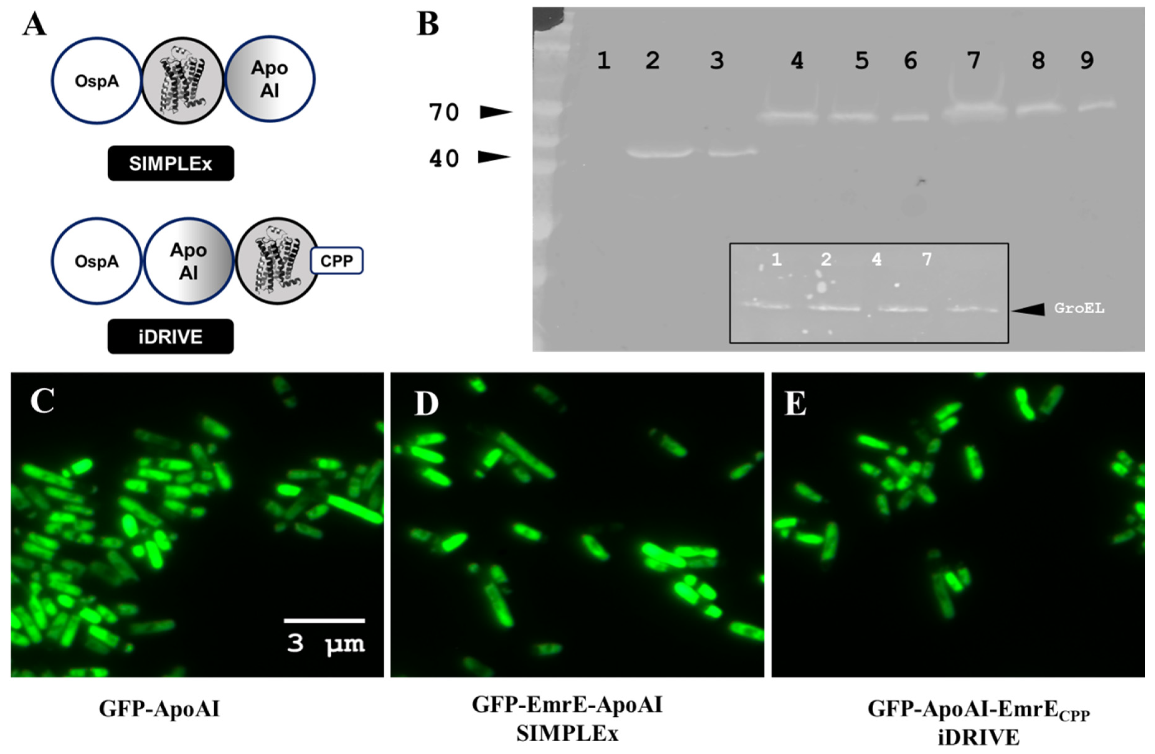
3.1. Design of iDRIVE (In Vivo Deployment of Recombinant Viable MPs)
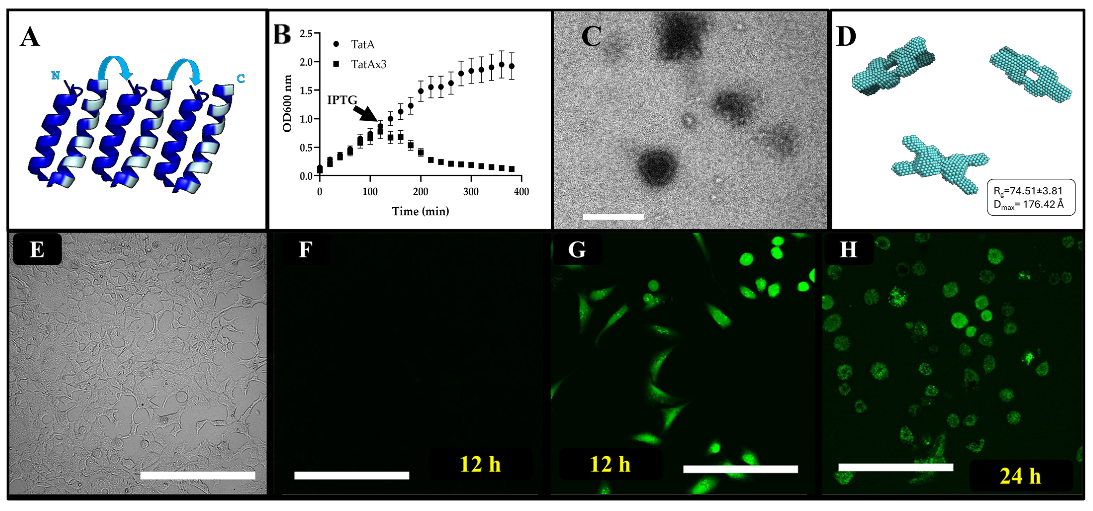
3.2. SIMPLEx and iDRIVE Pore-Forming Proteins Affect Cell Membranes
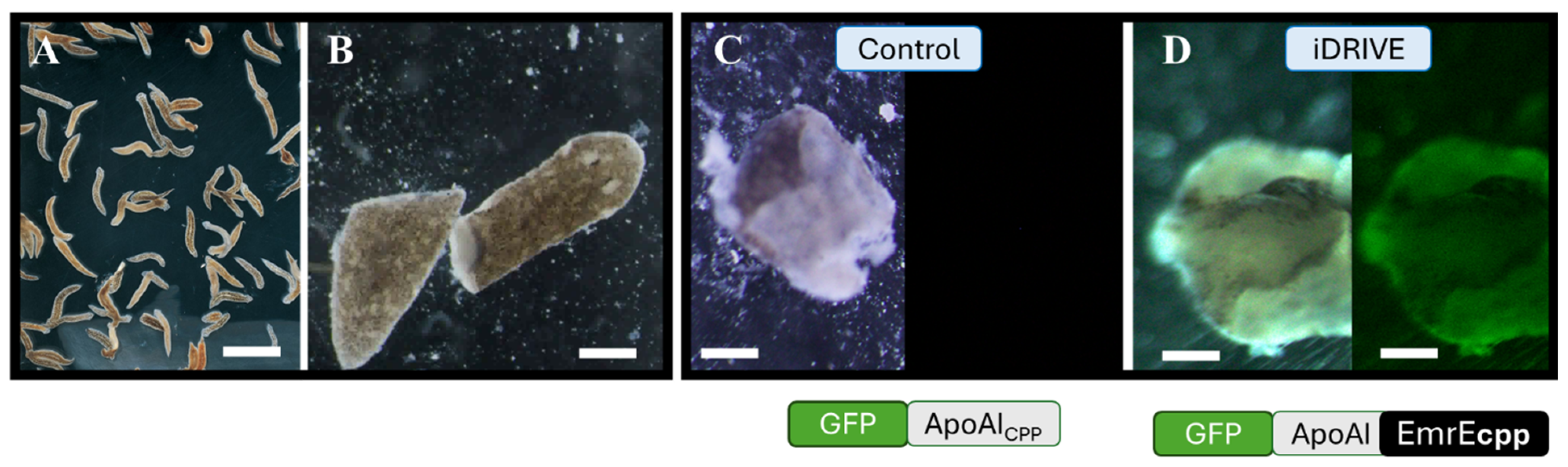
3.3. iDRIVE Interacts with Quiescent and Proliferating Cells In Vivo
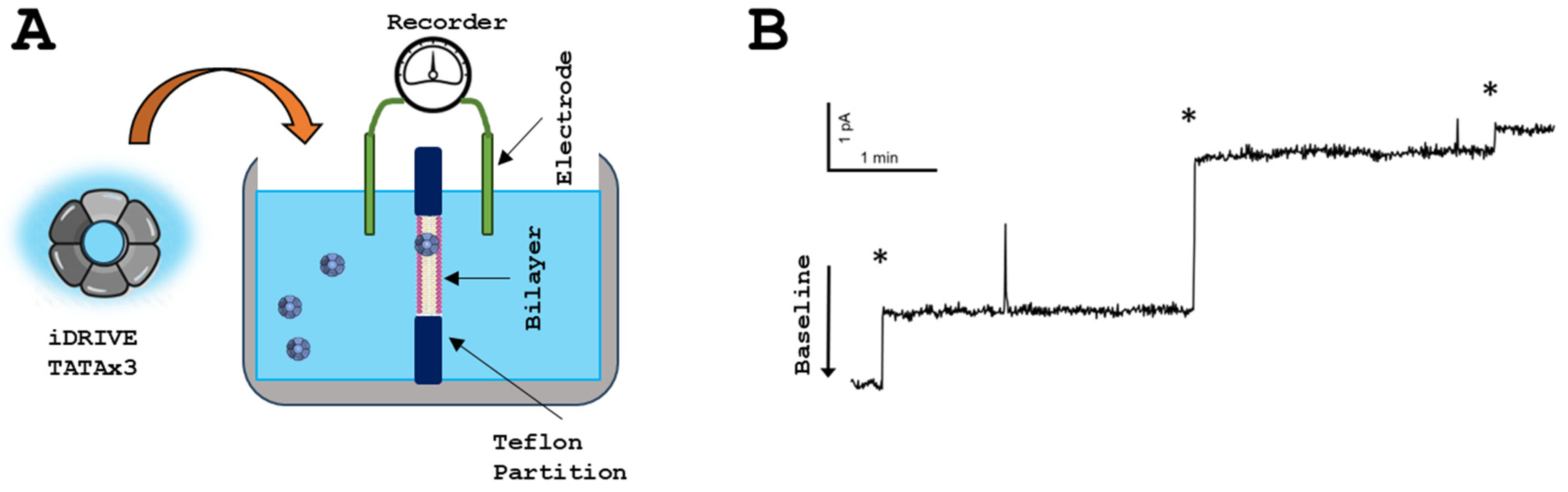
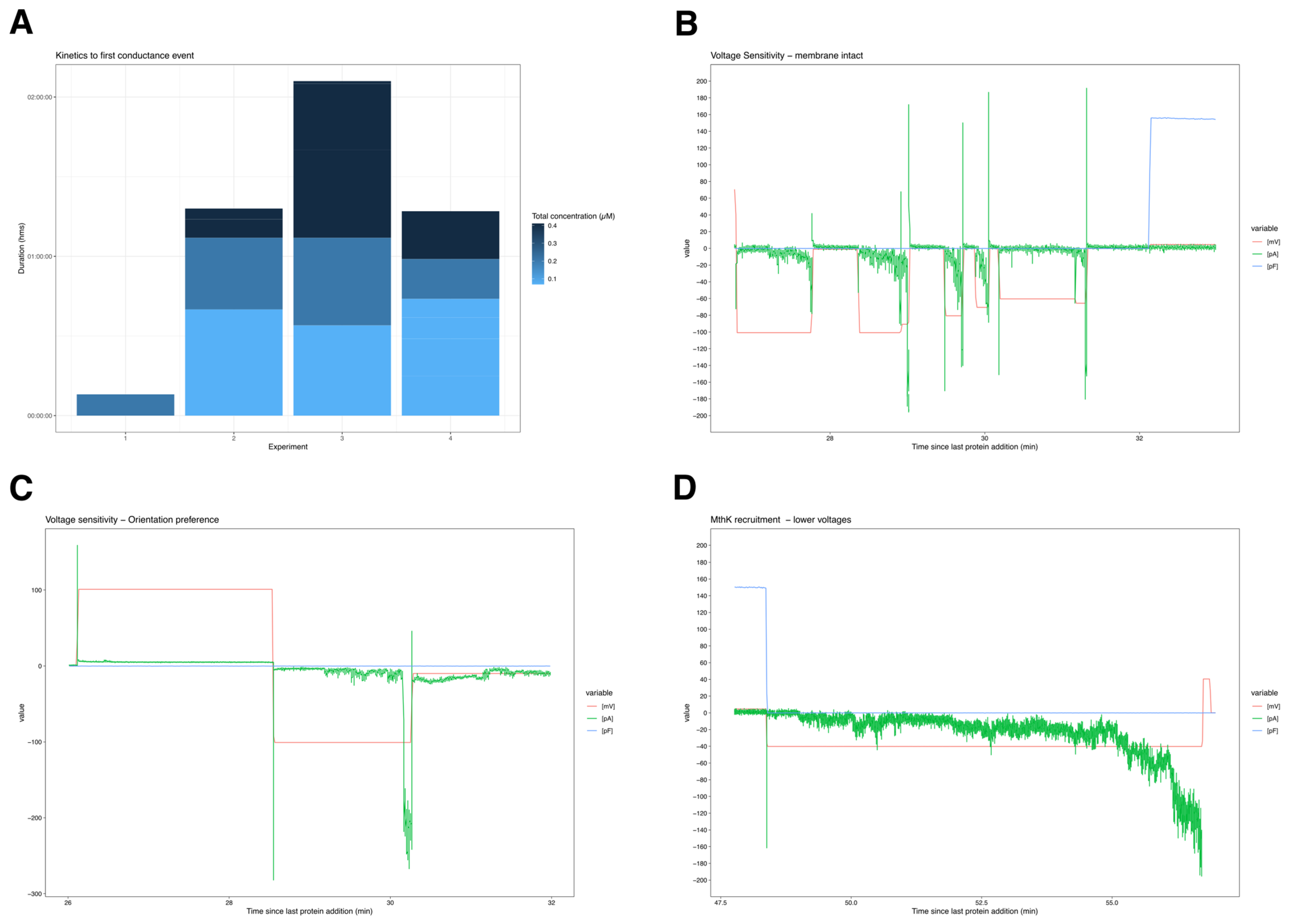
3.4. iDRIVE Allows MPs to Re-Insert into the Plasma Membrane: Planar Bilayer Interactions
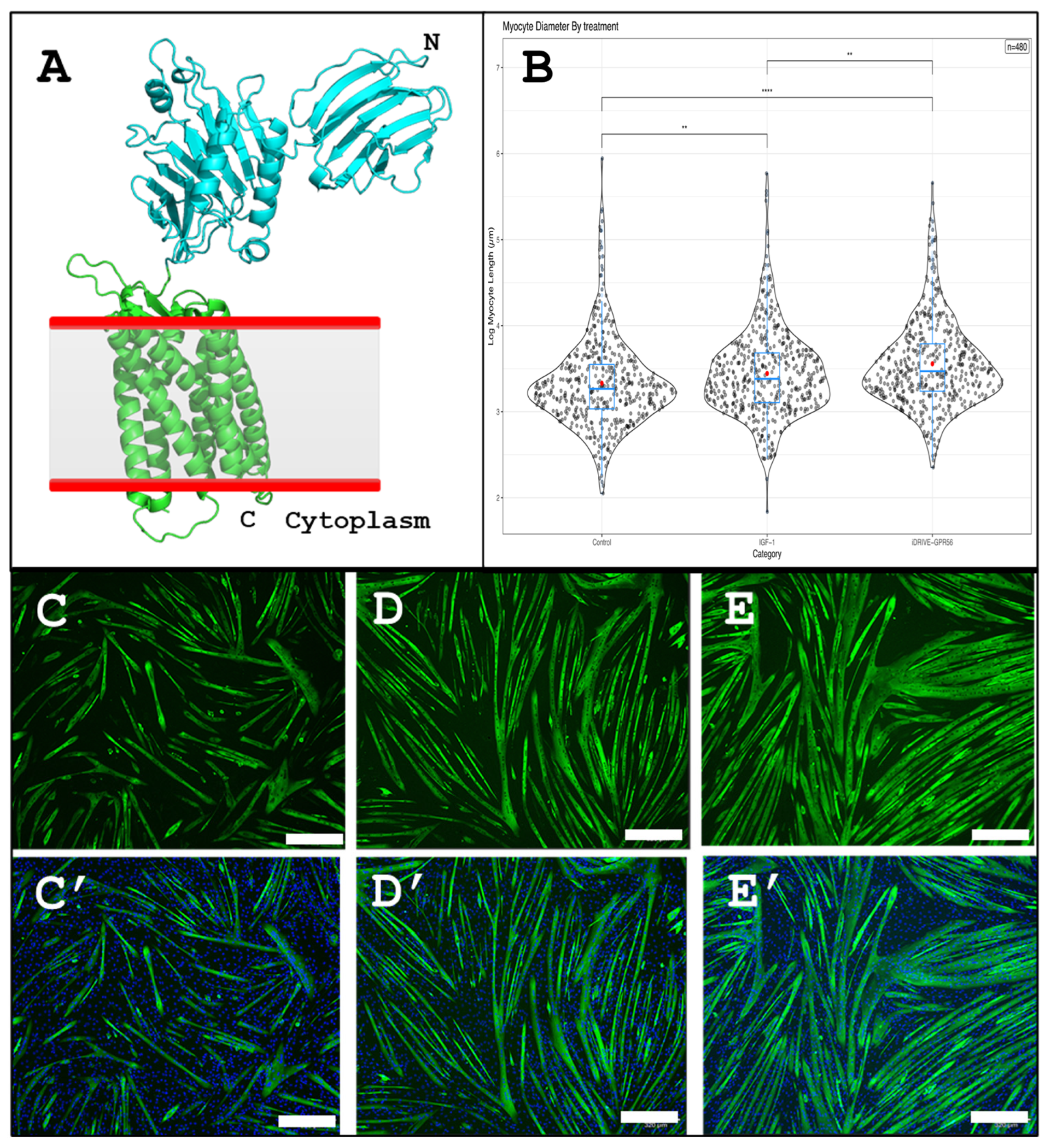
3.5. iDRIVE Allows MPs to Activate Signal Pathways In Vitro: The Case of Myotube Hypertrophy
3.6. iDRIVE Allows MPs to Activate Signal Pathways In Vitro: The Case of Embryoid Body Differentiation
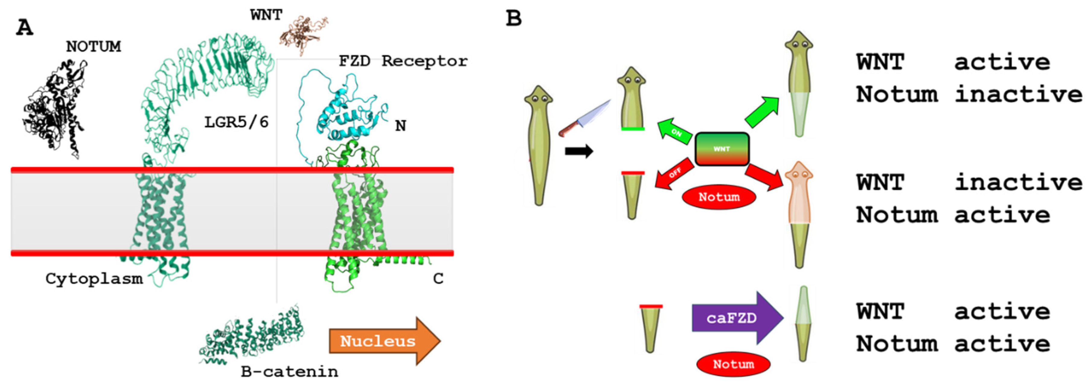
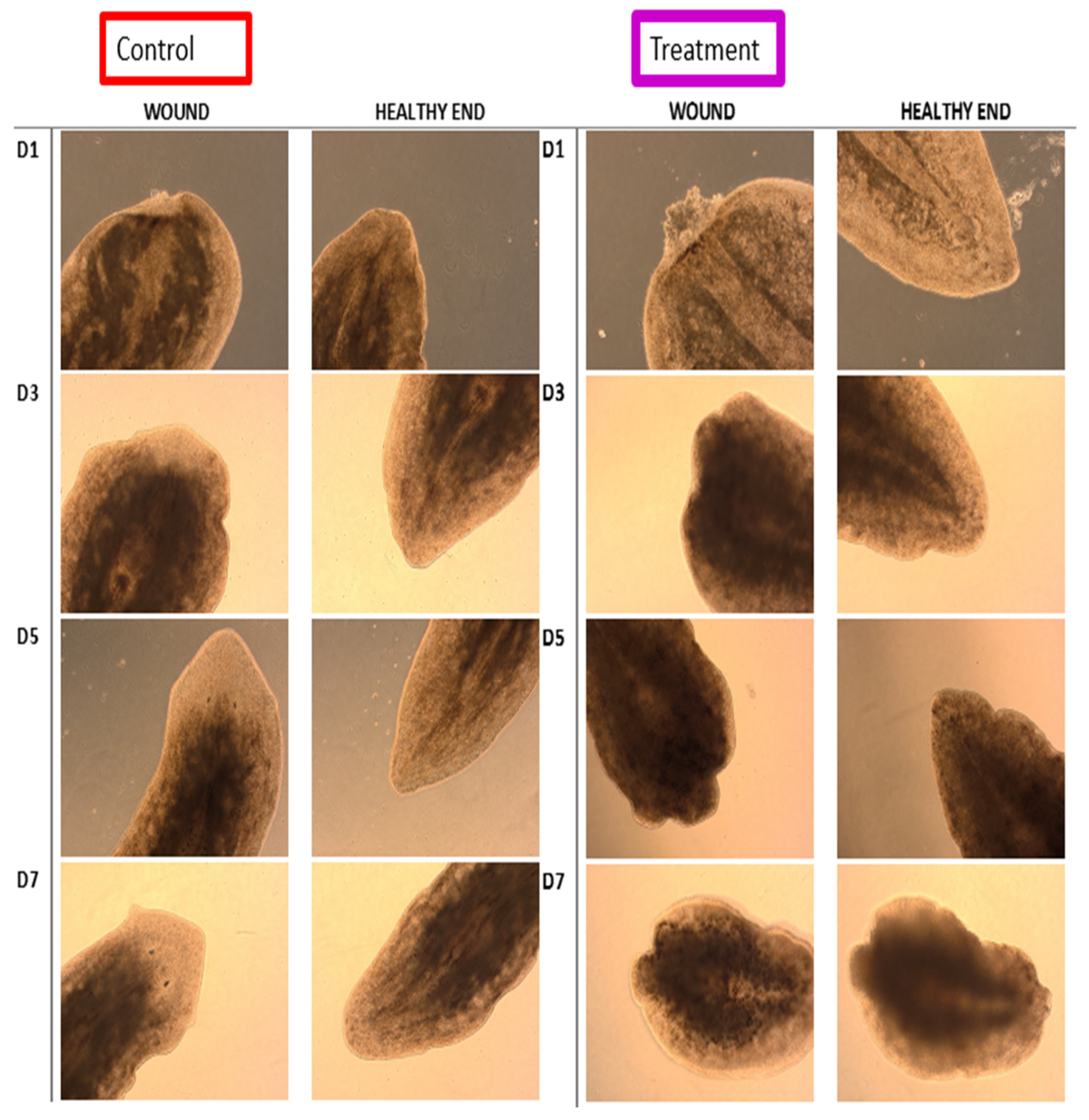
3.7. iDRIVE Allows MPs to Activate Signaling Pathways In Vivo: The Case of Planaria Regeneration
3.8. iDRIVE cahFZD7 and CHIR99021 Elicit Similar Differential Methylation Patterns Across CpG Regions—In Vitro HEK 293 Embryoid Bodies
3.8.1. Statistical Testing
3.8.2. Functional Enrichment
4. Discussion
5. Conclusions
Supplementary Materials
Author Contributions
Funding
Institutional Review Board Statement
Informed Consent Statement
Data Availability Statement
Acknowledgments
Conflicts of Interest
References
- Wallin, E.; von Heijne, G. Genome-wide analysis of integral membrane proteins from eubacterial, archaean, and eukaryotic organisms. Protein Sci. 1998, 7, 1029–1038. [Google Scholar] [CrossRef] [Scilit]
- Hedin, L.E.; Illergard, K.; Elofsson, A. An introduction to membrane proteins. J. Proteome Res. 2011, 10, 3324–3331. [Google Scholar] [CrossRef] [Scilit]
- Danielli, J.F.; Harvey, E.N. The tension at the surface of mackerel egg oil, with remarks on the nature of the cell surface. J. Cell. Comp. Physiol. 1935, 5, 483–494. [Google Scholar] [CrossRef] [Scilit]
- Tomita, M.; Furthmayr, H.; Marchesi, V.T. Primary structure of human erythrocyte glycophorin A. Isolation and characterization of peptides and complete amino acid sequence. Biochemistry 1978, 17, 4756–4770. [Google Scholar] [CrossRef] [Scilit] [PubMed]
- Tate, C.G. Overexpression of mammalian integral membrane proteins for structural studies. FEBS Lett. 2001, 504, 94–98. [Google Scholar] [CrossRef] [Scilit] [PubMed]
- Hedfalk, K. Further advances in the production of membrane proteins in Pichia pastoris. Bioengineered 2013, 4, 363–367. [Google Scholar] [CrossRef] [Scilit]
- Gulezian, E.; Crivello, C.; Bednenko, J.; Zafra, C.; Zhang, Y.; Colussi, P.; Hussain, S. Membrane protein production and formulation for drug discovery. Trends Pharmacol. Sci. 2021, 42, 657–674. [Google Scholar] [CrossRef] [Scilit]
- Swenson, C.S.; Mandava, G.; Thomas, D.M.; Moellering, R.E. Tackling Undruggable Targets with Designer Peptidomimetics and Synthetic Biologics. Chem. Rev. 2024, 124, 13020–13093. [Google Scholar] [CrossRef] [Scilit]
- Rawlings, A.E. Membrane proteins: Always an insoluble problem? Biochem. Soc. Trans. 2016, 44, 790–795. [Google Scholar] [CrossRef] [Scilit]
- Bayburt, T.H.; Grinkova, Y.V.; Sligar, S.G. Self-Assembly of Discoidal Phospholipid Bilayer Nanoparticles with Membrane Scaffold Proteins. Nano Lett. 2002, 2, 853–856. [Google Scholar] [CrossRef] [Scilit]
- Denisov, I.G.; Grinkova, Y.V.; Lazarides, A.A.; Sligar, S.G. Directed self-assembly of monodisperse phospholipid bilayer Nanodiscs with controlled size. J. Am. Chem. Soc. 2004, 126, 3477–3487. [Google Scholar] [CrossRef] [Scilit] [PubMed]
- Pina, A.S.; Lowe, C.R.; Roque, A.C. Challenges and opportunities in the purification of recombinant tagged proteins. Biotechnol. Adv. 2014, 32, 366–381. [Google Scholar] [CrossRef] [Scilit]
- Denisov, I.G.; Sligar, S.G. Nanodiscs for the study of membrane proteins. Curr. Opin. Struct. Biol. 2024, 87, 102844. [Google Scholar] [CrossRef] [Scilit]
- Knowles, T.J.; Finka, R.; Smith, C.; Lin, Y.P.; Dafforn, T.; Overduin, M. Membrane proteins solubilized intact in lipid containing nanoparticles bounded by styrene maleic acid copolymer. J. Am. Chem. Soc. 2009, 131, 7484–7485. [Google Scholar] [CrossRef] [Scilit]
- Broadbent, L.; Depping, P.; Lode, A.; Vaitsopoulou, A.; Hardy, D.; Ayub, H.; Mitchell-White, J.; Kerr, I.D.; Goddard, A.D.; Bill, R.M.; et al. Detergent-Free Membrane Protein Purification Using SMA Polymer; Methods in Molecular Biology; Humana: New York, NY, USA, 2022; Volume 2507, pp. 389–404. [Google Scholar]
- Scott, D.J.; Kummer, L.; Tremmel, D.; Pluckthun, A. Stabilizing membrane proteins through protein engineering. Curr. Opin. Chem. Biol. 2013, 17, 427–435. [Google Scholar] [CrossRef] [Scilit]
- Mizrachi, D.; Chen, Y.; Liu, J.; Peng, H.M.; Ke, A.; Pollack, L.; Turner, R.J.; Auchus, R.J.; DeLisa, M.P. Making water-soluble integral membrane proteins in vivo using an amphipathic protein fusion strategy. Nat. Commun. 2015, 6, 6826. [Google Scholar] [CrossRef] [Scilit]
- Zhang, S.; Tao, F.; Qing, R.; Tang, H.; Skuhersky, M.; Corin, K.; Tegler, L.; Wassie, A.; Wassie, B.; Kwon, Y.; et al. QTY code enables design of detergent-free chemokine receptors that retain ligand-binding activities. Proc. Natl. Acad. Sci. USA 2018, 115, E8652–E8659. [Google Scholar] [CrossRef] [Scilit]
- Jaroentomeechai, T.; Kwon, Y.H.; Liu, Y.; Young, O.; Bhawal, R.; Wilson, J.D.; Li, M.; Chapla, D.G.; Moremen, K.W.; Jewett, M.C.; et al. A universal glycoenzyme biosynthesis pipeline that enables efficient cell-free remodeling of glycans. Nat. Commun. 2022, 13, 6325. [Google Scholar] [CrossRef] [Scilit] [PubMed]
- Mihaljevic, L.; Kim, D.E.; Eisenach, H.E.; Bandawane, P.D.; Borst, A.J.; Courbet, A.; Bettin, E.; Liu, Q.; Weidle, C.; Majumder, S.; et al. Solubilization of Membrane Proteins using designed protein WRAPS. bioRxiv 2025. [Google Scholar] [CrossRef] [Scilit] [PubMed]
- Dean, M.R.P.; Duncan, E.M. Laboratory Maintenance and Propagation of Freshwater Planarians. Curr. Protoc. Microbiol. 2020, 59, e120. [Google Scholar] [CrossRef] [Scilit]
- Iuchi, K.; Oya, K.; Hosoya, K.; Sasaki, K.; Sakurada, Y.; Nakano, T.; Hisatomi, H. Different morphologies of human embryonic kidney 293T cells in various types of culture dishes. Cytotechnology 2020, 72, 131–140. [Google Scholar] [CrossRef] [Scilit]
- Behrens, M.A.; He, Y.; Oliveira, C.L.; Andersen, G.R.; Pedersen, J.S.; Nielsen, K.H. Structural analysis of RNA helicases with small-angle X-ray scattering. In Methods in Enzymology; Elsevier: Amsterdam, The Netherlands, 2012; Volume 511, pp. 191–212. [Google Scholar]
- Svergun, D. Determination of the regularization parameter in indirect-transform methods using perceptual criteria. J. Appl. Crystallogr. 1992, 25, 495–503. [Google Scholar] [CrossRef] [Scilit]
- Franke, D.; Svergun, D.I. DAMMIF, a program for rapid ab-initio shape determination in small-angle scattering. J. Appl. Crystallogr. 2009, 42, 342–346. [Google Scholar] [CrossRef] [Scilit] [PubMed]
- Zakharian, E. Recording of Ion Channel Activity in Planar Lipid Bilayer Experiments; Methods in Molecular Biology; Humana Press: Totowa, NJ, USA, 2013; Volume 998, pp. 109–118. [Google Scholar] [CrossRef] [Scilit]
- Jiang, Y.; Idikuda, V.; Chowdhury, S.; Chanda, B. Activation of the archaeal ion channel MthK is exquisitely regulated by temperature. Elife 2020, 9, e59055. [Google Scholar] [CrossRef] [Scilit]
- Aryee, M.J.; Jaffe, A.E.; Corrada-Bravo, H.; Ladd-Acosta, C.; Feinberg, A.P.; Hansen, K.D.; Irizarry, R.A. Minfi: A flexible and comprehensive Bioconductor package for the analysis of Infinium DNA methylation microarrays. Bioinformatics 2014, 30, 1363–1369. [Google Scholar] [CrossRef] [Scilit]
- Peters, T.J.; Meyer, B.; Ryan, L.; Achinger-Kawecka, J.; Song, J.; Campbell, E.M.; Qu, W.; Nair, S.; Loi-Luu, P.; Stricker, P.; et al. Characterisation and reproducibility of the HumanMethylationEPIC v2.0 BeadChip for DNA methylation profiling. BMC Genom. 2024, 25, 251. [Google Scholar] [CrossRef] [Scilit]
- Ritchie, M.E.; Phipson, B.; Wu, D.; Hu, Y.; Law, C.W.; Shi, W.; Smyth, G.K. limma powers differential expression analyses for RNA-sequencing and microarray studies. Nucleic Acids Res. 2015, 43, e47. [Google Scholar] [CrossRef] [Scilit]
- Gu, Z.; Eils, R.; Schlesner, M. Complex heatmaps reveal patterns and correlations in multidimensional genomic data. Bioinformatics 2016, 32, 2847–2849. [Google Scholar] [CrossRef] [Scilit] [PubMed]
- Murphy, S.M.; Kiely, M.; Jakeman, P.M.; Kiely, P.A.; Carson, B.P. Optimization of an in vitro bioassay to monitor growth and formation of myotubes in real time. Biosci. Rep. 2016, 36, e00330. [Google Scholar] [CrossRef] [Scilit] [PubMed]
- Tricerri, M.A.; Sanchez, S.A.; Arnulphi, C.; Durbin, D.M.; Gratton, E.; Jonas, A. Interaction of apolipoprotein A-I in three different conformations with palmitoyl oleoyl phosphatidylcholine vesicles. J. Lipid Res. 2002, 43, 187–197. [Google Scholar] [CrossRef] [Scilit]
- Davis, H.C.; Posey, N.D.; Tew, G.N. Protein Binding and Release by Polymeric Cell-Penetrating Peptide Mimics. Biomacromolecules 2022, 23, 57–66. [Google Scholar] [CrossRef] [Scilit]
- Malaby, A.W.; Chakravarthy, S.; Irving, T.C.; Kathuria, S.V.; Bilsel, O.; Lambright, D.G. Methods for analysis of size-exclusion chromatography-small-angle X-ray scattering and reconstruction of protein scattering. J. Appl. Crystallogr. 2015, 48, 1102–1113. [Google Scholar] [CrossRef] [Scilit] [PubMed]
- Walther, T.H.; Gottselig, C.; Grage, S.L.; Wolf, M.; Vargiu, A.V.; Klein, M.J.; Vollmer, S.; Prock, S.; Hartmann, M.; Afonin, S.; et al. Folding and self-assembly of the TatA translocation pore based on a charge zipper mechanism. Cell 2013, 152, 316–326. [Google Scholar] [CrossRef] [Scilit]
- Hutcheon, G.W.; Bolhuis, A. The archaeal twin-arginine translocation pathway. Biochem. Soc. Trans. 2003, 31, 686–689. [Google Scholar] [CrossRef] [Scilit] [PubMed]
- Gohlke, U.; Pullan, L.; McDevitt, C.A.; Porcelli, I.; de Leeuw, E.; Palmer, T.; Saibil, H.R.; Berks, B.C. The TatA component of the twin-arginine protein transport system forms channel complexes of variable diameter. Proc. Natl. Acad. Sci. USA 2005, 102, 10482–10486. [Google Scholar] [CrossRef] [Scilit]
- Flores-Romero, H.; Ros, U.; Garcia-Saez, A.J. Pore formation in regulated cell death. EMBO J. 2020, 39, e105753. [Google Scholar] [CrossRef] [Scilit]
- Doonan, F.; Cotter, T.G. Morphological assessment of apoptosis. Methods 2008, 44, 200–204. [Google Scholar] [CrossRef] [Scilit] [PubMed]
- Ho, C.J.; Samarasekera, G.; Rothe, K.; Xu, J.; Yang, K.C.; Leung, E.; Chan, M.; Jiang, X.; Gorski, S.M. Puncta intended: Connecting the dots between autophagy and cell stress networks. Autophagy 2021, 17, 1028–1033. [Google Scholar] [CrossRef] [Scilit]
- Snapp, E. Design and use of fluorescent fusion proteins in cell biology. Curr. Protoc. Cell Biol. 2005, 27, 21.4.1–21.4.13. [Google Scholar] [CrossRef] [Scilit]
- Yorimitsu, T.; Nair, U.; Yang, Z.; Klionsky, D.J. Endoplasmic reticulum stress triggers autophagy. J. Biol. Chem. 2006, 281, 30299–30304. [Google Scholar] [CrossRef] [Scilit]
- Alexandrov, A.I.; Grosfeld, E.V.; Dergalev, A.A.; Kushnirov, V.V.; Chuprov-Netochin, R.N.; Tyurin-Kuzmin, P.A.; Kireev, I.I.; Ter-Avanesyan, M.D.; Leonov, S.V.; Agaphonov, M.O. Analysis of novel hyperosmotic shock response suggests ‘beads in liquid’ cytosol structure. Biol. Open 2019, 8, bio044529. [Google Scholar] [CrossRef] [Scilit]
- Rink, J.C. Stem cell systems and regeneration in planaria. Dev. Genes Evol. 2013, 223, 67–84. [Google Scholar] [CrossRef] [Scilit]
- Scalf, S.M.; Wu, Q.; Guo, S. Molecular basis of cell fate plasticity—insights from the privileged cells. Curr. Opin. Genet. Dev. 2025, 93, 102354. [Google Scholar] [CrossRef] [Scilit]
- Wagner, D.E.; Wang, I.E.; Reddien, P.W. Clonogenic neoblasts are pluripotent adult stem cells that underlie planarian regeneration. Science 2011, 332, 811–816. [Google Scholar] [CrossRef] [Scilit] [PubMed]
- Zadek, B.; Nimigean, C.M. Calcium-dependent gating of MthK, a prokaryotic potassium channel. J. Gen. Physiol. 2006, 127, 673–685. [Google Scholar] [CrossRef] [Scilit]
- Li, Y.; Berke, I.; Chen, L.; Jiang, Y. Gating and inward rectifying properties of the MthK K+ channel with and without the gating ring. J. Gen. Physiol. 2007, 129, 109–120. [Google Scholar] [CrossRef] [Scilit]
- Thomson, A.S.; Rothberg, B.S. Voltage-dependent inactivation gating at the selectivity filter of the MthK K+ channel. J. Gen. Physiol. 2010, 136, 569–579. [Google Scholar] [CrossRef] [Scilit]
- Posson, D.J.; McCoy, J.G.; Nimigean, C.M. The voltage-dependent gate in MthK potassium channels is located at the selectivity filter. Nat. Struct. Mol. Biol. 2013, 20, 159–166. [Google Scholar] [CrossRef] [Scilit] [PubMed]
- Semsarian, C.; Sutrave, P.; Richmond, D.R.; Graham, R.M. Insulin-like growth factor (IGF-I) induces myotube hypertrophy associated with an increase in anaerobic glycolysis in a clonal skeletal-muscle cell model. Biochem. J. 1999, 339, 443–451. [Google Scholar] [CrossRef] [PubMed]
- White, J.P.; Wrann, C.D.; Rao, R.R.; Nair, S.K.; Jedrychowski, M.P.; You, J.S.; Martinez-Redondo, V.; Gygi, S.P.; Ruas, J.L.; Hornberger, T.A.; et al. G protein-coupled receptor 56 regulates mechanical overload-induced muscle hypertrophy. Proc. Natl. Acad. Sci. USA 2014, 111, 15756–15761. [Google Scholar] [CrossRef] [Scilit]
- Jallouli, R.; Moreno-Salinas, A.L.; Laniel, A.; Holleran, B.; Avet, C.; Jacob, J.; Hoang, T.; Lavoie, C.; Carmon, K.S.; Bouvier, M.; et al. G protein selectivity profile of GPR56/ADGRG1 and its effect on downstream effectors. Cell. Mol. Life Sci. 2024, 81, 383. [Google Scholar] [CrossRef] [Scilit]
- Seifert, R.; Wenzel-Seifert, K. Constitutive activity of G-protein-coupled receptors: Cause of disease and common property of wild-type receptors. Naunyn-Schmiedebergs Arch. Pharmacol. 2002, 366, 381–416. [Google Scholar] [CrossRef] [Scilit]
- Lala, T.; Hall, R.A. Adhesion G protein-coupled receptors: Structure, signaling, physiology, and pathophysiology. Physiol. Rev. 2022, 102, 1587–1624. [Google Scholar] [CrossRef] [Scilit]
- de Graaf, C.; Nijmeijer, S.; Wolf, S.; Ernst, O.P. 7TM Domain Structure of Adhesion GPCRs. In Adhesion G Protein-Coupled Receptors; Handbook of Experimental Pharmacology; Springer: Cham, Switzerland, 2016; Volume 234, pp. 43–66. [Google Scholar]
- Werner, H. The IGF1 Signaling Pathway: From Basic Concepts to Therapeutic Opportunities. Int. J. Mol. Sci. 2023, 24, 14882. [Google Scholar] [CrossRef] [Scilit]
- Cervera, J.; Levin, M.; Mafe, S. Morphology changes induced by intercellular gap junction blocking: A reaction-diffusion mechanism. Biosystems 2021, 209, 104511. [Google Scholar] [CrossRef] [Scilit]
- Cervera, J.; Levin, M.; Mafe, S. Top-down perspectives on cell membrane potential and protein transcription. Sci. Rep. 2025, 16. [Google Scholar] [CrossRef] [Scilit] [PubMed]
- Aberg, N.D.; Blomstrand, F.; Aberg, M.A.; Bjorklund, U.; Carlsson, B.; Carlsson-Skwirut, C.; Bang, P.; Ronnback, L.; Eriksson, P.S. Insulin-like growth factor-I increases astrocyte intercellular gap junctional communication and connexin43 expression in vitro. J. Neurosci. Res. 2003, 74, 12–22. [Google Scholar] [CrossRef] [Scilit]
- Langenhan, T.; Aust, G.; Hamann, J. Sticky signaling--adhesion class G protein-coupled receptors take the stage. Sci. Signal. 2013, 6, re3. [Google Scholar] [CrossRef] [Scilit] [PubMed]
- Prochnow, N.; Dermietzel, R. Connexons and cell adhesion: A romantic phase. Histochem. Cell Biol. 2008, 130, 71–77. [Google Scholar] [CrossRef] [Scilit] [PubMed]
- Sonavane, P.R.; Willert, K. Controlling Wnt Signaling Specificity and Implications for Targeting WNTs Pharmacologically. Handb. Exp. Pharmacol. 2021, 269, 3–28. [Google Scholar] [CrossRef] [Scilit]
- Martinez-Marin, D.; Stroman, G.C.; Fulton, C.J.; Pruitt, K. Frizzled receptors: Gatekeepers of Wnt signaling in development and disease. Front. Cell Dev. Biol. 2025, 13, 1599355. [Google Scholar] [CrossRef] [Scilit] [PubMed]
- Liu, J.; Xiao, Q.; Xiao, J.; Niu, C.; Li, Y.; Zhang, X.; Zhou, Z.; Shu, G.; Yin, G. Wnt/β-catenin signalling: Function, biological mechanisms, and therapeutic opportunities. Signal Transduct. Target. Ther. 2022, 7, 3. [Google Scholar] [CrossRef] [Scilit]
- Ai, Z.; Shao, J.; Wu, Y.; Yu, M.; Du, J.; Shi, X.; Shi, X.; Zhang, Y.; Guo, Z. CHIR99021 enhances Klf4 Expression through β-Catenin Signaling and miR-7a Regulation in J1 Mouse Embryonic Stem Cells. PLoS ONE 2016, 11, e0150936. [Google Scholar] [CrossRef] [Scilit]
- Ye, S.; Tan, L.; Yang, R.; Fang, B.; Qu, S.; Schulze, E.N.; Song, H.; Ying, Q.; Li, P. Pleiotropy of glycogen synthase kinase-3 inhibition by CHIR99021 promotes self-renewal of embryonic stem cells from refractory mouse strains. PLoS ONE 2012, 7, e35892. [Google Scholar] [CrossRef] [Scilit] [PubMed]
- Xu, L.; Chen, B.; Schihada, H.; Wright, S.C.; Turku, A.; Wu, Y.; Han, G.W.; Kowalski-Jahn, M.; Kozielewicz, P.; Bowin, C.F.; et al. Cryo-EM structure of constitutively active human Frizzled 7 in complex with heterotrimeric G(s). Cell Res. 2021, 31, 1311–1314. [Google Scholar] [CrossRef] [Scilit]
- Pettinato, G.; Wen, X.; Zhang, N. Engineering Strategies for the Formation of Embryoid Bodies from Human Pluripotent Stem Cells. Stem Cells Dev. 2015, 24, 1595–1609. [Google Scholar] [CrossRef] [Scilit]
- Voss, J.H.; Koszegi, Z.; Yan, Y.; Shorter, E.; Gratz, L.; Lanner, J.T.; Calebiro, D.; Schulte, G. WNT-induced association of Frizzled and LRP6 is not sufficient for the initiation of WNT/β-catenin signaling. Nat. Commun. 2025, 16, 4848. [Google Scholar] [CrossRef] [Scilit]
- Brueggemann, L.I.; Sullivan, J.M. HEK293S cells have functional retinoid processing machinery. J. Gen. Physiol. 2002, 119, 593–612. [Google Scholar] [CrossRef] [Scilit] [PubMed]
- Cheng, T.; Zhai, K.; Chang, Y.; Yao, G.; He, J.; Wang, F.; Kong, H.; Xin, H.; Wang, H.; Jin, M.; et al. CHIR99021 combined with retinoic acid promotes the differentiation of primordial germ cells from human embryonic stem cells. Oncotarget 2017, 8, 7814–7826. [Google Scholar] [CrossRef] [Scilit] [PubMed]
- Karami, A.; Tebyanian, H.; Goodarzi, V.; Shiri, S. Planarians: An In Vivo Model for Regenerative Medicine. Int. J. Stem Cells 2015, 8, 128–133. [Google Scholar] [CrossRef] [Scilit]
- Gurley, K.A.; Rink, J.C.; Alvarado, A.S. β-catenin defines head versus tail identity during planarian regeneration and homeostasis. Science 2008, 319, 323–327. [Google Scholar] [CrossRef] [Scilit] [PubMed]
- Kakugawa, S.; Langton, P.F.; Zebisch, M.; Howell, S.; Chang, T.H.; Liu, Y.; Feizi, T.; Bineva, G.; O’Reilly, N.; Snijders, A.P.; et al. Notum deacylates Wnt proteins to suppress signalling activity. Nature 2015, 519, 187–192. [Google Scholar] [CrossRef] [Scilit]
- Beane, W.S.; Morokuma, J.; Adams, D.S.; Levin, M. A chemical genetics approach reveals H,K-ATPase-mediated membrane voltage is required for planarian head regeneration. Chem. Biol. 2011, 18, 77–89. [Google Scholar] [CrossRef] [Scilit] [PubMed]
- Adler, C.E.; Alvarado, A.S. Systemic RNA Interference in Planarians by Feeding of dsRNA Containing Bacteria; Methods in Molecular Biology; Humana Press: New York, NY, USA, 2018; Volume 1774, pp. 445–454. [Google Scholar]
- Pascual-Carreras, E.; Marin-Barba, M.; Castillo-Lara, S.; Coronel-Cordoba, P.; Magri, M.S.; Wheeler, G.N.; Gomez-Skarmeta, J.L.; Abril, J.F.; Salo, E.; Adell, T. Wnt/β-catenin signalling is required for pole-specific chromatin remodeling during planarian regeneration. Nat. Commun. 2023, 14, 298. [Google Scholar] [CrossRef] [Scilit] [PubMed]
- Xue, C.; Chu, Q.; Shi, Q.; Zeng, Y.; Lu, J.; Li, L. Wnt signaling pathways in biology and disease: Mechanisms and therapeutic advances. Signal Transduct. Target. Ther. 2025, 10, 106. [Google Scholar] [CrossRef] [Scilit]
- Tao, Y.X. Constitutive activation of G protein-coupled receptors and diseases: Insights into mechanisms of activation and therapeutics. Pharmacol. Ther. 2008, 120, 129–148. [Google Scholar] [CrossRef] [Scilit]
- Gene Ontology, C. The Gene Ontology knowledgebase in 2026. Nucleic Acids Res. 2026, 54, D1779–D1792. [Google Scholar] [CrossRef] [Scilit]
- Ashburner, M.; Ball, C.A.; Blake, J.A.; Botstein, D.; Butler, H.; Cherry, J.M.; Davis, A.P.; Dolinski, K.; Dwight, S.S.; Eppig, J.T.; et al. Gene ontology: Tool for the unification of biology. Nat. Genet. 2000, 25, 25–29. [Google Scholar] [CrossRef] [Scilit]
- Liu, S.; Anderson, P.J.; Rajagopal, S.; Lefkowitz, R.J.; Rockman, H.A. G Protein-Coupled Receptors: A Century of Research and Discovery. Circ. Res. 2024, 135, 174–197. [Google Scholar] [CrossRef] [Scilit]
- Zhang, M.; Chen, T.; Lu, X.; Lan, X.; Chen, Z.; Lu, S. G protein-coupled receptors (GPCRs): Advances in structures, mechanisms, and drug discovery. Signal Transduct. Target. Ther. 2024, 9, 88. [Google Scholar] [CrossRef] [Scilit]
- Sriram, K.; Insel, P.A. G Protein-Coupled Receptors as Targets for Approved Drugs: How Many Targets and How Many Drugs? Mol. Pharmacol. 2018, 93, 251–258. [Google Scholar] [CrossRef] [Scilit]
- Fan, C.; Sukomon, N.; Flood, E.; Rheinberger, J.; Allen, T.W.; Nimigean, C.M. Ball-and-chain inactivation in a calcium-gated potassium channel. Nature 2020, 580, 288–293. [Google Scholar] [CrossRef] [Scilit]
- Kwon, Y.H.; Mihaljevic, L.; Kim, K.; Kim, D.E.; Donahue, T.C.; Bidstrup, E.J.; Bandi, C.K.; Sotomayor, B.; Hulbert, S.W.; Myers, K.A.; et al. Topological reprogramming transforms an integral membrane oligosaccharyltransferase into a water-soluble glycosylation catalyst. bioRxiv 2026. [Google Scholar] [CrossRef] [Scilit]
- Drake, M.T.; Shenoy, S.K.; Lefkowitz, R.J. Trafficking of G protein-coupled receptors. Circ. Res. 2006, 99, 570–582. [Google Scholar] [CrossRef] [Scilit] [PubMed]
- Tao, Y.X.; Conn, P.M. Chaperoning G protein-coupled receptors: From cell biology to therapeutics. Endocr. Rev. 2014, 35, 602–647. [Google Scholar] [CrossRef] [Scilit]
- Inoue, R.; Fukutani, Y.; Niwa, T.; Matsunami, H.; Yohda, M. Identification and Characterization of Proteins That Are Involved in RTP1S-Dependent Transport of Olfactory Receptors. Int. J. Mol. Sci. 2023, 24, 7829. [Google Scholar] [CrossRef] [Scilit] [PubMed]


Disclaimer/Publisher’s Note: The statements, opinions and data contained in all publications are solely those of the individual author(s) and contributor(s) and not of MDPI and/or the editor(s). MDPI and/or the editor(s) disclaim responsibility for any injury to people or property resulting from any ideas, methods, instructions or products referred to in the content. |
© 2026 by the authors. Licensee MDPI, Basel, Switzerland. This article is an open access article distributed under the terms and conditions of the Creative Commons Attribution (CC BY) license.
Share and Cite
Valencia-Amores, S.; Davila Aleman, I.; Jenkins, T.G.; Mizrachi, D. Beyond Small Molecules: Orchestrating Cell Fate with Engineered Water-Soluble Membrane Proteins. Biomolecules 2026, 16, 546. https://doi.org/10.3390/biom16040546
Valencia-Amores S, Davila Aleman I, Jenkins TG, Mizrachi D. Beyond Small Molecules: Orchestrating Cell Fate with Engineered Water-Soluble Membrane Proteins. Biomolecules. 2026; 16(4):546. https://doi.org/10.3390/biom16040546
Chicago/Turabian StyleValencia-Amores, Sebastian, Israel Davila Aleman, Timothy G. Jenkins, and Dario Mizrachi. 2026. "Beyond Small Molecules: Orchestrating Cell Fate with Engineered Water-Soluble Membrane Proteins" Biomolecules 16, no. 4: 546. https://doi.org/10.3390/biom16040546
APA StyleValencia-Amores, S., Davila Aleman, I., Jenkins, T. G., & Mizrachi, D. (2026). Beyond Small Molecules: Orchestrating Cell Fate with Engineered Water-Soluble Membrane Proteins. Biomolecules, 16(4), 546. https://doi.org/10.3390/biom16040546