Peptide Drugs in Gastrointestinal Tumors: Integrating Targeting, Delivery, and Therapeutic Actions for Synergistic Strategies
 , , ,
, , ,  and
and
Abstract
1. Introduction
2. Mechanism of Action of Peptide Drugs
2.1. Blocking Peptides
2.2. Delivery Peptides
2.3. Mimetic Peptides
3. Peptide Drugs in the Treatment of Gastrointestinal Malignant Tumors
3.1. Colorectal Cancer
3.2. Gastric Cancer
3.3. Pancreatic Ductal Adenocarcinoma
3.4. Hepatocellular Carcinoma
3.5. Esophageal Cancer
4. Optimization Strategies for Peptide Drugs
4.1. Strategies to Improve Delivery Efficiency
4.2. Strategies to Enhance Stability
5. Discussion
6. Conclusions
Author Contributions
Funding
Institutional Review Board Statement
Informed Consent Statement
Data Availability Statement
Conflicts of Interest
References
- Bliss, M. The history of insulin. Diabetes Care 1993, 16, 4–7. [Google Scholar] [CrossRef] [Scilit] [PubMed]
- Sims, E.K.; Carr, A.L.J.; Oram, R.A.; DiMeglio, L.A.; Evans-Molina, C. 100 years of insulin: Celebrating the past, present and future of diabetes therapy. Nat. Med. 2021, 27, 1154–1164. [Google Scholar] [CrossRef] [Scilit] [PubMed]
- Singh, I.R.; Aggarwal, N.; Srivastava, S.; Panda, J.J.; Mishra, J. Small Peptide-Based Nanodelivery Systems for Cancer Therapy and Diagnosis. J. Pharmacol. Exp. Ther. 2024, 390, 30–44. [Google Scholar] [CrossRef] [Scilit] [PubMed]
- Buddhiraju, H.S.; Yadav, D.N.; Dey, S.; Eswar, K.; Padmakumar, A.; Rengan, A.K. Advances in Peptide-Decorated Targeted Drug Delivery: Exploring Therapeutic Potential and Nanocarrier Strategies. ACS Appl. Bio Mater. 2024, 7, 4879–4893. [Google Scholar] [CrossRef] [Scilit]
- Ilangala, A.B.; Lechanteur, A.; Fillet, M.; Piel, G. Therapeutic peptides for chemotherapy: Trends and challenges for advanced delivery systems. Eur. J. Pharm. Biopharm. 2021, 167, 140–158. [Google Scholar] [CrossRef] [Scilit]
- Gilad, Y.; Firer, M.; Gellerman, G. Recent Innovations in Peptide Based Targeted Drug Delivery to Cancer Cells. Biomedicines 2016, 4, 11. [Google Scholar] [CrossRef] [Scilit]
- Sharma, K.; Sharma, K.K.; Sharma, A.; Jain, R. Peptide-based drug discovery: Current status and recent advances. Drug Discov. Today 2023, 28, 103464. [Google Scholar] [CrossRef] [Scilit]
- Muttenthaler, M.; King, G.F.; Adams, D.J.; Alewood, P.F. Trends in peptide drug discovery. Nat. Rev. Drug Discov. 2021, 20, 309–325. [Google Scholar] [CrossRef] [Scilit]
- Wang, L.; Wang, N.; Zhang, W.; Cheng, X.; Yan, Z.; Shao, G.; Wang, X.; Wang, R.; Fu, C. Therapeutic peptides: Current applications and future directions. Signal Transduct. Target. Ther. 2022, 7, 48. [Google Scholar] [CrossRef] [Scilit]
- Erak, M.; Bellmann-Sickert, K.; Els-Heindl, S.; Beck-Sickinger, A.G. Peptide chemistry toolbox—Transforming natural peptides into peptide therapeutics. Bioorg. Med. Chem. 2018, 26, 2759–2765. [Google Scholar] [CrossRef] [Scilit]
- Huo, Y.; Zhang, Z.; Dong, S.; Du, Y.; Ma, M. Advances in clinical studies of peptide drugs in stroke disease. Biochim. Biophys. Acta Mol. Basis Dis. 2025, 1871, 167970. [Google Scholar] [CrossRef] [Scilit] [PubMed]
- Arnold, M.; Abnet, C.C.; Neale, R.E.; Vignat, J.; Giovannucci, E.L.; McGlynn, K.A.; Bray, F. Global Burden of 5 Major Types of Gastrointestinal Cancer. Gastroenterology 2020, 159, 335–349.e15. [Google Scholar] [CrossRef] [Scilit] [PubMed]
- Bray, F.; Laversanne, M.; Sung, H.; Ferlay, J.; Siegel, R.L.; Soerjomataram, I.; Jemal, A. Global cancer statistics 2022: GLOBOCAN estimates of incidence and mortality worldwide for 36 cancers in 185 countries. CA Cancer J. Clin. 2024, 74, 229–263. [Google Scholar] [CrossRef] [Scilit] [PubMed]
- Liu, B.; Zhou, H.; Tan, L.; Siu, K.T.H.; Guan, X.-Y. Exploring treatment options in cancer: Tumor treatment strategies. Signal Transduct. Target. Ther. 2024, 9, 175. [Google Scholar] [CrossRef] [Scilit]
- Joshi, D.C.; Sharma, A.; Prasad, S.; Singh, K.; Kumar, M.; Sherawat, K.; Tuli, H.S.; Gupta, M. Novel therapeutic agents in clinical trials: Emerging approaches in cancer therapy. Discov. Oncol. 2024, 15, 342. [Google Scholar] [CrossRef] [Scilit]
- Khan, S.U.; Fatima, K.; Aisha, S.; Malik, F. Unveiling the mechanisms and challenges of cancer drug resistance. Cell Commun. Signal. CCS 2024, 22, 109. [Google Scholar] [CrossRef] [Scilit]
- Tabibzadeh, A.; Tameshkel, F.S.; Moradi, Y.; Soltani, S.; Moradi-Lakeh, M.; Ashrafi, G.H.; Motamed, N.; Zamani, F.; Motevalian, S.A.; Panahi, M.; et al. Signal transduction pathway mutations in gastrointestinal (GI) cancers: A systematic review and meta-analysis. Sci. Rep. 2020, 10, 18713. [Google Scholar] [CrossRef] [Scilit]
- Alves de Souza, S.M.; Hernández-Ledesma, B.; de Souza, T.L.F. Lunasin as a Promising Plant-Derived Peptide for Cancer Therapy. Int. J. Mol. Sci. 2022, 23, 9548. [Google Scholar] [CrossRef] [Scilit]
- Yu, Z.; Li, Y.; Li, Y.; Zhang, J.; Li, M.; Ji, L.; Tang, Y.; Zheng, Y.; Sheng, J.; Han, Q.; et al. Bufalin stimulates antitumor immune response by driving tumor-infiltrating macrophage toward M1 phenotype in hepatocellular carcinoma. J. Immunother. Cancer 2022, 10, e004297. [Google Scholar] [CrossRef] [Scilit]
- Wang, H.; Zhang, C.; Xu, L.; Zang, K.; Ning, Z.; Jiang, F.; Chi, H.; Zhu, X.; Meng, Z. Bufalin suppresses hepatocellular carcinoma invasion and metastasis by targeting HIF-1alpha via the PI3K/AKT/mTOR pathway. Oncotarget 2016, 7, 20193–20208. [Google Scholar] [CrossRef] [Scilit]
- Park, C.B.; Yi, K.S.; Matsuzaki, K.; Kim, M.S.; Kim, S.C. Structure-activity analysis of buforin II, a histone H2A-derived antimicrobial peptide: The proline hinge is responsible for the cell-penetrating ability of buforin II. Proc. Natl. Acad. Sci. USA 2000, 97, 8245–8250. [Google Scholar] [CrossRef] [Scilit]
- Armstrong, A.; Coburn, F.; Nsereko, Y.; Al Musaimi, O. Peptide-Drug Conjugates: A New Hope for Cancer. J. Pept. Sci. Off. Publ. Eur. Pept. Soc. 2025, 31, e70040. [Google Scholar] [CrossRef] [Scilit] [PubMed]
- Zhang, X.; Li, X.; Zhao, Y.; Zheng, Q.; Wu, Q.; Yu, Y. Nanocarrier system: An emerging strategy for bioactive peptide delivery. Front. Nutr. 2022, 9, 1050647. [Google Scholar] [CrossRef] [Scilit] [PubMed]
- Ghosh, D.; Peng, X.; Leal, J.; Mohanty, R. Peptides as drug delivery vehicles across biological barriers. J. Pharm. Investig. 2018, 48, 89–111. [Google Scholar] [CrossRef] [Scilit] [PubMed]
- Rehman, H.M.; Ahmad, S.; Sarwar, A.; Bashir, H. Unlocking the potential of tumor-targeting peptides in precision oncology. Oncol. Res. 2025, 33, 1547–1570. [Google Scholar] [CrossRef] [Scilit]
- Feng, Y.; Li, T.; Li, S.; Liu, Z.; Tang, Z.; Chen, C.; Zhou, C.; Lu, T.; Chen, J. Recent advances in peptide-drug conjugates as anticancer agents. Eur. J. Med. Chem. 2026, 304, 118482. [Google Scholar] [CrossRef] [Scilit]
- Nsereko, Y.; Armstrong, A.; Coburn, F.; Al Musaimi, O. Innovative Peptide Therapeutics in the Pipeline: Transforming Cancer Detection and Treatment. Int. J. Mol. Sci. 2025, 26, 6815. [Google Scholar] [CrossRef] [Scilit]
- Gouveia, M.J.; Campanhã, J.; Barbosa, F.; Vale, N. From Mechanism to Medicine: Peptide-Based Approaches for Cancer Diagnosis and Therapy. Biomolecules 2025, 16, 27. [Google Scholar] [CrossRef] [Scilit]
- Coburn, F.; Nsereko, Y.; Armstrong, A.; Al Musaimi, O. Peptide inhibitors: Breaking cancer code. Eur. J. Med. Chem. 2025, 297, 117961. [Google Scholar] [CrossRef] [Scilit]
- Chinnadurai, R.K.; Khan, N.; Meghwanshi, G.K.; Ponne, S.; Althobiti, M.; Kumar, R. Current research status of anti-cancer peptides: Mechanism of action, production, and clinical applications. Biomed. Pharmacother. 2023, 164, 114996. [Google Scholar] [CrossRef] [Scilit]
- Nhàn, N.T.T.; Yamada, T.; Yamada, K.H. Peptide-Based Agents for Cancer Treatment: Current Applications and Future Directions. Int. J. Mol. Sci. 2023, 24, 12931. [Google Scholar] [CrossRef] [Scilit]
- Bose, D.; Roy, L.; Chatterjee, S. Peptide therapeutics in the management of metastatic cancers. RSC Adv. 2022, 12, 21353–21373. [Google Scholar] [CrossRef] [Scilit] [PubMed]
- Zhang, Q.; Han, Z.; Tao, J.; Zhao, M.; Zhang, W.; Li, P.; Tang, L.; Gu, Y. An innovative peptide with high affinity to GPC3 for hepatocellular carcinoma diagnosis. Biomater. Sci. 2018, 7, 159–167. [Google Scholar] [CrossRef] [Scilit] [PubMed]
- Rizvi, S.F.A.; Zhang, L.; Zhang, H.; Fang, Q. Peptide-Drug Conjugates: Design, Chemistry, and Drug Delivery System as a Novel Cancer Theranostic. ACS Pharmacol. Transl. Sci. 2024, 7, 309–334. [Google Scholar] [CrossRef] [Scilit] [PubMed]
- Ghadiri, N.; Javidan, M.; Sheikhi, S.; Taştan, Ö.; Parodi, A.; Liao, Z.; Tayybi Azar, M.; Ganjalıkhani-Hakemi, M. Bioactive peptides: An alternative therapeutic approach for cancer management. Front. Immunol. 2024, 15, 1310443. [Google Scholar] [CrossRef] [Scilit]
- Afonso, A.L.; Cavaleiro, C.T.; Castanho, M.A.R.B.; Neves, V.; Cavaco, M. The Potential of Peptide-Based Inhibitors in Disrupting Protein-Protein Interactions for Targeted Cancer Therapy. Int. J. Mol. Sci. 2025, 26, 3117. [Google Scholar] [CrossRef] [Scilit]
- Stanfield, R.L.; Wilson, I.A. Protein-peptide interactions. Curr. Opin. Struct. Biol. 1995, 5, 103–113. [Google Scholar] [CrossRef] [Scilit]
- Ren, R.; Mayer, B.J.; Cicchetti, P.; Baltimore, D. Identification of a ten-amino acid proline-rich SH3 binding site. Science 1993, 259, 1157–1161. [Google Scholar] [CrossRef] [Scilit]
- Helmer, D.; Schmitz, K. Peptides and Peptide Analogs to Inhibit Protein-Protein Interactions. Adv. Exp. Med. Biol. 2016, 917, 147–183. [Google Scholar]
- Neduva, V.; Russell, R.B. Peptides mediating interaction networks: New leads at last. Curr. Opin. Biotechnol. 2006, 17, 465–471. [Google Scholar] [CrossRef] [Scilit]
- Wang, D.; Liao, W.; Arora, P.S. Enhanced metabolic stability and protein-binding properties of artificial alpha helices derived from a hydrogen-bond surrogate: Application to Bcl-xL. Angew. Chem. (Int. Ed. Engl.) 2005, 44, 6525–6529. [Google Scholar] [CrossRef] [Scilit]
- Guerlavais, V.; Sawyer, T.K.; Carvajal, L.; Chang, Y.S.; Graves, B.; Ren, J.-G.; Sutton, D.; Olson, K.A.; Packman, K.; Darlak, K.; et al. Discovery of Sulanemadlin (ALRN-6924), the First Cell-Permeating, Stabilized α-Helical Peptide in Clinical Development. J. Med. Chem. 2023, 66, 9401–9417. [Google Scholar] [CrossRef] [Scilit] [PubMed]
- Araste, F.; Abnous, K.; Hashemi, M.; Taghdisi, S.M.; Ramezani, M.; Alibolandi, M. Peptide-based targeted therapeutics: Focus on cancer treatment. J. Control. Release Off. J. Control. Release Soc. 2018, 292, 141–162. [Google Scholar] [CrossRef] [Scilit]
- Hoppenz, P.; Els-Heindl, S.; Beck-Sickinger, A.G. Peptide-Drug Conjugates and Their Targets in Advanced Cancer Therapies. Front. Chem. 2020, 8, 571. [Google Scholar] [CrossRef] [Scilit] [PubMed]
- Javid, H.; Oryani, M.A.; Rezagholinejad, N.; Esparham, A.; Tajaldini, M.; Karimi-Shahri, M. RGD peptide in cancer targeting: Benefits, challenges, solutions, and possible integrin-RGD interactions. Cancer Med. 2024, 13, e6800. [Google Scholar] [CrossRef] [Scilit] [PubMed]
- Zuo, H. iRGD: A Promising Peptide for Cancer Imaging and a Potential Therapeutic Agent for Various Cancers. J. Oncol. 2019, 2019, 9367845. [Google Scholar] [CrossRef] [Scilit]
- Lopes, R.; Shi, K.; Fonseca, N.A.; Gama, A.; Ramalho, J.S.; Almeida, L.; Moura, V.; Simões, S.; Tidor, B.; Moreira, J.N. Modelling the impact of nucleolin expression level on the activity of F3 peptide-targeted pH-sensitive pegylated liposomes containing doxorubicin. Drug Deliv. Transl. Res. 2022, 12, 629–646. [Google Scholar] [CrossRef] [Scilit]
- Blanco-Míguez, A.; Gutiérrez-Jácome, A.; Pérez-Pérez, M.; Pérez-Rodríguez, G.; Catalán-García, S.; Fdez-Riverola, F.; Lourenço, A.; Sánchez, B. From amino acid sequence to bioactivity: The biomedical potential of antitumor peptides. Protein Sci. A Publ. Protein Soc. 2016, 25, 1084–1095. [Google Scholar] [CrossRef] [Scilit]
- Chen, X.; Zhao, Z.; Laster, K.V.; Liu, K.; Dong, Z. Advancements in therapeutic peptides: Shaping the future of cancer treatment. Biochim. Biophys. Acta Rev. Cancer 2024, 1879, 189197. [Google Scholar]
- Liu, D.; Liu, L.; Li, X.; Wang, S.; Wu, G.; Che, X. Advancements and Challenges in Peptide-Based Cancer Vaccination: A Multidisciplinary Perspective. Vaccines 2024, 12, 950. [Google Scholar] [CrossRef] [Scilit] [PubMed]
- Stemmer, S.M.; Benjaminov, O.; Silverman, M.H.; Sandler, U.; Purim, O.; Sender, N.; Meir, C.; Oren-Apoteker, P.; Ohana, J.; Devary, Y. A phase I clinical trial of dTCApFs, a derivative of a novel human hormone peptide, for the treatment of advanced/metastatic solid tumors. Mol. Clin. Oncol. 2018, 8, 22–29. [Google Scholar] [CrossRef] [Scilit] [PubMed]
- Luna Vital, D.A.; González de Mejía, E.; Dia, V.P.; Loarca-Piña, G. Peptides in common bean fractions inhibit human colorectal cancer cells. Food Chem. 2014, 157, 347–355. [Google Scholar] [CrossRef] [Scilit]
- Vadevoo, S.M.P.; Gurung, S.; Lee, H.-S.; Gunassekaran, G.R.; Lee, S.-M.; Yoon, J.-W.; Lee, Y.-K.; Lee, B. Peptides as multifunctional players in cancer therapy. Exp. Mol. Med. 2023, 55, 1099–1109. [Google Scholar] [CrossRef] [Scilit]
- Wang, Y.; Zhang, L.; Liu, C.; Luo, Y.; Chen, D. Peptide-Mediated Nanocarriers for Targeted Drug Delivery: Developments and Strategies. Pharmaceutics 2024, 16, 240. [Google Scholar] [CrossRef] [Scilit]
- Cooper, B.M.; Iegre, J.; O’ Donovan, D.H.; Ölwegård Halvarsson, M.; Spring, D.R. Peptides as a platform for targeted therapeutics for cancer: Peptide-drug conjugates (PDCs). Chem. Soc. Rev. 2021, 50, 1480–1494. [Google Scholar] [CrossRef] [Scilit]
- Thongpon, P.; Tang, M.; Cong, Z. Peptide-Based Nanoparticle for Tumor Therapy. Biomedicines 2025, 13, 1415. [Google Scholar] [CrossRef] [Scilit]
- Cummings, A.; Foster, R.; Calman, L.; Permyakova, N.V.; Bridges, J.; Wiseman, T.; Corbett, T.; Smith, P.W.F.; Foster, C. Quality of life and health status in older adults (≥65 years) up to five years following colorectal cancer treatment: Findings from the ColoREctal Wellbeing (CREW) cohort study. PLoS ONE 2022, 17, e0270033. [Google Scholar]
- Zayegh, O.; Hmidi, Z.S.; Nawlo, A.A.; Al-Mouakeh, A.; Amin, B.; Banjah, B.; Chammout, A.; Alsayid, M. Awareness and Knowledge of Colorectal Cancer Screening Among Medical Students at the University of Aleppo: A Cross-Sectional Study. Avicenna J. Med. 2022, 12, 54–60. [Google Scholar] [CrossRef] [Scilit]
- La Vecchia, S.; Sebastián, C. Metabolic pathways regulating colorectal cancer initiation and progression. Semin. Cell Dev. Biol. 2020, 98, 63–70. [Google Scholar] [CrossRef] [Scilit] [PubMed]
- Liu, X.; Yang, K.; Li, Z.; Liu, J. MMP2 Polymorphisms and Colorectal Cancer Susceptibility in a Chinese Han Population. Int. J. Gen. Med. 2022, 15, 6009–6019. [Google Scholar] [CrossRef] [Scilit] [PubMed]
- Song, D.; Hu, F.; Huang, C.; Lan, J.; She, X.; Zhao, C.; Wu, H.; Liu, A.; Wu, Q.; Chen, Y.; et al. Tiam1 methylation by NSD2 promotes Rac1 signaling activation and colon cancer metastasis. Proc. Natl. Acad. Sci. USA 2023, 120, e2305684120. [Google Scholar] [CrossRef] [Scilit] [PubMed]
- Song, D.; Lan, J.; Chen, Y.; Liu, A.; Wu, Q.; Zhao, C.; Feng, Y.; Wang, J.; Luo, X.; Cao, Z.; et al. NSD2 promotes tumor angiogenesis through methylating and activating STAT3 protein. Oncogene 2021, 40, 2952–2967. [Google Scholar] [CrossRef] [Scilit]
- Bhandari, D.; Rafiq, S.; Gat, Y.; Gat, P.; Waghmare, R.; Kumar, V. A Review on Bioactive Peptides: Physiological Functions, Bioavailability and Safety. Int. J. Pept. Res. Ther. 2020, 26, 139–150. [Google Scholar] [CrossRef] [Scilit]
- Fernández-Tomé, S.; Hernández-Ledesma, B. Current state of art after twenty years of the discovery of bioactive peptide lunasin. Food Res. Int. 2019, 116, 71–78. [Google Scholar] [CrossRef] [Scilit]
- Fernández-Tomé, S.; Xu, F.; Han, Y.; Hernández-Ledesma, B.; Xiao, H. Inhibitory Effects of Peptide Lunasin in Colorectal Cancer HCT-116 Cells and Their Tumorsphere-Derived Subpopulation. Int. J. Mol. Sci. 2020, 21, 537. [Google Scholar] [CrossRef] [Scilit]
- Cicero, A.F.G.; Fogacci, F.; Colletti, A. Potential role of bioactive peptides in prevention and treatment of chronic diseases: A narrative review. Br. J. Pharmacol. 2017, 174, 1378–1394. [Google Scholar] [CrossRef] [Scilit]
- Galvez, A.F.; Chen, N.; Macasieb, J.; de Lumen, B.O. Chemopreventive property of a soybean peptide (lunasin) that binds to deacetylated histones and inhibits acetylation. Cancer Res. 2001, 61, 7473–7478. [Google Scholar]
- Hernández-Ledesma, B.; Hsieh, C.-C.; de Lumen, B.O. Relationship between lunasin’s sequence and its inhibitory activity of histones H3 and H4 acetylation. Mol. Nutr. Food Res. 2011, 55, 989–998. [Google Scholar] [CrossRef] [Scilit]
- Kaufman-Szymczyk, A.; Kaczmarek, W.; Fabianowska-Majewska, K.; Lubecka-Gajewska, K. Lunasin and Its Epigenetic Impact in Cancer Chemoprevention. Int. J. Mol. Sci. 2023, 24, 9187. [Google Scholar] [CrossRef] [Scilit] [PubMed]
- Wan, X.; Liu, H.; Sun, Y.; Zhang, J.; Chen, X.; Chen, N. Lunasin: A promising polypeptide for the prevention and treatment of cancer. Oncol. Lett. 2017, 13, 3997–4001. [Google Scholar] [CrossRef] [Scilit] [PubMed]
- Lule, V.K.; Garg, S.; Pophaly, S.D.; Hitesh; Tomar, S.K. Potential Health Benefits of Lunasin: A Multifaceted Soy-Derived Bioactive Peptide. J. Food Sci. 2015, 80, R485–R494. [Google Scholar] [PubMed]
- Matsuki, K.; Sasho, T.; Nakagawa, K.; Tahara, M.; Sugioka, K.; Ochiai, N.; Ogino, S.; Wada, Y.; Moriya, H. RGD peptide-induced cell death of chondrocytes and synovial cells. J. Orthop. Sci. 2008, 13, 524–532. [Google Scholar] [CrossRef] [Scilit]
- Cioce, V.; Margulies, I.M.; Sobel, M.E.; Castronovo, V. Interaction between the 67 kilodalton metastasis-associated laminin receptor and laminin. Kidney Int. 1993, 43, 30–37. [Google Scholar] [CrossRef] [Scilit]
- Carvalho, M.R.; Carvalho, C.R.; Maia, F.R.; Caballero, D.; Kundu, S.C.; Reis, R.L.; Oliveira, J.M. Peptide-Modified Dendrimer Nanoparticles for Targeted Therapy of Colorectal Cancer. Adv. Ther. 2019, 2, 1900132. [Google Scholar] [CrossRef] [Scilit]
- Kumazoe, M.; Tachibana, H. 67-kDa laminin receptor and cGMP induced cancer-selective apoptosis. BMC Pharmacol. Toxicol. 2013, 14, O33. [Google Scholar] [CrossRef] [Scilit]
- Zhao, Y.; Wang, Q.; Zeng, Y.; Xie, Y.; Zhou, J. Gastrin/CCK-B Receptor Signaling Promotes Cell Invasion and Metastasis by Upregulating MMP-2 and VEGF Expression in Gastric Cancer. J. Cancer 2022, 13, 134–145. [Google Scholar]
- Schmitz, F.; Otte, J.M.; Stechele, H.U.; Reimann, B.; Banasiewicz, T.; Fölsch, U.R.; Schmidt, W.E.; Herzig, K.H. CCK-B/gastrin receptors in human colorectal cancer. Eur. J. Clin. Investig. 2001, 31, 812–820. [Google Scholar]
- Gilliam, A.D.; Watson, S.A. G17DT: An antigastrin immunogen for the treatment of gastrointestinal malignancy. Expert Opin. Biol. Ther. 2007, 7, 397–404. [Google Scholar] [CrossRef] [Scilit]
- Smith, A.M.; Justin, T.; Michaeli, D.; Watson, S.A. Phase I/II study of G17-DT, an anti-gastrin immunogen, in advanced colorectal cancer. Clin. Cancer Res. 2000, 6, 4719–4724. [Google Scholar] [PubMed]
- Taniguchi, H.; Iwasa, S.; Yamazaki, K.; Yoshino, T.; Kiryu, C.; Naka, Y.; Liew, E.L.; Sakata, Y. Phase 1 study of OCV-C02, a peptide vaccine consisting of two peptide epitopes for refractory metastatic colorectal cancer. Cancer Sci. 2017, 108, 1013–1021. [Google Scholar] [CrossRef] [Scilit] [PubMed]
- Matsushita, N.; Aruga, A.; Inoue, Y.; Kotera, Y.; Takeda, K.; Yamamoto, M. Phase I clinical trial of a peptide vaccine combined with tegafur-uracil plus leucovorin for treatment of advanced or recurrent colorectal cancer. Oncol. Rep. 2013, 29, 951–959. [Google Scholar] [CrossRef] [Scilit] [PubMed]
- Kawamura, J.; Sugiura, F.; Sukegawa, Y.; Yoshioka, Y.; Hida, J.-I.; Hazama, S.; Okuno, K. Cytotoxic T lymphocyte response to peptide vaccination predicts survival in stage III colorectal cancer. Cancer Sci. 2018, 109, 1545–1551. [Google Scholar] [CrossRef] [Scilit]
- Hijikata, Y.; Okazaki, T.; Tanaka, Y.; Murahashi, M.; Yamada, Y.; Yamada, K.; Takahashi, A.; Inoue, H.; Kishimoto, J.; Nakanishi, Y.; et al. A phase I clinical trial of RNF43 peptide-related immune cell therapy combined with low-dose cyclophosphamide in patients with advanced solid tumors. PLoS ONE 2018, 13, e0187878. [Google Scholar] [CrossRef] [Scilit]
- Johnson, M.W. Etiology and treatment of macular edema. Am. J. Ophthalmol. 2009, 147, 11–21.e11. [Google Scholar] [CrossRef] [Scilit]
- Wang, X.; Wang, N.; Yang, Y.; Wang, X.; Liang, J.; Tian, X.; Zhang, H.; Leng, X. Polydopamine nanoparticles carrying tumor cell lysate as a potential vaccine for colorectal cancer immunotherapy. Biomater. Sci. 2019, 7, 3062–3075. [Google Scholar] [CrossRef] [Scilit]
- Valenzuela-Sánchez, F.; Valenzuela-Méndez, B.; Rodríguez-Gutiérrez, J.F.; Estella-García, Á.; González-García, M. New role of biomarkers: Mid-regional pro-adrenomedullin, the biomarker of organ failure. Ann. Transl. Med. 2016, 4, 329. [Google Scholar] [CrossRef] [Scilit]
- Tsuruma, T.; Hata, F.; Torigoe, T.; Furuhata, T.; Idenoue, S.; Kurotaki, T.; Yamamoto, M.; Yagihashi, A.; Ohmura, T.; Yamaguchi, K.; et al. Phase I clinical study of anti-apoptosis protein, survivin-derived peptide vaccine therapy for patients with advanced or recurrent colorectal cancer. J. Transl. Med. 2004, 2, 19. [Google Scholar] [CrossRef] [Scilit]
- Kameshima, H.; Tsuruma, T.; Kutomi, G.; Shima, H.; Iwayama, Y.; Kimura, Y.; Imamura, M.; Torigoe, T.; Takahashi, A.; Hirohashi, Y.; et al. Immunotherapeutic benefit of α-interferon (IFNα) in survivin2B-derived peptide vaccination for advanced pancreatic cancer patients. Cancer Sci. 2013, 104, 124–129. [Google Scholar] [CrossRef] [Scilit]
- Jiang, S.; Li, H.; Zhang, L.; Mu, W.; Zhang, Y.; Chen, T.; Wu, J.; Tang, H.; Zheng, S.; Liu, Y.; et al. Generic Diagramming Platform (GDP): A comprehensive database of high-quality biomedical graphics. Nucleic Acids Res. 2025, 53, D1670–D1676. [Google Scholar] [CrossRef] [Scilit] [PubMed]
- Hernández-Ledesma, B.; Hsieh, C.C.; de Lumen, B.O. Chemopreventive properties of Peptide Lunasin: A review. Protein Pept. Lett. 2013, 20, 424–432. [Google Scholar] [PubMed]
- de Lumen, B.O. Lunasin: A cancer-preventive soy peptide. Nutr. Rev. 2005, 63, 16–21. [Google Scholar] [CrossRef] [Scilit] [PubMed]
- Iwasa, S.; Taniguchi, H.; Yamazaki, K.; Yoshino, T.; Kiryu, C.; Liew, E.L.; Naka, Y.; Sakata, Y. Phase 1 trial of OCV-C02, a peptide vaccine for metastatic colorectal cancer patients refractory to all standard chemotherapies. J. Clin. Oncol. 2017, 35, 706. [Google Scholar] [CrossRef] [Scilit]
- Carvalho, M.R.; Reis, R.L.; Oliveira, J.M. Dendrimer nanoparticles for colorectal cancer applications. J. Mater. Chem. B 2020, 8, 1128–1138. [Google Scholar] [CrossRef] [Scilit]
- Hooi, J.K.Y.; Lai, W.Y.; Ng, W.K.; Suen, M.M.Y.; Underwood, F.E.; Tanyingoh, D.; Malfertheiner, P.; Graham, D.Y.; Wong, V.W.S.; Wu, J.C.Y.; et al. Global Prevalence of Helicobacter pylori Infection: Systematic Review and Meta-Analysis. Gastroenterology 2017, 153, 420–429. [Google Scholar] [CrossRef] [Scilit]
- Rüschoff, J.; Hanna, W.; Bilous, M.; Hofmann, M.; Osamura, R.Y.; Penault-Llorca, F.; Van De Vijver, M.; Viale, G. HER2 testing in gastric cancer: A practical approach. Mod. Pathol. 2012, 25, 637–650. [Google Scholar] [CrossRef] [Scilit]
- Saeed, A.; Park, R.; Sun, W. The integration of immune checkpoint inhibitors with VEGF targeted agents in advanced gastric and gastroesophageal adenocarcinoma: A review on the rationale and results of early phase trials. J. Hematol. Oncol. 2021, 14, 13. [Google Scholar] [CrossRef] [Scilit]
- Passarella, R.J.; Spratt, D.E.; van der Ende, A.E.; Phillips, J.G.; Wu, H.; Sathiyakumar, V.; Zhou, L.; Hallahan, D.E.; Harth, E.; Diaz, R. Targeted nanoparticles that deliver a sustained, specific release of Paclitaxel to irradiated tumors. Cancer Res. 2010, 70, 4550–4559. [Google Scholar] [CrossRef] [Scilit]
- Kapoor, V.; Dadey, D.Y.A.; Nguyen, K.; Wildman, S.A.; Hoye, K.; Khudanyan, A.; Bandara, N.; Rogers, B.E.; Thotala, D.; Hallahan, D.E. Tumor-Specific Binding of Radiolabeled PEGylated GIRLRG Peptide: A Novel Agent for Targeting Cancers. J. Nucl. Med. Off. Publ. Soc. Nucl. Med. 2016, 57, 1991–1997. [Google Scholar] [CrossRef] [Scilit]
- Duan, Y.; Xu, Y.; Dou, Y.; Xu, D. Helicobacter pylori and gastric cancer: Mechanisms and new perspectives. J. Hematol. Oncol. 2025, 18, 10. [Google Scholar] [CrossRef] [Scilit] [PubMed]
- Himaya, S.W.A.; Dewapriya, P.; Kim, S.-K. EGFR tyrosine kinase inhibitory peptide attenuates Helicobacter pylori-mediated hyper-proliferation in AGS enteric epithelial cells. Toxicol. Appl. Pharmacol. 2013, 269, 205–214. [Google Scholar] [CrossRef] [Scilit] [PubMed]
- Wang, X.; Li, Y.; Xu, G.; Liu, M.; Xue, L.; Liu, L.; Hu, S.; Zhang, Y.; Nie, Y.; Liang, S.; et al. Mechanism study of peptide GMBP1 and its receptor GRP78 in modulating gastric cancer MDR by iTRAQ-based proteomic analysis. BMC Cancer 2015, 15, 358. [Google Scholar] [CrossRef] [Scilit]
- Kang, J.; Zhao, G.; Lin, T.; Tang, S.; Xu, G.; Hu, S.; Bi, Q.; Guo, C.; Sun, L.; Han, S.; et al. A peptide derived from phage display library exhibits anti-tumor activity by targeting GRP78 in gastric cancer multidrug resistance cells. Cancer Lett. 2013, 339, 247–259. [Google Scholar] [CrossRef] [Scilit]
- Hui, X.; Han, Y.; Liang, S.; Liu, Z.; Liu, J.; Hong, L.; Zhao, L.; He, L.; Cao, S.; Chen, B.; et al. Specific targeting of the vasculature of gastric cancer by a new tumor-homing peptide CGNSNPKSC. J. Control. Release 2008, 131, 86–93. [Google Scholar] [CrossRef] [Scilit]
- Chen, B.; Cao, S.; Zhang, Y.; Wang, X.; Liu, J.; Hui, X.; Wan, Y.; Du, W.; Wang, L.; Wu, K.; et al. A novel peptide (GX1) homing to gastric cancer vasculature inhibits angiogenesis and cooperates with TNF alpha in anti-tumor therapy. BMC Cell Biol. 2009, 10, 63. [Google Scholar] [CrossRef] [Scilit]
- Wu, K.; Nie, Y.; Guo, C.; Chen, Y.; Ding, J.; Fan, D. Molecular basis of therapeutic approaches to gastric cancer. J. Gastroenterol. Hepatol. 2009, 24, 37–41. [Google Scholar] [CrossRef] [Scilit]
- Jian, Y.; Zhao, M.; Cao, J.; Fan, T.; Bu, W.; Yang, Y.; Li, W.; Zhang, W.; Qiao, Y.; Wang, J.; et al. A Gastric Cancer Peptide GX1-Modified Nano-Lipid Carriers Encapsulating Paclitaxel: Design and Evaluation of Anti-Tumor Activity. Drug Des. Dev. Ther. 2020, 14, 2355–2370. [Google Scholar] [CrossRef] [Scilit]
- Xiong, D.; Liu, Z.; Bian, T.; Li, J.; Huang, W.; Jing, P.; Liu, L.; Wang, Y.; Zhong, Z. GX1-mediated anionic liposomes carrying adenoviral vectors for enhanced inhibition of gastric cancer vascular endothelial cells. Int. J. Pharm. 2015, 496, 699–708. [Google Scholar] [CrossRef] [Scilit]
- Lu, L.; Li, Z.J.; Li, L.F.; Shen, J.; Zhang, L.; Li, M.X.; Xiao, Z.G.; Wang, J.H.; Cho, C.H. A novel vascular-targeting peptide for gastric cancer delivers low-dose TNFα to normalize the blood vessels and improve the anti-cancer efficiency of 5-fluorouracil. Peptides 2017, 97, 54–63. [Google Scholar] [CrossRef] [Scilit] [PubMed]
- Liang, S.; Lin, T.; Ding, J.; Pan, Y.; Dang, D.; Guo, C.; Zhi, M.; Zhao, P.; Sun, L.; Hong, L.; et al. Screening and identification of vascular-endothelial-cell-specific binding peptide in gastric cancer. J. Mol. Med. 2006, 84, 764–773. [Google Scholar] [CrossRef] [Scilit] [PubMed]
- Liu, L.; Yin, J.; Liu, C.; Guan, G.; Shi, D.; Wang, X.; Xu, B.; Tian, Z.; Zhao, J.; Nie, Y.; et al. In vivo molecular imaging of gastric cancer in human-murine xenograft models with confocal laser endomicroscopy using a tumor vascular homing peptide. Cancer Lett. 2015, 356, 891–898. [Google Scholar] [CrossRef] [Scilit] [PubMed]
- Tian, Z.; Liang, S.; Zhou, X.; Luo, H.; Tian, M.; Zhang, X.; Guo, C.; Zhang, J. Near-infrared-dye labeled tumor vascular-targeted dimer GEBP11 peptide for image-guided surgery in gastric cancer. Front. Oncol. 2022, 12, 885036. [Google Scholar] [CrossRef] [Scilit]
- Zhang, J.; Hu, H.; Liang, S.; Yin, J.; Hui, X.; Hu, S.; He, M.; Wang, J.; Wang, B.; Nie, Y.; et al. Targeted radiotherapy with tumor vascular homing trimeric GEBP11 peptide evaluated by multimodality imaging for gastric cancer. J. Control. Release 2013, 172, 322–329. [Google Scholar] [CrossRef] [Scilit]
- Su, T.; Wang, Y.; Wang, J.; Han, D.; Ma, S.; Cao, J.; Li, X.; Zhang, R.; Qiao, H.; Liang, J.; et al. In Vivo Magnetic Resonance and Fluorescence Dual-Modality Imaging of Tumor Angiogenesis in Rats Using GEBP11 Peptide Targeted Magnetic Nanoparticles. J. Biomed. Nanotechnol. 2016, 12, 1011–1022. [Google Scholar] [CrossRef] [Scilit]
- Mabeta, P.; Steenkamp, V. The VEGF/VEGFR Axis Revisited: Implications for Cancer Therapy. Int. J. Mol. Sci. 2022, 23, 15585. [Google Scholar] [CrossRef] [Scilit]
- Liu, Y.; Li, Y.; Wang, Y.; Lin, C.; Zhang, D.; Chen, J.; Ouyang, L.; Wu, F.; Zhang, J.; Chen, L. Recent progress on vascular endothelial growth factor receptor inhibitors with dual targeting capabilities for tumor therapy. J. Hematol. Oncol. 2022, 15, 89. [Google Scholar] [CrossRef] [Scilit]
- Fujiwara, Y.; Okada, K.; Omori, T.; Sugimura, K.; Miyata, H.; Ohue, M.; Kobayashi, S.; Takahashi, H.; Nakano, H.; Mochizuki, C.; et al. Multiple therapeutic peptide vaccines for patients with advanced gastric cancer. Int. J. Oncol. 2017, 50, 1655–1662. [Google Scholar] [CrossRef] [Scilit]
- Estrada, C.C.; Maldonado, A.; Mallipattu, S.K. Therapeutic Inhibition of VEGF Signaling and Associated Nephrotoxicities. J. Am. Soc. Nephrol. 2019, 30, 187–200. [Google Scholar] [CrossRef] [Scilit]
- Higashihara, Y.; Kato, J.; Nagahara, A.; Izumi, K.; Konishi, M.; Kodani, T.; Serizawa, N.; Osada, T.; Watanabe, S. Phase I clinical trial of peptide vaccination with URLC10 and VEGFR1 epitope peptides in patients with advanced gastric cancer. Int. J. Oncol. 2014, 44, 662–668. [Google Scholar] [CrossRef] [Scilit]
- Liu, D.; Li, H.; Ouyang, J. Roles of DEPDC1 in various types of cancer (Review). Oncol. Lett. 2024, 28, 518. [Google Scholar] [CrossRef] [Scilit] [PubMed]
- Zhan, W.; Li, H.; Zhou, Y.; Li, K.; Liu, C. In Vivo Imaging Guided GMBP1 Modified Nanoplatform for Real Time Monitoring and Photothermal Therapy of Multidrug Resistance in Gastric Cancer. J. Biomed. Nanotechnol. 2023, 19, 963–977. [Google Scholar] [CrossRef] [Scilit]
- Lei, Z.; Chai, N.; Tian, M.; Zhang, Y.; Wang, G.; Liu, J.; Tian, Z.; Yi, X.; Chen, D.; Li, X.; et al. Novel peptide GX1 inhibits angiogenesis by specifically binding to transglutaminase-2 in the tumorous endothelial cells of gastric cancer. Cell Death Dis. 2018, 9, 579. [Google Scholar] [CrossRef] [Scilit] [PubMed]
- Gobbi, P.G.; Bergonzi, M.; Comelli, M.; Villano, L.; Pozzoli, D.; Vanoli, A.; Dionigi, P. The prognostic role of time to diagnosis and presenting symptoms in patients with pancreatic cancer. Cancer Epidemiol. 2013, 37, 186–190. [Google Scholar] [CrossRef] [Scilit]
- Li, B.; Qiu, B.; Lee, D.S.M.; Walton, Z.E.; Ochocki, J.D.; Mathew, L.K.; Mancuso, A.; Gade, T.P.F.; Keith, B.; Nissim, I.; et al. Fructose-1,6-bisphosphatase opposes renal carcinoma progression. Nature 2014, 513, 251–255. [Google Scholar] [CrossRef] [Scilit]
- Jin, X.; Pan, Y.; Wang, L.; Ma, T.; Zhang, L.; Tang, A.H.; Billadeau, D.D.; Wu, H.; Huang, H. Fructose-1,6-bisphosphatase Inhibits ERK Activation and Bypasses Gemcitabine Resistance in Pancreatic Cancer by Blocking IQGAP1-MAPK Interaction. Cancer Res. 2017, 77, 4328–4341. [Google Scholar] [CrossRef] [Scilit]
- Dong, C.; Yuan, T.; Wu, Y.; Wang, Y.; Fan, T.W.M.; Miriyala, S.; Lin, Y.; Yao, J.; Shi, J.; Kang, T.; et al. Loss of FBP1 by Snail-Mediated Repression Provides Metabolic Advantages in Basal-like Breast Cancer. Cancer Cell 2013, 23, 316–331. [Google Scholar] [CrossRef] [Scilit]
- Hirata, H.; Sugimachi, K.; Komatsu, H.; Ueda, M.; Masuda, T.; Uchi, R.; Sakimura, S.; Nambara, S.; Saito, T.; Shinden, Y.; et al. Decreased Expression of Fructose-1,6-bisphosphatase Associates with Glucose Metabolism and Tumor Progression in Hepatocellular Carcinoma. Cancer Res. 2016, 76, 3265–3276. [Google Scholar] [CrossRef] [Scilit]
- Zhu, Y.; Shi, M.; Chen, H.; Gu, J.; Zhang, J.; Shen, B.; Deng, X.; Xie, J.; Zhan, X.; Peng, C. NPM1 activates metabolic changes by inhibiting FBP1 while promoting the tumorigenicity of pancreatic cancer cells. Oncotarget 2015, 6, 21443–21451. [Google Scholar] [CrossRef] [Scilit]
- Zhang, J.; Wang, J.; Xing, H.; Li, Q.; Zhao, Q.; Li, J. Down-regulation of FBP1 by ZEB1-mediated repression confers to growth and invasion in lung cancer cells. Mol. Cell. Biochem. 2016, 411, 331–340. [Google Scholar] [CrossRef] [Scilit] [PubMed]
- Zhou, C.; Zhu, X.; Liu, N.; Dong, X.; Zhang, X.; Huang, H.; Tang, Y.; Liu, S.; Hu, M.; Wang, M.; et al. B-lymphoid tyrosine kinase-mediated FAM83A phosphorylation elevates pancreatic tumorigenesis through interacting with β-catenin. Signal Transduct. Target. Ther. 2023, 8, 66. [Google Scholar] [CrossRef] [Scilit] [PubMed]
- Zhou, C.; Dong, X.; Li, S.; Xi, Y.; Liu, Y.; Qian, X.; Song, Z.; Zhou, L.; Zhang, R.; Lyu, H.; et al. Serine/threonine/tyrosine kinase 1 drives pancreatic carcinogenesis via GSK3β sequestration-mediated Wnt/β-catenin pathway hyperactivation. Signal Transduct. Target. Ther. 2025, 10, 205. [Google Scholar] [CrossRef] [Scilit] [PubMed]
- Zhou, C.; Dong, X.; Wang, M.; Qian, X.; Hu, M.; Liang, K.; Liang, Y.; Zhang, R.; Huang, Y.; Lyu, H.; et al. Phosphorylated STYK1 restrains the inhibitory role of EGFR in autophagy initiation and EGFR-TKIs sensitivity. Cell Insight 2022, 1, 100045. [Google Scholar] [CrossRef] [Scilit]
- Germini, D.; Farhat, R.; Dadon, L.; Lacroix, A.; Nemati, F.; Rebollo, A.; Decaudin, D.; Wiels, J.; Brenner, C. A translational study for biomarker identification of PEP-010, a pro-apoptotic peptide restoring apoptosis in cancer models. Biochim. Biophys. Acta Mol. Basis Dis. 2025, 1871, 167492. [Google Scholar] [CrossRef] [Scilit]
- Lacroix, A.; Farhat, R.; Robert, A.; Brenner, C.; Wiels, J.; Germini, D. The first-in-class pro-apoptotic peptide PEP-010 is effective in monotherapy and in combination with paclitaxel on resistant ovarian adenocarcinoma cell models. Front. Pharmacol. 2024, 15, 1444973. [Google Scholar] [CrossRef] [Scilit]
- Lacroix, A.; Swales, L.; Farhat, R.; Dadon, L.; Rashidova, G.; Lhoste, L.; Cartry, J.; Mathieu, J.R.R.; Boileve, A.; Jaulin, F.; et al. The pro-apoptotic peptide PEP-010 triggers apoptosis in pancreatic tumor models in monotherapy and in combination with gemcitabine independently of the KRAS status. Cancer Lett. 2025, 630, 217896. [Google Scholar] [CrossRef] [Scilit]
- Mondal, G.; Kumar, V.; Shukla, S.K.; Singh, P.K.; Mahato, R.I. EGFR-Targeted Polymeric Mixed Micelles Carrying Gemcitabine for Treating Pancreatic Cancer. Biomacromolecules 2016, 17, 301–313. [Google Scholar] [CrossRef] [Scilit]
- Springfeld, C.; Neoptolemos, J.P. CEND-1: A game changer for pancreatic cancer chemotherapy? Lancet Gastroenterol. Hepatol. 2022, 7, 900–902. [Google Scholar] [CrossRef] [Scilit]
- Laakkonen, P.; Åkerman, M.E.; Biliran, H.; Yang, M.; Ferrer, F.; Karpanen, T.; Hoffman, R.M.; Ruoslahti, E. Antitumor activity of a homing peptide that targets tumor lymphatics and tumor cells. Proc. Natl. Acad. Sci. USA 2004, 101, 9381–9386. [Google Scholar] [CrossRef] [Scilit]
- Sugahara, K.N.; Teesalu, T.; Karmali, P.P.; Kotamraju, V.R.; Agemy, L.; Girard, O.M.; Hanahan, D.; Mattrey, R.F.; Ruoslahti, E. Tissue-Penetrating Delivery of Compounds and Nanoparticles into Tumors. Cancer Cell 2009, 16, 510–520. [Google Scholar] [CrossRef] [Scilit] [PubMed]
- Teesalu, T.; Sugahara, K.N.; Kotamraju, V.R.; Ruoslahti, E. C-end rule peptides mediate neuropilin-1-dependent cell, vascular, and tissue penetration. Proc. Natl. Acad. Sci. USA 2009, 106, 16157–16162. [Google Scholar] [CrossRef] [Scilit]
- Pramanik, D.; Majeti, B.K.; Mondal, G.; Karmali, P.P.; Sistla, R.; Ramprasad, O.G.; Srinivas, G.; Pande, G.; Chaudhuri, A. Lipopeptide with a RGDK Tetrapeptide Sequence Can Selectively Target Genes to Proangiogenic α5β1 Integrin Receptor and Mouse Tumor Vasculature. J. Med. Chem. 2008, 51, 7298–7302. [Google Scholar] [CrossRef] [Scilit] [PubMed]
- Ruoslahti, E. Tumor penetrating peptides for improved drug delivery. Adv. Drug Deliv. Rev. 2017, 110-111, 3–12. [Google Scholar] [CrossRef] [Scilit] [PubMed]
- Järveläinen, H.A.; Schmithals, C.; von Harten, M.; Kakoschky, B.; Vogl, T.J.; Harris, S.; Henson, C.; Bullen-Clerkson, G.; Piiper, A. Assessment of the Pharmacokinetics, Disposition, and Duration of Action of the Tumour-Targeting Peptide CEND-1. Int. J. Mol. Sci. 2023, 24, 5700. [Google Scholar] [CrossRef] [Scilit]
- Dean, A.; Gill, S.; McGregor, M.; Broadbridge, V.; Järveläinen, H.A.; Price, T. Dual αV-integrin and neuropilin-1 targeting peptide CEND-1 plus nab-paclitaxel and gemcitabine for the treatment of metastatic pancreatic ductal adenocarcinoma: A first-in-human, open-label, multicentre, phase 1 study. Lancet Gastroenterol. Hepatol. 2022, 7, 943–951. [Google Scholar] [CrossRef] [Scilit]
- Jiang, Y.; Liu, S.; Zhang, Y.; Li, H.; He, H.; Dai, J.; Jiang, T.; Ji, W.; Geng, D.; Elzatahry, A.A.; et al. Magnetic mesoporous nanospheres anchored with LyP-1 as an efficient pancreatic cancer probe. Biomaterials 2017, 115, 9–18. [Google Scholar] [CrossRef] [Scilit]
- Fogal, V.; Richardson, A.D.; Karmali, P.P.; Scheffler, I.E.; Smith, J.W.; Ruoslahti, E. Mitochondrial p32 Protein Is a Critical Regulator of Tumor Metabolism via Maintenance of Oxidative Phosphorylation. Mol. Cell. Biol. 2017, 37, e00137. [Google Scholar] [CrossRef] [Scilit]
- Yagi, M.; Uchiumi, T.; Takazaki, S.; Okuno, B.; Nomura, M.; Yoshida, S.-i.; Kanki, T.; Kang, D. p32/gC1qR is indispensable for fetal development and mitochondrial translation: Importance of its RNA-binding ability. Nucleic Acids Res. 2012, 40, 9717–9737. [Google Scholar] [CrossRef] [Scilit]
- Marasini, R.; Nguyen, T.D.T.; Rayamajhi, S.; Aryal, S. Synthesis and characterization of a tumor-seeking LyP-1 peptide integrated lipid–polymer composite nanoparticle. Mater. Adv. 2020, 1, 469–480. [Google Scholar] [CrossRef] [Scilit]
- Bausch, D.; Thomas, S.; Mino-Kenudson, M.; Fernández-del, C.C.; Bauer, T.W.; Williams, M.; Warshaw, A.L.; Thayer, S.P.; Kelly, K.A. Plectin-1 as a novel biomarker for pancreatic cancer. Clin. Cancer Res. Off. J. Am. Assoc. Cancer Res. 2011, 17, 302–309. [Google Scholar] [CrossRef] [Scilit]
- Wu, Y.; Tang, Y.; Xie, S.; Zheng, X.; Zhang, S.; Mao, J.; Wang, B.; Hou, Y.; Hu, L.; Chai, K. Chimeric peptide supramolecular nanoparticles for plectin-1 targeted miRNA-9 delivery in pancreatic cancer. Theranostics 2020, 10, 1151–1165. [Google Scholar] [CrossRef] [Scilit]
- Vachutinsky, Y.; Oba, M.; Miyata, K.; Hiki, S.; Kano, M.R.; Nishiyama, N.; Koyama, H.; Miyazono, K.; Kataoka, K. Antiangiogenic gene therapy of experimental pancreatic tumor by sFlt-1 plasmid DNA carried by RGD-modified crosslinked polyplex micelles. J. Control. Release 2011, 149, 51–57. [Google Scholar] [CrossRef] [Scilit]
- Li, Z.; Zhao, R.; Wu, X.; Sun, Y.; Yao, M.; Li, J.; Xu, Y.; Gu, J. Identification and characterization of a novel peptide ligand of epidermal growth factor receptor for targeted delivery of therapeutics. FASEB J. 2005, 19, 1978–1985. [Google Scholar] [CrossRef] [Scilit] [PubMed]
- Xu, J.; Singh, A.; Amiji, M.M. Redox-responsive targeted gelatin nanoparticles for delivery of combination wt-p53 expressing plasmid DNA and gemcitabine in the treatment of pancreatic cancer. BMC Cancer 2014, 14, 75. [Google Scholar]
- Uribe, M.L.; Marrocco, I.; Yarden, Y. EGFR in Cancer: Signaling Mechanisms, Drugs, and Acquired Resistance. Cancers 2021, 13, 2748. [Google Scholar] [CrossRef] [Scilit]
- Tang, H.; Pan, Y.; Zhang, Y.; Tang, H. Challenges for the application of EGFR-targeting peptide GE11 in tumor diagnosis and treatment. J. Control. Release 2022, 349, 592–605. [Google Scholar] [CrossRef] [Scilit]
- Brett, B.T.; Smith, S.C.; Bouvier, C.V.; Michaeli, D.; Hochhauser, D.; Davidson, B.R.; Kurzawinski, T.R.; Watkinson, A.F.; Van Someren, N.; Pounder, R.E.; et al. Phase II study of anti-gastrin-17 antibodies, raised to G17DT, in advanced pancreatic cancer. J. Clin. Oncol. 2002, 20, 4225–4231. [Google Scholar]
- Gilliam, A.D.; Broome, P.; Topuzov, E.G.; Garin, A.M.; Pulay, I.; Humphreys, J.; Whitehead, A.; Takhar, A.; Rowlands, B.J.; Beckingham, I.J. An international multicenter randomized controlled trial of G17DT in patients with pancreatic cancer. Pancreas 2012, 41, 374–379. [Google Scholar] [CrossRef] [Scilit] [PubMed]
- Profile, A.R.D. Gastrin 17 vaccine—Aphton: Anti-gastrin 17 immunogen, G17DT. BioDrugs Clin. Immunother. Biopharm. Gene Ther. 2003, 17, 223–225. [Google Scholar]
- Oji, Y.; Nakamori, S.; Fujikawa, M.; Nakatsuka, S.-I.; Yokota, A.; Tatsumi, N.; Abeno, S.; Ikeba, A.; Takashima, S.; Tsujie, M.; et al. Overexpression of the Wilms’ tumor gene WT1 in pancreatic ductal adenocarcinoma. Cancer Sci. 2004, 95, 583–587. [Google Scholar] [CrossRef] [Scilit]
- Gugulothu, K.N.; Anvesh Sai, P.; Suraparaju, S.; Karuturi, S.P.; Pendli, G.; Kamma, R.B.; Nimmagadda, K.; Modepalli, A.; Mamilla, M.; Vashist, S. WT1 Cancer Vaccine in Advanced Pancreatic Cancer: A Systematic Review. Cureus 2024, 16, e56934. [Google Scholar] [CrossRef] [Scilit]
- Katsuda, M.; Miyazawa, M.; Ojima, T.; Katanuma, A.; Hakamada, K.; Sudo, K.; Asahara, S.; Endo, I.; Ueno, M.; Hara, K.; et al. A double-blind randomized comparative clinical trial to evaluate the safety and efficacy of dendritic cell vaccine loaded with WT1 peptides (TLP0-001) in combination with S-1 in patients with advanced pancreatic cancer refractory to standard chemotherapy. Trials 2019, 20, 242. [Google Scholar] [CrossRef] [Scilit]
- Oji, Y.; Hashimoto, N.; Tsuboi, A.; Murakami, Y.; Iwai, M.; Kagawa, N.; Chiba, Y.; Izumoto, S.; Elisseeva, O.; Ichinohasama, R.; et al. Association of WT1 IgG antibody against WT1 peptide with prolonged survival in glioblastoma multiforme patients vaccinated with WT1 peptide. Int. J. Cancer 2016, 139, 1391–1401. [Google Scholar] [CrossRef] [Scilit] [PubMed]
- Hein, K.Z.; Yao, S.; Fu, S. Wilms’ Tumor 1 (WT1): The Vaccine for Cancer. J. Immunother. Precis. Oncol. 2020, 3, 165–171. [Google Scholar] [CrossRef] [Scilit] [PubMed]
- Yanagisawa, R.; Koizumi, T.; Koya, T.; Sano, K.; Koido, S.; Nagai, K.; Kobayashi, M.; Okamoto, M.; Sugiyama, H.; Shimodaira, S. WT1-pulsed Dendritic Cell Vaccine Combined with Chemotherapy for Resected Pancreatic Cancer in a Phase I Study. Anticancer Res. 2018, 38, 2217–2225. [Google Scholar] [CrossRef] [Scilit] [PubMed]
- Kaida, M.; Morita-Hoshi, Y.; Soeda, A.; Wakeda, T.; Yamaki, Y.; Kojima, Y.; Ueno, H.; Kondo, S.; Morizane, C.; Ikeda, M.; et al. Phase 1 Trial of Wilms Tumor 1 (WT1) Peptide Vaccine and Gemcitabine Combination Therapy in Patients with Advanced Pancreatic or Biliary Tract Cancer. J. Immunother. 2011, 34, 92–99. [Google Scholar] [CrossRef] [Scilit]
- Kimura, Y.; Tsukada, J.; Tomoda, T.; Takahashi, H.; Imai, K.; Shimamura, K.; Sunamura, M.; Yonemitsu, Y.; Shimodaira, S.; Koido, S.; et al. Clinical and immunologic evaluation of dendritic cell-based immunotherapy in combination with gemcitabine and/or S-1 in patients with advanced pancreatic carcinoma. Pancreas 2012, 41, 195–205. [Google Scholar]
- Mayanagi, S.; Kitago, M.; Sakurai, T.; Matsuda, T.; Fujita, T.; Higuchi, H.; Taguchi, J.; Takeuchi, H.; Itano, O.; Aiura, K.; et al. Phase I pilot study of Wilms tumor gene 1 peptide-pulsed dendritic cell vaccination combined with gemcitabine in pancreatic cancer. Cancer Sci. 2015, 106, 397–406. [Google Scholar]
- Koido, S.; Homma, S.; Okamoto, M.; Takakura, K.; Mori, M.; Yoshizaki, S.; Tsukinaga, S.; Odahara, S.; Koyama, S.; Imazu, H.; et al. Treatment with chemotherapy and dendritic cells pulsed with multiple Wilms’ tumor 1 (WT1)-specific MHC class I/II-restricted epitopes for pancreatic cancer. Clin. Cancer Res. 2014, 20, 4228–4239. [Google Scholar]
- Kuroda, Y.; Suzuki, K.; Kunisada, Y.; Kawai, H.; Havia, H.; Järvinen, T.A.; Tsuji, M.; Sugahara, K.N. Abstract 2684: The iRGD tumor-penetrating peptide inhibits TGF-β activation mediated by an αvβ5 integrin-rich tumor microenvironment in pancreatic cancer. Cancer Res. 2025, 85, 2684. [Google Scholar] [CrossRef] [Scilit]
- Akashi, Y.; Oda, T.; Ohara, Y.; Miyamoto, R.; Kurokawa, T.; Hashimoto, S.; Enomoto, T.; Yamada, K.; Satake, M.; Ohkohchi, N. Anticancer effects of gemcitabine are enhanced by co-administered iRGD peptide in murine pancreatic cancer models that overexpressed neuropilin-1. Br. J. Cancer 2014, 110, 1481–1487. [Google Scholar] [CrossRef] [Scilit]
- Liu, X.; Jiang, J.; Ji, Y.; Lu, J.; Chan, R.; Meng, H. Targeted drug delivery using iRGD peptide for solid cancer treatment. Mol. Syst. Des. Eng. 2017, 2, 370–379. [Google Scholar] [CrossRef] [Scilit]
- Li, J.H.; McMillan, R.H.; Begum, A.; Gocke, C.B.; Matsui, W. IQGAP1 Maintains Pancreatic Ductal Adenocarcinoma Clonogenic Growth and Metastasis. Pancreas 2019, 48, 94–98. [Google Scholar] [CrossRef] [Scilit] [PubMed]
- Hallaji, M.; Allahyari, M.; Teimoori-Toolabi, L.; Yasami-Khiabani, S.; Golkar, M.; Fard-Esfahani, P. Targeted cancer treatment using a novel EGFR-specific Fc-fusion peptide based on GE11 peptide. Sci. Rep. 2025, 15, 5107. [Google Scholar] [CrossRef] [Scilit] [PubMed]
- Nishida, S.; Koido, S.; Takeda, Y.; Homma, S.; Komita, H.; Takahara, A.; Morita, S.; Ito, T.; Morimoto, S.; Hara, K.; et al. Wilms Tumor Gene (WT1) Peptide–based Cancer Vaccine Combined with Gemcitabine for Patients with Advanced Pancreatic Cancer. J. Immunother. 2014, 37, 105–114. [Google Scholar] [CrossRef] [Scilit] [PubMed]
- Nishida, S.; Ishikawa, T.; Kokura, S.; Egawa, S.; Koido, S.; Yasuda, H.; Yanagimoto, H.; Ishii, J.; Kanno, Y.; Oba, M.S.; et al. Randomized phase II study of WT1 peptide vaccine plus gemcitabine for advanced pancreatic ductal adenocarcinoma (PDAC): Clinical efficacy and immune response. J. Clin. Oncol. 2016, 34, 3085. [Google Scholar] [CrossRef] [Scilit]
- Ganne-Carrié, N.; Nahon, P. Differences between hepatocellular carcinoma caused by alcohol and other aetiologies. J. Hepatol. 2025, 82, 909–917. [Google Scholar] [CrossRef] [Scilit]
- Jiang, J.; Shiels, M.S.; Rivera, D.; Ghany, M.G.; Engels, E.A.; O’Brien, T.R. Trends in hepatocellular carcinoma and viral hepatitis treatment in older Americans. PLoS ONE 2024, 19, e0307746. [Google Scholar] [CrossRef] [Scilit]
- Iloabachie, C.; Jiang, Y.; Power, S.; Juneja, A.; Lund, M.; Lupu, G.; Badal, J.; Seligson, D.; Sanoff, H.; Mauro, D.; et al. S1991 Associations Between Liver Disease Etiology and Post-Liver Directed Treatment Mortality in HCC. Am. J. Gastroenterol. 2024, 119, S1425–S1426. [Google Scholar] [CrossRef] [Scilit]
- Su, R.-Y.; Xu, C.-H.; Guo, H.-J.; Meng, L.-J.; Zhuo, J.-Y.; Xu, N.; Li, H.-G.; He, C.-Y.; Zhang, X.-Y.; Lian, Z.-X.; et al. Oncogenic cholesterol rewires lipid metabolism in hepatocellular carcinoma via the CSNK2A1-IGF2R Ser2484 axis. J. Adv. Res. 2025, 76, 449–465. [Google Scholar] [CrossRef] [Scilit]
- Liu, B.H.; Jobichen, C.; Chia, C.S.B.; Chan, T.H.M.; Tang, J.P.; Chung, T.X.Y.; Li, J.; Poulsen, A.; Hung, A.W.; Koh-Stenta, X.; et al. Targeting cancer addiction for SALL4 by shifting its transcriptome with a pharmacologic peptide. Proc. Natl. Acad. Sci. USA 2018, 115, E7119–E7128. [Google Scholar] [CrossRef] [Scilit]
- Zhao, M.; Wang, Y.; Liu, Y.; Zhang, W.; Liu, Y.; Yang, X.; Cao, Y.; Wang, S. C7 peptide inhibits hepatocellular carcinoma metastasis by targeting the HGF/c-Met signaling pathway. Cancer Biol. Ther. 2019, 20, 1430–1442. [Google Scholar] [CrossRef] [Scilit]
- Xu, H.; Pan, L.; Ren, Y.; Yang, Y.; Huang, X.; Liu, Z. RGD-modified angiogenesis inhibitor HM-3 dose: Dual function during cancer treatment. Bioconjugate Chem. 2011, 22, 1386–1393. [Google Scholar] [CrossRef] [Scilit] [PubMed]
- Hu, L.; Wang, J.; Wang, Y.; Xu, H. An integrin αvβ3 antagonistic modified peptide inhibits tumor growth through inhibition of the ERK and AKT signaling pathways. Oncol. Rep. 2016, 36, 1953–1962. [Google Scholar] [CrossRef] [Scilit] [PubMed]
- Yassin, S.; Hu, J.; Xu, H.; Li, C.; Setrerrahmane, S. Corrigendum] In vitro and in vivo activities of an antitumor peptide HM-3: A special dose-efficacy relationship on an HCT-116 xenograft model in nude mice. Oncol. Rep. 2020, 43, 1349. [Google Scholar] [CrossRef] [Scilit] [PubMed]
- Hasan, A.M.; Cavalu, S.; Kira, A.Y.; Hamad, R.S.; Abdel-Reheim, M.A.; Elmorsy, E.A.; El-Kott, A.F.; Morsy, K.; AlSheri, A.S.; Negm, S.; et al. Localized Drug Delivery in Different Gastrointestinal Cancers: Navigating Challenges and Advancing Nanotechnological Solutions. Int. J. Nanomed. 2025, 20, 741–770. [Google Scholar] [CrossRef] [Scilit]
- Jiang, X.; Wang, X.; Shen, T.; Yao, Y.; Chen, M.; Li, Z.; Li, X.; Shen, J.; Kou, Y.; Chen, S.; et al. FAPI-04 PET/CT Using [(18)F]AlF Labeling Strategy: Automatic Synthesis, Quality Control, and In Vivo Assessment in Patient. Front. Oncol. 2021, 11, 649148. [Google Scholar] [CrossRef] [Scilit]
- Operti, M.C.; Bernhardt, A.; Pots, J.; Sincari, V.; Jager, E.; Grimm, S.; Engel, A.; Benedikt, A.; Hruby, M.; De Vries, I.J.M.; et al. Translating the Manufacture of Immunotherapeutic PLGA Nanoparticles from Lab to Industrial Scale: Process Transfer and In Vitro Testing. Pharmaceutics 2022, 14, 1690. [Google Scholar] [CrossRef] [Scilit]
- Chen, Y.; Zhang, K.; Zhang, R.; Wang, Z.; Yang, L.; Zhao, T.; Zhang, S.; Lin, Y.; Zhao, H.; Liu, Y.; et al. Targeting the SOX2/CDP protein complex with a peptide suppresses the malignant progression of esophageal squamous cell carcinoma. Cell Death Discov. 2023, 9, 399. [Google Scholar] [CrossRef] [Scilit]
- Zhang, W.; Song, J.; Zhang, B.; Liu, L.; Wang, K.; Wang, R. Design of acid-activated cell penetrating peptide for delivery of active molecules into cancer cells. Bioconjugate Chem. 2011, 22, 1410–1415. [Google Scholar] [CrossRef] [Scilit] [PubMed]
- Ye, G.; Luo, S.; Zafar, H.; Ge, H.; Liu, B.; Wang, N.; Jin, Y.; Wang, M.; Chen, X.; Ye, X. pH-sensitive supramolecular self-assembled peptide hydrogel for the treatment of esophageal cancer. Front. Pharmacol. 2024, 15, 1453422. [Google Scholar] [CrossRef] [Scilit]
- Vrettos, E.I.; Mező, G.; Tzakos, A.G. On the design principles of peptide–drug conjugates for targeted drug delivery to the malignant tumor site. Beilstein J. Org. Chem. 2018, 14, 930–954. [Google Scholar] [CrossRef] [Scilit]
- Dai, J.; Ashrafizadeh, M.; Aref, A.R.; Sethi, G.; Ertas, Y.N. Peptide-functionalized, -assembled and -loaded nanoparticles in cancer therapy. Drug Discov. Today 2024, 29, 103981. [Google Scholar] [CrossRef] [Scilit]
- Zhang, C.; Pan, D.; Li, J.; Hu, J.; Bains, A.; Guys, N.; Zhu, H.; Li, X.; Luo, K.; Gong, Q.; et al. Enzyme-responsive peptide dendrimer-gemcitabine conjugate as a controlled-release drug delivery vehicle with enhanced antitumor efficacy. Acta Biomater. 2017, 55, 153–162. [Google Scholar]
- Zeng, Y.; Shen, M.; Singhal, A.; Sevink, G.J.A.; Crone, N.; Boyle, A.L.; Kros, A. Enhanced Liposomal Drug Delivery Via Membrane Fusion Triggered by Dimeric Coiled-Coil Peptides. Small 2023, 19, e2301133. [Google Scholar] [CrossRef] [Scilit]
- De Jesus, I.S.; Vélez, J.A.C.; Pissinati, E.F.; Correia, J.T.M.; Rivera, D.G.; Paixao, M.W. Recent Advances in Photoinduced Modification of Amino Acids, Peptides, and Proteins. Chem. Rec. 2024, 24, e202300322. [Google Scholar] [CrossRef] [Scilit]
- Li, J.; Zhao, J.; Tan, T.; Liu, M.; Zeng, Z.; Zeng, Y.; Zhang, L.; Fu, C.; Chen, D.; Xie, T. Nanoparticle Drug Delivery System for Glioma and Its Efficacy Improvement Strategies: A Comprehensive Review. Int. J. Nanomed. 2020, 15, 2563–2582. [Google Scholar] [CrossRef] [Scilit]
- Li, Y.; Dautel, D.R.; Gray, M.A.; McKenna, M.E.; Champion, J.A. Rational design of elastin-like polypeptide fusion proteins to tune self-assembly and properties of protein vesicles. J. Mater. Chem. B 2023, 11, 6443–6452. [Google Scholar] [CrossRef] [Scilit] [PubMed]
- Li, X.; Yu, N.; Li, J.; Bai, J.; Ding, D.; Tang, Q.; Xu, H. Novel “Carrier-Free” Nanofiber Codelivery Systems with the Synergistic Antitumor Effect of Paclitaxel and Tetrandrine through the Enhancement of Mitochondrial Apoptosis. ACS Appl. Mater. Interfaces 2020, 12, 10096–10106. [Google Scholar] [CrossRef] [Scilit] [PubMed]
- Mirchandani, Y.; Patravale, V.B.; S, B. Solid lipid nanoparticles for hydrophilic drugs. J. Control. Release 2021, 335, 457–464. [Google Scholar] [CrossRef] [Scilit]
- Shi, L.; Wu, X.; Li, T.; Wu, Y.; Song, L.; Zhang, W.; Yin, L.; Wu, Y.; Han, W.; Yang, Y. An esterase-activatable prodrug formulated liposome strategy: Potentiating the anticancer therapeutic efficacy and drug safety. Nanoscale Adv. 2022, 4, 952–966. [Google Scholar] [CrossRef] [Scilit] [PubMed]
- Yamada, Y.; Harashima, H. Delivery of bioactive molecules to the mitochondrial genome using a membrane-fusing, liposome-based carrier, DF-MITO-Porter. Biomaterials 2012, 33, 1589–1595. [Google Scholar] [CrossRef] [Scilit]
- Wang, X.; Shao, N.; Zhang, Q.; Cheng, Y. Mitochondrial targeting dendrimer allows efficient and safe gene delivery. J. Mater. Chem. B 2014, 2, 2546–2553. [Google Scholar] [CrossRef] [Scilit]
- Huang, Q.; Chu, Z.; Wang, Z.; Li, Q.; Meng, S.; Lu, Y.; Ma, K.; Cui, S.; Hu, W.; Zhang, W.; et al. circCDK13-loaded small extracellular vesicles accelerate healing in preclinical diabetic wound models. Nat. Commun. 2024, 15, 3904. [Google Scholar] [CrossRef] [Scilit]
- Deb, A.; Gupta, S.; Mazumder, P.B. Exosomes: A new horizon in modern medicine. Life Sci. 2021, 264, 118623. [Google Scholar] [CrossRef] [Scilit]
- Xu, R.; Rai, A.; Chen, M.; Suwakulsiri, W.; Greening, D.W.; Simpson, R.J. Extracellular vesicles in cancer—Implications for future improvements in cancer care. Nat. Rev. Clin. Oncol. 2018, 15, 617–638. [Google Scholar] [CrossRef] [Scilit]
- Marki, A.; Ley, K. The expanding family of neutrophil-derived extracellular vesicles. Immunol. Rev. 2022, 312, 52–60. [Google Scholar] [CrossRef] [Scilit]
- van Niel, G.; D’Angelo, G.; Raposo, G. Shedding light on the cell biology of extracellular vesicles. Nat. Rev. Mol. Cell Biol. 2018, 19, 213–228. [Google Scholar] [CrossRef] [Scilit]
- Geng, Y.; Wang, M.; Wu, Z.; Jia, J.; Yang, T.; Yu, L. Research progress of circRNA in malignant tumour metabolic reprogramming. RNA Biol. 2023, 20, 641–651. [Google Scholar] [CrossRef] [Scilit] [PubMed]
- Jayasinghe, M.K.; Pirisinu, M.; Chen, H.; Yang, Y.; Peng, B.; Shi, J.; Le, M.T. Harnessing Extracellular Vesicles from Red Blood Cells for Targeted Delivery of Therapeutic Peptides and RNAs for Leukemia Treatment. Blood 2021, 138, 3980. [Google Scholar] [CrossRef] [Scilit]
- Pham, T.C.; Jayasinghe, M.K.; Pham, T.T.; Yang, Y.; Wei, L.; Usman, W.M.; Chen, H.; Pirisinu, M.; Gong, J.; Kim, S.; et al. Covalent conjugation of extracellular vesicles with peptides and nanobodies for targeted therapeutic delivery. J. Extracell. Vesicles 2021, 10, e12057. [Google Scholar] [CrossRef] [Scilit] [PubMed]
- Saraf, P.; Li, X.; Wrischnik, L.; Jasti, B. In Vitro and In Vivo Efficacy of Self-Assembling RGD Peptide Amphiphiles for Targeted Delivery of Paclitaxel. Pharm. Res. 2015, 32, 3087–3101. [Google Scholar] [CrossRef] [Scilit]
- Park, K.-Y.; Kim, J. Cyclic pentapeptide cRGDfK enhances the inhibitory effect of sunitinib on TGF-β1-induced epithelial-to-mesenchymal transition in human non-small cell lung cancer cells. PLoS ONE 2020, 15, e0232917. [Google Scholar] [CrossRef] [Scilit]
- Schneider, J.A.; Craven, T.W.; Kasper, A.C.; Yun, C.; Haugbro, M.; Briggs, E.M.; Svetlov, V.; Nudler, E.; Knaut, H.; Bonneau, R.; et al. Design of Peptoid-peptide Macrocycles to Inhibit the β-catenin TCF Interaction in Prostate Cancer. Nat. Commun. 2018, 9, 4396. [Google Scholar] [CrossRef] [Scilit]
- Tugyi, R.; Uray, K.; Iván, D.; Fellinger, E.; Perkins, A.; Hudecz, F. Partial D-amino acid substitution: Improved enzymatic stability and preserved Ab recognition of a MUC2 epitope peptide. Proc. Natl. Acad. Sci. USA 2005, 102, 413–418. [Google Scholar] [CrossRef] [Scilit]
- Ding, X.; Li, Y. Engineering Bispecific Peptides for Precision Immunotherapy and Beyond. Int. J. Mol. Sci. 2025, 26, 10082. [Google Scholar] [CrossRef] [Scilit]
- Popp, I.; Del Pozzo, L.; Waser, B.; Reubi, J.C.; Meyer, P.T.; Maecke, H.R.; Gourni, E. Approaches to improve metabolic stability of a statine-based GRP receptor antagonist. Nucl. Med. Biol. 2017, 45, 22–29. [Google Scholar] [CrossRef] [Scilit]
- Tornesello, A.L.; Buonaguro, L.; Tornesello, M.L.; Buonaguro, F.M. New Insights in the Design of Bioactive Peptides and Chelating Agents for Imaging and Therapy in Oncology. Molecules 2017, 22, 1282. [Google Scholar] [CrossRef] [Scilit] [PubMed]
- García Garayoa, E.; Rüegg, D.; Bläuenstein, P.; Zwimpfer, M.; Khan, I.U.; Maes, V.; Blanc, A.; Beck-Sickinger, A.G.; Tourwé, D.A.; Schubiger, P.A. Chemical and biological characterization of new Re(CO)3/[99mTc](CO)3 bombesin analogues. Nucl. Med. Biol. 2007, 34, 17–28. [Google Scholar] [CrossRef] [Scilit] [PubMed]
- Kapoor, V.; Singh, A.K.; Rogers, B.E.; Thotala, D.; Hallahan, D.E. PEGylated peptide to TIP1 is a novel targeting agent that binds specifically to various cancers in vivo. J. Control. Release 2019, 298, 194–201. [Google Scholar] [CrossRef] [Scilit] [PubMed]
- Rizzuti, B.; Bartucci, R.; Sportelli, L.; Guzzi, R. Fatty acid binding into the highest affinity site of human serum albumin observed in molecular dynamics simulation. Arch. Biochem. Biophys. 2015, 579, 18–25. [Google Scholar] [CrossRef] [Scilit]
- Sjöholm, Å. Liraglutide Therapy for Type 2 Diabetes: Overcoming Unmet Needs. Pharmaceuticals 2010, 3, 764–781. [Google Scholar] [CrossRef] [Scilit]
- Lau, J.; Bloch, P.; Schäffer, L.; Pettersson, I.; Spetzler, J.; Kofoed, J.; Madsen, K.; Knudsen, L.B.; McGuire, J.; Steensgaard, D.B.; et al. Discovery of the Once-Weekly Glucagon-Like Peptide-1 (GLP-1) Analogue Semaglutide. J. Med. Chem. 2015, 58, 7370–7380. [Google Scholar] [CrossRef] [Scilit]
- Cui, G.L.; Sandvik, A.K.; Munkvold, B.; Waldum, H.L. Glycine-extended gastrin-17 stimulates acid secretion only via CCK-2 receptor-induced histamine release in the totally isolated vascularly perfused rat stomach. Acta Physiol. Scand. 2002, 174, 125–130. [Google Scholar] [PubMed]
- Loregger, A.; Grandl, M.; Mejías-Luque, R.; Allgäuer, M.; Degenhart, K.; Haselmann, V.; Oikonomou, C.; Hatzis, P.; Janssen, K.-P.; Nitsche, U.; et al. The E3 ligase RNF43 inhibits Wnt signaling downstream of mutated β-catenin by sequestering TCF4 to the nuclear membrane. Sci. Signal. 2015, 8, ra90. [Google Scholar] [CrossRef] [Scilit] [PubMed]
- Bardwell, A.J.; Lagunes, L.; Zebarjedi, R.; Bardwell, L. The WW domain of the scaffolding protein IQGAP1 is neither necessary nor sufficient for binding to the MAPKs ERK1 and ERK2. J. Biol. Chem. 2017, 292, 8750–8761. [Google Scholar] [CrossRef] [Scilit]
- Tanos, B.E.; Yeaman, C.; Rodriguez-Boulan, E. An emerging role for IQGAP1 in tight junction control. Small GTPases 2018, 9, 375–383. [Google Scholar] [CrossRef] [Scilit]
- Klepach, A.; Tran, H.; Ahmad Mohammed, F.; ElSayed, M.E.H. Characterization and impact of peptide physicochemical properties on oral and subcutaneous delivery. Adv. Drug Deliv. Rev. 2022, 186, 114322. [Google Scholar] [CrossRef] [Scilit]
- Cui, J.; Wen, Z.; Zhang, W.; Wu, W. Recent Advances in Oral Peptide or Protein-Based Drug Liposomes. Pharmaceuticals 2022, 15, 1072. [Google Scholar] [CrossRef] [Scilit]
- Lee, M.F.; Poh, C.L. Strategies to improve the physicochemical properties of peptide-based drugs. Pharm. Res. 2023, 40, 617–632. [Google Scholar] [CrossRef] [Scilit]
- Li, A. Chemical Modification of Bioactive Peptides and Their Advances in Drug Innovation. BIO Web Conf. 2026, 214, 5. [Google Scholar] [CrossRef] [Scilit]
- Lv, S.; Sylvestre, M.; Prossnitz, A.N.; Yang, L.F.; Pun, S.H. Design of Polymeric Carriers for Intracellular Peptide Delivery in Oncology Applications. Chem. Rev. 2021, 121, 11653–11698. [Google Scholar] [CrossRef] [Scilit]
- Singh, D. Cell-penetrating Peptides are Widely Used Molecular Transporter for Peptide Delivery in Cancer: Recent Advancements. Ther. Deliv. 2023, 14, 401–405. [Google Scholar] [CrossRef] [Scilit]
- Madani, F.; Lindberg, S.; Langel, Ü.; Futaki, S.; Gräslund, A. Mechanisms of Cellular Uptake of Cell-Penetrating Peptides. J. Biophys. 2011, 2011, 414729. [Google Scholar] [CrossRef] [Scilit] [PubMed]
- Wang, C.; Wang, B.; Zhang, Q.; Zhang, S. Tumor microenvironment-responsive cell-penetrating peptides: Design principle and precision delivery. Colloids Surf. B Biointerfaces 2024, 242, 114100. [Google Scholar] [CrossRef] [Scilit]
- Nikitovic, D.; Kukovyakina, E.; Berdiaki, A.; Tzanakakis, A.; Luss, A.; Vlaskina, E.; Yagolovich, A.; Tsatsakis, A.; Kuskov, A. Enhancing Tumor Targeted Therapy: The Role of iRGD Peptide in Advanced Drug Delivery Systems. Cancers 2024, 16, 3768. [Google Scholar] [CrossRef] [Scilit] [PubMed]
- Wright, B. Changes that impact upon health care: What do you know? Accid. Emerg. Nurs. 2001, 9, 137. [Google Scholar] [CrossRef] [Scilit]
- Zane, D.; Feldman, P.L.; Sawyer, T.; Sobol, Z.; Hawes, J. Development and Regulatory Challenges for Peptide Therapeutics. Int. J. Toxicol. 2021, 40, 108–124. [Google Scholar] [CrossRef] [Scilit]
- Fuse, S. Accelerating innovation in peptide synthesis through continuous-flow. Org. Biomol. Chem. 2025, 23, 10628–10645. [Google Scholar] [CrossRef] [Scilit]
- Colalto, C. Aspects of complexity in quality and safety assessment of peptide therapeutics and peptide-related impurities. A regulatory perspective. Regul. Toxicol. Pharmacol. RTP 2024, 153, 105699. [Google Scholar] [CrossRef] [Scilit]



| Peptide Name | Peptide Sequence | Function | Type | Tumor |
|---|---|---|---|---|
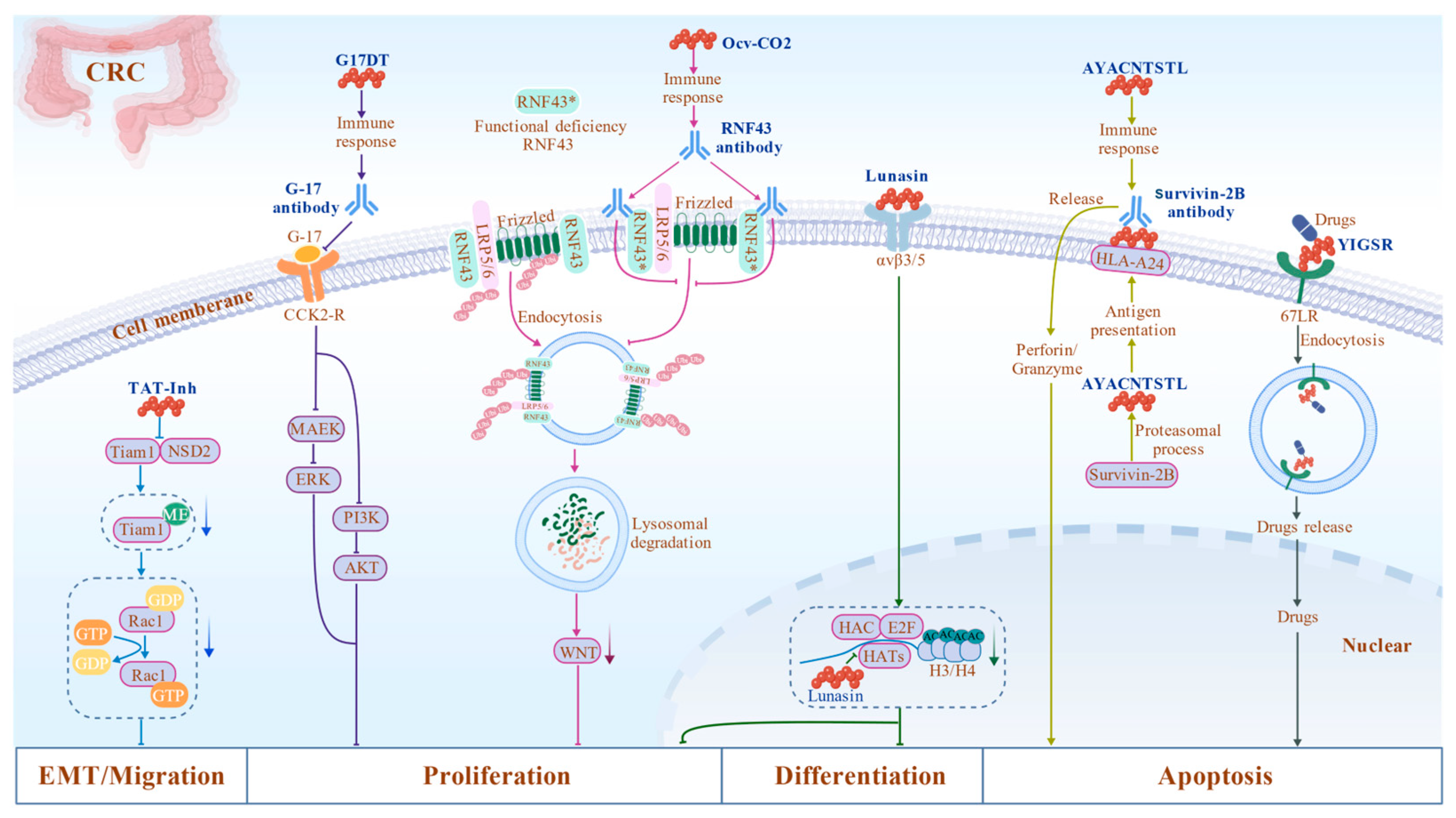
| TAT-Inh | YGRKKRRQRRRIVVDSRGFQKL | Inhibits Rac1 activity and suppresses EMT and metastasis by blocking NSD2/Tiam1 [61,62]. | Blocking peptides | CRC |
| Lunasin | SKWQHQQDSCRKQKQGVNLTPCEKHMEKIQGRGDDDDDDDD | Inhibit cancer cell proliferation and transformation by competitively binding to HATs [67,68]. | Blocking peptides | CRC |
| YIGSR | YIGSR | Targeted delivery to 67LR, combined with GEM to overcome resistance [73,74,75]. | Delivery peptides | CRC |
| G17DT | GPWLEEEEE | Induces anti-gastrin antibody production, blocks CCK2-R signaling, inhibits cell proliferation, and induces apoptosis [155,222]. | Mimetic peptides | CRC/GC |
| 0020OCV-C02 | NSQPVWLCL(RNF43-721) KLRQEVKQNL(TOMM34-299) | For mutant RNF43 [223], combination with UFT/LV therapy improves recurrence prognosis [80,81,82,83]. | Mimetic peptides | CRC |
| Survivin-2B | AYACNTSTL | HLA-A24 restrictive epitope peptide vaccine targeting invivor-2B can promote cell apoptosis [87]. | Mimetic peptides | CRC |
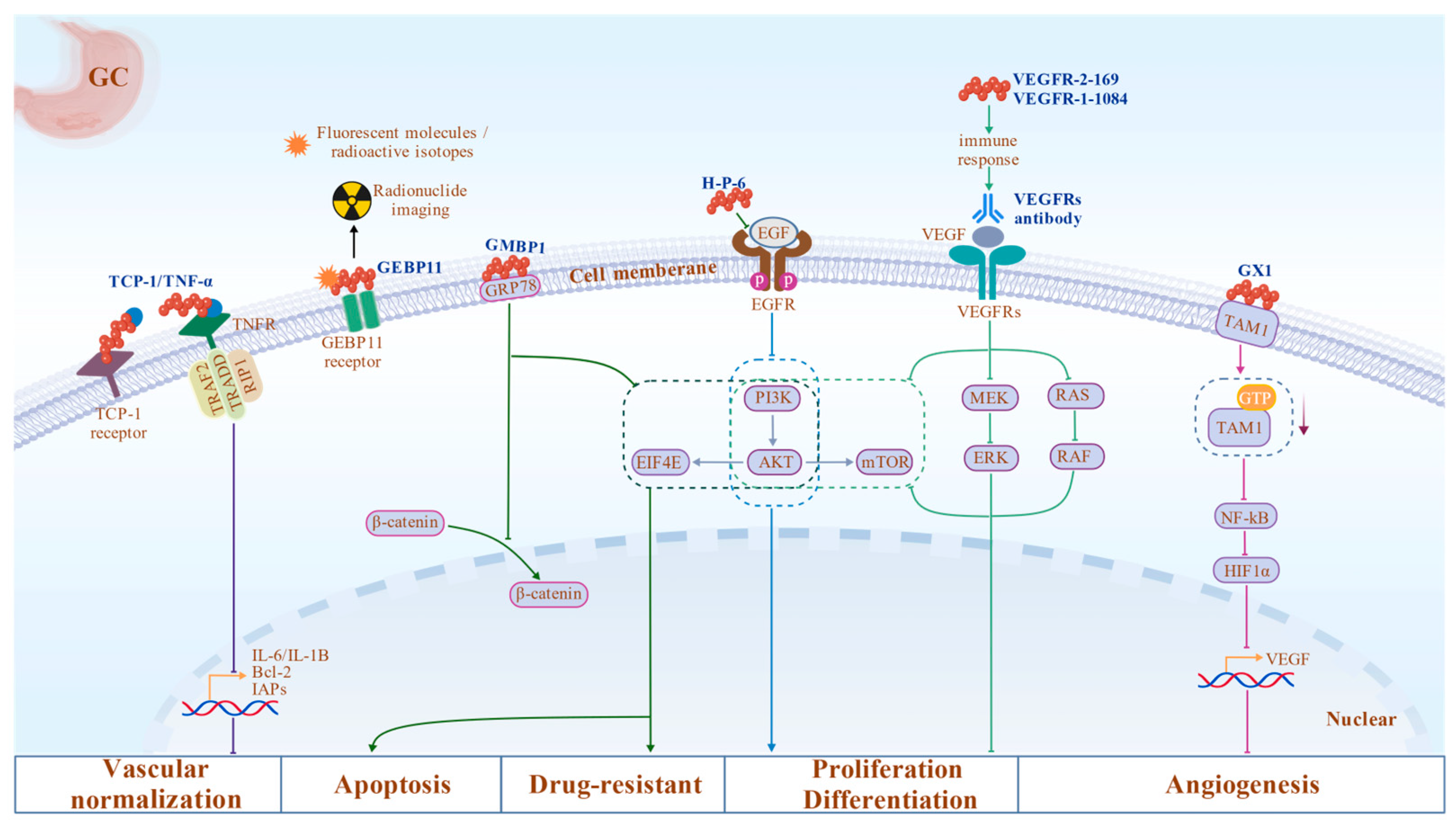
| GX1 Peptide | CGNSNPKSC(cyclic) | GX1-PTX-NLCs deliver PTX specifically to tumor vasculature, inhibiting angiogenesis and exerting antitumor effects [106,107]. | Blocking peptides | GC |
| H-P-6 | PQPKVLDS | Prevents H. pylori-induced proliferation by inhibiting EGFR signaling [100]. | Blocking peptides | GC |
| GMBP1 | ETAPLSTMLSPY | Reverse drug resistance and enhance chemotherapy sensitivity by binding to GRP78 [101,102]. | Blocking peptides | GC |
| TCP-1/TNF-α | CTPSPFSHC | Targeted delivery of TNFα to the GC vascular system, combined with chemotherapy to inhibit tumor growth [108]. | Delivery peptides | GC |
| GEBP11 | CTKNSYLMC | Targets vascular imaging to guide surgical resection [109,110,111,112,113]. | Delivery peptides | GC |
| VEGFR1-1084 VEGFR2-169 | SYGVLLWEI/RFVPDGNRI | Induce CTL response and inhibit angiogenesis [116]. | Mimetic peptides | GC |
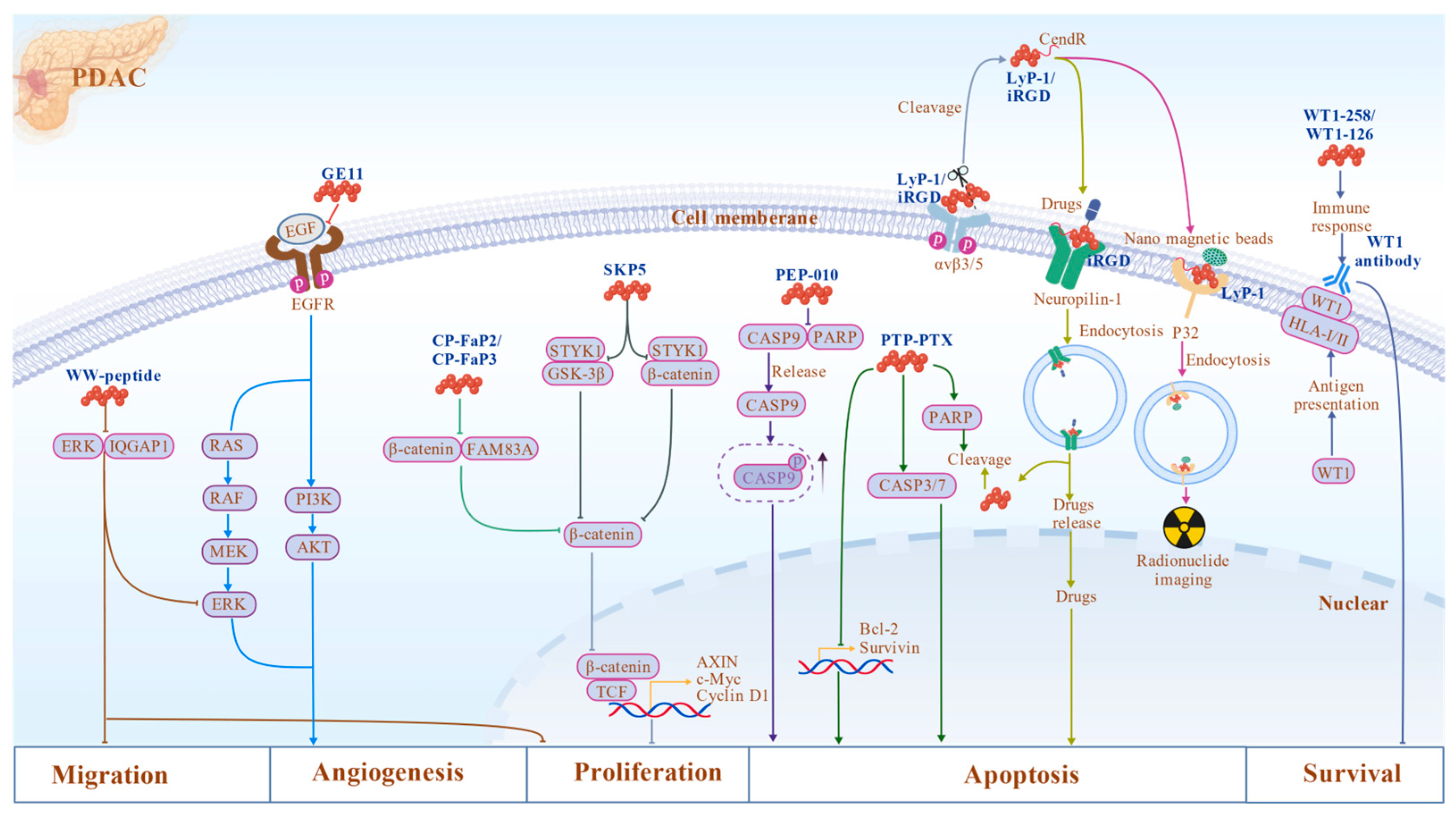
| WW Peptide | WWRRWWRR/(WWRR)n | Inhibits cell migration and metastasis by targeting IQGAP1-ERK complex [224,225]. | Blocking peptides | PDAC |
| CP-FaP2/ CP-FaP3 | YGRKKRRQRRR-LELQLRLQELQKQL/ YGRKKRRQRRR-QELQKQLLELQLRL | Disrupt FAM83A-β-catenin interaction, and inhibit Rac1 signaling and EMT [129]. | Blocking peptides | PDAC |
| SKP5 | RADVWSFGILLYEMV | Inhibit cell proliferation by blocking the Wnt signaling pathway [130,131]. | Blocking peptides | PDAC |
| GE11 | YHWYGYTPQNVI | Specifically binds to EGFR, acting as a delivery ligand to enhance the targeting precision of chemotherapeutic drugs [135]. | Mimetic peptides | PDAC |
| PEP-010 | VKKKKIKAEIKIYVETLDDIFEQWAHSEDL | Induces apoptosis by blocking caspase-9/PP2A interaction and releasing caspase-9 [132]. | Blocking peptides | PDAC |
| iRGD | CRGDKGPDC | Opens drug channels and enhances tissue permeability of nanomedicines by targeting vascular integrin αvβ3/β5 [136,137,138,139,140,141]. | Delivery peptides | PDAC |
| LyP-1 | CGNSNPKSC | Precise imaging and drug delivery by targeting p32 receptor [144,145,146]. | Delivery peptides | PDAC |
| PTP-PEG | KTLLPTP | Plectin-1-targeted delivery system enhances intracellular drug accumulation and therapeutic efficacy [148,149,150]. | Delivery peptides | PDAC |
| WT1-235/ WT1-126 | CMTWNQMNL/ RMFPNAPYL | Induce WT1-specific immune responses and reduce cell viability [159,160,161,162]. | Mimetic peptides | PDAC |
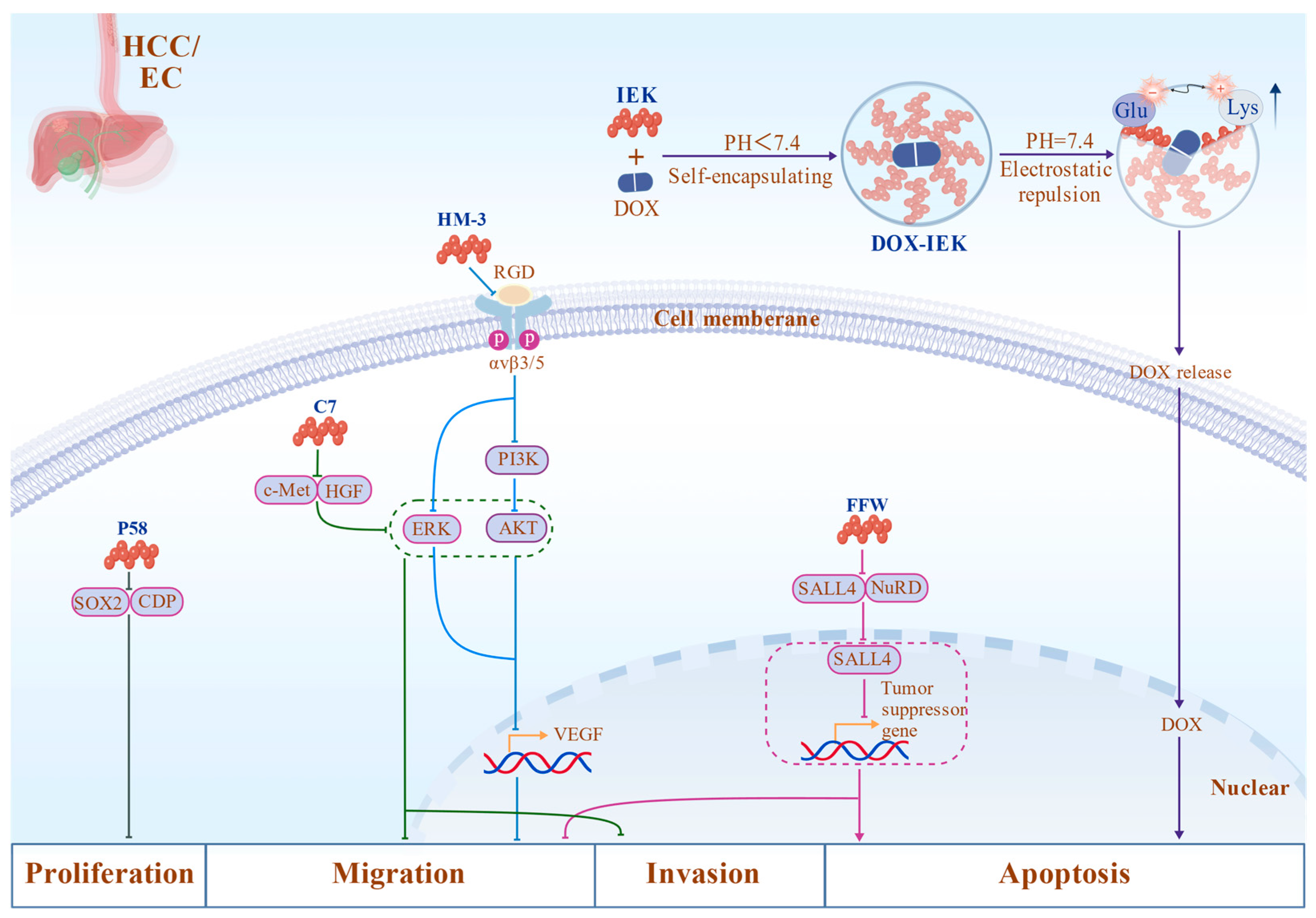
| C7 | CTPQTRPNC | Inhibits invasion and metastasis and reduces angiogenesis activity by competitively blocking HGF/c-Met [180]. | Blocking peptides | HCC |
| FFW | RRKFAKFQWI | Induces apoptosis, inhibits migration, and activates tumor suppressor genes by blocking SALL4/NuRD [179]. | Blocking peptides | HCC |
| HM-3 | IVRDRAAVPGGGRGD | Inhibits angiogenesis and metastasis and prolongs circulation by blocking αvβ3 integrin [181]. | Mimetic peptides | HCC |
| P58 | YLFAIYSFSSL | Inhibits cell proliferation, migration, and invasion, and induces apoptosis by blocking CDP/SOX2 [187]. | Blocking peptides | ESCC |
| DOX-IEK | IEIIIK | Serves as a drug delivery platform that facilitates pH-sensitive targeted release [189]. | Delivery peptides | ESCC |
Disclaimer/Publisher’s Note: The statements, opinions and data contained in all publications are solely those of the individual author(s) and contributor(s) and not of MDPI and/or the editor(s). MDPI and/or the editor(s) disclaim responsibility for any injury to people or property resulting from any ideas, methods, instructions or products referred to in the content. |
© 2026 by the authors. Licensee MDPI, Basel, Switzerland. This article is an open access article distributed under the terms and conditions of the Creative Commons Attribution (CC BY) license.
Share and Cite
Ouyang, Q.; Wu, G.; Chen, A.; Zhang, R.; Xiao, S.; Guo, D.; Zhang, Q.; Yan, C.; Chen, X.-Z.; Tang, J.; et al. Peptide Drugs in Gastrointestinal Tumors: Integrating Targeting, Delivery, and Therapeutic Actions for Synergistic Strategies. Biomolecules 2026, 16, 456. https://doi.org/10.3390/biom16030456
Ouyang Q, Wu G, Chen A, Zhang R, Xiao S, Guo D, Zhang Q, Yan C, Chen X-Z, Tang J, et al. Peptide Drugs in Gastrointestinal Tumors: Integrating Targeting, Delivery, and Therapeutic Actions for Synergistic Strategies. Biomolecules. 2026; 16(3):456. https://doi.org/10.3390/biom16030456
Chicago/Turabian StyleOuyang, Qian, Guifang Wu, Anyi Chen, Rui Zhang, Shuai Xiao, Dong Guo, Qi Zhang, Chaojun Yan, Xing-Zhen Chen, Jingfeng Tang, and et al. 2026. "Peptide Drugs in Gastrointestinal Tumors: Integrating Targeting, Delivery, and Therapeutic Actions for Synergistic Strategies" Biomolecules 16, no. 3: 456. https://doi.org/10.3390/biom16030456
APA StyleOuyang, Q., Wu, G., Chen, A., Zhang, R., Xiao, S., Guo, D., Zhang, Q., Yan, C., Chen, X.-Z., Tang, J., & Lyu, H. (2026). Peptide Drugs in Gastrointestinal Tumors: Integrating Targeting, Delivery, and Therapeutic Actions for Synergistic Strategies. Biomolecules, 16(3), 456. https://doi.org/10.3390/biom16030456