Macrophage Extracellular Vesicles: Therapeutic Strategies for Corneal Fibrosis in Rare Diseases
 ,
,  and
and
Abstract
1. Introduction
2. The Role of Immune Response, Macrophages and Nerves in Corneal Fibrosis
2.1. Corneal Inflammation Leading to Fibrosis
2.2. Involvement of Macrophages in Corneal Fibrosis
2.3. Involvement of Corneal Nerves in Corneal Fibrosis
3. Corneal Fibrosis in Rare Diseases
3.1. Rare Diseases and Epidermolysis Bullosa (EB)
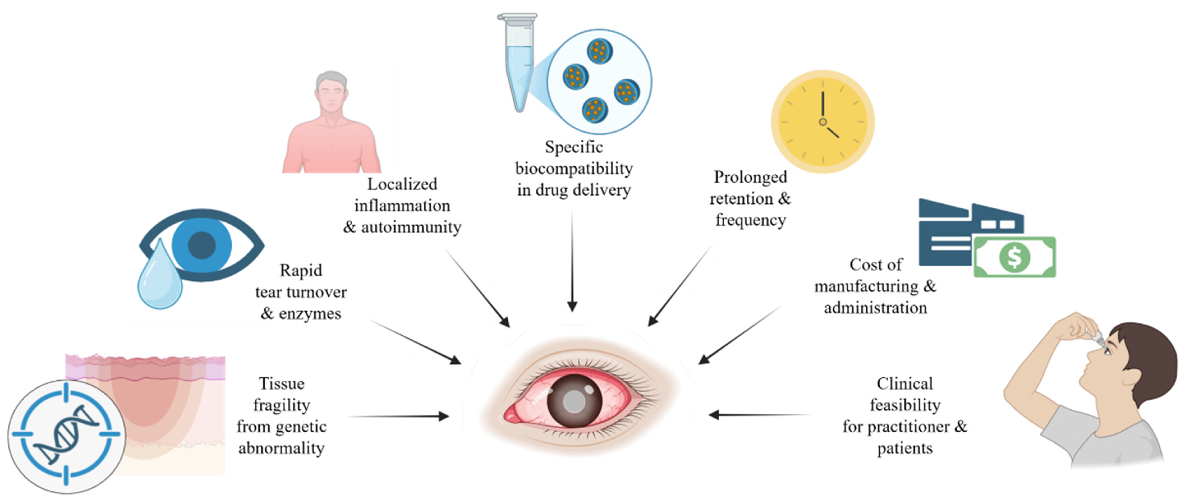
3.2. Considerations of Therapeutic Development for Rare Diseases
3.3. Current Ocular Treatments and Drug Delivery Options for Rare Diseases
3.4. Drug Delivery Options for Rare Diseases
4. Extracellular Vesicles for Corneal Fibrosis
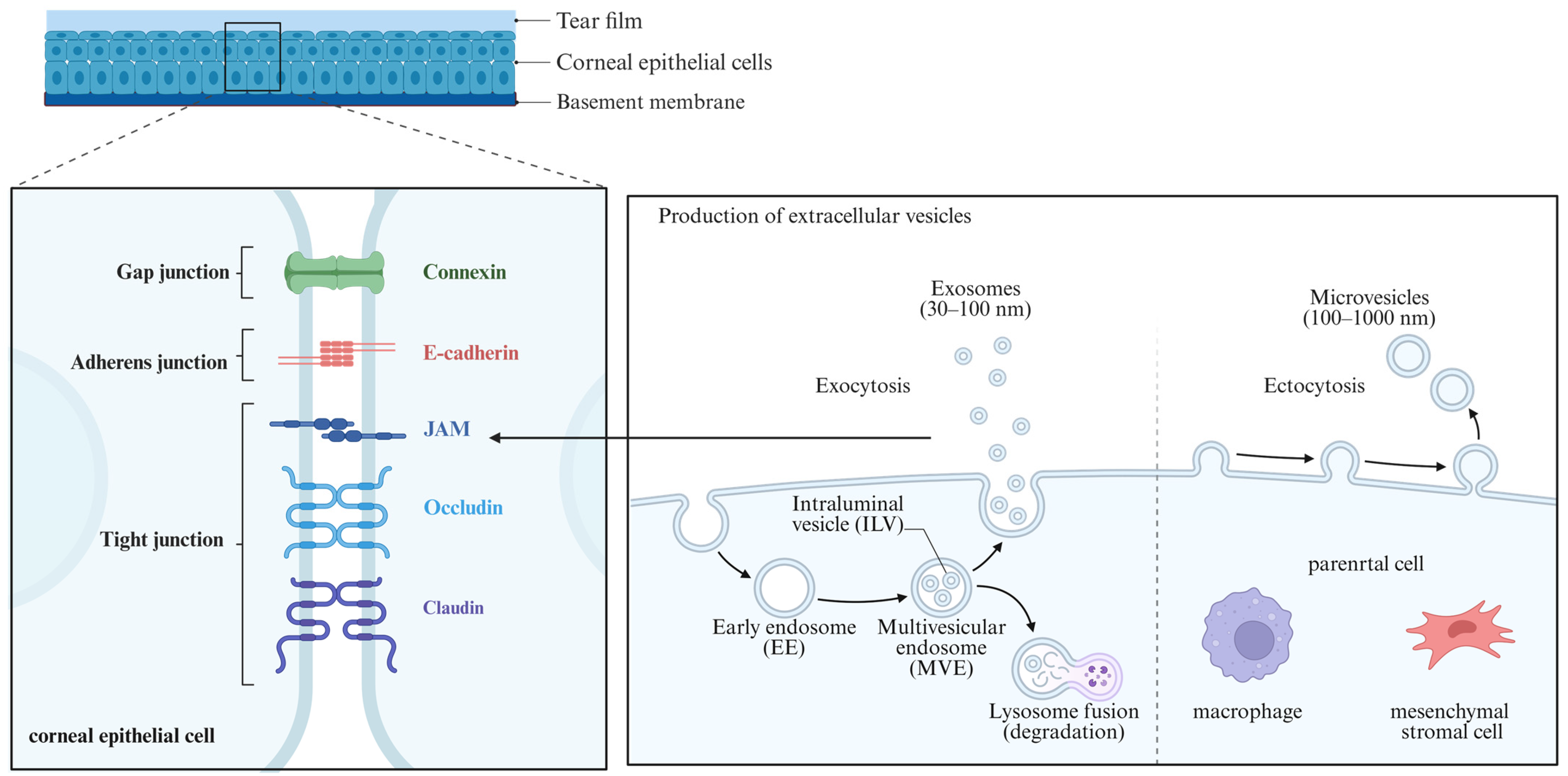
4.1. Involvement of EVs in Corneal Wound Healing
4.2. Specific EVs for Corneal Fibrosis
4.3. Current Developmental Progress of EVs for Corneal Fibrosis
4.4. Potential Delivery Methods of EVs for Corneal Fibrosis
5. Mac-EVs and Exosomes
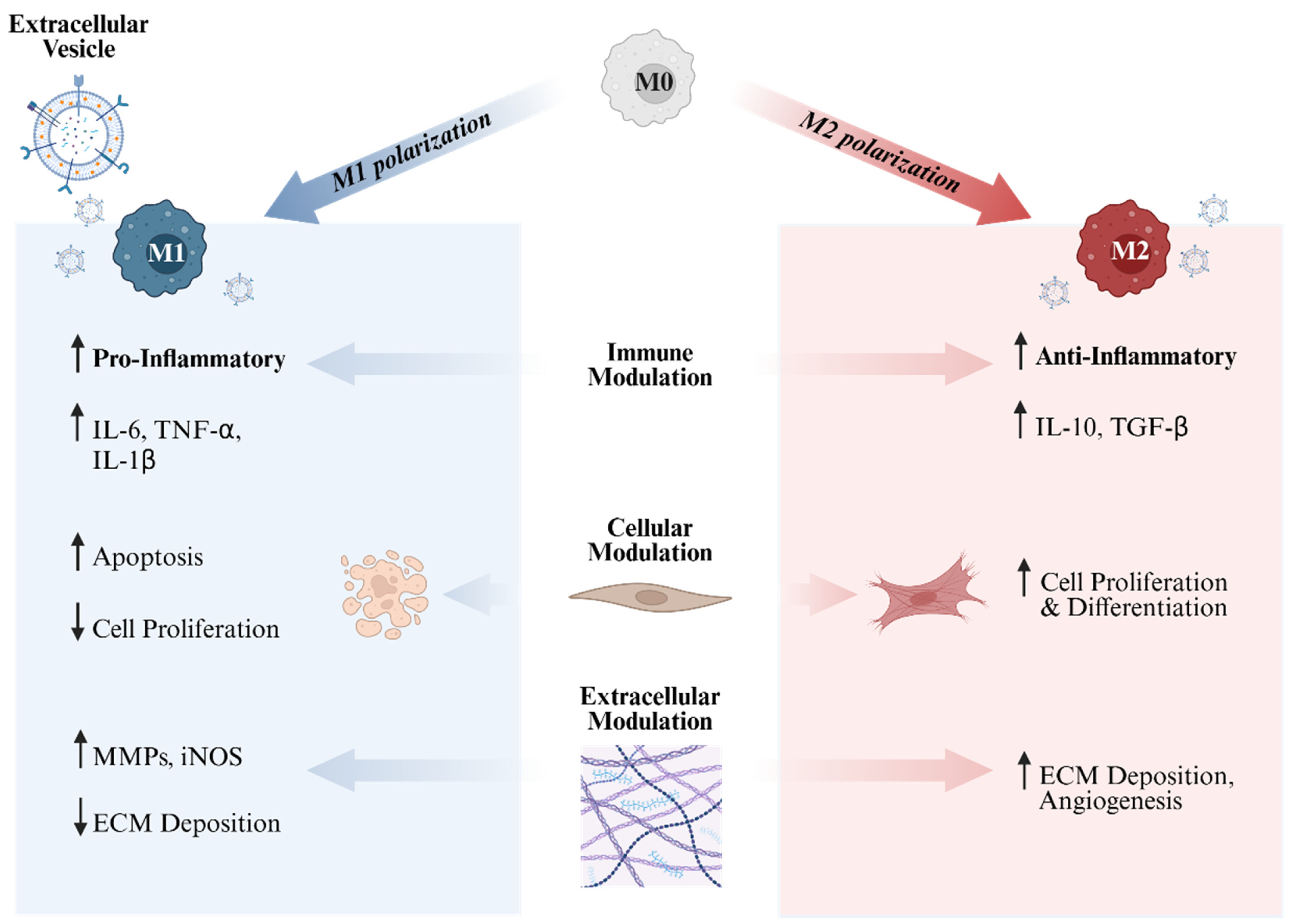
5.1. Types of Mac-EVs and Exosomes
5.2. Synthesis and Characterization of EVs
5.3. Potential Delivery Methods of Mac-EVs
5.4. Targeting EB-Associated Rare Diseases Using Mac-EVs
6. Conclusions
7. Future Directions
Author Contributions
Funding
Institutional Review Board Statement
Informed Consent Statement
Data Availability Statement
Acknowledgments
Conflicts of Interest
References
- Eghrari, A.O.; Riazuddin, S.A.; Gottsch, J.D. Overview of the Cornea: Structure, Function, and Development. Prog. Mol. Biol. Transl. Sci. 2015, 134, 7–23. [Google Scholar] [CrossRef] [Scilit] [PubMed]
- Meek, K.M.; Knupp, C. Corneal structure and transparency. Prog. Retin. Eye Res. 2015, 49, 1–16. [Google Scholar] [CrossRef] [Scilit] [PubMed]
- Fini, M.E. Keratocyte and fibroblast phenotypes in the repairing cornea. Prog. Retin. Eye Res. 1999, 18, 529–551. [Google Scholar] [CrossRef] [Scilit] [PubMed]
- Ljubimov, A.V.; Saghizadeh, M. Progress in corneal wound healing. Prog. Retin. Eye Res. 2015, 49, 17–45. [Google Scholar] [CrossRef] [Scilit]
- Whitcher, J.P.; Srinivasan, M.; Upadhyay, M.P. Corneal blindness: A global perspective. Bull. World Health Organ. 2001, 79, 214–221. [Google Scholar]
- Pascolini, D.; Mariotti, S.P. Global estimates of visual impairment: 2010. Br. J. Ophthalmol. 2012, 96, 614–618. [Google Scholar] [CrossRef] [Scilit]
- McKay, T.B.; Hutcheon, A.E.K.; Zieske, J.D. Biology of corneal fibrosis: Soluble mediators, integrins, and extracellular vesicles. Eye 2020, 34, 271–278. [Google Scholar] [CrossRef] [Scilit]
- Medeiros, C.S.; Marino, G.K.; Santhiago, M.R.; Wilson, S.E. The Corneal Basement Membranes and Stromal Fibrosis. Investig. Ophthalmol. Vis. Sci. 2018, 59, 4044–4053. [Google Scholar] [CrossRef] [Scilit]
- Roshandel, D.; Eslani, M.; Baradaran-Rafii, A.; Cheung, A.Y.; Kurji, K.; Jabbehdari, S.; Maiz, A.; Jalali, S.; Djalilian, A.R.; Holland, E.J. Current and emerging therapies for corneal neovascularization. Ocul. Surf. 2018, 16, 398–414. [Google Scholar] [CrossRef] [Scilit]
- Verma, S.; Moreno, I.Y.; Prinholato da Silva, C.; Sun, M.; Cheng, X.; Gesteira, T.F.; Coulson-Thomas, V.J. Endogenous TSG-6 modulates corneal inflammation following chemical injury. Ocul. Surf. 2024, 32, 26–38. [Google Scholar] [CrossRef] [Scilit]
- Downie, L.E.; Zhang, X.; Wu, M.; Karunaratne, S.; Loi, J.K.; Senthil, K.; Arshad, S.; Bertram, K.; Cunningham, A.L.; Carnt, N.; et al. Redefining the human corneal immune compartment using dynamic intravital imaging. Proc. Natl. Acad. Sci. USA 2023, 120, e2217795120. [Google Scholar] [CrossRef] [Scilit]
- Wu, M.; Fletcher, E.L.; Chinnery, H.R.; Downie, L.E.; Mueller, S.N. Redefining our vision: An updated guide to the ocular immune system. Nat. Rev. Immunol. 2024, 24, 896–911. [Google Scholar] [CrossRef] [Scilit]
- Nowell, C.S.; Odermatt, P.D.; Azzolin, L.; Hohnel, S.; Wagner, E.F.; Fantner, G.E.; Lutolf, M.P.; Barrandon, Y.; Piccolo, S.; Radtke, F. Chronic inflammation imposes aberrant cell fate in regenerating epithelia through mechanotransduction. Nat. Cell Biol. 2016, 18, 168–180. [Google Scholar] [CrossRef] [Scilit]
- Jadczyk-Sorek, K.; Garczorz, W.; Bubala-Stachowicz, B.; Francuz, T.; Mrukwa-Kominek, E. Matrix Metalloproteinases and the Pathogenesis of Recurrent Corneal Erosions and Epithelial Basement Membrane Dystrophy. Biology 2023, 12, 1263. [Google Scholar] [CrossRef] [Scilit] [PubMed]
- Jammes, M.; Tabasi, A.; Bach, T.; Ritter, T. Healing the cornea: Exploring the therapeutic solutions offered by MSCs and MSC-derived EVs. Prog. Retin. Eye Res. 2025, 105, 101325. [Google Scholar] [CrossRef] [Scilit] [PubMed]
- Kenyon, K.R.; Fogle, J.A.; Stone, D.L.; Stark, W.J. Regeneration of corneal epithelial basement membrane following thermal cauterization. Investig. Ophthalmol. Vis. Sci. 1977, 16, 292–301. [Google Scholar]
- Yang, G.N.; Roberts, P.K.; Gardner-Russell, J.; Shah, M.H.; Couper, T.A.; Zhu, Z.; Pollock, G.A.; Dusting, G.J.; Daniell, M. From bench to clinic: Emerging therapies for corneal scarring. Pharmacol. Ther. 2023, 242, 108349. [Google Scholar] [CrossRef] [Scilit]
- Wilson, S.E. Fibrosis Is a Basement Membrane-Related Disease in the Cornea: Injury and Defective Regeneration of Basement Membranes May Underlie Fibrosis in Other Organs. Cells 2022, 11, 309. [Google Scholar] [CrossRef] [Scilit]
- Tong, L.; Hodgkins, P.R.; Denyer, J.; Brosnahan, D.; Harper, J.; Russell-Eggitt, I.; Taylor, D.S.; Atherton, D. The eye in epidermolysis bullosa. Br. J. Ophthalmol. 1999, 83, 323–326. [Google Scholar] [CrossRef] [Scilit]
- Rousseau, A.; Prost-Squarcioni, C.; Doan, S.; Leroux-Villet, C.; Caux, F.; Hoang-Xuan, T.; Cochereau, I.; Gabison, E. Ocular involvement in epidermolysis bullosa acquisita with long-term follow-up. Br. J. Ophthalmol. 2020, 104, 235–240. [Google Scholar] [CrossRef] [Scilit]
- Fine, J.D.; Johnson, L.B.; Weiner, M.; Stein, A.; Cash, S.; Deleoz, J.; Devries, D.T.; Suchindran, C. Eye involvement in inherited epidermolysis bullosa: Experience of the National Epidermolysis Bullosa Registry. Am. J. Ophthalmol. 2004, 138, 254–262. [Google Scholar] [CrossRef] [Scilit] [PubMed]
- Bachir, Y.; Daruich, A.; Marie, C.; Robert, M.P.; Bremond-Gignac, D. Eye Involvement and Management in Inherited Epidermolysis Bullosa. Drugs 2022, 82, 1277–1285. [Google Scholar] [CrossRef] [Scilit] [PubMed]
- Ong, H.S.; Riau, A.K.; Yam, G.H.; Yusoff, N.; Han, E.J.Y.; Goh, T.W.; Lai, R.C.; Lim, S.K.; Mehta, J.S. Mesenchymal Stem Cell Exosomes as Immunomodulatory Therapy for Corneal Scarring. Int. J. Mol. Sci. 2023, 24, 7456. [Google Scholar] [CrossRef] [Scilit] [PubMed]
- Kalluri, R.; LeBleu, V.S. The biology, function, and biomedical applications of exosomes. Science 2020, 367, eaau6977. [Google Scholar] [CrossRef] [Scilit]
- Torricelli, A.A.; Singh, V.; Santhiago, M.R.; Wilson, S.E. The corneal epithelial basement membrane: Structure, function, and disease. Investig. Ophthalmol. Vis. Sci. 2013, 54, 6390–6400. [Google Scholar] [CrossRef] [Scilit]
- Lassance, L.; Marino, G.K.; Medeiros, C.S.; Thangavadivel, S.; Wilson, S.E. Fibrocyte migration, differentiation and apoptosis during the corneal wound healing response to injury. Exp. Eye Res. 2018, 170, 177–187. [Google Scholar] [CrossRef] [Scilit]
- Varki, R.; Sadowski, S.; Pfendner, E.; Uitto, J. Epidermolysis bullosa. I. Molecular genetics of the junctional and hemidesmosomal variants. J. Med. Genet. 2006, 43, 641–652. [Google Scholar] [CrossRef] [Scilit]
- Rocher, M.; Robert, P.Y.; Desmouliere, A. The myofibroblast, biological activities and roles in eye repair and fibrosis. A focus on healing mechanisms in avascular cornea. Eye 2020, 34, 232–240. [Google Scholar] [CrossRef] [Scilit]
- Jhanji, V.; Santra, M.; Riau, A.K.; Geary, M.L.; Yang, T.; Rubin, E.; Yusoff, N.; Dhaliwal, D.K.; Mehta, J.S.; Yam, G.H. Combined Therapy Using Human Corneal Stromal Stem Cells and Quiescent Keratocytes to Prevent Corneal Scarring after Injury. Int. J. Mol. Sci. 2022, 23, 6980. [Google Scholar] [CrossRef] [Scilit]
- Latta, L.; Figueiredo, F.C.; Ashery-Padan, R.; Collinson, J.M.; Daniels, J.; Ferrari, S.; Szentmáry, N.; Solá, S.; Shalom-Feuerstein, R.; Lako, M.; et al. Pathophysiology of aniridia-associated keratopathy: Developmental aspects and unanswered questions. Ocul. Surf. 2021, 22, 245–266. [Google Scholar] [CrossRef] [Scilit]
- Akhlaq, A.; Colon, C.; Cavalcanti, B.M.; Aggarwal, S.; Qazi, Y.; Cruzat, A.; Jersey, C.; Critser, D.B.; Watts, A.; Beyer, J.; et al. Density and distribution of dendritiform cells in the peripheral cornea of healthy subjects using in vivo confocal microscopy. Ocul. Surf. 2022, 26, 157–165. [Google Scholar] [CrossRef] [Scilit] [PubMed]
- Bedggood, P.; Wu, M.; Zhang, X.; Rajan, R.; Wu, C.Y.; Karunaratne, S.; Metha, A.B.; Mueller, S.N.; Chinnery, H.R.; Downie, L.E. Improved tracking of corneal immune cell dynamics using in vivo confocal microscopy. Biomed. Opt. Express 2024, 15, 6277–6298. [Google Scholar] [CrossRef] [Scilit] [PubMed]
- Wu, M.; Zhang, X.; Karunaratne, S.; Lee, J.H.; Lampugnani, E.R.; Selva, K.J.; Chung, A.W.; Mueller, S.N.; Chinnery, H.R.; Downie, L.E. Intravital Imaging of the Human Cornea Reveals the Differential Effects of Season on Innate and Adaptive Immune Cell Morphodynamics. Ophthalmology 2024, 131, 1185–1195. [Google Scholar] [CrossRef] [Scilit] [PubMed]
- Yu, F.X.; Lee, P.S.Y.; Yang, L.; Gao, N.; Zhang, Y.; Ljubimov, A.V.; Yang, E.; Zhou, Q.; Xie, L. The impact of sensory neuropathy and inflammation on epithelial wound healing in diabetic corneas. Prog. Retin. Eye Res. 2022, 89, 101039. [Google Scholar] [CrossRef] [Scilit]
- Mohan, R.R.; Kempuraj, D.; D’Souza, S.; Ghosh, A. Corneal stromal repair and regeneration. Prog. Retin. Eye Res. 2022, 91, 101090. [Google Scholar] [CrossRef] [Scilit]
- Varol, C.; Mildner, A.; Jung, S. Macrophages: Development and tissue specialization. Annu. Rev. Immunol. 2015, 33, 643–675. [Google Scholar] [CrossRef] [Scilit]
- Wieghofer, P.; Hagemeyer, N.; Sankowski, R.; Schlecht, A.; Staszewski, O.; Amann, L.; Gruber, M.; Koch, J.; Hausmann, A.; Zhang, P.; et al. Mapping the origin and fate of myeloid cells in distinct compartments of the eye by single-cell profiling. EMBO J. 2021, 40, e105123. [Google Scholar] [CrossRef] [Scilit]
- Hoeffel, G.; Wang, Y.; Greter, M.; See, P.; Teo, P.; Malleret, B.; Leboeuf, M.; Low, D.; Oller, G.; Almeida, F.; et al. Adult Langerhans cells derive predominantly from embryonic fetal liver monocytes with a minor contribution of yolk sac-derived macrophages. J. Exp. Med. 2012, 209, 1167–1181. [Google Scholar] [CrossRef] [Scilit]
- Ploeger, D.T.; Hosper, N.A.; Schipper, M.; Koerts, J.A.; de Rond, S.; Bank, R.A. Cell plasticity in wound healing: Paracrine factors of M1/M2 polarized macrophages influence the phenotypical state of dermal fibroblasts. Cell Commun. Signal 2013, 11, 29. [Google Scholar] [CrossRef] [Scilit]
- Brissette-Storkus, C.S.; Reynolds, S.M.; Lepisto, A.J.; Hendricks, R.L. Identification of a novel macrophage population in the normal mouse corneal stroma. Investig. Ophthalmol. Vis. Sci. 2002, 43, 2264–2271. [Google Scholar]
- Davies, L.C.; Jenkins, S.J.; Allen, J.E.; Taylor, P.R. Tissue-resident macrophages. Nat. Immunol. 2013, 14, 986–995. [Google Scholar] [CrossRef] [Scilit] [PubMed]
- Duffield, J.S.; Lupher, M.; Thannickal, V.J.; Wynn, T.A. Host responses in tissue repair and fibrosis. Annu. Rev. Pathol. 2013, 8, 241–276. [Google Scholar] [CrossRef] [Scilit] [PubMed]
- Mantovani, A.; Sica, A.; Sozzani, S.; Allavena, P.; Vecchi, A.; Locati, M. The chemokine system in diverse forms of macrophage activation and polarization. Trends Immunol. 2004, 25, 677–686. [Google Scholar] [CrossRef] [Scilit] [PubMed]
- Shapouri-Moghaddam, A.; Mohammadian, S.; Vazini, H.; Taghadosi, M.; Esmaeili, S.A.; Mardani, F.; Seifi, B.; Mohammadi, A.; Afshari, J.T.; Sahebkar, A. Macrophage plasticity, polarization, and function in health and disease. J. Cell. Physiol. 2018, 233, 6425–6440. [Google Scholar] [CrossRef] [Scilit]
- Hickman, E.; Smyth, T.; Cobos-Uribe, C.; Immormino, R.; Rebuli, M.E.; Moran, T.; Alexis, N.E.; Jaspers, I. Expanded characterization of in vitro polarized M0, M1, and M2 human monocyte-derived macrophages: Bioenergetic and secreted mediator profiles. PLoS ONE 2023, 18, e0279037. [Google Scholar] [CrossRef] [Scilit]
- Zhao, X.; Yang, L.; Pan, J.; Zeng, Z.; Zhang, T.; Yang, Y.; Zhang, J.; Chen, T.; Xiao, Z.; Pan, W. CXCL8 modulates M0 macrophage proliferation and polarization to influence tumor progression in cervical cancer. Sci. Rep. 2025, 15, 790. [Google Scholar] [CrossRef] [Scilit]
- Gordon, S. Alternative activation of macrophages. Nat. Rev. Immunol. 2003, 3, 23–35. [Google Scholar] [CrossRef] [Scilit]
- Li, Z.; Bratlie, K.M. Fibroblasts treated with macrophage conditioned medium results in phenotypic shifts and changes in collagen organization. Mater. Sci. Eng. C Mater. Biol. Appl. 2021, 122, 111915. [Google Scholar] [CrossRef] [Scilit]
- Martinez, F.O.; Gordon, S. The M1 and M2 paradigm of macrophage activation: Time for reassessment. F1000Prime Rep. 2014, 6, 13. [Google Scholar] [CrossRef] [Scilit]
- Lee, H.S.; Amouzegar, A.; Dana, R. Kinetics of Corneal Antigen Presenting Cells in Experimental Dry Eye Disease. BMJ Open Ophthalmol. 2017, 1, e000078. [Google Scholar] [CrossRef] [Scilit]
- Li, W.; Gurdziel, K.; Pitchaikannu, A.; Gupta, N.; Hazlett, L.D.; Xu, S. The miR-183/96/182 cluster is a checkpoint for resident immune cells and shapes the cellular landscape of the cornea. Ocul. Surf. 2023, 30, 17–41. [Google Scholar] [CrossRef] [Scilit] [PubMed]
- Chu, C.; Huang, Y.; Cao, L.; Ji, S.; Zhu, B.; Shen, Q. Role of macrophages in peritoneal dialysis-associated peritoneal fibrosis. Ren. Fail. 2025, 47, 2474203. [Google Scholar] [CrossRef] [Scilit] [PubMed]
- Gordon, S.; Taylor, P.R. Monocyte and macrophage heterogeneity. Nat. Rev. Immunol. 2005, 5, 953–964. [Google Scholar] [CrossRef] [Scilit] [PubMed]
- Ma, J.; Chen, T.; Mandelin, J.; Ceponis, A.; Miller, N.E.; Hukkanen, M.; Ma, G.F.; Konttinen, Y.T. Regulation of macrophage activation. Cell. Mol. Life Sci. 2003, 60, 2334–2346. [Google Scholar] [CrossRef] [Scilit]
- Liu, T.; Huang, T.; Li, J.; Li, A.; Li, C.; Huang, X.; Li, D.; Wang, S.; Liang, M. Optimization of differentiation and transcriptomic profile of THP-1 cells into macrophage by PMA. PLoS ONE 2023, 18, e0286056. [Google Scholar] [CrossRef] [Scilit]
- Tarique, A.A.; Logan, J.; Thomas, E.; Holt, P.G.; Sly, P.D.; Fantino, E. Phenotypic, functional, and plasticity features of classical and alternatively activated human macrophages. Am. J. Respir. Cell Mol. Biol. 2015, 53, 676–688. [Google Scholar] [CrossRef] [Scilit]
- Bonini, S.; Rama, P.; Olzi, D.; Lambiase, A. Neurotrophic keratitis. Eye 2003, 17, 989–995. [Google Scholar] [CrossRef] [Scilit]
- Maher, C.; Chen, Z.; Zhou, Y.; You, J.; Sutton, G.; Wallace, G. Innervation in corneal bioengineering. Acta Biomater. 2024, 189, 73–87. [Google Scholar] [CrossRef] [Scilit]
- Marfurt, C.F.; Cox, J.; Deek, S.; Dvorscak, L. Anatomy of the human corneal innervation. Exp. Eye Res. 2010, 90, 478–492. [Google Scholar] [CrossRef] [Scilit]
- Wu, M.; Hill, L.J.; Downie, L.E.; Chinnery, H.R. Neuroimmune crosstalk in the cornea: The role of immune cells in corneal nerve maintenance during homeostasis and inflammation. Prog. Retin. Eye Res. 2022, 91, 101105. [Google Scholar] [CrossRef] [Scilit]
- Tervo, K.; Tervo, T.; Eränkö, L.; Eränkö, O. Substance P immunoreactive nerves in the rodent cornea. Neurosci. Lett. 1981, 25, 95–97. [Google Scholar] [CrossRef] [Scilit] [PubMed]
- Shaheen, B.S.; Bakir, M.; Jain, S. Corneal nerves in health and disease. Surv. Ophthalmol. 2014, 59, 263–285. [Google Scholar] [CrossRef] [Scilit] [PubMed]
- Słoniecka, M.; Danielson, P. Substance P induces fibrotic changes through activation of the RhoA/ROCK pathway in an in vitro human corneal fibrosis model. J. Mol. Med. 2019, 97, 1477–1489. [Google Scholar] [CrossRef] [Scilit] [PubMed]
- Schmid, E.; Leierer, J.; Doblinger, A.; Laslop, A.; Fischer-Colbrie, R.; Humpel, C.; Theodorsson, E.; Teuchner, B.; Lalehabbasi, D.; Dragosits, E.; et al. Neurokinin a is a main constituent of sensory neurons innervating the anterior segment of the eye. Investig. Ophthalmol. Vis. Sci. 2005, 46, 268–274. [Google Scholar] [CrossRef] [Scilit]
- Nakamura, M.; Nishida, T.; Ofuji, K.; Reid, T.W.; Mannis, M.J.; Murphy, C.J. Synergistic effect of substance P with epidermal growth factor on epithelial migration in rabbit cornea. Exp. Eye Res. 1997, 65, 321–329. [Google Scholar] [CrossRef] [Scilit]
- Sacchetti, M.; Micera, A.; Lambiase, A.; Speranza, S.; Mantelli, F.; Petrachi, G.; Bonini, S.; Bonini, S. Tear levels of neuropeptides increase after specific allergen challenge in allergic conjunctivitis. Mol. Vis. 2011, 17, 47–52. [Google Scholar]
- Ramappa, M.; Chaurasia, S.; Chakrabarti, S.; Kaur, I. Congenital corneal anesthesia. J. AAPOS 2014, 18, 427–432. [Google Scholar] [CrossRef] [Scilit]
- Fine, J.-D. Epidemiology of Inherited Epidermolysis Bullosa Based on Incidence and Prevalence Estimates From the National Epidermolysis Bullosa Registry. JAMA Dermatol. 2016, 152, 1231–1238. [Google Scholar] [CrossRef] [Scilit]
- Kiritsi, D.; Has, C.; Bruckner-Tuderman, L. Laminin 332 in junctional epidermolysis bullosa. Cell Adh. Migr. 2013, 7, 135–141. [Google Scholar] [CrossRef] [Scilit]
- Kivirikko, S.; McGrath, J.A.; Pulkkinen, L.; Uitto, J.; Christiano, A.M. Mutational hotspots in the LAMB3 gene in the lethal (Herlitz) type of junctional epidermolysis bullosa. Hum. Mol. Genet. 1996, 5, 231–237. [Google Scholar] [CrossRef] [Scilit]
- Kwong, A.; Cogan, J.; Hou, Y.; Antaya, R.; Hao, M.; Kim, G.; Lincoln, V.; Chen, Q.; Woodley, D.T.; Chen, M. Gentamicin Induces Laminin 332 and Improves Wound Healing in Junctional Epidermolysis Bullosa Patients with Nonsense Mutations. Mol. Ther. 2020, 28, 1327–1338. [Google Scholar] [CrossRef] [Scilit] [PubMed]
- Tanaka, A.C.; Robbins, C.C.; Wiss, K.; Chen, V.M. Frequent corneal abrasions precede scarring and vision loss in epidermolysis bullosa: An international patient survey. J. Am. Acad. Dermatol. 2020, 83, 917–919. [Google Scholar] [CrossRef] [Scilit] [PubMed]
- Chen, V.M.; Mehta, N.; Robbins, C.C.; Noh, E.; Pramil, V.; Duker, J.S.; Waheed, N.K. Anterior-segment spectral domain optical coherence tomography in epidermolysis bullosa. Ocul. Surf. 2020, 18, 912–919. [Google Scholar] [CrossRef] [Scilit] [PubMed]
- Has, C.; Nystrom, A.; Saeidian, A.H.; Bruckner-Tuderman, L.; Uitto, J. Epidermolysis bullosa: Molecular pathology of connective tissue components in the cutaneous basement membrane zone. Matrix Biol. 2018, 71–72, 313–329. [Google Scholar] [CrossRef] [Scilit]
- Fine, J.D.; Bruckner-Tuderman, L.; Eady, R.A.; Bauer, E.A.; Bauer, J.W.; Has, C.; Heagerty, A.; Hintner, H.; Hovnanian, A.; Jonkman, M.F.; et al. Inherited epidermolysis bullosa: Updated recommendations on diagnosis and classification. J. Am. Acad. Dermatol. 2014, 70, 1103–1126. [Google Scholar] [CrossRef] [Scilit]
- Chen, V.M.; Kumar, V.S.S.; Noh, E.; Perloff, J. Validation and Repeatability of the Epidermolysis Bullosa Eye Disease Index in Dystrophic Epidermolysis Bullosa. Cornea 2023, 42, 74–79. [Google Scholar] [CrossRef] [Scilit]
- Ouyang, H.; Xue, Y.; Lin, Y.; Zhang, X.; Xi, L.; Patel, S.; Cai, H.; Luo, J.; Zhang, M.; Zhang, M.; et al. WNT7A and PAX6 define corneal epithelium homeostasis and pathogenesis. Nature 2014, 511, 358–361. [Google Scholar] [CrossRef] [Scilit]
- Ihnatko, R.; Eden, U.; Fagerholm, P.; Lagali, N. Congenital Aniridia and the Ocular Surface. Ocul. Surf. 2016, 14, 196–206. [Google Scholar] [CrossRef] [Scilit]
- Jacobson, A.; Mian, S.I.; Bohnsack, B.L. Clinical outcomes and visual prognostic factors in congenital aniridia. BMC Ophthalmol. 2022, 22, 235. [Google Scholar] [CrossRef] [Scilit]
- Messmer, E.M.; Kenyon, K.R.; Rittinger, O.; Janecke, A.R.; Kampik, A. Ocular manifestations of keratitis-ichthyosis-deafness (KID) syndrome. Ophthalmology 2005, 112, e1–e6. [Google Scholar] [CrossRef] [Scilit]
- Shah, S.R.; Karia, U.K.; Shah, B.J.; Vyas, H.R. Keratitis–Ichthyosis–Deafness: A Rare Kid in Clinical Practice. Indian J. Paediatr. Dermatol. 2022, 23, 251–253. [Google Scholar] [CrossRef] [Scilit]
- Mimura, T.; Amano, S.; Fukuoka, S.; Honda, N.; Arita, R.; Ochiai, M.; Yanagisawa, M.; Usui, T.; Ono, K.; Araki, F.; et al. In vivo confocal microscopy of hereditary sensory and autonomic neuropathy. Curr. Eye Res. 2008, 33, 940–945. [Google Scholar] [CrossRef] [Scilit] [PubMed]
- Haga, N.; Kubota, M.; Miwa, Z. Epidemiology of hereditary sensory and autonomic neuropathy type IV and V in Japan. Am. J. Med. Genet. Part A 2013, 161, 871–874. [Google Scholar]
- Hirsch, T.; Rothoeft, T.; Teig, N.; Bauer, J.W.; Pellegrini, G.; De Rosa, L.; Scaglione, D.; Reichelt, J.; Klausegger, A.; Kneisz, D.; et al. Regeneration of the entire human epidermis using transgenic stem cells. Nature 2017, 551, 327–332. [Google Scholar] [CrossRef] [Scilit]
- Kueckelhaus, M.; Rothoeft, T.; De Rosa, L.; Yeni, B.; Ohmann, T.; Maier, C.; Eitner, L.; Metze, D.; Losi, L.; Secone Seconetti, A.; et al. Transgenic Epidermal Cultures for Junctional Epidermolysis Bullosa—5-Year Outcomes. N. Engl. J. Med. 2021, 385, 2264–2270. [Google Scholar] [CrossRef] [Scilit]
- Gade, S.; So, Y.; Mishra, D.; Baviskar, S.M.; Assiri, A.A.; Glover, K.; Sheshala, R.; Vora, L.K.; Thakur, R.R.S. Ocular Drug Delivery: Emerging Approaches and Advances. Pharmaceutics 2025, 17, 599. [Google Scholar] [CrossRef] [Scilit]
- Sultana, S.; Yusuf, M.; Sharma, V. Nanovesicular Drug Delivery Systems for Rare Ocular Diseases: Advances, Challenges, and Future Directions. AAPS PharmSciTech 2025, 26, 197. [Google Scholar] [CrossRef] [Scilit]
- Agarwal, P.; Rupenthal, I.D. In vitro and ex vivo corneal penetration and absorption models. Drug Deliv. Transl. Res. 2016, 6, 634–647. [Google Scholar] [CrossRef] [Scilit]
- Mohan, R.R.; Rodier, J.T.; Sharma, A. Corneal gene therapy: Basic science and translational perspective. Ocul. Surf. 2013, 11, 150–164. [Google Scholar] [CrossRef] [Scilit]
- Tovar Vetencourt, A.; Sayed-Ahmed, I.; Gomez, J.; Chen, H.; Agostini, B.; Carroll, K.; Parry, T.; Krishnan, S.; Sabater, A.L. Ocular Gene Therapy in a Patient with Dystrophic Epidermolysis Bullosa. N. Engl. J. Med. 2024, 390, 530–535. [Google Scholar] [CrossRef] [Scilit]
- Wong, C.H.; Li, D.; Wang, N.; Gruber, J.; Lo, A.W.; Conti, R.M. The estimated annual financial impact of gene therapy in the United States. Gene Ther. 2023, 30, 761–773. [Google Scholar] [CrossRef] [Scilit]
- Khan, A.; Riaz, R.; Ashraf, S.; Akilimali, A. Revolutionary breakthrough: FDA approves Vyjuvek, the first topical gene therapy for dystrophic epidermolysis bullosa. Ann. Med. Surg. 2023, 85, 6298–6301. [Google Scholar] [CrossRef] [Scilit]
- Raymakers, A.J.N.; Kesselheim, A.S.; Mostaghimi, A.; Feldman, W.B. Estimated Spending on Beremagene Geperpavec for Dystrophic Epidermolysis Bullosa. JAMA Dermatol. 2024, 160, 297–302. [Google Scholar] [CrossRef] [Scilit] [PubMed]
- Barrett, D.; Cannon, P.M.; Mingozzi, F.; Porteus, M.; Rivière, I.; Flotte, T.R. Overcoming barriers to commercially pre-viable gene and cell therapies for rare and ultra-rare diseases. Mol. Ther. 2025, 33, 5316–5326. [Google Scholar] [CrossRef] [Scilit] [PubMed]
- Kim, M.; Choi, K.; Lin, A.; Kim, J. Current and Future Cornea Chip Models for Advancing Ophthalmic Research and Therapeutics. Adv. Biol. 2025, 9, e2400571. [Google Scholar] [CrossRef] [Scilit] [PubMed]
- Verbeeck, J.; Geroldinger, M.; Nyberg, J.; Thiel, K.E.; Hooker, A.C.; Bathke, A.C.; Bauer, J.W.; Molenberghs, G.; Laimer, M.; Zimmermann, G. Reflection on clinical and methodological issues in rare disease clinical trials. Orphanet J. Rare Dis. 2025, 20, 277. [Google Scholar] [CrossRef] [Scilit]
- Figueira, E.C.; Murrell, D.F.; Coroneo, M.T. Ophthalmic involvement in inherited epidermolysis bullosa. Dermatol. Clin. 2010, 28, 143–152. [Google Scholar] [CrossRef] [Scilit]
- Fini, M.E.; Schwartz, S.G.; Gao, X.; Jeong, S.; Patel, N.; Itakura, T.; Price, M.O.; Price, F.W., Jr.; Varma, R.; Stamer, W.D. Steroid-induced ocular hypertension/glaucoma: Focus on pharmacogenomics and implications for precision medicine. Prog. Retin. Eye Res. 2017, 56, 58–83. [Google Scholar] [CrossRef] [Scilit]
- Rigas, B.; Huang, W.; Honkanen, R. NSAID-induced corneal melt: Clinical importance, pathogenesis, and risk mitigation. Surv. Ophthalmol. 2020, 65, 1–11. [Google Scholar] [CrossRef] [Scilit]
- Castiglia, D.; Fortugno, P.; Condorelli, A.G.; Barresi, S.; De Luca, N.; Pizzi, S.; Neri, I.; Graziano, C.; Trojan, D.; Ponzin, D.; et al. A Novel Phenotype of Junctional Epidermolysis Bullosa with Transient Skin Fragility and Predominant Ocular Involvement Responsive to Human Amniotic Membrane Eyedrops. Genes 2021, 12, 716. [Google Scholar] [CrossRef] [Scilit]
- Korkmaz, I.; Kocamanoglu, M.; Gurdal, M.; Arici, M.; Yaman, B.; Palamar, M.; Egrilmez, S.; Yildirim, N.; Barut-Selver, O. Clinically correlated dose of the amniotic membrane extract is superior to its transplantation in corneal wound healing. Mol. Vis. 2024, 30, 188–199. [Google Scholar]
- Kelly, T.-L.; Williams, K.A.; Coster, D.J.; Australian Corneal Graft Registry. Corneal Transplantation for Keratoconus: A Registry Study. Arch. Ophthalmol. 2011, 129, 691–697. [Google Scholar] [CrossRef] [Scilit] [PubMed]
- Thanos, M.; Pauklin, M.; Steuhl, K.-P.; Meller, D. Ocular Surface Reconstruction With Cultivated Limbal Epithelium in a Patient With Unilateral Stem Cell Deficiency Caused by Epidermolysis Bullosa Dystrophica Hallopeau-Siemens. Cornea 2010, 29, 462–464. [Google Scholar] [CrossRef] [Scilit] [PubMed]
- Singh, S.; Ray, A.; Gupta, N.; Sethi, N. Anaesthetic management of a rare case of paediatric epidermolysis bullosa. Indian J. Anaesth. 2019, 63, 73–74. [Google Scholar] [CrossRef] [Scilit] [PubMed]
- Medsinge, A.; Gajdosova, E.; Moore, W.; Nischal, K.K. Management of inflammatory corneal melt leading to central perforation in children: A retrospective study and review of literature. Eye 2016, 30, 593–601. [Google Scholar] [CrossRef] [Scilit]
- Sravani, N.A.-O.; Mohamed, A.A.-O.; Sangwan, V.A.-O. Type 1 Boston Keratoprosthesis for Limbal Stem Cell Deficiency in Epidermolysis Bullosa. Ocul. Immunol. Inflamm. 2019, 27, 282–284. [Google Scholar] [CrossRef] [Scilit]
- Liu, X.; Chen, Z.; Bai, J.; Li, X.; Chen, X.; Li, Z.; Pan, H.; Li, S.; Gao, Q.; Zhao, N.; et al. Multifunctional Hydrogel Eye Drops for Synergistic Treatment of Ocular Inflammatory Disease. ACS Nano 2023, 17, 25377–25390. [Google Scholar] [CrossRef] [Scilit]
- Huang, J.; Jiang, T.; Qie, J.; Cheng, X.; Wang, Y.; Ye, Y.; Yang, Z.; Yan, H.; Yao, K.; Han, H. Biologically inspired bioactive hydrogels for scarless corneal repair. Sci. Adv. 2024, 10, eadt1643. [Google Scholar] [CrossRef] [Scilit]
- Moxon, S.R.; Vincent, R.C.; Taylor, A.; Cassidy, B.; Moakes, R.J.A.; Butt, G.F.; Wallace, G.R.; Metcalfe, A.D.; Williams, R.L.; Barnes, N.M.; et al. Structuring of gellan hydrogel enables the production of inherently antifibrotic, lubricating eye drops. Acta Biomater. 2025, 205, 260–269. [Google Scholar] [CrossRef] [Scilit]
- Qian, S.; Wang, X.; Guo, Y.; He, W.; Yang, J.; Chen, H.; Li, R.; Su, L.; Wang, X.; Shao, Y.; et al. Synchronous Sterilization and Immunoreaction Termination for Corneal Transparency Protection in Treating Pseudomonas aeruginosa Induced Bacterial Keratitis. Adv. Mater. 2025, 37, e2419209. [Google Scholar] [CrossRef] [Scilit]
- Baig, M.S.; Karade, S.K.; Ahmad, A.; Khan, M.A.; Haque, A.; Webster, T.J.; Faiyazuddin, M.; Al-Qahtani, N.H. Lipid-based nanoparticles: Innovations in ocular drug delivery. Front. Mol. Biosci. 2024, 11, 1421959. [Google Scholar] [CrossRef] [Scilit]
- Wu, Y.; Tao, Q.; Xie, J.; Lu, L.; Xie, X.; Zhang, Y.; Jin, Y. Advances in Nanogels for Topical Drug Delivery in Ocular Diseases. Gels 2023, 9, 292. [Google Scholar] [CrossRef] [Scilit]
- Xiang, Y.; Qiu, Z.; Ding, Y.; Du, M.; Gao, N.; Cao, H.; Zuo, H.; Cheng, H.; Gao, X.; Zheng, S.; et al. Dexamethasone-loaded ROS stimuli-responsive nanogels for topical ocular therapy of corneal neovascularization. J. Control. Release 2024, 372, 874–884. [Google Scholar] [CrossRef] [Scilit] [PubMed]
- Santra, M.; Geary, M.L.; Rubin, E.; Hsu, M.Y.S.; Funderburgh, M.L.; Chandran, C.; Du, Y.; Dhaliwal, D.K.; Jhanji, V.; Yam, G.H. Good manufacturing practice production of human corneal limbus-derived stromal stem cells and in vitro quality screening for therapeutic inhibition of corneal scarring. Stem Cell Res. Ther. 2024, 15, 11. [Google Scholar] [CrossRef] [Scilit] [PubMed]
- Shojaati, G.; Khandaker, I.; Funderburgh, M.L.; Mann, M.M.; Basu, R.; Stolz, D.B.; Geary, M.L.; Dos Santos, A.; Deng, S.X.; Funderburgh, J.L. Mesenchymal Stem Cells Reduce Corneal Fibrosis and Inflammation via Extracellular Vesicle-Mediated Delivery of miRNA. Stem Cells Transl. Med. 2019, 8, 1192–1201. [Google Scholar] [CrossRef] [Scilit] [PubMed]
- Muntz, A.; Marasini, S.; Wang, M.T.M.; Craig, J.P. Prophylactic action of lipid and non-lipid tear supplements in adverse environmental conditions: A randomised crossover trial. Ocul. Surf. 2020, 18, 920–925. [Google Scholar] [CrossRef] [Scilit]
- Tigabu, A.; Willcox, M.D.P.; Stapleton, F. Phenotypic and genotypic profiling of antimicrobial resistance genes and virulence factors in Pseudomonas aeruginosa isolates from keratitis patients. Ocul. Surf. 2025, 38, 392–420. [Google Scholar] [CrossRef] [Scilit]
- Osei Duah Junior, I.; Ampong, J.; Danquah, C.A. Mechanisms and Evolution of Antimicrobial Resistance in Ophthalmology: Surveillance, Clinical Implications, and Future Therapies. Antibiotics 2025, 14, 1167. [Google Scholar] [CrossRef] [Scilit]
- Grzybowski, A.; Kanclerz, P. Early postoperative intraocular pressure elevation following cataract surgery. Curr. Opin. Ophthalmol. 2019, 30, 56–62. [Google Scholar] [CrossRef] [Scilit]
- Tripodi, S.; Maggio, E.; Arena, F.; Pertile, G. A Case of Corneal Melting Associated with Topical Diclofenac After Phacovitrectomy for Macular Pucker in a Patient with Rheumatoid Arthritis. Int. Med. Case Rep. J. 2025, 18, 1399–1406. [Google Scholar] [CrossRef] [Scilit]
- Klingeborn, M.; Dismuke, W.M.; Bowes Rickman, C.; Stamer, W.D. Roles of exosomes in the normal and diseased eye. Prog. Retin. Eye Res. 2017, 59, 158–177. [Google Scholar] [CrossRef] [Scilit]
- Tran, P.H.L.; Xiang, D.; Tran, T.T.D.; Yin, W.; Zhang, Y.; Kong, L.; Chen, K.; Sun, M.; Li, Y.; Hou, Y.; et al. Exosomes and Nanoengineering: A Match Made for Precision Therapeutics. Adv. Mater. 2020, 32, e1904040. [Google Scholar] [CrossRef] [Scilit] [PubMed]
- Matsubara, M.; Zieske, J.D.; Fini, M.E. Mechanism of basement membrane dissolution preceding corneal ulceration. Investig. Ophthalmol. Vis. Sci. 1991, 32, 3221–3237. [Google Scholar]
- Zieske, J.D.; Higashijima, S.C.; Spurr-Michaud, S.J.; Gipson, I.K. Biosynthetic responses of the rabbit cornea to a keratectomy wound. Investig. Ophthalmol. Vis. Sci. 1987, 28, 1668–1677. [Google Scholar]
- Yeung, V.; Zhang, T.C.; Yuan, L.; Parekh, M.; Cortinas, J.A.; Delavogia, E.; Hutcheon, A.E.K.; Guo, X.; Ciolino, J.B. Extracellular Vesicles Secreted by Corneal Myofibroblasts Promote Corneal Epithelial Cell Migration. Int. J. Mol. Sci. 2022, 23, 3136. [Google Scholar] [CrossRef] [Scilit]
- McKay, T.B.; Hutcheon, A.E.K.; Zieske, J.D.; Ciolino, J.B. Extracellular Vesicles Secreted by Corneal Epithelial Cells Promote Myofibroblast Differentiation. Cells 2020, 9, 1080. [Google Scholar] [CrossRef] [Scilit]
- Suanno, G.; Genna, V.G.; Maurizi, E.; Dieh, A.A.; Griffith, M.; Ferrari, G. Cell therapy in the cornea: The emerging role of microenvironment. Prog. Retin. Eye Res. 2024, 102, 101275. [Google Scholar] [CrossRef] [Scilit]
- Yi, X.J.; Wang, Y.; Yu, F.S.X. Corneal epithelial tight junctions and their response to lipopolysaccharide challenge. Investig. Ophthalmol. Vis. Sci. 2000, 41, 4093–4100. [Google Scholar]
- Cholkar, K.; Dasari, S.R.; Pal, D.; Mitra, A.K. 1—Eye: Anatomy, physiology and barriers to drug delivery. In Ocular Transporters and Receptors; Mitra, A.K., Ed.; Woodhead Publishing: Cambridge, UK, 2013; pp. 1–36. [Google Scholar]
- Xu, G.; Jin, J.; Fu, Z.; Wang, G.; Lei, X.; Xu, J.; Wang, J. Extracellular vesicle-based drug overview: Research landscape, quality control and nonclinical evaluation strategies. Signal Transduct. Target. Ther. 2025, 10, 255. [Google Scholar] [CrossRef] [Scilit]
- Zieske, J.D.; Hutcheon, A.E.K.; Guo, X. Extracellular Vesicles and Cell–Cell Communication in the Cornea. Anat. Rec. 2020, 303, 1727–1734. [Google Scholar] [CrossRef] [Scilit]
- Yeung, V.; Boychev, N.; Farhat, W.; Ntentakis, D.P.; Hutcheon, A.E.K.; Ross, A.E.; Ciolino, J.B. Extracellular Vesicles in Corneal Fibrosis/Scarring. Int. J. Mol. Sci. 2022, 23, 5921. [Google Scholar] [CrossRef] [Scilit]
- van de Wakker, S.A.-O.; Bauzá-Martinez, J.A.-O.; Ríos Arceo, C.; Manjikian, H.; Snijders Blok, C.J.B.; Roefs, M.A.-O.; Willms, E.A.-O.; Maas, R.A.-O.; Pronker, M.A.-O.; de Jong, O.A.-O.; et al. Size matters: Functional differences of small extracellular vesicle subpopulations in cardiac repair responses. J. Extracell. Vesicles 2024, 13, 12396. [Google Scholar] [CrossRef] [Scilit] [PubMed]
- Caponnetto, F.; Manini, I.; Skrap, M.; Palmai-Pallag, T.; Di Loreto, C.; Beltrami, A.P.; Cesselli, D.; Ferrari, E. Size-dependent cellular uptake of exosomes. Nanomed. Nanotechnol. Biol. Med. 2017, 13, 1011–1020. [Google Scholar] [CrossRef] [Scilit] [PubMed]
- Wang, L.; Xu, X.; Chen, Q.; Wei, Y.; Wei, Z.; Jin, Z.B.; Liang, Q. Extracellular Vesicle MicroRNAs From Corneal Stromal Stem Cell Enhance Stemness of Limbal Epithelial Stem Cells by Targeting the Notch Pathway. Investig. Ophthalmol. Vis. Sci. 2023, 64, 42. [Google Scholar] [CrossRef] [Scilit] [PubMed]
- Yam, G.H.; Yang, T.; Geary, M.L.; Santra, M.; Funderburgh, M.; Rubin, E.; Du, Y.; Sahel, J.A.; Jhanji, V.; Funderburgh, J.L. Human corneal stromal stem cells express anti-fibrotic microRNA-29a and 381-5p—A robust cell selection tool for stem cell therapy of corneal scarring. J. Adv. Res. 2023, 45, 141–155. [Google Scholar] [CrossRef] [Scilit]
- Kumar, A.; Yun, H.; Funderburgh, M.L.; Du, Y. Regenerative therapy for the Cornea. Prog. Retin. Eye Res. 2022, 87, 101011. [Google Scholar] [CrossRef] [Scilit]
- Alió del Barrio, J.L.; De la Mata, A.; De Miguel, M.P.; Arnalich-Montiel, F.; Nieto-Miguel, T.; El Zarif, M.; Cadenas-Martín, M.; López-Paniagua, M.; Galindo, S.; Calonge, M.; et al. Corneal Regeneration Using Adipose-Derived Mesenchymal Stem Cells. Cells 2022, 11, 2549. [Google Scholar] [CrossRef] [Scilit]
- Tang, Q.; Lu, B.; He, J.; Chen, X.; Fu, Q.; Han, H.; Luo, C.; Yin, H.; Qin, Z.; Lyu, D.; et al. Exosomes-loaded thermosensitive hydrogels for corneal epithelium and stroma regeneration. Biomaterials 2022, 280, 121320. [Google Scholar] [CrossRef] [Scilit]
- Bonelli, F.; Moosavizadeh, S.; Fasolo, E.; Di Nella, A.; Barbaro, V.; Zorzi, I.; Krampera, M.; Tothova, J.D.; Ponzin, D.; Ritter, T.; et al. Development and optimization of an ex vivo model of corneal epithelium damage with 1-heptanol: Investigating the influence of donor clinical parameters and MSC-sEV treatment on healing capacity. Ocul. Surf. 2025, 36, 224–236. [Google Scholar] [CrossRef] [Scilit]
- Shan, Y.; Yu, M.; Dai, H.; Zhu, X.; Wang, F.; You, Y.; Cao, H.; Sheng, L.; Zhao, J.; Tang, L.; et al. The role of macrophage-derived Exosomes in reversing peritoneal fibrosis: Insights from Astragaloside IV. Phytomedicine 2024, 129, 155683. [Google Scholar] [CrossRef] [Scilit]
- Lee, S.H.; Koh, A.; Lee, S.J.; Lee, H.; Kim, K.W. M2a Macrophage-Dominant Microenvironment in Inflammation Attenuation and Wound Healing of Human Corneal Endothelial Cells. Investig. Ophthalmol. Vis. Sci. 2025, 66, 35. [Google Scholar] [CrossRef] [Scilit]
- Ghosh, A.; Singh, V.K.; Singh, V.; Basu, S.; Pati, F. Recent Advancements in Molecular Therapeutics for Corneal Scar Treatment. Cells 2022, 11, 3310. [Google Scholar] [CrossRef] [Scilit] [PubMed]
- Mikula, E.; Winkler, M.; Juhasz, T.; Brown, D.J.; Shoa, G.; Tran, S.; Kenney, M.C.; Jester, J.V. Axial mechanical and structural characterization of keratoconus corneas. Exp. Eye Res. 2018, 175, 14–19. [Google Scholar] [CrossRef] [Scilit] [PubMed]
- Samaeekia, R.; Rabiee, B.; Putra, I.; Shen, X.; Park, Y.J.; Hematti, P.; Eslani, M.; Djalilian, A.R. Effect of Human Corneal Mesenchymal Stromal Cell-derived Exosomes on Corneal Epithelial Wound Healing. Investig. Ophthalmol. Vis. Sci. 2018, 59, 5194–5200. [Google Scholar] [CrossRef] [Scilit] [PubMed]
- Reis, L.M.; Seese, S.E.; Costakos, D.; Semina, E.V. Congenital anterior segment ocular disorders: Genotype-phenotype correlations and emerging novel mechanisms. Prog. Retin. Eye Res. 2024, 102, 101288. [Google Scholar] [CrossRef] [Scilit]
- Jumelle, C.; Gholizadeh, S.; Annabi, N.; Dana, R. Advances and limitations of drug delivery systems formulated as eye drops. J. Control. Release 2020, 321, 1–22. [Google Scholar] [CrossRef] [Scilit]
- Bahr, M.; Amer, M.; Abo-El-Sooud, K.; Kamel, S.; Fouly, M.; Abdallah, A.N.; Shamaa, A.; El-Tookhy, O. Effect of Topical application of Lyophilized Xenogenous Mesenchymal Stem Cell-Derived Extracellular Vesicles on Central and Peripheral Corneal Ulcers Healing in Rabbits. Adv. Anim. Vet. Sci. 2022, 11, 132–140. [Google Scholar] [CrossRef] [Scilit]
- Bachu, R.D.; Chowdhury, P.; Al-Saedi, Z.H.F.; Karla, P.K.; Boddu, S.H.S. Ocular Drug Delivery Barriers-Role of Nanocarriers in the Treatment of Anterior Segment Ocular Diseases. Pharmaceutics 2018, 10, 28. [Google Scholar] [CrossRef] [Scilit]
- Saccu, G.; Menchise, V.; Gai, C.; Bertolin, M.; Ferrari, S.; Giordano, C.; Manco, M.; Dastrù, W.; Tolosano, E.; Bussolati, B.; et al. Bone Marrow Mesenchymal Stromal/Stem Cell-Derived Extracellular Vesicles Promote Corneal Wound Repair by Regulating Inflammation and Angiogenesis. Cells 2022, 11, 3892. [Google Scholar] [CrossRef] [Scilit]
- Sun, X.; Song, W.; Teng, L.; Huang, Y.; Liu, J.; Peng, Y.; Lu, X.; Yuan, J.; Zhao, X.; Zhao, Q.; et al. MiRNA 24-3p-rich exosomes functionalized DEGMA-modified hyaluronic acid hydrogels for corneal epithelial healing. Bioact. Mater. 2023, 25, 640–656. [Google Scholar] [CrossRef] [Scilit]
- Wei, R.; Wang, Y.; Feng, Z.; Liu, R.; Liu, C.; Hu, X.; Liu, Y.; Kong, B.; Zhou, X.; Li, M. Self-healing adhesive oxidized guar gum hydrogel loaded with mesenchymal stem cell exosomes for corneal wound healing. J. Nanobiotechnol. 2025, 23, 321. [Google Scholar] [CrossRef] [Scilit] [PubMed]
- Liu, X.; Li, X.; Wu, G.; Qi, P.; Zhang, Y.; Liu, Z.; Li, X.; Yu, Y.; Ye, X.; Li, Y.; et al. Umbilical Cord Mesenchymal Stem Cell-Derived Small Extracellular Vesicles Deliver miR-21 to Promote Corneal Epithelial Wound Healing through PTEN/PI3K/Akt Pathway. Stem Cells Int. 2022, 2022, 1252557. [Google Scholar] [CrossRef] [Scilit] [PubMed]
- Hu, S.; Wang, Z.; Jin, C.; Chen, Q.; Fang, Y.; Jin, J.; Chen, J.; Lu, L.; Tian, H.; Xu, J. Human amniotic epithelial cell-derived extracellular vesicles provide an extracellular matrix-based microenvironment for corneal injury repair. J. Tissue Eng. 2022, 13, 20417314221122123. [Google Scholar] [CrossRef] [Scilit] [PubMed]
- Rosenquist Lybecker, J.; Van de Ven, A.; Braesch-Andersen, K.; Juriga, D.; Norein, N.; Hansson, P.; Samanta, A. Comprehensive Characterization and Sustained, On-Demand Delivery of Corneal Epithelial Extracellular Vesicles for Healing Corneal Epithelium. ChemRxiv 2024. [Google Scholar] [CrossRef] [Scilit]
- Rosenquist Lybecker, J.; Van de Ven, A.; Braesch-Andersen, K.; Juriga, D.; Norein, N.; Hansson, P.; Samanta, A. Hydrogel-Mediated Sustained Delivery of Corneal Epithelial Extracellular Vesicles: A Strategy for Enhanced Corneal Regeneration. ACS Omega 2025, 10, 37081–37095. [Google Scholar] [CrossRef] [Scilit]
- Dastjerdi, M.H.; Sadrai, Z.; Saban, D.R.; Zhang, Q.; Dana, R. Corneal penetration of topical and subconjunctival bevacizumab. Investig. Ophthalmol. Vis. Sci. 2011, 52, 8718–8723. [Google Scholar] [CrossRef] [Scilit]
- Aghaei, H.; Kheirkhah, A.; Alizadeh, A.M.; Es’haghi, A.; Aliakbar-Navahi, R.; Keikha, Z.; Chaibakhsh, S. Local ocular safety of the subconjunctival injection of cetuximab in rabbits. BMC Ophthalmol. 2023, 23, 155. [Google Scholar] [CrossRef] [Scilit]
- Harding, C.; Heuser, J.; Stahl, P. Receptor-mediated endocytosis of transferrin and recycling of the transferrin receptor in rat reticulocytes. J. Cell Biol. 1983, 97, 329–339. [Google Scholar] [CrossRef] [Scilit]
- György, B.; Szabó, T.G.; Pásztói, M.; Pál, Z.; Misják, P.; Aradi, B.; László, V.; Pállinger, E.; Pap, E.; Kittel, A.; et al. Membrane vesicles, current state-of-the-art: Emerging role of extracellular vesicles. Cell. Mol. Life Sci. 2011, 68, 2667–2688. [Google Scholar] [CrossRef] [Scilit]
- Rasaei, R.; Tyagi, A.; Rasaei, S.; Lee, S.J.; Yang, S.R.; Kim, K.S.; Ramakrishna, S.; Hong, S.H. Human pluripotent stem cell-derived macrophages and macrophage-derived exosomes: Therapeutic potential in pulmonary fibrosis. Stem Cell Res. Ther. 2022, 13, 433. [Google Scholar] [CrossRef] [Scilit]
- Johnstone, R.M.; Adam, M.; Hammond, J.R.; Orr, L.; Turbide, C. Vesicle formation during reticulocyte maturation. Association of plasma membrane activities with released vesicles (exosomes). J. Biol. Chem. 1987, 262, 9412–9420. [Google Scholar] [CrossRef] [Scilit]
- Cui, Y.; Hong, S.; Xia, Y.; Li, X.; He, X.; Hu, X.; Li, Y.; Wang, X.; Lin, K.; Mao, L. Melatonin Engineering M2 Macrophage-Derived Exosomes Mediate Endoplasmic Reticulum Stress and Immune Reprogramming for Periodontitis Therapy. Adv. Sci. 2023, 10, e2302029. [Google Scholar] [CrossRef] [Scilit]
- Dillac, L.; El Dika, L.; Ullah, R.; Kubiak, J.Z.; Kloc, M. Macrophage Cell Cycle. Results Probl. Cell Differ. 2024, 74, 119–134. [Google Scholar] [CrossRef] [Scilit] [PubMed]
- Njock, M.S.; Cheng, H.S.; Dang, L.T.; Nazari-Jahantigh, M.; Lau, A.C.; Boudreau, E.; Roufaiel, M.; Cybulsky, M.I.; Schober, A.; Fish, J.E. Endothelial cells suppress monocyte activation through secretion of extracellular vesicles containing antiinflammatory microRNAs. Blood 2015, 125, 3202–3212. [Google Scholar] [CrossRef] [Scilit]
- Kwak, G.; Cheng, J.; Kim, H.; Song, S.; Lee, S.J.; Yang, Y.; Jeong, J.H.; Lee, J.E.; Messersmith, P.B.; Kim, S.H. Sustained Exosome-Guided Macrophage Polarization Using Hydrolytically Degradable PEG Hydrogels for Cutaneous Wound Healing: Identification of Key Proteins and MiRNAs, and Sustained Release Formulation. Small 2022, 18, e2200060. [Google Scholar] [CrossRef] [Scilit] [PubMed]
- Wang, Y.; Li, C.; Zhao, R.; Qiu, Z.; Shen, C.; Wang, Z.; Liu, W.; Zhang, W.; Ge, J.; Shi, B. CircUbe3a from M2 macrophage-derived small extracellular vesicles mediates myocardial fibrosis after acute myocardial infarction. Theranostics 2021, 11, 6315–6333. [Google Scholar] [CrossRef] [Scilit]
- Mathieu, M.; Martin-Jaular, L.; Lavieu, G.; Théry, C. Specificities of secretion and uptake of exosomes and other extracellular vesicles for cell-to-cell communication. Nat. Cell Biol. 2019, 21, 9–17. [Google Scholar] [CrossRef] [Scilit]
- Li, C.; Deng, C.; Wang, S.; Dong, X.; Dai, B.; Guo, W.; Guo, Q.; Feng, Y.; Xu, H.; Song, X.; et al. A novel role for the ROS-ATM-Chk2 axis mediated metabolic and cell cycle reprogramming in the M1 macrophage polarization. Redox Biol. 2024, 70, 103059. [Google Scholar] [CrossRef] [Scilit]
- Harper, J.W.; Adami, G.R.; Wei, N.; Keyomarsi, K.; Elledge, S.J. The p21 Cdk-interacting protein Cip1 is a potent inhibitor of G1 cyclin-dependent kinases. Cell 1993, 75, 805–816. [Google Scholar] [CrossRef] [Scilit]
- Yao, M.Y.; Zhang, W.H.; Ma, W.T.; Liu, Q.H.; Xing, L.H.; Zhao, G.F. microRNA-328 in exosomes derived from M2 macrophages exerts a promotive effect on the progression of pulmonary fibrosis via FAM13A in a rat model. Exp. Mol. Med. 2019, 51, 1–16. [Google Scholar] [CrossRef] [Scilit]
- Cui, H.; He, Y.; Chen, S.; Zhang, D.; Yu, Y.; Fan, C. Macrophage-Derived miRNA-Containing Exosomes Induce Peritendinous Fibrosis after Tendon Injury through the miR-21-5p/Smad7 Pathway. Mol. Ther. Nucleic Acids 2019, 14, 114–130. [Google Scholar] [CrossRef] [Scilit] [PubMed]
- Govindappa, P.K.; Patil, M.; Garikipati, V.N.S.; Verma, S.K.; Saheera, S.; Narasimhan, G.; Zhu, W.; Kishore, R.; Zhang, J.; Krishnamurthy, P. Targeting exosome-associated human antigen R attenuates fibrosis and inflammation in diabetic heart. Faseb J. 2020, 34, 2238–2251. [Google Scholar] [CrossRef] [Scilit] [PubMed]
- Wang, C.; Zhang, C.; Liu, L.; Chen, B.; Li, Y.; Du, J. Macrophage-Derived mir-155-Containing Exosomes Suppress Fibroblast Proliferation and Promote Fibroblast Inflammation during Cardiac Injury. Mol. Ther. 2017, 25, 192–204. [Google Scholar] [CrossRef] [Scilit] [PubMed]
- Zhu, Q.J.; Zhu, M.; Xu, X.X.; Meng, X.M.; Wu, Y.G. Exosomes from high glucose-treated macrophages activate glomerular mesangial cells via TGF-β1/Smad3 pathway in vivo and in vitro. Faseb J. 2019, 33, 9279–9290. [Google Scholar] [CrossRef] [Scilit]
- Kim, H.; Wang, S.Y.; Kwak, G.; Yang, Y.; Kwon, I.C.; Kim, S.H. Exosome-Guided Phenotypic Switch of M1 to M2 Macrophages for Cutaneous Wound Healing. Adv. Sci. 2019, 6, 1900513. [Google Scholar] [CrossRef] [Scilit]
- Xu, J.; Chen, P.; Luan, X.; Yuan, X.; Wei, S.; Li, Y.; Guo, C.; Wu, X.; Di, G. The NLRP3 Activation in Infiltrating Macrophages Contributes to Corneal Fibrosis by Inducing TGF-beta1 Expression in the Corneal Epithelium. Investig. Ophthalmol. Vis. Sci. 2022, 63, 15. [Google Scholar] [CrossRef] [Scilit]
- Lan, C.; Liu, G.; Huang, L.; Wang, X.; Tan, J.; Wang, Y.; Fan, N.; Zhu, Y.; Yu, M.; Liu, X. Forkhead Domain Inhibitor-6 Suppresses Corneal Neovascularization and Subsequent Fibrosis After Alkali Burn in Rats. Investig. Ophthalmol. Vis. Sci. 2022, 63, 14. [Google Scholar] [CrossRef] [Scilit]
- Wei, F.; Liu, H.; Wang, Y.; Li, Y.; Han, S. Engineering macrophages and their derivatives: A new hope for antitumor therapy. Biomed. Pharmacother. 2024, 177, 116925. [Google Scholar] [CrossRef] [Scilit]
- Wu, T.; Liu, Y.; Cao, Y.; Liu, Z. Engineering Macrophage Exosome Disguised Biodegradable Nanoplatform for Enhanced Sonodynamic Therapy of Glioblastoma. Adv. Mater. 2022, 34, e2110364. [Google Scholar] [CrossRef] [Scilit]
- Zhen, X.; Li, Y.; Yuan, W.; Zhang, T.; Li, M.; Huang, J.; Kong, N.; Xie, X.; Wang, S.; Tao, W. Biointerface-Engineered Hybrid Nanovesicles for Targeted Reprogramming of Tumor Microenvironment. Adv. Mater. 2024, 36, e2401495. [Google Scholar] [CrossRef] [Scilit]
- Gowri Rangaswamy, G.; Sri Murugan, P.V.; Moon-Chang, B.; Byungheon, L. M1 macrophage exosomes engineered to foster M1 polarization and target the IL-4 receptor inhibit tumor growth by reprogramming tumor-associated macrophages into M1-like macrophages. Biomaterials 2021, 278, 121137. [Google Scholar] [CrossRef] [Scilit]
- Jing, Z.; Zhang, G.; Cai, Y.; Liang, J.; Lv, L.; Dang, X. Engineered extracellular vesicle-delivered TGF-β inhibitor for attenuating osteoarthritis by targeting subchondral bone. J. Tissue Eng. 2024, 15, 20417314241257781. [Google Scholar] [CrossRef] [Scilit]
- Théry, C.; Witwer, K.W.; Aikawa, E.; Alcaraz, M.J.; Anderson, J.D.; Andriantsitohaina, R.; Antoniou, A.; Arab, T.; Archer, F.; Atkin-Smith, G.K.; et al. Minimal information for studies of extracellular vesicles 2018 (MISEV2018): A position statement of the International Society for Extracellular Vesicles and update of the MISEV2014 guidelines. J. Extracell. Vesicles 2018, 7, 1535750. [Google Scholar] [CrossRef] [Scilit] [PubMed]
- Lőrincz, Á.M.; Timár, C.I.; Marosvári, K.A.; Veres, D.S.; Otrokocsi, L.; Kittel, Á.; Ligeti, E. Effect of storage on physical and functional properties of extracellular vesicles derived from neutrophilic granulocytes. J. Extracell. Vesicles 2014, 3, 25465. [Google Scholar] [CrossRef] [Scilit] [PubMed]
- Lötvall, J.; Hill, A.F.; Hochberg, F.; Buzás, E.I.; Di Vizio, D.; Gardiner, C.; Gho, Y.S.; Kurochkin, I.V.; Mathivanan, S.; Quesenberry, P.; et al. Minimal experimental requirements for definition of extracellular vesicles and their functions: A position statement from the International Society for Extracellular Vesicles. J. Extracell. Vesicles 2014, 3, 26913. [Google Scholar] [CrossRef] [Scilit]
- Gardiner, C.; Di Vizio, D.; Sahoo, S.; Théry, C.; Witwer, K.W.; Wauben, M.; Hill, A.F. Techniques used for the isolation and characterization of extracellular vesicles: Results of a worldwide survey. J. Extracell. Vesicles 2016, 5, 32945. [Google Scholar] [CrossRef] [Scilit]
- Konoshenko, M.Y.; Lekchnov, E.A.; Vlassov, A.V.; Laktionov, P.P. Isolation of Extracellular Vesicles: General Methodologies and Latest Trends. Biomed. Res. Int. 2018, 2018, 8545347. [Google Scholar] [CrossRef] [Scilit]
- Théry, C.; Amigorena, S.; Raposo, G.; Clayton, A. Isolation and characterization of exosomes from cell culture supernatants and biological fluids. Curr. Protoc. Cell Biol. 2006, 30, 3–22. [Google Scholar] [CrossRef] [Scilit]
- Webber, J.; Clayton, A. How pure are your vesicles? J. Extracell. Vesicles 2013, 2, 19861. [Google Scholar] [CrossRef] [Scilit]
- Tao, S.; Fan, J.; Li, J.; Wu, Z.; Yao, Y.; Wang, Z.; Wu, Y.; Liu, X.; Xiao, Y.; Wei, H. Extracellular vesicles derived from Lactobacillus johnsonii promote gut barrier homeostasis by enhancing M2 macrophage polarization. J. Adv. Res. 2025, 69, 545–563. [Google Scholar] [CrossRef] [Scilit]
- Cavallaro, S.; Hååg, P.; Viktorsson, K.; Krozer, A.; Fogel, K.; Lewensohn, R.; Linnros, J.; Dev, A. Comparison and optimization of nanoscale extracellular vesicle imaging by scanning electron microscopy for accurate size-based profiling and morphological analysis. Nanoscale Adv. 2021, 3, 3053–3063. [Google Scholar] [CrossRef] [Scilit] [PubMed]
- Yaker, L.; Tebani, A.; Lesueur, C.; Dias, C.; Jung, V.; Bekri, S.; Guerrera, I.C.; Kamel, S.; Ausseil, J.; Boullier, A. Extracellular Vesicles From LPS-Treated Macrophages Aggravate Smooth Muscle Cell Calcification by Propagating Inflammation and Oxidative Stress. Front. Cell Dev. Biol. 2022, 10, 823450. [Google Scholar] [CrossRef] [Scilit] [PubMed]
- Lin, F.; Luo, H.; Wang, J.; Li, Q.; Zha, L. Macrophage-derived extracellular vesicles as new players in chronic non-communicable diseases. Front. Immunol. 2024, 15, 1479330. [Google Scholar] [CrossRef] [Scilit] [PubMed]
- Li, S.; Chen, L.; Fu, Y. Nanotechnology-based ocular drug delivery systems: Recent advances and future prospects. J. Nanobiotechnol. 2023, 21, 232. [Google Scholar] [CrossRef] [Scilit]
- Maulvi, F.A.; Soni, T.G.; Shah, D.O. A review on therapeutic contact lenses for ocular drug delivery. Drug Deliv. 2016, 23, 3017–3026. [Google Scholar] [CrossRef] [Scilit]
- Turk, S.E.; Dhaiban, S.; Yasmeen, N.; Mansoori, M.A.; Pappa, A.M.; Sajini, A.; Butt, H. Exosome-Loaded Contact Lenses: A Novel Approach for Sustained Ocular Drug Delivery. Nano Select 2025, 6, e70045. [Google Scholar] [CrossRef] [Scilit]
- Kooijmans, S.A.A.; de Jong, O.G.; Schiffelers, R.M. Exploring interactions between extracellular vesicles and cells for innovative drug delivery system design. Adv. Drug Deliv. Rev. 2021, 173, 252–278. [Google Scholar] [CrossRef] [Scilit]
- Chen, S.; Mienaltowski, M.J.; Birk, D.E. Regulation of corneal stroma extracellular matrix assembly. Exp. Eye Res. 2015, 133, 69–80. [Google Scholar] [CrossRef] [Scilit]
- Buzás, E.I.; Tóth, E.Á.; Sódar, B.W.; Szabó-Taylor, K.É. Molecular interactions at the surface of extracellular vesicles. Semin. Immunopathol. 2018, 40, 453–464. [Google Scholar] [CrossRef] [Scilit]
- Willcox, M.D.; Argüeso, P.; Georgiev, G.A.; Holopainen, J.M.; Laurie, G.W.; Millar, T.J.; Papas, E.B.; Rolland, J.P.; Schmidt, T.A.; Stahl, U. TFOS DEWS II tear film report. Ocul. Surf. 2017, 15, 366–403. [Google Scholar] [CrossRef] [Scilit]
- Wiklander, O.P.; Nordin, J.Z.; O’Loughlin, A.; Gustafsson, Y.; Corso, G.; Mäger, I.; Vader, P.; Lee, Y.; Sork, H.; Seow, Y. Extracellular vesicle in vivo biodistribution is determined by cell source, route of administration and targeting. J. Extracell. Vesicles 2015, 4, 26316. [Google Scholar] [CrossRef] [Scilit]
- Narauskaitė, D.; Vydmantaitė, G.; Rusteikaitė, J.; Sampath, R.; Rudaitytė, A.; Stašytė, G.; Aparicio Calvente, M.I.; Jekabsone, A. Extracellular Vesicles in Skin Wound Healing. Pharmaceuticals 2021, 14, 811. [Google Scholar] [CrossRef] [Scilit]
- Lv, K.; Wang, Y.; Lou, P.; Liu, S.; Zhou, P.; Yang, L.; Lu, Y.; Cheng, J.; Liu, J. Extracellular vesicles as advanced therapeutics for the resolution of organ fibrosis: Current progress and future perspectives. Front. Immunol. 2022, 13, 1042983. [Google Scholar] [CrossRef] [Scilit]
- Silva, R.M.; Rosa, S.S.; Santos, J.A.L.; Azevedo, A.M.; Fernandes-Platzgummer, A. Enabling Mesenchymal Stromal Cells and Their Extracellular Vesicles Clinical Availability-A Technological and Economical Evaluation. J. Extracell. Biol. 2025, 4, e70037. [Google Scholar] [CrossRef] [Scilit]
- Li, M.; Wang, T.; Tian, H.; Wei, G.; Zhao, L.; Shi, Y. Macrophage-derived exosomes accelerate wound healing through their anti-inflammation effects in a diabetic rat model. Artif. Cells Nanomed. Biotechnol. 2019, 47, 3793–3803. [Google Scholar] [CrossRef] [Scilit]
- Xia, W.; Liu, Y.; Jiang, X.; Li, M.; Zheng, S.; Zhang, Z.; Huang, X.; Luo, S.; Khoong, Y.; Hou, M.; et al. Lean adipose tissue macrophage derived exosome confers immunoregulation to improve wound healing in diabetes. J. Nanobiotechnol. 2023, 21, 128. [Google Scholar] [CrossRef] [Scilit]
- Zhou, X.; Brown, B.A.; Siegel, A.P.; El Masry, M.S.; Zeng, X.; Song, W.; Das, A.; Khandelwal, P.; Clark, A.; Singh, K.; et al. Exosome-Mediated Crosstalk between Keratinocytes and Macrophages in Cutaneous Wound Healing. ACS Nano 2020, 14, 12732–12748. [Google Scholar] [CrossRef] [Scilit] [PubMed]
- McBride, J.D.; Rodriguez-Menocal, L.; Candanedo, A.; Guzman, W.; Garcia-Contreras, M.; Badiavas, E.V. Dual mechanism of type VII collagen transfer by bone marrow mesenchymal stem cell extracellular vesicles to recessive dystrophic epidermolysis bullosa fibroblasts. Biochimie 2018, 155, 50–58. [Google Scholar] [CrossRef] [Scilit] [PubMed]



| Category | Specific Methods/Materials | Main Functions | Limitations or Considerations | Reference |
|---|---|---|---|---|
| Supportive treatments | Preservative-free artificial tears, gels, ointments | Reduce mechanical friction; improve tear film stability | Short duration; require frequent application | [116] |
| Antibiotic eye drops | Reduce infection risk after epithelial defects | Risk of antibiotic resistance; need regular drug rotation | [117,118] | |
| Topical corticosteroids | Anti-inflammatory; delay fibrosis progression | Require IOP monitoring; not suitable for long-term use | [119] | |
| Non-steroidal anti-inflammatory drugs (NSAIDs) | Pain control; mild inflammation relief | Limited anti-inflammatory effect; possible corneal irritation | [99,120] | |
| Amniotic membrane extract eye drops | Anti-inflammatory; promote epithelial repair; symptom improvement reported in JEB | Contain growth factors (EGF/FGF/TGF-β) | [100,101] | |
| Advanced formulations and delivery platforms | Hydrogels | High adhesion and water content; form a protective layer and provide drug sustained release | Batch variability (especially natural polysaccharides); crosslinkers may irritate ocular surface; tear film may affect gel network stability | [108] |
| Nanoparticles | Tunable structure; improve drug solubility and penetration; allow co-loading of multiple agents | Limited effective retention due to blinking and tear turnover; limited drug-loading capacity | [110] | |
| Liposomes/Cationic nanostructured lipid carriers (CNLC) | Strong adhesion to ocular surface; increase local drug concentration; prolong retention time | Cationic charge enhances adhesion and penetration, but high surface charge may cause epithelial irritation and cytotoxicity | [111] | |
| Nanogels | Combine advantages of hydrogels and nanoparticles; allow multi-drug loading; can respond to ROS or pH for “on-demand” release | Complex synthesis systems; batch-to-batch consistency difficult to ensure | [113] |
| Category | EV Source | Model | Key Molecules/Pathway | Reference |
|---|---|---|---|---|
| MSC-EV | Induced pluripotent stem cell-derived mesenchymal stromal cells | In vitro: human corneal epithelial cells (hCEpiCs); In vivo: rat anterior stromal injury | miR-432-5p ↓ TRAM2 → ↓ Collagen I/V → ↓ ECM deposition | [139] |
| Bone marrow-derived mesenchymal stromal cell | In vitro: hCEpiCs; Ex vivo: human cornea | Not specified | [140] | |
| CSSC-EV | Human corneal stromal stem cell | In vivo: mouse stromal injury model; In vitro: human keratocytes | EV miRNA is required for anti-fibrotic function | [115] |
| Human corneal stromal stem cell | In vivo: mouse corneal injury model; In vitro: keratocyte | miR-29a, miR-381 ↓ inflammation (iNOS, MCP1, CXCL10) &TGF-β1-induced fibrosis (Col3A1, SPARC, MCP1, FN-EDA, αSMA) | [136] | |
| Mac-EV | Rat Peritoneal macrophage | In vivo: rat peritoneal fibrosis; In vitro: human mesothelial cell | miR-204-5p ↓ FOXC1→ ↓ ECM deposition | [141] |
| M2-macrophage | In vitro: hCEnCs | ↓inflammation (IL-6/IL-1β/ICAM-1) | [142] |
| Delivery Method | EV Cargo | Producing Cell | Model | Results | Year | Reference |
|---|---|---|---|---|---|---|
| Topical eye drops | Not specified | Human corneal mesenchymal stromal cells | Corneal epithelial debridement wound (mouse) | Accelerated epithelial wound closure | 2018 | [145] |
| Not specified | Human bone marrow-derived mesenchymal stromal/stem cells | Alkali burn-induced corneal injury (mouse) | Enhanced corneal wound repair | 2022 | [150] | |
| Not specified | Canine mesenchymal stem cells | Stromal ulcer–induced corneal fibrosis (rabbit) | Improved epithelialization and reduced stromal fibrosis | 2022 | [148] | |
| Hydrogel-based delivery | Not specified | Human corneal stromal stem cells | Stromal injury-induced fibrosis (mouse) | Markedly reduced corneal stromal scarring and fibrosis | 2022 | [115] |
| miR-24-3p | Rabbit adipose-derived mesenchymal stem cells | Alkali burn-induced corneal injury (rabbit) | Accelerated corneal epithelial defect healing and reduced stromal fibrosis | 2023 | [151] | |
| Not specified | Mesenchymal stem cells | Corneal defect injury (rabbit) | Rapid epithelialization and reduced fibrosis and inflammatory response | 2025 | [152] | |
| Subconjunctival injection | miR-21 | Human umbilical cord mesenchymal stem cells | Corneal epithelial injury (mouse) | Enhanced corneal epithelial wounds recovery | 2022 | [153] |
| miR-24-3p | Rabbit adipose-derived mesenchymal stem cells | Corneal epithelial defect injury (rabbit) | Accelerated corneal epithelial wound recovery | 2023 | [151] | |
| Combined (topical eye drops + subconjunctival injection) | miR-27a-3p, miR-27b-3p | Human amniotic epithelial cells | Alkali burn-induced corneal injury (rabbit) | Accelerated corneal epithelial wound healing | 2022 | [154] |
Disclaimer/Publisher’s Note: The statements, opinions and data contained in all publications are solely those of the individual author(s) and contributor(s) and not of MDPI and/or the editor(s). MDPI and/or the editor(s) disclaim responsibility for any injury to people or property resulting from any ideas, methods, instructions or products referred to in the content. |
© 2026 by the authors. Licensee MDPI, Basel, Switzerland. This article is an open access article distributed under the terms and conditions of the Creative Commons Attribution (CC BY) license.
Share and Cite
Li, H.; Loewinger, A.-S.; Roshandel, D.; Fang, Y.; You, J.; Daniell, M.; Yang, G.N. Macrophage Extracellular Vesicles: Therapeutic Strategies for Corneal Fibrosis in Rare Diseases. Biomolecules 2026, 16, 346. https://doi.org/10.3390/biom16030346
Li H, Loewinger A-S, Roshandel D, Fang Y, You J, Daniell M, Yang GN. Macrophage Extracellular Vesicles: Therapeutic Strategies for Corneal Fibrosis in Rare Diseases. Biomolecules. 2026; 16(3):346. https://doi.org/10.3390/biom16030346
Chicago/Turabian StyleLi, Haiming, Anne-Sophie Loewinger, Danial Roshandel, Yuan Fang, Jingjing You, Mark Daniell, and Gink N. Yang. 2026. "Macrophage Extracellular Vesicles: Therapeutic Strategies for Corneal Fibrosis in Rare Diseases" Biomolecules 16, no. 3: 346. https://doi.org/10.3390/biom16030346
APA StyleLi, H., Loewinger, A.-S., Roshandel, D., Fang, Y., You, J., Daniell, M., & Yang, G. N. (2026). Macrophage Extracellular Vesicles: Therapeutic Strategies for Corneal Fibrosis in Rare Diseases. Biomolecules, 16(3), 346. https://doi.org/10.3390/biom16030346