Review

MYC and Metabolomics: Can We Use What We Know for DLBCL Subtyping and Diagnosis?

by
Adrian Florentin Suman
,
Davide De Luca
,
Melania Gaggini
* and
Francesco Cucco
*
Institute of Clinical Physiology, National Research Council, 56124 Pisa, Italy
*
Authors to whom correspondence should be addressed.
These authors contributed equally to this work.
Biomolecules 2025, 15(9), 1346; https://doi.org/10.3390/biom15091346
Submission received: 28 July 2025 / Revised: 12 September 2025 / Accepted: 17 September 2025 / Published: 20 September 2025

Abstract

Diffuse large B-cell lymphoma (DLBCL) is a molecular and clinical heterogenous entity, and, over the past 30 years, many efforts have been made in trying to dissect this diverseness and identify biomarkers capable of efficiently stratifying DLBCL patients and spotting the ones showing a worse clinical outcome. Despite the achievement in this research field, only a few biomarkers have been validated and introduced in a clinical setting. Among those, approximately 5–15% of DLBCL cases harbor MYC gene translocations, often involving immunoglobulin genes as a translocation partner, and concomitant point mutations, correlating with a poor response to standard therapies. However, given the difficulty in detecting these abnormalities requiring specialized techniques and high-quality specimens, the use of metabolomics (i.e., the study of small metabolites in body fluids and tissues) can offer a useful alternative for the identification of high-risk DLBCL patients. Amino acids (AAs) are metabolites essential in the process of tumorigenesis and can increase immune escape and drug resistance. Therefore, we review the use of metabolomics to improve the diagnosis and prognosis in DLBCL patients in relation to the MYC role in the regulation of amino acid metabolism, as these metabolites may be used as potential biomarkers in a clinical environment.

1. Introduction

DLBCL is a group of nodal and extranodal large B-cell lymphomas that account for about 30% of adult lymphomas and include a heterogeneous variety of clinically and molecularly different cases [1,2]. The standard therapy is R-CHOP (rituximab plus cyclophosphamide, doxorubicin, vincristine, and prednisone) chemoimmunotherapy, which achieves a five-year survival rate in about 70% of patients [2]. The proportion of patients relapsing after R-CHOP poorly respond to other therapies. How to predict patients who do not respond to therapies and treat them according to their molecular architecture is a huge unmet clinical need. DLBCL carrying rearrangements of MYC and B-cell lymphoma 2 (BCL2) genes and/or B-cell lymphoma 6 (BCL6), detected by fluorescence in situ hybridization (FISH), were introduced in the 2017 World Health Organization (WHO) classification update as defining alterations of a novel category named “High Grade B-cell lymphoma with MYC, BCL2 and/or BCL6 rearrangements”, known to poorly respond to R-CHOP. This category was maintained by the most recent classifications [1,2] including exclusively high-grade B-cell lymphoma (HGBL) with MYC and BCL2 rearrangements (MYC/BCL2). Furthermore, when the MYC rearrangement partner is represented by the immunoglobulin genes (IG), double hit lymphomas (MYC/BCL2-DH) are characterized by a worse clinical outcome, but the reason for this is unclear [3]. This may be explained, at least partially, by the finding of frequent MYC point mutations in DLBCL cases with a MYC/IG gene translocation (~65%), compared with cases harboring a MYC/non-IG rearrangement (~13%) [4]. The MYC mutation profile, in those cases, is consistent with the one found in Burkitt Lymphoma (BL), where almost all the cases are characterized by the same translocation [5]. Among the DLBCL MYC point mutations, there is a clear hotspot affecting the MYC phosphorylation site (T58) and its adjacent residues [4]. These hotspot mutations are critical for F-Box and WD Repeat Domain-Containing 7 (FBXW7)-mediated proteasome degradation and have been proven to increase the MYC protein half-life in vitro [6]. In addition, these mutations have been found to significantly co-occur with MYC/IG genes translocations, correlating with unfavorable clinical outcomes and higher levels of MYC protein expression by immunohistochemistry (IHC) [4]. This evidence suggests that MYC upregulation is a driver event in lymphoma development and maintenance, and this is often linked to the pro-proliferative phenotype typically induced by MYC aberrant expression/activity [7]. However, MYC is a key factor in regulating multiple aspects of cell biology including cell cycle progression, apoptosis, DNA replication, protein synthesis, and metabolism [8,9]. Recently, it has been demonstrated that there is a harmful interaction between MYC inhibition and mitochondrial metabolism, since the disruption of mitochondrial complex I creates a unique vulnerability in MYC-inhibited cells [10]. Furthermore, important interactions with MYC and Hypoxia-Inducible Factor-1 alpha (HIF-1α) have been observed, which promote glucose and glutamine metabolism and the activation of antigen presentation on regulatory T cells and their subsequent metabolic reprogramming [11]. Moreover, MYC is involved in the regulation of the AAs metabolism, which correlates with tumor development and progression, since cancer cells have a consistent demand for AAs to provide substrates for energy production [8,12]. MYC activates critical transporters for essential amino acids, including branched amino acids (BCAAs), in particular, solute carriers (SLC) LAT1, LAT3, and Solute Carrier Family 1 Member 5 (SLC1A5). MYC also targets branched-chain amino acid transaminase 1 (BCAT1), which catalyzes the decomposition of BCAAs [13]. In MYC-driven B-cell lymphomas, MYC enhances the translation of glutaminase (GLS), which converts glutamine (Gln) into glutamate, which is then further metabolized in the TCA cycle, providing intermediates for energy synthesis (ATP), a fundamental process for the high proliferation rate of these cancerous cells [14]. These and other aspects of the MYC biology in lymphomas with a focus on DLBCL and AAs metabolism will be discussed more in depth in the subsequent chapters of this review.

2. DLBCL Molecular Subtyping

DLBCL is a heterogenous entity, and many efforts have been made by researchers in the past 30 years to dissect this complexity, particularly at the molecular level.
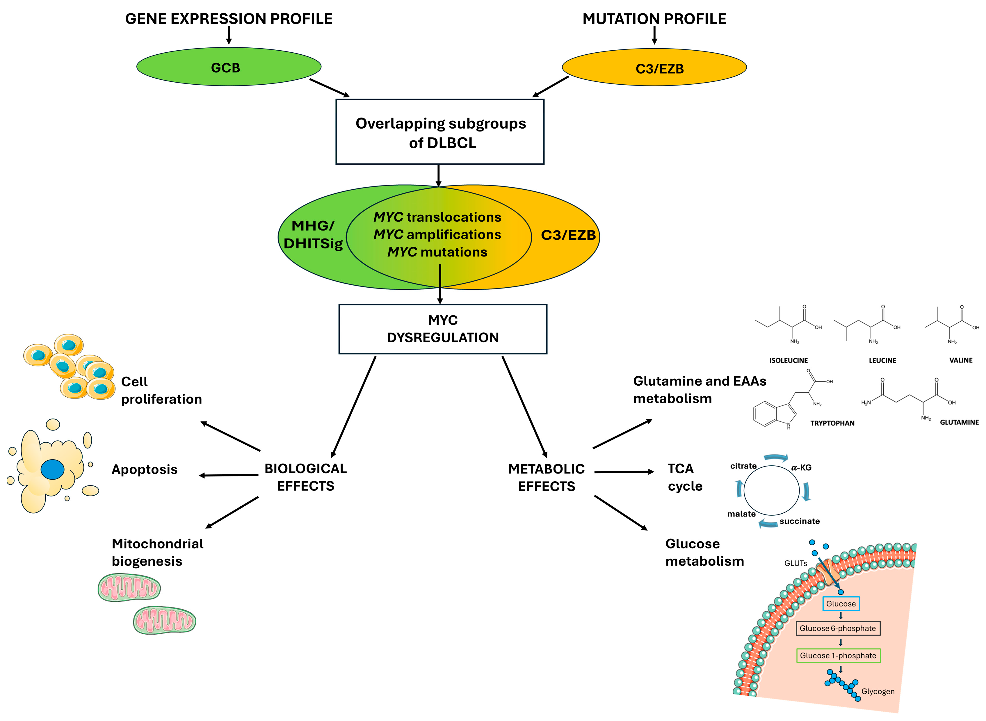
The first successful attempt was a classification based on the gene expression profile (GEP), which identified DLBCL cases sharing similar expression signatures with two specific stages of B-cell maturation: germinal center B-cell (GCB) and activated B-cell (ABC) [15]. This early classification identified the Cell-Of-Origin (COO) of these lymphomas and showed promising results in the clinic, with the ABC subtype displaying a worse outcome within an R-CHOP regimen [16]. More recently, advances in the DLBCL GEP-based classification allowed the refinement of this kind of molecular subtyping up to the identification of the so-called Molecular High-Grade (MHG) subtype, which represented a further step in the stratification of the GCB group [17] (Figure 1). Indeed, the MHG has a gene expression signature like the centroblast in the dark zone of the germinal center (GC) and distinguishable from one of the centrocytes of the GC light zone [17]. Moreover, the MHG signature was originally designed to distinguish DLBCL from BL, which is a (mostly) pediatric and aggressive type of lymphoma [18]. Consistently, the MHG group in the clinic showed a worse prognosis among the GEP subtypes, and this discovery was particularly relevant as the MHG largely belonged to the GCB group, which globally showed a favorable outcome [17]. The same results have been achieved independently by another group by using a slightly different GEP approach, defining the Double-Hit Gene Expression Signature (DHITSig), since cases with this signature were like HGBL-MYC/BCL2 rather than BL and centroblasts [19]. However, about half of the cases with MHG or DHITSig do not present MYC and BCL2 rearrangements [17,19], indicating that additional mechanisms are likely involved and can be caught by the application of specific gene expression signatures.
DLBCL GEP-subtypes also showed distinct mutational profiles [20], and this raised the possibility of using genetic alterations for the identification of well-defined groups. To this aim, two independent studies used a whole exome sequencing (WES) approach applied to large cohorts of DLBCL cases and defined a few genetic categories which largely overlapped [21,22]. Further refinement of this classification identified the EZH2-mutated and BCL2-translocated (EZB)-MYC+ group, which was characterized by a BL-like mutation profile and ribosomal protein expression signature, therefore sharing molecular features with the MHG/DHITSig GEP group [23] (Figure 1). Altogether, this evidence suggests that DLBCL molecular profiling could potentially be used in the clinic for precise diagnosis and treatment decisions. However, to date, these analyses are not easily appliable in routine clinical practice, and more simplified markers are required.
Figure 1. Similar MYC gene alterations leading to MYC deregulation were found by two different classification systems in overlapping subsets of DLBCL cases. Two independent molecular classification systems, gene expression profiles, and mutational profiles were identified in DLBCL, the MHG/DHITSig group, and the EZB-MYC+ group, respectively. These subsets of patients share MYC locus alterations, e.g., chromosomal rearrangement, copy number gain, and point mutations, which lead to MYC dysregulation; indeed, these genetic abnormalities promote MYC overexpression or enhance its constitutive activity, with dramatic impact both on cellular proliferation and metabolic reprogramming.
Biomolecules 15 01346 g001

3. MYC-Driven Lymphomas

Genetic rearrangements involving MYC locus are a predominant mechanism leading to MYC overexpression in multiple hematological cancers [24]. MYC translocations were firstly observed in BL, where 90–95% of these alterations are represented by the t(8;14) (q24;q32) rearrangement. This translocation normally puts MYC under the control of the Ig heavy-chain (IgH) gene’s strong regulatory elements, causing uncontrolled MYC expression. The remaining 5–10% of BL cases present t(2;8) (p12;q24) and t(8;22) (q24;q11) rearrangements [25,26]. MYC translocations are not only observed in BL, but also in other B-cell malignancies. Indeed, in DLBCL, MYC translocations are found in 5–15% of patients, rising to 50% when considering DLBCL cases which also present BCL2 and/or BCL6 rearrangements [27]. Moreover, a EUROMYC retrospective study found a high frequency of MYC translocations in HIV-associated large B-cell lymphomas, consistent with their aggressive nature, and suggesting the necessity of differential and more intensive therapies [28].
Aside from chromosomal rearrangements, MYC deregulation in lymphomas is also caused by other genetic alterations such as MYC copy number gains. Zak and colleagues investigated the prognostic impact of MYC amplifications in a cohort of 396 large B-cell lymphoma (LBCL) patients. The authors observed that 20.9% of cases presented MYC amplification, with a correlation between a high copy number gain (>7 MYC copies) and worse prognosis [29]. Comprehensively, MYC chromosomal translocations and MYC amplifications are also found in other aggressive hematological neoplasms such as plasmablastic lymphoma, transformed follicular lymphoma, and lymphoblastic leukemia [27,28].
Even if MYC dysregulation in lymphomas is mostly associated with chromosomal translocations and, to a lesser extent, copy number gains, MYC point mutations represent another type of MYC alteration, which have been firstly observed in BL [30]. MYC point mutations have been traditionally considered a minor oncogenic factor with respect to MYC amplifications and chromosomal rearrangements due to their relative low frequency. However, in the last 20 years, the routine use of next-generation sequencing approaches allowed for the identification of MYC point mutations in various cancer types, including both solid and non-solid tumors, often in association with poor outcomes [31,32,33]. The origin of these mutations in lymphomas is uncertain, but MYC is a predicted target of the aberrant somatic hypermutation machinery (aSHM), which displays a high affinity for the genomic regions around the transcription start site (TSS) of transcriptionally active genes. Indeed, the distribution of MYC point mutations in lymphomas recapitulate an aSHM mutation-driven signature [34]. Despite these considerations, the functional consequences of MYC point mutations are still mostly uncharacterized and need further investigation, although there are some exceptions such as the cluster of hotspot mutations affecting the Myc Box I (MBI) domain. Multiple experimental evidence showed that MBI point mutations are critical for MYC turnover and function; indeed, these mutations have been shown to affect the FBXW7-mediated proteasome degradation, increasing the MYC protein half-life in vitro [35,36,37].
In general, B-cell lymphomas driven by MYC dysregulation are characterized by genomic instability and metabolic reprogramming to support rapid cell proliferation. MYC alterations in hematological cancers differ from the ones observed in solid tumors, e.g., the former predominantly present MYC translocations and point mutations, while the latter are mostly characterized by MYC amplifications [24].

4. MYC Dysregulation: From Gene Expression to Metabolic Reprogramming

The MYC gene encodes for the MYC proto-oncogene, a transcription factor that regulates the expression of 10–15% of the genome [38]. At the protein structure level, it is organized in an N-terminal transactivation domain and a C-terminal basic helix-loop-helix–leucine zipper (bHLH-LZ) domain. In particular, the bHLH-LZ is crucial for the heterodimerization with partner proteins, such as MYC Associated Factor X (MAX), for transcription activation [39]. MYC-MAX complexes bind consensus DNA sequences, the E-boxes (5′-CACGTG-3′) domains in the promoter of target genes involved in many cellular processes, including cell metabolism, cell growth, and survival, to regulate their expression [39].
Within healthy cells, MYC expression is controlled both at the transcriptional and post-transcriptional level to finely tune, respectively, the production of its messenger RNA (mRNA) and its translation rate. These control mechanisms are essential, because even small perturbations in MYC expression and activity display a high oncogenic capacity [40]. Indeed, MYC promotes the activation of genes involved in the G1–S phase transition within the cell cycle, whereas it represses the activity of cell cycle inhibitors such as Cyclin-Dependent Kinase Inhibitor 2B (CDKN2B). Therefore, MYC overexpression results in the reinforcement of these mechanisms, ultimately leading to an increased cell proliferation rate [41]. On the other hand, normal cells react to MYC hyperactivity by triggering apoptosis; in fact, uncontrolled and sustained MYC expression can lead to proliferative stress and the subsequent activation of apoptotic processes, such as the Alternate Reading Frame (ARF)–mouse double minute 2 homolog (MDM2)–Tumor Protein p53 (TP53) axis [41]. Therefore, additional genetic alterations are necessary to allow MYC to exert its oncogenic activity in cancer cells, e.g., the enhanced expression of antiapoptotic factors such as BCL2 (through chromosomal translocations) or mutations in checkpoint genes like TP53 [42] (Figure 1).
In addition, MYC regulates mitochondrial biogenesis by the expression of nuclear-encoded mitochondrial genes involved in mitochondrial mass regulation. Among these genes, the most relevant are the mitochondrial transcription factor A (TFAM), the peroxisome proliferator-activated receptor gamma coactivator 1-alpha (PPARGC1A), and several members of the Translocase of the Outer Mitochondrial Membrane (TOM) and Translocase of the Inner Mitochondrial Membrane (TIM) import machinery [43]. In particular, the TIM/TOM machinery is a sophisticated system for recognition and translocation across the outer and inner mitochondrial membranes of almost 1000 precursors of proteins involved in mitochondrial structure and function [43] (Figure 1).
Exploring the MYC-regulated metabolic activity within cells, MYC controls nutrient acquisition to produce ATP and key cellular metabolites to increase cell mass and trigger DNA replication and cell division programs by regulating glutaminolysis, nucleotide synthesis, and glycolysis [43]. Indeed, glycolytic enzymes, e.g., Glucose Transporter 1 (GLUT1), Hexokinase 2 (HK2), Phosphofructokinase (PFKM), Enolase 1 (ENO1), and Lactate Dehydrogenase A (LDHA), are upregulated by MYC, which also promotes alternative splicing of the pyruvate kinase-encoding gene, favoring the Pyruvate Kinase M2 (PKM2) isoform [44]. Therefore, MYC enhances the uptake of glucose with the consequent production of lactate to generate rapid high levels of energy, i.e., the Warburg effect [44]. Furthermore, MYC upregulates GLS and the AAs transporter, e.g., Solute Carrier Family 1 Member 5 (SLC1A5), to increase the glutamine uptake and, by supporting glutamine anaplerosis, MYC contributes to the replenishment of intermediates of the tricarboxylic acid (TCA) cycle [45]. Given MYC’s role in the metabolic regulation of nutrient uptake to meet the high energy requirements for tumor proliferation, specific metabolites regulated by MYC are currently studied through metabolomic techniques to identify potential diagnostic and prognostic markers in solid and non-solid tumors. In the next chapter we will review the use of these applications in B-cell lymphomas with a particular focus on DLBCL.

5. The Use of Metabolomic Techniques in B-Cell Lymphoma for Diagnosis and Prognosis

Cancer cells have a dysregulated metabolism due to uncontrolled proliferation. The most obvious metabolic modification in tumors is the increase in glucose consumption, which results in more lactate production, even when oxygen is present, as was widely demonstrated many years ago [46,47]. However, tumor cells have been shown to support cancer growth, survival, and invasion by increasing the uptake of other nutrients (such as Gln and fatty acids) [48]. The study of metabolic alterations through metabolomics is a powerful tool that can identify cancer metabolites and drivers of tumorigenesis. Metabolomics is based on high specificity and sensibility techniques that can detect and quantify a wide spectrum of metabolites [49]. Metabolomics studies can be divided into two classes: targeted and untargeted. The targeted workflow focuses on the quantitative measurement of predefined groups of known metabolites, often driven by specific biological hypotheses. Untargeted metabolomics aims to detect as many metabolites as possible in a sample, including unknown compounds, without a prior hypothesis [50]. For the metabolomic analysis of biological fluids, there are different techniques, and the platform of choice depends on experimental goals, sample traits, and sensitivity. The most analytical platforms used are Gas Chromatography–Mass Spectrometry (GC-MS), Liquid Chromatography–Mass Spectrometry (LC-MS), and nuclear magnetic resonance (NMR) spectroscopy (Table 1) [51,52].
Metabolites play a role as signaling molecules, energy precursors, and cofactors, and they also regulate various processes and enzymatic cascades [59,60,61]. Additionally, metabolites, particularly AAs, are involved in the biological processes of tumorigenesis, cancer cell invasion, cancer stem cell pluripotency, insulin sensitivity, epigenetic regulation, and other cellular processes [62]. To date, there are few studies that concern metabolomic analysis and AA profiles in DLBCL. Regarding the mass spectrometry techniques applied to metabolomics, we report below some works on the evaluation of AAs related to diagnosis or prognosis in DLBCL. The use of the GC-MS technique has been used in some studies on subjects with DLBCL in different types of matrices (i.e., plasma, urine). Barberini et al. used untargeted GC-MS to compare the metabolic profile by case/control status and across the major lymphoma subtypes. They showed that glycine was more abundant in the plasma samples of DLBCL compared to the controls [53]. In another study, by the use of untargeted GC-MS, the urine samples of patients with DLBCL and healthy individuals were investigated, and the results showed that glycine, serine, threonine, and their metabolisms, as well as aminoacyl-tRNA biosynthesis, were the metabolites with the highest capacity to discriminate healthy subjects from DLBCL patients. Therefore, these metabolites could be exploited as potential diagnostic biomarkers and targets for therapeutic purposes. [54]. Moreover, an untargeted plasma metabolomics analysis by the GC-MS technique was carried out in the healthy controls (Ctrl), DLBCL patients that were recently diagnosed (ND), and DLBCL patients who achieved complete remission (CR) as part of another study. The aim was to explore the metabolic characteristics of peripheral plasma derived from DLBCL patients and the Ctrl and identify metabolic pathways and biomarkers associated with the diagnosis and prognosis prediction of DLBCL. The results showed that the level of glucose and aspartate in plasma was decreased in ND patients and elevated in CR patients, indicating that glucose and aspartate may be involved in the pathogenesis of DLBCL [55]. The lower level of aspartate in ND patients may be related to disorders of the asparagine metabolism. Furthermore, a decrease in methionine and cysteine as well as in branched-chain amino acids was observed in the plasma of ND patients, and this alteration was closely associated with a worse prognosis [55]. The GC-MS technique was employed in another study for the detection of metabolic alterations, which could be specifically assigned to the DLBCL Cell-Of-Origin subtypes, and with the aim to identify some metabolites associated with the clinical outcomes. Unfortunately, metabolomics profiling did not distinguish the GCB subtype from the non-GCB subtypes; nevertheless, serum metabolomic profiling reported that valine and pyroglutamic acid could be candidate biomarkers for the prognosis of DLBCL [56]. LC-MS was used to perform untargeted metabolomic studies on serum samples from patients with newly diagnosed DLBCL and whose age–gender matched healthy controls to verify whether the differential metabolites had diagnostic value in DLBCL patients. The metabolic profile of DLBCL patients was distinct from the healthy controls, indicating a differential metabolism in the disease condition. The enrichment analyses showed that differential metabolites were mainly enriched in the D-Glutamine and D-glutamate metabolism. In particular, the Gln and glutamate metabolism are essential for cell survival and proliferation, since DLBCL cells are largely dependent on the concentration of Gln, which is converted to glutamate by GLS [50]. Glutamate, in turn, is used for various purposes: it provides nitrogen for nucleotide and AA synthesis, generates alpha-ketoglutarate (α-KG) for the TCA cycle, and even acts as a signaling molecule [57]. The use of NMR-based serum metabolomics can help to identify a signature for patients with DLBCL who are at high risk of failing immunochemotherapy, leading to more personalized treatment. The serum metabolomic signature at the time of diagnosis, analyzed by NMR spectroscopy, seems to differ between DLBCL patients with refractory disease or early relapse (REF/REL) and cured (CURED) patients. Some AAs (lysine and arginine) were significantly higher in REF/REL patients. In contrast, the AAs aspartate, valine, and ornithine were found to be higher in the cured patient group [58].

6. MYC-Driven Amino Acid Metabolism in Cancer Cells

Animal cells frequently exhibit an increased demand for essential amino acids (EAAs), because cells cannot synthesize EAAs de novo or in insufficient amounts, requiring these from diet [63]. AAs, as carbon and nitrogen donors, allow to get rid of the nutrition limitation and therefore play an important role in tumorigenesis. AAs have a regulatory function in tumors, participating in signaling pathways, the tumor microenvironment, and epigenetic modifications [64]. AAs metabolism, both EAAs and non-essential amino acids (NEAAs), are regulated by MYC, which also modulates metabolic reprogramming, cancer cell anabolism, metabolic enzymes expression, mitochondrial respiration, glycolysis, and fatty acid and nucleotide synthesis in different types of tumors [65]. In cancer cells, MYC plays a substantial role in promoting the transcription of the L-type amino acid transporter (LAT) (Na+-independent transporters), which transports neutral AAs into cells. LAT expression has been found to be increased in many cancers, and it is essential for cell growth and protein translation regulation through the mTORC1 pathway [66,67]. AAs are essential for the activation of mTORC1, which in turn increases lipid and protein synthesis [68]. The transport of neutral AAs into cells is mediated by a membrane transporter family, grouped into two sub-families, specifically SLC7 (LAT1 and LAT2) and SLC43 (LAT3 and LAT4). MYC selectively upregulates LAT1 and LAT3 expression and transcription [67,69]. In Burkitt’s lymphomas, oncogenic MYC and LAT1/LAT3 transporters create positive feedback called MYC-LAT1/LAT3. MYC activates LAT1 and LAT3 expression and promotes effective EAAs uptake into tumor cells (Figure 2) [70]. Efficient EAAs incorporation, in turn, promotes MYC synthesis, constituting a feedforward activation loop that reinforces MYC-regulated oncogenic programs, conferring advantages to acquiring nutrients and utilizing nutrients during malignant transformation. Consistently, in MYC-overexpressing tumor cells, LAT1/LAT3 inhibition compromises metabolic reprogramming and, in particular, the depletion of LAT1 significantly inhibits glucose and Gln uptake [69]. MYC can also regulate NEAAs metabolism, in particular, the AAs Gln, proline, and serine. In cancer cells, Gln is the most abundant AA in blood circulation, acting as major fuel, and anaplerosis metabolites in tumors, driving the TCA cycle to sustain mitochondrial ATP production. Anaplerotic metabolism of Gln generates α-KG and subsequently oxaloacetate (OAA), fueling the TCA cycle through a series of biochemical reactions termed glutaminolysis [71]. Gln maintains redox homeostasis in numerous metabolic processes and is essential for the proliferation of many cancer cells, regulating mitochondrial functions and macromolecular synthesis [72]. MYC promotes Gln uptake by activating the expression of glutamine transporter SLC1A5 [69,73]. Glutaminolysis is a process catalyzed by GLS1 or GLS2 to produce glutamate and fuel the TCA cycle (Figure 2). GLS1 and GLS2 are isozymes that play opposite roles in tumor development. GLS1 has oncogenic properties, while GLS2 has been described as a tumor suppressor [74]. In human P-493 B lymphoma cells, MYC transcriptionally represses miR-23a/b to enhance the translation of GLS1, leading to an increase in glutaminolysis. P-493 B-cell growth was diminished significantly by Gln withdrawal, that also led to the decrease in ATP levels, reflecting diminished cellular oxygen consumption [14]. Cancer cells need free AAs because of their highly anabolic state; for this reason, the autophagy–lysosome circuit is upregulated in cancer cells [75], and Transcription Factor EB (TFEB), a master regulator of the autophagy–lysosome pathway, sustains AA pools via protein breakdown. In acute myeloid leukemia, MYC suppresses autophagy by antagonizing the expression and function of TFEB, and this suppression circuit is necessary for the maintenance of a malignant state [76,77]. In normal B cells and in premalignant and neoplastic B cells of Em-Myc transgenic mice, the autophagy pathway is suppressed by MYC. In this regard, given the suppression of autophagy driven by MYC, it was evaluated whether there were changes in intracellular AA pools in normal versus premalignant Em-Myc pre-B cells. Concentrations of AAs were significantly increased (BCAAs, Gln, glutamate, glycine, and arginine) in premalignant Em-Myc pre-B cells versus normal pre-B cells, indicating that MYC-expressing B cells have compensatory mechanisms to maintain AA pools [70]. In view of all these considerations, altered AA metabolisms are common in tumors, particularly lymphomas; thus, targeting molecules in AAs’ metabolic signaling pathways has the potential to treat cancer.

7. Conclusions

Metabolism alteration in cancer cells is one of the most important aspects in the discrimination between healthy and cancerous cells. As mentioned, AAs contribute to tumorigenesis, acting as a source of energy for tumor growth, driving alterations in the cellular metabolism to meet the increased demand for nutrients [76]. Targeting AA metabolisms, trying to inhibit the transporters and enzymes that regulate the supply of AAs, could be an important approach in cancer therapy. Gln is the most consumed amino acid, and its availability can limit tumor cell proliferation; it is utilized for the biosynthesis of NEAAs, and is also the major source of α-ketoglutarate in the TCA cycle, besides the fact that its intermediate glutamate functions as an exchange factor for the import of EAAs [78]. Tumors driven by MYC or KRAS are particularly dependent on exogenous Gln. Glutaminolysis and the de novo biosynthesis of Gln are upregulated in several cancers via common oncogenes/tumor suppressors including MYC and p53 [79]. Gln synthesis is often due to the upregulation of Gln synthetase, whereas enhanced glutaminolysis is caused by increased GLS activity [80]. Various inhibitors of GLS have been proposed to counteract glutamine catabolism, in particular CB-389, a potent, selective, and orally bioavailable inhibitor of both splice variants of glutaminase, which showed significant antitumor activity in two xenograft models [81]. Moreover, MYC upregulates LAT1 and LAT3 expression and transcription, sustaining EAA metabolism in tumor cells. The therapeutic approach aimed at inhibiting LAT1 would offer an opportunity to unleash the functional association between MYC and LAT1, leading to tumor suppression. JPH203, a specific LAT1 inhibitor, can play a role as a novel anti-tumor agent and can be evaluated as an MYC-selective cancer therapeutic in future clinical trials [82].
The metabolomic approach for the study of DLBCL can help in discovering novel metabolic biomarkers crucial for patients’ stratification and potentially for the development of new treatment procedures. In clinical practice, this is translated into the proximal goal of obtaining a rapid, precise, and inexpensive method for early diagnosis and prognosis prediction. To this aim, the selection of metabolites that are associated with heterogenous and not easily treatable tumors such as DLBCL will positively influence diagnosis. For the high-risk DLBCL cases with a strong MYC-driven expression signature, the selection of some key AAs influenced by MYC, such as Gln, glutamate, tyrosine, tryptophan, and BCAAs, could be initially evaluated for a general screening and later applied in routine enzymatic tests for a rapid diagnosis. The development of these metabolic “sensors” in DLBCL would be particularly important since the dissection of the molecular complexity of these cases currently requires a combination of multiple high-tech platforms, which, unfortunately, are far from being found in standardized clinical diagnostic settings. In the longer term, the use of metabolomics focused on AA metabolisms could lead to the development of new targeted treatment strategies in high-risk DLBCL as well as in other MYC-driven malignancies. Therefore, targeting AA metabolisms, trying to inhibit the transporters and enzymes that regulate the supply of AAs, could be an important therapeutic approach for clinically aggressive cancers.

Author Contributions

A.F.S., D.D.L., M.G. and F.C. wrote the article, A.F.S., D.D.L. and M.G. made the pictures and the table, A.F.S., D.D.L., M.G. and F.C. revised the article. All authors have read and agreed to the published version of the manuscript.

Funding

This research received no external funding.

Institutional Review Board Statement

Not applicable.

Informed Consent Statement

Not applicable.

Data Availability Statement

No new data was created.

Acknowledgments

Figure 1 and Figure 2 was partly generated using Servier Medical Art (https://smart.servier.com (accessed on 23 May 2025)), licensed under CC BY 4.0 (https://creativecommons.org/licenses/by/4.0/ (accessed on 23 May 2025)).

Conflicts of Interest

The authors declare no conflicts of interest.

Abbreviations

The following abbreviations are used in this manuscript:
DLBCLDiffuse large B-cell lymphoma
AAsAmino acids
BCL2B-cell lymphoma 2
BCL6B-cell lymphoma 6
FISHFluorescence in situ hybridization
WHOWorld Health Organization
BLBurkitt lymphoma
IHCImmunohistochemistry
HIF-1αHypoxia-inducible factor-1 alpha
BCAAsBranched amino acids
SLCSolute carrier
SLC1A5Solute carrier family 1 member 5
BCAT1Branched-chain amino acid transaminase 1
TCATricarboxylic acid cycle
GEPGene expression profile
GCBGerminal center B-cell
ABCActivated B-cell
COOCell-of-origin
MHGMolecular high-grade
GCGerminal center
DHITSigDouble-hit gene expression signature
HGBLHigh grade B-cell lymphoma
WESWhole exome sequencing
CSRClass-switch recombination
LBCLsLarge B-cell lymphomas
aSHMAberrant somatic hypermutation machinery
TSSTranscription start site
IgImmunoglobulin
IgHImmunoglobulin heavy chain
MCLMantle cell lymphoma
MBIMYC Box I
FBXW7F-Box and WD repeat domain containing 7
NMRNuclear magnetic resonance
bHLH-LZbasic helix-loop-helix leucine zipper
CDKN2BCyclin dependent kinase inhibitor 2B
ARFAlternate reading frame
MDM2Mouse double minute 2 homolog
TP53Tumor protein p53
TFAMTranscription factor A
PPARGC1APeroxisome proliferator-activated receptor gamma coactivator 1-alpha
HK2Hexokinase 2
PFKMPhosphofructokinase
ENO1Enolase 1
LDHALactate dehydrogenase A
PKM2Pyruvate kinase M2
LC-MSLiquid chromatography–mass spectrometry
GC-MSGas chromatography–mass spectrometry
NDRecently diagnosed
CRComplete remission
α-KGAlpha-ketoglutarate
REFRefractory disease
RELEarly relapse
EAAsEssential amino acids
NEAAsNon-essential amino acids
LATL-type amino acid transporter
OAAOxaloacetate
GLS1Glutaminase 1
GLS2Glutaminase 2
TFEBTranscription factor EB
GlnGlutamine

References

  1. Campo, E.; Jaffe, E.S.; Cook, J.R.; Quintanilla-Martinez, L.; Swerdlow, S.H.; Anderson, K.C.; Brousset, P.; Cerroni, L.; de Leval, L.; Dirnhofer, S.; et al. The International Consensus Classification of Mature Lymphoid Neoplasms: A Report from the Clinical Advisory Committee. Blood 2022, 140, 1229–1253. [Google Scholar] [CrossRef]
  2. Alaggio, R.; Amador, C.; Anagnostopoulos, I.; Attygalle, A.D.; Araujo, I.B.d.O.; Berti, E.; Bhagat, G.; Borges, A.M.; Boyer, D.; Calaminici, M.; et al. The 5th Edition of the World Health Organization Classification of Haematolymphoid Tumours: Lymphoid Neoplasms. Leukemia 2022, 36, 1720–1748. [Google Scholar] [CrossRef]
  3. Rosenwald, A.; Bens, S.; Advani, R.; Barrans, S.; Copie-Bergman, C.; Elsensohn, M.-H.; Natkunam, Y.; Calaminici, M.; Sander, B.; Baia, M.; et al. Prognostic Significance of MYC Rearrangement and Translocation Partner in Diffuse Large B-Cell Lymphoma: A Study by the Lunenburg Lymphoma Biomarker Consortium. J. Clin. Oncol. 2019, 37, 3359–3368. [Google Scholar] [CrossRef] [PubMed]
  4. Cucco, F.; Barrans, S.; Sha, C.; Clipson, A.; Crouch, S.; Dobson, R.; Chen, Z.; Thompson, J.S.; Care, M.A.; Cummin, T.; et al. Distinct Genetic Changes Reveal Evolutionary History and Heterogeneous Molecular Grade of DLBCL with MYC/BCL2 Double-Hit. Leukemia 2020, 34, 1329–1341. [Google Scholar] [CrossRef] [PubMed]
  5. Grande, B.M.; Gerhard, D.S.; Jiang, A.; Griner, N.B.; Abramson, J.S.; Alexander, T.B.; Allen, H.; Ayers, L.W.; Bethony, J.M.; Bhatia, K.; et al. Genome-Wide Discovery of Somatic Coding and Noncoding Mutations in Pediatric Endemic and Sporadic Burkitt Lymphoma. Blood 2019, 133, 1313–1324. [Google Scholar] [CrossRef]
  6. Salghetti, S.E.; Young Kim, S.; Tansey, W.P. Destruction of Myc by Ubiquitin-mediated Proteolysis: Cancer-associated and Transforming Mutations Stabilize Myc. EMBO J. 1999, 18, 717–726. [Google Scholar] [CrossRef]
  7. Ott, G.; Rosenwald, A.; Campo, E. Understanding MYC-Driven Aggressive B-Cell Lymphomas: Pathogenesis and Classification. Blood 2013, 122, 3884–3891. [Google Scholar] [CrossRef]
  8. Wu, J.; Meng, F.; Ran, D.; Song, Y.; Dang, Y.; Lai, F.; Yang, L.; Deng, M.; Song, Y.; Zhu, J. The Metabolism and Immune Environment in Diffuse Large B-Cell Lymphoma. Metabolites 2023, 13, 734. [Google Scholar] [CrossRef]
  9. Dhanasekaran, R.; Deutzmann, A.; Mahauad-Fernandez, W.D.; Hansen, A.S.; Gouw, A.M.; Felsher, D.W. The MYC Oncogene—The Grand Orchestrator of Cancer Growth and Immune Evasion. Nat. Rev. Clin. Oncol. 2022, 19, 23–36. [Google Scholar] [CrossRef]
  10. Yang, W.; Guo, Q.; Quan, S.; Chalmers, Z.R.; Parker, J.B.; Truica, M.; Dufficy, M.F.; Kerber, M.M.; Vasan, K.; Gupta, D.G.; et al. Impaired Mitochondrial Metabolism Is a Critical Cancer Vulnerability for MYC Inhibitors. Sci. Adv. 2025, 11, eadw5228. [Google Scholar] [CrossRef]
  11. Venkatraman, S.; Balasubramanian, B.; Thuwajit, C.; Meller, J.; Tohtong, R.; Chutipongtanate, S. Targeting MYC at the Intersection between Cancer Metabolism and Oncoimmunology. Front. Immunol. 2024, 15, 1324045. [Google Scholar] [CrossRef]
  12. Vettore, L.; Westbrook, R.L.; Tennant, D.A. New Aspects of Amino Acid Metabolism in Cancer. Br. J. Cancer 2020, 122, 150–156. [Google Scholar] [CrossRef] [PubMed]
  13. Zhou, W.; Feng, X.; Ren, C.; Jiang, X.; Liu, W.; Huang, W.; Liu, Z.; Li, Z.; Zeng, L.; Wang, L.; et al. Over-Expression of BCAT1, a c-Myc Target Gene, Induces Cell Proliferation, Migration and Invasion in Nasopharyngeal Carcinoma. Mol. Cancer 2013, 12, 53. [Google Scholar] [CrossRef]
  14. Gao, P.; Tchernyshyov, I.; Chang, T.-C.; Lee, Y.-S.; Kita, K.; Ochi, T.; Zeller, K.I.; De Marzo, A.M.; Van Eyk, J.E.; Mendell, J.T.; et al. C-Myc Suppression of miR-23a/b Enhances Mitochondrial Glutaminase Expression and Glutamine Metabolism. Nature 2009, 458, 762–765. [Google Scholar] [CrossRef]
  15. Alizadeh, A.A.; Eisen, M.B.; Davis, R.E.; Ma, C.; Lossos, I.S.; Rosenwald, A.; Boldrick, J.C.; Sabet, H.; Tran, T.; Yu, X.; et al. Distinct Types of Diffuse Large B-Cell Lymphoma Identified by Gene Expression Profiling. Nature 2000, 403, 503–511. [Google Scholar] [CrossRef]
  16. Shipp, M.A.; Ross, K.N.; Tamayo, P.; Weng, A.P.; Kutok, J.L.; Aguiar, R.C.T.; Gaasenbeek, M.; Angelo, M.; Reich, M.; Pinkus, G.S.; et al. Diffuse Large B-Cell Lymphoma Outcome Prediction by Gene-Expression Profiling and Supervised Machine Learning. Nat. Med. 2002, 8, 68–74. [Google Scholar] [CrossRef] [PubMed]
  17. Sha, C.; Barrans, S.; Cucco, F.; Bentley, M.A.; Care, M.A.; Cummin, T.; Kennedy, H.; Thompson, J.S.; Uddin, R.; Worrillow, L.; et al. Molecular High-Grade B-Cell Lymphoma: Defining a Poor-Risk Group That Requires Different Approaches to Therapy. J. Clin. Oncol. 2019, 37, 202–212. [Google Scholar] [CrossRef]
  18. Sha, C.; Barrans, S.; Care, M.A.; Cunningham, D.; Tooze, R.M.; Jack, A.; Westhead, D.R. Transferring Genomics to the Clinic: Distinguishing Burkitt and Diffuse Large B Cell Lymphomas. Genome Med. 2015, 7, 64. [Google Scholar] [CrossRef]
  19. Ennishi, D.; Jiang, A.; Boyle, M.; Collinge, B.; Grande, B.M.; Ben-Neriah, S.; Rushton, C.; Tang, J.; Thomas, N.; Slack, G.W.; et al. Double-Hit Gene Expression Signature Defines a Distinct Subgroup of Germinal Center B-Cell-Like Diffuse Large B-Cell Lymphoma. J. Clin. Oncol. 2019, 37, 190–201. [Google Scholar] [CrossRef] [PubMed]
  20. Reddy, A.; Zhang, J.; Davis, N.S.; Moffitt, A.B.; Love, C.L.; Waldrop, A.; Leppa, S.; Pasanen, A.; Meriranta, L.; Karjalainen-Lindsberg, M.-L.; et al. Genetic and Functional Drivers of Diffuse Large B Cell Lymphoma. Cell 2017, 171, 481–494.e15. [Google Scholar] [CrossRef]
  21. Schmitz, R.; Wright, G.W.; Huang, D.W.; Johnson, C.A.; Phelan, J.D.; Wang, J.Q.; Roulland, S.; Kasbekar, M.; Young, R.M.; Shaffer, A.L.; et al. Genetics and Pathogenesis of Diffuse Large B-Cell Lymphoma. N. Engl. J. Med. 2018, 378, 1396–1407. [Google Scholar] [CrossRef]
  22. Chapuy, B.; Stewart, C.; Dunford, A.J.; Kim, J.; Kamburov, A.; Redd, R.A.; Lawrence, M.S.; Roemer, M.G.M.; Li, A.J.; Ziepert, M.; et al. Molecular Subtypes of Diffuse Large B Cell Lymphoma Are Associated with Distinct Pathogenic Mechanisms and Outcomes. Nat. Med. 2018, 24, 679–690. [Google Scholar] [CrossRef]
  23. Wright, G.W.; Huang, D.W.; Phelan, J.D.; Coulibaly, Z.A.; Roulland, S.; Young, R.M.; Wang, J.Q.; Schmitz, R.; Morin, R.D.; Tang, J.; et al. A Probabilistic Classification Tool for Genetic Subtypes of Diffuse Large B Cell Lymphoma with Therapeutic Implications. Cancer Cell 2020, 37, 551–568.e14. [Google Scholar] [CrossRef]
  24. Sewastianik, T.; Prochorec-Sobieszek, M.; Chapuy, B.; Juszczyński, P. MYC Deregulation in Lymphoid Tumors: Molecular Mechanisms, Clinical Consequences and Therapeutic Implications. Biochim. Biophys. Acta 2014, 1846, 457–467. [Google Scholar] [CrossRef]
  25. Taub, R.; Kirsch, I.; Morton, C.; Lenoir, G.; Swan, D.; Tronick, S.; Aaronson, S.; Leder, P. Translocation of the C-Myc Gene into the Immunoglobulin Heavy Chain Locus in Human Burkitt Lymphoma and Murine Plasmacytoma Cells. Proc. Natl. Acad. Sci. USA 1982, 79, 7837–7841. [Google Scholar] [CrossRef]
  26. Molyneux, E.M.; Rochford, R.; Griffin, B.; Newton, R.; Jackson, G.; Menon, G.; Harrison, C.J.; Israels, T.; Bailey, S. Burkitt’s Lymphoma. Lancet 2012, 379, 1234–1244. [Google Scholar] [CrossRef] [PubMed]
  27. Swerdlow, S.H.; Campo, E.; Pileri, S.A.; Harris, N.L.; Stein, H.; Siebert, R.; Advani, R.; Ghielmini, M.; Salles, G.A.; Zelenetz, A.D.; et al. The 2016 revision of the World Health Organization classification of lymphoid neoplasms. Blood 2016, 127, 2375–2390. [Google Scholar] [CrossRef] [PubMed]
  28. Pagani, C.; Rusconi, C.; Dalla Pria, A.; Ravano, E.; Schommers, P.; Bastos-Oreiro, M.; Verga, L.; Gini, G.; Spina, M.; Arcaini, L.; et al. MYC Rearrangements in HIV-Associated Large B-Cell Lymphomas: EUROMYC, a European Retrospective Study. Blood Adv. 2024, 8, 968–977. [Google Scholar] [CrossRef] [PubMed]
  29. Zak, T.; Santana-Santos, L.; Gao, J.; Behdad, A.; Aqil, B.; Wolniak, K.; Lu, X.; Ji, P.; Chen, Q.; Chen, Y.-H.; et al. Prognostic Significance of Copy Number Gains of MYC Detected by Fluorescence in Situ Hybridization in Large B-Cell Lymphoma. Leuk. Lymphoma 2024, 65, 26–36. [Google Scholar] [CrossRef]
  30. Rabbitts, T.H.; Hamlyn, P.H.; Baer, R. Altered Nucleotide Sequences of a Translocated C-Myc Gene in Burkitt Lymphoma. Nature 1983, 306, 760–765. [Google Scholar] [CrossRef]
  31. Schaub, F.X.; Dhankani, V.; Berger, A.C.; Trivedi, M.; Richardson, A.B.; Shaw, R.; Zhao, W.; Zhang, X.; Ventura, A.; Liu, Y.; et al. Pan-Cancer Alterations of the MYC Oncogene and Its Proximal Network across the Cancer Genome Atlas. Cell Syst. 2018, 6, 282–300.e2. [Google Scholar] [CrossRef]
  32. Tate, J.G.; Bamford, S.; Jubb, H.C.; Sondka, Z.; Beare, D.M.; Bindal, N.; Boutselakis, H.; Cole, C.G.; Creatore, C.; Dawson, E.; et al. COSMIC: The Catalogue Of Somatic Mutations In Cancer. Nucleic Acids Res. 2019, 47, D941–D947. [Google Scholar] [CrossRef]
  33. De Luca, D.; Munafò, C.; Lorenzi, L.; Cucco, F. MYC Point Mutations in Cancer: A Reboot and a Sequel. Clin. Cancer Res. 2025. accepted. [Google Scholar] [CrossRef] [PubMed]
  34. Khodabakhshi, A.H.; Morin, R.D.; Fejes, A.P.; Mungall, A.J.; Mungall, K.L.; Bolger-Munro, M.; Johnson, N.A.; Connors, J.M.; Gascoyne, R.D.; Marra, M.A.; et al. Recurrent Targets of Aberrant Somatic Hypermutation in Lymphoma. Oncotarget 2012, 3, 1308–1319. [Google Scholar] [CrossRef]
  35. Hemann, M.T.; Bric, A.; Teruya-Feldstein, J.; Herbst, A.; Nilsson, J.A.; Cordon-Cardo, C.; Cleveland, J.L.; Tansey, W.P.; Lowe, S.W. Evasion of the P53 Tumour Surveillance Network by Tumour-Derived MYC Mutants. Nature 2005, 436, 807–811. [Google Scholar] [CrossRef] [PubMed]
  36. Arthur, N.B.J.; Christensen, K.A.; Mannino, K.; Ruzinova, M.B.; Kumar, A.; Gruszczynska, A.; Day, R.B.; Erdmann-Gilmore, P.; Mi, Y.; Sprung, R.; et al. Missense Mutations in Myc Box I Influence Nucleocytoplasmic Transport to Promote Leukemogenesis. Clin. Cancer Res. 2024, 30, 3622–3639. [Google Scholar] [CrossRef]
  37. Freie, B.; Carroll, P.A.; Varnum-Finney, B.J.; Ramsey, E.L.; Ramani, V.; Bernstein, I.; Eisenman, R.N. A Germline Point Mutation in the MYC-FBW7 Phosphodegron Initiates Hematopoietic Malignancies. Genes. Dev. 2024, 38, 253–272. [Google Scholar] [CrossRef]
  38. Papadimitropoulou, A.; Makri, M.; Zoidis, G. MYC the Oncogene from Hell: Novel Opportunities for Cancer Therapy. Eur. J. Med. Chem. 2024, 267, 116194. [Google Scholar] [CrossRef] [PubMed]
  39. Nie, Z.; Guo, C.; Das, S.K.; Chow, C.C.; Batchelor, E.; Simons, S.S.; Levens, D. Dissecting Transcriptional Amplification by MYC. Elife 2020, 9, e52483. [Google Scholar] [CrossRef]
  40. Levens, D. You Don’t Muck with MYC. Genes. Cancer 2010, 1, 547–554. [Google Scholar] [CrossRef]
  41. Mainwaring, O.J.; Weishaupt, H.; Zhao, M.; Rosén, G.; Borgenvik, A.; Breinschmid, L.; Verbaan, A.D.; Richardson, S.; Thompson, D.; Clifford, S.C.; et al. ARF Suppression by MYC but Not MYCN Confers Increased Malignancy of Aggressive Pediatric Brain Tumors. Nat. Commun. 2023, 14, 1221. [Google Scholar] [CrossRef]
  42. Clipson, A.; Barrans, S.; Zeng, N.; Crouch, S.; Grigoropoulos, N.F.; Liu, H.; Kocialkowski, S.; Wang, M.; Huang, Y.; Worrillow, L.; et al. The Prognosis of MYC Translocation Positive Diffuse Large B-Cell Lymphoma Depends on the Second Hit. J. Pathol. Clin. Res. 2015, 1, 125–133. [Google Scholar] [CrossRef]
  43. Purhonen, J.; Klefström, J.; Kallijärvi, J. MYC-an Emerging Player in Mitochondrial Diseases. Front. Cell Dev. Biol. 2023, 11, 1257651. [Google Scholar] [CrossRef]
  44. Zhao, J.; Jin, D.; Huang, M.; Ji, J.; Xu, X.; Wang, F.; Zhou, L.; Bao, B.; Jiang, F.; Xu, W.; et al. Glycolysis in the Tumor Microenvironment: A Driver of Cancer Progression and a Promising Therapeutic Target. Front. Cell Dev. Biol. 2024, 12, 1416472. [Google Scholar] [CrossRef]
  45. Jin, J.; Byun, J.-K.; Choi, Y.-K.; Park, K.-G. Targeting Glutamine Metabolism as a Therapeutic Strategy for Cancer. Exp. Mol. Med. 2023, 55, 706–715. [Google Scholar] [CrossRef]
  46. Warburg, O. On the Origin of Cancer Cells. Science 1956, 123, 309–314. [Google Scholar] [CrossRef] [PubMed]
  47. Koppenol, W.H.; Bounds, P.L.; Dang, C.V. Otto Warburg’s Contributions to Current Concepts of Cancer Metabolism. Nat. Rev. Cancer 2011, 11, 325–337. [Google Scholar] [CrossRef] [PubMed]
  48. Reynolds, M.R.; Lane, A.N.; Robertson, B.; Kemp, S.; Liu, Y.; Hill, B.G.; Dean, D.C.; Clem, B.F. Control of Glutamine Metabolism by the Tumor Suppressor Rb. Oncogene 2014, 33, 556–566. [Google Scholar] [CrossRef] [PubMed]
  49. Clish, C.B. Metabolomics: An Emerging but Powerful Tool for Precision Medicine. Cold Spring Harb. Mol. Case Stud. 2015, 1, a000588. [Google Scholar] [CrossRef]
  50. Alfaifi, A.; Refai, M.Y.; Alsaadi, M.; Bahashwan, S.; Malhan, H.; Al-Kahiry, W.; Dammag, E.; Ageel, A.; Mahzary, A.; Albiheyri, R.; et al. Metabolomics: A New Era in the Diagnosis or Prognosis of B-Cell Non-Hodgkin’s Lymphoma. Diagnostics 2023, 13, 861. [Google Scholar] [CrossRef]
  51. Li, H.; Tennessen, J.M. Methods for Studying the Metabolic Basis of Drosophila Development. Wiley Interdiscip. Rev. Dev. Biol. 2017, 6, e280. [Google Scholar] [CrossRef]
  52. Want, E.J.; Cravatt, B.F.; Siuzdak, G. The Expanding Role of Mass Spectrometry in Metabolite Profiling and Characterization. Chembiochem 2005, 6, 1941–1951. [Google Scholar] [CrossRef]
  53. Barberini, L.; Noto, A.; Fattuoni, C.; Satta, G.; Zucca, M.; Cabras, M.G.; Mura, E.; Cocco, P. The Metabolomic Profile of Lymphoma Subtypes: A Pilot Study. Molecules 2019, 24, 2367. [Google Scholar] [CrossRef] [PubMed]
  54. Bueno Duarte, G.H.; de Piloto Fernandes, A.M.A.; Silva, A.A.R.; Zamora-Obando, H.R.; Amaral, A.G.; de Sousa Mesquita, A.; Schmidt-Filho, J.; Cordeiro de Lima, V.C.; D’Almeida Costa, F.; Andrade, V.P.; et al. Gas Chromatography-Mass Spectrometry Untargeted Profiling of Non-Hodgkin’s Lymphoma Urinary Metabolite Markers. Anal. Bioanal. Chem. 2020, 412, 7469–7480. [Google Scholar] [CrossRef]
  55. Fei, F.; Zheng, M.; Xu, Z.; Sun, R.; Chen, X.; Cao, B.; Li, J. Plasma Metabolites Forecast Occurrence and Prognosis for Patients With Diffuse Large B-Cell Lymphoma. Front. Oncol. 2022, 12, 894891. [Google Scholar] [CrossRef] [PubMed]
  56. Mi, M.; Liu, Z.; Zheng, X.; Wen, Q.; Zhu, F.; Li, J.; Mungur, I.D.; Zhang, L. Serum Metabolomic Profiling Based on GC/MS Helped to Discriminate Diffuse Large B-Cell Lymphoma Patients with Different Prognosis. Leuk. Res. 2021, 111, 106693. [Google Scholar] [CrossRef] [PubMed]
  57. Zhang, Y.; Lv, L.; Wang, C.; Kong, R.; Lu, T.; Ding, M.; Jiang, Y.; Wang, X.; Zhou, X. Comprehensive Metabolome Analysis Identified Novel Biomarkers for the Diagnosis and Prognosis of Diffuse Large B-Cell Lymphoma. Blood 2022, 140, 11934–11935. [Google Scholar] [CrossRef]
  58. Stenson, M.; Pedersen, A.; Hasselblom, S.; Nilsson-Ehle, H.; Karlsson, B.G.; Pinto, R.; Andersson, P.-O. Serum Nuclear Magnetic Resonance-Based Metabolomics and Outcome in Diffuse Large B-Cell Lymphoma Patients—A Pilot Study. Leuk Lymphoma 2016, 57, 1814–1822. [Google Scholar] [CrossRef]
  59. Zhou, Y.; Hu, G.; Wang, M.C. Host and Microbiota Metabolic Signals in Aging and Longevity. Nat Chem Biol 2021, 17, 1027–1036. [Google Scholar] [CrossRef]
  60. Shigeta, K.; Hasegawa, M.; Hishiki, T.; Naito, Y.; Baba, Y.; Mikami, S.; Matsumoto, K.; Mizuno, R.; Miyajima, A.; Kikuchi, E.; et al. IDH2 Stabilizes HIF-1α-Induced Metabolic Reprogramming and Promotes Chemoresistance in Urothelial Cancer. EMBO J 2023, 42, e110620. [Google Scholar] [CrossRef]
  61. Chen, D.; Xia, S.; Zhang, R.; Li, Y.; Famulare, C.A.; Fan, H.; Wu, R.; Wang, M.; Zhu, A.C.; Elf, S.E.; et al. Lysine Acetylation Restricts Mutant IDH2 Activity to Optimize Transformation in AML Cells. Mol Cell 2021, 81, 3833–3847.e11. [Google Scholar] [CrossRef] [PubMed]
  62. Showalter, M.R.; Cajka, T.; Fiehn, O. Epimetabolites: Discovering Metabolism beyond Building and Burning. Curr Opin Chem Biol 2017, 36, 70–76. [Google Scholar] [CrossRef] [PubMed]
  63. Hou, Y.; Yin, Y.; Wu, G. Dietary Essentiality of “Nutritionally Non-Essential Amino Acids” for Animals and Humans. Exp. Biol. Med. 2015, 240, 997–1007. [Google Scholar] [CrossRef]
  64. Chen, J.; Cui, L.; Lu, S.; Xu, S. Amino Acid Metabolism in Tumor Biology and Therapy. Cell Death Dis. 2024, 15, 42. [Google Scholar] [CrossRef] [PubMed]
  65. Huang, H.; Weng, H.; Zhou, H.; Qu, L. Attacking C-Myc: Targeted and Combined Therapies for Cancer. Curr. Pharm. Des. 2014, 20, 6543–6554. [Google Scholar] [CrossRef]
  66. Wang, Q.; Holst, J. L-Type Amino Acid Transport and Cancer: Targeting the mTORC1 Pathway to Inhibit Neoplasia. Am. J. Cancer Res. 2015, 5, 1281–1294. [Google Scholar]
  67. Babu, E.; Kanai, Y.; Chairoungdua, A.; Kim, D.K.; Iribe, Y.; Tangtrongsup, S.; Jutabha, P.; Li, Y.; Ahmed, N.; Sakamoto, S.; et al. Identification of a Novel System L Amino Acid Transporter Structurally Distinct from Heterodimeric Amino Acid Transporters. J. Biol. Chem. 2003, 278, 43838–43845. [Google Scholar] [CrossRef]
  68. Semba, R.D.; Trehan, I.; Gonzalez-Freire, M.; Kraemer, K.; Moaddel, R.; Ordiz, M.I.; Ferrucci, L.; Manary, M.J. Perspective: The Potential Role of Essential Amino Acids and the Mechanistic Target of Rapamycin Complex 1 (mTORC1) Pathway in the Pathogenesis of Child Stunting. Adv. Nutr. 2016, 7, 853–865. [Google Scholar] [CrossRef]
  69. Yue, M.; Jiang, J.; Gao, P.; Liu, H.; Qing, G. Oncogenic MYC Activates a Feedforward Regulatory Loop Promoting Essential Amino Acid Metabolism and Tumorigenesis. Cell Rep. 2017, 21, 3819–3832. [Google Scholar] [CrossRef]
  70. Fernandez, M.R.; Schaub, F.X.; Yang, C.; Li, W.; Yun, S.; Schaub, S.K.; Dorsey, F.C.; Liu, M.; Steeves, M.A.; Ballabio, A.; et al. Disrupting the MYC-TFEB Circuit Impairs Amino Acid Homeostasis and Provokes Metabolic Anergy. Cancer Res. 2022, 82, 1234–1250. [Google Scholar] [CrossRef]
  71. DeBerardinis, R.J.; Chandel, N.S. Fundamentals of Cancer Metabolism. Sci. Adv. 2016, 2, e1600200. [Google Scholar] [CrossRef]
  72. Pi, M.; Kuang, H.; Yue, C.; Yang, Q.; Wu, A.; Li, Y.; Assaraf, Y.G.; Yang, D.-H.; Wu, S. Targeting Metabolism to Overcome Cancer Drug Resistance: A Promising Therapeutic Strategy for Diffuse Large B Cell Lymphoma. Drug Resist. Updates 2022, 61, 100822. [Google Scholar] [CrossRef] [PubMed]
  73. Wise, D.R.; DeBerardinis, R.J.; Mancuso, A.; Sayed, N.; Zhang, X.-Y.; Pfeiffer, H.K.; Nissim, I.; Daikhin, E.; Yudkoff, M.; McMahon, S.B.; et al. Myc Regulates a Transcriptional Program That Stimulates Mitochondrial Glutaminolysis and Leads to Glutamine Addiction. Proc. Natl. Acad. Sci. USA 2008, 105, 18782–18787. [Google Scholar] [CrossRef] [PubMed]
  74. Ramirez-Peña, E.; Arnold, J.; Shivakumar, V.; Joseph, R.; Vidhya Vijay, G.; den Hollander, P.; Bhangre, N.; Allegakoen, P.; Prasad, R.; Conley, Z.; et al. The Epithelial to Mesenchymal Transition Promotes Glutamine Independence by Suppressing GLS2 Expression. Cancers 2019, 11, 1610. [Google Scholar] [CrossRef]
  75. Guo, J.Y.; Chen, H.-Y.; Mathew, R.; Fan, J.; Strohecker, A.M.; Karsli-Uzunbas, G.; Kamphorst, J.J.; Chen, G.; Lemons, J.M.S.; Karantza, V.; et al. Activated Ras Requires Autophagy to Maintain Oxidative Metabolism and Tumorigenesis. Genes. Dev. 2011, 25, 460–470. [Google Scholar] [CrossRef] [PubMed]
  76. Yun, S.; Vincelette, N.D.; Yu, X.; Watson, G.W.; Fernandez, M.R.; Yang, C.; Hitosugi, T.; Cheng, C.-H.; Freischel, A.R.; Zhang, L.; et al. TFEB Links MYC Signaling to Epigenetic Control of Myeloid Differentiation and Acute Myeloid Leukemia. Blood Cancer Discov. 2021, 2, 162–185. [Google Scholar] [CrossRef]
  77. Annunziata, I.; van de Vlekkert, D.; Wolf, E.; Finkelstein, D.; Neale, G.; Machado, E.; Mosca, R.; Campos, Y.; Tillman, H.; Roussel, M.F.; et al. MYC Competes with MiT/TFE in Regulating Lysosomal Biogenesis and Autophagy through an Epigenetic Rheostat. Nat. Commun. 2019, 10, 3623. [Google Scholar] [CrossRef]
  78. Choi, B.-H.; Coloff, J.L. The Diverse Functions of Non-Essential Amino Acids in Cancer. Cancers 2019, 11, 675. [Google Scholar] [CrossRef]
  79. Yang, L.; Venneti, S.; Nagrath, D. Glutaminolysis: A Hallmark of Cancer Metabolism. Annu. Rev. Biomed. Eng. 2017, 19, 163–194. [Google Scholar] [CrossRef]
  80. Zhang, J.; Pavlova, N.N.; Thompson, C.B. Cancer Cell Metabolism: The Essential Role of the Nonessential Amino Acid, Glutamine. EMBO J. 2017, 36, 1302–1315. [Google Scholar] [CrossRef]
  81. Gross, M.I.; Demo, S.D.; Dennison, J.B.; Chen, L.; Chernov-Rogan, T.; Goyal, B.; Janes, J.R.; Laidig, G.J.; Lewis, E.R.; Li, J.; et al. Antitumor Activity of the Glutaminase Inhibitor CB-839 in Triple-Negative Breast Cancer. Mol. Cancer Ther. 2014, 13, 890–901. [Google Scholar] [CrossRef] [PubMed]
  82. Oda, K.; Hosoda, N.; Endo, H.; Saito, K.; Tsujihara, K.; Yamamura, M.; Sakata, T.; Anzai, N.; Wempe, M.F.; Kanai, Y.; et al. L-Type Amino Acid Transporter 1 Inhibitors Inhibit Tumor Cell Growth. Cancer Sci. 2010, 101, 173–179. [Google Scholar] [CrossRef] [PubMed]
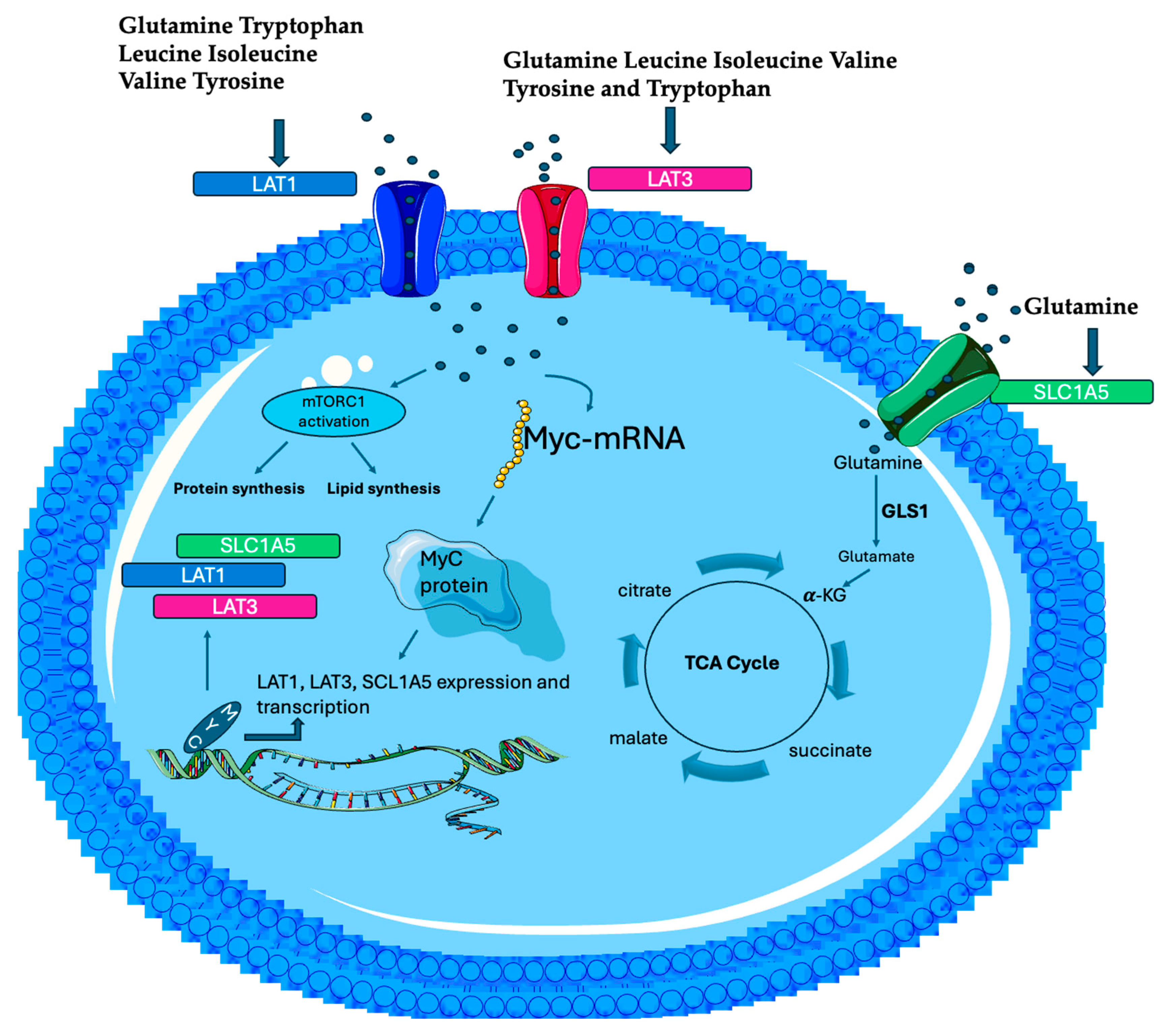
Figure 2. MYC selectively activates LAT1, LAT3, and SLC1A5 expression and transcription. LAT1 and LAT 3 import into the cell EAAs, more specifically, BCAAs (leucine, valine, and isoleucine), tryptophan, as well as glutamine. The increase in EAAs, in turn, stimulates MYC mRNA translation, leading to more MYC protein, constituting a feedforward activation loop to promote the EAAs’ transport and tumorigenesis. MYC promotes Gln uptake by activating the expression of glutamine transporter SLC1A5 and GLS1 or GL2 convert Gln into glutamate, that is then further metabolized in the TCA cycle, providing intermediates for energy (ATP). Moreover, EAAs activate mTORC1 pathways that promote protein and lipid synthesis, crucial processes for growth and repair processes.
Biomolecules 15 01346 g002
Table 1. Comparative overview of the advantages and disadvantages and the most recurrent metabolites detectable by analytical metabolomic techniques.
TechnologyAdvantagesDisadvantagesMost Metabolites
Detected
Metabolites and References
GC-MSHigh sensibility, resolution, and reproducibility
Variety of commercial libraries
Separation efficiency and quantitative accuracy
Requires derivatization
Only suitable for small and volatile compounds
Low-molecular-weight molecules, volatile and polar such as alcohols, aldehydes ketones, lactate pyruvate amino acids, sugars, and free fatty acidsGlicyne, serine threonine glucose, aspartate, methionine, and cysteine valine pyroglutammic acid [53,54,55,56]
LC-MSHigh sensitivity and specificity detects trace amounts of metabolites
Does not require derivatization
Suitable for polar and non-polar compounds
Wide applicability
Ion suppression may affect accuracy
Less standardization and reproducibility than GC-MS
High-molecular-weight molecules, non-volatile and less polar such as complex lipids (phospholipids sphingolipids triglycerides), peptides, proteins, and energy metabolites (NAD+/NAD)Glutamine,
glutamate [57]
NMR
Spectroscopy
Non-destructive
Simple sample preparation
Highly reproducible
Detailed structural information
Lower sensitivity than LC-MS and GC-MS
Longer and more complex analysis times
Global profiling of lipids, proteins, nucleotides, and derivates (ADP, ATP, and AMP), urine metabolites, and small bioactive molecules (neurotransmitters)Lysine, arginine [58]
Disclaimer/Publisher’s Note: The statements, opinions and data contained in all publications are solely those of the individual author(s) and contributor(s) and not of MDPI and/or the editor(s). MDPI and/or the editor(s) disclaim responsibility for any injury to people or property resulting from any ideas, methods, instructions or products referred to in the content.