Review

Targeting Mitochondrial Quality Control for the Treatment of Triple-Negative Breast Cancer: From Molecular Mechanisms to Precision Therapy

1
Medical College, Hunan University of Chinese Medicine, Changsha 410208, China
2
College of Acupuncture and Moxibustion, Hunan University of Chinese Medicine, Changsha 410208, China
*
Authors to whom correspondence should be addressed.
These authors contributed equally to this work.
Biomolecules 2025, 15(7), 970; https://doi.org/10.3390/biom15070970
Submission received: 16 May 2025 / Revised: 20 June 2025 / Accepted: 3 July 2025 / Published: 5 July 2025
(This article belongs to the Section Cellular Biochemistry)

Abstract

Breast cancer is the leading threat to the health of women, with a rising global incidence linked to social and psychological factors. Among its subtypes, triple-negative breast cancer (TNBC), which lacks estrogen receptor (ER), progesterone receptor (PR), and human epidermal growth factor receptor 2 (HER2) expression, is highly heterogeneous with early metastasis and a poor prognosis, making it the most challenging subtype. Mounting evidence shows that the mitochondrial quality control (MQC) system is vital for maintaining cellular homeostasis. Dysfunction of the MQC is tied to tumor cell invasiveness, metastasis, and chemoresistance. This paper comprehensively reviews the molecular link between MQC and TNBC development. We focused on how abnormal MQC affects TNBC progression by influencing chemoresistance, immune evasion, metastasis, and cancer stemness. On the basis of current studies, new TNBC treatment strategies targeting key MQC nodes have been proposed. These findings increase the understanding of TNBC pathogenesis and offer a theoretical basis for overcoming treatment challenges, providing new research angles and intervention targets for effective precision therapy for TNBC.

1. Introduction

According to the global cancer statistics released by the Global Cancer Observatory (GCO) in 2022, the incidence of breast cancer ranks second globally, only after lung cancer, with a mortality rate as high as 6.9% [1]. This poses a serious threat to the health of women worldwide. TNBC, as the most aggressive subtype of breast cancer, has higher incidence and mortality rates than other subtypes. It lacks the three targets of ER, PR, and HER2, and accounts for approximately 15% of all breast cancer cases [2]. It is worth noting that TNBC has particularly prominent aggressive biological behavior. Epidemiological data show that about 46% of TNBC patients will develop distant metastasis, with a median survival of only 13.3 months after metastasis. The metastasis often involves organs such as the brain and viscera, and the recurrence rate within 3 years after surgery is as high as 25% [3]. This high risk of metastasis and recurrence makes TNBC one of the most challenging types of cancer to treat clinically.
The molecular heterogeneity of TNBC is the core of its therapeutic challenge. The classification methods vary depending on the population studied. This heterogeneity results in traditional treatment strategies being unable to cover all patients, and most patients respond poorly to existing therapies. The current treatment of TNBC mainly relies on surgery, chemotherapy, and radiotherapy [4]. However, it still faces multiple challenges. For example, TNBC has a high resistance to anthracyclines, taxanes, and other chemotherapeutic drugs. The mechanisms involve the coordinated action of multiple factors, such as drug efflux mediated by ABC transporters, dormancy of CSCs, hypoxic microenvironment, and evasion of apoptosis [5]. Moreover, although TNBC has a relatively high immunogenicity, the objective response rate of monotherapy with programmed death-1 (PD-1)/programmed death-ligand 1 (PD-L1) inhibitors is less than 25% [6]. Additionally, the survival benefit from the combination of immune checkpoint inhibitors (ICIs) is limited [6]. Due to the lack of expression of ER, PR, and HER2, TNBC is unresponsive to endocrine therapy and HER2-targeted drugs [7]. Although Poly (ADP-ribose) polymerase (PARP) inhibitors have shown some efficacy in patients with Breast cancer susceptibility gene (BRCA) mutations, they are only applicable to a minority of subtypes [8].
In recent years, it has been confirmed that the abnormality of MQC is an important driving factor for the malignant phenotype of TNBC. Disruption of MQC can mediate chemoresistance and distant metastasis through pathways such as enhancing OXPHOS metabolism, inhibiting mitophagy, and promoting epithelial–mesenchymal transition (EMT) [9,10]. Moreover, the imbalance of MQC can lead to upregulation of PD-L1 expression and mitochondrial translocation, thereby inducing T-cell exhaustion and immune evasion [11]. Based on the above mechanisms, targeting key nodes of MQC has become a new paradigm for reversing the therapeutic challenges of TNBC. At the same time, combination therapies and novel targeted drugs have also shown significant potential in preclinical studies. In the future, with the advancement of molecular classification techniques and in-depth multiomics research, precision treatment strategies are gradually becoming mainstream. Exploring the interactions between MQC and other signaling pathways, as well as the clinical translation of novel targeted drugs, may become potential directions for breaking through the treatment of TNBC.

2. Triple-Negative Breast Cancer

TNBC is a subtype of breast cancer characterized by high malignancy, propensity for recurrence, and poor prognosis [4]. Currently, the diagnosis of TNBC mainly relies on immunohistochemical (IHC) testing, which confirms the absence of expression of ER, PR, and HER2 to make the diagnosis [8]. However, this diagnostic method is limited in that it fails to provide detailed information on tumor heterogeneity, which is crucial for precision treatment. Recent studies have shown that radiomics has potential application value in the diagnosis and heterogeneity analysis of TNBC. Lin Jiang et al., [10], using the Fudan classification (FUSCC classification) as the gold standard, analyzed the data of 860 breast cancer patients (including 246 TNBC patients) and found that the combined model of radiomics and IHC staining data had a higher area under the receiver operating characteristic curve (AUC) in the basal-like immune-suppressed (BLIS) subtype than the individual radiomics and IHC models . This finding provides a new idea for non-invasive precise subtyping.
The “triple-negative” feature of TNBC differentiates it from luminal and HER2-positive breast cancers, rendering endocrine therapy and anti-HER2 treatment ineffective for TNBC [4]. This significantly limits the treatment options for TNBC. Therefore, research on TNBC subtype classification based on molecular characteristics and differences in signaling pathways is of great significance. At present, there are three internationally influential TNBC classification methods: (1) FUSCC classification [12,13], (2) Lehmann classification system [14,15,16], and (3) Burstein classification [17,18,19]. These classification methods, by more finely dividing TNBC subtypes, lay the foundation for providing more precise treatment plans for patients. Given the particularity and therapeutic challenges of TNBC, researchers are committed to identifying new therapeutic targets, such as Mucin 1 (MUC1) [20] and Nudix-type motif 5 (NUDT5) [21]. This article will systematically summarize the relevant targets of MQC-targeted therapy for TNBC, providing a reference for the therapeutic prospects of TNBC.

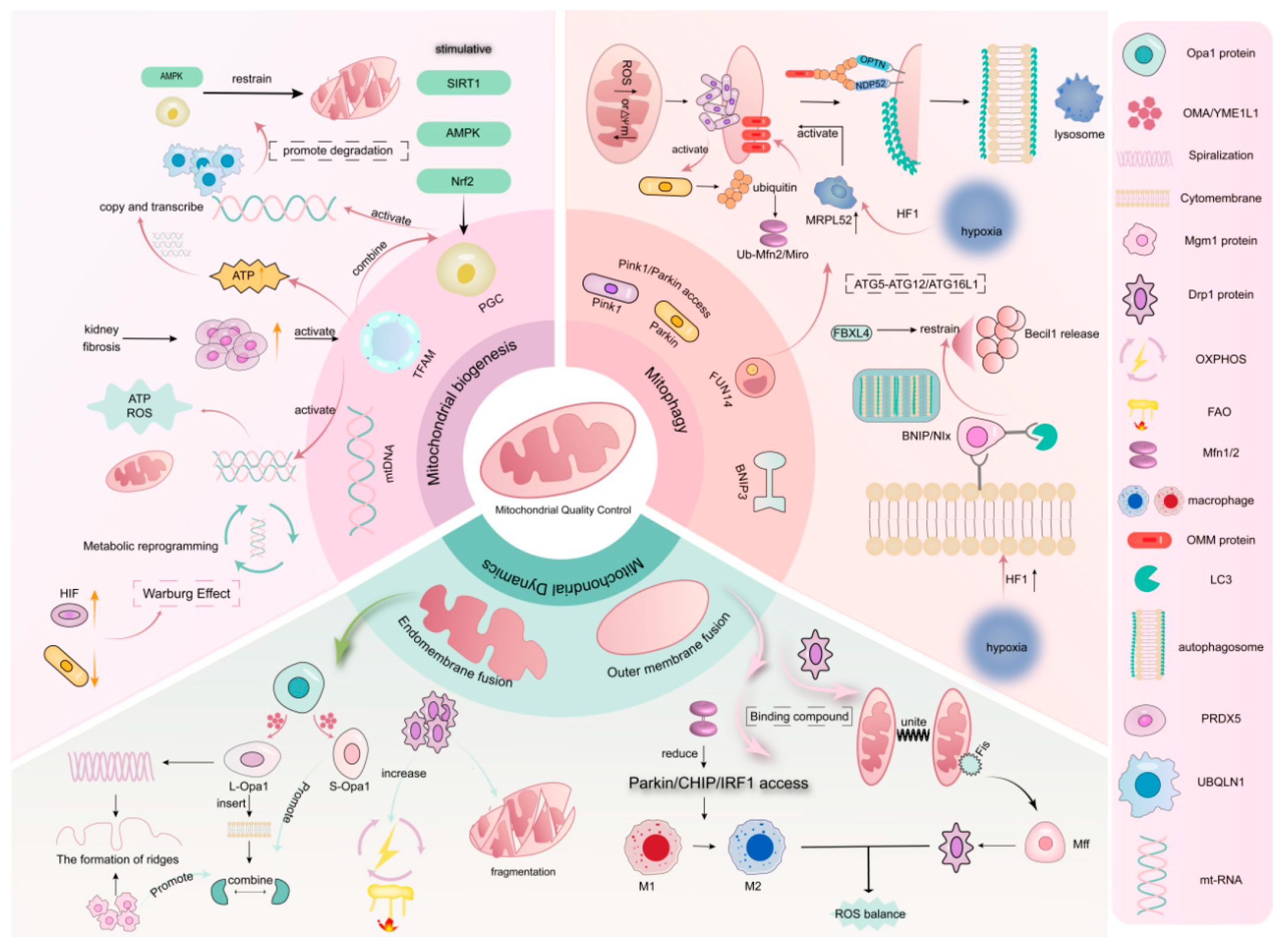
3. Mitochondrial Quality Control

Mitochondria, as the energy factories and metabolic regulation centers of the cell, often participate in the occurrence and progression of various diseases when their functions are disrupted. In recent years, an increasing number of studies have shown that MQC is not only involved in energy metabolism and the production of important substances, but also plays a significant role in processes such as apoptosis, oxidative stress, immune regulation, and metabolic reprogramming. It can also shape the tumor microenvironment. Currently, it has become a major research hotspot in the treatment of TNBC [4,22]. The MQC system, through processes such as mitochondrial biogenesis, autophagy, and dynamics, forms a dynamic and sophisticated regulatory network to maintain the stability of mitochondrial structure and function. A detailed exploration of the occurrence and specific mechanisms of MQC (Figure 1) helps to understand the onset of diseases and provides a new perspective for the intervention and treatment of TNBC.

3.1. Mitochondrial Biogenesis

Mitochondrial biogenesis, as the final step in MQC, mainly regulates cellular homeostasis by increasing the number of mitochondria in the cytoplasm [23]. Mitochondria have their own set of DNA, namely mitochondria DNA (mtDNA), where mitochondrial transcription factor A (TFAM) plays a key regulatory role in the replication and transcription of mtDNA. When cells are under stress, TFAM can clear mtDNA through the autophagy pathway, thereby avoiding its induction of inflammatory responses [24]. Studies have found that peroxiredoxin 5 (PRDX5) can enhance the function of TFAM, thereby promoting the production of ATP in mitochondria and the recovery of mtDNA copy number, ultimately alleviating respiratory suppression in diseases [25]. Secondly, peroxisome proliferator-activated receptor gamma coactivator 1α (PGC-1α), as a transcriptional coactivator, can regulate the expression of mitochondrial proteins. For example, PGC-1α controls the transcription of mtDNA by binding to the TFAM promoter and promotes mitochondrial biogenesis. It also coactivates nuclear respiratory factor 1 (Nrf1) and Nrf2 to promote the expression of genes related to the antioxidant system [26]. Therefore, studying the biogenesis mediated by the PGC-1α/TFAM pathway may be of great significance for the occurrence and development of diseases. Fan et al. [27,28] found through experiments that PGC-1α can serve as a target for inhibiting mitochondrial gene expression, thereby intervening in the further metastasis of TNBC to improve prognosis. AMP-activated protein kinase (AMPK) can act as an activator to enhance the expression of PGC-1α, promote biogenesis, and reduce mitochondrial damage, thereby inhibiting cell death and exerting a certain protective effect on organ systems to improve patient survival.
It is worth noting that mitochondria, as the main regulators of pyroptosis, release large amounts of reactive oxygen species (ROS) when damaged, thereby triggering a large number of inflammatory responses. The damage to mitochondria is mainly due to the disruption of processes such as mitochondrial biogenesis that provide energy demands. Therefore, mitochondrial biogenesis is closely related to ROS. Studies have shown that [29] the levels of certain protein structures, such as ubiquilin 1 (UBQLN1), can regulate the production and degradation of PGC-1α, leading to a reduction in mitochondrial biogenesis. In this process, the decrease in ROS levels also plays an important role. It can be seen that PGC-1α plays an important, pivotal role in regulating mitochondrial function. Similarly, to adapt to environmental changes, tumor cells generally adopt the process of metabolic reprogramming to meet the growth needs of cells, that is, the abnormal initiation of glycolysis known as the “Warburg effect” [30]. This change often optimizes the biosynthetic process and improves energy utilization efficiency by inducing the expression of related factors or proteins [31]. This suggests that by acting on related genes and metabolic pathways to regulate mitochondrial biogenesis and antioxidant defense processes, it may be possible to effectively intervene in the tumor’s survival environment.

3.2. Mitophagy

Mitochondrial autophagy, an adaptive metabolic response that clears damaged or excess mitochondria, prevents ROS accumulation and cell death [32]. This type of autophagy also holds an important position in the proliferation of cancer cells. For example, when FUN14 domain-containing protein 1 (FUNDC1) mediates Parkin-independent mitophagy, it restores autophagosome biogenesis by promoting the formation of the ATG5-ATG12/ATG16L1 complex. However, it also acts as a tethering protein of the mitochondrial–ER contact component, altering its structure to meet the energy demands of rapid tumor cell proliferation [33], and targeting this pathway can have tumor-suppressive effects by inducing ROS-related mitochondrial changes. Conversely, high FUNDC1 expression is linked to poor breast cancer prognosis [34].
In recent breast cancer studies, Ursolic acid (UA) was found to enable transcription factor EB (TFEB) nuclear entry in a partial mammalian target of rapamycin (mTOR)-dependent manner and prevent its ubiquitination. Once in the nucleus, TFEB activates the mitochondrial autophagy–lysosome pathway to remove damaged mitochondria, reducing inflammatory factors and showing therapeutic potential [35].

3.2.1. PINK1/Parkin Signaling Pathway

The PTEN induced kinase 1 (PINK1)/Parkin pathway, a key mechanism for mitochondrial autophagy, is activated when the mitochondrial membrane potential (Δψm) is dissipated or under oxidative stress, leading to PINK1 accumulation. However, mild oxidative stress may not trigger this pathway [36]. Once activated, PINK1 phosphorylates and ubiquitinates mitochondrial surfaces, recruiting Parkin to initiate autophagy. Parkin ubiquitinates outer mitochondrial membrane proteins, marking mitochondria for degradation via autophagosome–lysosome fusion. Additionally, PINK1/Parkin ubiquitinates the mitochondrial transport protein Miro, halting mitochondrial movement. Under oxidative stress, this pathway also promotes the formation of mitochondrial-derived vesicles (MDVs) for mitochondrial repair [37].
The ubiquitin–proteasome system (UPS), particularly the E3 ubiquitin ligase Parkin, plays a significant role in mitochondrial autophagy. Parkin mediates the ubiquitination of mitofusin 2 (MFN2) on the outer mitochondrial membrane (OMM), especially after PINK1 phosphorylates MFN2. This process reduces mitochondrial fusion, increases fission, and promotes autophagosome formation [38]. The downregulation of another ubiquitin ligase CRL4CUL4A/DDB1 can also significantly enhance this process. Since it can maintain cellular energy balance, this process can also inhibit the proliferation of cancer cells [39].
In cancer cells, hypoxia-induced HIF-1 upregulates the mitochondrial ribosomal protein (MRPL52), promoting PINK1/Parkin-dependent mitochondrial autophagy to clear ROS and protect cancer cells [40]. Similarly, Fanconi anemia complementation group C (FANCC) overexpression enhances this pathway, reducing necroptosis. FANCC interacts with TANK-binding kinase 1(TBK1), which stabilizes PINK1 and facilitates Parkin recruitment [41].

3.2.2. BNIP Signaling Pathway

BCL2-interacting protein 3 (BNIP3) and its homolog NIX/BNIP3L are BH3-only domain-containing proteins critical for receptor-mediated mitophagy, a process that is independent of mitochondrial ubiquitination. These proteins function as tumor suppressors and are tightly linked to mitochondrial homeostasis. Upon binding to BCL2, BNIP3/NIX liberates Beclin1, increasing its availability for autophagosome formation [42]. Notably, BNIP3 stabilization is regulated by calnexin (CANX) overexpression, which activates mitogen-activated protein kinase (MAPK) signaling to amplify protective mitophagy. MAPK, as a direct effector of oncogenic signaling pathways, plays a key role in the growth, metastasis, drug resistance, and stemness of tumor cells. Studies have found that ROS-mediated mitophagy is detrimental to the synthesis of hormones such as progesterone and can even affect the body’s hormone levels [43,44].
As a downstream effector of HIF-1α, BNIP3 is essential for hypoxia/reoxygenation (H/R)-induced mitophagy. HIF-1α knockout attenuates mitophagy, exacerbating ROS accumulation and apoptosis in acute kidney injury (AKI), whereas BNIP3 overexpression rescues this phenotype, highlighting its cytoprotective role [45]. This suggests that BNIP3 can provide an advantage for the survival and progression of cancer cells by promoting the autophagy process and eliminating damaged mitochondria, which is also one of the sources of intracellular ROS. This argument has been confirmed in relevant experiments [46].
NIX, a paralog of BNIP3, is indispensable for mitochondrial clearance during erythroid differentiation. Enhancing NIX-mediated mitophagy accelerates erythroid maturation [47]. Both BNIP3 and NIX are OMM proteins whose expression is induced by HIF-1α. These proteins interact with core autophagy components (e.g., WIPI2/3) through LC3-interacting regions (LIRs), facilitating autophagosome engulfment of damaged mitochondria and ROS mitigation. Furthermore, WIPI family proteins engage with the minimal essential region (MER) required for autophagosome formation. Intriguingly, F-box and leucine-rich repeat protein 4 (FBXL4) acts as a key suppressor of NIX/BNIP3-dependent mitophagy, whereas HIF-1α drives BNIP3 upregulation under hypoxia [48,49].

3.3. Mitochondrial Dynamics

3.3.1. Mitochondrial Fusion

Mitochondrial dynamics, primarily comprising mitochondrial fusion and fission, are crucial for maintaining the structural and functional integrity of mitochondria. In TNBC, mitochondrial fusion is a highly coordinated dynamic process involving both inner and outer membrane fusion, with the participation of multiple proteins. This process plays key roles in material transport, population homogenization, and oxidative phosphorylation, significantly contributing to cellular functional regulation and adaptability [50]. Mitochondrial fusion has emerged as a novel therapeutic target for various diseases.
The optic atrophy 1 (OPA1) protein is central to inner mitochondrial membrane (IMM) fusion [51]. OPA1 has enzymatic activities, including GTPase and metalloprotease functions. Under physiological conditions, OPA1 is hydrolyzed in the mitochondrial matrix by the metalloproteases OMA1 and YME1L1 into long isoforms (L-OPA1) anchored to the IMM and soluble short isoforms (S-OPA1) localized in the intermembrane space. L-OPA1 mediates initial membrane contact through its hydrophobic domain inserted into the IMM and has recently been implicated in tumor cell metabolism and proliferation [52]. S-OPA1, which is confined to the intermembrane space, interacts with L-OPA1 to facilitate IMM fusion [53].
To increase fusion efficiency, after Opa1 is hydrolyzed, it forms a helical structure under the action of GTPase, which draws adjacent IMMs closer and generates membrane protrusions to enable docking. These junctional sites may also influence crista formation [54]. OMM fusion is driven primarily by Mfn1/Mfn2, which mediate trans-interactions between adjacent mitochondria. GTP hydrolysis facilitates the formation of “tethered complexes,” ultimately completing fusion [55]. Recent studies [56] have demonstrated that the downregulation of Mfn1 can trigger the Parkin/CHIP/IRF1 pathway to enhance the expression of IL-12, which plays a positive role in the activation of immune responses.
Additionally, the mitochondrial metalloprotease OMA1 critically regulates fusion dynamics [57]. During membrane depolarization, OMA1 collaborates with yeast mitochondrial escape 1-like protease (YME1L) to cleave OPA1, shifting the L-OPA1/S-OPA1 ratio and modulating fusion capacity. Furthermore, the OPA1 homolog Mgm1 protein also has GTPase activity. Its unique structural features enable the inner mitochondrial membranes to fuse with each other under the energy provided by GTPase hydrolysis. It also induces membrane bending through multimodal polymerization and binding to multiple membrane sites to adapt to mitochondrial membrane fusion and cristae formation [58]. It is worth mentioning that studies have already found that [59] the roles of key proteins such as Opa1 and OMA in mitochondrial fusion may provide new targets for the treatment of TNBC. By regulating the activity or expression levels of these proteins, it may be possible to affect the metabolic reprogramming and drug resistance of TNBC cells.

3.3.2. Mitochondrial Division

Mitochondrial fission, a crucial aspect of mitochondrial dynamics. This process plays essential roles in maintaining mitochondrial network morphology, regulating organelle size, and participating in cellular metabolic regulation and stress responses [60]. Dynamin-related protein 1 (Drp1), encoded by the DNM1L gene, serves as a pivotal GTPase that drives mitochondrial fission. The phosphorylation of Drp1 at Ser616, which is catalyzed by various protein kinases, constitutes a critical regulatory mechanism for fission initiation [61]. Through GTP hydrolysis, Drp1 releases the substantial energy required for mitochondrial membrane constriction and subsequent organelle separation.
During fission initiation, cytoplasmic Drp1 undergoes activation and subsequently translocates to predetermined fission sites under the regulation of specific signaling proteins. At these sites, Drp1 oligomerizes to form annular structures encircling mitochondrial membranes, facilitating membrane constriction through mechanical force generation. Notably, this process is sensitive to changes in the mitochondrial metabolic status and membrane potential. Studies have found that [62] the GTPase activity and changes in membrane potential involved in this process may also affect the proliferation and survival of TNBC cells, thereby intervening in the progression of the disease. The mitochondrial fission machinery involves the coordinated action of mitochondrial fission factor (Mff) and mitochondrial fission 1 protein (Fis1), both of which are localized to the outer mitochondrial membrane but play distinct functional roles. Mff primarily facilitates Drp1 recruitment and oligomerization at fission sites through direct protein–protein interactions, with substantial evidence confirming its critical role in precise Drp1 localization [63]. In contrast, Fis1 functions as an essential membrane protein for mitochondrial and peroxisomal fission in both mammalian and yeast cells, serving as a molecular scaffold that assists in Drp1 recruitment and facilitates oligomeric ring assembly [64]. Experimental data suggest synergistic cooperation between Mff and Fis1 in promoting Drp1-mediated fission, with functional complementarity observed under varying physiological conditions [65]. Emerging research highlights the metabolic implications of Drp1-regulated fission, particularly its capacity to increase the oxidative phosphorylation (OXPHOS) efficiency and FAO capacity. Furthermore, Drp1 has regulatory effects on the homeostasis of cellular ROS and the functional modulation of macrophages. These mechanisms collectively influence tumor metabolic reprogramming and antitumor immunity, suggesting potential therapeutic applications in oncology [66].

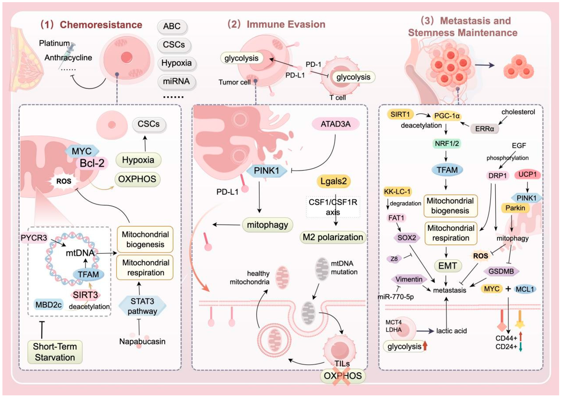
4. Relationship Between MQC and TNBC

Mitochondria, as the “power source” of the cell, exist in the cytoplasm through the process of endosymbiosis with bacteria. Its main functions revolve around energy supply and biosynthesis [67], and it also plays an important role in the proliferation, metastasis, and invasion of TNBC cells. In mitochondria, the TCA cycle is the core hub of energy metabolism, where intermediates generated from nutrients produce reducing equivalents through oxidation and key metabolites for biosynthetic reactions [68]. Based on this, the OXPHOS metabolic pathway transports electrons through the electron transport chain (ETC) in the inner membrane to generate ATP. Given that the proteins and complexes in this pathway can serve as targets for cancer therapy, a large number of related inhibitors have been developed in recent years [69].
As our understanding of cell death modes continues to evolve, mitochondria have once again been revealed to be closely related to biological processes such as ROS generation, ferroptosis, apoptosis, and calcium ion homeostasis [70]. This means that the important role of mitochondria in the progression of TNBC has once again been consolidated. Numerous studies have shown that when MQC is impaired, mitochondria are highly susceptible to oxidative stress damage, which induces ROS production and thereby promotes lipid peroxidation, mediating a form of cell death characterized by iron accumulation and lipid peroxidation—ferroptosis [71]. In addition, during apoptosis, the increase in mitochondrial outer membrane permeability (MOMP) releases cytochrome C, which promotes the formation of apoptotic bodies, the cleavage and activation of caspase-9, and the activation of caspase-3 [72]. So, how does MQC affect the survival and death of TNBC cells? In the following text, we further explore this question from three main aspects: chemoresistance, immune evasion, metastasis, and stemness maintenance (Figure 2 and Table 1).

4.1. Impact of MQC Abnormalities on the Progression and Prognosis of TNBC

4.1.1. Chemotherapy Resistance

Triple-negative breast cancer is characterized by high recurrence rates, aggressive behavior, and poor survival outcomes [73,74]. Systemic adjuvant–neoadjuvant chemotherapy (e.g., anthracyclines, taxanes, and platinum-based agents) remains the primary treatment for newly diagnosed patients [75]. However, chemotherapy, which is currently the sole systemic option for TNBC, faces considerable limitations, including severe side effects and inevitable chemoresistance [76]. Overcoming chemoresistance is critical to improving prognosis and extending survival [73]. The proposed mechanisms of chemoresistance, as summarized by Nedeljković M et al. [5], involve multifactorial cooperation, including: (1) drug efflux via ATP-binding cassette (ABC) transporters, (2) survival of quiescent CSCs post-chemotherapy, (3) hypoxic tumor microenvironments, (4) apoptosis evasion, (5) unique miRNA expression profiles, and (6) intratumoral heterogeneity.
Accumulating evidence highlights mitochondria as central regulators of development, stem cell fate, and metabolic reprogramming [77]. Mitochondria-driven metabolic remodeling increases tumor cell adaptation to hypoxia and nutrient deprivation, directly or indirectly promoting survival and progression [78]. Specifically, TNBC CSCs exhibit a preference for OXPHOS, which is linked to mitochondrial biogenesis and quality control [10,79]. MYC transcription factors and antiapoptotic Bcl-2 family proteins (e.g., MCL-1) are enriched in chemoresistant TNBC cells, where they cooperatively enhance OXPHOS, activate hypoxia pathways, and sustain CSC populations to drive chemoresistance [10].
Mitochondria, as the central hub of cellular metabolism, depend on genetic components encoded by both the nuclear genome and mtDNA. Consequently, mtDNA mutations or transcriptional defects are key drivers of chemotherapy resistance in TNBC cells. Yijie Hao et al. [80] reported that methyl-CpG-binding domain protein 2c (MBD2c) is highly enriched in the mitochondrial noncoding region (NCR) of TNBC cells, where it activates mitochondrial TFAM via SIRT3-mediated deacetylation, increasing mtDNA transcription and respiration. MBD2c inhibition increases cisplatin sensitivity, implicating mtDNA regulation in chemoresistance [80].
Although upstream mechanisms (mtDNA transcription, respiration) are well characterized, downstream responses to mitochondrial dysfunction, such as ROS accumulation and damaged mitochondrial clearance, remain underexplored. The STAT3 inhibitor napabucasin enhances paclitaxel sensitivity in TNBC by suppressing STAT3 signaling, impairing mitochondrial complex I activity and ATP production, and inducing ROS accumulation [81]. Additionally, the natural compound Guangsangon E (GSE), which is isolated from mulberries, inhibits TNBC growth by inducing mitochondrial dysfunction and mitophagy, leading to ROS overload. These findings highlight mitochondrial-targeted strategies to combat TNBC chemoresistance.

4.1.2. Immune Evasion

TNBC, characterized by high PD-L1 expression and abundant tumor-infiltrating lymphocytes (TILs), presents immunogenic features that make it a potential candidate for immune checkpoint inhibitor (ICI) therapy [82]. However, clinical data reveal the limited efficacy of anti-PD-L1 monotherapy, with objective response rates less than 25% [6], and combined chemotherapy strategies fail to significantly improve overall survival [83]. Recent studies have implicated dysregulated MQC in tumor metabolic reprogramming, redox homeostasis, and immune evasion, with MQC abnormalities closely linked to TNBC immunotherapy resistance. In TNBC, cancer cells upregulate PD-L1 to suppress T-cell effector functions, facilitating immune escape [11]. Furthermore, PD-L1/PD-1 interactions enhance tumor cell glycolytic metabolism while inhibiting T-cell glycolysis, conferring survival advantages in nutrient-deprived tumor microenvironments [84].
Jernej Repas et al. [85] demonstrated that MDA-MB-231 cells treated with metformin and 2-deoxy-D-glucose exhibit morphologically intact mitochondria with increased volume and enhanced mitochondrial quality. Notably, this combination significantly suppresses glycosylated PD-L1, yielding potent antitumor effects, and their research further confirms the important role of PD-L1 in the immune evasion process of TNBC tumor tissue. However, the mechanistic link between mitochondrial quality control and glycosylated PD-L1 remains unexplored [85]. Additionally, paclitaxel-resistant TNBC cells upregulate ATPase family AAA domain-containing protein 3A (ATAD3A), which inhibits PINK1-dependent mitophagy, reduces mitochondrial PD-L1 localization, and promotes plasma membrane accumulation of PD-L1 to amplify immune evasion [86]. This finding underscores the interplay between chemoresistance and immune escape in TNBC, warranting further exploration of this mechanistic network.
In TNBC, MQC dysregulation also modulates mtDNA dynamics and intercellular mitochondrial transfer to drive immune evasion. Intriguingly, TNBC cells utilize bidirectional mitochondrial transfer, exporting mtDNA-mutated mitochondria to TILs via extracellular vesicles (EVs) while importing healthy mitochondria from T cells, to induce T-cell metabolic reprogramming (e.g., impaired OXPHOS and reduced mitochondrial quality) and functional exhaustion [87]. This process establishes an “immune escape highway”. Immune evasion mechanisms extend beyond T cells; for example, galectin-2 (Lgals2) promotes M2 macrophage polarization via the CSF1–CSF1R axis, facilitating mitochondrial transfer and immunosuppression [88].
In summary, TNBC exploits MQC dysregulation through metabolic reprogramming, intercellular mitochondrial trafficking, and immune cell polarization to sustain immune evasion, perpetuating a vicious chemoresistance–immunosuppression cycle. Current research remains fragmented, with unclear clinical translation pathways. Future efforts should integrate multiomics data to map the MQC-immune evasion regulatory network and conduct prospective trials to validate strategies targeting this axis, potentially breaking the resistance-immune escape synergy in TNBC.

4.1.3. Metastasis and Stemness Maintenance

TNBC is more aggressive and has a greater metastatic propensity than other breast cancer subtypes [89]. The survival and motility of metastatic cells depend on mitochondrial respiration and OXPHOS, suggesting that mitochondrial-targeted therapies could exploit this dependency to improve TNBC prognosis [90]. EMT, a critical process conferring metastatic and invasive capacities, involves E-cadherin downregulation and mesenchymal marker upregulation. Aberrant E-cadherin promoter methylation disrupts the regulation of cellular motility in TNBC [91]. Reduced miR-770-5p expression is correlated with TNBC proliferation and metastasis. Fei et al. [92] demonstrated that by using the antimicrobial peptide merecidin to act on the cytoplasm of tumor cells in nude mice, increasing the expression of miR-770-5p can negatively regulate vimentin, thereby delaying the metastasis and proliferation of breast cancer cells. Mitochondrial dysfunction is linked to EMT progression, with SIRT1 modulating mitochondrial biogenesis and energy metabolism to regulate EMT outcomes [9]. As a deacetylation substrate of SIRT1, PGC-1α activates the transcription factors NRF-1/NRF-2 to control TFAM and TFB1M expression, driving mitochondrial biogenesis. Studies have shown that PGC-1α overexpression promotes TNBC metastasis via the NRF-1/TFAM axis [93]. Unlike ER+ breast cancers, TNBC displays glycolytic preference and reduced OXPHOS activity owing to oncogenic mutations, mitochondrial defects, transcriptional dysregulation and hypoxic–nutrient-deprived microenvironments [94]. This metabolic heterogeneity, exemplified by the Warburg effect [30], enables rapid tumor growth under nutrient stress. In some TNBC cases, stromal cells exhibit glycolytic activity to supply lactate to tumor cells (reverse Warburg effect), with low GLUT1/MCT4 in tumor cells, high MCT4 in the stroma, and elevated LDHA driving metastasis [95].
TNBC cells maintain stem-like properties through specific mechanisms, preserving undifferentiated states to form new tumor foci and drive recurrence. Residual CSCs post-treatment contribute to poor prognosis and involve multiple signaling pathways [96,97]. Patients with chemoresistant TNBC exhibit MYC/MCL1 coamplification. Lee et al. [10] reported that siRNA-mediated MYC/MCL1 knockdown reduces ALDH+ cell formation, mitochondrial respiration (OCR), ROS levels, and CD44high/CD24low populations in MDA-MB-436 and SUM159PT cells, whereas overexpression enhances stemness. ALDH+ cells, potential CSC targets, drive TNBC progression [98]. KK-LC-1 (CT83/Cxorf61), which is overexpressed in TNBC tissues, is correlated with migration, invasion, and wound-healing capacities [99]. Bu et al. [100] revealed that KK-LC-1 maintains stemness via the FAT1-Hippo-YAP1/SOX2 axis, promoting FAT1 ubiquitination to activate SOX2 and ALDH1A1 transcription. Mitochondrial quality control factors interact with ALDH+ expression to influence TNBC progression. Epidermal growth factor (EGF) phosphorylates DRP1 via the ERK/AMPK pathway in TNBC cells. DRP1 upregulation correlates with the ALDH+ cell proportion, OXPHOS, and FAO, fueling CSC energy demands [101]. Activated DRP1 induces mitochondrial fission, driving metabolic reprogramming, ROS modulation, and stemness-mediated metastasis [102]. DRP1 elevation coincides with SOD2 upregulation, which scavenges excess ROS to sustain CSCs and promote EMT [103]. Mitochondrial fission-induced ROS activate the NF-κB and HIF-1α pathways, increasing the expression of stemness genes (e.g., OCT4 and NANOG) [104]. Xu et al. [105] reported that hypoxia-induced HIF-2α is critical for TNBC progression, where HIF-2α upregulation promotes miR-141-mediated MALAT1 suppression, impairing autophagy and metastasis.
Table 1. MQCs drive the malignant phenotype of TNBC.
MechanismFunctionAuthorReferences
Chemotherapy resistanceDrug efflux; relatively quiescent CSCs; hypoxic microenvironment; evasion of apoptosis; unique miRNA expression profile; intratumoral heterogeneity in TNBC.Nedeljković M.[5]
Mitochondria-driven metabolic reprogramming promotes tumor survival and disease progression, and cancer stem cells in TNBC tend to favor OXPHOS metabolism.Missiroli S.[10,78,79]
MYC and MCL are enriched in drug-resistant TNBC cells, and they enhance mitochondrial OXPHOS, activate the hypoxia pathway, and mediate drug-resistant characteristics.Lee K.M.[10]
MBD2c activates TFAM through SIRT3-mediated deacetylation, thereby upregulating the transcription of mtDNA and mitochondrial respiration. Meanwhile, inhibiting MBD2c can significantly enhance the sensitivity to cisplatin.Hao Y.[80]
Immune evasionCells treated with metformin and 2-deoxy-D-glucose show mitochondria with normal morphology and improved quality under electron microscopy. Glycosylated PD-L1 in these cells is significantly inhibited, and they exhibit potent anti-tumor efficacy.Repas J.[85]
In paclitaxel-resistant TNBC cells, the expression of ATAD3A is upregulated, inhibiting mitophagy and leading to a decrease in mitochondrial PD-L1, which enhances immune evasion.Xie X.Q.[86]
Cancer cells in TNBC induce metabolic reprogramming and functional exhaustion of T cells through a bidirectional mitochondrial transfer mechanism.Ikeda H.[87]
Lgals2 can regulate the CSF1/CSF1R axis to induce M2 polarization of macrophages, promoting mitochondrial transfer and immune evasion.Ji P.[88]
Metastasis and stemness maintenanceEMT reduces E-cadherin, and the abnormal methylation of the E-cadherin promoter affects the regulation of tumor cell motility and invasiveness.Noyan S.[91]
Using the antimicrobial peptide merecidin to increase the expression of miR-770-5p can negatively regulate vimentin, thereby delaying the metastasis and proliferation of breast cancer cells.Ma F.[92]
SIRT1 controls the outcome of EMT by regulating mitochondrial biogenesis and energy metabolism.Zhang J.[9]
PGC-1α is deacetylated and activated by SIRT1, and it binds with the factors NRF-1 and NRF-2 to regulate the expression of genes such as TFAM and TFB1M, affecting the process of mitochondrial biogenesis. When PGC-1α is highly expressed, it drives OXPHOS, thereby promoting the metastasis of TNBC.Fan S.[93]
TNBC metabolism is characterized by a preference for glycolysis and low OXPHOS activity, a shift associated with tumor metabolic reprogramming and the tumor microenvironment.Wang Z.[94]
Low GLUT1/MCT4 in tumor cells, high MCT4 in stromal cells, and high expression of LDHA play a key role in the reverse Warburg effect, which is of great significance for the reversal of TNBC metastasis.Cheng S.Y.[95]
Reducing the expression of MYC and MCL1 via siRNA increases the formation of ALDH+ cells.Lee K.M.[10]
ALDH+ cells, as potential targets for CSC-directed therapy in TNBC, can intervene in the development process of TNBC through various mechanisms.Liu C.[98]
KK-LC-1 is highly expressed in breast tissue and is significantly associated with the migration, invasion, and scratch healing abilities of TNBC cells.Zhu X.[99]
KK-LC-1 promotes the ubiquitination and degradation of FAT1, activates the transcription of SOX2 and ALDH1A1, thereby enhancing the self-renewal and invasive capacity of ALDH+ stem cells.Bu J.[100]
EGF phosphorylates DRP1, and high expression of DRP1 is positively correlated with the proportion of ALDH+ cells, OXPHOS, and FAO levels, providing energy for CSCs.Weiner-Gorzel K.[101]
Activated DRP1 induces the mitochondrial fission process, promoting the stemness and metastasis of TNBC through metabolic reprogramming and ROS regulation. Meanwhile, the increase in DRP1 is accompanied by upregulation of SOD2, which can clear excessive ROS and maintain the survival of CSCs.Shome R.[102,103]
The mitochondrial fission process leads to increased ROS levels, stabilizes HIF, and simultaneously activates the NF-κB and HIF-1α pathways, upregulating stemness genes such as OCT4 and NANOG, thereby promoting transcription.Altea-Manzano P.[104]
Hypoxia induces the upregulation of HIF-2α expression, thereby promoting miR-141 to target and bind to MALAT1, inhibiting its mediation of cell autophagy, degradation, and metastasis.Xu F.[105]

4.2. Therapeutic Strategies for TNBC Targeting Mitochondrial Quality Control

The relentless pursuit of effective therapeutic strategies for TNBC continues, with MQC-targeted approaches emerging as a promising frontier in the management of this recalcitrant malignancy. Accumulating evidence highlights the therapeutic potential of several agents and novel targets, offering new possibilities for improving TNBC treatment outcomes and prognosis (Figure 3 and Table 2).

4.2.1. Available Drug Regimens

Multiple clinical or experimental drugs have demonstrated efficacy in MQC-targeted TNBC therapy. Park et al. [106] revealed that FAO activates the Src oncogenic pathway in TNBC, whereas low-dose metformin potentiates AMPK–ACC–FAO signaling to further stimulate Src kinase activity. Their study demonstrated that metformin combined with dasatinib (a Src inhibitor) suppresses TNBC tumor growth and metastasis through mitochondrial–oncogene crosstalk. Rossi et al. [107] reported that BET inhibitors induce an imbalance in mitochondrial dynamics by downregulating fission proteins and upregulating fusion proteins, thereby promoting TNBC cell apoptosis. Chen et al. [108] developed KLA-modified liposomes coloaded with 5-fluorouracil and paclitaxel (KLA-5-FU/PTX Lps), which exhibited robust mitochondrial targeting, induced severe mitochondrial dysfunction, and triggered apoptosis in TNBC-bearing murine models. Zhang et al. [109] reported that isotoosendanin (ITSN) promotes MYH9 degradation via the Smad2/3–GOT2–MYH9 axis, inhibiting MYH9-mediated mitochondrial fission and pseudopod formation to suppress TNBC metastasis. Baek et al. [59] demonstrated that DNA-damaging chemotherapy elevates OPA1 levels, driving mitochondrial fusion and oxidative phosphorylation. Subsequent OPA1 inhibition using MYLS22 suppressed residual TNBC cell regrowth, providing insights into overcoming chemoresistance via MQC modulation. The above research summaries all focus on the midstream and upstream links of the MQC mechanism. However, as downstream responses secondary to mitochondrial dysfunction, such as the clearance of accumulated ROS and damaged mitochondria, what mechanisms could serve as new targets? In the research of related drugs, some scholars have found that Napabucasin, as a STAT3 inhibitor, can significantly enhance the sensitivity to paclitaxel by inhibiting the STAT3 signaling pathway and impairing mitochondrial function (such as reducing the activity of complex I and ATP levels), which leads to ROS accumulation. This study also provides a feasible solution for addressing the problem of chemotherapy resistance in TNBC [81]. In addition, the natural compound GSE isolated from mulberry has been proven to promote ROS accumulation and inhibit TNBC cell growth by inducing mitochondrial dysfunction and mitophagy [110].

4.2.2. Traditional Chinese Medicine and Novel Therapies

Natural product-derived compounds exhibit remarkable potential in MQC-targeted TNBC therapy. Cruz et al. [111] reported that α-mangostin reduces mitochondrial complex I activity and mitochondrial mass, inducing lethal mitochondrial dysfunction in TNBC cells. Wang et al. [112] demonstrated that astragaloside IV (As) enhances mitochondrial biogenesis and activity in tumor-infiltrating lymphocytes (TILs), whereas oxymatrine promotes TIL infiltration into tumors, synergistically ameliorating the immunosuppressive tumor microenvironment. Falcone et al. [113] proposed that high-dose resveratrol disrupts mitochondrial function via long-chain acylcarnitine accumulation, suggesting its utility in targeting aggressive breast cancers. Hong et al. [114] revealed that the ginsenoside Rk1 induces ROS generation, inhibits the PI3K/Akt pathway, and reduces the mitochondrial membrane potential, exerting potent anti-TNBC activity with minimal organ toxicity. Chen et al. [115] engineered a pH-sensitive AS1411-triptolide conjugate (AS-TP) that selectively activates mitochondrial apoptosis pathways in TNBC cells while mitigating systemic toxicity, highlighting its dual advantages in efficacy and safety.
Nanotechnology-based drug delivery systems have revolutionized MQC-targeted therapeutic approaches. Xiao et al. [116] developed doxycycline (Doxy)- and chlorin e6 (Ce6)-coloaded nanoparticles, where Doxy-induced mitochondrial dysfunction enhances Ce6-mediated photodynamic therapy, suppresses autophagic flux, and upregulates MHC-I expression to promote antigen presentation and CTL activation. Zhang et al. [117] constructed Au@Zn/CeO (AZC)-engineered Escherichia coli for targeted delivery to CSC-enriched TNBC regions. The AZC system induces mitochondrial dysfunction, ROS overproduction, and photothermal ablation, effectively eliminating CSCs. These findings validate the feasibility of MQC-targeted nanotherapeutics in TNBC treatment, with CSC-specific mitochondrial targeting emerging as a promising direction [118].

4.2.3. Therapeutic Targets of TNBC by Targeting MQC

Novel MQC-related therapeutic targets have been identified. In the preceding text, we mentioned that overexpression of PGC-1α can promote the metastasis of TNBC tumor cells. Regarding this, Fan et al. [93] demonstrated that cancer-associated fibroblasts (CAFs) activate the PGC-1α/ERRα axis to increase mitochondrial biogenesis in TNBC cells. Shikonin (an extract from the root of the lithospermum plant) inhibits this process via PGC-1α phosphorylation, reducing mitochondrial quantity and ATP production to suppress metastasis. In addition, Faeghh et al. [119] discovered through experiments that cholesterol can induce the high expression of ERRα and enhance its targeting function, as well as strengthen its interaction with PGC-1α. This process has a stimulating effect on the transcription, proliferation, and metastasis of TNBC cells. Therefore, reducing the cholesterol levels in TNBC patients may have a positive impact on their prognosis. Jiang et al. [120] revealed that phosphorylated caveolin-1 (Cav-1) suppresses mitophagy initiation in a phosphorylation-dependent manner, leading to damaged mitochondrial accumulation and mtROS overproduction in MDA-MB-231 cells, suggesting that Cav-1 phosphorylation is therapeutically vulnerable. Ding et al. [121] established a TNBC prognostic model based on nine mitochondrial autophagy-related genes (MRGs), including MRPS5 and PYCR1, where elevated MRG expression was correlated with reduced patient survival. Regarding prognosis, as mentioned earlier, stem cells, as one of the key factors of chemoresistance, the further revelation of the close association between mitochondrial metabolic remodeling and stem cell enrichment in the aforementioned studies is also a key mechanism of TNBC chemoresistance. Ioannis S Pateras et al. [122] found that short-term starvation can make TNBC cells more sensitive to chemotherapy. Mechanistically, in the short-term starvation and chemotherapy group, the expression of mtDNA in tumor cells was downregulated, which in turn inhibited mitochondrial biogenesis, damaged mitochondrial respiration, and altered their metabolic profiles, causing ROS accumulation and upregulating the sensitivity of tumor cells to chemotherapeutic drugs [122]. Although this study is currently limited to in vitro and in vivo experiments, it is expected to bring new treatment options for clinical TNBC in the future and improve the long-term prognosis of TNBC. Feifei Zhuang et al. [123] found that pyrroline-5-carboxylate reductase 3 (PYCR3) is significantly upregulated in TNBC tumor tissues, and it promotes drug resistance by increasing mtDNA copy number and mitochondrial respiration. Knockdown of PYCR3 has been proven to reverse the acquired resistance to the chemotherapeutic drug doxorubicin. This study provides a new therapeutic target for solving the problem of chemoresistance in TNBC. Uncoupling protein 1 (UCP1), a member of the mitochondrial anion carrier protein (MACP) family, plays an important role in the regulation of membrane potential and lipid metabolism balance in the occurrence and development of various cancers [124,125]. Studies have found that [126] UCP1 can increase the expression of PINK1 and Parkin proteins, regulate the autophagy and apoptosis of mitochondria, and at the same time impair the pro-invasive effect of the pyroptosis protein GSDMB on TNBC, implying its positive role in inhibiting the proliferation and metastasis of TNBC.
In conclusion, MQC-targeted strategies, spanning mitochondrial biogenesis regulation, dynamic modulation, and mitophagy manipulation, provide innovative avenues for TNBC therapy. The optimization of validated agents through improved formulations, combination regimens, and nanotechnology-enhanced delivery systems holds promise for enhancing efficacy while minimizing toxicity. Future research should prioritize elucidating the mechanistic roles of MQC regulators in TNBC pathogenesis and translating these discoveries into clinical applications. The integration of advanced drug delivery platforms with mitochondrial-targeted therapies may overcome treatment resistance and improve patient outcomes.
Table 2. Mitochondria-related therapeutic targets.
Research StrategyMechanism of ActionDrug/TargetResearch ModelKey Findings
Regulation of mitochondrial metabolismActivates the AMPK-ACC-FAO signaling pathway and inhibits the Src kinase pathway [106]Metformin + DasatinibTNBC mouse modelSuppresses tumor growth and metastasis
Reduces mitochondrial complex I activity, inducing mitochondrial dysfunction [111]α-CurcuminTNBC cell linesInduces cancer cell death
Inhibition of the STAT3 signaling pathway and impairment of mitochondrial function (such as reducing the activity of complex I and ATP levels) [81]NapabucasinTNBC cell modelSignificantly enhance the sensitivity to paclitaxel
Mitochondrial dynamics regulationInhibits MYH9-regulated mitochondrial fission [109]Isotoosendanin (ITSN)TNBC metastasis modelInhibits tumor cell metastasis
Inhibits mitochondrial fission, promotes fusion, causing dynamic abnormalities [107]BET inhibitorsTNBC cellular modelsPromotes tumor cell apoptosis
Inhibit OPA1-mediated mitochondrial fusion [59]OPA1 inhibitor (MYLS22)Chemotherapy-resistant TNBC modelInhibit the regrowth of residual tumors
Nanodrug delivery systemDisrupts mitochondrial function, inhibits autophagy, enhances antigen presentation [108]KLA-5-FU/PTX liposomesHuman breast cancer mouse modelExhibits significant antitumor activity
Photodynamic therapy combined with mitochondrial dysfunction [116]Doxy/Ce6 nanodrugsTumor modelsEnhances photodynamic efficacy and promotes immune recognition
Induces mitochondrial dysfunction, ROS accumulation, and photothermal therapy [117]Au@Zn/CeO engineered bacteriaTNBC stem cell-enriched modelsEliminates CSCs
Mitochondrial biogenesis targetingInhibits the PGC-1α/ERRα axis, reducing mitochondrial biogenesis [93]Shikonin (Lithospermum extract)TNBC cell metastasis modelSuppresses ATP production and tumor metastasis
Mitophagic regulationIncrease the expression of PINK1 and Parkin proteins, while impairing the pro-invasive effect of the pyroptosis protein GSDMB on TNBC [126]UCP1TNBC mouse modelHas a positive effect on inhibiting the proliferation and metastasis of TNBC
Phosphorylates Cav-1 to restrict mitophagy, leading to damaged mitochondrial accumulation [120]Cavity protein-1 (Cav-1) phosphorylationTNBC cell modelIncreases mtROS and suppresses cancer cell survival
Induce mitochondrial dysfunction and mitophagy, promoting ROS accumulationGSEHuman breast cancer mouse modelInhibit TNBC cell growth
Prognostic models and gene targetsMitochondrial autophagy-related genes (MRGs) expression correlates with prognosis [121]MRPS5, PYCR1, C20orf27 (9 MRGs)TNBC patient cohortHigh MRG expression significantly reduces patient survival rates
Increase mtDNA copy number and mitochondrial respiration to promote drug resistance [124]PYCR3TNBC cell metastasis modelKnockdown of PYCR3 can reverse the acquired resistance of TNBC to the chemotherapeutic drug doxorubicin.

5. Summary and Outlook

TNBC represents the most aggressive breast cancer subtype with a dismal prognosis, posing significant therapeutic challenges because of the absence of effective molecular targets, high heterogeneity, robust invasiveness, and low survival rates. Recent advances highlight the pivotal role of MQC in TNBC pathogenesis. Elucidating the interplay between MQC dysregulation and TNBC malignancy is critical for unraveling molecular mechanisms and developing novel therapeutic strategies. Mitochondria, as central hubs of cellular energy metabolism and signaling platforms, directly influence cell survival and proliferation. In TNBC, MQC dysfunction drives malignant progression through multiple pathways: aberrant mitochondrial fission activates the AMPK–EMT pathway to increase metastatic potential; defective mitophagy contributes to chemoresistance; dysregulated biogenesis reprograms metabolic networks for energy acquisition; and imbalanced dynamics disrupt the immune microenvironment, fostering immune tolerance. These observations underscore the central role of MQC in TNBC biology.
Current therapeutic strategies targeting MQC have gained momentum. Agents such as metformin combined with Src inhibitors, α-curcumin, and BET inhibitors suppress TNBC growth via distinct mechanisms. Concurrently, advancements in nanodrug delivery systems, including drug-loaded nanoparticles and engineered bacteria-based carriers, have demonstrated promising antitumor efficacy in preclinical models. However, clinical translation faces challenges: the complexity and heterogeneity of MQC regulatory networks necessitate deeper mechanistic exploration; the efficacy of MQC-targeted drugs requires validation in large-scale clinical trials; and the molecular diversity of TNBC demands biomarker-guided precision therapies.
Future research should prioritize (1) identifying novel MQC-related therapeutic targets, (2) developing mitochondrial-targeted molecules and nanocarriers, (3) optimizing combination therapies to overcome monotherapy limitations, and (4) accelerating translational studies to bridge preclinical and clinical gaps.
In conclusion, MQC plays a central role in TNBC progression. Targeting MQC pathways offers transformative potential for TNBC treatment. Continued research and innovation in this field may substantially improve patient outcomes and survival.

Author Contributions

W.P.: Manuscript drafting, Figure/table preparation, Writing—review and editing, Supervision; L.D.: Manuscript drafting, Figure/table preparation, Writing—review and editing; M.L. (Mingxiao Li): Writing—review and editing, Figure/table preparation; S.C.: Writing—review and editing; Y.X.: Writing–review and editing; Y.Y.: Writing—review and editing; M.D.: Figure/table preparation, Investigation; M.M.: Table preparation, Format revision; Y.M.: Guidance, Funding support; M.L. (Mi Liu): Guidance, Funding support. All authors have read and agreed to the published version of the manuscript.

Funding

This work was supported by the science and technology innovation Program of Hunan Province (No. 2024JK2132, No. 2024RC1061), the State Administration of Traditional Chinese Medicine 2022 Youth Qihuang Scholars Training Program (National Letter of Traditional Chinese Medicine Education [2022] 256), the Acupuncture Bioinformation and Smart Wellness Innovation and Entrepreneurship Education Center of Hunan University of Chinese Medicine (No. [2021] 356, Hunan Provincial Department of Education Notice), and the 2024 Provincial College Students Innovation and Entrepreneurship Training Program Project (No. S202410541114) at Hunan University of Chinese Medicine.

Institutional Review Board Statement

Not applicable.

Informed Consent Statement

Not applicable.

Data Availability Statement

Not applicable.

Conflicts of Interest

The authors declare that they have no conflicts of interest.

References

  1. Bray, F.; Laversanne, M.; Sung, H.; Ferlay, J.; Siegel, R.L.; Soerjomataram, I.; Jemal, A. Global cancer statistics 2022: GLOBOCAN estimates of incidence and mortality worldwide for 36 cancers in 185 countries. CA Cancer J. Clin. 2024, 74, 229–263. [Google Scholar] [CrossRef] [PubMed]
  2. Bianchini, G.; Balko, J.M.; Mayer, I.A.; Sanders, M.E.; Gianni, L. Triple-negative breast cancer: Challenges and opportunities of a heterogeneous disease. Nat. Rev. Clin. Oncol. 2016, 13, 674–690. [Google Scholar] [CrossRef] [PubMed]
  3. Lin, N.U.; Claus, E.; Sohl, J.; Razzak, A.R.; Arnaout, A.; Winer, E.P. Sites of distant recurrence and clinical outcomes in patients with metastatic triple-negative breast cancer: High incidence of central nervous system metastases. Cancer 2008, 113, 2638–2645. [Google Scholar] [CrossRef]
  4. Qian, J.; Ma, Y.; Tahaney, W.M.; Moyer, C.L.; Lanier, A.; Hill, J.; Coleman, D.; Koupaei, N.; Hilsenbeck, S.G.; Savage, M.I.; et al. The novel phosphatase NUDT5 is a critical regulator of triple-negative breast cancer growth. Breast Cancer Res. 2024, 26, 23. [Google Scholar] [CrossRef]
  5. Nedeljkovic, M.; Damjanovic, A. Mechanisms of Chemotherapy Resistance in Triple-Negative Breast Cancer-How We Can Rise to the Challenge. Cells 2019, 8, 957. [Google Scholar] [CrossRef] [PubMed]
  6. Voorwerk, L.; Slagter, M.; Horlings, H.M.; Sikorska, K.; van de Vijver, K.K.; de Maaker, M.; Nederlof, I.; Kluin, R.J.C.; Warren, S.; Ong, S.; et al. Immune induction strategies in metastatic triple-negative breast cancer to enhance the sensitivity to PD-1 blockade: The TONIC trial. Nat. Med. 2019, 25, 920–928. [Google Scholar] [CrossRef] [PubMed]
  7. Das, A.; Mandal, S.K.; Arya, S.; Samanta, S.K.; Kumar, A.; Panchpuri, M.; Abdel-Gawad, H.; Dwibedi, V.; Das, N.; Bose, S.; et al. Therapeutic approach for triple-negative breast Cancer through poly (ADP-ribose) Polymerase-1 inhibitors: Current update. Bioorganic Chem. 2025, 163, 108558. [Google Scholar] [CrossRef]
  8. Barchiesi, G.; Roberto, M.; Verrico, M.; Vici, P.; Tomao, S.; Tomao, F. Emerging Role of PARP Inhibitors in Metastatic Triple Negative Breast Cancer. Current Scenario and Future Perspectives. Front. Oncol. 2021, 11, 769280. [Google Scholar] [CrossRef]
  9. Zhang, J.; Peng, J.; Kong, D.; Wang, X.; Wang, Z.; Liu, J.; Yu, W.; Wu, H.; Long, Z.; Zhang, W.; et al. Silent information regulator 1 suppresses epithelial-to-mesenchymal transition in lung cancer cells via its regulation of mitochondria status. Life Sci. 2021, 280, 119716. [Google Scholar] [CrossRef]
  10. Lee, K.M.; Giltnane, J.M.; Balko, J.M.; Schwarz, L.J.; Guerrero-Zotano, A.L.; Hutchinson, K.E.; Nixon, M.J.; Estrada, M.V.; Sanchez, V.; Sanders, M.E.; et al. MYC and MCL1 Cooperatively Promote Chemotherapy-Resistant Breast Cancer Stem Cells via Regulation of Mitochondrial Oxidative Phosphorylation. Cell Metab. 2017, 26, 633–647.e7. [Google Scholar] [CrossRef]
  11. Schmid, P.; Rugo, H.S.; Adams, S.; Schneeweiss, A.; Barrios, C.H.; Iwata, H.; Dieras, V.; Henschel, V.; Molinero, L.; Chui, S.Y.; et al. Atezolizumab plus nab-paclitaxel as first-line treatment for unresectable, locally advanced or metastatic triple-negative breast cancer (IMpassion130): Updated efficacy results from a randomised, double-blind, placebo-controlled, phase 3 trial. Lancet Oncol. 2020, 21, 44–59. [Google Scholar] [CrossRef]
  12. Jie, H.; Ma, W.; Huang, C. Diagnosis, Prognosis, and Treatment of Triple-Negative Breast Cancer: A Review. Breast Cancer 2025, 17, 265–274. [Google Scholar] [CrossRef]
  13. Jiang, Y.Z.; Ma, D.; Suo, C.; Shi, J.; Xue, M.; Hu, X.; Xiao, Y.; Yu, K.D.; Liu, Y.R.; Yu, Y.; et al. Genomic and Transcriptomic Landscape of Triple-Negative Breast Cancers: Subtypes and Treatment Strategies. Cancer Cell 2019, 35, 428–440.e5. [Google Scholar] [CrossRef] [PubMed]
  14. Vtorushin, S.; Dulesova, A.; Krakhmal, N. Luminal androgen receptor (LAR) subtype of triple-negative breast cancer: Molecular, morphological, and clinical features. J. Zhejiang Univ. Sci. B 2022, 23, 617–624. [Google Scholar] [CrossRef] [PubMed]
  15. Popovic, L.S.; Matovina-Brko, G.; Popovic, M.; Punie, K.; Cvetanovic, A.; Lambertini, M. Targeting triple-negative breast cancer: A clinical perspective. Oncol. Res. 2023, 31, 221–238. [Google Scholar] [CrossRef]
  16. Lehmann, B.D.; Jovanović, B.; Chen, X.; Estrada, M.V.; Johnson, K.N.; Shyr, Y.; Moses, H.L.; Sanders, M.E.; Pietenpol, J.A. Refinement of Triple-Negative Breast Cancer Molecular Subtypes: Implications for Neoadjuvant Chemotherapy Selection. PLoS ONE 2016, 11, e0157368. [Google Scholar] [CrossRef] [PubMed]
  17. Manjunath, M.; Choudhary, B. Triple-negative breast cancer: A run-through of features, classification and current therapies. Oncol. Lett. 2021, 22, 512. [Google Scholar] [CrossRef]
  18. Goldhirsch, A.; Winer, E.P.; Coates, A.S.; Gelber, R.D.; Piccart-Gebhart, M.; Thürlimann, B.; Senn, H.J. Personalizing the treatment of women with early breast cancer: Highlights of the St Gallen International Expert Consensus on the Primary Therapy of Early Breast Cancer 2013. Ann. Oncol. 2013, 24, 2206–2223. [Google Scholar] [CrossRef]
  19. Agostinetto, E.; Losurdo, A.; Nader-Marta, G.; Santoro, A.; Punie, K.; Barroso, R.; Popovic, L.; Solinas, C.; Kok, M.; de Azambuja, E.; et al. Progress and pitfalls in the use of immunotherapy for patients with triple negative breast cancer. Expert Opin. Investig. Drugs 2022, 31, 567–591. [Google Scholar] [CrossRef]
  20. Mizumoto, S.; Inubushi, S.; Miki, M.; Nakamura, H.; Baba, M.; Yamashita, Y.; Yamamoto, M.; Inoue, S.; Tanino, H.; Kunihisa, T. Target-Oriented Classification of Triple-negative Breast Cancer. Anticancer. Res. 2023, 43, 5067–5072. [Google Scholar] [CrossRef]
  21. Jiang, L.; You, C.; Xiao, Y.; Wang, H.; Su, G.H.; Xia, B.Q.; Zheng, R.C.; Zhang, D.D.; Jiang, Y.Z.; Gu, Y.J.; et al. Radiogenomic analysis reveals tumor heterogeneity of triple-negative breast cancer. Cell Rep. Med. 2022, 3, 100694. [Google Scholar] [CrossRef]
  22. Futamura, M.; Tokumaru, Y.; Takabe, K.; Arakawa, H.; Asano, Y.; Mori, R.; Mase, J.; Nakakami, A.; Yoshida, K. MIEAP, a p53-downstream gene, is associated with suppression of breast cancer cell proliferation and better survival. Am. J. Cancer Res. 2021, 11, 6060–6073. [Google Scholar]
  23. Liao, Y.; Octaviani, S.; Tian, Z.; Wang, S.R.; Huang, C.; Huang, J. Mitochondrial quality control in hematopoietic stem cells: Mechanisms, implications, and therapeutic opportunities. Stem Cell Res. Ther. 2025, 16, 180. [Google Scholar] [CrossRef]
  24. Liu, H.; Zhen, C.; Xie, J.; Luo, Z.; Zeng, L.; Zhao, G.; Lu, S.; Zhuang, H.; Fan, H.; Li, X.; et al. TFAM is an autophagy receptor that limits inflammation by binding to cytoplasmic mitochondrial DNA. Nat. Cell Biol. 2024, 26, 878–891. [Google Scholar] [CrossRef] [PubMed]
  25. Xu, L.H.; Tan, R.Z.; Lin, J.Y.; Li, T.; Jia, J.; Wu, L.H.; Wang, R.; He, Y.H.; Su, H.W.; Li, P.; et al. Chaihuang Yishen Granule ameliorates mitochondrial homeostasis by upregulating PRDX5/TFAM axis to inhibit renal fibrosis in CKD. Phytomedicine 2025, 139, 156426. [Google Scholar] [CrossRef]
  26. Guo, B.; Zheng, C.; Cao, J.; Luo, F.; Li, H.; Hu, S.; Lee, S.M.; Yang, X.; Zhang, G.; Zhang, Z.; et al. Tetramethylpyrazine nitrone exerts neuroprotection via activation of PGC-1alpha/Nrf2 pathway in parkinson’s disease models. J. Adv. Res. 2024, 64, 195–211. [Google Scholar] [CrossRef]
  27. Li, B.; Yu, K.; Zhou, X.; Sun, J.; Qi, L.; Li, W.; Yang, T.; Li, W.; Wang, N.; Gu, X.; et al. Increased TSPO alleviates neuropathic pain by preventing pyroptosis via the AMPK-PGC-1α pathway. J. Headache Pain 2025, 26, 16. [Google Scholar] [CrossRef] [PubMed]
  28. Hu, Y.; Chen, X.; Zhao, Q.; Li, G.; Zhang, H.; Ma, Z.; Yu, H.; Zeng, Q.; Zhang, H.; Xu, D. Berberine improves cardiac insufficiency through AMPK/PGC-1α signaling-mediated mitochondrial homeostasis and apoptosis in HFpEF mice. Int. Immunopharmacol. 2025, 155, 114613. [Google Scholar] [CrossRef] [PubMed]
  29. Xu, J.; Ji, L.; Ruan, Y.; Wan, Z.; Lin, Z.; Xia, S.; Tao, L.; Zheng, J.; Cai, L.; Wang, Y.; et al. UBQLN1 mediates sorafenib resistance through regulating mitochondrial biogenesis and ROS homeostasis by targeting PGC1β in hepatocellular carcinoma. Signal Transduct. Target. Ther. 2021, 6, 190. [Google Scholar] [CrossRef]
  30. Liu, S.; Li, Y.; Yuan, M.; Song, Q.; Liu, M. Correlation between the Warburg effect and progression of triple-negative breast cancer. Front. Oncol. 2022, 12, 1060495. [Google Scholar] [CrossRef]
  31. Fang, Y.; Zhang, Q.; Lv, C.; Guo, Y.; He, Y.; Guo, P.; Wei, Z.; Xia, Y.; Dai, Y. Mitochondrial fusion induced by transforming growth factor-β1 serves as a switch that governs the metabolic reprogramming during differentiation of regulatory T cells. Redox Biol. 2023, 62, 102709. [Google Scholar] [CrossRef] [PubMed]
  32. Sun, L.L.; Dai, Y.F.; You, M.X.; Li, C.H. Choline dehydrogenase interacts with SQSTM1 to activate mitophagy and promote coelomocyte survival in Apostichopus japonicus following Vibrio splendidus infection. Zool. Res. 2023, 44, 905–918. [Google Scholar] [CrossRef] [PubMed]
  33. He, W.; Sun, Z.; Tong, G.; Zeng, L.; He, W.; Chen, X.; Zhen, C.; Chen, P.; Tan, N.; He, P. FUNDC1 alleviates doxorubicin-induced cardiotoxicity by restoring mitochondrial-endoplasmic reticulum contacts and blocked autophagic flux. Theranostics 2024, 14, 3719–3738. [Google Scholar] [CrossRef] [PubMed]
  34. Li, J.; Agarwal, E.; Bertolini, I.; Seo, J.H.; Caino, M.C.; Ghosh, J.C.; Kossenkov, A.V.; Liu, Q.; Tang, H.Y.; Goldman, A.R.; et al. The mitophagy effector FUNDC1 controls mitochondrial reprogramming and cellular plasticity in cancer cells. Sci. Signal. 2020, 13, eaaz8240. [Google Scholar] [CrossRef]
  35. Zheng, B.; Wang, Y.; Zhou, B.; Qian, F.; Liu, D.; Ye, D.; Zhou, X.; Fang, L. Urolithin A inhibits breast cancer progression via activating TFEB-mediated mitophagy in tumor macrophages. J. Adv. Res. 2025, 69, 125–138. [Google Scholar] [CrossRef]
  36. Ma, K.Y.; Fokkens, M.R.; Reggiori, F.; Mari, M.; Verbeek, D.S. Parkinson’s disease-associated VPS35 mutant reduces mitochondrial membrane potential and impairs PINK1/Parkin-mediated mitophagy. Transl. Neurodegener. 2021, 10, 19. [Google Scholar] [CrossRef] [PubMed]
  37. Wang, Z.; Chan, S.W.; Zhao, H.; Miu, K.K.; Chan, W.Y. Outlook of PINK1/Parkin signaling in molecular etiology of Parkinson’s disease, with insights into Pink1 knockout models. Zool. Res. 2023, 44, 559–576. [Google Scholar] [CrossRef]
  38. Liu, Y.; Yao, C.; Sheng, B.; Zhi, S.; Chen, X.; Ding, P.; Zhang, J.; Tao, Z.; Li, W.; Zhuang, Z.; et al. Inhibition of USP30 Promotes Mitophagy by Regulating Ubiquitination of MFN2 by Parkin to Attenuate Early Brain Injury After SAH. Transl. Stroke Res. 2025, 16, 448–466. [Google Scholar] [CrossRef]
  39. Lee, K.J.; Mann, E.; Wright, G.; Piett, C.G.; Nagel, Z.D.; Gassman, N.R. Exploiting DNA repair defects in triple negative breast cancer to improve cell killing. Ther. Adv. Med. Oncol. 2020, 12, 1758835920958354. [Google Scholar] [CrossRef]
  40. Li, X.; Wang, M.; Li, S.; Chen, Y.; Wang, M.; Wu, Z.; Sun, X.; Yao, L.; Dong, H.; Song, Y.; et al. HIF-1-induced mitochondrial ribosome protein L52: A mechanism for breast cancer cellular adaptation and metastatic initiation in response to hypoxia. Theranostics 2021, 11, 7337–7359. [Google Scholar] [CrossRef]
  41. Xia, M.; Li, C.; Chen, J.; Wu, C.; Zhang, J.; Hong, H.; Jiang, J.; Xu, G.; Qian, Z.; Cui, Z. Activation of FANCC attenuates mitochondrial ROS-driven necroptosis by targeting TBK1-dependent mitophagy in astrocytes after spinal cord injury. Theranostics 2025, 15, 4188–4211. [Google Scholar] [CrossRef] [PubMed]
  42. Zhou, S.; Liu, Y.; Zhang, Q.; Xu, H.; Fang, Y.; Chen, X.; Fu, J.; Yuan, Y.; Li, Y.; Yuan, L.; et al. Human menstrual blood-derived stem cells reverse sorafenib resistance in hepatocellular carcinoma cells through the hyperactivation of mitophagy. Stem Cell Res. Ther. 2023, 14, 58. [Google Scholar] [CrossRef]
  43. Zhu, H.L.; Shi, X.T.; Xu, X.F.; Zhou, G.X.; Xiong, Y.W.; Yi, S.J.; Liu, W.B.; Dai, L.M.; Cao, X.L.; Xu, D.X.; et al. Melatonin protects against environmental stress-induced fetal growth restriction via suppressing ROS-mediated GCN2/ATF4/BNIP3-dependent mitophagy in placental trophoblasts. Redox Biol. 2021, 40, 101854. [Google Scholar] [CrossRef]
  44. Liu, X.; Zhao, J.; Sun, Q.; Xue, Z.; Tang, Z.; Liu, W.; Liu, J.; Miao, B.; Su, N.; He, Y.; et al. Calnexin promotes glioblastoma progression by inducing protective mitophagy through the MEK/ERK/BNIP3 pathway. Theranostics 2025, 15, 2624–2648. [Google Scholar] [CrossRef] [PubMed]
  45. Fu, Z.J.; Wang, Z.Y.; Xu, L.; Chen, X.H.; Li, X.X.; Liao, W.T.; Ma, H.K.; Jiang, M.D.; Xu, T.T.; Xu, J.; et al. HIF-1α-BNIP3-mediated mitophagy in tubular cells protects against renal ischemia/reperfusion injury. Redox Biol. 2020, 36, 101671. [Google Scholar] [CrossRef]
  46. Chiu, H.W.; Yeh, Y.L.; Wang, Y.C.; Huang, W.J.; Ho, S.Y.; Lin, P.; Wang, Y.J. Combination of the novel histone deacetylase inhibitor YCW1 and radiation induces autophagic cell death through the downregulation of BNIP3 in triple-negative breast cancer cells in vitro and in an orthotopic mouse model. Mol. Cancer 2016, 15, 46. [Google Scholar] [CrossRef]
  47. Kuhikar, R.; Khan, N.; Philip, J.; Melinkeri, S.; Kale, V.; Limaye, L. Transforming growth factor β1 accelerates and enhances in vitro red blood cell formation from hematopoietic stem cells by stimulating mitophagy. Stem Cell Res. Ther. 2020, 11, 71. [Google Scholar] [CrossRef]
  48. Sulkshane, P.; Ram, J.; Thakur, A.; Reis, N.; Kleifeld, O.; Glickman, M.H. Ubiquitination and receptor-mediated mitophagy converge to eliminate oxidation-damaged mitochondria during hypoxia. Redox Biol. 2021, 45, 102047. [Google Scholar] [CrossRef] [PubMed]
  49. Clague, M.J.; Urbe, S. Diverse routes to mitophagy governed by ubiquitylation and mitochondrial import. Trends Cell Biol. 2025, 35, 527–538. [Google Scholar] [CrossRef]
  50. Zacharioudakis, E.; Gavathiotis, E. Mitochondrial dynamics proteins as emerging drug targets. Trends Pharmacol. Sci. 2023, 44, 112–127. [Google Scholar] [CrossRef]
  51. Ban, T.; Ishihara, T.; Kohno, H.; Saita, S.; Ichimura, A.; Maenaka, K.; Oka, T.; Mihara, K.; Ishihara, N. Molecular basis of selective mitochondrial fusion by heterotypic action between OPA1 and cardiolipin. Nat. Cell Biol. 2017, 19, 856–863. [Google Scholar] [CrossRef]
  52. Wang, Y.; Wang, Y.; Liu, W.; Ding, L.; Zhang, X.; Wang, B.; Tong, Z.; Yue, X.; Li, C.; Xu, L.; et al. TIM-4 orchestrates mitochondrial homeostasis to promote lung cancer progression via ANXA2/PI3K/AKT/OPA1 axis. Cell Death Dis. 2023, 14, 141. [Google Scholar] [CrossRef]
  53. Ban, T.; Kohno, H.; Ishihara, T.; Ishihara, N. Relationship between OPA1 and cardiolipin in mitochondrial inner-membrane fusion. Biochim. Biophys. Acta Bioenerg. 2018, 1859, 951–957. [Google Scholar] [CrossRef] [PubMed]
  54. Olichon, A.; Baricault, L.; Gas, N.; Guillou, E.; Valette, A.; Belenguer, P.; Lenaers, G. Loss of OPA1 perturbates the mitochondrial inner membrane structure and integrity, leading to cytochrome c release and apoptosis. J. Biol. Chem. 2003, 278, 7743–7746. [Google Scholar] [CrossRef] [PubMed]
  55. Cao, Y.L.; Meng, S.; Chen, Y.; Feng, J.X.; Gu, D.D.; Yu, B.; Li, Y.J.; Yang, J.Y.; Liao, S.; Chan, D.C.; et al. MFN1 structures reveal nucleotide-triggered dimerization critical for mitochondrial fusion. Nature 2017, 542, 372–376. [Google Scholar] [CrossRef] [PubMed]
  56. Zhao, M.; Li, J.; Chen, F.; Han, Y.; Chen, D.; Hu, H. Engineering nanoparticles boost TNBC therapy by CD24 blockade and mitochondrial dynamics regulation. J. Control. Release 2023, 355, 211–227. [Google Scholar] [CrossRef]
  57. Chan, D.C. Mitochondrial Dynamics and Its Involvement in Disease. Annu. Rev. Pathol. 2020, 15, 235–259. [Google Scholar] [CrossRef]
  58. Yan, L.; Qi, Y.; Ricketson, D.; Li, L.; Subramanian, K.; Zhao, J.; Yu, C.; Wu, L.; Sarsam, R.; Wong, M.; et al. Structural analysis of a trimeric assembly of the mitochondrial dynamin-like GTPase Mgm1. Proc. Natl. Acad. Sci. USA 2020, 117, 4061–4070. [Google Scholar] [CrossRef]
  59. Baek, M.L.; Lee, J.; Pendleton, K.E.; Berner, M.J.; Goff, E.B.; Tan, L.; Martinez, S.A.; Mahmud, I.; Wang, T.; Meyer, M.D.; et al. Mitochondrial structure and function adaptation in residual triple negative breast cancer cells surviving chemotherapy treatment. Oncogene 2023, 42, 1117–1131. [Google Scholar] [CrossRef]
  60. Ji, W.K.; Chakrabarti, R.; Fan, X.; Schoenfeld, L.; Strack, S.; Higgs, H.N. Receptor-mediated Drp1 oligomerization on endoplasmic reticulum. J. Cell Biol. 2017, 216, 4123–4139. [Google Scholar] [CrossRef]
  61. Kashatus, J.A.; Nascimento, A.; Myers, L.J.; Sher, A.; Byrne, F.L.; Hoehn, K.L.; Counter, C.M.; Kashatus, D.F. Erk2 phosphorylation of Drp1 promotes mitochondrial fission and MAPK-driven tumor growth. Mol. Cell 2015, 57, 537–551. [Google Scholar] [CrossRef]
  62. Humphries, B.A.; Cutter, A.C.; Buschhaus, J.M.; Chen, Y.C.; Qyli, T.; Palagama, D.S.W.; Eckley, S.; Robison, T.H.; Bevoor, A.; Chiang, B.; et al. Enhanced mitochondrial fission suppresses signaling and metastasis in triple-negative breast cancer. Breast Cancer Res. 2020, 22, 60. [Google Scholar] [CrossRef]
  63. Jin, J.Y.; Wei, X.X.; Zhi, X.L.; Wang, X.H.; Meng, D. Drp1-dependent mitochondrial fission in cardiovascular disease. Acta Pharmacol. Sin. 2021, 42, 655–664. [Google Scholar] [CrossRef]
  64. Garrido-Bazan, V.; Pardo, J.P.; Aguirre, J. DnmA and FisA Mediate Mitochondria and Peroxisome Fission, and Regulate Mitochondrial Function, ROS Production and Development in Aspergillus nidulans. Front. Microbiol. 2020, 11, 837. [Google Scholar] [CrossRef]
  65. Liu, R.; Chan, D.C. The mitochondrial fission receptor Mff selectively recruits oligomerized Drp1. Mol. Biol. Cell 2015, 26, 4466–4477. [Google Scholar] [CrossRef]
  66. Yu, S.; Cao, Z.; Cai, F.; Yao, Y.; Chang, X.; Wang, X.; Zhuang, H.; Hua, Z.C. ADT-OH exhibits anti-metastatic activity on triple-negative breast cancer by combinatorial targeting of autophagy and mitochondrial fission. Cell Death Dis. 2024, 15, 463. [Google Scholar] [CrossRef] [PubMed]
  67. Liu, Y.; Sun, Y.; Guo, Y.; Shi, X.; Chen, X.; Feng, W.; Wu, L.L.; Zhang, J.; Yu, S.; Wang, Y.; et al. An Overview: The Diversified Role of Mitochondria in Cancer Metabolism. Int. J. Biol. Sci. 2023, 19, 897–915. [Google Scholar] [CrossRef] [PubMed]
  68. Arnold, P.K.; Jackson, B.T.; Paras, K.I.; Brunner, J.S.; Hart, M.L.; Newsom, O.J.; Alibeckoff, S.P.; Endress, J.; Drill, E.; Sullivan, L.B.; et al. A non-canonical tricarboxylic acid cycle underlies cellular identity. Nature 2022, 603, 477–481. [Google Scholar] [CrossRef] [PubMed]
  69. Kalyanaraman, B.; Cheng, G.; Hardy, M.; You, M. OXPHOS-targeting drugs in oncology: New perspectives. Expert. Opin. Ther. Targets 2023, 27, 939–952. [Google Scholar] [CrossRef]
  70. Walkon, L.L.; Strubbe-Rivera, J.O.; Bazil, J.N. Calcium Overload and Mitochondrial Metabolism. Biomolecules 2022, 12, 1891. [Google Scholar] [CrossRef]
  71. Li, J.; Jia, Y.C.; Ding, Y.X.; Bai, J.; Cao, F.; Li, F. The crosstalk between ferroptosis and mitochondrial dynamic regulatory networks. Int. J. Biol. Sci. 2023, 19, 2756–2771. [Google Scholar] [CrossRef] [PubMed]
  72. Hall-Younger, E.; Tait, S.W. Mitochondria and cell death signalling. Curr. Opin. Cell Biol. 2025, 94, 102510. [Google Scholar] [CrossRef]
  73. Obidiro, O.; Battogtokh, G.; Akala, E.O. Triple Negative Breast Cancer Treatment Options and Limitations: Future Outlook. Pharmaceutics 2023, 15, 1796. [Google Scholar] [CrossRef] [PubMed]
  74. Dent, R.; Trudeau, M.; Pritchard, K.I.; Hanna, W.M.; Kahn, H.K.; Sawka, C.A.; Lickley, L.A.; Rawlinson, E.; Sun, P.; Narod, S.A. Triple-negative breast cancer: Clinical features and patterns of recurrence. Clin. Cancer Res. 2007, 13, 4429–4434. [Google Scholar] [CrossRef] [PubMed]
  75. O’Reilly, E.A.; Gubbins, L.; Sharma, S.; Tully, R.; Guang, M.H.; Weiner-Gorzel, K.; McCaffrey, J.; Harrison, M.; Furlong, F.; Kell, M.; et al. The fate of chemoresistance in triple negative breast cancer (TNBC). BBA Clin. 2015, 3, 257–275. [Google Scholar] [CrossRef]
  76. Mosca, L.; Ilari, A.; Fazi, F.; Assaraf, Y.G.; Colotti, G. Taxanes in cancer treatment: Activity, chemoresistance and its overcoming. Drug Resist. Updat. 2021, 54, 100742. [Google Scholar] [CrossRef]
  77. Nunnari, J.; Suomalainen, A. Mitochondria: In sickness and in health. Cell 2012, 148, 1145–1159. [Google Scholar] [CrossRef]
  78. Missiroli, S.; Perrone, M.; Genovese, I.; Pinton, P.; Giorgi, C. Cancer metabolism and mitochondria: Finding novel mechanisms to fight tumours. EBioMedicine 2020, 59, 102943. [Google Scholar] [CrossRef]
  79. Li, F.; Wang, Y.; Zeller, K.I.; Potter, J.J.; Wonsey, D.R.; O’Donnell, K.A.; Kim, J.W.; Yustein, J.T.; Lee, L.A.; Dang, C.V. Myc stimulates nuclearly encoded mitochondrial genes and mitochondrial biogenesis. Mol. Cell. Biol. 2005, 25, 6225–6234. [Google Scholar] [CrossRef]
  80. Hao, Y.; Zhou, Z.; Liu, R.; Shen, S.; Liu, H.; Zhou, Y.; Sun, Y.; Mao, Q.; Zhang, T.; Li, S.T.; et al. Mitochondria-localized MBD2c facilitates mtDNA transcription and drug resistance. Nat. Chem. Biol. 2024, 21, 926–938. [Google Scholar] [CrossRef]
  81. Yuan, L.; Zhu, Y.; Guan, G.; Liu, M. Napabucasin targets resistant triple negative breast cancer through suppressing STAT3 and mitochondrial function. Cancer Chemother. Pharmacol. 2025, 95, 51. [Google Scholar] [CrossRef]
  82. Maeda, T.; Hiraki, M.; Jin, C.; Rajabi, H.; Tagde, A.; Alam, M.; Bouillez, A.; Hu, X.; Suzuki, Y.; Miyo, M.; et al. MUC1-C Induces PD-L1 and Immune Evasion in Triple-Negative Breast Cancer. Cancer Res. 2018, 78, 205–215. [Google Scholar] [CrossRef]
  83. Chang, C.H.; Qiu, J.; O’Sullivan, D.; Buck, M.D.; Noguchi, T.; Curtis, J.D.; Chen, Q.; Gindin, M.; Gubin, M.M.; van der Windt, G.J.; et al. Metabolic Competition in the Tumor Microenvironment Is a Driver of Cancer Progression. Cell 2015, 162, 1229–1241. [Google Scholar] [CrossRef] [PubMed]
  84. Qin, G.; Wang, X.; Ye, S.; Li, Y.; Chen, M.; Wang, S.; Qin, T.; Zhang, C.; Li, Y.; Long, Q.; et al. NPM1 upregulates the transcription of PD-L1 and suppresses T cell activity in triple-negative breast cancer. Nat. Commun. 2020, 11, 1669. [Google Scholar] [CrossRef] [PubMed]
  85. Repas, J.; Zupin, M.; Vodlan, M.; Veranic, P.; Gole, B.; Potocnik, U.; Pavlin, M. Dual Effect of Combined Metformin and 2-Deoxy-D-Glucose Treatment on Mitochondrial Biogenesis and PD-L1 Expression in Triple-Negative Breast Cancer Cells. Cancers 2022, 14, 1343. [Google Scholar] [CrossRef]
  86. Xie, X.Q.; Yang, Y.; Wang, Q.; Liu, H.F.; Fang, X.Y.; Li, C.L.; Jiang, Y.Z.; Wang, S.; Zhao, H.Y.; Miao, J.Y.; et al. Targeting ATAD3A-PINK1-mitophagy axis overcomes chemoimmunotherapy resistance by redirecting PD-L1 to mitochondria. Cell Res. 2023, 33, 215–228. [Google Scholar] [CrossRef] [PubMed]
  87. Ikeda, H.; Kawase, K.; Nishi, T.; Watanabe, T.; Takenaga, K.; Inozume, T.; Ishino, T.; Aki, S.; Lin, J.; Kawashima, S.; et al. Immune evasion through mitochondrial transfer in the tumour microenvironment. Nature 2025, 638, 225–236. [Google Scholar] [CrossRef]
  88. Ji, P.; Gong, Y.; Jin, M.L.; Wu, H.L.; Guo, L.W.; Pei, Y.C.; Chai, W.J.; Jiang, Y.Z.; Liu, Y.; Ma, X.Y.; et al. In vivo multidimensional CRISPR screens identify Lgals2 as an immunotherapy target in triple-negative breast cancer. Sci. Adv. 2022, 8, eabl8247. [Google Scholar] [CrossRef]
  89. Varzaru, V.B.; Vlad, T.; Popescu, R.; Vlad, C.S.; Moatar, A.E.; Cobec, I.M. Triple-Negative Breast Cancer: Molecular Particularities Still a Challenge. Diagnostics 2024, 14, 1875. [Google Scholar] [CrossRef]
  90. Passaniti, A.; Kim, M.S.; Polster, B.M.; Shapiro, P. Targeting mitochondrial metabolism for metastatic cancer therapy. Mol. Carcinog. 2022, 61, 827–838. [Google Scholar] [CrossRef]
  91. Noyan, S.; Ozketen, A.A.; Gurdal, H.; Dedeoglu, B.G. miR-770-5p regulates EMT and invasion in TNBC cells by targeting DNMT3A. Cell Signal 2021, 83, 109996. [Google Scholar] [CrossRef]
  92. Ma, F.; Song, J.; He, M.; Wang, X. The Antimicrobial Peptide Merecidin Inhibit the Metastasis of Triple-Negative Breast Cancer by Obstructing EMT via miR-30d-5p/Vimentin. Technol. Cancer Res. Treat. 2024, 23, 15330338241281310. [Google Scholar] [CrossRef]
  93. Fan, S.; Yan, X.; Hu, X.; Liu, X.; Zhao, S.; Zhang, Y.; Zhou, X.; Shen, X.; Qi, Q.; Chen, Y. Shikonin blocks CAF-induced TNBC metastasis by suppressing mitochondrial biogenesis through GSK-3β/NEDD4-1 mediated phosphorylation-dependent degradation of PGC-1α. J. Exp. Clin. Cancer Res. 2024, 43, 180. [Google Scholar] [CrossRef]
  94. Wang, Z.; Jiang, Q.; Dong, C. Metabolic reprogramming in triple-negative breast cancer. Cancer Biol. Med. 2020, 17, 44–59. [Google Scholar] [CrossRef] [PubMed]
  95. Cheng, S.Y.; Yang, Y.C.; Ting, K.L.; Wen, S.Y.; Viswanadha, V.P.; Huang, C.Y.; Kuo, W.W. Lactate dehydrogenase downregulation mediates the inhibitory effect of diallyl trisulfide on proliferation, metastasis, and invasion in triple-negative breast cancer. Environ. Toxicol. 2017, 32, 1390–1398. [Google Scholar] [CrossRef] [PubMed]
  96. Hua, Z.; White, J.; Zhou, J. Cancer stem cells in TNBC. Semin. Cancer Biol. 2022, 82, 26–34. [Google Scholar] [CrossRef]
  97. Castaneda, M.; den Hollander, P.; Werden, S.; Ramirez-Pena, E.; Vasaikar, S.V.; Kuburich, N.A.; Gould, C.; Soundararajan, R.; Mani, S.A. beta-Catenin Drives the FOXC2-Mediated Epithelial-Mesenchymal Transition and Acquisition of Stem Cell Properties. Cancers 2025, 17, 1114. [Google Scholar] [CrossRef] [PubMed]
  98. Liu, C.; Qiang, J.; Deng, Q.; Xia, J.; Deng, L.; Zhou, L.; Wang, D.; He, X.; Liu, Y.; Zhao, B.; et al. ALDH1A1 Activity in Tumor-Initiating Cells Remodels Myeloid-Derived Suppressor Cells to Promote Breast Cancer Progression. Cancer Res. 2021, 81, 5919–5934. [Google Scholar] [CrossRef]
  99. Zhu, X.; Bu, J.; Zhu, T.; Jiang, Y. Targeting KK-LC-1 inhibits malignant biological behaviors of triple-negative breast cancer. J. Transl. Med. 2023, 21, 184. [Google Scholar] [CrossRef]
  100. Bu, J.; Zhang, Y.; Wu, S.; Li, H.; Sun, L.; Liu, Y.; Zhu, X.; Qiao, X.; Ma, Q.; Liu, C.; et al. KK-LC-1 as a therapeutic target to eliminate ALDH+ stem cells in triple negative breast cancer. Nat. Commun. 2023, 14, 2602. [Google Scholar] [CrossRef]
  101. Weiner-Gorzel, K.; Murphy, M. Mitochondrial dynamics, a new therapeutic target for Triple Negative Breast Cancer. Biochim. Biophys. Acta Rev. Cancer 2021, 1875, 188518. [Google Scholar] [CrossRef] [PubMed]
  102. Shome, R.; Ghosh, S.S. Tweaking EMT and MDR dynamics to constrain triple-negative breast cancer invasiveness by EGFR and Wnt/β-catenin signaling regulation. Cell. Oncol 2021, 44, 405–422. [Google Scholar] [CrossRef]
  103. Huang, S.Q.; Cao, K.X.; Wang, C.L.; Chen, P.L.; Chen, Y.X.; Zhang, Y.T.; Yu, S.H.; Bai, Z.X.; Guo, S.; Liao, M.X.; et al. Decreasing mitochondrial fission ameliorates HIF-1α-dependent pathological retinal angiogenesis. Acta Pharmacol. Sin. 2024, 45, 1438–1450. [Google Scholar] [CrossRef] [PubMed]
  104. Altea-Manzano, P.; Doglioni, G.; Liu, Y.; Cuadros, A.M.; Nolan, E.; Fernandez-Garcia, J.; Wu, Q.; Planque, M.; Laue, K.J.; Cidre-Aranaz, F.; et al. A palmitate-rich metastatic niche enables metastasis growth via p65 acetylation resulting in pro-metastatic NF-κB signaling. Nat. Cancer 2023, 4, 344–364. [Google Scholar] [CrossRef]
  105. Xu, F.; Hu, Y.; Gao, J.; Wang, J.; Xie, Y.; Sun, F.; Wang, L.; Miyamoto, A.; Xia, O.; Zhang, C. HIF-1α/Malat1/miR-141 Axis Activates Autophagy to Increase Proliferation, Migration, and Invasion in Triple-negative Breast Cancer. Curr. Cancer Drug Targets 2023, 23, 363–378. [Google Scholar] [CrossRef]
  106. Park, J.H.; Jung, K.H.; Jia, D.; Yang, S.; Attri, K.S.; Ahn, S.; Murthy, D.; Samanta, T.; Dutta, D.; Ghidey, M.; et al. Biguanides antithetically regulate tumor properties by the dose-dependent mitochondrial reprogramming-driven c-Src pathway. Cell Rep. Med. 2025, 6, 101941. [Google Scholar] [CrossRef] [PubMed]
  107. Rossi, T.; Iorio, E.; Chirico, M.; Pisanu, M.E.; Amodio, N.; Cantafio, M.E.G.; Perrotta, I.; Colciaghi, F.; Fiorillo, M.; Gianferrari, A.; et al. BET inhibitors (BETi) influence oxidative phosphorylation metabolism by affecting mitochondrial dynamics leading to alterations in apoptotic pathways in triple-negative breast cancer (TNBC) cells. Cell Prolif. 2024, 57, e13730. [Google Scholar] [CrossRef]
  108. Chen, T.; Chen, H.; Jiang, Y.; Yan, Q.; Zheng, S.; Wu, M. Co-Delivery of 5-Fluorouracil and Paclitaxel in Mitochondria-Targeted KLA-Modified Liposomes to Improve Triple-Negative Breast Cancer Treatment. Pharmaceuticals 2022, 15, 881. [Google Scholar] [CrossRef]
  109. Zhang, J.N.; Zhang, Z.; Huang, Z.L.; Guo, Q.; Wu, Z.Q.; Ke, C.; Lu, B.; Wang, Z.T.; Ji, L.L. Isotoosendanin inhibits triple-negative breast cancer metastasis by reducing mitochondrial fission and lamellipodia formation regulated by the Smad2/3-GOT2-MYH9 signaling axis. Acta Pharmacol. Sin. 2024, 45, 2672–2683. [Google Scholar] [CrossRef]
  110. Tanner, L.; Haynes, R.K.; Wiesner, L. Accumulation of TB-Active Compounds in Murine Organs Relevant to Infection by Mycobacterium tuberculosis. Front. Pharmacol. 2020, 11, 724. [Google Scholar] [CrossRef]
  111. Cruz-Gregorio, A.; Aranda-Rivera, A.K.; Aparicio-Trejo, O.E.; Medina-Campos, O.N.; Sciutto, E.; Fragoso, G.; Pedraza-Chaverri, J. α-Mangostin induces oxidative damage, mitochondrial dysfunction, and apoptosis in a triple-negative breast cancer model. Phytother. Res. 2023, 37, 3394–3407. [Google Scholar] [CrossRef]
  112. Wang, H.; Wei, L.; Mao, D.; Che, X.; Ye, X.; Liu, Y.; Chen, Y. Combination of oxymatrine (Om) and astragaloside IV (As) enhances the infiltration and function of TILs in triple-negative breast cancer (TNBC). Int. Immunopharmacol. 2023, 125, 111026. [Google Scholar] [CrossRef] [PubMed]
  113. Falcone, I.G.; Rushing, B.R. Untargeted Metabolomics Reveals Acylcarnitines as Major Metabolic Targets of Resveratrol in Breast Cancer Cells. Metabolites 2025, 15, 250. [Google Scholar] [CrossRef]
  114. Hong, Y.; Fan, D. Ginsenoside Rk1 induces cell cycle arrest and apoptosis in MDA-MB-231 triple negative breast cancer cells. Toxicology 2019, 418, 22–31. [Google Scholar] [CrossRef] [PubMed]
  115. Chen, Y.; Yang, J.; Wang, C.; Wang, T.; Zeng, Y.; Li, X.; Zuo, Y.; Chen, H.; Zhang, C.; Cao, Y.; et al. Aptamer-functionalized triptolide with release controllability as a promising targeted therapy against triple-negative breast cancer. J. Exp. Clin. Cancer Res. 2024, 43, 207. [Google Scholar] [CrossRef] [PubMed]
  116. Xiao, H.; Li, X.; Li, B.; Yang, S.; Qin, J.; Han, S.; Ren, J.; Shuai, X. Nanodrug Inducing Autophagy Inhibition and Mitochondria Dysfunction for Potentiating Tumor Photo-Immunotherapy. Small 2023, 19, e2300280. [Google Scholar] [CrossRef]
  117. Zhang, W.; Jiang, Y.; Liu, L.; Shen, H.; Huang, X.; Zheng, W.; Chu, Z.; Wang, W.; Guo, Y.; Qian, H. Implantable Microneedles Loaded with Nanoparticles Surface Engineered Escherichia coli for Efficient Eradication of Triple-Negative Breast Cancer Stem Cells. Nano Lett. 2025, 25, 2041–2051. [Google Scholar] [CrossRef]
  118. Duan, H.; Liu, Y.; Gao, Z.; Huang, W. Recent advances in drug delivery systems for targeting cancer stem cells. Acta Pharm. Sin. B 2021, 11, 55–70. [Google Scholar] [CrossRef]
  119. Ghanbari, F.; Mader, S.; Philip, A. Cholesterol as an Endogenous Ligand of ERRα Promotes ERRα-Mediated Cellular Proliferation and Metabolic Target Gene Expression in Breast Cancer Cells. Cells 2020, 9, 1765. [Google Scholar] [CrossRef]
  120. Jiang, Y.; Krantz, S.; Qin, X.; Li, S.; Gunasekara, H.; Kim, Y.M.; Zimnicka, A.; Bae, M.; Ma, K.; Toth, P.T.; et al. Caveolin-1 controls mitochondrial damage and ROS production by regulating fission-fusion dynamics and mitophagy. Redox Biol. 2022, 52, 102304. [Google Scholar] [CrossRef]
  121. Ding, P.; Pei, S.; Qu, Z.; Yang, Y.; Liu, Q.; Kong, X.; Wang, Z.; Wang, J.; Fang, Y. Single-cell sequencing unveils mitophagy-related prognostic model for triple-negative breast cancer. Front. Immunol. 2024, 15, 1489444. [Google Scholar] [CrossRef] [PubMed]
  122. Pateras, I.S.; Williams, C.; Gianniou, D.D.; Margetis, A.T.; Avgeris, M.; Rousakis, P.; Legaki, A.I.; Mirtschink, P.; Zhang, W.; Panoutsopoulou, K.; et al. Short term starvation potentiates the efficacy of chemotherapy in triple negative breast cancer via metabolic reprogramming. J. Transl. Med. 2023, 21, 169. [Google Scholar] [CrossRef] [PubMed]
  123. Zhuang, F.; Huang, S.; Liu, L. PYCR3 modulates mtDNA copy number to drive proliferation and doxorubicin resistance in triple-negative breast cancer. Int. J. Biochem. Cell Biol. 2024, 171, 106581. [Google Scholar] [CrossRef]
  124. Kawashima, M.; Bensaad, K.; Zois, C.E.; Barberis, A.; Bridges, E.; Wigfield, S.; Lagerholm, C.; Dmitriev, R.I.; Tokiwa, M.; Toi, M.; et al. Correction to: Disruption of hypoxia-inducible fatty acid binding protein 7 induces beige fat-like differentiation and thermogenesis in breast cancer cells. Cancer Metab. 2020, 8, 18. [Google Scholar] [CrossRef] [PubMed]
  125. Bertholet, A.M.; Kirichok, Y. UCP1: A transporter for H+ and fatty acid anions. Biochimie 2017, 134, 28–34. [Google Scholar] [CrossRef]
  126. Xia, J.; Chu, C.; Li, W.; Chen, H.; Xie, W.; Cheng, R.; Hu, K.; Li, X. Mitochondrial Protein UCP1 Inhibits the Malignant Behaviors of Triple-negative Breast Cancer through Activation of Mitophagy and Pyroptosis. Int. J. Biol. Sci. 2022, 18, 2949–2961. [Google Scholar] [CrossRef]
Figure 1. This schematic diagram comprehensively illustrates the regulatory network of mitochondrial quality control, including the regulation of mtDNA during mitochondrial biogenesis by the SIRT1–AMPK–PGC1α–TFAM signaling axis; the activation mechanisms of two key autophagy pathways, PINK1/Parkin and BNIP3/NIX; and the dynamic balance between mitochondrial fusion mediated by MFN1/2-OPA1 and Drp1-Mff/Fis1-regulated fission. The diagram details important molecular events, such as UBQLN1-mediated degradation of PGC-1α and Parkin-mediated ubiquitination of MFN2, highlighting the pathophysiological importance of these pathways in tumor metabolic reprogramming and neurodegenerative diseases. The image is based on the Adobe Illustrator (2020) platform.
Biomolecules 15 00970 g001
Figure 2. Mechanistic network of MQC abnormalities driving the malignant phenotype of TNBC. This diagram integrates the link between MQC imbalance and key TNBC pathological processes (chemoresistance, immune evasion, metastasis, and stemness maintenance). (1) Chemoresistance mechanisms such as mitochondrial metabolic remodeling (enhanced OXPHOS and abnormal mtDNA transcription), CSC enrichment (via MYC/MCL1 activation), ROS accumulation, and targeted strategies (short-term starvation with chemotherapy and the STAT3 inhibitor napabucasin); (2) immune evasion processes such as upregulated PD-L1 (from ATAD3A-induced mitophagy inhibition), mitochondrial transfer (EV-mediated healthy mitochondria uptake), T-cell metabolic suppression (impaired glycolysis), and interventions (metformin with 2–DG and Lgals2/CSF1R axis inhibition); (3) metastasis and stemness maintenance involving enhanced EMT (SIRT1–PGC–1α/OXPHOS axis-driven), metabolic heterogeneity (reverse Warburg effect), stem cell traits (ALDH+/SOX2 activation), and targeted therapies (Z8-mediated FAT1-Hippo pathway inhibition). ROS and hypoxia, depicted throughout, synergistically promote malignant phenotypes. The key molecules and pathways are described as follows: mtDNA; ABC transporters; CSCs; TILs; PD-L1; ATAD3A; Lgals2; KK-LC-1; DRP1; UCP1; napabucasin (a STAT3 pathway inhibitor); Z8 (a KK-LC-1 pathway blocker); MBD2c; PYCR3; TFAM; SIRT3; MCL1; LDHA; and MCT4. The figure was drawn on the FigDraw (https://www.figdraw.com/static/index.html#/paint_user_home, accessed date: 5 May 2025) platform.
Biomolecules 15 00970 g002
Figure 3. Drug-target mechanism map for the treatment of TNBC. This schematic diagram integrates the multiple types of targets involved in the treatment of TNBC and the corresponding drugs and mechanisms of action. The diagram shows the following core elements: drugs can release the immune escape mechanism of tumor cells and enhance the capture and killing of tumor cells by immune cells by acting on relevant targets on tumor cells or T cells; at the same time, drugs can also inhibit the growth, proliferation and differentiation of tumor cells by altering the metabolic pathway of tumor cells or the process of genetic material expression. The figure was created using the FigDraw (https://www.figdraw.com/static/index.html#/paint_user_home, Export date: 16 June 2025) platform.
Biomolecules 15 00970 g003
Disclaimer/Publisher’s Note: The statements, opinions and data contained in all publications are solely those of the individual author(s) and contributor(s) and not of MDPI and/or the editor(s). MDPI and/or the editor(s) disclaim responsibility for any injury to people or property resulting from any ideas, methods, instructions or products referred to in the content.

