Review

Macropinocytosis: Both a Target and a Tool for Cancer Therapy

School of Medicine, Jiangsu University, 301 Xuefu Road, Zhenjiang 212013, China
*
Author to whom correspondence should be addressed.
Biomolecules 2025, 15(7), 936; https://doi.org/10.3390/biom15070936 (registering DOI)
Submission received: 23 May 2025 / Revised: 18 June 2025 / Accepted: 24 June 2025 / Published: 26 June 2025
(This article belongs to the Section Molecular Medicine)

Abstract

Macropinocytosis is a non-selective, clathrin-independent endocytic process that facilitates bulk internalization of extracellular fluid and its dissolved components (including proteins, lipids, and nucleotides) through plasma membrane remodeling and the subsequent formation of macropinosomes. This evolutionarily conserved cellular process plays important roles in nutrient supply, immune response, and metabolism. Particularly, cancer cells exploit activated macropinocytosis to obtain nutrients for supporting proliferation and survival under nutritional stress. Thus, macropinocytosis emerges as an important target for cancer therapy. Furthermore, as activated macropinocytosis constitutively uptakes extracellular fluids into cancer cells, it has been utilized for delivering anti-tumor drugs in cancer therapy. In this review, we systematically addressed progress in cancer therapeutic strategies in both targeting macropinocytosis and utilizing macropinocytosis as an anti-cancer drug delivering tool, including therapeutic applications with macropinocytosis inhibitors; metabolic modulators; methuosis (the macropinocytosis-associated cell death) inducers; and macropinocytosis-mediated anti-cancer drug delivery strategies such as nanoparticles, viral vectors, extracellular vesicles, and targeted conjugates. We conclude that developing targeted macropinocytosis anti-cancer drugs and exploring macropinocytosis-dependent anti-cancer drug delivery systems open new avenues for cancer therapy.

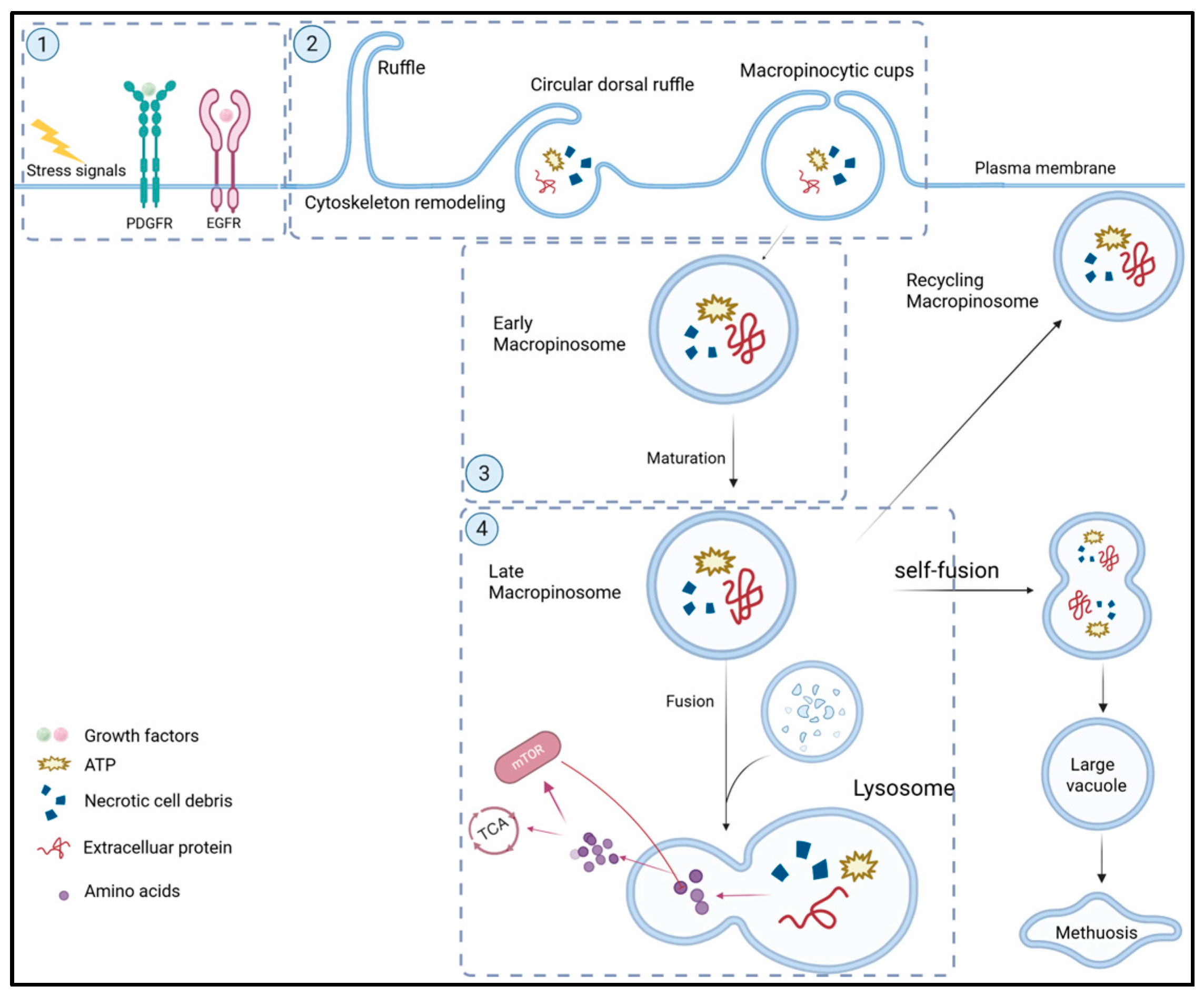
1. Overview of Macropinocytosis

Macropinocytosis is an evolutionarily conserved cellular process. Macropinocytosis, a non-coating plasma membrane endocytosis, differs from the clathrin-dependent endocytic pathway. As shown in Figure 1, macropinocytosis contains four stages: (1) initiation and activation; (2) formation of macropinosomes; (3) maturation; and (4) degradation and recycling of macropinosomes [1]. Macropinocytosis is initiated and activated by nutritional stress signals, extracellular stimuli such as growth factors, or chemokines [2,3]. These signals primarily activate downstream signaling pathways involving small GTPases RAS and RAC1 and inositol phospholipid kinases PI3K and PI4K. These pathways modulate the actin cytoskeleton, which drives plasma membrane remodeling [4,5] and leads to the formation of cup-shaped membrane ruffles [6,7]. Subsequently, these ruffles extend distally and wrap the extracellular fluids containing proteins, lipids, and nucleotides by either fusing with each other or folding back onto the plasma membrane, ultimately forming macropinosomes [2]. During maturation, early macropinosomes shrink in size regulated by ion channels to become late macropinosomes [1] and are eventually transported to lysosomes for degradation or recycled back to the plasma membrane. The degraded biomolecules provide nutrients for cancer cell growth. Notably, macropinocytosis in cancer cells with oncogenic RAS mutations is constitutively activated and utilized for supporting tumor growth [8]. Thus, oncogenic RAS mutation-bearing tumors are the most suitable for targeted therapies harnessing macropinocytosis.
There are multiple excellent review articles published about the molecular mechanism underlying macropinocytosis and the signaling pathways regulating macropinocytosis [2,9,10]. Thus, here we focus on a review of the current progress in macropinoctosis-related cancer therapeutic strategies, including both targeting macropinocytosis to inhibit tumor growth and utilizing macropinocytosis as an anti-cancer drug delivery tool. The content of these two aspects includes therapeutic applications with macropinocytic inhibitors; methuosis-inducing agents; and the macropinocytosis-mediated anti-cancer drug delivery strategies such as nanoparticle-conjugated anti-cancer drugs, virus-loaded anti-cancer protein cDNAs, and exo-vesicle (liposome)-loaded anti-cancer drugs. In conclusion, macropinocytosis offers new cancer therapeutic strategies and techniques based on the cellular function of macropinocytosis in providing nutrients for tumor growth and delivering extracellular fluids into cancer cells.

2. Roles of Macropinocytosis in Cancers

2.1. Providing Nutrients for Cancer Cell Growth

(a)
Uptake of proteins and ATP: It has been observed that the majority of biomolecules in macropinosomes are proteins [11]. These proteins are digested into free amino acids in lysosomes that are used to synthesize new proteins or catabolized to generate ATP for energy [6]. The main protein internalized by macropinocytosis is extracellular albumin, the most abundant protein in human plasma (~50%) [12]. In addition, given that albumin serves as a carrier protein, albumin-bound molecules such as fatty acids (FAs) and cholesterol, which are essential components for maintaining the structural integrity of the cell membrane, are also internalized by macropinocytosis [12]. Apparently, macropinocytosis enables tumor cells to sustain their metabolic and biosynthetic needs and promotes tumor cell growth under nutritional stress conditions [11,13].
It has been observed that extracellular ATP is extensively internalized by macropinocytosis in cancer cells [14,15]. This internalized ATP provides an important source of energy for tumor proliferation, survival, and resistance to targeted therapies [15]. Consequently, suppressing macropinocytosis effectively diminishes extracellular ATP uptake in cancer cells, resulting in depleting the cells’ energy reservoirs and impeding tumor progression in vivo. Emerging evidence further reveals an important role of ATP in driving epithelial–mesenchymal transition (EMT) in a macropinocytosis-dependent manner [15].
(b)
Uptake of necrotic cell debris: In addition to extracellular proteins and ATP, cancer cells assimilate other extracellular bioactive macromolecules through macropinocytosis. Tumor cell necrosis occurs under hypoxic or nutrient-deprived conditions and generates abundant cell debris containing proteins, fatty acids (FAs), nucleotides, and triacylglycerols (TAGs) [16,17]. These necrotic fragments are captured by neighbor tumor cells via macropinocytosis and processed by lysosomes to produce nutrients for fueling tumor cell growth. In addition, the uptake of necrotic cells by macropinocytosis may also contribute to resistance against antimetabolite chemotherapeutics [18].

2.2. Counteracting Oxidative Stress

Malignant tumor cells frequently exhibit disrupted redox homeostasis due to their heightened metabolic activity combined with microenvironmental hypoxia, leading to the sustained accumulation of reactive oxygen species (ROS) [19,20]. To counteract oxidative stress, cancer cells have evolved multiple defensive mechanisms, including macropinocytosis [2]. Macropinocytosis alleviates ROS-induced cellular damage through the internalization of exogenous anti-oxidants (e.g., glutathione precursors) and redox-regulating metabolites (e.g., cysteine) for endogenous anti-oxidant synthesis [21].
It has been shown that pharmacological blockade of autophagy in pancreatic ductal adenocarcinoma (PDAC) undergoes metabolic reprogramming by upregulating macropinocytosis activity, and the nutrient acquisition switches from autophagy to macropinocytosis [21]. This adaptive metabolic reprogramming is mediated by the nuclear factor erythroid 2-related factor 2 (NRF2) anti-oxidation pathway that upregulates macropinocytotic genes to activate the salvage pathway. NRF2 is a known transcriptional regulator in response to oxidative stress, governing the expression of genes involved in anti-oxidation and redox homeostasis [2,22]. The activation of macropinocytosis by NRF2 upon the inhibition of autophagy may represent a new signaling pathway that counteracts cellular nutritional and/or oxidative stress.

2.3. Promoting Immune Escape of Cancers

Macropinocytosis plays a key role in cancer immune escape by regulating the tumor microenvironment (TME) and immune cell function [23,24]. First, cancer cells utilize macropinocytosis to internalize extracellular proteins, which are lysosomally degraded to generate immunosuppressive metabolites, particularly lactate and adenosine, via enhanced glycolytic flux and purine salvage pathways [25]. The accumulation of lactate in the tumor microenvironment promotes immune escape of tumor cells by reducing immune cell activity and inhibiting the production of inflammatory mediators [26]. Secondly, macropinocytosis may affect the immune escape process by regulating the expression of immune checkpoint molecules (e.g., PD-L1). It was observed that macropinocytosis in cancer cells activates the mTOR signaling pathway, which promotes the expression of PD-L1 and inhibits T cell activation and function, thereby leading to immune escape of cancer cells [27,28,29]. In addition, macropinocytosis compromises the antigen-presenting function of tumor-associated macrophages (TAMs) and dendritic cells (DCs), reduces their immune surveillance capacity against cancer cells and further promotes immune escape through the internalization of the antigenic molecules [30,31]. Therefore, targeting macropinocytosis has become a new strategy to enhance the efficacy of immunotherapy. For example, the combination of macropinocytosis inhibitors with immune checkpoint inhibitors (e.g., anti-PD-1/PD-L1 antibodies) can produce synergistic anti-tumor effects [32,33]. This strategy provides a new direction for cancer therapy by simultaneously blocking the macropinocytosis-mediated immune escape pathway and enhancing the anti-tumor activity of immune cells.

2.4. Induction of Cancer Cell Death (Methuosis) upon Excessive Macropinocytosis

The term “methuosis” was first defined by Overmeyer et al. in glioblastoma cells with oncogenic RAS hyperactivation that stimulates excessive macropinocytosis and leads to cancer cell death. Thus, methuosis is a form of cell death that is induced by the over-activation of macropinocytosis and characterized by the formation of large vacuoles [34]. The large vacuoles [34], which are derived from the fusion of macropinosomes upon the excessive activation of macropinocytosis, are unable to be transported to lysosomes. The lack of lysosomal degradation results in the expansion and accumulation of the large vacuoles in cells [35]. Consequently, the accumulation of large vacuoles segregates essential nutrients and organelles, leading to metabolic stress and energy depletion. Furthermore, the expansion of large vacuoles disrupts cellular structure and function and ultimately leads to cell death [36,37,38]. Methuosis differs from apoptosis as it does not have significant nuclear condensation during the process and is not dependent on the activation of caspase family members [39]. In addition, methuosis does not exhibit the increase in membrane permeability seen in necrosis at its early stage [34]. Methuosis has been observed in cells from multiple types of cancers [40,41,42], offering a novel perspective on cancer therapeutic interventions. However, the precise molecular mechanisms underlying methuosis and the regulatory pathways of methuosis are yet to be fully elucidated.

3. The Role of Macropinocytosis in Cancer Therapy

Macropinocytosis is an important cellular process for cancer cell growth and survival under nutritional stress. Targeting macropinocytosis emerges as a promising therapeutic strategy for cancers, especially for cancers with oncogenic RAS mutants that have active macropinocytosis. On the other hand, macropinocytosis, as a process that uptakes extracellular fluids into cells, can be used for anti-cancer drug delivery in cancer therapy. Thus, macropinocytosis has dual roles in cancer therapeutic applications: both as a therapeutic target and as a therapeutic tool.

3.1. Targeting Macropinocytosis for Cancer Therapy

3.1.1. Application of Macropinocytosis Inhibitors for Cancer Therapy

The development of inhibitors of macropinocytosis as targeted cancer therapeutic drugs may effectively impede the macropinocytosis-dependent oncogenic pathways and disrupt the metabolic activity of cancer cells. As shown in Table 1, several inhibitors of macropinocytosis have been developed. Most of the inhibitors attack the early stage (initiation and activation stage) of macropinocytosis.
The small GTPase RAC1, a pivotal regulator in the initiation of macropinocytosis, has emerged as a significant drug target. Research findings have demonstrated that the inhibition of RAC1 activity can effectively impede the process of macropinocytosis. Currently, two compounds, NSC23766 and EHT1864, have been extensively utilized as RAC1 inhibitors in preclinical studies, exhibiting high specificity in inhibiting RAC1 activity and demonstrating therapeutic benefits in RAS-mutant cancers, including pancreatic cancer (PDAC) and non-small-cell lung cancer (NSCLC) [43,44]. Among them, NSC23766 specifically inhibits the interaction between RAC1 and guanine nucleotide exchange factor (GEF), preventing the conversion of RAC1-GDP to RAC1-GTP. In contrast, EHT1864 binds RAC1 isoforms (RAC1b, RAC2, RAC3) with a high affinity, inducing nucleotide dissociation [44,45]. Recent studies have found that the N,N′-disubstituted guanidino analog 1D-242 significantly inhibits RAC1-mediated TNFα-induced NF-κB nuclear translocation during non-small-cell lung cancer (NSCLC) cell proliferation and migration [46]. Furthermore, the E3 ubiquitin ligase MG53 has been shown to impede RAC1 activity through a direct targeting approach, leading to ubiquitination modification at the Lys5 site. This, in turn, has been observed to hinder the progression of hepatocellular carcinoma (HCC) [47].
In addition to the direct targeting of RAC1, the PI3K/AKT/mTOR signaling pathway, as an important regulatory pathway of macropinocytosis, has also become an important target for drug development. A notable example is the natural product Wortmannin, derived from Penicillium, which has been shown to inhibit cell growth and induce apoptosis in cancer cells by targeting the PI3K/AKT pathway [48]. LY294002, a synthetic compound designed based on the flavonoid quercetin, functions as a PI3K inhibitor, blocking PI3K/AKT signaling, inhibiting glycolysis, and interfering with ATP production, thereby inducing apoptosis in cancer cells [49].
Other types of macropinocytosis inhibitors have also demonstrated promising applications. 5-(N-Ethyl-N-isopropyl)-amiloride (EIPA), an Na+/H+ ion channel inhibitor, has been shown to inhibit the activities of RAC1 and Cdc42 by lowering the submembranous pH, thereby blocking macropinocytosis and actin polymerization [50]. Cytochalasin D, an inhibitor of actin polymerization, effectively inhibited membrane ruffling and macropinocytosis in fibroblasts [9]. These studies provide a significant theoretical foundation and experimental basis for the development of novel targeted drugs for the therapy of macropinocytosis-associated cancers.
Table 1. Macropinocytosis-targeted drugs or agents for cancer therapy.
Inhibitor TypeDrugMechanismCancer TypeReferences
Macropinocytosis inhibitorsNSC23766Inhibition of Rac1 activityRas-mutant
tumors
[44]
EHT1864Inhibition of Rac1 activityRas-mutant
tumors
[45]
1D-242Inhibition of Rac1-mediated nuclear translocationRas-mutant
tumors
[46]
MG53Ubiquitination modification at Lys5Ras-mutant
tumors
[47]
WortmanninInhibition of PI3K/AKT pathwayRas-mutant
tumors
[48]
EIPALowering the pH value below the plasma membraneRas-mutant
tumors
[49]
LY294002Blocking PI3K/AKT signalingRas-mutant
tumors
[50]
Cytochalasin DInhibition of actin polymerizationRas-mutant
tumors
[9]
NaN3Blocking ATP dependenceRas-mutant
tumors
[51]
PoziotinibTargeting EGFRKras-mutant
tumors
[52]
IvermectinInhibiting PAK1Kras-mutant
tumors
[52]
Tyrphostin A9Targeting PDGFRKras-mutant
tumors
[53]
LY2090314Inhibiting GSK-3 Kras-mutant
tumors
[54]
Pyrvinium pamoateInhibiting Wnt/β-cateninKras-mutant
tumors
[55]
Metabolic
regulators
CQ, HCQInhibition of cytoprotective autophagyPancreatic cancer, etc.[56]
Lys05Lysosomal deacidificationGlioblastoma[57]
Bafilomycin A1V-ATPase inhibitorsVarious cancers[58]
TelaglenastatInhibition of glutamine conversionMelanoma[59]
Methuosis
inducers
MIPP,
MOMIPP
Inducing plasma membrane shrinkageGlioblastoma, Breast cancer[60,61]
Bacoside AInduction of macropinocytosisGlioblastoma[62]
Nerve growth factorInduction of macropinocytosisMedulloblastoma[63]
TrehaloseForced induction of macropinocytosisGlioblastoma[64]
12AInduction of macropinocytosisBreast cancer, etc.[65]

3.1.2. Macropinocytosis-Associated Metabolic Regulators

Following the process of macropinocytosis, cancer cells can metabolize internalized proteins, carbohydrates, and other nutrients to generate energy and synthesize precursors for cellular biosynthesis, which participate in various metabolic processes within the cell. Targeting these metabolic pathways can suppress macropinocytosis, deplete energy reserves, and ultimately induce cell apoptosis. The potential drugs used to target macropinocytosis-associated metabolic pathways are listed in Table 1. Notably, autophagy and macropinocytosis exhibit cross-regulation in metabolic processes. The inhibition of autophagy may impede the degradation and recycling of internalized substances in macropinocytosis, consequently resulting in the death of cancer cells due to nutrient deprivation. Among autophagy inhibitors, chloroquine (CQ) and hydroxychloroquine (HCQ), which are widely used to alleviate acute and chronic inflammatory diseases, have been shown to inhibit cytoprotective autophagy, thereby enhancing the sensitivity of cancer cells to chemotherapeutic drugs [56]. The disruption of lysosomes, pivotal organelles in cellular autophagy and macropinocytosis, can markedly inhibit the degradation of internalized substances. Dimeric chloroquine (Lys05), a novel lysosomal inhibitor, has been shown to accumulate within and deacidify the lysosomes of both cells and tumors and to prevent the degradation of nutrients in macropinosomes [57]. Bafilomycin A1, a macrolide antibiotic, functions as a specific and potent inhibitor of V-ATPases, thereby preventing lysosomal acidification and, consequently, impeding the degradation of substances, affecting cancer cell proliferation [58]. Furthermore, L-glutamine (Gln) is imperative for cancer cell proliferation, and macropinocytosis is a key metabolic pathway for glutamine acquisition by cancer cells. Consequently, the inhibition of glutamine synthesis may prove effective in impeding the glutamatergic signaling of cancer cells. Telaglenastat (CB-839), a highly potent glutaminase inhibitor (GLSi) with a favorable systemic tolerance, has been observed to significantly reduce the glutamine to α-ketoglutarate (α-KG) conversion in tumor cells while concomitantly enhancing the activity of T cell-mediated immunotherapy in the context of combating melanoma [59]. These studies provide a significant theoretical foundation and experimental validation for the development of metabolic modulation-based strategies for macropinocytosis-targeted therapy.

3.1.3. Methuosis Inducers

The over-activation of macropinocytosis causes the accumulation of macropinosome-derived large vacuoles, leading to methuosis, a form of cell death associated with excessive macropinocytosis. The dysregulation of ion channels and transporter proteins in the vacuolar membrane increases permeability and osmotic imbalance and promotes vacuole expansion [35]. In addition, lysosomal dysfunction prevents the degradation of vesicle contents, leading to further vesicle enlargement and cell rupture. The accumulation of large vacuoles segregates essential nutrients and organelles, leading to metabolic stress and energy depletion. Large vesicles can displace cytoplasmic organelles and disrupt cellular structure and function, ultimately leading to cell death. Therefore, inducing the over-activation of macropinocytosis to promote methuosis becomes a promising cancer therapeutic strategy. Potential methuosis inducers are shown in Table 1. Silva-Pavez et al. found that treatment with the casein kinase 2 (CK2) inhibitor silmitasertib induced colorectal cancer cells to produce large numbers of macropinocytosis-derived vacuoles, ultimately leading to methuosis [66]. Similarly, Indole-Based Chalcones (e.g., MIPP and MOMIPP) are able to rapidly induce plasma membrane contraction and promote the formation of macropinosomes. These newly formed macropinosomes neither participate in membrane recycling nor fuse with lysosomes, leading to the vacuolization of cells and thus exerting anti-cancer effects [60,61]. Recent studies have shown that natural trehalose triggers early RAS activation in NF1-deficient U373-MG cells, and this signaling can subsequently stimulate macropinocytosis, which causes the cells to become overstimulated and ultimately undergo methuosis, leading to cell death [64]. A pyridine-pyrimidine-indole-carbohydrazide derivative, 12A, was designed and synthesized by Wu et al. as a novel inducer, which has antiproliferative and vacuolization-inducing effects on cancer cells similar to those of MOMIPP. Notably, the vacuoles induced by this compound were macropinosome-derived rather than autophagosome-derived. In the MDA-MB-231 xenograft mouse model, 12A exhibited significant tumor growth inhibition, making it a potential candidate for the development of novel anti-cancer therapeutic strategies [65].

3.1.4. Targeting Macropinocytosis for Drug Resistance

Chemotherapy is one of the primary clinical approaches for cancer management. However, chemo-resistance poses a persistent challenge during chemotherapeutic interventions. During chemotherapy treatment, certain cancer cells upregulate the expression of ATP-binding cassette (ABC) efflux transporters, including P-glycoprotein (P-gp/ABCB1), multidrug resistance protein 2 (MRP2/ABCC2), and breast cancer resistance protein (BCRP/ABCG2) [67]. These membrane transporters actively expel diverse chemotherapeutic compounds through ATP-dependent mechanisms, thus causing resistance to chemo-drug treatment by reducing intracellular drug accumulation and diminishing its cytotoxic efficacy [68,69]. Macropinocytosis uptakes extracellular ATP (eATP) and causes a significant increase in intracellular ATP levels. This mechanism directly potentiates the efflux activity of primary ABC multidrug transporters, accelerating the extracellular transport of chemotherapeutic drugs, which consequently diminishes intracellular drug retention [70]. Furthermore, numerous chemotherapeutic agents, including gemcitabine [71], 5-fluorouracil [72], doxorubicin [73], and platinum drugs, are nucleotide-targeted therapies by either targeting key enzymes in the nucleotide biosynthetic pathway or damaging DNA [74]. Macropinocytosis circumvents these nucleotide-targeted therapies via the uptake of extracellular proteins that produce free amino acids for metabolic precursors in nucleotide biosynthesis [75]. Thus, macropinocytosis plays an important role in chemo-resistance. The pharmacological inhibition of macropinocytosis (e.g., EIPA) blocks the macropinocytosis-mediated adaptive pathways for chemo-resistance and synergizes with chemotherapy by the dual suppression of nutrient salvage and drug efflux. For example, in Ras-mutant tumors, the combination of a macropinocytosis inhibitor with a chemotherapeutic drug (e.g., gemcitabine) synergistically suppresses tumor progression while delaying acquired resistance [76].
Both macropinocytosis and autophagy are stress-adaptive pathways. When autophagy inhibitors are used to treat pancreatic ductal adenocarcinoma (PDAC), macropinocytosis is activated via the NRF2 pathway to mitigate the cytotoxic effect caused by the inhibition of autophagy, thereby causing therapeutic resistance. Therefore, treatment with inhibitors of both autophagy and macropinocytosis (e.g., chloroquine) significantly enhances the anti-tumor effect [21].

3.2. Utilizing Macropinocytosis for Anti-Cancer Drug Delivery

In the field of cancer therapy, the development of drug delivery systems has become one of the key strategies to improve therapeutic efficacy and minimize side effects. Macropinocytosis is a non-selective endocytotic process by which cells can take up large amounts of extracellular fluid and dissolved substances. Thus, macropinocytosis offers an effective way to deliver conventional chemo drugs into cells. The macropinocytosis-mediated drug delivery systems have two forms: carrier-based formulations and conjugate-mediated targeted delivery systems.

3.2.1. The Macropinocytosis-Mediated Carrier Delivery System

A carrier formulation delivery system represents the delivery of drugs that need to be packaged in individual carriers, such as exosomes, nanoparticle carriers, and viral carriers that carry the drug into the carrier cavity to deliver the drug molecule. Table 2 shows a list of the current macropinocytosis-mediated carrier-based anti-cancer drug delivery methods.
(a)
Nanoparticles
Lipid nanoparticles (LNPs) are the most widely used delivery carrier. LNPs consist of phospholipid bilayers with low toxicity and low immunogenicity and are capable of encapsulating both hydrophilic and hydrophobic drugs. Gilleron et al. demonstrated by image-based analysis that LNPs are mainly used for cellular internalization through clathrin-mediated endocytosis and macropinocytosis [94]. Currently, liposome-based drug delivery is mainly focused on two major areas: chemotherapeutic drugs (e.g., adriamycin) and nucleic acid drugs (e.g., siRNA). Doxil, a doxorubicin formulation of polyethylene glycol-coated liposomes has been successfully used in the clinic [77]. In the laboratory research phase, several innovative studies have demonstrated the potential of liposomal delivery systems. For example, Wu et al. constructed the fibroblast activation protein (FAP)-responsive liposome FrLip@R, which efficiently delivered all-trans retinoic acid (ATRA) to pancreatic stellate cells (PSCs) in a pancreatic ductal adenocarcinoma (PDAC) model by macropinocytosis. It successfully returned activated PSCs to a quiescent state, disrupting the tumor stromal barrier, and significantly enhanced intratumoral drug delivery efficiency [78].
Zheng et al. developed fucoidan-encapsulated pH-sensitive liposomes (FU-GEM PSLs) for the targeted delivery of gemcitabine, which showed good results in the treatment of pancreatic cancer [79]. In terms of nucleic acid drug delivery, small interfering RNAs (siRNAs) have emerged as a promising strategy for liposomes. The siRNA molecules can effectively reduce the expression of oncogenes or other disease-causing genes in cancer cells by silencing specific genes by promoting the degradation of target mRNAs. Yang et al. successfully developed a lipid complex that can deliver siRNAs to glioma safely, efficiently, and selectively via the nasal route. This delivery system was preferentially internalized by glioma cells along with cellular debris through macropinocytosis, which significantly increased the accumulation of therapeutic agents in tumor tissues [81]. Yan et al. constructed a cancer cell membrane-fused liposome (CLip) containing an siATG5-loaded calcium phosphate (CaP) core, termed CLip@siATG5. This system can effectively regulate the metabolism of pancreatic cancer cells and significantly improve the delivery efficiency of chemotherapeutic drugs [80].
In addition to LNPs, other types of nanoparticles have also been developed as novel drug delivery platforms (Table 2), especially for targeted delivery applications. Among them, albumin and lipoprotein are important carriers for macropinocytosis-mediated therapies due to their unique biological properties. As an ideal drug carrier, albumin is characterized by biodegradability, good biocompatibility, low immunogenicity, etc., and the presence of multiple drug binding sites on its molecular surface makes it suitable for loading multiple therapeutic drugs. In clinical applications, nanoparticle albumin-conjugated paclitaxel (nab-paclitaxel, nab-PTX) has been successfully used for targeted therapy of KRAS-mutant cancers (e.g., pancreatic cancer and breast cancer) [82]. Studies have shown that KRAS-mutant cancer cells exhibit sensitivity to a variety of albumin-conjugated drugs, including adriamycin [95,96], anti-EGFR antibodies [83], and human β-defensin-2 [84]. Lipoproteins, as another important carrier, are composed of lipids and apolipoproteins and are mainly involved in lipid transport in the organism. In recent years, lipoproteins with a small particle size (e.g., low-density lipoprotein, LDL, and high-density lipoprotein, HDL) have been widely used in the development of drug delivery systems. Similar to albumin, lipoproteins have excellent biocompatibility, biodegradability, and non-immunogenicity and are capable of being internalized by tumor cells as nutrients through macropinocytosis. However, the clinical application of natural lipoproteins is limited by low yields and complex purification processes. To overcome these limitations, biomimetic lipoprotein systems have been developed. For example, paclitaxel associated with LDL-like nanoparticles has entered clinical trials (NCT04148833) [85]. The use of HDL nanoparticles encapsulated with salinomycin can enhance the drug uptake of cervical cancer stem cells (CSCs) through macropinocytosis and effectively kill tumor cells at lower concentrations [86]. In addition, combining calcium phosphate encapsulating siRNA with apolipoprotein E3-reconstituted high-density lipoprotein can achieve efficient intracellular delivery of siRNA through macropinocytosis [87].
In addition to albumin and lipoprotein nanoparticles, a variety of other types of nanoparticles have shown unique promise for application in cancer therapy through macropinocytosis. These nanoparticles utilize different mechanisms of action and offer diverse strategic options for cancer therapy. Ruthenium nanoparticles are novel anti-cancer nanomaterials that act by disrupting the cellular redox balance and the actin cytoskeleton. This dual mechanism of action induces oxidative stress, leading to cytoskeletal reorganization, which in turn impairs the nutrient uptake capacity of cancer cells and ultimately promotes cancer cell death [97,98]. In the field of gene therapy, Su et al. found that the overexpression of heat shock protein A9 (HSPA9/GRP75) significantly enhanced macropinocytosis in ovarian cancer cells. This property synergized with concentrated Tat peptide–plasmid DNA complex (Tat-pDNA) nanoparticles with added Ca2+ to significantly enhance the cytotoxic effect on tumor cells [99]. In addition, miRNA-34a-encapsulated nanoparticles could be efficiently taken up by cancer cells via macropinocytosis, thereby inhibiting tumor growth. Liposome–polycation–peptide nanoparticle (LPP) mRNA vaccines based on poly (β-amino ester) polymer mRNA encapsulated in lipid shells have shown excellent delivery efficiency in the presence of macropinocytosis, especially in dendritic cells [88]. In terms of traditional chemotherapy drug delivery, paclitaxel-loaded poly(lactic-co-glycolic acid) nanoparticles exhibited dual cytotoxicity effects on breast cancer cells by macropinocytosis, which retained the anti-cancer activity of paclitaxel itself and enhanced the targeted delivery effect of the nanoparticles [89].
(b)
Virus vectors
Adenovirus, lentivirus, and other viral vectors have been used as the anti-cancer drug carriers in the macropinocytosis-mediated drug delivery [90,91] (Table 2). Viral particles first activate signaling pathways that trigger actin-mediated membrane ruffling, followed by the formation of a macropinosome on the plasma membrane, which in turn leads to the internalization of viral particles into the cell. By exploiting this natural infectious ability and efficient intracellular delivery property, drugs can be delivered inside cancer cells by virus vectors. It was observed that lentiviruses employ macropinocytosis as their primary invasion route. For example, human immunodeficiency virus type 1 (HIV-1) enters macrophages through a CCR5 (C-C chemokine receptor 5)-triggered macropinocytosis-like mechanism, subsequently completing its uncoating within the endolysosomal pathway [100]. Macropinocytosis is one of the known endocytic routes for adeno-associated virus (AAV) that is used for anti-tumor drug delivery [101,102]. In addition, nanoparticle–oncolytic virus–polymer composites can be efficiently taken up by cancer cells through macropinocytosis, enhancing targeting and immune activation against cancer cells [91]. Moon et al. utilized a pH-sensitive and bio-reducible polymer (PPCBA) to complex with an oncolytic adenovirus (Ad), forming Ad-PPCBA. The Ad-PPCBA targets the acidic and hypoxic tumor microenvironment and overcomes coxsackie and adenovirus receptor (CAR)-dependent entry into target cells, showing potential for treating both primary and metastatic tumors [91].
(c)
Exosomes
Exosomes, a class of nanoscale extracellular vesicles (40–150 nm) with a lipid bilayer structure, mediate intercellular communication under physiological and pathological conditions. These natural nanocarriers can be taken up by cells through various endocytosis pathways, including macropinocytosis, which significantly improves the internalization efficiency of exosomes and provides new ideas for drug delivery. Kamerkar et al. found that enhanced macropinocytosis facilitates exosome uptake in Kras-mutant cancers. Compared to synthetic liposomes, exosomes showed superior performance in delivering RNA interference (RNAi) molecules and inhibiting tumor growth [103]. Deng et al. fused CLT (Celastrol)-loaded PEGylated lipids with DC2.4 cell membranes (M-LIP-CLT) to construct a delivery system targeting Kras-mutated pancreatic cancer [92]. Nakase et al. explored the application of exosomes in protein delivery. By encapsulating the ribosome-inactivating protein saporin within exosomes, they successfully inhibited cytoplasmic protein synthesis in target cells, thereby effectively suppressing the growth of tumor cells [93].

3.2.2. Macropinocytosis-Mediated Conjugated Targeted Delivery Systems

Unlike a carrier-based delivery system, a conjugated targeted delivery system employs a linker to covalently conjugate a drug to a molecule that recognizes the specific target protein or intracellular site, thus delivering the drug via macropinocytosis to the target protein or intracellular site. Current conjugated targeted delivery systems includes two delivery methods: PDCs (Peptide–Drug Conjugates) and ADCs (Antibody–Drug Conjugates). Table 3 shows a list of the current macropinocytosis-mediated conjugated targeted delivery systems.
(a)
Antibody–Drug Conjugates (ADCs)
The design of antibody–drug conjugates (ADCs) is based on the concept of the differential expression of tumor antigens between tumors and normal tissues, allowing for the specific delivery of chemotherapeutic agents to the target tumors. ADCs are known to enter cancer cells through macropinocytosis-mediated internalization, thereby inhibiting tumor cell growth and proliferation. AGS-16C3F, an ADC that targets ectonucleotide pyrophosphatase/phosphodiesterase 3 (ENPP3), is currently in development for treating metastatic renal cell carcinoma [104,105].
(b)
Peptide–Drug Conjugates (PDCs)
Peptide–drug conjugates (PDCs) are new types of drug delivery methods, in which the anti-cancer drug is covalently conjugated to the designed peptide via linkers. The PDC delivery techniques are based on the principle of the interaction of the peptides with the target tumor molecule or the induction of macropinocytosis by the peptides. PDCs can achieve precise drug delivery in cancer cells, improve therapeutic efficacy, and reduce toxicity to normal cells [106]. In the application of cell-penetrating peptides (CPPs), these short peptides that mimic the structure of viral peptides facilitate the transmembrane transport of exogenous molecules by inducing macropinocytosis. Among them, the TAT peptide, a peptide derived from HIV-1, is the most widely studied CPP, and when nanoparticles are modified with TAT for drug delivery, they carry a “bystander cargo” that can also be internalized by the cell through macropinocytosis. This property simplifies the drug formulation process as no additional modification of the drug molecule is required [109]. In addition, arginine-rich CPPs have shown promising applications in macropinocytosis-mediated drug delivery. For example, octa-arginine peptide (R8) has been successfully used for siRNA and antigen delivery, while arginine 12-mer peptide (R12) induces macropinocytosis activity and promotes receptor internalization by targeting CXCR4 [107,108,110].

3.2.3. Current Clinical Application of the Macropinocytosis-Mediated Anti-Cancer Drug Delivery for Cancer Therapy

In current clinical applications, macropinocytosis, a non-selective endocytic pathway, is being widely explored for the delivery of anti-cancer drugs, especially in KRAS-mutated cancers (e.g., lung cancer), and shows significant potential. The application of macropinocytosis in lung cancer therapy has focused on the development of nanocarrier systems capable of efficiently entering cancer cells via the macropinocytosis pathway, thereby increasing the concentration of drug in the cancer cells while reducing the toxic effects on normal cells. For example, Abraxane (albumin-bound paclitaxel, PTX) is a typical example of a drug that utilizes the macropinocytosis effect for drug delivery [40,111]. Compared to conventional paclitaxel, Abraxane offers the advantages of higher tumor uptake, fewer side effects, and a shorter administration time. Abraxane is currently approved by the U.S. Food and Drug Administration (FDA) and the European Medicines Agency (EMA) for the treatment of advanced breast cancer, pancreatic cancer, and non-small-cell lung cancer (NSCLC) [112].
In ovarian cancer therapy, a research team developed Tat/pDNA/C16TAB (T-P-C) nanoparticles. This delivery vehicle employs the cationic surfactant C16TAB to condense Tat/pDNA nanocomplexes, forming irregular particles characterized by a small size, positive surface charge, and high pDNA encapsulation efficiency. The mechanism underlying the delivery involves the Arf6 GTPase/Rab signaling axis-dependent activation of the macropinocytosis pathway [113].
Although macropinocytosis has great potential for drug delivery, it still faces several challenges in clinical application. For example, how to activate macropinocytosis to improve drug delivery efficiency and how to avoid drug degradation in the macropinocytosis pathway are critical issues that need to be addressed. In the future, through in-depth research on the mechanism of macropinocytosis and the combination of nanotechnology innovations, it is expected that more-efficient and safer anti-cancer therapeutic solutions will be developed, bringing new hope to lung cancer and other cancer patients.

4. Conclusions

Macropinocytosis has great potential in cancer therapy. Macropinocytosis enables cancer cells to take up exogenous nutrients and maintain malignant proliferation; thus, it is an important therapeutic target. As macropinocytosis is capable of endocytosing extracellular fluids into cells and active tumors, particularly tumors with oncogenic RAS mutations, it can be used as a tool for the delivery of anti-cancer drugs specifically to tumors. The current cancer therapeutic research on macropinocytosis includes targeting macropinocytosis, inducing methuosis, and delivering anti-cancer drugs by macropinocytosis. Currently, the clinical application of macropinocytosis for cancer therapy is in the development stage and faces many challenges. To fully exploit macropinocytosis in cancer therapy, the following aspects need to be improved or completed. First, the specificity of drug delivery must be improved. Although cancer cells usually have higher macropinocytosis activity, normal cells can also take up carrier-loaded or conjugated drugs via macropinocytosis. Therefore, how to improve the specificity of macropinocytosis-mediated drug delivery is an important direction for future research. Second, novel drug carriers for cancers must be developed to improve the delivery efficiency and biocompatibility. Third, the regulation mechanism underlying micropinocytosis must be further understood. Although it is known that RAS, RAC1, PI3K, and other signaling pathways regulate the activation of macropinocytosis, the regulatory signaling pathways for the late stages of macropinocytosis, including macropinosome maturation, recycling and degradation, remain incomplete. Fourth, the clinical application of macropinocytosis for clinical cancer therapy must be accelerated. Although macropinocytosis has shown significant effects in laboratory studies, its clinical application is still lacking. More clinical trials are needed to validate the safety and efficacy of macropinocytosis-mediated drug delivery systems. Finally, macropinocytosis targeting must be combined with other cancer therapeutic techniques (e.g., chemotherapy, radiotherapy, immunotherapy, etc.), which may be valuable for improving the efficiency of current cancer therapy.

Author Contributions

M.Z. searched the literature and wrote the original draft; L.Z. and Y.Z. designed and drew Figure 1. A.S. and G.S. assisted in the manuscript preparation and reviewed the manuscript. Q.L. supervised and participated in the writing and revision of this manuscript. All authors have read and agreed to the published version of the manuscript.

Funding

This research was funded by the National Natural Science Foundation of China. (No. 82172942 and No. 81871888 to Q.L.).

Institutional Review Board Statement

Not applicable.

Informed Consent Statement

Not applicable.

Data Availability Statement

Not applicable.

Acknowledgments

We would like to thank Wannian Yang for his critical reading and comments on this manuscript.

Conflicts of Interest

The authors declare no conflicts of interest.

Abbreviations

The following abbreviations are used in this manuscript:
ABCATP-binding cassette
AdAdenovirus
ADCsAntibody–drug conjugates
ATRAAll-trans retinoic acid
BCRPBreast cancer resistance protein
CK2Casein kinase 2
CPPsCell-penetrating peptides
CQChloroquine
CSCsCancer stem cells
DCsDendritic cells
eATPExtracellular ATP
EMTEpithelial–mesenchymal transition
FAPFibroblast activation protein
FAsFatty acids
FDAFood and Drug Administration
GEFGuanine nucleotide exchange factor
GlnL-glutamine
GLSiGlutaminase inhibitor
HCQHydroxychloroquine
HDLHigh-density lipoprotein
HEP-2Human epithelioma-2
HCCHepatocellular carcinoma
LDLLow-density lipoprotein
LNPsLipid nanoparticles
MKMegakaryocyte
MPMacropinocytosis
MRP2Multidrug resistance protein 2
mSCLCMetastatic small-cell lung cancer
NRF2Nuclear factor erythroid 2-related factor 2
NSCLCNon-small-cell lung cancer
PDACPancreatic ductal adenocarcinoma
PDCsPeptide–drug conjugates
P-gpP-Glycoprotein
PPCBApH-sensitive and bio-reducible polymer
PSCsPancreatic stellate cells
PTXPaclitaxel
EMAEuropean Medicines Agency
ROSReactive oxygen species
TAGsTriacylglycerols
TAMsTumor-associated macrophages
TMETumor microenvironment

References

  1. Wu, Y.; Hu, X.; Wei, Z.; Lin, Q. Cellular Regulation of Macropinocytosis. Int. J. Mol. Sci. 2024, 25, 6963. [Google Scholar] [CrossRef] [PubMed]
  2. Lambies, G.; Commisso, C. Macropinocytosis and Cancer: From Tumor Stress to Signaling Pathways. In Macropinocytosis; Commisso, C., Ed.; Subcellular Biochemistry; Springer International Publishing: Cham, Switzerland, 2022; Volume 98, pp. 15–40. ISBN 978-3-030-94003-4. [Google Scholar]
  3. Yang, C.; Zhang, F.; Chen, F.; Chang, Z.; Zhao, Y.; Shao, D.; Sun, W.; Dong, W.; Wang, Z. Biomimetic Nanovaccines Potentiating Dendritic Cell Internalization via CXCR4-Mediated Macropinocytosis. Adv. Healthc. Mater. 2023, 12, 2202064. [Google Scholar] [CrossRef] [PubMed]
  4. Hall, A. Ras-Related GTPases and the Cytoskeleton. Mol. Biol. Cell 1992, 3, 475–479. [Google Scholar] [CrossRef] [PubMed]
  5. Salloum, G.; Jakubik, C.T.; Erami, Z.; Heitz, S.D.; Bresnick, A.R.; Backer, J.M. PI3Kβ Is Selectively Required for Growth Factor-Stimulated Macropinocytosis. Cell Sci. 2019, 132, jcs231639. [Google Scholar] [CrossRef]
  6. Recouvreux, M.V.; Commisso, C. Macropinocytosis: A Metabolic Adaptation to Nutrient Stress in Cancer. Front. Endocrinol. 2017, 8, 261. [Google Scholar] [CrossRef]
  7. Swanson, J.A. Shaping Cups into Phagosomes and Macropinosomes. Nat. Rev. Mol. Cell Biol. 2008, 9, 639–649. [Google Scholar] [CrossRef]
  8. Murugan, A.K.; Grieco, M.; Tsuchida, N. RAS Mutations in Human Cancers: Roles in Precision Medicine. Semin. Cancer Biol. 2019, 59, 23–35. [Google Scholar] [CrossRef]
  9. Xu, G.; Zhang, Q.; Cheng, R.; Qu, J.; Li, W. Survival Strategies of Cancer Cells: The Role of Macropinocytosis in Nutrient Acquisition, Metabolic Reprogramming, and Therapeutic Targeting. Autophagy 2025, 21, 693–718. [Google Scholar] [CrossRef]
  10. Zhang, Y.; Commisso, C. Macropinocytosis in Cancer: A Complex Signaling Network. Trends Cancer 2019, 5, 332–334. [Google Scholar] [CrossRef]
  11. Commisso, C.; Davidson, S.M.; Soydaner-Azeloglu, R.G.; Parker, S.J.; Kamphorst, J.J.; Hackett, S.; Grabocka, E.; Nofal, M.; Drebin, J.A.; Thompson, C.B.; et al. Macropinocytosis of Protein Is an Amino Acid Supply Route in Ras-Transformed Cells. Nature 2013, 497, 633–637. [Google Scholar] [CrossRef]
  12. Commisso, C.; Debnath, J. Macropinocytosis Fuels Prostate Cancer. Cancer Discov. 2018, 8, 800–802. [Google Scholar] [CrossRef] [PubMed]
  13. Chen, L.; Cheng, D.; Chu, J.; Zhang, T.; Dong, Z.; Lou, H.; Zhu, L.; Liu, Y. A Novel Method to Image Macropinocytosis in Vivo. Front. Neurosci. 2018, 12, 324. [Google Scholar] [CrossRef] [PubMed]
  14. Vaidya, F.U.; Sufiyan Chhipa, A.; Mishra, V.; Gupta, V.K.; Rawat, S.G.; Kumar, A.; Pathak, C. Molecular and Cellular Paradigms of Multidrug Resistance in Cancer. Cancer Rep. 2022, 5, e1291. [Google Scholar] [CrossRef]
  15. Qian, Y.; Wang, X.; Liu, Y.; Li, Y.; Colvin, R.A.; Tong, L.; Wu, S.; Chen, X. Extracellular ATP Is Internalized by Macropinocytosis and Induces Intracellular ATP Increase and Drug Resistance in Cancer Cells. Cancer Lett. 2014, 351, 242–251. [Google Scholar] [CrossRef]
  16. Tsuchihara, K.; Fujii, S.; Esumi, H. Autophagy and Cancer: Dynamism of the Metabolism of Tumor Cells and Tissues. Cancer Lett. 2009, 278, 130–138. [Google Scholar] [CrossRef]
  17. Petan, T.; Jarc, E.; Jusović, M. Lipid Droplets in Cancer: Guardians of Fat in a Stressful World. Molecules 2018, 23, 1941. [Google Scholar] [CrossRef]
  18. Jayashankar, V.; Edinger, A.L. Macropinocytosis Confers Resistance to Therapies Targeting Cancer Anabolism. Nat. Commun. 2020, 11, 1121. [Google Scholar] [CrossRef]
  19. Gorrini, C.; Harris, I.S.; Mak, T.W. Modulation of Oxidative Stress as an Anticancer Strategy. Nat. Rev. Drug Discov. 2013, 12, 931–947. [Google Scholar] [CrossRef]
  20. DeNicola, G.M.; Karreth, F.A.; Humpton, T.J.; Gopinathan, A.; Wei, C.; Frese, K.; Mangal, D.; Yu, K.H.; Yeo, C.J.; Calhoun, E.S.; et al. Oncogene-Induced Nrf2 Transcription Promotes ROS Detoxification and Tumorigenesis. Nature 2011, 475, 106–109. [Google Scholar] [CrossRef]
  21. Su, H.; Yang, F.; Fu, R.; Li, X.; French, R.; Mose, E.; Pu, X.; Trinh, B.; Kumar, A.; Liu, J.; et al. Cancer Cells Escape Autophagy Inhibition via NRF2-Induced Macropinocytosis. Cancer Cell 2021, 39, 678–693.e11. [Google Scholar] [CrossRef]
  22. Li, H.; Zhang, Q.; Li, W.; Li, H.; Bao, J.; Yang, C.; Wang, A.; Wei, J.; Chen, S.; Jin, H. Role of Nrf2 in the Antioxidation and Oxidative Stress Induced Developmental Toxicity of Honokiol in Zebrafish. Toxicol. Appl. Pharmacol. 2019, 373, 48–61. [Google Scholar] [CrossRef] [PubMed]
  23. Ding, X.; Yao, T.; Liu, X.; Fan, Z.; Liu, Y. A Macropinocytosis-Related Gene Signature Predicts the Prognosis and Immune Microenvironment in Hepatocellular Carcinoma. Front. Oncol. 2023, 13, 1143013. [Google Scholar] [CrossRef] [PubMed]
  24. Wu, B.; Wang, Q.; Shi, X.; Jiang, M. Targeting Endocytosis and Cell Communications in the Tumor Immune Microenvironment. Cell Commun. Signal. 2022, 20, 161. [Google Scholar] [CrossRef]
  25. Galassi, C.; Chan, T.A.; Vitale, I.; Galluzzi, L. The Hallmarks of Cancer Immune Evasion. Cancer Cell 2024, 42, 1825–1863. [Google Scholar] [CrossRef]
  26. Gao, Y.; Zhou, H.; Liu, G.; Wu, J.; Yuan, Y.; Shang, A. Tumor Microenvironment: Lactic Acid Promotes Tumor Development. Immunol. Res. 2022, 2022, 3119375. [Google Scholar] [CrossRef]
  27. Meng, D.; Yang, Q.; Jeong, M.-H.; Curukovic, A.; Tiwary, S.; Melick, C.H.; Lama-Sherpa, T.D.; Wang, H.; Huerta-Rosario, M.; Urquhart, G.; et al. SNAT7 Regulates mTORC1 via Macropinocytosis. Proc. Natl. Acad. Sci. USA 2022, 119, e2123261119. [Google Scholar] [CrossRef]
  28. Matsuzaka, Y.; Yashiro, R. Molecular Docking and Intracellular Translocation of Extracellular Vesicles for Efficient Drug Delivery. Int. J. Mol. Sci. 2022, 23, 12971. [Google Scholar] [CrossRef]
  29. Koh, V.; Chakrabarti, J.; Torvund, M.; Steele, N.; Hawkins, J.A.; Ito, Y.; Wang, J.; Helmrath, M.A.; Merchant, J.L.; Ahmed, S.A.; et al. Hedgehog Transcriptional Effector GLI Mediates mTOR-Induced PD-L1 Expression in Gastric Cancer Organoids. Cancer Lett. 2021, 518, 59–71. [Google Scholar] [CrossRef]
  30. Palm, W. Metabolic Functions of Macropinocytosis. Philos. Trans. R. Soc. Lond. B Biol. Sci. 2019, 374, 20180285. [Google Scholar] [CrossRef]
  31. Jhunjhunwala, S.; Hammer, C.; Delamarre, L. Antigen Presentation in Cancer: Insights into Tumour Immunogenicity and Immune Evasion. Nat. Rev. Cancer 2021, 21, 298–312. [Google Scholar] [CrossRef]
  32. Gide, T.N.; Quek, C.; Menzies, A.M.; Tasker, A.T.; Shang, P.; Holst, J.; Madore, J.; Lim, S.Y.; Velickovic, R.; Wongchenko, M.; et al. Distinct Immune Cell Populations Define Response to Anti-PD-1 Monotherapy and Anti-PD-1/Anti-CTLA-4 Combined Therapy. Cancer Cell 2019, 35, 238–255.e6. [Google Scholar] [CrossRef] [PubMed]
  33. Butterfield, L.H.; Najjar, Y.G. Immunotherapy Combination Approaches: Mechanisms, Biomarkers and Clinical Observations. Nat. Rev. Immunol. 2024, 24, 399–416. [Google Scholar] [CrossRef] [PubMed]
  34. Maltese, W.A.; Overmeyer, J.H. Methuosis: Nonapoptotic Cell Death Associated with Vacuolization of Macropinosome and Endosome Compartments. Am. J. Pathol. 2014, 184, 1630–1642. [Google Scholar] [CrossRef]
  35. Park, W.; Wei, S.; Kim, B.-S.; Kim, B.; Bae, S.-J.; Chae, Y.C.; Ryu, D.; Ha, K.-T. Diversity and Complexity of Cell Death: A Historical Review. Exp. Mol. Med. 2023, 55, 1573–1594. [Google Scholar] [CrossRef] [PubMed]
  36. Zheng, S.; Li, Y.; Shao, Y.; Li, L.; Song, F. Osmotic Pressure and Its Biological Implications. Int. J. Mol. Sci. 2024, 25, 3310. [Google Scholar] [CrossRef]
  37. Roffay, C.; Molinard, G.; Kim, K.; Urbanska, M.; Andrade, V.; Barbarasa, V.; Nowak, P.; Mercier, V.; García-Calvo, J.; Matile, S.; et al. Passive Coupling of Membrane Tension and Cell Volume during Active Response of Cells to Osmosis. Proc. Natl. Acad. Sci. USA 2021, 118, e2103228118. [Google Scholar] [CrossRef]
  38. Yan, Q.; Gomis Perez, C.; Karatekin, E. Cell Membrane Tension Gradients, Membrane Flows, and Cellular Processes. Physiology 2024, 39, 231–245. [Google Scholar] [CrossRef]
  39. Overmeyer, J.H.; Kaul, A.; Johnson, E.E.; Maltese, W.A. Active Ras Triggers Death in Glioblastoma Cells through Hyperstimulation of Macropinocytosis. Mol. Cancer Res. 2008, 6, 965–977. [Google Scholar] [CrossRef]
  40. Puccini, J.; Badgley, M.A.; Bar-Sagi, D. Exploiting Cancer’s Drinking Problem: Regulation and Therapeutic Potential of Macropinocytosis. Trends Cancer 2022, 8, 54–64. [Google Scholar] [CrossRef]
  41. Bielsa, N.; Casasampere, M.; Abad, J.L.; Enrich, C.; Delgado, A.; Fabriàs, G.; Lizcano, J.M.; Casas, J. Methuosis Contributes to Jaspine-B-Induced Cell Death. Int. J. Mol. Sci. 2022, 23, 7257. [Google Scholar] [CrossRef]
  42. Bartoszewska, E.; Florek, K.; Zagórski, K.; Gachowska, M.; Wietrzyk, A.; Hutny, A.; Nowakowska-Toporowska, A.; Kulbacka, J. Methuosis, Alkaliptosis, and Oxeiptosis and Their Significance in Anticancer Therapy. Cells 2024, 13, 2095. [Google Scholar] [CrossRef] [PubMed]
  43. Onesto, C.; Shutes, A.; Picard, V.; Schweighoffer, F.; Der, C.J. Characterization of EHT 1864, a Novel Small Molecule Inhibitor of Rac Family Small GTPases. Methods Enzymol. 2008, 439, 111–129. [Google Scholar] [CrossRef]
  44. Gao, Y.; Dickerson, J.B.; Guo, F.; Zheng, J.; Zheng, Y. Rational Design and Characterization of a Rac GTPase-Specific Small Molecule Inhibitor. Proc. Natl. Acad. Sci. USA 2004, 101, 7618–7623. [Google Scholar] [CrossRef]
  45. Shutes, A.; Onesto, C.; Picard, V.; Leblond, B.; Schweighoffer, F.; Der, C.J. Specificity and Mechanism of Action of EHT 1864, a Novel Small Molecule Inhibitor of Rac Family Small GTPases. Biol. Chem. 2007, 282, 35666–35678. [Google Scholar] [CrossRef]
  46. Ciarlantini, M.S.; Barquero, A.; Bayo, J.; Wetzler, D.; Dodes Traian, M.M.; Bucci, H.A.; Fiore, E.J.; Gandolfi Donadío, L.; Defelipe, L.; Turjanski, A.; et al. Development of an Improved Guanidine-Based Rac1 Inhibitor with in Vivo Activity against Non-Small Cell Lung Cancer. ChemMedChem 2021, 16, 1011–1021. [Google Scholar] [CrossRef]
  47. Ma, X.; Ma, X.; Zhu, L.; Zhao, Y.; Chen, M.; Li, T.; Lin, Y.; Ma, D.; Sun, C.; Han, L. The E3 Ubiquitin Ligase MG53 Inhibits Hepatocellular Carcinoma by Targeting RAC1 Signaling. Oncogenesis 2022, 11, 40. [Google Scholar] [CrossRef]
  48. Bani, N.; Rahmani, F.; Shakour, N.; Amerizadeh, F.; Khalili-Tanha, G.; Khazaei, M.; Hassanian, S.M.; Kerachian, M.A.; Abbaszadegan, M.R.; Mojarad, M.; et al. Wortmannin Inhibits Cell Growth and Induces Apoptosis in Colorectal Cancer Cells by Suppressing the PI3K/AKT Pathway. Anticancer. Agents Med. Chem. 2024, 24, 916–927. [Google Scholar] [CrossRef]
  49. Huang, A.; Zeng, P.; Li, Y.; Lu, W.; Lai, Y. LY294002 Is a Promising Inhibitor to Overcome Sorafenib Resistance in FLT3-ITD Mutant AML Cells by Interfering with PI3K/Akt Signaling Pathway. Front. Oncol. 2021, 11, 782065. [Google Scholar] [CrossRef]
  50. Koivusalo, M.; Welch, C.; Hayashi, H.; Scott, C.C.; Kim, M.; Alexander, T.; Touret, N.; Hahn, K.M.; Grinstein, S. Amiloride Inhibits Macropinocytosis by Lowering Submembranous pH and Preventing Rac1 and Cdc42 Signaling. Cell Biol. 2010, 188, 547–563. [Google Scholar] [CrossRef]
  51. Ida, H.; Taira, N.; Azuma, K.; Kumatani, A.; Akishiba, M.; Futaki, S.; Takahashi, Y.; Shiku, H. Surface Morphology Live-Cell Imaging Reveals How Macropinocytosis Inhibitors Affect Membrane Dynamics. Electrochim. Acta 2023, 441, 141783. [Google Scholar] [CrossRef]
  52. Brambillasca, S.; Cera, M.R.; Andronache, A.; Dey, S.K.; Fagá, G.; Fancelli, D.; Frittoli, E.; Pasi, M.; Robusto, M.; Varasi, M.; et al. Novel Selective Inhibitors of Macropinocytosis-Dependent Growth in Pancreatic Ductal Carcinoma. Biomed. Pharmacother. 2024, 177, 116991. [Google Scholar] [CrossRef] [PubMed]
  53. Park, S.J.; Park, Y.J.; Shin, J.H.; Kim, E.S.; Hwang, J.J.; Jin, D.-H.; Kim, J.C.; Cho, D.-H. A Receptor Tyrosine Kinase Inhibitor, Tyrphostin A9 Induces Cancer Cell Death through Drp1 Dependent Mitochondria Fragmentation. Biochem. Biophys. Res. Commun. 2011, 408, 465–470. [Google Scholar] [CrossRef] [PubMed]
  54. Zamek-Gliszczynski, M.J.; Abraham, T.L.; Alberts, J.J.; Kulanthaivel, P.; Jackson, K.A.; Chow, K.H.; McCann, D.J.; Hu, H.; Anderson, S.; Furr, N.A.; et al. Pharmacokinetics, Metabolism, and Excretion of the Glycogen Synthase Kinase-3 Inhibitor LY2090314 in Rats, Dogs, and Humans: A Case Study in Rapid Clearance by Extensive Metabolism with Low Circulating Metabolite Exposure. Drug Metab. Dispos. 2013, 41, 714–726. [Google Scholar] [CrossRef] [PubMed]
  55. Xu, L.; Zhang, L.; Hu, C.; Liang, S.; Fei, X.; Yan, N.; Zhang, Y.; Zhang, F. WNT Pathway Inhibitor Pyrvinium Pamoate Inhibits the Self-Renewal and Metastasis of Breast Cancer Stem Cells. Int. J. Oncol. 2016, 48, 1175–1186. [Google Scholar] [CrossRef]
  56. Ferreira, P.M.P.; de Sousa, R.W.R.; Ferreira, J.R.d.O.; Militão, G.C.G.; Bezerra, D.P. Chloroquine and Hydroxychloroquine in Antitumor Therapies Based on Autophagy-Related Mechanisms. Pharmacol. Res. 2021, 168, 105582. [Google Scholar] [CrossRef]
  57. Amaravadi, R.K.; Winkler, J.D. Lys05: A New Lysosomal Autophagy Inhibitor. Autophagy 2012, 8, 1383–1384. [Google Scholar] [CrossRef]
  58. Wang, R.; Wang, J.; Hassan, A.; Lee, C.-H.; Xie, X.-S.; Li, X. Molecular Basis of V-ATPase Inhibition by Bafilomycin A1. Nat. Commun. 2021, 12, 1782. [Google Scholar] [CrossRef]
  59. Varghese, S.; Pramanik, S.; Williams, L.J.; Hodges, H.R.; Hudgens, C.W.; Fischer, G.M.; Luo, C.K.; Knighton, B.; Tan, L.; Lorenzi, P.L.; et al. The Glutaminase Inhibitor CB-839 (Telaglenastat) Enhances the Antimelanoma Activity of T-Cell-Mediated Immunotherapies. Mol. Cancer Ther. 2021, 20, 500–511. [Google Scholar] [CrossRef]
  60. Robinson, M.W.; Overmeyer, J.H.; Young, A.M.; Erhardt, P.W.; Maltese, W.A. Synthesis and Evaluation of Indole-Based Chalcones as Inducers of Methuosis, a Novel Type of Nonapoptotic Cell Death. Med. Chem. 2012, 55, 1940–1956. [Google Scholar] [CrossRef]
  61. Huang, W.; Sun, X.; Li, Y.; He, Z.; Li, L.; Deng, Z.; Huang, X.; Han, S.; Zhang, T.; Zhong, J.; et al. Discovery and Identification of Small Molecules as Methuosis Inducers with in Vivo Antitumor Activities. Med. Chem. 2018, 61, 5424–5434. [Google Scholar] [CrossRef]
  62. John, S.; Sivakumar, K.C.; Mishra, R. Bacoside A Induces Tumor Cell Death in Human Glioblastoma Cell Lines through Catastrophic Macropinocytosis. Front. Mol. Neurosci. 2017, 10, 171. [Google Scholar] [CrossRef] [PubMed]
  63. Li, C.; MacDonald, J.I.S.; Talebian, A.; Leuenberger, J.; Seah, C.; Pasternak, S.H.; Michnick, S.W.; Meakin, S.O. Unravelling the Mechanism of TrkA-Induced Cell Death by Macropinocytosis in Medulloblastoma Daoy Cells. Mol. Cell. Biol. 2016, 36, 2596–2611. [Google Scholar] [CrossRef] [PubMed]
  64. Del Bello, B.; Gamberucci, A.; Marcolongo, P.; Maellaro, E. The Autophagy Inducer Trehalose Stimulates Macropinocytosis in NF1-Deficient Glioblastoma Cells. Cancer Cell Int. 2022, 22, 232. [Google Scholar] [CrossRef]
  65. Wu, J.; Hu, H.; Ao, M.; Cui, Z.; Zhou, X.; Qin, J.; Guo, Y.; Chen, J.; Xue, Y.; Fang, M. Design, Synthesis, and Biological Evaluation of 5-((4-(Pyridin-3-Yl)Pyrimidin-2-Yl)Amino)-1H-Indole-2-Carbohydrazide Derivatives: The Methuosis Inducer 12A as a Novel and Selective Anticancer Agent. Enzym. Inhib. Med. Chem. 2021, 36, 1436–1453. [Google Scholar] [CrossRef] [PubMed]
  66. Silva-Pavez, E.; Villar, P.; Trigo, C.; Caamaño, E.; Niechi, I.; Pérez, P.; Muñoz, J.P.; Aguayo, F.; Burzio, V.A.; Varas-Godoy, M.; et al. CK2 Inhibition with Silmitasertib Promotes Methuosis-like Cell Death Associated to Catastrophic Massive Vacuolization of Colorectal Cancer Cells. Cell Death Dis. 2019, 10, 73. [Google Scholar] [CrossRef]
  67. Schinkel, A.H.; Jonker, J.W. Mammalian Drug Efflux Transporters of the ATP Binding Cassette (ABC) Family: An Overview. Adv. Drug Deliv. Rev. 2003, 55, 3–29. [Google Scholar] [CrossRef]
  68. Wang, J.-Q.; Yang, Y.; Cai, C.-Y.; Teng, Q.-X.; Cui, Q.; Lin, J.; Assaraf, Y.G.; Chen, Z.-S. Multidrug Resistance Proteins (MRPs): Structure, Function and the Overcoming of Cancer Multidrug Resistance. Drug Resist. Updat. 2021, 54, 100743. [Google Scholar] [CrossRef]
  69. Zhang, H.; Xu, H.; Ashby, C.R.; Assaraf, Y.G.; Chen, Z.-S.; Liu, H.-M. Chemical Molecular-Based Approach to Overcome Multidrug Resistance in Cancer by Targeting P-Glycoprotein (P-Gp). Med. Res. Rev. 2021, 41, 525–555. [Google Scholar] [CrossRef]
  70. Zhang, H.; Chen, X. Abstract 405: A New Mechanism of Drug Resistance in Cancer: Extracellular ATP-Induced Resistance by Macropinocytosis-Mediated Internalization and Redox Changes. Cancer Res. 2022, 82, 405. [Google Scholar] [CrossRef]
  71. Toschi, L.; Finocchiaro, G.; Bartolini, S.; Gioia, V.; Cappuzzo, F. Role of Gemcitabine in Cancer Therapy. Future Oncol. Lond. Engl. 2005, 1, 7–17. [Google Scholar] [CrossRef]
  72. Longley, D.B.; Harkin, D.P.; Johnston, P.G. 5-Fluorouracil: Mechanisms of Action and Clinical Strategies. Nat. Rev. Cancer 2003, 3, 330–338. [Google Scholar] [CrossRef] [PubMed]
  73. Blum, R.H.; Carter, S.K. Adriamycin. A New Anticancer Drug with Significant Clinical Activity. Ann. Intern. Med. 1974, 80, 249–259. [Google Scholar] [CrossRef] [PubMed]
  74. Zhang, C.; Xu, C.; Gao, X.; Yao, Q. Platinum-Based Drugs for Cancer Therapy and Anti-Tumor Strategies. Theranostics 2022, 12, 2115–2132. [Google Scholar] [CrossRef] [PubMed]
  75. Wu, H.-L.; Gong, Y.; Ji, P.; Xie, Y.-F.; Jiang, Y.-Z.; Liu, G.-Y. Targeting Nucleotide Metabolism: A Promising Approach to Enhance Cancer Immunotherapy. Hematol. Oncol. 2022, 15, 45. [Google Scholar] [CrossRef]
  76. Khine, H.E.E.; Suriya, U.; Rungrotmongkol, T.; Chamni, S.; Lu, Y.; Bénard, A.; Lan, B.; Mukhopadhyay, D.; Chang, D.; Biankin, A.; et al. Jorunnamycin A Induces Apoptosis in Pancreatic Ductal Adenocarcinoma Cells, Spheroids, and Patient-Derived Organoids by Modulating KRAS-Mediated Survival Pathways. Sci. Rep. 2025, 15, 11376. [Google Scholar] [CrossRef]
  77. Hubert, A.; Lyass, O.; Pode, D.; Gabizon, A. Doxil (Caelyx): An Exploratory Study with Pharmacokinetics in Patients with Hormone-Refractory Prostate Cancer. Anticancer Drugs 2000, 11, 123–127. [Google Scholar] [CrossRef]
  78. Wu, Z.; Wu, Y.; Wang, M.; Chen, D.; Lv, J.; Yan, J.; Zhou, D.; Pang, Y.; Liang, H.; Zhang, D.; et al. FAP-Activated Liposomes Achieved Specific Macropinocytosis Uptake by Pancreatic Stellate Cells for Efficient Desmoplasia Reversal. Chem. Eng. 2024, 495, 153369. [Google Scholar] [CrossRef]
  79. Zheng, Z.; Li, M.; Yang, J.; Zhou, X.; Chen, Y.; Silli, E.K.; Tang, J.; Gong, S.; Yuan, Y.; Zong, Y.; et al. Growth Inhibition of Pancreatic Cancer by Targeted Delivery of Gemcitabine via Fucoidan-Coated pH-Sensitive Liposomes. Int. J. Biol. Macromol. 2024, 277, 134517. [Google Scholar] [CrossRef]
  80. Yan, J.; Wang, M.; Lv, S.; Chen, D.; Wu, Z.; Zhou, D.; Zhang, S.; Lv, J.; Xu, K.; Xu, C.; et al. SiATG5-Loaded Cancer Cell Membrane-Fused Liposomes Induced Increased Uptake of Albumin-Bound Chemotherapeutics by Pancreatic Cancer Cells. Control. Release 2024, 367, 620–636. [Google Scholar] [CrossRef]
  81. Hu, Y.; Jiang, K.; Wang, D.; Yao, S.; Lu, L.; Wang, H.; Song, J.; Zhou, J.; Fan, X.; Wang, Y.; et al. Core-Shell Lipoplexes Inducing Active Macropinocytosis Promote Intranasal Delivery of c-Myc siRNA for Treatment of Glioblastoma. Acta Biomater. 2022, 138, 478–490. [Google Scholar] [CrossRef]
  82. Li, R.; Ng, T.S.C.; Wang, S.J.; Prytyskach, M.; Rodell, C.B.; Mikula, H.; Kohler, R.H.; Garlin, M.A.; Lauffenburger, D.A.; Parangi, S.; et al. Therapeutically Reprogrammed Nutrient Signalling Enhances Nanoparticulate Albumin Bound Drug Uptake and Efficacy in KRAS-Mutant Cancer. Nat. Nanotechnol. 2021, 16, 830–839. [Google Scholar] [CrossRef] [PubMed]
  83. Wang, X.; Sheng, W.; Wang, Y.; Li, L.; Li, Y.; Zhang, S.; Liu, X.; Chen, S.; Zhen, Y. A Macropinocytosis-Intensifying Albumin Domain-Based scFv Antibody and Its Conjugate Directed against K-Ras Mutant Pancreatic Cancer. Mol. Pharm. 2018, 15, 2403–2412. [Google Scholar] [CrossRef] [PubMed]
  84. Du, Y.; Shang, B.-Y.; Sheng, W.-J.; Zhang, S.-H.; Li, Y.; Miao, Q.-F.; Zhen, Y.-S. A Recombinantly Tailored β-Defensin That Displays Intensive Macropinocytosis-Mediated Uptake Exerting Potent Efficacy against K-Ras Mutant Pancreatic Cancer. Oncotarget 2016, 7, 58418–58434. [Google Scholar] [CrossRef] [PubMed]
  85. Maranhão, R.C.; Vital, C.G.; Tavoni, T.M.; Graziani, S.R. Clinical Experience with Drug Delivery Systems as Tools to Decrease the Toxicity of Anticancer Chemotherapeutic Agents. Expert Opin. Drug Deliv. 2017, 14, 1217–1226. [Google Scholar] [CrossRef]
  86. Yin, X.; Lu, Y.; Zou, M.; Wang, L.; Zhou, X.; Zhang, Y.; Su, M. Synthesis and Characterization of Salinomycin-Loaded High-Density Lipoprotein and Its Effects on Cervical Cancer Cells and Cervical Cancer Stem Cells. Int. J. Nanomed. 2021, 16, 6367–6382. [Google Scholar] [CrossRef]
  87. Huang, J.-L.; Jiang, G.; Song, Q.-X.; Gu, X.; Hu, M.; Wang, X.-L.; Song, H.-H.; Chen, L.-P.; Lin, Y.-Y.; Jiang, D.; et al. Lipoprotein-Biomimetic Nanostructure Enables Efficient Targeting Delivery of siRNA to Ras-Activated Glioblastoma Cells via Macropinocytosis. Nat. Commun. 2017, 8, 15144. [Google Scholar] [CrossRef]
  88. Persano, S.; Guevara, M.L.; Li, Z.; Mai, J.; Ferrari, M.; Pompa, P.P.; Shen, H. Lipopolyplex Potentiates Anti-Tumor Immunity of mRNA-Based Vaccination. Biomaterials 2017, 125, 81–89. [Google Scholar] [CrossRef]
  89. Al-Humaidi, R.B.; Fayed, B.; Shakartalla, S.B.; Jagal, J.; Jayakumar, M.N.; Al Shareef, Z.M.; Sharif, S.I.; Noreddin, A.; Semreen, M.H.; Omar, H.A.; et al. Optimum Inhibition of MCF-7 Breast Cancer Cells by Efficient Targeting of the Macropinocytosis Using Optimized Paclitaxel-Loaded Nanoparticles. Life Sci. 2022, 305, 120778. [Google Scholar] [CrossRef]
  90. Xiang, H.-L.; Chen, Y.; Wang, J.-W.; Wang, H.-J.; Gao, X.-F.; Li, H.; Mao, S.-J. Enhancing Cytotoxicity of Daunorubicin on Drug-resistant Leukaemia Cells with Microparticle-Mediated Drug Delivery System. J. Microencapsul. 2019, 36, 291–304. [Google Scholar] [CrossRef]
  91. Moon, C.Y.; Choi, J.-W.; Kasala, D.; Jung, S.-J.; Kim, S.W.; Yun, C.-O. Dual Tumor Targeting with pH-Sensitive and Bioreducible Polymer-Complexed Oncolytic Adenovirus. Biomaterials 2015, 41, 53–68. [Google Scholar] [CrossRef]
  92. Deng, L.; Zhang, H.; Zhang, Y.; Luo, S.; Du, Z.; Lin, Q.; Zhang, Z.; Zhang, L. An Exosome-Mimicking Membrane Hybrid Nanoplatform for Targeted Treatment toward Kras-Mutant Pancreatic Carcinoma. Biomater. Sci. 2021, 9, 5599–5611. [Google Scholar] [CrossRef] [PubMed]
  93. Nakase, I.; Kobayashi, N.B.; Takatani-Nakase, T.; Yoshida, T. Active Macropinocytosis Induction by Stimulation of Epidermal Growth Factor Receptor and Oncogenic Ras Expression Potentiates Cellular Uptake Efficacy of Exosomes. Sci. Rep. 2015, 5, 10300. [Google Scholar] [CrossRef] [PubMed]
  94. Gilleron, J.; Querbes, W.; Zeigerer, A.; Borodovsky, A.; Marsico, G.; Schubert, U.; Manygoats, K.; Seifert, S.; Andree, C.; Stöter, M.; et al. Image-Based Analysis of Lipid Nanoparticle-Mediated siRNA Delivery, Intracellular Trafficking and Endosomal Escape. Nat. Biotechnol. 2013, 31, 638–646. [Google Scholar] [CrossRef]
  95. Liu, H.; Sun, M.; Liu, Z.; Kong, C.; Kong, W.; Ye, J.; Gong, J.; Huang, D.C.S.; Qian, F. KRAS-Enhanced Macropinocytosis and Reduced FcRn-Mediated Recycling Sensitize Pancreatic Cancer to Albumin-Conjugated Drugs. Control. Release 2019, 296, 40–53. [Google Scholar] [CrossRef]
  96. Liu, H.; Qian, F. Exploiting Macropinocytosis for Drug Delivery into KRAS Mutant Cancer. Theranostics 2022, 12, 1321–1332. [Google Scholar] [CrossRef]
  97. Xia, F.; Hu, X.; Zhang, B.; Wang, X.; Guan, Y.; Lin, P.; Ma, Z.; Sheng, J.; Ling, D.; Li, F. Ultrasmall Ruthenium Nanoparticles with Boosted Antioxidant Activity Upregulate Regulatory T Cells for Highly Efficient Liver Injury Therapy. Small 2022, 18, e2201558. [Google Scholar] [CrossRef]
  98. Zhou, Y.; Yu, Q.; Qin, X.; Bhavsar, D.; Yang, L.; Chen, Q.; Zheng, W.; Chen, L.; Liu, J. Improving the Anticancer Efficacy of Laminin Receptor-Specific Therapeutic Ruthenium Nanoparticles (RuBB-Loaded EGCG-RuNPs) via ROS-Dependent Apoptosis in SMMC-7721 Cells. ACS Appl. Mater. Interfaces 2016, 8, 15000–15012. [Google Scholar] [CrossRef]
  99. Su, L.; Sun, Z.; Qi, F.; Su, H.; Qian, L.; Li, J.; Zuo, L.; Huang, J.; Yu, Z.; Li, J.; et al. GRP75-Driven, Cell-Cycle-Dependent Macropinocytosis of Tat/pDNA-Ca2+ Nanoparticles Underlies Distinct Gene Therapy Effect in Ovarian Cancer. Nanobiotechnology 2022, 20, 340. [Google Scholar] [CrossRef]
  100. Gobeil, L.-A.; Lodge, R.; Tremblay, M.J. Macropinocytosis-like HIV-1 Internalization in Macrophages Is CCR5 Dependent and Leads to Efficient but Delayed Degradation in Endosomal Compartments. J. Virol. 2013, 87, 735–745. [Google Scholar] [CrossRef]
  101. Meier, O.; Greber, U.F. Adenovirus Endocytosis. J. Gene Med. 2004, 6, S152–S163. [Google Scholar] [CrossRef]
  102. Shan, L.; Cui, S.; Du, C.; Wan, S.; Qian, Z.; Achilefu, S.; Gu, Y. A Paclitaxel-Conjugated Adenovirus Vector for Targeted Drug Delivery for Tumor Therapy. Biomaterials 2012, 33, 146–162. [Google Scholar] [CrossRef] [PubMed]
  103. Kamerkar, S.; LeBleu, V.S.; Sugimoto, H.; Yang, S.; Ruivo, C.F.; Melo, S.A.; Lee, J.J.; Kalluri, R. Exosomes Facilitate Therapeutic Targeting of Oncogenic KRAS in Pancreatic Cancer. Nature 2017, 546, 498–503. [Google Scholar] [CrossRef] [PubMed]
  104. Zhao, H.; Atkinson, J.; Gulesserian, S.; Zeng, Z.; Nater, J.; Ou, J.; Yang, P.; Morrison, K.; Coleman, J.; Malik, F.; et al. Modulation of Macropinocytosis-Mediated Internalization Decreases Ocular Toxicity of Antibody-Drug Conjugates. Cancer Res. 2018, 78, 2115–2126. [Google Scholar] [CrossRef]
  105. Zhao, H.; Gulesserian, S.; Ganesan, S.K.; Ou, J.; Morrison, K.; Zeng, Z.; Robles, V.; Snyder, J.; Do, L.; Aviña, H.; et al. Inhibition of Megakaryocyte Differentiation by Antibody-Drug Conjugates (ADCs) Is Mediated by Macropinocytosis: Implications for ADC-Induced Thrombocytopenia. Mol. Cancer Ther. 2017, 16, 1877–1886. [Google Scholar] [CrossRef]
  106. Jiang, G.; Chen, H.; Huang, J.; Song, Q.; Chen, Y.; Gu, X.; Jiang, Z.; Huang, Y.; Lin, Y.; Feng, J.; et al. Tailored Lipoprotein-Like miRNA Delivery Nanostructure Suppresses Glioma Stemness and Drug Resistance through Receptor-Stimulated Macropinocytosis. Adv. Sci. 2020, 7, 1903290. [Google Scholar] [CrossRef]
  107. Nakamura, T.; Moriguchi, R.; Kogure, K.; Shastri, N.; Harashima, H. Efficient MHC Class I Presentation by Controlled Intracellular Trafficking of Antigens in Octaarginine-Modified Liposomes. Mol. Ther. Am. Soc. Gene Ther. 2008, 16, 1507–1514. [Google Scholar] [CrossRef]
  108. Nakase, I.; Akita, H.; Kogure, K.; Gräslund, A.; Langel, Ü.; Harashima, H.; Futaki, S. Efficient Intracellular Delivery of Nucleic Acid Pharmaceuticals Using Cell-Penetrating Peptides. Acc. Chem. Res. 2012, 45, 1132–1139. [Google Scholar] [CrossRef]
  109. Wei, Y.; Tang, T.; Pang, H.-B. Cellular Internalization of Bystander Nanomaterial Induced by TAT-Nanoparticles and Regulated by Extracellular Cysteine. Nat. Commun. 2019, 10, 3646. [Google Scholar] [CrossRef]
  110. Tanaka, G.; Nakase, I.; Fukuda, Y.; Masuda, R.; Oishi, S.; Shimura, K.; Kawaguchi, Y.; Takatani-Nakase, T.; Langel, U.; Gräslund, A.; et al. CXCR4 Stimulates Macropinocytosis: Implications for Cellular Uptake of Arginine-Rich Cell-Penetrating Peptides and HIV. Chem. Biol. 2012, 19, 1437–1446. [Google Scholar] [CrossRef]
  111. Cullis, J.; Siolas, D.; Avanzi, A.; Barui, S.; Maitra, A.; Bar-Sagi, D. Macropinocytosis of Nab-Paclitaxel Drives Macrophage Activation in Pancreatic Cancer. Cancer Immunol. Res. 2017, 5, 182–190. [Google Scholar] [CrossRef]
  112. Holder, J.E.; Ferguson, C.; Oliveira, E.; Lodeiro, C.; Trim, C.M.; Byrne, L.J.; Bertolo, E.; Wilson, C.M. The Use of Nanoparticles for Targeted Drug Delivery in Non-Small Cell Lung Cancer. Front. Oncol. 2023, 13, 1154318. [Google Scholar] [CrossRef] [PubMed]
  113. Sun, Z.; Huang, J.; Su, L.; Li, J.; Qi, F.; Su, H.; Chen, Y.; Zhang, Q.; Zhang, Q.; Li, Z.; et al. Arf6-Mediated Macropinocytosis-Enhanced Suicide Gene Therapy of C16TAB-Condensed Tat/pDNA Nanoparticles in Ovarian Cancer. Nanoscale 2021, 13, 14538–14551. [Google Scholar] [CrossRef] [PubMed]
Figure 1. Process of macropinocytosis. The process of macropinocytosis has 4 stages: (1) Initiation and activation: Macropinocytosis is initiated and activated by cellular signals that induce cytoskeletal remodeling to form plasma membrane ruffles. (2) Formation of macropinosomes: The plasma membrane ruffles fold into cup-shaped structures that encapsulate extracellular nutritional fluids and form early macropinosomes. (3) Maturation: The early macropinosomes undergo volume adjustment to become late macropinosomes. (4) Degradation and recycling: The matured macropinosomes either fuse with lysosomes to release nutrients or recycle back to the plasma membrane. Excessive macropinocytosis may generate large vacuoles that lead to non-apoptotic cell death called methuosis.
Biomolecules 15 00936 g001
Table 2. Macropinocytosis-mediated carrier delivery systems.
SubtypeCarrierPayloadsCancer TypeReference
NanoparticlesLiposomesAdriamycinKaposi sarcoma[77]
All-trans retinoic acidPancreatic ductal adenocarcinoma[78]
GemcitabinePancreatic cancer[79]
siRNA(ATG5)Pancreatic cancer[80]
siRNAGlioma[81]
AlbuminPaclitaxelMultiple types of cancer cells[82]
AdriamycinPancreatic cancer[83]
Anti-EGFR antibodiesPancreatic cancer[83]
β-Defensin-2Multiple types of cancer cells[84]
LipoproteinPaclitaxelMultiple types of cancer cells[85]
SalinomycinCervical cancer[86]
siRNAGlioblastoma[87]
Lipid shellmRNAMelanoma[88]
AdriamycinBreast cancer, etc.[89]
Virus vectorsLentivirussiRNALeukemia[90]
Oncolytic virusesPPCBAPrimary tumor and metastatic tumors[91]
ExosomesExosomesCelastrolKras-mutant tumors[92]
ExosomesSaporinRas-mutant tumors[93]
Table 3. Macropinocytosis-mediated conjugated targeted delivery systems.
SubtypeCarrierPayloadsCancer TypeReferences
Antibody–Drug Conjugates (ADCs)AntibodyChemotherapy drugsMultiple types of cancer cells[104,105]
Peptide–Drug Conjugates (PDCs)Elastin-like peptidemiRNA-34aGlioma[106]
R8NAPMultiple types of cancer cells[107,108]
R12NAPMultiple types of cancer cells[107,108]
Disclaimer/Publisher’s Note: The statements, opinions and data contained in all publications are solely those of the individual author(s) and contributor(s) and not of MDPI and/or the editor(s). MDPI and/or the editor(s) disclaim responsibility for any injury to people or property resulting from any ideas, methods, instructions or products referred to in the content.

